
Electrospun Shape Memory Polymer Micro-/Nanofibers and Tailoring Their Roles for Biomedical Applications


Abstract
:
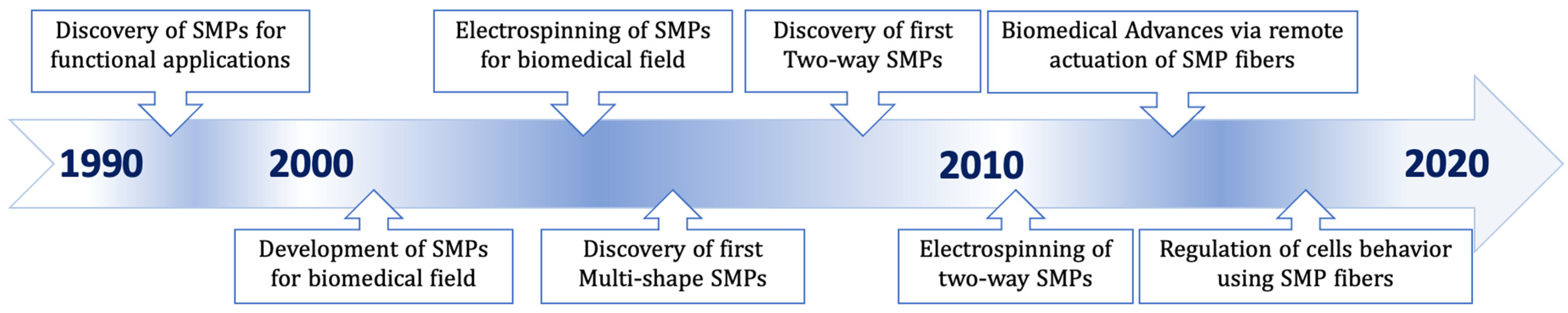
1. Introduction
2. Architecture of SMPs
3. Triple and Multiple SMPs
4. Reversible SMPs
4.1. Reversible Thermally Induced SMPs
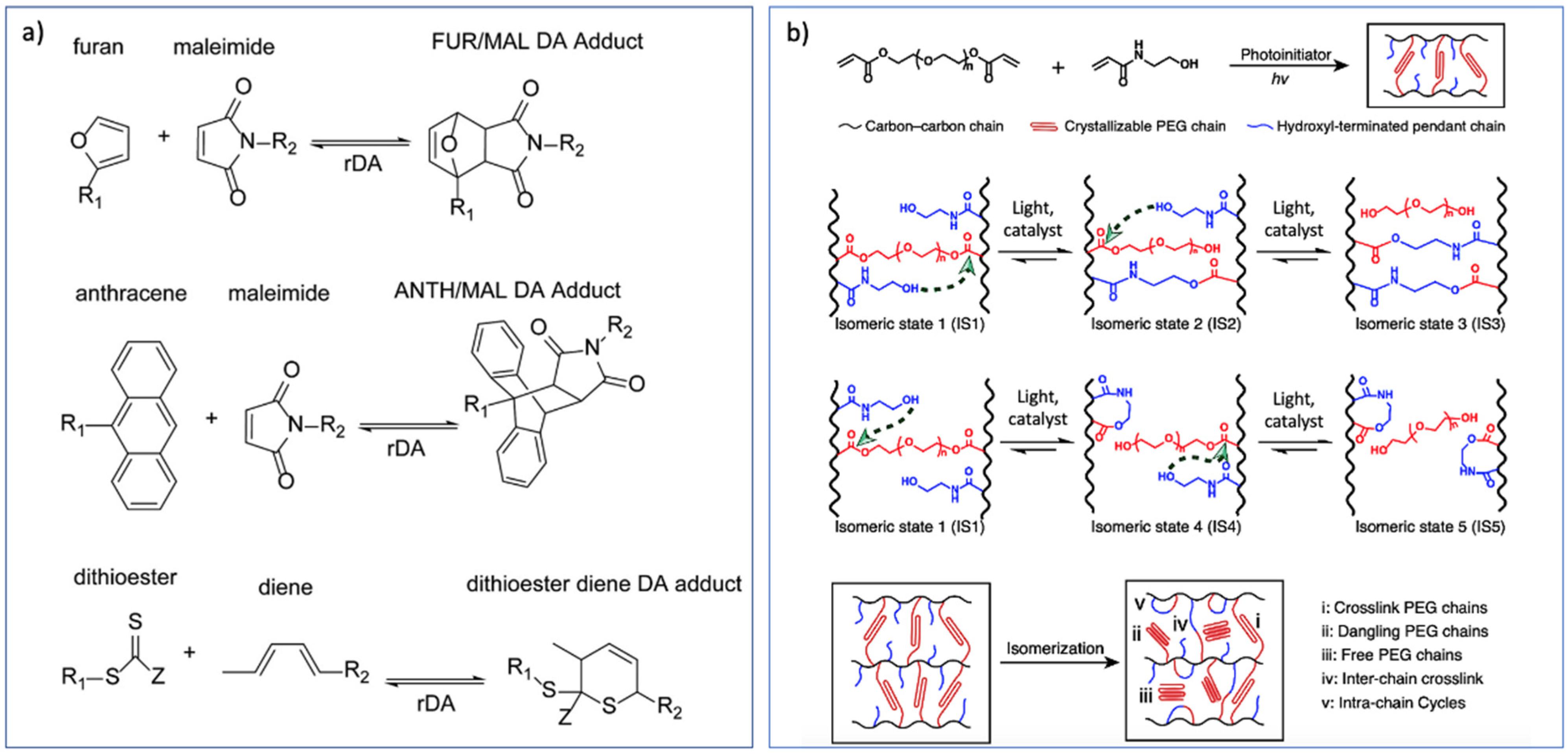
4.2. Reversible SMPs Based on Thermo- and Light-Activated Covalent Bonds
5. Stimulus-Responsive Methods
6. SMPs with Biomimetic Micro-/Nanofibrous Structures
7. Development of Electrospinning Process for SMPs
7.1. Conventional Electrospinning Process
7.2. Coaxial Electrospinning
7.3. Electrospinning of SMPs with Functional Fillers
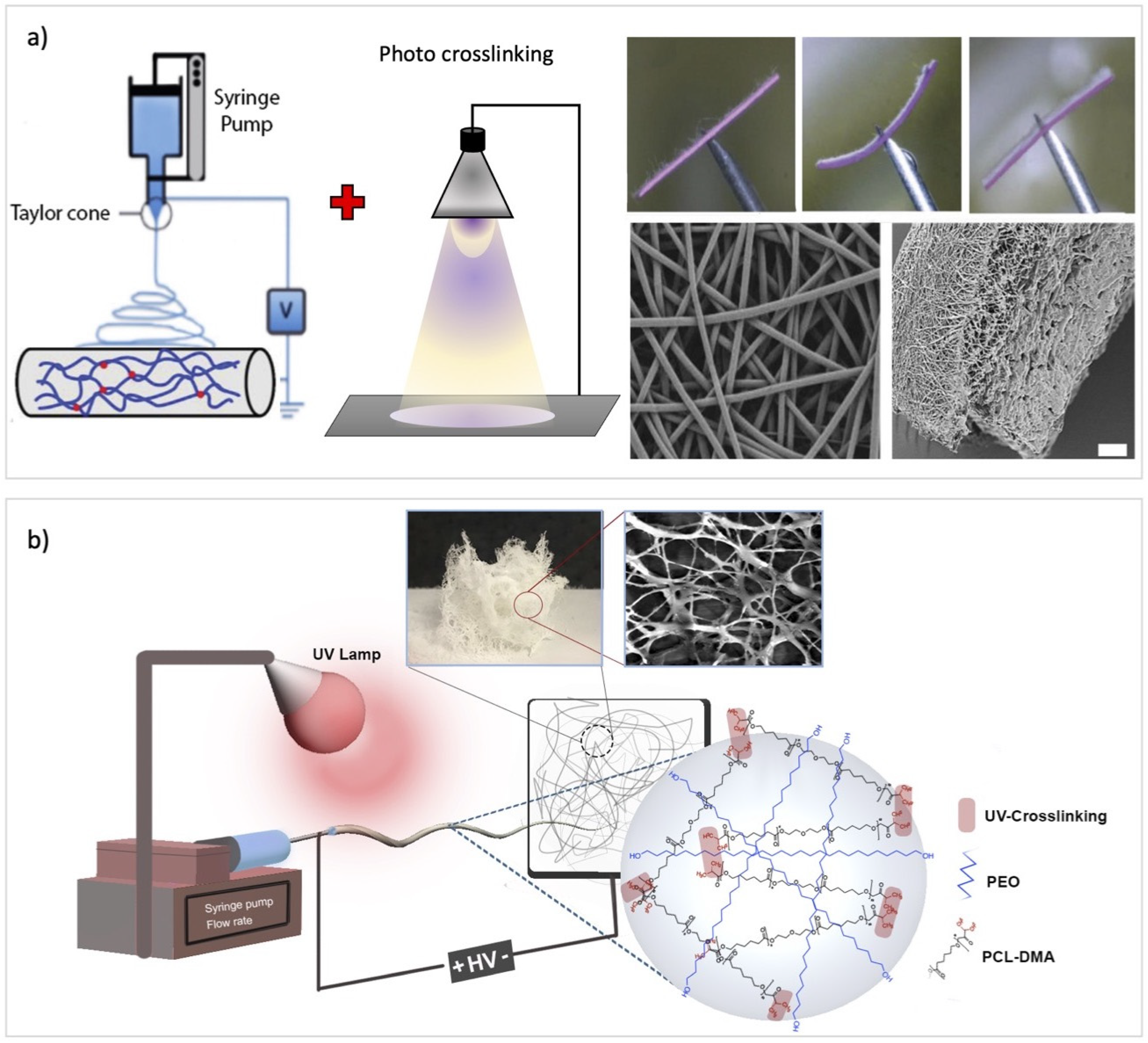
7.4. Electrospinning along with UV Irradiation
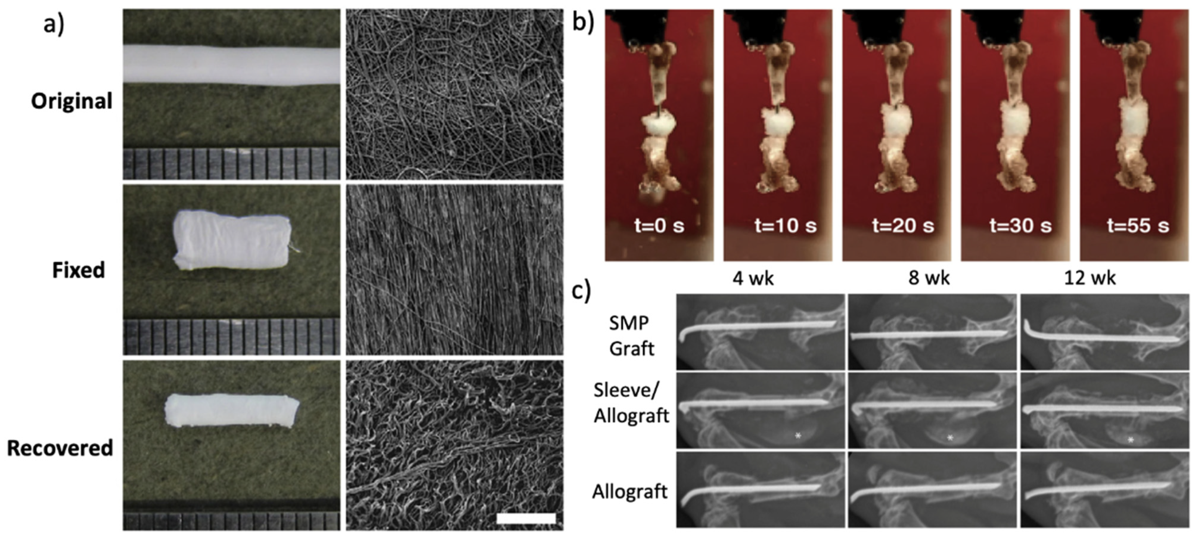
8. Biomedical Applications for SMP Fibers
9. Regulation of Cell Behaviors Using Biomimetic SMP Nanofibers
10. Conclusions and Future Challenges
Author Contributions
Funding
Conflicts of Interest
References
- Chen, H.-M.; Wang, L.; Zhou, S.-B. Recent Progress in Shape Memory Polymers for Biomedical Applications. Chin. J. Polym. Sci. 2018, 36, 905–917. [Google Scholar] [CrossRef]
- Mu, T.; Liu, L.; Lan, X.; Liu, Y.; Leng, J. Shape memory polymers for composites. Compos. Sci. Technol. 2018, 160, 169–198. [Google Scholar] [CrossRef]
- Lee, H.-F.; Yu, H. Study of electroactive shape memory polyurethane–carbon nanotube hybrids. Soft Matter 2011, 7, 3801–3807. [Google Scholar] [CrossRef]
- Huang, W.M.; Yang, B.; Zhao, Y.; Ding, Z. Thermo-moisture responsive polyurethane shape-memory polymer and composites: A review. J. Mater. Chem. 2010, 20, 3367–3381. [Google Scholar] [CrossRef]
- Zhao, W.; Liu, L.; Zhang, F.; Leng, J.; Liu, Y. Shape memory polymers and their composites in biomedical applications. Mater. Sci. Eng. C 2019, 97, 864–883. [Google Scholar] [CrossRef] [PubMed]
- Hu, J.; Chen, S. A review of actively moving polymers in textile applications. J. Mater. Chem. 2010, 20, 3346–3355. [Google Scholar] [CrossRef]
- Lan, X.; Liu, Y.; Lv, H.; Wang, X.; Leng, J.; Du, S. Fiber reinforced shape-memory polymer composite and its application in a deployable hinge. Smart Mater. Struct. 2009, 18, 024002. [Google Scholar] [CrossRef] [Green Version]
- Leng, J.; Lv, H.; Liu, Y.; Du, S. Electroactivate shape-memory polymer filled with nanocarbon particles and short carbon fibers. Appl. Phys. Lett. 2007, 91, 144105. [Google Scholar] [CrossRef]
- Cha, D.I.; Kim, H.Y.; Lee, K.H.; Jung, Y.C.; Cho, J.W.; Chun, B.C. Electrospun nonwovens of shape-memory polyurethane block copolymers. J. Appl. Polym. Sci. 2005, 96, 460–465. [Google Scholar] [CrossRef]
- Tan, L.; Gan, L.; Hu, J.; Zhu, Y.; Han, J. Functional shape memory composite nanofibers with graphene oxide filler. Compos. Part A Appl. Sci. Manuf. 2015, 76, 115–123. [Google Scholar] [CrossRef]
- Zhuo, H.; Hu, J.; Chen, S. Electrospun polyurethane nanofibres having shape memory effect. Mater. Lett. 2008, 62, 2074–2076. [Google Scholar] [CrossRef]
- Zhuo, H.; Hu, J.; Chen, S. Study of the thermal properties of shape memory polyurethane nanofibrous nonwoven. J. Mater. Sci. 2011, 46, 3464–3469. [Google Scholar] [CrossRef]
- Chen, H.; Cao, X.; Zhang, J.; Zhang, J.; Ma, Y.; Shi, G.; Ke, Y.; Tong, D.; Jiang, L. Electrospun shape memory film with reversible fibrous structure. J. Mater. Chem. 2012, 22, 22387–22391. [Google Scholar] [CrossRef]
- Zhang, J.-N.; Ma, Y.-M.; Zhang, J.-J.; Xu, D.; Yang, Q.-L.; Guan, J.-G.; Cao, X.-Y.; Jiang, L. Microfiber SMPU film affords quicker shape recovery than the bulk one. Mater. Lett. 2011, 65, 3639–3642. [Google Scholar] [CrossRef]
- Greiner, A.; Wendorff, J.H. Electrospinning: A Fascinating Method for the Preparation of Ultrathin Fibers. Angew. Chem. Int. Ed. 2007, 46, 5670–5703. [Google Scholar] [CrossRef]
- Ko, Y.-I.; Kim, B.-S.; Bae, J.-S.; Kim, Y.-A.; Kim, I.-S. Silicone-coated elastomeric polylactide nanofiber filaments: Mechanical properties and shape memory behavior. Rsc Adv. 2013, 3, 20091–20098. [Google Scholar] [CrossRef]
- Iregui, A.; Irusta, L.; Llorente, O.; Martin, L.; Calvo-Correas, T.; Eceiza, A.; González, A. Electrospinning of cationically polymerized epoxy/polycaprolactone blends to obtain shape memory fibers (SMF). Eur. Polym. J. 2017, 94, 376–383. [Google Scholar] [CrossRef]
- Chung, Y.C.; Yang, K.; Choi, J.W.; Chun, B.C. Characterisation and application of polyurethane copolymers grafted with photoluminescent dyes. Coloration Technol. 2014, 130, 305–313. [Google Scholar] [CrossRef]
- Zhuo, H.T.; Hu, J.L.; Chen, S.J. Coaxial electrospun polyurethane core-shell nanofibers for shape memory and antibacterial nanomaterials. Express Polym. Lett. 2011, 5, 182–187. [Google Scholar] [CrossRef]
- Li, W.; Yan, Z.; Ren, J.; Qu, X. Manipulating cell fate: Dynamic control of cell behaviors on functional platforms. Chem. Soc. Rev. 2018, 47, 8639–8684. [Google Scholar] [CrossRef]
- Taylor, C.T.; Colgan, S.P. Regulation of immunity and inflammation by hypoxia in immunological niches. Nat. Rev. Immunol. 2017, 17, 774–785. [Google Scholar] [CrossRef] [PubMed]
- Gajewski, T.F.; Schreiber, H.; Fu, Y.-X. Innate and adaptive immune cells in the tumor microenvironment. Nat. Immunol. 2013, 14, 1014–1022. [Google Scholar] [CrossRef] [Green Version]
- Hynes, R.O. The Extracellular Matrix: Not Just Pretty Fibrils. Science 2009, 326, 1216. [Google Scholar] [CrossRef] [PubMed] [Green Version]
- Yang, G.; Li, X.; He, Y.; Ma, J.; Ni, G.; Zhou, S. From nano to micro to macro: Electrospun hierarchically structured polymeric fibers for biomedical applications. Prog. Polym. Sci. 2018, 81, 80–113. [Google Scholar] [CrossRef]
- Lu, T.; Hu, H.; Li, Y.; Jiang, Q.; Su, J.; Lin, H.; Xiao, Y.; Zhu, X.; Zhang, X. Bioactive scaffolds based on collagen filaments with tunable physico-chemical and biological features. Soft Matter 2020, 16, 4540–4548. [Google Scholar] [CrossRef]
- Kawai, T.; Matsuda, T. Plastic Molded Articles with Shape Memory Property. U.S. patent US 4,950,258, 21 August 1990. [Google Scholar]
- Lendlein, A.; Langer, R. Biodegradable, Elastic Shape-Memory Polymers for Potential Biomedical Applications. Science 2002, 296, 1673. [Google Scholar] [CrossRef]
- Bellin, I.; Kelch, S.; Langer, R.; Lendlein, A. Polymeric triple-shape materials. Proc. Natl. Acad. Sci. USA 2006, 103, 18043. [Google Scholar] [CrossRef] [Green Version]
- Chen, S.; Hu, J.; Zhuo, H.; Zhu, Y. Two-way shape memory effect in polymer laminates. Mater. Lett. 2008, 62, 4088–4090. [Google Scholar] [CrossRef]
- Chung, T.; Romo-Uribe, A.; Mather, P.T. Two-Way Reversible Shape Memory in a Semicrystalline Network. Macromolecules 2008, 41, 184–192. [Google Scholar] [CrossRef]
- Boland, E.D.; Coleman, B.D.; Barnes, C.P.; Simpson, D.G.; Wnek, G.E.; Bowlin, G.L. Electrospinning polydioxanone for biomedical applications. Acta Biomater. 2005, 1, 115–123. [Google Scholar] [CrossRef]
- Gong, T.; Li, W.; Chen, H.; Wang, L.; Shao, S.; Zhou, S. Remotely actuated shape memory effect of electrospun composite nanofibers. Acta Biomater. 2012, 8, 1248–1259. [Google Scholar] [CrossRef]
- Tseng, L.-F.; Mather, P.T.; Henderson, J.H. Shape-memory-actuated change in scaffold fiber alignment directs stem cell morphology. Acta Biomater. 2013, 9, 8790–8801. [Google Scholar] [CrossRef] [PubMed]
- Peterson, G.I.; Dobrynin, A.V.; Becker, M.L. Biodegradable Shape Memory Polymers in Medicine. Adv. Healthc. Mater. 2017, 6, 1700694. [Google Scholar] [CrossRef] [PubMed] [Green Version]
- Zare, M.; Prabhakaran, M.P.; Parvin, N.; Ramakrishna, S. Thermally-induced two-way shape memory polymers: Mechanisms, structures, and applications. Chem. Eng. J. 2019, 374, 706–720. [Google Scholar] [CrossRef]
- Leng, J.; Lan, X.; Liu, Y.; Du, S. Shape-memory polymers and their composites: Stimulus methods and applications. Prog. Mater. Sci. 2011, 56, 1077–1135. [Google Scholar] [CrossRef]
- Wang, Y.; Li, Y.; Luo, Y.; Huang, M.; Liang, Z. Synthesis and characterization of a novel biodegradable thermoplastic shape memory polymer. Mater. Lett. 2009, 63, 347–349. [Google Scholar] [CrossRef]
- Zhang, L.; Huang, M.; Yu, R.; Huang, J.; Dong, X.; Zhang, R.; Zhu, J. Bio-based shape memory polyurethanes (Bio-SMPUs) with short side chains in the soft segment. J. Mater. Chem. A 2014, 2, 11490–11498. [Google Scholar] [CrossRef]
- Behl, M.; Lendlein, A. Triple-shape polymers. J. Mater. Chem. 2010, 20, 3335–3345. [Google Scholar] [CrossRef]
- Liu, Y. Polymerization-induced phase separation and resulting thermomechanical properties of thermosetting/reactive nonlinear polymer blends: A review. J. Appl. Polym. Sci. 2013, 127, 3279–3292. [Google Scholar] [CrossRef]
- Torbati, A.H.; Nejad, H.B.; Ponce, M.; Sutton, J.P.; Mather, P.T. Properties of triple shape memory composites prepared via polymerization-induced phase separation. Soft Matter 2014, 10, 3112–3121. [Google Scholar] [CrossRef]
- Leibler, L. Theory of Microphase Separation in Block Copolymers. Macromolecules 1980, 13, 1602–1617. [Google Scholar] [CrossRef]
- Luo, Y.; Guo, Y.; Gao, X.; Li, B.-G.; Xie, T. A General Approach Towards Thermoplastic Multishape-Memory Polymers via Sequence Structure Design. Adv. Mater. 2013, 25, 743–748. [Google Scholar] [CrossRef]
- Li, J.; Liu, T.; Xia, S.; Pan, Y.; Zheng, Z.; Ding, X.; Peng, Y. A versatile approach to achieve quintuple-shape memory effect by semi-interpenetrating polymer networks containing broadened glass transition and crystalline segments. J. Mater. Chem. 2011, 21, 12213–12217. [Google Scholar] [CrossRef]
- Luo, X.; Mather, P.T. Triple-Shape Polymeric Composites (TSPCs). Adv. Funct. Mater. 2010, 20, 2649–2656. [Google Scholar] [CrossRef]
- Zhang, F.; Zhang, Z.; Liu, Y.; Lu, H.; Leng, J. The quintuple-shape memory effect in electrospun nanofiber membranes. Smart Mater. Struct. 2013, 22, 085020. [Google Scholar] [CrossRef]
- Meng, H.; Mohamadian, H.; Stubblefield, M.; Jerro, D.; Ibekwe, S.; Pang, S.-S.; Li, G. Various shape memory effects of stimuli-responsive shape memory polymers. Smart Mater. Struct. 2013, 22, 093001. [Google Scholar] [CrossRef]
- Qin, H.; Mather, P.T. Combined One-Way and Two-Way Shape Memory in a Glass-Forming Nematic Network. Macromolecules 2009, 42, 273–280. [Google Scholar] [CrossRef]
- Chen, S.; Hu, J.; Zhuo, H. Properties and mechanism of two-way shape memory polyurethane composites. Compos. Sci. Technol. 2010, 70, 1437–1443. [Google Scholar] [CrossRef]
- Tamagawa, H. Thermo-responsive two-way shape changeable polymeric laminate. Mater. Lett. 2010, 64, 749–751. [Google Scholar] [CrossRef]
- Wu, Y.; Hu, J.; Han, J.; Zhu, Y.; Huang, H.; Li, J.; Tang, B. Two-way shape memory polymer with “switch–spring” composition by interpenetrating polymer network. J. Mater. Chem. A 2014, 2, 18816–18822. [Google Scholar] [CrossRef]
- Fei, F.L.; Zhi, R.M.; Qiu, Z.M.; Dong, C.X. A Facile Approach Toward Scalable Fabrication of Reversible Shape-Memory Polymers with Bonded Elastomer Microphases as Internal Stress Provider. Macromol. Rapid Commun. 2017, 38, 1700124. [Google Scholar]
- Gall, K.; Dunn, M.L.; Liu, Y.; Finch, D.; Lake, M.; Munshi, N.A. Shape memory polymer nanocomposites. Acta Mater. 2002, 50, 5115–5126. [Google Scholar] [CrossRef]
- Pandini, S.; Agnelli, S.; Merlettini, A.; Gualandi, C.; Focarete, M.L.; Paderni, K.; Messori, M.; Toselli, M. Two-Way Shape Memory Behavior of Electrospun Non-Woven Mats Prepared from Sol-Gel Crosslinked Poly(ε-Caprolactone). Adv. Sci. Technol. 2017, 97, 100–105. [Google Scholar] [CrossRef]
- Zare, M.; Parvin, N.; Prabhakaran, M.P.; Mohandesi, J.A.; Ramakrishna, S. Highly porous 3D sponge-like shape memory polymer for tissue engineering application with remote actuation potential. Compos. Sci. Technol. 2019, 184, 107874. [Google Scholar] [CrossRef]
- Lewis, C.L.; Dell, E.M. A review of shape memory polymers bearing reversible binding groups. J. Polym. Sci. Part B Polym. Phys. 2016, 54, 1340–1364. [Google Scholar] [CrossRef]
- Raquez, J.M.; Vanderstappen, S.; Meyer, F.; Verge, P.; Alexandre, M.; Thomassin, J.M.; Jerome, C.; Dubois, P. Design of cross-linked semicrystalline poly(epsilon-caprolactone)-based networks with one-way and two-way shape-memory properties through Diels-Alder reactions. Chemistry 2011, 17, 10135–10143. [Google Scholar] [CrossRef] [PubMed]
- Defize, T.; Riva, R.; Raquez, J.-M.; Dubois, P.; Jérôme, C.; Alexandre, M. Thermoreversibly Crosslinked Poly(ε-caprolactone) as Recyclable Shape-Memory Polymer Network. Macromol. Rapid Commun. 2011, 32, 1264–1269. [Google Scholar] [CrossRef]
- Zeng, C.; Seino, H.; Ren, J.; Hatanaka, K.; Yoshie, N. Bio-Based Furan Polymers with Self-Healing Ability. Macromolecules 2013, 46, 1794–1802. [Google Scholar] [CrossRef]
- Zeng, C.; Seino, H.; Ren, J.; Yoshie, N. Polymers with Multishape Memory Controlled by Local Glass Transition Temperature. Acs Appl. Mater. Interfaces 2014, 6, 2753–2758. [Google Scholar] [CrossRef]
- Michal, B.T.; Jaye, C.A.; Spencer, E.J.; Rowan, S.J. Inherently Photohealable and Thermal Shape-Memory Polydisulfide Networks. ACS Macro Lett. 2013, 2, 694–699. [Google Scholar] [CrossRef]
- Xiao, L.; Wei, M.; Zhan, M.; Zhang, J.; Xie, H.; Deng, X.; Yang, K.; Wang, Y. Novel triple-shape PCU/PPDO interpenetrating polymer networks constructed by self-complementary quadruple hydrogen bonding and covalent bonding. Polym. Chem. 2014, 5, 2231–2241. [Google Scholar] [CrossRef]
- Niu, Y.; Zhang, P.; Zhang, J.; Xiao, L.; Yang, K.; Wang, Y. Poly(p-dioxanone)–poly(ethylene glycol) network: Synthesis, characterization, and its shape memory effect. Polym. Chem. 2012, 3, 2508–2516. [Google Scholar] [CrossRef]
- Dong, J.; Weiss, R.A. Shape Memory Behavior of Zinc Oleate-Filled Elastomeric Ionomers. Macromolecules 2011, 44, 8871–8879. [Google Scholar] [CrossRef]
- Merline, J.D.; Nair, C.P.R.; Gouri, C.; Shrisudha, T.; Ninan, K.N. Shape memory characterization of polytetra methylene oxide/poly (acrylic acid-co-acrylonitrile) complexed gel. J. Mater. Sci. 2007, 42, 5897–5902. [Google Scholar] [CrossRef]
- Kumpfer, J.R.; Rowan, S.J. Thermo-, Photo-, and Chemo-Responsive Shape-Memory Properties from Photo-Cross-Linked Metallo-Supramolecular Polymers. J. Am. Chem. Soc. 2011, 133, 12866–12874. [Google Scholar] [CrossRef] [PubMed]
- Lendlein, A.; Jiang, H.; Jünger, O.; Langer, R. Light-induced shape-memory polymers. Nature 2005, 434, 879–882. [Google Scholar] [CrossRef] [PubMed]
- Nagata, M.; Kitazima, I. Photocurable biodegradable poly(ε-caprolactone)/poly(ethylene glycol) multiblock copolymers showing shape-memory properties. Colloid Polym. Sci. 2006, 284, 380–386. [Google Scholar] [CrossRef]
- Nagata, M.; Sato, Y. Synthesis and properties of photocurable biodegradable multiblock copolymers based on poly(ε-caprolactone) and poly(L-lactide) segments. J. Polym. Sci. Part A Polym. Chem. 2005, 43, 2426–2439. [Google Scholar] [CrossRef]
- He, J.; Zhao, Y.; Zhao, Y. Photoinduced bending of a coumarin-containing supramolecular polymer. Soft Matter 2009, 5, 308–310. [Google Scholar] [CrossRef]
- Kumar, G.S.; Neckers, D.C. Photochemistry of azobenzene-containing polymers. Chem. Rev. 1989, 89, 1915–1925. [Google Scholar] [CrossRef]
- Lee, K.M.; Koerner, H.; Vaia, R.A.; Bunning, T.J.; White, T.J. Light-activated shape memory of glassy, azobenzene liquid crystalline polymer networks. Soft Matter 2011, 7, 4318–4324. [Google Scholar] [CrossRef]
- Zhang, X.; Zhou, Q.; Liu, H.; Liu, H. UV light induced plasticization and light activated shape memory of spiropyran doped ethylene-vinyl acetate copolymers. Soft Matter 2014, 10, 3748–3754. [Google Scholar] [CrossRef]
- Miao, W.; Zou, W.; Jin, B.; Ni, C.; Zheng, N.; Zhao, Q.; Xie, T. On demand shape memory polymer via light regulated topological defects in a dynamic covalent network. Nat. Commun. 2020, 11, 4257. [Google Scholar] [CrossRef]
- Hu, J.; Zhu, Y.; Huang, H.; Lu, J. Recent advances in shape–memory polymers: Structure, mechanism, functionality, modeling and applications. Prog. Polym. Sci. 2012, 37, 1720–1763. [Google Scholar] [CrossRef]
- Xie, F.; Huang, L.; Liu, Y.; Leng, J. Synthesis and characterization of high temperature cyanate-based shape memory polymers with functional polybutadiene/acrylonitrile. Polymer 2014, 55, 5873–5879. [Google Scholar] [CrossRef]
- Du, H.; Song, Z.; Wang, J.; Liang, Z.; Shen, Y.; You, F. Microwave-induced shape-memory effect of silicon carbide/poly(vinyl alcohol) composite. Sens. Actuators A Phys. 2015, 228, 1–8. [Google Scholar] [CrossRef]
- Yoonessi, M.; Shi, Y.; Scheiman, D.A.; Lebron-Colon, M.; Tigelaar, D.M.; Weiss, R.A.; Meador, M.A. Graphene Polyimide Nanocomposites; Thermal, Mechanical, and High-Temperature Shape Memory Effects. ACS Nano 2012, 6, 7644–7655. [Google Scholar] [CrossRef]
- Lu, H.; Liang, F.; Gou, J.; Leng, J.; Du, S. Synergistic effect of Ag nanoparticle-decorated graphene oxide and carbon fiber on electrical actuation of polymeric shape memory nanocomposites. Smart Mater. Struct. 2014, 23, 085034. [Google Scholar] [CrossRef]
- Tandon, G.P.; Goecke, K.; Cable, K.; Baur, J. Environmental Durability of Fabric-Reinforced Shape-Memory Polymer Composites. J. Intell. Mater. Syst. Struct. 2010, 21, 1365–1381. [Google Scholar] [CrossRef]
- Lu, H.; Gou, J.; Leng, J.; Du, S. Magnetically aligned carbon nanotube in nanopaper enabled shape-memory nanocomposite for high speed electrical actuation. Appl. Phys. Lett. 2011, 98, 174105. [Google Scholar] [CrossRef] [Green Version]
- Kumar, U.N.; Kratz, K.; Heuchel, M.; Behl, M.; Lendlein, A. Shape-Memory Nanocomposites with Magnetically Adjustable Apparent Switching Temperatures. Adv. Mater. 2011, 23, 4157–4162. [Google Scholar] [CrossRef] [PubMed]
- Mohr, R.; Kratz, K.; Weigel, T.; Lucka-Gabor, M.; Moneke, M.; Lendlein, A. Initiation of shape-memory effect by inductive heating of magnetic nanoparticles in thermoplastic polymers. Proc. Natl. Acad. Sci. USA 2006, 103, 3540. [Google Scholar] [CrossRef] [PubMed] [Green Version]
- Li, G.; Fei, G.; Xia, H.; Han, J.; Zhao, Y. Spatial and temporal control of shape memory polymers and simultaneous drug release using high intensity focused ultrasound. J. Mater. Chem. 2012, 22, 7692–7696. [Google Scholar] [CrossRef]
- Li, G.; Yan, Q.; Xia, H.; Zhao, Y. Therapeutic-Ultrasound-Triggered Shape Memory of a Melamine-Enhanced Poly(vinyl alcohol) Physical Hydrogel. ACS Appl. Mater. Interfaces 2015, 7, 12067–12073. [Google Scholar] [CrossRef] [PubMed]
- Fang, Y.; Ni, Y.; Leo, S.-Y.; Taylor, C.; Basile, V.; Jiang, P. Reconfigurable photonic crystals enabled by pressure-responsive shape-memory polymers. Nat. Commun. 2015, 6, 7416. [Google Scholar] [CrossRef] [Green Version]
- Huang, W.M.; Yang, B.; An, L.; Li, C.; Chan, Y.S. Water-driven programmable polyurethane shape memory polymer: Demonstration and mechanism. Appl. Phys. Lett. 2005, 86, 114105. [Google Scholar] [CrossRef]
- Lu, H.; Leng, J.; Du, S. A phenomenological approach for the chemo-responsive shape memory effect in amorphous polymers. Soft Matter 2013, 9, 3851–3858. [Google Scholar] [CrossRef]
- Xiao, R.; Guo, J.; Safranski, D.L.; Nguyen, T.D. Solvent-driven temperature memory and multiple shape memory effects. Soft Matter 2015, 11, 3977–3985. [Google Scholar] [CrossRef]
- Wang, W.; Lu, H.; Liu, Y.; Leng, J. Sodium dodecyl sulfate/epoxy composite: Water-induced shape memory effect and its mechanism. J. Mater. Chem. A 2014, 2, 5441–5449. [Google Scholar] [CrossRef]
- Dallmeyer, I.; Chowdhury, S.; Kadla, J.F. Preparation and Characterization of Kraft Lignin-Based Moisture-Responsive Films with Reversible Shape-Change Capability. Biomacromolecules 2013, 14, 2354–2363. [Google Scholar] [CrossRef]
- He, Z.; Satarkar, N.; Xie, T.; Cheng, Y.-T.; Hilt, J.Z. Remote Controlled Multishape Polymer Nanocomposites with Selective Radiofrequency Actuations. Adv. Mater. 2011, 23, 3192–3196. [Google Scholar] [CrossRef]
- Li, W.; Liu, Y.; Leng, J. Selectively actuated multi-shape memory effect of a polymer multicomposite. J. Mater. Chem. A 2015, 3, 24532–24539. [Google Scholar] [CrossRef]
- Zhao, Q.; Qi, H.J.; Xie, T. Recent progress in shape memory polymer: New behavior, enabling materials, and mechanistic understanding. Prog. Polym. Sci. 2015, 49–50, 79–120. [Google Scholar]
- Jiang, S.; Liu, F.; Lerch, A.; Ionov, L.; Agarwal, S. Unusual and Superfast Temperature-Triggered Actuators. Adv. Mater. 2015, 27, 4865–4870. [Google Scholar] [CrossRef]
- Wang, J.; Li, J.; Li, N.; Guo, X.; He, L.; Cao, X.; Zhang, W.; He, R.; Qian, Z.; Cao, Y.; et al. A Bottom-Up Approach to Dual Shape-Memory Effects. Chem. Mater. 2015, 27, 2439–2448. [Google Scholar] [CrossRef]
- Wischke, C.; Schossig, M.; Lendlein, A. Shape-Memory Effect of Micro-/Nanoparticles from Thermoplastic Multiblock Copolymers. Small 2014, 10, 83–87. [Google Scholar] [CrossRef] [PubMed]
- Singhal, P.; Rodriguez, J.N.; Small, W.; Eagleston, S.; Van de Water, J.; Maitland, D.J.; Wilson, T.S. Ultra low density and highly crosslinked biocompatible shape memory polyurethane foams. J. Polym. Sci. Part B Polym. Phys. 2012, 50, 724–737. [Google Scholar] [CrossRef] [PubMed] [Green Version]
- Guillaume, O.; Daly, A.; Lennon, K.; Gansau, J.; Buckley, S.F.; Buckley, C.T. Shape-memory porous alginate scaffolds for regeneration of the annulus fibrosus: Effect of TGF-β3 supplementation and oxygen culture conditions. Acta Biomater. 2014, 10, 1985–1995. [Google Scholar] [CrossRef] [PubMed]
- Almeida, H.V.; Sathy, B.N.; Dudurych, I.; Buckley, C.T.; O’Brien, F.J.; Kelly, D.J. Anisotropic Shape-Memory Alginate Scaffolds Functionalized with Either Type I or Type II Collagen for Cartilage Tissue Engineering. Tissue Eng. Part A 2016, 23, 55–68. [Google Scholar] [CrossRef]
- Gao, H.-L.; Lu, Y.; Mao, L.-B.; An, D.; Xu, L.; Gu, J.-T.; Long, F.; Yu, S.-H. A shape-memory scaffold for macroscale assembly of functional nanoscale building blocks. Mater. Horiz. 2014, 1, 69–73. [Google Scholar] [CrossRef]
- Correia, C.O.; Leite, Á.J.; Mano, J.F. Chitosan/bioactive glass nanoparticles scaffolds with shape memory properties. Carbohydr. Polym. 2015, 123, 39–45. [Google Scholar] [CrossRef]
- Jiang, L.B.; Su, D.H.; Liu, P.; Ma, Y.Q.; Shao, Z.Z.; Dong, J. Shape-memory collagen scaffold for enhanced cartilage regeneration: Native collagen versus denatured collagen. Osteoarthr. Cartil. 2018, 26, 1389–1399. [Google Scholar] [CrossRef] [Green Version]
- Liu, X.; Zhao, K.; Gong, T.; Song, J.; Bao, C.; Luo, E.; Weng, J.; Zhou, S. Delivery of Growth Factors Using a Smart Porous Nanocomposite Scaffold to Repair a Mandibular Bone Defect. Biomacromolecules 2014, 15, 1019–1030. [Google Scholar] [CrossRef]
- Ebara, M.; Uto, K.; Idota, N.; Hoffman, J.M.; Aoyagi, T. Shape-Memory Surface with Dynamically Tunable Nano-Geometry Activated by Body Heat. Adv. Mater. 2012, 24, 273–278. [Google Scholar] [CrossRef] [PubMed]
- Bao, M.; Lou, X.; Zhou, Q.; Dong, W.; Yuan, H.; Zhang, Y. Electrospun Biomimetic Fibrous Scaffold from Shape Memory Polymer of PDLLA-co-TMC for Bone Tissue Engineering. ACS Appl. Mater. Interfaces 2014, 6, 2611–2621. [Google Scholar] [CrossRef] [PubMed]
- Bao, M.; Wang, X.; Yuan, H.; Lou, X.; Zhao, Q.; Zhang, Y. HAp incorporated ultrafine polymeric fibers with shape memory effect for potential use in bone screw hole healing. J. Mater. Chem. B 2016, 4, 5308–5320. [Google Scholar] [CrossRef] [PubMed]
- Wang, C.; Zhou, Y.; Wang, M. In situ delivery of rhBMP-2 in surface porous shape memory scaffolds developed through cryogenic 3D plotting. Mater. Lett. 2017, 189, 140–143. [Google Scholar] [CrossRef]
- Chung, S.E.; Park, C.H.; Yu, W.-R.; Kang, T.J. Thermoresponsive shape memory characteristics of polyurethane electrospun web. J. Appl. Polym. Sci. 2011, 120, 492–500. [Google Scholar] [CrossRef]
- Du, L.; Yang, S.; Li, W.; Li, H.; Feng, S.; Zeng, R.; Yu, B.; Xiao, L.; Nie, H.-Y.; Tu, M. Scaffold composed of porous vancomycin-loaded poly(lactide-co-glycolide) microspheres: A controlled-release drug delivery system with shape-memory effect. Mater. Sci. Eng. C 2017, 78, 1172–1178. [Google Scholar] [CrossRef]
- Yoon, J.J.; Kim, J.H.; Park, T.G. Dexamethasone-releasing biodegradable polymer scaffolds fabricated by a gas-foaming/salt-leaching method. Biomaterials 2003, 24, 2323–2329. [Google Scholar] [CrossRef]
- Lv, T.; Cheng, Z.; Zhang, D.; Zhang, E.; Zhao, Q.; Liu, Y.; Jiang, L. Superhydrophobic Surface With Shape Memory Micro/Nanostructure and Its Application in Rewritable Chip for Droplet Storage. ACS Nano 2016, 10, 9379–9386. [Google Scholar] [CrossRef]
- Lv, T.; Cheng, Z.; Zhang, E.; Kang, H.; Liu, Y.; Jiang, L. Self-Restoration of Superhydrophobicity on Shape Memory Polymer Arrays with Both Crushed Microstructure and Damaged Surface Chemistry. Small 2017, 13, 1503402. [Google Scholar] [CrossRef]
- Lee, E.M.; Smith, K.; Gall, K.; Boyan, B.D.; Schwartz, Z. Change in surface roughness by dynamic shape-memory acrylate networks enhances osteoblast differentiation. Biomaterials 2016, 110, 34–44. [Google Scholar] [CrossRef] [PubMed]
- Han, Y.; Liu, Y.; Wang, W.; Leng, J.; Jin, P. Controlled wettability based on reversible micro-cracking on a shape memory polymer surface. Soft Matter 2016, 12, 2708–2714. [Google Scholar] [CrossRef] [PubMed]
- Nahavandizadeh, N.; Rezaei, M. Preparation of Shape Memory Polyurethane/Hydroxyapatite Nanocomposite Scaffolds by Electrospinning Method and Investigation of Their Microstructure and Physical-Mechanical Properties. Polym.-Plast. Technol. Mater. 2020, 59, 1562–1573. [Google Scholar] [CrossRef]
- Lv, H.; Tang, D.; Sun, Z.; Gao, J.; Yang, X.; Jia, S.; Peng, J. Electrospun PCL-based polyurethane/HA microfibers as drug carrier of dexamethasone with enhanced biodegradability and shape memory performances. Colloid Polym. Sci. 2020, 298, 103–111. [Google Scholar] [CrossRef]
- Xiao, Y.; Gong, T.; Zhou, S. The functionalization of multi-walled carbon nanotubes by in situ deposition of hydroxyapatite. Biomaterials 2010, 31, 5182–5190. [Google Scholar] [CrossRef] [PubMed]
- Peng, Q.; Cheng, J.; Lu, S.; Li, Y. Electrospun hyperbranched polylactic acid–modified cellulose nanocrystals/polylactic acid for shape memory membranes with high mechanical properties. Polym. Adv. Technol. 2019, 31, 15–24. [Google Scholar] [CrossRef]
- Liu, Y.; Li, Y.; Chen, H.; Yang, G.; Zheng, X.; Zhou, S. Water-induced shape-memory poly(d,l-lactide)/microcrystalline cellulose composites. Carbohydr. Polym. 2014, 104, 101–108. [Google Scholar] [CrossRef]
- Zheng, X.; Zhou, S.; Yu, X.; Li, X.; Feng, B.; Qu, S.; Weng, J. Effect of In vitro degradation of poly(D,L-lactide)/β-tricalcium composite on its shape-memory properties. J. Biomed. Mater. Res. Part B Appl. Biomater. 2008, 86, 170–180. [Google Scholar] [CrossRef]
- Yu, X.; Zhou, S.; Zheng, X.; Xiao, Y.; Guo, T. Influence of in Vitro Degradation of a Biodegradable Nanocomposite on Its Shape Memory Effect. J. Phys. Chem. C 2009, 113, 17630–17635. [Google Scholar] [CrossRef]
- Yang, Q.; Fan, J.; Li, G. Artificial muscles made of chiral two-way shape memory polymer fibers. Appl. Phys. Lett. 2016, 109, 183701. [Google Scholar] [CrossRef]
- Kaursoin, J.; Agrawal, A.K. Melt spun thermoresponsive shape memory fibers based on polyurethanes: Effect of drawing and heat-setting on fiber morphology and properties. J. Appl. Polym. Sci. 2007, 103, 2172–2182. [Google Scholar] [CrossRef]
- Ji, F.; Zhu, Y.; Hu, J.; Liu, Y.; Yeung, L.-Y.; Ye, G. Smart polymer fibers with shape memory effect. Smart Mater. Struct. 2006, 15, 1547–1554. [Google Scholar] [CrossRef]
- Zhang, F.; Zhang, Z.; Liu, Y.; Cheng, W.; Huang, Y.; Leng, J. Thermosetting epoxy reinforced shape memory composite microfiber membranes: Fabrication, structure and properties. Compos. Part A Appl. Sci. Manuf. 2015, 76, 54–61. [Google Scholar] [CrossRef]
- Huang, Z.-M.; Zhang, Y.Z.; Kotaki, M.; Ramakrishna, S. A review on polymer nanofibers by electrospinning and their applications in nanocomposites. Compos. Sci. Technol. 2003, 63, 2223–2253. [Google Scholar] [CrossRef]
- Doshi, J.; Reneker, D.H. Electrospinning process and applications of electrospun fibers. J. Electrost. 1995, 35, 151–160. [Google Scholar] [CrossRef]
- Tashi, Z.; Zare, M.; Parvin, N. Application of phytic-acid as an in-situ crosslinking agent in electrospun gelatin-based scaffolds for skin tissue engineering. Mater. Lett. 2020, 264, 127275. [Google Scholar] [CrossRef]
- Frenot, A.; Chronakis, I.S. Polymer nanofibers assembled by electrospinning. Curr. Opin. Colloid Interface Sci. 2003, 8, 64–75. [Google Scholar] [CrossRef]
- Ghaseminezhad, K.; Zare, M.; Lashkarara, S.; Yousefzadeh, M.; Aghazadeh Mohandesi, J. Fabrication of althea officinalis loaded electrospun nanofibrous scaffold for potential application of skin tissue engineering. J. Appl. Polym. Sci. 2020, 137, 48587. [Google Scholar] [CrossRef]
- Meng, Q.; Hu, J.; Zhu, Y.; Lu, J.; Liu, Y. Morphology, phase separation, thermal and mechanical property differences of shape memory fibres prepared by different spinning methods. Smart Mater. Struct. 2007, 16, 1192–1197. [Google Scholar] [CrossRef]
- Zhuo, H.; Hu, J.; Chen, S.; Yeung, L. Preparation of polyurethane nanofibers by electrospinning. J. Appl. Polym. Sci. 2008, 109, 406–411. [Google Scholar] [CrossRef]
- Zhang, F.; Zhang, Z.; Liu, Y.; Leng, J. Shape memory properties of electrospun nafion nanofibers. Fibers Polym. 2014, 15, 534–539. [Google Scholar] [CrossRef]
- Zhang, F.; Zhang, Z.; Liu, Y.; Leng, J. Fabrication of Shape Memory Nanofibers by Electrospinning Method; SPIE: Bellingham, DC, USA, 2013; Volume 8687. [Google Scholar]
- Matsumoto, H.; Ishiguro, T.; Konosu, Y.; Minagawa, M.; Tanioka, A.; Richau, K.; Kratz, K.; Lendlein, A. Shape-memory properties of electrospun non-woven fabrics prepared from degradable polyesterurethanes containing poly(ω-pentadecalactone) hard segments. Eur. Polym. J. 2012, 48, 1866–1874. [Google Scholar] [CrossRef] [Green Version]
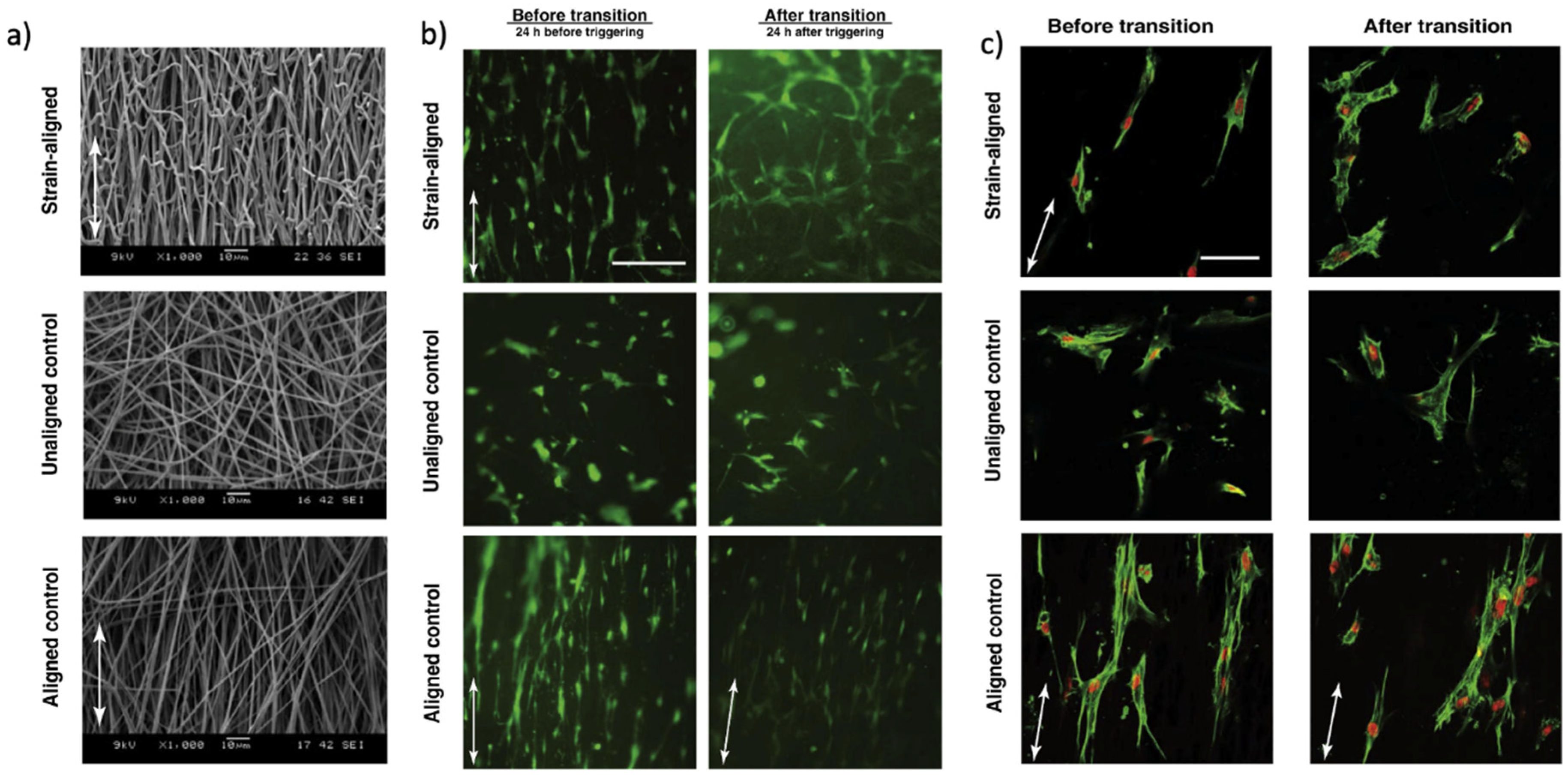
- Wang, J.; Quach, A.; Brasch, M.E.; Turner, C.E.; Henderson, J.H. On-command on/off switching of progenitor cell and cancer cell polarized motility and aligned morphology via a cytocompatible shape memory polymer scaffold. Biomaterials 2017, 140, 150–161. [Google Scholar] [CrossRef]
- Yoo, H.J.; Mahapatra, S.S.; Cho, J.W. High-Speed Actuation and Mechanical Properties of Graphene-Incorporated Shape Memory Polyurethane Nanofibers. J. Phys. Chem. C 2014, 118, 10408–10415. [Google Scholar] [CrossRef]
- Ang, J.Y.; Chan, B.Q.Y.; Kai, D.; Loh, X.J. Engineering Porous Water-Responsive Poly(PEG/PCL/PDMS Urethane) Shape Memory Polymers. Macromol. Mater. Eng. 2017, 302, 1700174. [Google Scholar] [CrossRef]
- Zhang, F.; Xia, Y.; Wang, L.; Liu, L.; Liu, Y.; Leng, J. Conductive Shape Memory Microfiber Membranes with Core–Shell Structures and Electroactive Performance. ACS Appl. Mater. Interfaces 2018, 10, 35526–35532. [Google Scholar] [CrossRef]
- Joon-Sung, A.; Woong-Ryeol, Y.; Ji Ho, Y.; Hee Youk, R. In situ temperature tunable pores of shape memory polyurethane membranes. Smart Mater. Struct. 2011, 20, 105024. [Google Scholar]
- Luo, C.J.; Edirisinghe, M. Core-Liquid-Induced Transition from Coaxial Electrospray to Electrospinning of Low-Viscosity Poly(lactide-co-glycolide) Sheath Solution. Macromolecules 2014, 47, 7930–7938. [Google Scholar] [CrossRef]
- Zhang, Q.; Kratz, K.; Lendlein, A. Shape-memory properties of degradable electrospun scaffolds based on hollow microfibers. Polym. Adv. Technol. 2015, 26, 1468–1475. [Google Scholar] [CrossRef]
- Liu, Y.; Li, Y.; Yang, G.; Zheng, X.; Zhou, S. Multi-Stimulus-Responsive Shape-Memory Polymer Nanocomposite Network Cross-Linked by Cellulose Nanocrystals. Acs Appl. Mater. Interfaces 2015, 7, 4118–4126. [Google Scholar] [CrossRef]
- Jiang, Z.-C.; Xiao, Y.-Y.; Kang, Y.; Pan, M.; Li, B.-J.; Zhang, S. Shape Memory Polymers Based on Supramolecular Interactions. Acs Appl. Mater. Interfaces 2017, 9, 20276–20293. [Google Scholar] [CrossRef]
- Hao, B.; Li, Y.; Xiao, X.; Dai, W.; Chen, H.; Chen, S. A facile photo-polymerization method for reconfigurable shape memory polymers. Mater. Lett. 2019, 254, 214–217. [Google Scholar] [CrossRef]
- Yao, Y.; Luo, Y.; Lu, H.; Wang, B. Remotely actuated porous composite membrane with shape memory property. Compos. Struct. 2018, 192, 507–515. [Google Scholar] [CrossRef]
- Zhang, Q.; Rudolph, T.; Benitez, A.J.; Gould, O.E.C.; Behl, M.; Kratz, K.; Lendlein, A. Temperature-controlled reversible pore size change of electrospun fibrous shape-memory polymer actuator based meshes. Smart Mater. Struct. 2019, 28, 055037. [Google Scholar] [CrossRef]
- Pandini, S.; Agnelli, S.; Merlettini, A.; Chiellini, F.; Gualandi, C.; Paderni, K.; Focarete, M.L.; Messori, M.; Toselli, M. Mutifunctional Electrospun Nonwoven Mats with Two-Way Shape Memory Behavior Prepared from Sol–Gel Crosslinked Poly(ε-Caprolactone). Macromol. Mater. Eng. 2017, 302, 1600519. [Google Scholar] [CrossRef] [Green Version]
- Zhang, K.; Wang, S.; Zhou, C.; Cheng, L.; Gao, X.; Xie, X.; Sun, J.; Wang, H.; Weir, M.D.; Reynolds, M.A.; et al. Advanced smart biomaterials and constructs for hard tissue engineering and regeneration. Bone Res. 2018, 6, 31. [Google Scholar] [CrossRef] [PubMed]
- Torbati, A.H.; Mather, R.T.; Reeder, J.E.; Mather, P.T. Fabrication of a light-emitting shape memory polymeric web containing indocyanine green. J. Biomed. Mater. Res. Part B Appl. Biomater. 2014, 102, 1236–1243. [Google Scholar] [CrossRef]
- He, J.; Huang, T.; Gan, L.; Zhou, Z.; Jiang, B.; Wu, Y.; Wu, F.; Gu, Z. Collagen-infiltrated porous hydroxyapatite coating and its osteogenic properties: In vitro and in vivo study. J. Biomed. Mater. Res. Part A 2012, 100A, 1706–1715. [Google Scholar] [CrossRef]
- Baker, R.M.; Tseng, L.-F.; Iannolo, M.T.; Oest, M.E.; Henderson, J.H. Self-deploying shape memory polymer scaffolds for grafting and stabilizing complex bone defects: A mouse femoral segmental defect study. Biomaterials 2016, 76, 388–398. [Google Scholar] [CrossRef]
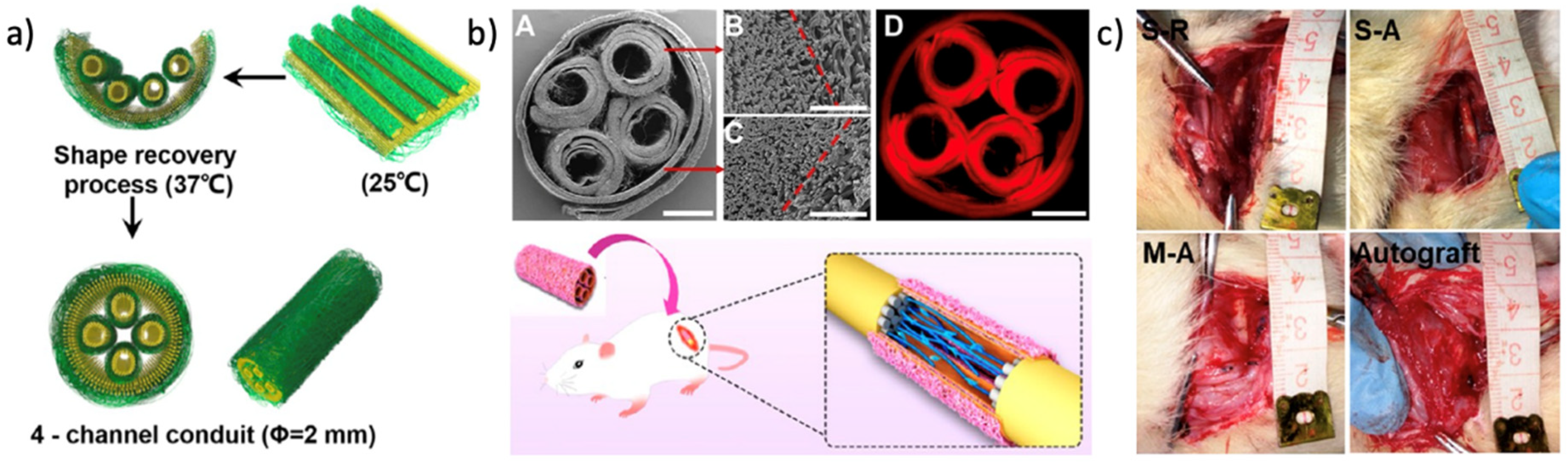
- Wang, J.; Xiong, H.; Zhu, T.; Liu, Y.; Pan, H.; Fan, C.; Zhao, X.; Lu, W.W. Bioinspired Multichannel Nerve Guidance Conduit Based on Shape Memory Nanofibers for Potential Application in Peripheral Nerve Repair. ACS Nano 2020, 14, 12579–12595. [Google Scholar] [CrossRef]
- Hsieh, C.-H.; Mohd Razali, N.A.; Lin, W.-C.; Yu, Z.-W.; Istiqomah, D.; Kotsuchibashi, Y.; Su, H.-H. Development of Thermo-Responsive Polycaprolactone–Polydimethylsiloxane Shrinkable Nanofibre Mesh. Nanomaterials 2020, 10, 1427. [Google Scholar] [CrossRef]
- Wang, X.; Yan, H.; Shen, Y.; Tang, H.; Yi, B.; Qin, C.; Zhang, Y. Shape Memory and Osteogenesis Capabilities of the Electrospun Poly(3-Hydroxybutyrate-co-3-Hydroxyvalerate) Modified Poly(l-Lactide) Fibrous Mats. Tissue Eng. Part A 2020, 27, 142–152. [Google Scholar] [CrossRef] [PubMed]
- Bil, M.; Kijeńska-Gawrońska, E.; Głodkowska-Mrówka, E.; Manda-Handzlik, A.; Mrówka, P. Design and in vitro evaluation of electrospun shape memory polyurethanes for self-fitting tissue engineering grafts and drug delivery systems. Mater. Sci. Eng. C 2020, 110, 110675. [Google Scholar] [CrossRef] [PubMed]
- Guan, X.; Xia, H.; Ni, Q.-Q. Shape memory polyurethane-based electrospun yarns for thermo-responsive actuation. J. Appl. Polym. Sci. 2021, 138, 50565. [Google Scholar] [CrossRef]
- Guan, X.; Dong, Y.; Xia, H.; Yao, J.; Ni, Q.-Q. Mechanical and shape memory performance of shape memory polyurethane-based aligned nanofibers. Polym. Test. 2020, 91, 106778. [Google Scholar] [CrossRef]
- Chen, M.; Li, L.; Xia, L.; Zhang, F.; Jiang, S.; Hu, H.; Li, X.; Wang, H. Temperature Responsive Shape-Memory Scaffolds with Circumferentially Aligned Nanofibers for Guiding Smooth Muscle Cell Behavior. Macromol. Biosci. 2020, 20, 1900312. [Google Scholar] [CrossRef] [PubMed]
- Iregui, A.; Irusta, L.; Martin, L.; González, A. Analysis of the Process Parameters for Obtaining a Stable Electrospun Process in Different Composition Epoxy/Poly ε-Caprolactone Blends with Shape Memory Properties. Polymers 2019, 11, 475. [Google Scholar] [CrossRef] [Green Version]
- Niiyama, E.; Tanabe, K.; Uto, K.; Kikuchi, A.; Ebara, M. Shape-Memory Nanofiber Meshes with Programmable Cell Orientation. Fibers 2019, 7, 20. [Google Scholar] [CrossRef] [Green Version]
- Leonés, A.; Sonseca, A.; López, D.; Fiori, S.; Peponi, L. Shape memory effect on electrospun PLA-based fibers tailoring their thermal response. Eur. Polym. J. 2019, 117, 217–226. [Google Scholar] [CrossRef]
- Sabzi, M.; Ranjbar-Mohammadi, M.; Zhang, Q.; Kargozar, S.; Leng, J.; Akhtari, T.; Abbasi, R. Designing triple-shape memory polymers from a miscible polymer pair through dual-electrospinning technique. J. Appl. Polym. Sci. 2019, 136, 47471. [Google Scholar] [CrossRef]
- Dong, Y. Clay montmorillonite-poly(e-caprolactone) electrospun microfiber/epoxy composites with triple shape memory effect. Pigment Resin Technol. 2018, 47, 29–37. [Google Scholar] [CrossRef]
- Zhao, Q.; Wang, J.; Cui, H.; Chen, H.; Wang, Y.; Du, X. Programmed Shape-Morphing Scaffolds Enabling Facile 3D Endothelialization. Adv. Funct. Mater. 2018, 28, 1801027. [Google Scholar] [CrossRef]
- Shirole, A.; Sapkota, J.; Foster, E.J.; Weder, C. Shape Memory Composites Based on Electrospun Poly(vinyl alcohol) Fibers and a Thermoplastic Polyether Block Amide Elastomer. ACS Appl. Mater. Interfaces 2016, 8, 6701–6708. [Google Scholar] [CrossRef] [PubMed]
- Budun, S.; İşgören, E.; Erdem, R.; Yüksek, M. Morphological and mechanical analysis of electrospun shape memory polymer fibers. Appl. Surf. Sci. 2016, 380, 294–300. [Google Scholar] [CrossRef]
- Merlettini, A.; Pandini, S.; Agnelli, S.; Gualandi, C.; Paderni, K.; Messori, M.; Toselli, M.; Focarete, M.L. Facile fabrication of shape memory poly(ε-caprolactone) non-woven mat by combining electrospinning and sol–gel reaction. RSC Adv. 2016, 6, 43964–43974. [Google Scholar] [CrossRef] [Green Version]
- Kai, D.; Tan, M.J.; Prabhakaran, M.P.; Chan, B.Q.Y.; Liow, S.S.; Ramakrishna, S.; Loh, X.J. Biocompatible electrically conductive nanofibers from inorganic-organic shape memory polymers. Colloids Surf. B Biointerfaces 2016, 148, 557–565. [Google Scholar] [CrossRef]
- Yao, Y.; Wei, H.; Wang, J.; Lu, H.; Leng, J.; Hui, D. Fabrication of hybrid membrane of electrospun polycaprolactone and polyethylene oxide with shape memory property. Compos. Part B Eng. 2015, 83, 264–269. [Google Scholar] [CrossRef]
- Yao, Y.; Wang, J.; Lu, H.; Xu, B.; Fu, Y.; Liu, Y.; Leng, J. Thermosetting epoxy resin/thermoplastic system with combined shape memory and self-healing properties. Smart Mater. Struct. 2015, 25, 015021. [Google Scholar] [CrossRef]
- Birjandi Nejad, H.; Robertson, J.M.; Mather, P.T. Interwoven polymer composites via dual-electrospinning with shape memory and self-healing properties. Mrs Commun. 2015, 5, 211–221. [Google Scholar] [CrossRef]
- Wang, X.; Xie, J.; Yang, L.; Lou, X.X.; Zhang, Y. Fabrication of Dex-loaded shape memory polymer based composite nanofibers for potential bone tissue engineering. J. Donghua Univ. (Engl. Ed.) 2014, 31, 709–713. [Google Scholar]
- Zhang, F.; Zhang, Z.; Liu, Y.; Leng, J. Electrospun nanofiber membranes for electrically activated shape memory nanocomposites. Smart Mater. Struct. 2014, 23, 065020. [Google Scholar] [CrossRef]
- Gu, X.; Mather, P.T. Water-triggered shape memory of multiblock thermoplastic polyurethanes (TPUs). RSC Adv. 2013, 3, 15783–15791. [Google Scholar] [CrossRef]
- Rana, S.; Kim, S.D.; Cho, J.W. Conducting core-sheath nanofibers for electroactive shape-memory applications. Polym. Adv. Technol. 2013, 24, 609–614. [Google Scholar] [CrossRef]
- Fejős, M.; Molnár, K.; Karger-Kocsis, J. Epoxy/Polycaprolactone Systems with Triple-Shape Memory Effect: Electrospun Nanoweb with and without Graphene Versus Co-Continuous Morphology. Materials 2013, 6, 4489–4504. [Google Scholar] [CrossRef] [Green Version]
- Kratz, K.; Habermann, R.; Becker, T.; Richau, K.; Lendlein, A. Shape-Memory Properties and Degradation Behavior of Multifunctional Electro-Spun Scaffolds. Int. J. Artif. Organs 2011, 34, 225–230. [Google Scholar] [CrossRef]
- McDowell, J.J.; Zacharia, N.S.; Puzzo, D.; Manners, I.; Ozin, G.A. Electroactuation of Alkoxysilane-Functionalized Polyferrocenylsilane Microfibers. J. Am. Chem. Soc. 2010, 132, 3236–3237. [Google Scholar] [CrossRef] [PubMed]
- Fu, G.D.; Xu, L.Q.; Yao, F.; Zhang, K.; Wang, X.F.; Zhu, M.F.; Nie, S.Z. Smart Nanofibers from Combined Living Radical Polymerization, “Click Chemistry”, and Electrospinning. ACS Appl. Mater. Interfaces 2009, 1, 239–243. [Google Scholar] [CrossRef]
- Meng, Q.; Hu, J. Self-organizing alignment of carbon nanotube in shape memory segmented fiber prepared by in situ polymerization and melt spinning. Compos. Part A Appl. Sci. Manuf. 2008, 39, 314–321. [Google Scholar] [CrossRef]
- Meng, Q.; Hu, J.; Zhu, Y.; Lu, J.; Liu, Y. Polycaprolactone-based shape memory segmented polyurethane fiber. J. Appl. Polym. Sci. 2007, 106, 2515–2523. [Google Scholar] [CrossRef]
- Kim, J.; Hayward, R.C. Mimicking dynamic in vivo environments with stimuli-responsive materials for cell culture. Trends Biotechnol. 2012, 30, 426–439. [Google Scholar] [CrossRef] [PubMed]
- Stevens, M.M.; George, J.H. Exploring and Engineering the Cell Surface Interface. Science 2005, 310, 1135. [Google Scholar] [CrossRef] [PubMed]
- Lam, M.T.; Clem, W.C.; Takayama, S. Reversible on-demand cell alignment using reconfigurable microtopography. Biomaterials 2008, 29, 1705–1712. [Google Scholar] [CrossRef] [PubMed] [Green Version]
- Adamo, L.; Naveiras, O.; Wenzel, P.L.; McKinney-Freeman, S.; Mack, P.J.; Gracia-Sancho, J.; Suchy-Dicey, A.; Yoshimoto, M.; Lensch, M.W.; Yoder, M.C.; et al. Biomechanical forces promote embryonic haematopoiesis. Nature 2009, 459, 1131–1135. [Google Scholar] [CrossRef] [Green Version]
- Wang, C.; Yue, H.; Feng, Q.; Xu, B.; Bian, L.; Shi, P. Injectable Nanoreinforced Shape-Memory Hydrogel System for Regenerating Spinal Cord Tissue from Traumatic Injury. ACS Appl. Mater. Interfaces 2018, 10, 29299–29307. [Google Scholar] [CrossRef] [PubMed]


| SMP Components | Stimulation | Fabrication Method | Application | Research Team |
|---|---|---|---|---|
| Poly (ε-caprolactone) Polydimethylsiloxane | 37 °C | Simple electrospinning | Medical shrinkable tubing and wire | Hsieh et al., 2020 [155] |
| Poly (lactide-co-trimethylene carbonate) | 37 °C | Simple electrospinning | Peripheral Nerve Repair | Wang et al., 2020 [154] |
| Poly (3-Hydroxybutyrate-co-3-Hydroxyvalerate) Modified Poly(l-Lactide) | 37 °C | Simple electrospinning | Bone tissue engineering | Wang et al., 2020 [156] |
| D,l-lactide-co-glycolide diol poly (ε-caprolactone) diols poly-l-lactide diol | 42 °C | Simple electrospinning | Drug delivery | Bil et al., 2020 [157] |
| Poly (ε-caprolactone) Hydroxyapatite | 37–45 °C | Simple electrospinning | Drug carrier | Lv et al., 2020 [117] |
| Polyurethane Hydroxyapatite | 50 °C | Simple electrospinning | Tissue engineering | Nahavandizadeh et al., 2020 [116] |
| Polyurethane | 65 °C | Simple electrospinning | Actuator | Guan et al., 2020 [158,159] |
| Poly (lactide–glycolide–trimethylene carbonate) | 37 °C | Simple electrospinning | Regulating cell behavior | Chen et al., 2019 [160] |
| Polylactic acid Cellulose nanocrystals | 57.1 °C | Simple electrospinning | Biology basic membranes | Peng et al., 2019 [119] |
| Poly (ε-caprolactone) Epoxy | 42 °C | Electrospinning + UV irradiation | Sensors and membranes | Iregui et al., 2019 [161] |
| Poly (ε-caprolactone) Polyethylene oxide | 39 °C | Electrospinning + UV irradiation | Tissue engineering | Zare et al., 2019 [55] |
| Poly (ε-caprolactone) | 37 °C | Simple electrospinning | Regulating cell behavior | Niiyama et al., 2019 [162] |
| Poly (lactic acid) | 40 °C | Simple electrospinning | Tissue engineering | Leones et al., 2019 [163] |
| Poly (lactic acid) Poly (vinyl acetate) | 38–41 °C | Dual electrospinning | Bone tissue engineering | Sabzi et al., 2019 [164] |
| Poly (ε-caprolactone) Clay montmorillonite Epoxy | 40 °C | Simple electrospinning | Tissue engineering | Dong et al., 2018 [165] |
| Poly (ε-caprolactone) Gelatin methacrylate | 37 °C | Simple electrospinning | Vascular grafts | Zhao et al., 2018 [166] |
| Poly (lactic acid) | 70 °C | Simple electrospinning | Sensors and actuators | Zhang et al., 2018 [140] |
| Poly (lactic acid) | Electricity | Coaxial electrospinning | Actuator | Zhang et al., 2018 [140] |
| Poly (ethylene glycol) Poly (ε-caprolactone) Poly (dimethylsiloxane) | Water & Heat 35 °C | Simple electrospinning | Tissue engineering | Ang et al., 2017 [139] |
| Poly (ε-caprolactone) | 55 °C | Simple electrospinning | Sensors and actuators | Pandini et al., 2017 [54] |
| Poly (lactic acid) | 37 °C | Simple electrospinning | Regulating cell behavior | Wang et al., 2017 [137] |
| Poly (ε-caprolactone) Poly (ethylene glycol) | 37 °C | Simple electrospinning | Bone graft substitutes | Baker et al., 2016 [153] |
| Poly (lactide-trimethylene carbonate) Hydroxyapatite | 43.5 °C | Coaxial electrospinning | Bone tissue engineering | Bao et al., 2016 [107] |
| Poly (vinyl alcohol) Polyether block amide Elastomer | 85 °C | Simple electrospinning | Sensors and actuators | Shirole et al., 2016 [167] |
| Ethylene glycol Ethylene oxide Polypropylene oxide | 38.06 °C | Simple electrospinning | Tissue engineering | Budun et al., 2016 [168] |
| Triethoxysilane-terminated poly (ε-caprolactone) | 37 °C | Electrospinning + sol–gel | Tissue engineering | Merlettini et al., 2016 [169] |
| Polydimethylsiloxane Poly (ε-caprolactone) | 38 °C | Simple electrospinning | Nerve tissue engineering | Dan et al., 2016 [170] |
| Poly (ε-caprolactone) Polyethylene oxide | 55 °C | Simple electrospinning | Tissue engineering | Yao et al., 2015 [171] |
| Poly (N-isopropylacrylamide) | 35 °C | Simple electrospinning | Actuator | Jiang et al., 2015 [95] |
| Poly (ε-caprolactone) Epoxy | 63.8 °C | Simple electrospinning | Self-healing capability | Yao et al., 2015 [172] |
| Poly (ε-caprolactone) diol Graphene oxide | 37.48 °C | Simple electrospinning | Wound healing | Tan et al., 2015 [10] |
| Co-polyetherester-urethane | 40–45 °C | Coaxial electrospinning | Tissue engineering | Zhang et al., 2015 [143] |
| Poly (ε-caprolactone) Epoxy | 42.3 °C | coaxial electrospinning | Tissue engineering | Zhang et al., 2015 [126] |
| Poly (vinyl acetate) Poly (ε-caprolactone) | 16 and 55 °C | Dual electrospinning | Sensors and actuators | Birjandi et al., 2015 [173] |
| Poly (ε-caprolactone) Graphene | 50 °C | Simple electrospinning | Sensors and actuators | Yoo et al., 2014 [138] |
| Poly (lactide trimethylene carbonate) | 39.7 °C | Coaxial electrospinning | Drug delivery | Xianliu et al., 2014 [174] |
| Poly (vinyl acetate) | 50 °C | Simple electrospinning | Sensors and actuators | Torbati et al., 2014 [153] |
| Poly (lactide-trimethylene carbonate) | 39 °C | Simple electrospinning | Bone tissue engineering | Bao et al., 2014 [106] |
| Polyacrylonitrile (PAN) | Electricity | Simple electrospinning | Tissue engineering | Zhang et al., 2014 [175] |
| Poly (ε-caprolactone) Polyethylene oxide | Water | Simple electrospinning | Water responsive actuator | Gu et al., 2013 [176] |
| Poly (ε-caprolactone) diol | 36.5 °C | Simple electrospinning | Electroactive application | Rana et al., 2013 [177] |
| POSS polylactide/caprolactone copolymer | 40 °C | Simple electrospinning | Regulating Cell behavior | Tseng et al., 2013 [33] |
| Epoxy Poly (ε-caprolactone) | 30 and 60 °C | Simple electrospinning | Sensors and actuators | Fejos et al., 2013 [178] |
| Lignin | Moisture | Simple electrospinning | Actuator | Dallmeyer et al., 2013 [91] |
| Poly (ε-caprolactone) multiwalled carbon nanotubes Fe3O4 | 40 °C Magnetic field | Simple electrospinning | Tissue engineering | Gong et al., 2012 [32] |
| Poly (ω-pentadecalactone) Poly (ε-caprolactone) | 53 °C | Simple electrospinning | Tissue engineering | Matsumoto et al., 2012 [136] |
| Poly (ε-caprolactone) diol | 45.5–47.5 °C | Simple electrospinning | Tissue engineering | Chen et al., 2012 [13] |
| Poly (ε-caprolactone) diol | 38 °C | Simple electrospinning | Intelligent clothing | Chung et al., 2011 [109] |
| Poly (p-dioxanone) Poly (ε-caprolactone) | 32–35 °C | Simple electrospinning | Tissue engineering | Kratz et al., 2011 [179] |
| Poly (ε-caprolactone) | 50.5 °C | Simple electrospinning | Actuator | Zhang et al., 2011 [14] |
| Poly ferrocenyl methyl vinyl silane | Electricity | Simple electrospinning | Electric Actuator | McDowell et al., 2010 [180] |
| 4-vinyl- benzyl chloride glycidyl methacrylate | UV irradiation | Simple electrospinning | Smart drug delivery | Fu et al., 2009 [181] |
| Poly (ε-caprolactone) diol | 50 °C | Melt spinning | Sensors and actuators | Meng et al., 2008 [182] |
| Polyester polyol-based polyurethane | 55 °C | Melt spinning | Sensors and actuators | Kaursoin et al., 2007 [124] |
| Poly (ε-caprolactone) diol | 36.20 °C | Wet spinning | Tissue engineering | Meng et al., 2007 [183] |
| Research Group | Micro-/Nanofibrous SMP | Analyses of Cell Behavior |
|---|---|---|
| Chen et al., 2019 [160] | Poly(lactide–glycolide)/chitosan | Regulating cell adhesion, proliferation, and morphology |
| Niiyama et al., 2019 [162] | Poly(ε-caprolactone) with hexamethylene diisocyanate/1,4-butanediol | Altering human mesenchymal stem cell alignment and orientation |
| Tseng et al., 2013 [33] | POSS containing polylactide/caprolactone copolymer | Controlling cell alignment and morphology |
| Wang et al., 2020 [156] | Poly(3-Hydroxybutyrate-co-3-Hydroxyvalerate) Modified Poly(l-Lactide) | Enhanced osteogenesis-inducing ability in bone mesenchymal stem cells |
| Wang et al., 2018 [188] | Poly(D,L-lactic acid-co-trimethyl carbonate | Providing the necessary support and guidance for motor neuron differentiation Improving the viability of embryonic stem cells and their differentiation toward motor neurons |
| Zhao et al., 2018 [166] | Poly-ε-caprolactone and gelatin methacrylate | Supporting homogeneous endothelial cell attachment Offering a visible approach for facile 3D endothelialization |
| Wang et al., 2017 [137] | Poly-DL-lactic acid-based polyurethane | On-command guidance of polarized cell motility and alignment |
Publisher’s Note: MDPI stays neutral with regard to jurisdictional claims in published maps and institutional affiliations. |
© 2021 by the authors. Licensee MDPI, Basel, Switzerland. This article is an open access article distributed under the terms and conditions of the Creative Commons Attribution (CC BY) license (https://creativecommons.org/licenses/by/4.0/).
Share and Cite
Zare, M.; Davoodi, P.; Ramakrishna, S. Electrospun Shape Memory Polymer Micro-/Nanofibers and Tailoring Their Roles for Biomedical Applications. Nanomaterials 2021, 11, 933. https://doi.org/10.3390/nano11040933
Zare M, Davoodi P, Ramakrishna S. Electrospun Shape Memory Polymer Micro-/Nanofibers and Tailoring Their Roles for Biomedical Applications. Nanomaterials. 2021; 11(4):933. https://doi.org/10.3390/nano11040933
Chicago/Turabian StyleZare, Mohadeseh, Pooya Davoodi, and Seeram Ramakrishna. 2021. "Electrospun Shape Memory Polymer Micro-/Nanofibers and Tailoring Their Roles for Biomedical Applications" Nanomaterials 11, no. 4: 933. https://doi.org/10.3390/nano11040933