
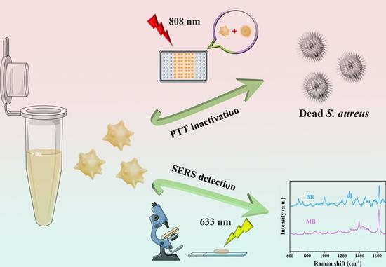
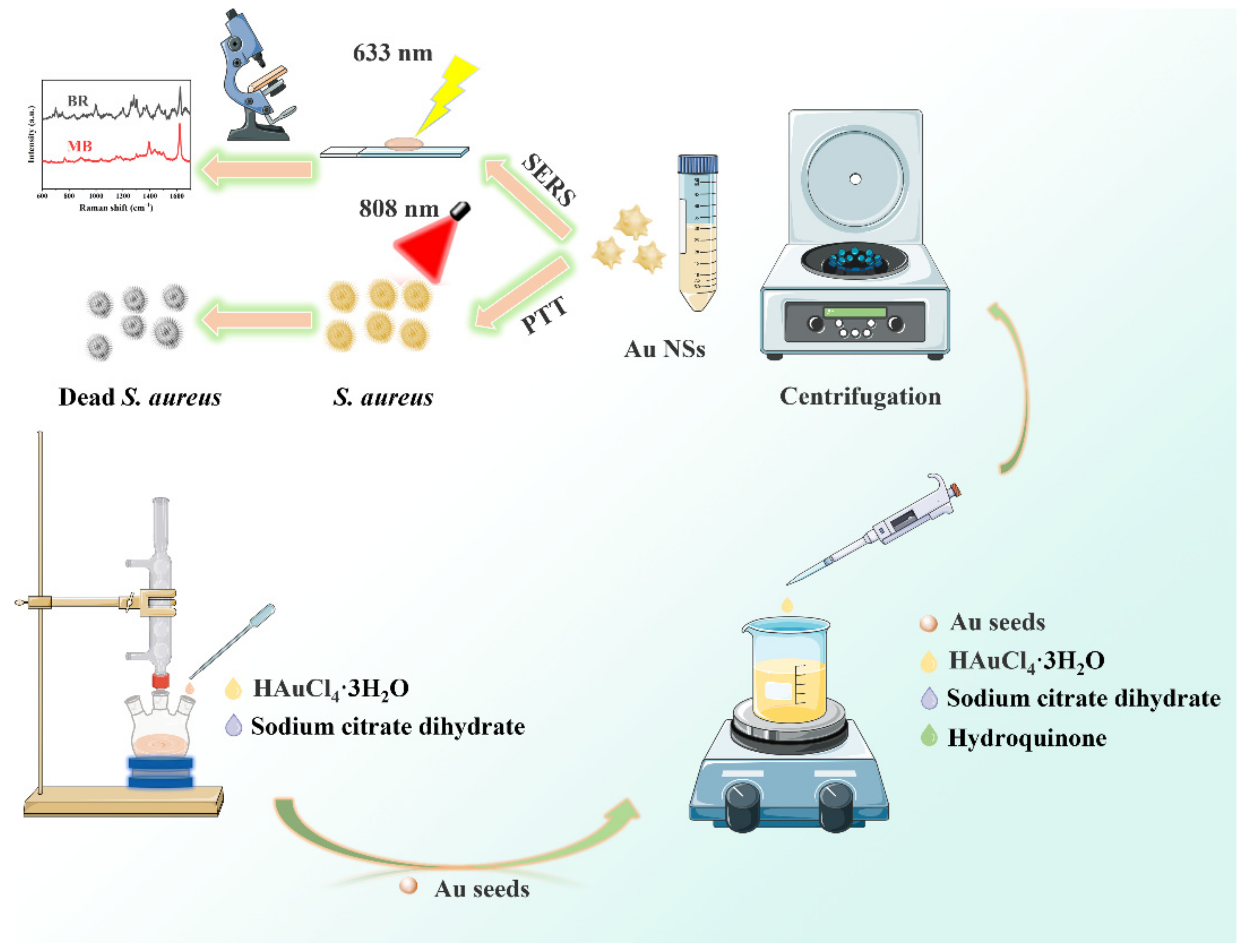
Multifunctional Plasmon-Tunable Au Nanostars and Their Applications in Highly Efficient Photothermal Inactivation and Ultra-Sensitive SERS Detection
Abstract
:
1. Introduction
2. Experimental Section
2.1. Materials and Instruments
2.2. Fabrication of Au NSs
2.3. Photothermal Conversion Performance of Au NSs
2.4. PTT of S. aureus
2.5. SERS Measurements and FDTD Algorithm Method
3. Results and Discussion
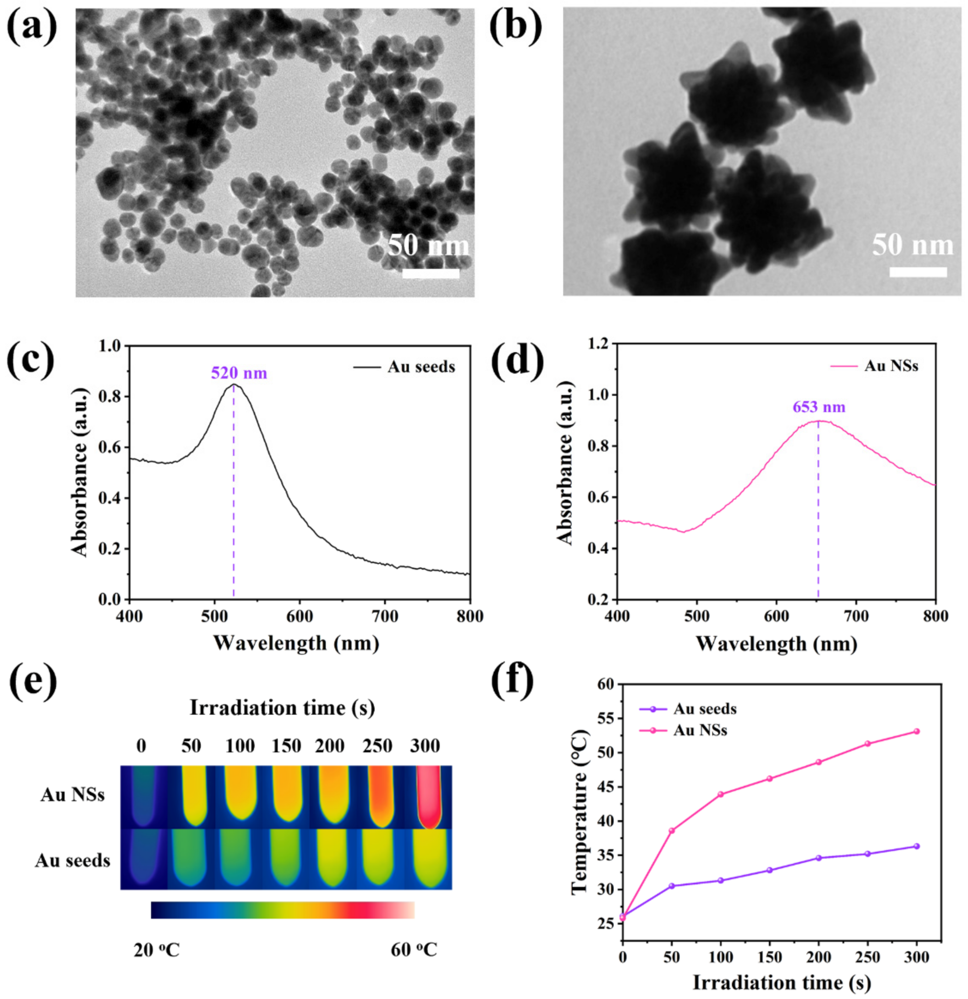
3.1. Fabrication and Characterization of Au NSs
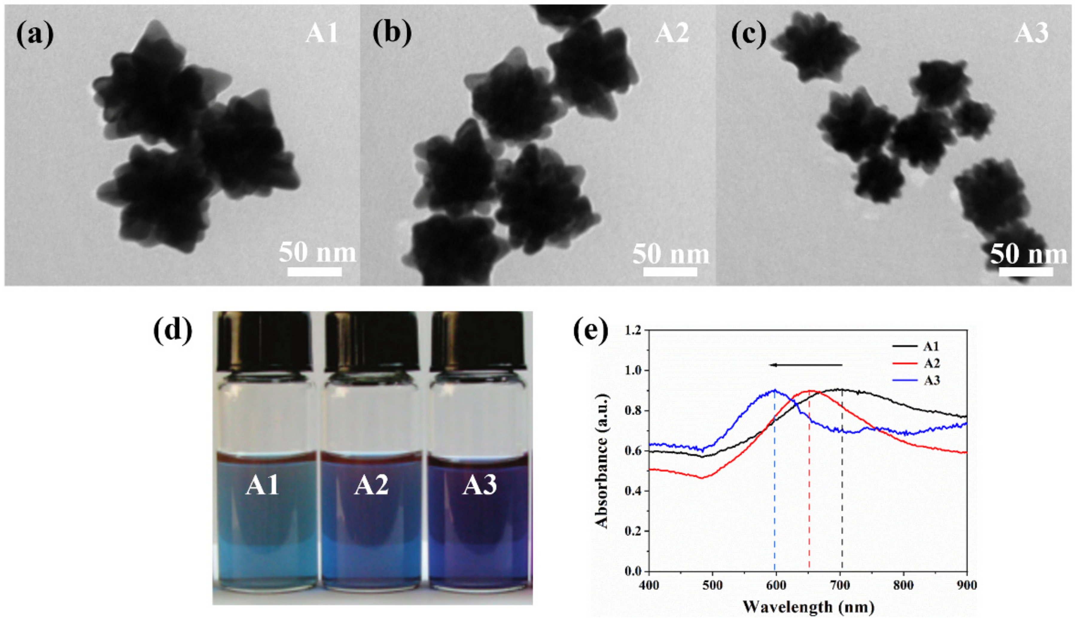
3.2. Effects of Different Au Seed Additions on Sizes and Optical Properties of Au NSs
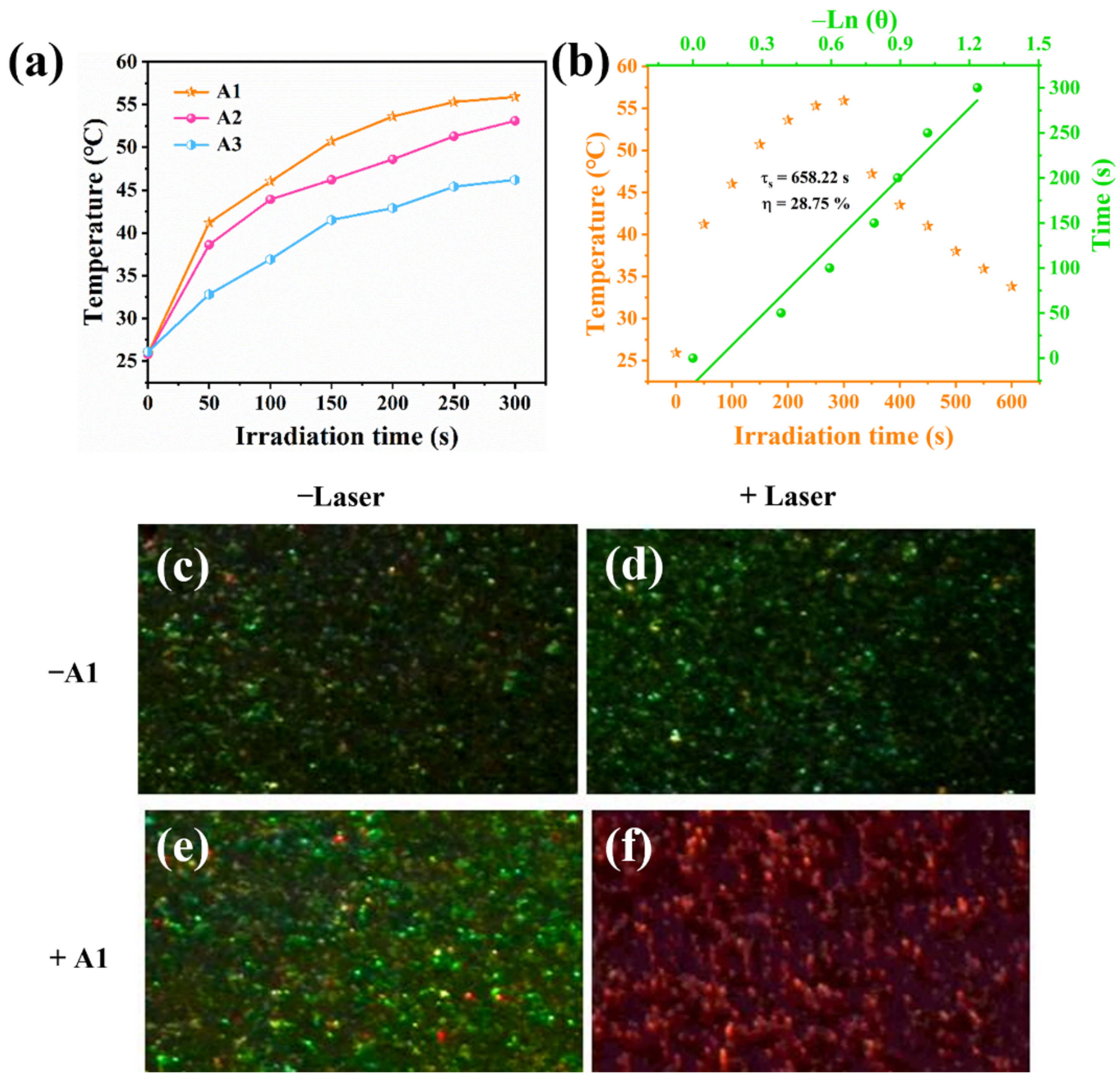
3.3. Photothermal Effects of Au NSs and PTT of S. aureus by Au NSs
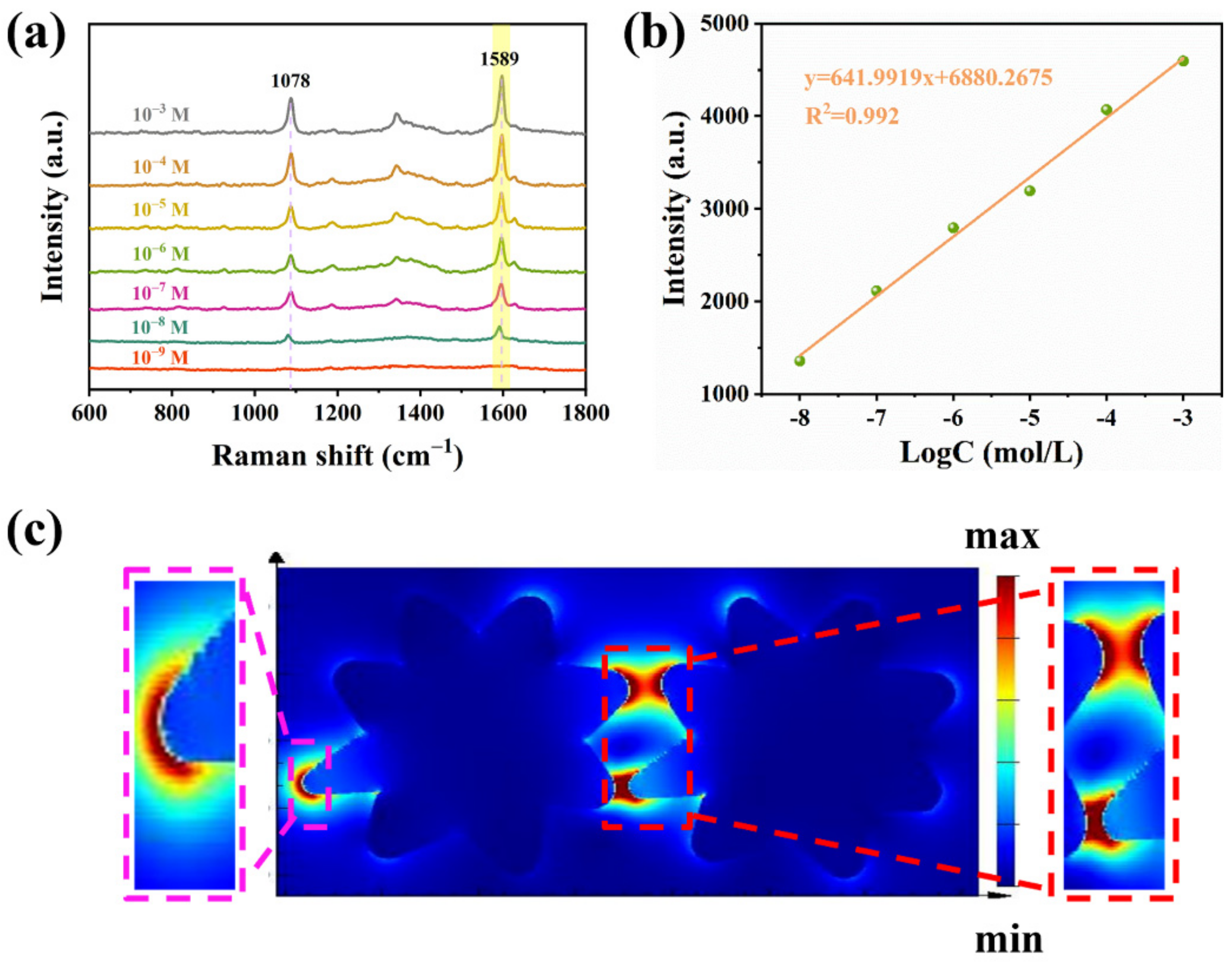
3.4. Mechanism of SERS Enhancement
3.5. Application of A1 in Detection of Different Types of Pollutants
4. Conclusions
Supplementary Materials
Author Contributions
Funding
Data Availability Statement
Conflicts of Interest
References
- Peng, Y.; Xiong, B.; Peng, L.; Li, H.; He, Y.; Yeung, E.S. Recent advances in optical imaging with anisotropic plasmonic nanoparticles. Anal. Chem. 2015, 87, 200–215. [Google Scholar] [CrossRef] [PubMed]
- Dissanayake, N.M.; Arachchilage, J.S.; Samuels, T.A.; Obare, S.O. Highly sensitive plasmonic metal nanoparticle-based sensors for the detection of organophosphorus pesticides. Talanta 2019, 200, 218–227. [Google Scholar] [CrossRef]
- Raj, S.I.; Jaiswal, A.; Uddin, I. Ultrasmall Aqueous Starch-Capped CuS Quantum Dots with Tunable Localized Surface Plasmon Resonance and Composition for The Selective and Sensitive Detection of Mercury(ii) Ions. RSC Adv. 2020, 10, 14050–14059. [Google Scholar] [CrossRef]
- Wang, H.; Cui, J.; Arshad, A.; Xu, S.; Wang, L. A Visual Photothermal Paper Sensor for H2S Recognition through Rational Modulation LSPR Wavelength of Plasmonics. Sci. China Chem. 2018, 61, 368–374. [Google Scholar] [CrossRef]
- Qi, P.; Luo, Y.; Shi, B.; Li, W.; Liu, D.; Zheng, L.; Liu, Z.; Hou, Y.; Fang, Z. Phonon scattering and exciton localization: Molding exciton flux in two dimensional disorder energy landscape. eLight 2021, 1, 6. [Google Scholar] [CrossRef]
- Espinosa, A.; Silva, A.K.; Sanchez-Iglesias, A.; Grzelczak, M.; Pechoux, C.; Desboeufs, K.; Liz-Marzan, L.M.; Wilhelm, C. Cancer Cell Internalization of Gold Nanostars Impacts Their Photothermal Efficiency In Vitro and In Vivo: Toward a Plasmonic Thermal Fingerprint in Tumoral Environment. Adv. Healthc. Mater. 2016, 5, 1040–1048. [Google Scholar] [CrossRef]
- Espinosa, A.; Kolosnjaj-Tabi, J.; Abou-Hassan, A.; Plan Sangnier, A.; Curcio, A.; Silva, A.K.A.; Di Corato, R.; Neveu, S.; Pellegrino, T.; Liz-Marzán, L.M.; et al. Magnetic (Hyper) Thermia or Photothermia? Progressive Comparison of Iron Oxide and Gold Nanoparticles Heating in Water, in Cells, and In Vivo. Adv. Funct. Mater. 2018, 28, 1803660. [Google Scholar] [CrossRef]
- Blum, O.; Shaked, N.T. Prediction of photothermal phase signatures from arbitrary plasmonic nanoparticles and experimental verification. Light-Sci. Appl. 2015, 4, e322. [Google Scholar] [CrossRef] [Green Version]
- Pan, Z.Y.; Gao, P.F.; Jing, C.J.; Zhou, J.; Liang, W.T.; Lei, G.; Feng, W.; Li, Y.F.; Huang, C.Z. Microscopic electron counting during plasmon-driven photocatalytic proton coupled electron transfer on a single silver nanoparticle. Appl. Catal. B-Environ. 2021, 291, 120090. [Google Scholar] [CrossRef]
- Wang, X.; Ji, J.; Liu, T.; Liu, Y.; Qiao, L.; Liu, B. Plasmonic Colloidosome-Based Single Cell Detector: A Strategy for Individual Cell Secretion Sensing. Anal. Chem. 2019, 91, 2260–2265. [Google Scholar] [CrossRef]
- Wu, R.; Jin, Q.; Storey, C.; Collins, J.; Gomard, G.; Lemmer, U.; Canham, L.; Kling, R.; Kaplan, A. Gold nanoplasmonic particles in tunable porous silicon 3D scaffolds for ultra-low concentration detection by SERS. Nanoscale Horiz. 2021, 6, 781–790. [Google Scholar] [CrossRef] [PubMed]
- Jia, K.; Xie, J.; He, X.; Zhang, D.; Hou, B.; Li, X.; Zhou, X.; Hong, Y.; Liu, X. Polymeric micro-reactors mediated synthesis and assembly of Ag nanoparticles into cube-like superparticles for SERS application. Chem. Eng. J. 2020, 395, 125123. [Google Scholar] [CrossRef]
- Chikkaraddy, R.; Xomalis, A.; Jakob, L.A.; Baumberg, J.J. Mid-infrared-perturbed molecular vibrational signatures in plasmonic nanocavities. Light-Sci. Appl. 2022, 11, 19. [Google Scholar] [CrossRef] [PubMed]
- Stoev, I.D.; Seelbinder, B.; Erben, E.; Maghelli, N.; Kreysing, M. Highly sensitive force measurements in an optically generated, harmonic hydrodynamic trap. eLight 2021, 1, 7. [Google Scholar] [CrossRef]
- Mosquera, J.; Zhao, Y.; Jang, H.J.; Xie, N.; Xu, C.; Kotov, N.A.; Liz-Marzán, L.M. Plasmonic Nanoparticles with Supramolecular Recognition. Adv. Funct. Mater. 2019, 30, 1902082. [Google Scholar] [CrossRef]
- Said, Z.; Arora, S.; Farooq, S.; Sundar, L.S.; Li, C.; Allouhi, A. Recent advances on improved optical, thermal, and radiative characteristics of plasmonic nanofluids: Academic insights and perspectives. Sol. Energy Mater. Sol. Cells 2022, 236, 111504. [Google Scholar] [CrossRef]
- Sun, G.; Li, N.; Wang, D.; Xu, G.; Zhang, X.; Gong, H.; Li, D.; Li, Y.; Pang, H.; Gao, M.; et al. A Novel 3D Hierarchical Plasmonic Functional Cu@Co3O4@Ag Array as Intelligent SERS Sensing Platform with Trace Droplet Rapid Detection Ability for Pesticide Residue Detection on Fruits and Vegetables. Nanomaterials 2021, 11, 3460. [Google Scholar] [CrossRef] [PubMed]
- Akter, M.; Sikder, M.T.; Rahman, M.M.; Ullah, A.; Hossain, K.F.B.; Banik, S.; Hosokawa, T.; Saito, T.; Kurasaki, M. A systematic review on silver nanoparticles-induced cytotoxicity: Physicochemical properties and perspectives. J. Adv. Res. 2018, 9, 1–16. [Google Scholar] [CrossRef] [PubMed]
- Liao, C.; Li, Y.; Tjong, S.C. Bactericidal and Cytotoxic Properties of Silver Nanoparticles. Int. J. Mol. Sci. 2019, 20, 449. [Google Scholar] [CrossRef] [Green Version]
- Lee, D.; So, S.; Hu, G.; Kim, M.; Badloe, T.; Cho, H.; Kim, J.; Kim, H.; Qiu, C.-W.; Rho, J. Hyperbolic metamaterials: Fusing artificial structures to natural 2D materials. eLight 2022, 2, 1. [Google Scholar] [CrossRef]
- Li, J.; Liu, L.; Ai, Y.; Liu, Y.; Sun, H.; Liang, Q. Self-Polymerized Dopamine-Decorated Au NPs and Coordinated with Fe-MOF as a Dual Binding Sites and Dual Signal-Amplifying Electrochemical Aptasensor for the Detection of CEA. ACS Appl. Mater. Inter. 2020, 12, 5500–5510. [Google Scholar] [CrossRef] [PubMed]
- Guler, U.; Shalaev, V.M.; Boltasseva, A. Nanoparticle plasmonics: Going practical with transition metal nitrides. Mater. Today 2015, 18, 227–237. [Google Scholar] [CrossRef]
- Ali Dheyab, M.; Abdul Aziz, A.; Jameel, M.S.; Moradi Khaniabadi, P. Recent Advances in Synthesis, Medical Applications and Challenges for Gold-Coated Iron Oxide: Comprehensive Study. Nanomaterials 2021, 11, 2147. [Google Scholar] [CrossRef] [PubMed]
- Kunwar, S.; Pandey, P.; Lee, J. Enhanced Localized Surface Plasmon Resonance of Fully Alloyed AgAuPdPt, AgAuPt, AuPt, AgPt, and Pt Nanocrystals: Systematical Investigation on the Morphological and LSPR Properties of Mono-, Bi-, Tri-, and Quad-Metallic Nanoparticles. ACS Omega 2019, 4, 17340–17351. [Google Scholar] [CrossRef] [PubMed] [Green Version]
- Lee, S.; Sun, Y.; Cao, Y.; Kang, S.H. Plasmonic nanostructure-based bioimaging and detection techniques at the single-cell level. TrAC-Trend. Anal. Chem. 2019, 117, 58–68. [Google Scholar] [CrossRef]
- Acimovic, S.S.; Sipova, H.; Emilsson, G.; Dahlin, A.B.; Antosiewicz, T.J.; Kall, M. Superior LSPR substrates based on electromagnetic decoupling for on-a-chip high-throughput label-free biosensing. Light-Sci. Appl. 2017, 6, e17042. [Google Scholar] [CrossRef]
- Tzschoppe, M.; Huck, C.; Hotzel, F.; Gunther, B.; Mamiyev, Z.; Butkevich, A.; Ulrich, C.; Gade, L.H.; Pucci, A. How adsorbates alter the metallic behavior of quasi-1D electron systems of the Si(553)-Au surface. J. Phys.-Condens. Matter. 2019, 31, 195001. [Google Scholar] [CrossRef]
- Mamiyev, Z.; Fink, C.; Holtgrewe, K.; Pfnur, H.; Sanna, S. Enforced long-range order in 1D wires by coupling to higher dimensions. Phys. Rev. Lett. 2021, 126, 106101. [Google Scholar] [CrossRef] [PubMed]
- Kim, J.; Agrawal, A.; Krieg, F.; Bergerud, A.; Milliron, D.J. The Interplay of Shape and Crystalline Anisotropies in Plasmonic Semiconductor Nanocrystals. Nano Lett. 2016, 16, 3879–3884. [Google Scholar] [CrossRef]
- Li, T.; Gao, R.; Zhang, X.; Zhang, Y. Designing the Hotspots Distribution by Anisotropic Growth. Molecules 2021, 26, 187. [Google Scholar] [CrossRef]
- Mamiyev, Z.; Tzschoppe, M.; Huck, C.; Pucci, A.; Pfnür, H. Plasmon standing waves by oxidation of Si(553)-Au. J. Phys. Chem. C 2019, 123, 9400–9406. [Google Scholar] [CrossRef] [Green Version]
- Zhu, D.; Liu, Y.; Liu, M.; Liu, X.; Prasad, P.N.; Swihart, M.T. Galvanic replacement synthesis of multi-branched gold nanocrystals for photothermal cancer therapy. J. Mater. Chem. B 2020, 8, 5491–5499. [Google Scholar] [CrossRef]
- Xu, L.; Ding, Q. Magnetic field induced high-density SERS active assembly of Fe3O4@Au nanostars in a glass capillary for food colorant detection. Anal. Methods 2021, 13, 5487–5492. [Google Scholar] [CrossRef]
- Chen, H.; Kou, X.; Yang, Z.; Ni, W.; Wang, J. Shape- and Size-Dependent Refractive Index Sensitivity of Gold Nanoparticles. Langmuir 2008, 24, 5233–5237. [Google Scholar] [CrossRef]
- Li, D.; Zhang, Y.; Wen, S.; Song, Y.; Tang, Y.; Zhu, X.; Shen, M.; Mignani, S.; Majoral, J.P.; Zhao, Q.; et al. Construction of polydopamine-coated gold nanostars for CT imaging and enhanced photothermal therapy of tumors: An innovative theranostic strategy. J. Mater. Chem. B 2016, 4, 4216–4226. [Google Scholar] [CrossRef]
- Sau, T.K.; Rogach, A.L.; Doblinger, M.; Feldmann, J. One-step high-yield aqueous synthesis of size-tunable multispiked gold nanoparticles. Small 2011, 7, 2188–2194. [Google Scholar] [CrossRef]
- Qi, X.; Wang, X.; Dong, Y.; Xie, J.; Gui, X.; Bai, J.; Duan, J.; Liu, J.; Yao, H. Fast synthesis of gold nanostar SERS substrates based on ion-track etched membrane by one-step redox reaction. Spectrochim. Acta. A. Mol. Biomol. Spectrosc. 2022, 272, 120955. [Google Scholar] [CrossRef]
- Xia, Y.; Gilroy, K.D.; Peng, H.C.; Xia, X. Seed-Mediated Growth of Colloidal Metal Nanocrystals. Angew. Chem. Int. Edit. 2017, 56, 60–95. [Google Scholar] [CrossRef]
- Kang, H.J.; Bari, G.; Lee, T.G.; Khan, T.T.; Park, J.W.; Hwang, H.J.; Cho, S.Y.; Jun, Y.S. Microporous Carbon Nanoparticles for Lithium-Sulfur Batteries. Nanomaterials 2020, 10, 2012. [Google Scholar] [CrossRef]
- Guo, J.; Zeng, F.; Guo, J.; Ma, X. Preparation and application of microfluidic SERS substrate: Challenges and future perspectives. J. Mater. Sci. Technol. 2020, 37, 96–103. [Google Scholar] [CrossRef]
- Messina, T.C.; Srijanto, B.R.; Collier, C.P.; Kravchenko, I.I.; Richards, C.I. Gold Ion Beam Milled Gold Zero-Mode Waveguides. Nanomaterials 2022, 12, 1755. [Google Scholar] [CrossRef] [PubMed]
- Jin, Z.; Janoschka, D.; Deng, J.; Ge, L.; Dreher, P.; Frank, B.; Hu, G.; Ni, J.; Yang, Y.; Li, J.; et al. Phyllotaxis-inspired nanosieves with multiplexed orbital angular momentum. eLight 2021, 1, 5. [Google Scholar] [CrossRef]
- Guo, Z.; Zeng, Y.; Meng, F.; Qu, H.; Zhang, S.; Hu, S.; Fan, S.; Zeng, H.; Cao, R.; Prasad, P.N.; et al. In-situ neutron-transmutation for substitutional doping in 2D layered indium selenide based phototransistor. eLight 2022, 2, 9. [Google Scholar] [CrossRef]
- Sanna Angotzi, M.; Musinu, A.; Mameli, V.; Ardu, A.; Cara, C.; Niznansky, D.; Xin, H.L.; Cannas, C. Spinel Ferrite Core-Shell Nanostructures by a Versatile Solvothermal Seed-Mediated Growth Approach and Study of Their Nanointerfaces. ACS Nano 2017, 11, 7889–7900. [Google Scholar] [CrossRef] [PubMed]
- Zhang, C.; Wang, D.; Huang, S.; Yang, J.; Liu, J.; Fang, J. Nonlinear Optical Response of Gold Nanobipyramids for a Doubly Q-Switched Ho-Doped Laser at a Wavelength of 2.1 microm. Nanomaterials 2021, 11, 535. [Google Scholar] [CrossRef]
- Wang, J.; Liang, D.; Jin, Q.; Feng, J.; Tang, X. Bioorthogonal SERS Nanotags as a Precision Theranostic Platform for in Vivo SERS Imaging and Cancer Photothermal Therapy. Bioconjug. Chem. 2020, 31, 182–193. [Google Scholar] [CrossRef]
- Yang, J.; Dai, D.; Lou, X.; Ma, L.; Wang, B.; Yang, Y.W. Supramolecular nanomaterials based on hollow mesoporous drug carriers and macrocycle-capped CuS nanogates for synergistic chemo-photothermal therapy. Theranostics 2020, 10, 615–629. [Google Scholar] [CrossRef]
- Kumar, A.V.P.; Dubey, S.K.; Tiwari, S.; Puri, A.; Hejmady, S.; Gorain, B.; Kesharwani, P. Recent advances in nanoparticles mediated photothermal therapy induced tumor regression. Int. J. Pharm. 2021, 606, 120848. [Google Scholar] [CrossRef]
- Bao, X.; Yuan, Y.; Chen, J.; Zhang, B.; Li, D.; Zhou, D.; Jing, P.; Xu, G.; Wang, Y.; Hola, K.; et al. In vivo theranostics with near-infrared-emitting carbon dots-highly efficient photothermal therapy based on passive targeting after intravenous administration. Light-Sci. Appl. 2018, 7, 91. [Google Scholar] [CrossRef] [Green Version]
- Wang, Y.; Wei, G.; Zhang, X.; Huang, X.; Zhao, J.; Guo, X.; Zhou, S. Multistage Targeting Strategy Using Magnetic Composite Nanoparticles for Synergism of Photothermal Therapy and Chemotherapy. Small 2018, 14, e1702994. [Google Scholar] [CrossRef]
- Zhang, Y.; Xue, C.; Li, P.; Cui, S.; Cui, D.; Jin, H. Metal-organic framework engineered corn-like SERS active Ag@Carbon with controllable spacing distance for tracking trace amount of organic compounds. J. Hazard. Mater. 2022, 424, 127686. [Google Scholar] [CrossRef] [PubMed]
- Wang, X.; Guo, L. SERS Activity of Semiconductors: Crystalline and Amorphous Nanomaterials. Angew. Chem. Int. Edit. 2020, 59, 4231–4239. [Google Scholar] [CrossRef]
- Li, L.; Yang, J.; Wei, J.; Jiang, C.; Liu, Z.; Yang, B.; Zhao, B.; Song, W. SERS monitoring of photoinduced-enhanced oxidative stress amplifier on Au@carbon dots for tumor catalytic therapy. Light-Sci. Appl. 2022, 11, 286. [Google Scholar] [CrossRef]
- Chen, G.Y.; Sun, Y.B.; Shi, P.C.; Liu, T.; Li, Z.H.; Luo, S.H.; Wang, X.C.; Cao, X.Y.; Ren, B.; Liu, G.K.; et al. Revealing unconventional host-guest complexation at nanostructured interface by surface-enhanced Raman spectroscopy. Light-Sci. Appl. 2021, 10, 85. [Google Scholar] [CrossRef]
- Wang, X.; Wang, J.; Qiu, L.; Wang, C.; Lei, X.; Cui, P.; Zhou, S.; Zhao, D.; Ni, X.; Jiang, P.; et al. Gelatinase-Responsive Photothermal Nanotherapy Based on Au Nanostars Functionalized with Antimicrobial Peptides for Treating Staphylococcus aureus Infections. ACS Appl. Nano Mater. 2022, 5, 8324–8333. [Google Scholar] [CrossRef]
- Pan, Y.; Ma, X.; Liu, C.; Xing, J.; Zhou, S.; Parshad, B.; Schwerdtle, T.; Li, W.; Wu, A.; Haag, R. Retinoic Acid-Loaded Dendritic Polyglycerol-Conjugated Gold Nanostars for Targeted Photothermal Therapy in Breast Cancer Stem Cells. ACS Nano 2021, 15, 15069–15084. [Google Scholar] [CrossRef]
- Zhang, L.; Yang, X.Q.; Wei, J.S.; Li, X.; Wang, H.; Zhao, Y.D. Intelligent gold nanostars for in vivo CT imaging and catalase-enhanced synergistic photodynamic & photothermal tumor therapy. Theranostics 2019, 9, 5424–5442. [Google Scholar]
- Yuan, H.; Khoury, C.G.; Wilson, C.M.; Grant, G.A.; Bennett, A.J.; Vo-Dinh, T. In vivo particle tracking and photothermal ablation using plasmon-resonant gold nanostars. Nanomed.-Nanotechnol. 2012, 8, 1355–1363. [Google Scholar] [CrossRef] [Green Version]
- Wei, Y.; Zhang, X.; Liu, Z.; Chen, H.-S.; Yang, P. Site-selective modification of AgPt on multibranched Au nanostars for plasmon-enhanced hydrogen evolution and methanol oxidation reaction in visible to near-infrared region. J. Power Sources 2019, 425, 17–26. [Google Scholar] [CrossRef]
- Tran, V.; Thiel, C.; Svejda, J.T.; Jalali, M.; Walkenfort, B.; Erni, D.; Schlucker, S. Probing the SERS brightness of individual Au nanoparticles, hollow Au/Ag nanoshells, Au nanostars and Au core/Au satellite particles: Single-particle experiments and computer simulations. Nanoscale 2018, 10, 21721–21731. [Google Scholar] [CrossRef]
- Ma, W.; Sun, M.; Xu, L.; Wang, L.; Kuang, H.; Xu, C. A SERS active gold nanostar dimer for mercury ion detection. Chem. Commun. (Camb.) 2013, 49, 4989–4991. [Google Scholar] [CrossRef] [PubMed]
- Fan, G.; Gao, X.; Xu, S.; Li, X.; Zhang, Q.; Dai, C.; Xue, Q.; Wang, H. Engineering an Au nanostar-based liquid phase interfacial ratiometric SERS platform with programmable entropy-driven DNA circuits to detect protein biomarkers in clinical samples. Chem. Commun. (Camb.) 2022, 58, 407–410. [Google Scholar] [CrossRef] [PubMed]
- Indrasekara, A.S.; Meyers, S.; Shubeita, S.; Feldman, L.C.; Gustafsson, T.; Fabris, L. Gold nanostar substrates for SERS-based chemical sensing in the femtomolar regime. Nanoscale 2014, 6, 8891–8899. [Google Scholar] [CrossRef] [PubMed]
- Rodrıguez-Lorenzo, L.; Alvarez-Puebla, R.A.; Pastoriza-Santos, I.; Mazzucco, S.; Stephan, O.; Kociak, M.; Liz-Marzan, L.M.; Abajo, F.J. Zeptomol Detection Through Controlled Ultrasensitive Surface-Enhanced Raman Scattering. J. Am. Chem. Soc. 2009, 131, 4616–4618. [Google Scholar] [CrossRef] [PubMed]
- Huang, Z.; Meng, G.; Hu, X.; Pan, Q.; Huo, D.; Zhou, H.; Ke, Y.; Wu, N. Plasmon-tunable Au@Ag core-shell spiky nanoparticles for surface-enhanced Raman scattering. Nano Res. 2018, 12, 449–455. [Google Scholar] [CrossRef]
- Yu, X.; Zhong, Y.; Sun, Y.; Chen, Y. Controllable Preparation of Plasmonic Gold Nanostars for Enhanced Photothermal and SERS Effects. Chem. Res. Chin. Univ. 2020, 36, 1284–1291. [Google Scholar] [CrossRef]
- Chang, Y.X.; Zhang, N.N.; Xing, Y.C.; Zhang, Q.; Oh, A.; Gao, H.M.; Zhu, Y.; Baik, H.; Kim, B.; Yang, Y.; et al. Gold Nanotetrapods with Unique Topological Structure and Ultranarrow Plasmonic Band as Multifunctional Therapeutic Agents. J. Phys. Chem. Lett. 2019, 10, 4505–4510. [Google Scholar] [CrossRef]
- Wu, R.; Min, Q.; Guo, J.; Zheng, T.; Jiang, L.; Zhu, J.J. Sequential Delivery and Cascade Targeting of Peptide Therapeutics for Triplexed Synergistic Therapy with Real-Time Monitoring Shuttled by Magnetic Gold Nanostars. Anal. Chem. 2019, 91, 4608–4617. [Google Scholar] [CrossRef]
- Pu, Y.; Zhao, Y.; Zheng, P.; Li, M. Elucidating the Growth Mechanism of Plasmonic Gold Nanostars with Tunable Optical and Photothermal Properties. Inorg. Chem. 2018, 57, 8599–8607. [Google Scholar] [CrossRef]
- Xu, S.; Wang, Y.; Sun, Y.; Shan, G.; Chen, Y.; Liu, Y. The detection of copper ions based on photothermal effect of cysteine modified Au nanorods. Sensor. Actuat. B-Chem. 2017, 248, 761–768. [Google Scholar] [CrossRef]
- Rycenga, M.; Wang, Z.; Gordon, E.; Cobley, C.M.; Schwartz, A.G.; Lo, C.S.; Xia, Y. Probing the photothermal effect of gold-based nanocages with surface-enhanced Raman scattering (SERS). Angew. Chem. Int. Edit. 2009, 121, 10108–10111. [Google Scholar] [CrossRef]
- Ma, H.; Liu, Z.; Wei, Y.; Jiang, L. Controlled morphology evolution of branched Au nanostructures and their shape-dependent catalytic and photo-thermal properties. Colloid. Surface A 2019, 582, 123889. [Google Scholar] [CrossRef]
- Yin, T.; Li, Y.; Bian, K.; Zhu, R.; Liu, Z.; Niu, K.; Liu, H.; Gao, Z.; Gao, D. Self-assembly synthesis of vapreotidegold hybrid nanoflower for photothermal antitumor activity. Mat. Sci. Eng. C-Mater. 2018, 93, 716–723. [Google Scholar] [CrossRef] [PubMed]
- Wang, H.; Zhou, S.; Guo, L.; Wang, Y.; Feng, L. Intelligent Hybrid Hydrogels for Rapid In Situ Detection and Photothermal Therapy of Bacterial Infection. ACS Appl. Mater. Inter. 2020, 12, 39685–39694. [Google Scholar] [CrossRef]
- Yang, L.; Chen, S.; Wei, H.; Luo, Y.; Cong, F.; Li, W.; Hong, L.; Su, J. Low-Temperature Photothermal Therapy Based on Borneol-Containing Polymer-Modified MXene Nanosheets. ACS Appl. Mater. Inter. 2022, 14, 45178–45188. [Google Scholar] [CrossRef]
- Li, S.; Xu, P.; Ren, Z.; Zhang, B.; Du, Y.; Han, X.; Mack, N.H.; Wang, H.L. Fabrication of thorny Au nanostructures on polyaniline surfaces for sensitive surface-enhanced Raman spectroscopy. ACS Appl. Mater. Inter. 2013, 5, 49–54. [Google Scholar] [CrossRef]
- Wang, Y.; Zhang, M.; Ma, H.; Su, H.; Li, A.; Ruan, W.; Zhao, B. Surface Plasmon Resonance from Gallium-Doped Zinc Oxide Nanoparticles and Their Electromagnetic Enhancement Contribution to Surface-Enhanced Raman Scattering. ACS Appl. Mater. Inter. 2021, 13, 35038–35045. [Google Scholar] [CrossRef]
- Lin, S.; Guan, H.; Liu, Y.; Huang, S.; Li, J.; Hasi, W.; Xu, Y.; Zou, J.; Dong, B. Binary Plasmonic Assembly Films with Hotspot-Type-Dependent Surface-Enhanced Raman Scattering Properties. ACS Appl. Mater. Inter. 2021, 13, 53289–53299. [Google Scholar] [CrossRef]
- Li, J.F.; Zhang, Y.J.; Ding, S.Y.; Panneerselvam, R.; Tian, Z.Q. Core-Shell Nanoparticle-Enhanced Raman Spectroscopy. Chem. Rev. 2017, 117, 5002–5069. [Google Scholar] [CrossRef]
- Zhang, W.; Gu, P.; Wang, Z.; Ai, B.; Zhou, Z.; Zhao, Z.; Li, C.; Shi, Z.; Zhang, G. Integrated “Hot Spots”: Tunable Sub-10 nm Crescent Nanogap Arrays. Adv. Opt. Mater. 2019, 7, 1901337. [Google Scholar] [CrossRef]
- Zhang, Q.; Zhang, Y.; Chen, H.; Zhang, L.; Li, P.; Xiao, H.; Wu, W. One-dimensional nanohybrids based on cellulose nanocrystals and their SERS performance. Carbohydr. Polym. 2022, 284, 119140. [Google Scholar] [CrossRef]
- Huang, J.; Zhou, T.; Zhao, W.; Cui, S.; Guo, R.; Li, D.; Reddy Kadasala, N.; Han, D.; Jiang, Y.; Liu, Y.; et al. Multifunctional magnetic Fe3O4/Cu2O-Ag nanocomposites with high sensitivity for SERS detection and efficient visible light-driven photocatalytic degradation of polycyclic aromatic hydrocarbons (PAHs). J. Colloid Interf. Sci. 2022, 628, 315–326. [Google Scholar] [CrossRef]
- Huang, J.; Zhou, T.; Zhao, W.; Zhang, M.; Zhang, Z.; Lai, W.; Kadasala, N.R.; Liu, H.; Liu, Y. Magnetic-Core–Shell–Satellite Fe3O4-Au@Ag@(Au@Ag) Nanocomposites for Determination of Trace Bisphenol A Based on Surface-Enhanced Resonance Raman Scattering (SERRS). Nanomaterials 2022, 12, 3322. [Google Scholar] [CrossRef]
- Lin, M.-H.; Sun, L.; Kong, F.; Lin, M. Rapid detection of paraquat residues in green tea using surface-enhanced Raman spectroscopy (SERS) coupled with gold nanostars. Food Control 2021, 130, 108280. [Google Scholar] [CrossRef]
- Ye, Y.; Qi, X.; Wang, H.; Zhao, B.; Xu, L.; Zhang, Y.; Wang, X.; Zhou, N. A surface-enhanced Raman scattering aptasensor for Escherichia coli detection based on high-performance 3D substrate and hot spot effect. Anal. Chim. Acta 2022, 1221, 340141. [Google Scholar] [CrossRef]
- Boyack, R.; Le Ru, E.C. Investigation of particle shape and size effects in SERS using T-matrix calculations. Phys. Chem. Chem. Phys. 2009, 11, 7398–7405. [Google Scholar] [CrossRef]
- Li, J.J.; Wu, C.; Zhao, J.; Weng, G.J.; Zhu, J.; Zhao, J.W. Synthesis and SERS activity of super-multibranched Au-Ag nanostructure via silver coating-induced aggregation of nanostars. Spectrochim. Acta. A. Mol. Biomol. Spectrosc. 2018, 204, 380–387. [Google Scholar] [CrossRef]
- Meng, X.; Dyer, J.; Huo, Y.; Jiang, C. Greater SERS Activity of Ligand-Stabilized Gold Nanostars with Sharp Branches. Langmuir 2020, 36, 3558–3564. [Google Scholar] [CrossRef]
- Chen, L.; Zhu, Y.; Cui, Y.; Dai, R.; Shan, Z.; Chen, H. Fabrication of starch-based high-performance adsorptive hydrogels using a novel effective pretreatment and adsorption for cationic methylene blue dye: Behavior and mechanism. Chem. Eng. J. 2021, 405, 126953. [Google Scholar] [CrossRef]
- Fadillah, G.; Saleh, T.A.; Wahyuningsih, S.; Ninda Karlina Putri, E.; Febrianastuti, S. Electrochemical removal of methylene blue using alginate-modified graphene adsorbents. Chem. Eng. J. 2019, 378, 122140. [Google Scholar] [CrossRef]
- Pan, X.; Li, L.; Lin, H.; Tan, J.; Wang, H.; Liao, M.; Chen, C.; Shan, B.; Chen, Y.; Li, M. A graphene oxide-gold nanostar hybrid based-paper biosensor for label-free SERS detection of serum bilirubin for diagnosis of jaundice. Biosens. Bioelectron. 2019, 145, 111713. [Google Scholar] [CrossRef] [PubMed]
- Su, R.; Quan, Y.; Yang, S.; Hu, M.; Yang, J.; Gao, M. Destroying the symmetric structure to promote phase transition: Improving the SERS performance and catalytic activity of MoS2 nanoflowers. J. Alloys Compd. 2021, 886, 161268. [Google Scholar] [CrossRef]
- Ji, M.; Xu, M.; Zhang, W.; Yang, Z.; Huang, L.; Liu, J.; Zhang, Y.; Gu, L.; Yu, Y.; Hao, W.; et al. Structurally Well-Defined Au@Cu2-xS Core-Shell Nanocrystals for Improved Cancer Treatment Based on Enhanced Photothermal Efficiency. Adv. Mater. 2016, 28, 3094–3101. [Google Scholar] [CrossRef] [PubMed]
- Chen, X.; Zhang, M.; Li, S.; Li, L.; Zhang, L.; Wang, T.; Yu, M.; Mou, Z.; Wang, C. Facile synthesis of polypyrrole@metal-organic framework core-shell nanocomposites for dual-mode imaging and synergistic chemo-photothermal therapy of cancer cells. J. Mater. Chem. B 2017, 5, 1772–1778. [Google Scholar] [CrossRef]
- Yu, Y.; Chi, B.; Lin, L.; Yang, Z.; He, Q.; Xu, Z.; Yi, C.; Wang, J. Microwave-assisted preparation of paramagnetic zwitterionic amphiphilic copolymer hybrid molybdenum disulfide for T1-weighted magnetic resonance imaging-guided photothermal therapy. J. Mater. Chem. B 2018, 6, 6391–6398. [Google Scholar] [CrossRef]
- Chen, Z.; Xia, Q.; Zhou, Y.; Li, X.; Qi, L.; Feng, Q.; Liu, R.; Chen, W. 2-Dicyanomethylenethiazole based NIR absorbing organic nanoparticles for photothermal therapy and photoacoustic imaging. J. Mater. Chem. B 2019, 7, 3950–3957. [Google Scholar] [CrossRef]

Publisher’s Note: MDPI stays neutral with regard to jurisdictional claims in published maps and institutional affiliations. |
© 2022 by the authors. Licensee MDPI, Basel, Switzerland. This article is an open access article distributed under the terms and conditions of the Creative Commons Attribution (CC BY) license (https://creativecommons.org/licenses/by/4.0/).
Share and Cite
Zhou, T.; Huang, J.; Zhao, W.; Guo, R.; Cui, S.; Li, Y.; Zhang, X.; Liu, Y.; Zhang, Q. Multifunctional Plasmon-Tunable Au Nanostars and Their Applications in Highly Efficient Photothermal Inactivation and Ultra-Sensitive SERS Detection. Nanomaterials 2022, 12, 4232. https://doi.org/10.3390/nano12234232
Zhou T, Huang J, Zhao W, Guo R, Cui S, Li Y, Zhang X, Liu Y, Zhang Q. Multifunctional Plasmon-Tunable Au Nanostars and Their Applications in Highly Efficient Photothermal Inactivation and Ultra-Sensitive SERS Detection. Nanomaterials. 2022; 12(23):4232. https://doi.org/10.3390/nano12234232
Chicago/Turabian StyleZhou, Tianxiang, Jie Huang, Wenshi Zhao, Rui Guo, Sicheng Cui, Yuqing Li, Xiaolong Zhang, Yang Liu, and Qi Zhang. 2022. "Multifunctional Plasmon-Tunable Au Nanostars and Their Applications in Highly Efficient Photothermal Inactivation and Ultra-Sensitive SERS Detection" Nanomaterials 12, no. 23: 4232. https://doi.org/10.3390/nano12234232