
Advances in Nanocarrier Systems for Overcoming Formulation Challenges of Curcumin: Current Insights

 , ,
, ,  , , ,
, , ,  , and
, and
Abstract
1. Introduction
2. Physicochemical Properties
3. Pharmacokinetics
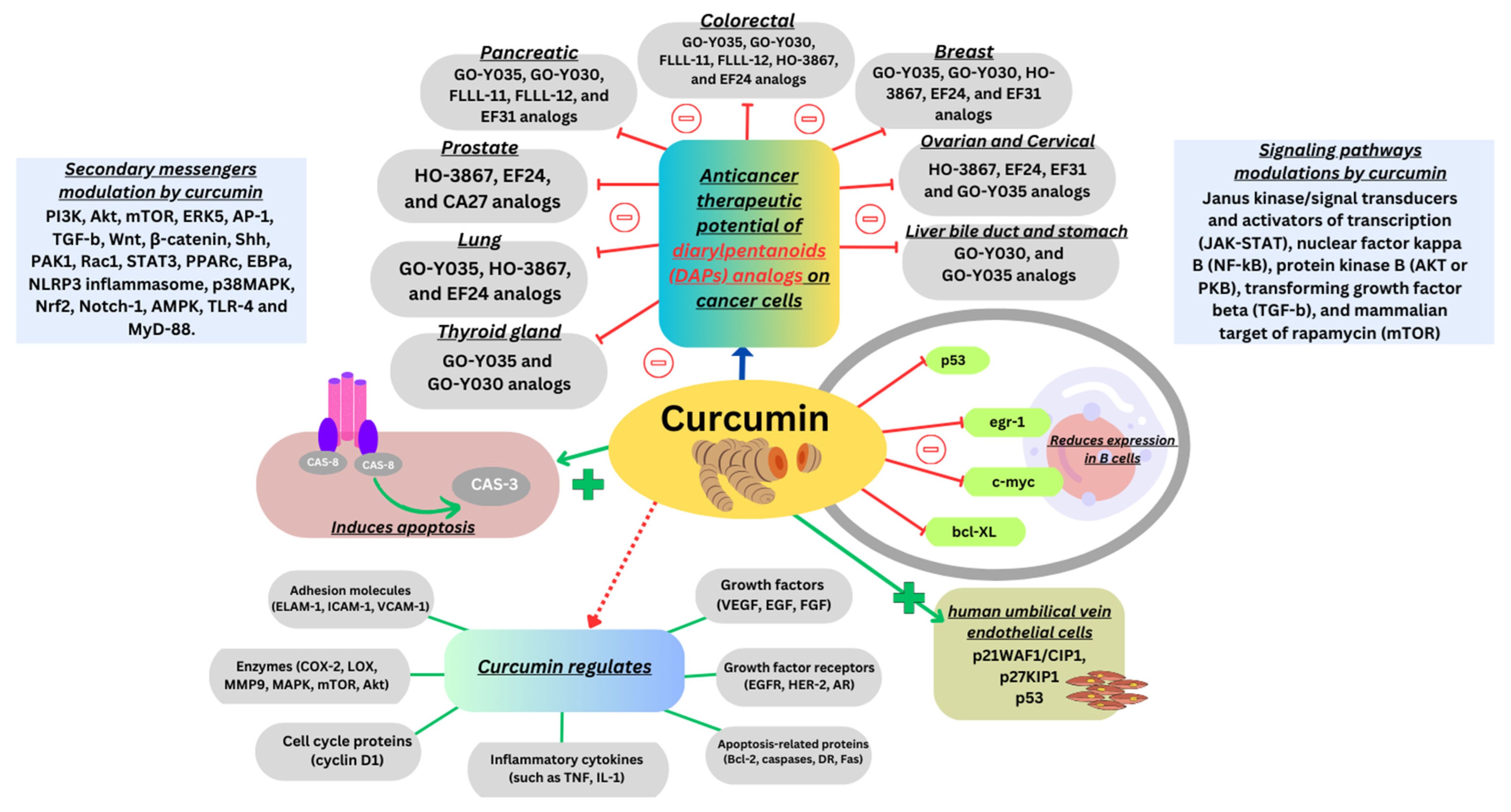
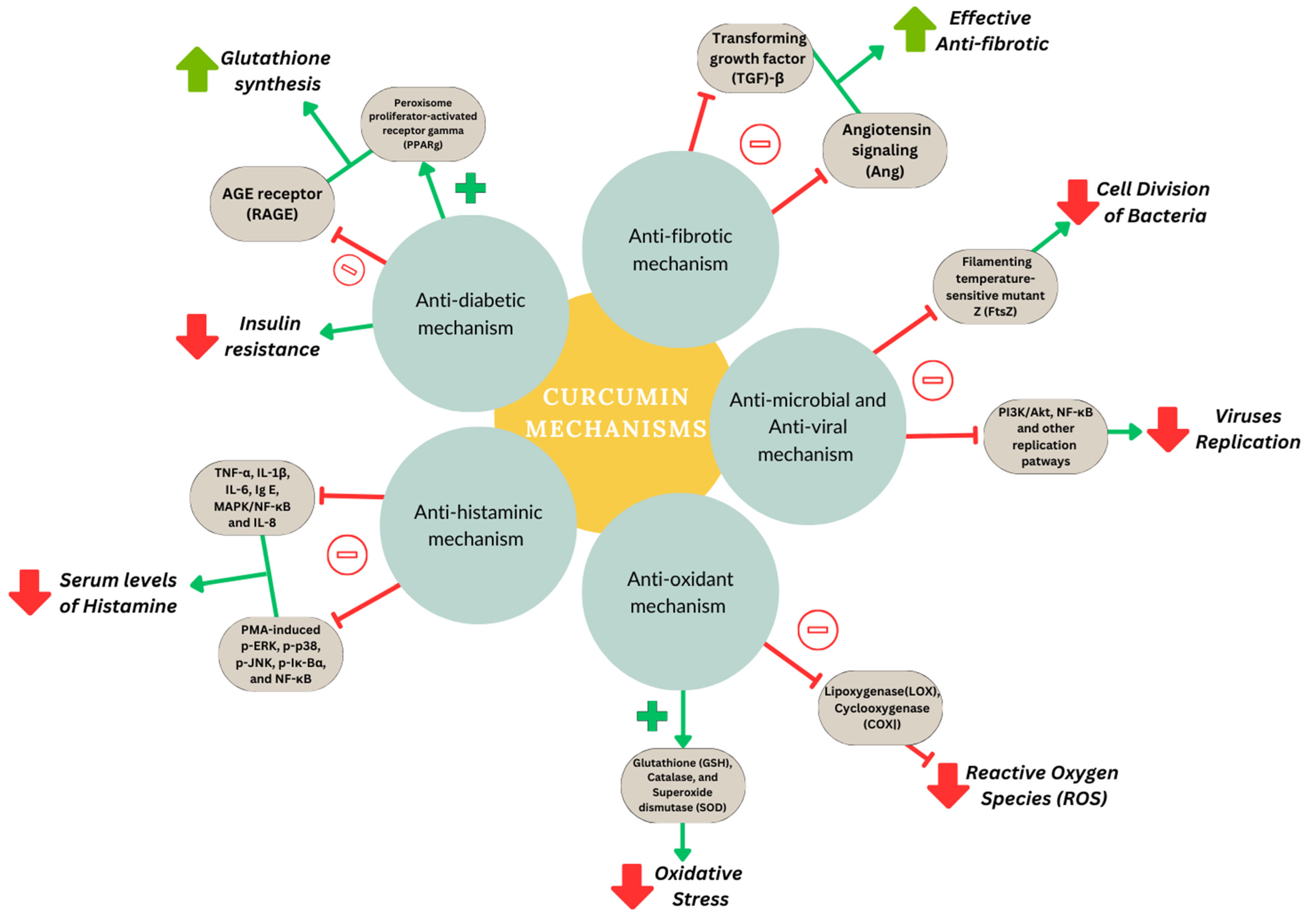
4. Molecular Pathways
5. Role of Nanoparticles in Curcumin Delivery
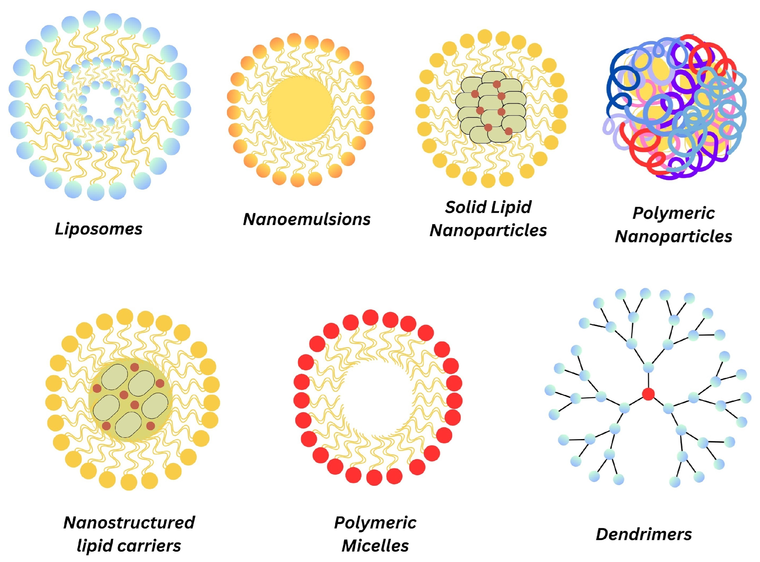
5.1. Liposomes
5.2. Nanoemulsions
5.3. Solid Lipid Nanoparticles
5.4. Nanostructured Lipid Carriers
5.5. Polymeric Micelles
5.6. Dendrimers
5.7. Polymeric Nanoparticles
6. Clinical Translation and Future Perspectives
7. Conclusions
Author Contributions
Funding
Data Availability Statement
Acknowledgments
Conflicts of Interest
References
- El-Saadony, M.T.; Yang, T.; Korma, S.A.; Sitohy, M.; Abd El-Mageed, T.A.; Selim, S.; Al Jaouni, S.K.; Salem, H.M.; Mahmmod, Y.; Soliman, S.M.; et al. Impacts of turmeric and its principal bioactive curcumin on human health: Pharmaceutical, medicinal, and food applications: A comprehensive review. Front. Nutr. 2022, 9, 1040259. [Google Scholar] [CrossRef] [PubMed]
- Tomeh, M.A.; Hadianamrei, R.; Zhao, X. A Review of Curcumin and Its Derivatives as Anticancer Agents. Int. J. Mol. Sci. 2019, 20, 1033. [Google Scholar] [CrossRef] [PubMed]
- Giordano, A.; Tommonaro, G. Curcumin and Cancer. Nutrients 2019, 11, 2376. [Google Scholar] [CrossRef] [PubMed]
- Den Hartogh, D.J.; Gabriel, A.; Tsiani, E. Antidiabetic Properties of Curcumin I: Evidence from In Vitro Studies. Nutrients 2020, 12, 118. [Google Scholar] [CrossRef] [PubMed]
- Lv, H.; Wang, Y.; Yang, X.; Ling, G.; Zhang, P. Application of curcumin nanoformulations in Alzheimer’s disease: Prevention, diagnosis and treatment. Nutr. Neurosci. 2023, 26, 727–742. [Google Scholar] [CrossRef] [PubMed]
- Weng, W.; Goel, A. Curcumin and colorectal cancer: An update and current perspective on this natural medicine. Semin. Cancer Biol. 2022, 80, 73–86. [Google Scholar] [CrossRef] [PubMed]
- Chamani, S.; Moossavi, M.; Naghizadeh, A.; Abbasifard, M.; Majeed, M.; Johnston, T.P.; Sahebkar, A. Immunomodulatory effects of curcumin in systemic autoimmune diseases. Phytother. Res. 2022, 36, 1616–1632. [Google Scholar] [CrossRef] [PubMed]
- Yang, S.; Sun, Y.; Kapilevich, L.; Zhang, X.; Huang, Y. Protective effects of curcumin against osteoporosis and its molecular mechanisms: A recent review in preclinical trials. Front. Pharmacol. 2023, 14, 1249418. [Google Scholar] [CrossRef] [PubMed]
- Cox, F.F.; Misiou, A.; Vierkant, A.; Ale-Agha, N.; Grandoch, M.; Haendeler, J.; Altschmied, J. Protective Effects of Curcumin in Cardiovascular Diseases—Impact on Oxidative Stress and Mitochondria. Cells 2022, 11, 342. [Google Scholar] [CrossRef] [PubMed]
- Lavian, S.; Mardaneh, P.; Bagherniya, M.; Emami, S.A.; Butler, A.E.; Sahebkar, A. The Effect of Synthetic Curcumin Analogues on Obesity, Diabetes and Cardiovascular Disease: A Literature Review. Curr. Med. Chem. 2023, 30, 3979–3992. [Google Scholar] [CrossRef] [PubMed]
- Farzaei, M.H.; Zobeiri, M.; Parvizi, F.; El-Senduny, F.F.; Marmouzi, I.; Coy-Barrera, E.; Naseri, R.; Nabavi, S.M.; Rahimi, R.; Abdollahi, M. Curcumin in Liver Diseases: A Systematic Review of the Cellular Mechanisms of Oxidative Stress and Clinical Perspective. Nutrients 2018, 10, 855. [Google Scholar] [CrossRef] [PubMed]
- Kunnumakkara, A.B.; Bordoloi, D.; Harsha, C.; Banik, K.; Gupta, S.C.; Aggarwal, B.B. Curcumin mediates anticancer effects by modulating multiple cell signaling pathways. Clin. Sci. 2017, 131, 1781–1799. [Google Scholar] [CrossRef] [PubMed]
- Panknin, T.M.; Howe, C.L.; Hauer, M.; Bucchireddigari, B.; Rossi, A.M.; Funk, J.L. Curcumin Supplementation and Human Disease: A Scoping Review of Clinical Trials. Int. J. Mol. Sci. 2023, 24, 4476. [Google Scholar] [CrossRef] [PubMed]
- Cacciola, N.A.; Cuciniello, R.; Petillo, G.D.; Piccioni, M.; Filosa, S.; Crispi, S. An Overview of the Enhanced Effects of Curcumin and Chemotherapeutic Agents in Combined Cancer Treatments. Int. J. Mol. Sci. 2023, 24, 12587. [Google Scholar] [CrossRef] [PubMed]
- Nelson, K.M.; Dahlin, J.L.; Bisson, J.; Graham, J.; Pauli, G.F.; Walters, M.A. The essential medicinal chemistry of curcumin: Miniperspective. J. Med. Chem. 2017, 60, 1620–1637. [Google Scholar] [CrossRef] [PubMed]
- Kotha, R.R.; Luthria, D.L. Curcumin: Biological, Pharmaceutical, Nutraceutical, and Analytical Aspects. Molecules 2019, 24, 2930. [Google Scholar] [CrossRef] [PubMed]
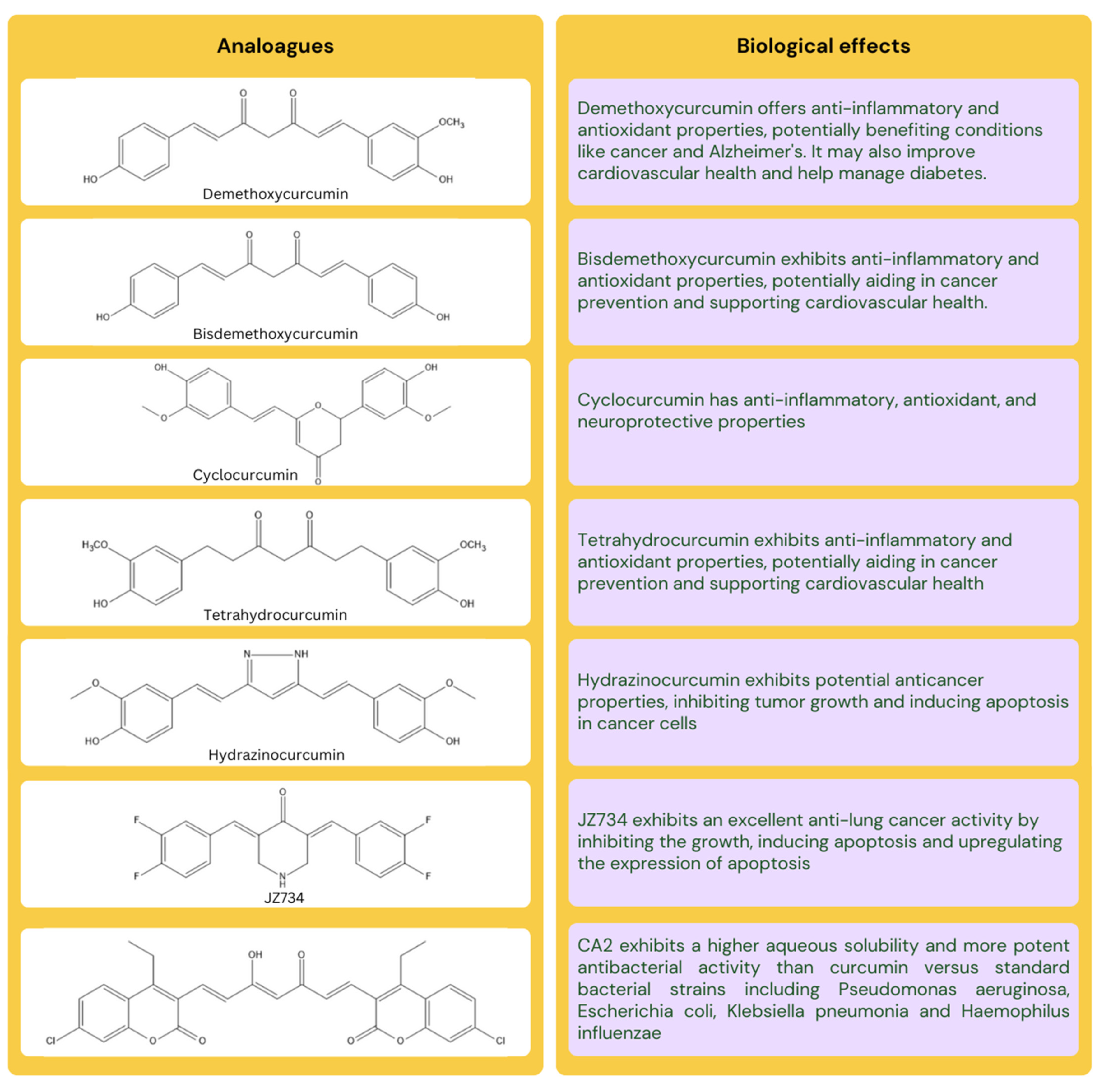
- Fu, M.; Chen, L.; Zhang, L.; Yu, X.; Yang, Q. Cyclocurcumin, a curcumin derivative, exhibits immune-modulating ability and is a potential compound for the treatment of rheumatoid arthritis as predicted by the MM-PBSA method. Int. J. Mol. Med. 2017, 39, 1164–1172. [Google Scholar] [CrossRef] [PubMed]
- Satyavert; Gupta, S.; Choudhury, H.; Jacob, S.; Nair, A.B.; Dhanawat, M.; Munjal, K. Pharmacokinetics and tissue distribution of hydrazinocurcumin in rats. Pharmacol. Rep. 2021, 73, 1734–1743. [Google Scholar] [CrossRef] [PubMed]
- Jäger, R.; Lowery, R.P.; Calvanese, A.V.; Joy, J.M.; Purpura, M.; Wilson, J.M. Comparative absorption of curcumin formulations. Nutr. J. 2014, 13, 11. [Google Scholar] [CrossRef] [PubMed]
- Zhang, S.; Asghar, S.; Yu, F.; Hu, Z.; Ping, Q.; Chen, Z.; Shao, F.; Xiao, Y. The enhancement of N-acetylcysteine on intestinal absorption and oral bioavailability of hydrophobic curcumin. Eur. J. Pharm. Sci. Off. J. Eur. Fed. Pharm. Sci. 2020, 154, 105506. [Google Scholar] [CrossRef] [PubMed]
- Sharma, R.A.; Euden, S.A.; Platton, S.L.; Cooke, D.N.; Shafayat, A.; Hewitt, H.R.; Marczylo, T.H.; Morgan, B.; Hemingway, D.; Plummer, S.M.; et al. Phase I clinical trial of oral curcumin: Biomarkers of systemic activity and compliance. Clin. Cancer Res. Off. J. Am. Assoc. Cancer Res. 2004, 10, 6847–6854. [Google Scholar] [CrossRef] [PubMed]
- Yang, K.Y.; Lin, L.C.; Tseng, T.Y.; Wang, S.C.; Tsai, T.H. Oral bioavailability of curcumin in rat and the herbal analysis from Curcuma longa by LC-MS/MS. J. Chromatogr. B Anal. Technol. Biomed. Life Sci. 2007, 853, 183–189. [Google Scholar] [CrossRef] [PubMed]
- Mahale, J.; Singh, R.; Howells, L.M.; Britton, R.G.; Khan, S.M.; Brown, K. Detection of Plasma Curcuminoids from Dietary Intake of Turmeric-Containing Food in Human Volunteers. Mol. Nutr. Food Res. 2018, 62, e1800267. [Google Scholar] [CrossRef] [PubMed]
- Schiborr, C.; Eckert, G.P.; Rimbach, G.; Frank, J. A validated method for the quantification of curcumin in plasma and brain tissue by fast narrow-bore high-performance liquid chromatography with fluorescence detection. Anal. Bioanal. Chem. 2010, 397, 1917–1925. [Google Scholar] [CrossRef] [PubMed]
- Hundshammer, C.; Schön, C.; Kimura, M.; Furune, T.; Terao, K.; Elgeti, D.; Mohr, R. Enhanced metabolic bioavailability of tetrahydrocurcumin after oral supplementation of a γ-cyclodextrin curcumin complex. J. Funct. Foods 2021, 79, 104410. [Google Scholar] [CrossRef]
- Di Meo, F.; Margarucci, S.; Galderisi, U.; Crispi, S.; Peluso, G. Curcumin, Gut Microbiota, and Neuroprotection. Nutrients 2019, 11, 2426. [Google Scholar] [CrossRef] [PubMed]
- Shishodia, S. Molecular mechanisms of curcumin action: Gene expression. BioFactors 2013, 39, 37–55. [Google Scholar] [CrossRef] [PubMed]
- Farghadani, R.; Naidu, R. Curcumin: Modulator of Key Molecular Signaling Pathways in Hormone-Independent Breast Cancer. Cancers 2021, 13, 3427. [Google Scholar] [CrossRef] [PubMed]
- Paulraj, F.; Abas, F.; Lajis, N.H.; Othman, I.; Naidu, R. Molecular Pathways Modulated by Curcumin Analogue, Diarylpentanoids in Cancer. Biomolecules 2019, 9, 270. [Google Scholar] [CrossRef] [PubMed]
- Joshi, P.; Joshi, S.; Semwal, D.; Bisht, A.; Paliwal, S.; Dwivedi, J.; Sharma, S. Curcumin: An Insight into Molecular Pathways Involved in Anticancer Activity. Mini Rev. Med. Chem. 2021, 21, 2420–2457. [Google Scholar] [CrossRef] [PubMed]
- Molla, S.; Hembram, K.C.; Chatterjee, S.; Nayak, D.; Sethy, C.; Pradhan, R.; Kundu, C.N. PARP inhibitor Olaparib Enhances the Apoptotic Potentiality of Curcumin by Increasing the DNA Damage in Oral Cancer Cells through Inhibition of BER Cascade. Pathol. Oncol. Res. 2020, 26, 2091–2103. [Google Scholar] [CrossRef] [PubMed]
- Chen, X.; Chen, X.; Shi, X.; Gao, Z.; Guo, Z. Curcumin attenuates endothelial cell fibrosis through inhibiting endothelial—Interstitial transformation. Clin. Exp. Pharmacol. Physiol. 2020, 47, 1182–1192. [Google Scholar] [CrossRef] [PubMed]
- Rai, M.; Ingle, A.P.; Pandit, R.; Paralikar, P.; Anasane, N.; Santos, C.A.D. Curcumin and curcumin-loaded nanoparticles: Antipathogenic and antiparasitic activities. Expert Rev. Anti-Infect. Ther. 2020, 18, 367–379. [Google Scholar] [CrossRef] [PubMed]
- Abrahams, S.; Haylett, W.L.; Johnson, G.; Carr, J.A.; Bardien, S. Antioxidant effects of curcumin in models of neurodegeneration, aging, oxidative and nitrosative stress: A review. Neuroscience 2019, 406, 1–21. [Google Scholar] [CrossRef] [PubMed]
- Hotsumi, M.; Tajiri, M.; Nikaido, Y.; Sato, T.; Makabe, K.; Konno, H. Design, synthesis, and evaluation of a water soluble C5-monoketone type curcumin analogue as a potent amyloid β aggregation inhibitor. Bioorganic Med. Chem. Lett. 2019, 29, 2157–2161. [Google Scholar] [CrossRef] [PubMed]
- Ardebili, A.; Pouriayevali, M.H.; Aleshikh, S.; Zahani, M.; Ajorloo, M.; Izanloo, A.; Siyadatpanah, A.; Razavi Nikoo, H.; Wilairatana, P.; Coutinho, H.D.M. Antiviral Therapeutic Potential of Curcumin: An Update. Molecules 2021, 26, 6994. [Google Scholar] [CrossRef] [PubMed]
- Zhang, N.; Li, H.; Jia, J.; He, M. Anti-inflammatory effect of curcumin on mast cell-mediated allergic responses in ovalbumin-induced allergic rhinitis mouse. Cell. Immunol. 2015, 298, 88–95. [Google Scholar] [CrossRef] [PubMed]
- Rashwan, A.K.; Karim, N.; Xu, Y.; Hanafy, N.A.N.; Li, B.; Mehanni, A.E.; Taha, E.M.; Chen, W. An updated and comprehensive review on the potential health effects of curcumin-encapsulated micro/nanoparticles. Crit. Rev. Food Sci. Nutr. 2023, 63, 9731–9751. [Google Scholar] [CrossRef] [PubMed]
- Gorain, B.; Al-Dhubiab, B.E.; Nair, A.; Kesharwani, P.; Pandey, M.; Choudhury, H. Multivesicular Liposome: A Lipid-based Drug Delivery System for Efficient Drug Delivery. Curr. Pharm. Des. 2021, 27, 4404–4415. [Google Scholar] [CrossRef] [PubMed]
- Nsairat, H.; Khater, D.; Sayed, U.; Odeh, F.; Al Bawab, A.; Alshaer, W. Liposomes: Structure, composition, types, and clinical applications. Heliyon 2022, 8, e09394. [Google Scholar] [CrossRef] [PubMed]
- Morsy, M.A.; Nair, A.B. Prevention of rat liver fibrosis by selective targeting of hepatic stellate cells using hesperidin carriers. Int. J. Pharm. 2018, 552, 241–250. [Google Scholar] [CrossRef] [PubMed]
- Moballegh Nasery, M.; Abadi, B.; Poormoghadam, D.; Zarrabi, A.; Keyhanvar, P.; Khanbabaei, H.; Ashrafizadeh, M.; Mohammadinejad, R.; Tavakol, S.; Sethi, G. Curcumin Delivery Mediated by Bio-Based Nanoparticles: A Review. Molecules 2020, 25, 689. [Google Scholar] [CrossRef] [PubMed]
- Nisini, R.; Poerio, N.; Mariotti, S.; De Santis, F.; Fraziano, M. The Multirole of Liposomes in Therapy and Prevention of Infectious Diseases. Front. Immunol. 2018, 9, 155. [Google Scholar] [CrossRef] [PubMed]
- Cheng, C.; Peng, S.; Li, Z.; Zou, L.; Liu, W.; Liu, C. Improved bioavailability of curcumin in liposomes prepared using a pH-driven, organic solvent-free, easily scalable process. RSC Adv. 2017, 7, 25978–25986. [Google Scholar] [CrossRef]
- Song, J.W.; Liu, Y.S.; Guo, Y.R.; Zhong, W.X.; Guo, Y.P.; Guo, L. Nano—Liposomes Double Loaded with Curcumin and Tetrandrine: Preparation, Characterization, Hepatotoxicity and Anti—Tumor Effects. Int. J. Mol. Sci. 2022, 23, 6858. [Google Scholar] [CrossRef] [PubMed]
- Ng, Z.Y.; Wong, J.Y.; Panneerselvam, J.; Madheswaran, T.; Kumar, P.; Pillay, V.; Hsu, A.; Hansbro, N.; Bebawy, M.; Wark, P.; et al. Assessing the potential of liposomes loaded with curcumin as a therapeutic intervention in asthma. Colloids Surf. B Biointerfaces 2018, 172, 51–59. [Google Scholar] [CrossRef] [PubMed]
- Kolter, M.; Wittmann, M.; Köll-Weber, M.; Süss, R. The suitability of liposomes for the delivery of hydrophobic drugs—A case study with curcumin. Eur. J. Pharm. Biopharm. 2019, 140, 20–28. [Google Scholar] [CrossRef] [PubMed]
- Martí Coma-Cros, E.; Biosca, A.; Lantero, E.; Manca, M.L.; Caddeo, C.; Gutiérrez, L.; Ramírez, M.; Borgheti-Cardoso, L.N.; Manconi, M.; Fernàndez-Busquets, X. Antimalarial Activity of Orally Administered Curcumin Incorporated in Eudragit(®)-Containing Liposomes. Int. J. Mol. Sci. 2018, 19, 1361. [Google Scholar] [CrossRef] [PubMed]
- Peng, S.; Zou, L.; Liu, W.; Liu, C.; McClements, D.J. Fabrication and Characterization of Curcumin-Loaded Liposomes Formed from Sunflower Lecithin: Impact of Composition and Environmental Stress. J. Agric. Food Chem. 2018, 66, 12421–12430. [Google Scholar] [CrossRef] [PubMed]
- Cuomo, F.; Cofelice, M.; Venditti, F.; Ceglie, A.; Miguel, M.; Lindman, B.; Lopez, F. In-vitro digestion of curcumin loaded chitosan-coated liposomes. Colloids Surf. B Biointerfaces 2018, 168, 29–34. [Google Scholar] [CrossRef]
- Feng, T.; Wei, Y.; Lee, R.J.; Zhao, L. Liposomal curcumin and its application in cancer. Int. J. Nanomed. 2017, 12, 6027–6044. [Google Scholar] [CrossRef] [PubMed]
- Defoirdt, T. Quorum-Sensing Systems as Targets for Antivirulence Therapy. Trends Microbiol. 2018, 26, 313–328. [Google Scholar] [CrossRef] [PubMed]
- Ding, T.; Li, T.; Li, J. Impact of curcumin liposomes with anti-quorum sensing properties against foodborne pathogens Aeromonas hydrophila and Serratia grimesii. Microb. Pathog. 2018, 122, 137–143. [Google Scholar] [CrossRef] [PubMed]
- Nair, A.B.; Kumar, S.; Dalal, P.; Nagpal, C.; Dalal, S.; Rao, R.; Sreeharsha, N.; Jacob, S. Novel Dermal Delivery Cargos of Clobetasol Propionate: An Update. Pharmaceutics 2022, 14, 383. [Google Scholar] [CrossRef] [PubMed]
- He, J.; Chen, W.; Chen, S.; Shi, M.; Lin, L.; Zhang, Y.; Hong, B. Microemulsion co-delivering curcumin and DHA-rich algal oil alleviates nonalcoholic fatty liver disease. J. Funct. Foods 2024, 112, 105998. [Google Scholar] [CrossRef]
- Hassan, S.F.; Asghar, S.; Ullah Khan, I.; Munir, R.; Khalid, S.H. Curcumin Encapsulation in Geranium Oil Microemulsion Elevates Its Antibacterial, Antioxidant, Anti-Inflammatory, and Anticancer Activities. ACS Omega 2024, 9, 5624–5636. [Google Scholar] [CrossRef] [PubMed]
- Gorain, B.; Choudhury, H.; Nair, A.B.; Dubey, S.K.; Kesharwani, P. Theranostic application of nanoemulsions in chemotherapy. Drug Discov. Today 2020, 25, 1174–1188. [Google Scholar] [CrossRef] [PubMed]
- Ahmed, K.; Li, Y.; McClements, D.J.; Xiao, H. Nanoemulsion- and emulsion-based delivery systems for curcumin: Encapsulation and release properties. Food Chem. 2012, 132, 799–807. [Google Scholar] [CrossRef]
- Zheng, Z.; Sun, Y.; Liu, Z.; Zhang, M.; Li, C.; Cai, H. The effect of curcumin and its nanoformulation on adjuvant-induced arthritis in rats. Drug Des. Dev. Ther. 2015, 9, 4931–4942. [Google Scholar] [CrossRef]
- Marwa, A.; Iskandarsyah; Jufri, M. Nanoemulsion curcumin injection showed significant anti-inflammatory activities on carrageenan-induced paw edema in Sprague-Dawley rats. Heliyon 2023, 9, e15457. [Google Scholar] [CrossRef] [PubMed]
- Liu, W.; Wang, J.; Zhang, C.; Bao, Z.; Wu, L. Curcumin nanoemulsions inhibit oral squamous cell carcinoma cell proliferation by PI3K/Akt/mTOR suppression and miR-199a upregulation: A preliminary study. Oral Dis. 2023, 29, 3183–3192. [Google Scholar] [CrossRef] [PubMed]
- Guan, Y.B.; Zhou, S.Y.; Zhang, Y.Q.; Wang, J.L.; Tian, Y.D.; Jia, Y.Y.; Sun, Y.J. Therapeutic effects of curcumin nanoemulsions on prostate cancer. J. Huazhong Univ. Sci. Technology. Med. Sci. = Hua Zhong Ke Ji Da Xue Xue Bao. Yi Xue Ying De Wen Ban = Huazhong Keji Daxue Xuebao. Yixue Yingdewen Ban 2017, 37, 371–378. [Google Scholar] [CrossRef] [PubMed]
- Al Fatease, A.; Alqahtani, A.; Khan, B.A.; Mohamed, J.M.M.; Farhana, S.A. Preparation and characterization of a curcumin nanoemulsion gel for the effective treatment of mycoses. Sci. Rep. 2023, 13, 22730. [Google Scholar] [CrossRef] [PubMed]
- Teimouri, A.; Jafarpour Azami, S.; Hashemi Hafshejani, S.; Ghanimatdan, M.; Bahreini, M.S.; Alimi, R.; Sadjjadi, S.M. Protoscolicidal effects of curcumin nanoemulsion against protoscoleces of Echinococcus granulosus. BMC Complement. Med. Ther. 2023, 23, 124. [Google Scholar] [CrossRef] [PubMed]
- Vitória Minzoni de Souza Iacia, M.; Eduarda Ferraz Mendes, M.; Cristiny de Oliveira Vieira, K.; Cristine Marques Ruiz, G.; José Leopoldo Constantino, C.; da Silva Martin, C.; Eloizo Job, A.; Alborghetti Nai, G.; Kretli Winkelstroter Eller, L. Evaluation of curcumin nanoemulsion effect to prevent intestinal damage. Int. J. Pharm. 2024, 650, 123683. [Google Scholar] [CrossRef] [PubMed]
- Nasr, M.; Abd-Allah, H.; Ahmed-Farid, O.A.H.; Bakeer, R.M.; Hassan, N.S.; Ahmed, R.F. A comparative study between curcumin and curcumin nanoemulsion on high-fat, high-fructose diet-induced impaired spermatogenesis in rats. J. Pharm. Pharmacol. 2022, 74, 268–281. [Google Scholar] [CrossRef] [PubMed]
- Zheng, B.; Zhang, X.; Peng, S.; Julian McClements, D. Impact of curcumin delivery system format on bioaccessibility: Nanocrystals, nanoemulsion droplets, and natural oil bodies. Food Funct. 2019, 10, 4339–4349. [Google Scholar] [CrossRef] [PubMed]
- Jiang, T.; Liao, W.; Charcosset, C. Recent advances in encapsulation of curcumin in nanoemulsions: A review of encapsulation technologies, bioaccessibility and applications. Food Res. Int. 2020, 132, 109035. [Google Scholar] [CrossRef] [PubMed]
- Teixé-Roig, J.; Oms-Oliu, G.; Odriozola-Serrano, I.; Martín-Belloso, O. Enhancing the Gastrointestinal Stability of Curcumin by Using Sodium Alginate-Based Nanoemulsions Containing Natural Emulsifiers. Int. J. Mol. Sci. 2022, 24, 498. [Google Scholar] [CrossRef]
- Barchitta, M.; Maugeri, A.; Favara, G.; Magnano San Lio, R.; Evola, G.; Agodi, A.; Basile, G. Nutrition and Wound Healing: An Overview Focusing on the Beneficial Effects of Curcumin. Int. J. Mol. Sci. 2019, 20, 1119. [Google Scholar] [CrossRef] [PubMed]
- Zhao, Y.; Dai, C.; Wang, Z.; Chen, W.; Liu, J.; Zhuo, R.; Yu, A.; Huang, S. A novel curcumin-loaded composite dressing facilitates wound healing due to its natural antioxidant effect. Drug Des. Dev. Ther. 2019, 13, 3269–3280. [Google Scholar] [CrossRef] [PubMed]
- Yang, R.; Wang, J.; Zhou, Z.; Qi, S.; Ruan, S.; Lin, Z.; Xin, Q.; Lin, Y.; Chen, X.; Xie, J. Curcumin promotes burn wound healing in mice by upregulating caveolin-1 in epidermal stem cells. Phytother. Res. 2019, 33, 422–430. [Google Scholar] [CrossRef] [PubMed]
- Dai, X.; Liu, J.; Zheng, H.; Wichmann, J.; Hopfner, U.; Sudhop, S.; Prein, C.; Shen, Y.; Machens, H.-G.; Schilling, A.F. Nano-formulated curcumin accelerates acute wound healing through Dkk-1-mediated fibroblast mobilization and MCP-1-mediated anti-inflammation. NPG Asia Mater. 2017, 9, e368. [Google Scholar] [CrossRef]
- Kumari, M.; Nanda, D.K. Potential of Curcumin nanoemulsion as antimicrobial and wound healing agent in burn wound infection. Burn. J. Int. Soc. Burn Inj. 2023, 49, 1003–1016. [Google Scholar] [CrossRef] [PubMed]
- Thomas, L.; Zakir, F.; Mirza, M.A.; Anwer, M.K.; Ahmad, F.J.; Iqbal, Z. Development of Curcumin loaded chitosan polymer based nanoemulsion gel: In vitro, ex vivo evaluation and in vivo wound healing studies. Int. J. Biol. Macromol. 2017, 101, 569–579. [Google Scholar] [CrossRef] [PubMed]
- Ahmad, N.; Ahmad, R.; Al-Qudaihi, A.; Alaseel, S.E.; Fita, I.Z.; Khalid, M.S.; Pottoo, F.H. Preparation of a novel curcumin nanoemulsion by ultrasonication and its comparative effects in wound healing and the treatment of inflammation. RSC Adv. 2019, 9, 20192–20206. [Google Scholar] [CrossRef] [PubMed]
- Zhang, Y.; Wang, Y.; Li, Y.; Yang, Y.; Jin, M.; Lin, X.; Zhuang, Z.; Guo, K.; Zhang, T.; Tan, W. Application of Collagen-Based Hydrogel in Skin Wound Healing. Gels 2023, 9, 185. [Google Scholar] [CrossRef] [PubMed]
- Krebs, J.; Stealey, S.; Brown, A.; Krohn, A.; Zustiak, S.P.; Case, N. Carrageenan-Based Crowding and Confinement Combination Approach to Increase Collagen Deposition for In Vitro Tissue Development. Gels 2023, 9, 705. [Google Scholar] [CrossRef] [PubMed]
- Pathan, I.B.; Munde, S.J.; Shelke, S.; Ambekar, W.; Mallikarjuna Setty, C. Curcumin loaded fish scale collagen-HPMC nanogel for wound healing application: Ex-vivo and In-vivo evaluation. Int. J. Polym. Mater. Polym. Biomater. 2019, 68, 165–174. [Google Scholar] [CrossRef]
- Shaker, D.S.; Ishak, R.A.; Ghoneim, A.; Elhuoni, M.A. Nanoemulsion: A review on mechanisms for the transdermal delivery of hydrophobic and hydrophilic drugs. Sci. Pharm. 2019, 87, 17. [Google Scholar] [CrossRef]
- Ding, T.; Li, T.; Wang, Z.; Li, J. Curcumin liposomes interfere with quorum sensing system of Aeromonas sobria and in silico analysis. Sci. Rep. 2017, 7, 8612. [Google Scholar] [CrossRef]
- Kaur, A.; Saxena, Y.; Bansal, R.; Gupta, S.; Tyagi, A.; Sharma, R.K.; Ali, J.; Panda, A.K.; Gabrani, R.; Dang, S. Intravaginal Delivery of Polyphenon 60 and Curcumin Nanoemulsion Gel. AAPS PharmSciTech 2017, 18, 2188–2202. [Google Scholar] [CrossRef] [PubMed]
- Itzia Azucena, R.C.; José Roberto, C.L.; Martin, Z.R.; Rafael, C.Z.; Leonardo, H.H.; Gabriela, T.P.; Araceli, C.R. Drug Susceptibility Testing and Synergistic Antibacterial Activity of Curcumin with Antibiotics against Enterotoxigenic Escherichia coli. Antibiotics 2019, 8, 43. [Google Scholar] [CrossRef] [PubMed]
- Dai, C.; Wang, Y.; Sharma, G.; Shen, J.; Velkov, T.; Xiao, X. Polymyxins—Curcumin Combination Antimicrobial Therapy: Safety Implications and Efficacy for Infection Treatment. Antioxidants 2020, 9, 506. [Google Scholar] [CrossRef] [PubMed]
- Khedr, S.I.; Gomaa, M.M.; Mogahed, N.; Gamea, G.A.; Khodear, G.A.M.; Sheta, E.; Soliman, N.A.H.; El Saadany, A.A.; Salama, A.M. Trichinella spiralis: A new parasitic target for curcumin nanoformulas in mice models. Parasitol. Int. 2024, 98, 102810. [Google Scholar] [CrossRef] [PubMed]
- Rahman, M.A.; Mittal, V.; Wahab, S.; Alsayari, A.; Bin Muhsinah, A.; Almaghaslah, D. Intravenous Nanocarrier for Improved Efficacy of Quercetin and Curcumin against Breast Cancer Cells: Development and Comparison of Single and Dual Drug—Loaded Formulations Using Hemolysis, Cytotoxicity and Cellular Uptake Studies. Membranes 2022, 12, 713. [Google Scholar] [CrossRef] [PubMed]
- Ishaq, M.; Khan, M.F.; Verma, G.; Rathi, A.; Adil, M.; Faizan, M.; Najmi, A.K.; Akhtar, M.; Al Kamaly, O.; Alshawwa, S.Z.; et al. Curcumin Nanoemulsion: Unveiling Cardioprotective Effects via ACE Inhibition and Antioxidant Properties in Hypertensive Rats. Medicina 2023, 59, 1748. [Google Scholar] [CrossRef] [PubMed]
- Vaz, G.R.; Carrasco, M.C.F.; Batista, M.M.; Barros, P.A.B.; Oliveira, M.D.C.; Muccillo-Baisch, A.L.; Yurgel, V.C.; Buttini, F.; Soares, F.A.A.; Cordeiro, L.M.; et al. Curcumin and Quercetin-Loaded Lipid Nanocarriers: Development of Omega-3 Mucoadhesive Nanoemulsions for Intranasal Administration. Nanomaterials 2022, 12, 1073. [Google Scholar] [CrossRef] [PubMed]
- Đoković, J.B.; Demisli, S.; Savić, S.M.; Marković, B.D.; Cekić, N.D.; Randjelovic, D.V.; Mitrović, J.R.; Lunter, D.J.; Papadimitriou, V.; Xenakis, A.; et al. The Impact of the Oil Phase Selection on Physicochemical Properties, Long-Term Stability, In Vitro Performance and Injectability of Curcumin-Loaded PEGylated Nanoemulsions. Pharmaceutics 2022, 14, 1666. [Google Scholar] [CrossRef]
- Jacob, S.; Nair, A.B.; Boddu, S.H.S.; Abuhijjleh, R.K.; Selvaraju, K.; Babu, T.; Gorain, B.; Shah, J.; Morsy, M.A. The emerging role of lipid nanosystems and nanomicelles in liver diseases. Eur. Rev. Med. Pharmacol. Sci. 2023, 27, 8651–8680. [Google Scholar] [CrossRef] [PubMed]
- Nair, A.B.; Shah, J.; Al-Dhubiab, B.E.; Jacob, S.; Patel, S.S.; Venugopala, K.N.; Morsy, M.A.; Gupta, S.; Attimarad, M.; Sreeharsha, N. Clarithromycin solid lipid nanoparticles for topical ocular therapy: Optimization, evaluation and in vivo studies. Pharmaceutics 2021, 13, 523. [Google Scholar] [CrossRef] [PubMed]
- Wang, W.; Chen, T.; Xu, H.; Ren, B.; Cheng, X.; Qi, R.; Liu, H.; Wang, Y.; Yan, L.; Chen, S.; et al. Curcumin-Loaded Solid Lipid Nanoparticles Enhanced Anticancer Efficiency in Breast Cancer. Molecules 2018, 23, 1578. [Google Scholar] [CrossRef] [PubMed]
- Rahman, M.A.; Ali, A.; Rahamathulla, M.; Salam, S.; Hani, U.; Wahab, S.; Warsi, M.H.; Yusuf, M.; Ali, A.; Mittal, V.; et al. Fabrication of Sustained Release Curcumin-Loaded Solid Lipid Nanoparticles (Cur-SLNs) as a Potential Drug Delivery System for the Treatment of Lung Cancer: Optimization of Formulation and In Vitro Biological Evaluation. Polymers 2023, 15, 542. [Google Scholar] [CrossRef] [PubMed]
- Jourghanian, P.; Ghaffari, S.; Ardjmand, M.; Haghighat, S.; Mohammadnejad, M. Sustained release Curcumin loaded Solid Lipid Nanoparticles. Adv. Pharm. Bull. 2016, 6, 17–21. [Google Scholar] [CrossRef]
- Gupta, T.; Singh, J.; Kaur, S.; Sandhu, S.; Singh, G.; Kaur, I.P. Enhancing Bioavailability and Stability of Curcumin Using Solid Lipid Nanoparticles (CLEN): A Covenant for Its Effectiveness. Front. Bioeng. Biotechnol. 2020, 8, 879. [Google Scholar] [CrossRef]
- Sadegh Malvajerd, S.; Azadi, A.; Izadi, Z.; Kurd, M.; Dara, T.; Dibaei, M.; Sharif Zadeh, M.; Akbari Javar, H.; Hamidi, M. Brain Delivery of Curcumin Using Solid Lipid Nanoparticles and Nanostructured Lipid Carriers: Preparation, Optimization, and Pharmacokinetic Evaluation. ACS Chem. Neurosci. 2019, 10, 728–739. [Google Scholar] [CrossRef]
- Rosa, A.; Nieddu, M.; Pitzanti, G.; Pireddu, R.; Lai, F.; Cardia, M.C. Impact of solid lipid nanoparticles on 3T3 fibroblasts viability and lipid profile: The effect of curcumin and resveratrol loading. J. Appl. Toxicol. 2023, 43, 272–286. [Google Scholar] [CrossRef] [PubMed]
- Patra, S.; Dey, J.; Chakraborty, A. Physicochemical Characterization, Stability, and In Vitro Evaluation of Curcumin-Loaded Solid Lipid Nanoparticles Prepared Using Biocompatible Synthetic Lipids. ACS Appl. Bio. Mater. 2023, 6, 2785–2794. [Google Scholar] [CrossRef] [PubMed]
- Yeo, S.; Kim, M.J.; Shim, Y.K.; Yoon, I.; Lee, W.K. Solid Lipid Nanoparticles of Curcumin Designed for Enhanced Bioavailability and Anticancer Efficiency. ACS Omega 2022, 7, 35875–35884. [Google Scholar] [CrossRef]
- Campisi, A.; Sposito, G.; Pellitteri, R.; Santonocito, D.; Bisicchia, J.; Raciti, G.; Russo, C.; Nardiello, P.; Pignatello, R.; Casamenti, F.; et al. Effect of Unloaded and Curcumin-Loaded Solid Lipid Nanoparticles on Tissue Transglutaminase Isoforms Expression Levels in an Experimental Model of Alzheimer’s Disease. Antioxidants 2022, 11, 1863. [Google Scholar] [CrossRef] [PubMed]
- Hazzah, H.A.; Farid, R.M.; Nasra, M.M.; Zakaria, M.; Gawish, Y.; El-Massik, M.A.; Abdallah, O.Y. A new approach for treatment of precancerous lesions with curcumin solid-lipid nanoparticle—Loaded gels: In vitro and clinical evaluation. Drug Deliv. 2016, 23, 1409–1419. [Google Scholar] [CrossRef] [PubMed]
- Ma, Z.; Wang, N.; He, H.; Tang, X. Pharmaceutical strategies of improving oral systemic bioavailability of curcumin for clinical application. J. Control. Release 2019, 316, 359–380. [Google Scholar] [CrossRef] [PubMed]
- Trevaskis, N.L.; Lee, G.; Escott, A.; Phang, K.L.; Hong, J.; Cao, E.; Katneni, K.; Charman, S.A.; Han, S.; Charman, W.N.; et al. Intestinal Lymph Flow, and Lipid and Drug Transport Scale Allometrically From Pre-clinical Species to Humans. Front. Physiol. 2020, 11, 458. [Google Scholar] [CrossRef] [PubMed]
- Sabet, S.; Rashidinejad, A.; Melton, L.D.; McGillivray, D.J. Recent advances to improve curcumin oral bioavailability. Trends Food Sci. Technol. 2021, 110, 253–266. [Google Scholar] [CrossRef]
- Ban, C.; Jo, M.; Park, Y.H.; Kim, J.H.; Han, J.Y.; Lee, K.W.; Kweon, D.H.; Choi, Y.J. Enhancing the oral bioavailability of curcumin using solid lipid nanoparticles. Food Chem. 2020, 302, 125328. [Google Scholar] [CrossRef] [PubMed]
- Baek, J.S.; Cho, C.W. Surface modification of solid lipid nanoparticles for oral delivery of curcumin: Improvement of bioavailability through enhanced cellular uptake, and lymphatic uptake. Eur. J. Pharm. Biopharm. 2017, 117, 132–140. [Google Scholar] [CrossRef]
- Smith, T.; Affram, K.; Nottingham, E.L.; Han, B.; Amissah, F.; Krishnan, S.; Trevino, J.; Agyare, E. Application of smart solid lipid nanoparticles to enhance the efficacy of 5-fluorouracil in the treatment of colorectal cancer. Sci. Rep. 2020, 10, 16989. [Google Scholar] [CrossRef] [PubMed]
- Guorgui, J.; Wang, R.; Mattheolabakis, G.; Mackenzie, G.G. Curcumin formulated in solid lipid nanoparticles has enhanced efficacy in Hodgkin’s lymphoma in mice. Arch. Biochem. Biophys. 2018, 648, 12–19. [Google Scholar] [CrossRef] [PubMed]
- Rodrigues, F.C.; Anil Kumar, N.V.; Thakur, G. Developments in the anticancer activity of structurally modified curcumin: An up-to-date review. Eur. J. Med. Chem. 2019, 177, 76–104. [Google Scholar] [CrossRef] [PubMed]
- Prasad, S.; Tyagi, A.K.; Aggarwal, B.B. Recent developments in delivery, bioavailability, absorption and metabolism of curcumin: The golden pigment from golden spice. Cancer Res. Treat. 2014, 46, 2–18. [Google Scholar] [CrossRef] [PubMed]
- Li, K.; Pi, C.; Wen, J.; He, Y.; Yuan, J.; Shen, H.; Zhao, W.; Zeng, M.; Song, X.; Lee, R.J.; et al. Formulation of the novel structure curcumin derivative—Loaded solid lipid nanoparticles: Synthesis, optimization, characterization and anti-tumor activity screening in vitro. Drug Deliv. 2022, 29, 2044–2057. [Google Scholar] [CrossRef] [PubMed]
- Jacob, S.; Nair, A.B.; Shah, J.; Gupta, S.; Boddu, S.H.S.; Sreeharsha, N.; Joseph, A.; Shinu, P.; Morsy, M.A. Lipid Nanoparticles as a Promising Drug Delivery Carrier for Topical Ocular Therapy—An Overview on Recent Advances. Pharmaceutics 2022, 14, 533. [Google Scholar] [CrossRef] [PubMed]
- Wang, X.; Shi, H.; Huang, S.; Zhang, Y.; He, X.; Long, Q.; Qian, B.; Zhong, Y.; Qi, Z.; Zhao, Q.; et al. Localized delivery of anti-inflammatory agents using extracellular matrix-nanostructured lipid carriers hydrogel promotes cardiac repair post-myocardial infarction. Biomaterials 2023, 302, 122364. [Google Scholar] [CrossRef] [PubMed]
- Elkhateeb, O.; Badawy, M.E.I.; Tohamy, H.G.; Abou-Ahmed, H.; El-Kammar, M.; Elkhenany, H. Curcumin-infused nanostructured lipid carriers: A promising strategy for enhancing skin regeneration and combating microbial infection. BMC Vet. Res. 2023, 19, 206. [Google Scholar] [CrossRef] [PubMed]
- Varshosaz, J.; Jandaghian, S.; Mirian, M.; Sajjadi, S.E. Co-delivery of rituximab targeted curcumin and imatinib nanostructured lipid carriers in non-Hodgkin lymphoma cells. J. Liposome Res. 2021, 31, 64–78. [Google Scholar] [CrossRef] [PubMed]
- Wang, F.; Ye, X.; Zhai, D.; Dai, W.; Wu, Y.; Chen, J.; Chen, W. Curcumin-loaded nanostructured lipid carrier induced apoptosis in human HepG2 cells through activation of the DR5/caspase-mediated extrinsic apoptosis pathway. Acta Pharm. 2020, 70, 227–237. [Google Scholar] [CrossRef] [PubMed]
- Vijayakumar, A.; Baskaran, R.; Baek, J.H.; Sundaramoorthy, P.; Yoo, B.K. In Vitro Cytotoxicity and Bioavailability of Ginsenoside-Modified Nanostructured Lipid Carrier Containing Curcumin. AAPS PharmSciTech 2019, 20, 88. [Google Scholar] [CrossRef] [PubMed]
- Gupta, M.K.; Sansare, V.; Shrivastava, B.; Jadhav, S.; Gurav, P. Fabrication and evaluation of mannose decorated curcumin loaded nanostructured lipid carriers for hepatocyte targeting: In vivo hepatoprotective activity in Wistar rats. Curr. Res. Pharmacol. Drug Discov. 2022, 3, 100083. [Google Scholar] [CrossRef] [PubMed]
- Xu, M.; Li, G.; Zhang, H.; Chen, X.; Li, Y.; Yao, Q.; Xie, M. Sequential delivery of dual drugs with nanostructured lipid carriers for improving synergistic tumor treatment effect. Drug Deliv. 2020, 27, 983–995. [Google Scholar] [CrossRef] [PubMed]
- Rubab, S.; Naeem, K.; Rana, I.; Khan, N.; Afridi, M.; Ullah, I.; Shah, F.A.; Sarwar, S.; Din, F.U.; Choi, H.I.; et al. Enhanced neuroprotective and antidepressant activity of curcumin-loaded nanostructured lipid carriers in lipopolysaccharide-induced depression and anxiety rat model. Int. J. Pharm. 2021, 603, 120670. [Google Scholar] [CrossRef] [PubMed]
- Singhvi, G.; Patil, S.; Girdhar, V.; Dubey, S.K. Nanocarriers for topical drug delivery: Approaches and advancements. Nanosci. Nanotechnol. Asia 2019, 9, 329–336. [Google Scholar] [CrossRef]
- Sarhadi, S.; Gholizadeh, M.; Moghadasian, T.; Golmohammadzadeh, S. Moisturizing effects of solid lipid nanoparticles (SLN) and nanostructured lipid carriers (NLC) using deionized and magnetized water by in vivo and in vitro methods. Iran. J. Basic Med. Sci. 2020, 23, 337–343. [Google Scholar] [CrossRef] [PubMed]
- Calderon-Jacinto, R.; Matricardi, P.; Gueguen, V.; Pavon-Djavid, G.; Pauthe, E.; Rodriguez-Ruiz, V. Dual Nanostructured Lipid Carriers/Hydrogel System for Delivery of Curcumin for Topical Skin Applications. Biomolecules 2022, 12, 780. [Google Scholar] [CrossRef] [PubMed]
- Pinto, T.C.; Martins, A.J.; Pastrana, L.; Pereira, M.C.; Cerqueira, M.A. Oleogel-Based Systems for the Delivery of Bioactive Compounds in Foods. Gels 2021, 7, 86. [Google Scholar] [CrossRef] [PubMed]
- Zhang, J.; Chuesiang, P.; Kim, J.T.; Shin, G.H. The role of nanostructured lipid carriers and type of biopolymers on the lipid digestion and release rate of curcumin from curcumin-loaded oleogels. Food Chem. 2022, 392, 133306. [Google Scholar] [CrossRef]
- Kamel, A.E.; Fadel, M.; Louis, D. Curcumin-loaded nanostructured lipid carriers prepared using Peceol™ and olive oil in photodynamic therapy: Development and application in breast cancer cell line. Int. J. Nanomed. 2019, 14, 5073–5085. [Google Scholar] [CrossRef] [PubMed]
- Chen, Y.; Pan, L.; Jiang, M.; Li, D.; Jin, L. Nanostructured lipid carriers enhance the bioavailability and brain cancer inhibitory efficacy of curcumin both in vitro and in vivo. Drug Deliv. 2016, 23, 1383–1392. [Google Scholar] [CrossRef] [PubMed]
- Rapalli, V.K.; Kaul, V.; Waghule, T.; Gorantla, S.; Sharma, S.; Roy, A.; Dubey, S.K.; Singhvi, G. Curcumin loaded nanostructured lipid carriers for enhanced skin retained topical delivery: Optimization, scale-up, in-vitro characterization and assessment of ex-vivo skin deposition. Eur. J. Pharm. Sci. Off. J. Eur. Fed. Pharm. Sci. 2020, 152, 105438. [Google Scholar] [CrossRef]
- Rawal, S.; Patel, B.; Patel, M.M. Fabrication, optimisation and in vitro evaluation of docetaxel and curcumin Co-loaded nanostructured lipid carriers for improved antitumor activity against non-small cell lung carcinoma. J. Microencapsul. 2020, 37, 543–556. [Google Scholar] [CrossRef] [PubMed]
- Jeon, Y.; Sym, S.J.; Yoo, B.K.; Baek, J.H. Long-term Survival, Tolerability, and Safety of First-Line Bevacizumab and FOLFIRI in Combination With Ginsenoside-Modified Nanostructured Lipid Carrier Containing Curcumin in Patients With Unresectable Metastatic Colorectal Cancer. Integr. Cancer Ther. 2022, 21, 15347354221105498. [Google Scholar] [CrossRef] [PubMed]
- Gonçalves, R.F.S.; Martins, J.T.; Abrunhosa, L.; Baixinho, J.; Matias, A.A.; Vicente, A.A.; Pinheiro, A.C. Lipid-based nanostructures as a strategy to enhance curcumin bioaccessibility: Behavior under digestion and cytotoxicity assessment. Food Res. Int. 2021, 143, 110278. [Google Scholar] [CrossRef] [PubMed]
- Obinu, A.; Porcu, E.P.; Piras, S.; Ibba, R.; Carta, A.; Molicotti, P.; Migheli, R.; Dalpiaz, A.; Ferraro, L.; Rassu, G.; et al. Solid Lipid Nanoparticles as Formulative Strategy to Increase Oral Permeation of a Molecule Active in Multidrug-Resistant Tuberculosis Management. Pharmaceutics 2020, 12, 1132. [Google Scholar] [CrossRef] [PubMed]
- Wolska, E.; Sznitowska, M.; Krzemińska, K.; Ferreira Monteiro, M. Analytical Techniques for the Assessment of Drug-Lipid Interactions and the Active Substance Distribution in Liquid Dispersions of Solid Lipid Microparticles (SLM) Produced de novo and Reconstituted from Spray-Dried Powders. Pharmaceutics 2020, 12, 664. [Google Scholar] [CrossRef] [PubMed]
- Sakellari, G.I.; Zafeiri, I.; Batchelor, H.; Spyropoulos, F. Formulation design, production and characterisation of solid lipid nanoparticles (SLN) and nanostructured lipid carriers (NLC) for the encapsulation of a model hydrophobic active. Food Hydrocoll. Health 2021, 1, 100024. [Google Scholar] [CrossRef] [PubMed]
- Perumal, S.; Atchudan, R.; Lee, W. A Review of Polymeric Micelles and Their Applications. Polymers 2022, 14, 2510. [Google Scholar] [CrossRef] [PubMed]
- Kotta, S.; Aldawsari, H.M.; Badr-Eldin, S.M.; Nair, A.B.; Yt, K. Progress in Polymeric Micelles for Drug Delivery Applications. Pharmaceutics 2022, 14, 1636. [Google Scholar] [CrossRef] [PubMed]
- Nishiyama, N.; Matsumura, Y.; Kataoka, K. Development of polymeric micelles for targeting intractable cancers. Cancer Sci. 2016, 107, 867–874. [Google Scholar] [CrossRef] [PubMed]
- Bholakant, R.; Dong, B.; Zhou, X.; Huang, X.; Zhao, C.; Huang, D.; Zhong, Y.; Qian, H.; Chen, W.; Feijen, J. Multi-functional polymeric micelles for chemotherapy-based combined cancer therapy. J. Mater. Chem. B 2021, 9, 8718–8738. [Google Scholar] [CrossRef] [PubMed]
- Muddineti, O.S.; Shah, A.; Rompicharla, S.V.K.; Ghosh, B.; Biswas, S. Cholesterol-grafted chitosan micelles as a nanocarrier system for drug-siRNA co-delivery to the lung cancer cells. Int. J. Biol. Macromol. 2018, 118, 857–863. [Google Scholar] [CrossRef] [PubMed]
- Le, T.T.; Kim, D. Folate-PEG/Hyd-curcumin/C18-g-PSI micelles for site specific delivery of curcumin to colon cancer cells via Wnt/β-catenin signaling pathway. Mater. Sci. Eng. C Mater. Biol. Appl. 2019, 101, 464–471. [Google Scholar] [CrossRef] [PubMed]
- Gong, F.; Chen, D.; Teng, X.; Ge, J.; Ning, X.; Shen, Y.L.; Li, J.; Wang, S. Curcumin-Loaded Blood-Stable Polymeric Micelles for Enhancing Therapeutic Effect on Erythroleukemia. Mol. Pharm. 2017, 14, 2585–2594. [Google Scholar] [CrossRef]
- Liu, Y.; Chen, F.; Zhang, K.; Wang, Q.; Chen, Y.; Luo, X. pH-Responsive reversibly cross-linked micelles by phenol–yne click via curcumin as a drug delivery system in cancer chemotherapy. J. Mater. Chem. B 2019, 7, 3884–3893. [Google Scholar] [CrossRef]
- Lu, C.; Jiang, L.; Xu, W.; Yu, F.; Xia, W.; Pan, M.; Zhou, W.; Pan, X.; Wu, C.; Liu, D. Poly(ethylene glycol) crosslinked multi-armed poly(ε-benzyloxycarbonyl-L-lysine)s as super-amphiphiles: Synthesis, self-assembly, and evaluation as efficient delivery systems for poorly water-soluble drugs. Colloids Surf. B Biointerfaces 2019, 182, 110384. [Google Scholar] [CrossRef] [PubMed]
- Tima, S.; Okonogi, S.; Ampasavate, C.; Berkland, C.; Anuchapreeda, S. FLT3-specific curcumin micelles enhance activity of curcumin on FLT3-ITD overexpressing MV4-11 leukemic cells. Drug Dev. Ind. Pharm. 2019, 45, 498–505. [Google Scholar] [CrossRef] [PubMed]
- Dai, L.; Li, X.; Yao, M.; Niu, P.; Yuan, X.; Li, K.; Chen, M.; Fu, Z.; Duan, X.; Liu, H.; et al. Programmable prodrug micelle with size-shrinkage and charge-reversal for chemotherapy-improved IDO immunotherapy. Biomaterials 2020, 241, 119901. [Google Scholar] [CrossRef] [PubMed]
- Zhou, Y.; Zhou, C.; Zou, Y.; Jin, Y.; Han, S.; Liu, Q.; Hu, X.; Wang, L.; Ma, Y.; Liu, Y. Multi pH-sensitive polymer-drug conjugate mixed micelles for efficient co-delivery of doxorubicin and curcumin to synergistically suppress tumor metastasis. Biomater. Sci. 2020, 8, 5029–5046. [Google Scholar] [CrossRef] [PubMed]
- Lübtow, M.M.; Nelke, L.C.; Seifert, J.; Kühnemundt, J.; Sahay, G.; Dandekar, G.; Nietzer, S.L.; Luxenhofer, R. Drug induced micellization into ultra-high capacity and stable curcumin nanoformulations: Physico-chemical characterization and evaluation in 2D and 3D in vitro models. J. Control. Release Off. J. Control. Release Soc. 2019, 303, 162–180. [Google Scholar] [CrossRef] [PubMed]
- Shi, Y.; Lammers, T.; Storm, G.; Hennink, W.E. Physico-Chemical Strategies to Enhance Stability and Drug Retention of Polymeric Micelles for Tumor-Targeted Drug Delivery. Macromol. Biosci. 2017, 17, 160. [Google Scholar] [CrossRef]
- Bresseleers, J.; Bagheri, M.; Storm, G.; Metselaar, J.M.; Hennink, W.E.; Meeuwissen, S.A.; van Hest, J.C.M. Scale-Up of the Manufacturing Process To Produce Docetaxel-Loaded mPEG-b-p(HPMA-Bz) Block Copolymer Micelles for Pharmaceutical Applications. Org. Process Res. Dev. 2019, 23, 2707–2715. [Google Scholar] [CrossRef] [PubMed]
- Sheybanifard, M.; Beztsinna, N.; Bagheri, M.; Buhl, E.M.; Bresseleers, J.; Varela-Moreira, A.; Shi, Y.; van Nostrum, C.F.; van der Pluijm, G.; Storm, G.; et al. Systematic evaluation of design features enables efficient selection of Π electron-stabilized polymeric micelles. Int. J. Pharm. 2020, 584, 119409. [Google Scholar] [CrossRef]
- Bagheri, M.; Fens, M.H.; Kleijn, T.G.; Capomaccio, R.B.; Mehn, D.; Krawczyk, P.M.; Scutigliani, E.M.; Gurinov, A.; Baldus, M.; van Kronenburg, N.C.H.; et al. In Vitro and In Vivo Studies on HPMA-Based Polymeric Micelles Loaded with Curcumin. Mol. Pharm. 2021, 18, 1247–1263. [Google Scholar] [CrossRef] [PubMed]
- Wang, Y.; Yu, J.; Cui, R.; Lin, J.; Ding, X. Curcumin in Treating Breast Cancer: A Review. J. Lab. Autom. 2016, 21, 723–731. [Google Scholar] [CrossRef]
- Liu, L.; Sun, L.; Wu, Q.; Guo, W.; Li, L.; Chen, Y.; Li, Y.; Gong, C.; Qian, Z.; Wei, Y. Curcumin loaded polymeric micelles inhibit breast tumor growth and spontaneous pulmonary metastasis. Int. J. Pharm. 2013, 443, 175–182. [Google Scholar] [CrossRef] [PubMed]
- Ganguly, R.; Kunwar, A.; Dutta, B.; Kumar, S.; Barick, K.C.; Ballal, A.; Aswal, V.K.; Hassan, P.A. Heat-induced solubilization of curcumin in kinetically stable pluronic P123 micelles and vesicles: An exploit of slow dynamics of the micellar restructuring processes in the aqueous pluronic system. Colloids Surf. B Biointerfaces 2017, 152, 176–182. [Google Scholar] [CrossRef] [PubMed]
- Feng, R.; Deng, P.; Song, Z.; Chu, W.; Zhu, W.; Teng, F.; Zhou, F. Glycyrrhetinic acid-modified PEG-PCL copolymeric micelles for the delivery of curcumin. React. Funct. Polym. 2017, 111, 30–37. [Google Scholar] [CrossRef]
- Sarika, P.R.; James, N.R.; Kumar, P.R.; Raj, D.K. Galactosylated alginate-curcumin micelles for enhanced delivery of curcumin to hepatocytes. Int. J. Biol. Macromol. 2016, 86, 1–9. [Google Scholar] [CrossRef] [PubMed]
- Zhou, Q.; Zhang, L.; Yang, T.; Wu, H. Stimuli-responsive polymeric micelles for drug delivery and cancer therapy. Int. J. Nanomed. 2018, 13, 2921–2942. [Google Scholar] [CrossRef] [PubMed]
- Tzankova, V.; Gorinova, C.; Kondeva-Burdina, M.; Simeonova, R.; Philipov, S.; Konstantinov, S.; Petrov, P.; Galabov, D.; Yoncheva, K. In vitro and in vivo toxicity evaluation of cationic PDMAEMA-PCL-PDMAEMA micelles as a carrier of curcumin. Food Chem. Toxicol. Int. J. Publ. Br. Ind. Biol. Res. Assoc. 2016, 97, 1–10. [Google Scholar] [CrossRef] [PubMed]
- Jia, F.; Chibhabha, F.; Yang, Y.; Kuang, Y.; Zhang, Q.; Ullah, S.; Liang, Z.; Xie, M.; Li, F. Detection and monitoring of the neuroprotective behavior of curcumin micelles based on an AIEgen probe. J. Mater. Chem. B 2021, 9, 731–745. [Google Scholar] [CrossRef] [PubMed]
- Wang, J.; Liu, Q.; Yang, L.; Xia, X.; Zhu, R.; Chen, S.; Wang, M.; Cheng, L.; Wu, X.; Wang, S. Curcumin-Loaded TPGS/F127/P123 Mixed Polymeric Micelles for Cervical Cancer Therapy: Formulation, Characterization, and In Vitro and In Vivo Evaluation. J. Biomed. Nanotechnol. 2017, 13, 1631–1646. [Google Scholar] [CrossRef] [PubMed]
- Wan Mohd Tajuddin, W.N.B.; Lajis, N.H.; Abas, F.; Othman, I.; Naidu, R. Mechanistic Understanding of Curcumin’s Therapeutic Effects in Lung Cancer. Nutrients 2019, 11, 2989. [Google Scholar] [CrossRef] [PubMed]
- Zhu, W.T.; Liu, S.Y.; Wu, L.; Xu, H.L.; Wang, J.; Ni, G.X.; Zeng, Q.B. Delivery of curcumin by directed self-assembled micelles enhances therapeutic treatment of non-small-cell lung cancer. Int. J. Nanomed. 2017, 12, 2621–2634. [Google Scholar] [CrossRef] [PubMed]
- Teeuwssen, M.; Fodde, R. Wnt Signaling in Ovarian Cancer Stemness, EMT, and Therapy Resistance. J. Clin. Med. 2019, 8, 1658. [Google Scholar] [CrossRef] [PubMed]
- Yen, H.Y.; Tsao, C.W.; Lin, Y.W.; Kuo, C.C.; Tsao, C.H.; Liu, C.Y. Regulation of carcinogenesis and modulation through Wnt/β-catenin signaling by curcumin in an ovarian cancer cell line. Sci. Rep. 2019, 9, 17267. [Google Scholar] [CrossRef]
- Hu, Y.; Ran, M.; Wang, B.; Lin, Y.; Cheng, Y.; Zheng, S. Co-Delivery of Docetaxel and Curcumin via Nanomicelles for Enhancing Anti-Ovarian Cancer Treatment. Int. J. Nanomed. 2020, 15, 9703–9715. [Google Scholar] [CrossRef] [PubMed]
- Zoi, V.; Galani, V.; Lianos, G.D.; Voulgaris, S.; Kyritsis, A.P.; Alexiou, G.A. The Role of Curcumin in Cancer Treatment. Biomedicines 2021, 9, 1086. [Google Scholar] [CrossRef] [PubMed]
- Tima, S.; Anuchapreeda, S.; Ampasavate, C.; Berkland, C.; Okonogi, S. Stable curcumin-loaded polymeric micellar formulation for enhancing cellular uptake and cytotoxicity to FLT3 overexpressing EoL-1 leukemic cells. Eur. J. Pharm. Biopharm. 2017, 114, 57–68. [Google Scholar] [CrossRef] [PubMed]
- Yoncheva, K.; Kamenova, K.; Perperieva, T.; Hadjimitova, V.; Donchev, P.; Kaloyanov, K.; Konstantinov, S.; Kondeva-Burdina, M.; Tzankova, V.; Petrov, P. Cationic triblock copolymer micelles enhance antioxidant activity, intracellular uptake and cytotoxicity of curcumin. Int. J. Pharm. 2015, 490, 298–307. [Google Scholar] [CrossRef] [PubMed]
- Ashrafizadeh, M.; Ahmadi, Z.; Mohamamdinejad, R.; Yaribeygi, H.; Serban, M.C.; Orafai, H.M.; Sahebkar, A. Curcumin Therapeutic Modulation of the Wnt Signaling Pathway. Curr. Pharm. Biotechnol. 2020, 21, 1006–1015. [Google Scholar] [CrossRef] [PubMed]
- Caspi, M.; Wittenstein, A.; Kazelnik, M.; Shor-Nareznoy, Y.; Rosin-Arbesfeld, R. Therapeutic targeting of the oncogenic Wnt signaling pathway for treating colorectal cancer and other colonic disorders. Adv. Drug Deliv. Rev. 2021, 169, 118–136. [Google Scholar] [CrossRef] [PubMed]
- Vallée, A.; Lecarpentier, Y.; Vallée, J.N. Curcumin: A therapeutic strategy in cancers by inhibiting the canonical WNT/β-catenin pathway. J. Exp. Clin. Cancer Res. 2019, 38, 323. [Google Scholar] [CrossRef] [PubMed]
- Hu, Y.; He, Y.; Ji, J.; Zheng, S.; Cheng, Y. Tumor Targeted Curcumin Delivery by Folate-Modified MPEG-PCL Self-Assembly Micelles for Colorectal Cancer Therapy. Int. J. Nanomed. 2020, 15, 1239–1252. [Google Scholar] [CrossRef] [PubMed]
- Woraphatphadung, T.; Sajomsang, W.; Rojanarata, T.; Ngawhirunpat, T.; Tonglairoum, P.; Opanasopit, P. Development of Chitosan-Based pH-Sensitive Polymeric Micelles Containing Curcumin for Colon-Targeted Drug Delivery. AAPS PharmSciTech 2018, 19, 991–1000. [Google Scholar] [CrossRef] [PubMed]
- Boddu, S.H.S.; Bhagav, P.; Karla, P.K.; Jacob, S.; Adatiya, M.D.; Dhameliya, T.M.; Ranch, K.M.; Tiwari, A.K. Polyamide/Poly(Amino Acid) Polymers for Drug Delivery. J. Funct. Biomater. 2021, 12, 58. [Google Scholar] [CrossRef] [PubMed]
- Kokaz, S.F.; Deb, P.K.; Borah, P.; Bania, R.; Venugopala, K.N.; Nair, A.B.; Singh, V.; Al-Shar’i, N.A.; Hourani, W.; Gupta, G.; et al. Dendrimers: Properties and Applications in Biomedical Field. In Nanoengineering of Biomaterials; Wiley Online Library: Hoboken, NJ, USA, 2022; pp. 215–243. [Google Scholar]
- Yallapu, M.M.; Ebeling, M.C.; Chauhan, N.; Jaggi, M.; Chauhan, S.C. Interaction of curcumin nanoformulations with human plasma proteins and erythrocytes. Int. J. Nanomed. 2011, 6, 2779–2790. [Google Scholar] [CrossRef]
- Debnath, S.; Saloum, D.; Dolai, S.; Sun, C.; Averick, S.; Raja, K.; Fata, J.E. Dendrimer-curcumin conjugate: A water soluble and effective cytotoxic agent against breast cancer cell lines. Anticancer Agents Med. Chem. 2013, 13, 1531–1539. [Google Scholar] [CrossRef] [PubMed]
- Gallien, J.; Srinageshwar, B.; Gallo, K.; Holtgrefe, G.; Koneru, S.; Otero, P.S.; Bueno, C.A.; Mosher, J.; Roh, A.; Kohtz, D.S.; et al. Curcumin Loaded Dendrimers Specifically Reduce Viability of Glioblastoma Cell Lines. Molecules 2021, 26, 6050. [Google Scholar] [CrossRef] [PubMed]
- Alibolandi, M.; Hoseini, F.; Mohammadi, M.; Ramezani, P.; Einafshar, E.; Taghdisi, S.M.; Ramezani, M.; Abnous, K. Curcumin-entrapped MUC-1 aptamer targeted dendrimer-gold hybrid nanostructure as a theranostic system for colon adenocarcinoma. Int. J. Pharm. 2018, 549, 67–75. [Google Scholar] [CrossRef] [PubMed]
- Kianamiri, S.; Dinari, A.; Sadeghizadeh, M.; Rezaei, M.; Daraei, B.; Bahsoun, N.E.; Nomani, A. Mitochondria-Targeted Polyamidoamine Dendrimer-Curcumin Construct for Hepatocellular Cancer Treatment. Mol. Pharm. 2020, 17, 4483–4498. [Google Scholar] [CrossRef]
- Begines, B.; Ortiz, T.; Pérez-Aranda, M.; Martínez, G.; Merinero, M.; Argüelles-Arias, F.; Alcudia, A. Polymeric Nanoparticles for Drug Delivery: Recent Developments and Future Prospects. Nanomaterials 2020, 10, 1403. [Google Scholar] [CrossRef]
- Nair, A.B.; Shah, J.; Al-Dhubiab, B.E.; Patel, S.S.; Morsy, M.A.; Patel, V.; Chavda, V.; Jacob, S.; Sreeharsha, N.; Shinu, P. Development of asialoglycoprotein receptor-targeted nanoparticles for selective delivery of gemcitabine to hepatocellular carcinoma. Molecules 2019, 24, 4566. [Google Scholar] [CrossRef] [PubMed]
- Chopra, M.; Jain, R.; Dewangan, A.K.; Varkey, S.; Mazumder, S. Design of curcumin loaded polymeric nanoparticles-optimization, formulation and characterization. J. Nanosci. Nanotechnol. 2016, 16, 9432–9442. [Google Scholar] [CrossRef]
- Elmowafy, M.; Shalaby, K.; Elkomy, M.H.; Alsaidan, O.A.; Gomaa, H.A.M.; Abdelgawad, M.A.; Mostafa, E.M. Polymeric Nanoparticles for Delivery of Natural Bioactive Agents: Recent Advances and Challenges. Polymers 2023, 15, 1123. [Google Scholar] [CrossRef] [PubMed]
- Hajba-Horváth, E.; Fodor-Kardos, A.; Shah, N.; Wacker, M.G.; Feczkó, T. Sustainable Stabilizer-Free Nanoparticle Formulations of Valsartan Using Eudragit(®) RLPO. Int. J. Mol. Sci. 2021, 22, 13069. [Google Scholar] [CrossRef] [PubMed]
- Umerska, A.; Gaucher, C.; Oyarzun-Ampuero, F.; Fries-Raeth, I.; Colin, F.; Villamizar-Sarmiento, M.G.; Maincent, P.; Sapin-Minet, A. Polymeric Nanoparticles for Increasing Oral Bioavailability of Curcumin. Antioxidants 2018, 7, 46. [Google Scholar] [CrossRef] [PubMed]
- Akl, M.A.; Kartal-Hodzic, A.; Oksanen, T.; Ismael, H.R.; Afouna, M.M.; Yliperttula, M.; Samy, A.M.; Viitala, T. Factorial design formulation optimization and in vitro characterization of curcumin-loaded PLGA nanoparticles for colon delivery. J. Drug Deliv. Sci. Technol. 2016, 32, 10–20. [Google Scholar] [CrossRef]
- Dias, L.D.; Blanco, K.C.; Mfouo-Tynga, I.S.; Inada, N.M.; Bagnato, V.S. Curcumin as a photosensitizer: From molecular structure to recent advances in antimicrobial photodynamic therapy. J. Photochem. Photobiol. C Photochem. Rev. 2020, 45, 100384. [Google Scholar] [CrossRef]
- Trigo Gutierrez, J.K.; Zanatta, G.C.; Ortega, A.L.M.; Balastegui, M.I.C.; Sanitá, P.V.; Pavarina, A.C.; Barbugli, P.A.; Mima, E.G.O. Encapsulation of curcumin in polymeric nanoparticles for antimicrobial Photodynamic Therapy. PLoS ONE 2017, 12, e0187418. [Google Scholar] [CrossRef] [PubMed]
- Annabi, N.; Tamayol, A.; Shin, S.R.; Ghaemmaghami, A.M.; Peppas, N.A.; Khademhosseini, A. Surgical Materials: Current Challenges and Nano-enabled Solutions. Nano Today 2014, 9, 574–589. [Google Scholar] [CrossRef] [PubMed]
- Shende, P.; Gupta, H. Formulation and comparative characterization of nanoparticles of curcumin using natural, synthetic and semi-synthetic polymers for wound healing. Life Sci. 2020, 253, 117588. [Google Scholar] [CrossRef] [PubMed]
- Xiong, K.; Zhang, Y.; Wen, Q.; Luo, J.; Lu, Y.; Wu, Z.; Wang, B.; Chen, Y.; Zhao, L.; Fu, S. Co-delivery of paclitaxel and curcumin by biodegradable polymeric nanoparticles for breast cancer chemotherapy. Int. J. Pharm. 2020, 589, 119875. [Google Scholar] [CrossRef] [PubMed]
- Pontes-Quero, G.M.; Benito-Garzón, L.; Pérez Cano, J.; Aguilar, M.R.; Vázquez-Lasa, B. Amphiphilic polymeric nanoparticles encapsulating curcumin: Antioxidant, anti-inflammatory and biocompatibility studies. Mater. Sci. Eng. C Mater. Biol. Appl. 2021, 121, 111793. [Google Scholar] [CrossRef] [PubMed]
- Kumari, M.; Sharma, N.; Manchanda, R.; Gupta, N.; Syed, A.; Bahkali, A.H.; Nimesh, S. PGMD/curcumin nanoparticles for the treatment of breast cancer. Sci. Rep. 2021, 11, 3824. [Google Scholar] [CrossRef] [PubMed]
- El-Naggar, M.E.; Al-Joufi, F.; Anwar, M.; Attia, M.F.; El-Bana, M.A. Curcumin-loaded PLA-PEG copolymer nanoparticles for treatment of liver inflammation in streptozotocin-induced diabetic rats. Colloids Surf. B Biointerfaces 2019, 177, 389–398. [Google Scholar] [CrossRef] [PubMed]
- Mansouri, K.; Rasoulpoor, S.; Daneshkhah, A.; Abolfathi, S.; Salari, N.; Mohammadi, M.; Rasoulpoor, S.; Shabani, S. Clinical effects of curcumin in enhancing cancer therapy: A systematic review. BMC Cancer 2020, 20, 791. [Google Scholar] [CrossRef] [PubMed]
- Di Meo, F.; Filosa, S.; Madonna, M.; Giello, G.; Di Pardo, A.; Maglione, V.; Baldi, A.; Crispi, S. Curcumin C3 complex®/Bioperine® has antineoplastic activity in mesothelioma: An in vitro and in vivo analysis. J. Exp. Clin. Cancer Res. 2019, 38, 360. [Google Scholar] [CrossRef] [PubMed]
- Gbolahan, O.B.; O’Neil, B.H.; McRee, A.J.; Sanoff, H.K.; Fallon, J.K.; Smith, P.C.; Ivanova, A.; Moore, D.T.; Dumond, J.; Asher, G.N. A phase I evaluation of the effect of curcumin on dose-limiting toxicity and pharmacokinetics of irinotecan in participants with solid tumors. Clin. Transl. Sci. 2022, 15, 1304–1315. [Google Scholar] [CrossRef] [PubMed]


| Methods | Procedure | Highlights | Reference |
|---|---|---|---|
| Thin film hydration | Phospholipid and curcumin were initially dissolved in absolute ethanol, followed by the removal of ethanol through rotary evaporation. The formed film was hydrated with ultrapure water for 30 min to obtain curcumin-loaded liposomes. | The diffraction pattern of thin film curcumin showed a crystalline structure. Liposomes were unstable during storage (4 °C for 1 month) and exhibited leakage of curcumin. The transformation levels of the formulation were 43.4%, and the bioaccessibility was 55.4%, similar to the pH-driven method (54.1%). | [44] |
| Thin film hydration | Soybean lecithin, cholesterol, DSPE–MPEG 2000, curcumin, and tetrandrine were dissolved in methanol and chloroform in a round-bottom flask and evaporated slowly leading to the formation of yellowish clear film. Ultrapure water was added to the blend and subjected to ultrasonication, followed by extrusion and filtration to remove any remaining drugs. The resulting liposomes were then transformed into nanoliposomes. | Nanoliposomes showed nanosized (<100 nm) vesicles, high drug entrapment capacity, loading efficiency and release characteristics. In vivo studies demonstrated the formulation has no significant toxicity on zebrafish. The tumor cytotoxicity test confirmed that nanoliposomes had a strong inhibitory effect on a variety of cancer cells. Furthermore, it enhanced the physical and chemical characteristics of both drugs, rendering them safer and more effective. | [45] |
| Thin film hydration | A round-bottom flask containing cholesterol (50 mg), Tween 80 (50 mg), and curcumin (1 mg) was dissolved in chloroform (10 mL) and evaporated (using Rotavapor at 50 °C) to develop a thin film. The thin film was hydrated with water (10 mL) and the flask was subjected to manual shaking (30 min) and sonication (3 min) using a bath sonicator. | Formulated curcumin carriers exhibited size, zeta potential and encapsulation efficiency of ~271.3, −61.0 mV, and 81.1%, respectively. Both concentrations of curcumin-loaded liposomes (1 and 5 μg/mL) demonstrated a substantial (p < 0.05) decrease in the expression levels of pro-inflammatory markers (IL-6, IL-8, IL-1β, and TNF-α) against the positive control group. Liposomal curcumin at 1μg/mL exhibited a more pronounced reduction in inflammatory markers compared to the concentration of 5 μg/mL. | [46] |
| Thin film hydration with hand extrusion | Lipid films were generated via rotary evaporation and hydrated with HEPES-buffered saline. Formed liposomes underwent 41 passages through an 80 nm polycarbonate membrane during extrusion. Non-incorporated crystalline or aggregated curcumin was eliminated through centrifugation for 15 min. | The findings indicate that the inclusion of the fluid phospholipid dioleoylphosphatidylcholine in liposomes enhanced the aqueous solubility and stability of curcumin. The in vivo effectiveness of curcumin achieved through reduced decomposition and accumulation in tumor tissue was hindered by the premature release of curcumin. Cytotoxicity and uptake experiments distinctly demonstrated a diminished effectiveness of curcumin liposomes. | [47] |
| pH driven | Ultrapure water, curcumin, and phospholipid were combined and stirred for a minimum of 4 h at room temperature. Subsequently, the solution’s pH was raised to 12 using 4 M NaOH. After 20 min of stirring, the pH was fixed at 5.3 with 4 M HCl, and the resulting pH-driven curcumin liposomes were collected. | Under alkaline conditions (pH 12), the mean vesicle size and zeta potential of the liposomes reduced from 980.5 to 220.3 nm and −53.7 to −91.9 mV, and the drug loading was ~3.0%, respectively. At acidic pH (5.3), encapsulation efficiency, average particle size, polydispersity index, and the zeta potential of the liposomes were measured as approximately ~62.8%, 217.5 nm, 0.248, and −53.1 mV, respectively. Microscopy images revealed spherical and multilamellar vesicle structures. The formulation demonstrated high bioaccessibility and sustained stability throughout storage. | [44] |
| Freeze-drying | The combination of the dispersions curcumin–hyaluronan in water and curcumin–Eudragit S100 in ethanol with soy phosphatidylcholine (Phospholipon® 90, P90G, Pfizer, New York, NY, USA) using sonication resulted in the formation of Eudragit–hyaluronan liposomes. Similarly, Eudragit-nutriosomes were produced by mixing dispersions of curcumin–Eudragit S100 (Evonik Industries AG, Darmstadt, Germany) in ethanol and curcumin–Nutriose FM06 (Roquette, Lestrem, France) in water with soy phosphatidylcholine using sonication. The resultant curcumin-loaded liposome vesicles underwent freezing (−80 °C) and freeze-drying 24 h at 90 °C at 0 mm Hg. Samples were then rehydrated with water and sonicated to obtain a curcumin concentration of 10 mg/mL. | The antimalarial efficacy of orally administered curcumin encapsulated in Eudragit-nutriosomes was demonstrated in Plasmodium yoelii-infected mice. The investigation revealed that highly stable nutriosomes, consisting of P90G (160 mg/mL), curcumin (10 mg/mL), and Nutriose (50 mg/mL), improved the accumulation of curcumin in the intestines and facilitated absorption to enhance curcumin bioavailability. | [48] |
| Methods | Surfactant, Co-Surfactant and Oil | Biological Action | Key Findings | Reference |
|---|---|---|---|---|
| High-pressure homogenization | Solutol-HS 15, soyabean oil | Antiarthritic activity | Curcumin NEs reduced NF-κB expression and inhibited the release of inflammatory facilitators, including TNF-α and IL-1β. An optimized formulation demonstrated that the AUC and Cmax were more than three-fold greater than those obtained with the nanosuspension, indicating improved bioavailability in rats. Both intravenous injection and oral delivery of nanoformulations yielded comparable therapeutic effects in rheumatoid arthritis. | [59] |
| Hot homogenization technique | Sodium oleate, lecithin, palm oil, medium chain coconut oil | Anti-inflammatory activity | Curcumin NEs administered at doses of 20 and 40 mg/kg in male rats with carrageenan-induced paw edema resulted in a 33% and 56% inhibition of paw edema, respectively, at 5 h. Notably, the inhibition of edema volume by the 40 mg/kg dose of curcumin nanoemulsion was comparable with the standard drug ketorolac at a dose of 2.7 mg/kg. | [60] |
| Interfacial pre-polymer deposition and spontaneous nano-emulsification | Medium chain-triglyceride, soy phospholipids, Poloxamer 188 | Antineoplastic activity | Curcumin NEs can be considered a promising therapeutic option for oral squamous cell carcinoma, potentially inhibiting cell proliferation via the downregulation of PI3K/Akt/mTOR pathway and the upregulation of miR-199a. CUR-NEs effectively counteract the impact of a miR-199a inhibitor on the cell proliferation of carcinoma cells and the phases of cell cycle multiplication in a timely manner. | [61] |
| Self-micro emulsification | Cremophor RH40, glycerol, medium chain triglyceride | Antineoplastic activity | Particle size, zeta potential, and PDI values for curcumin NEs were ~35 nm, –8.54 mV, and 0.132, respectively. The formulation exhibited significant inhibitory effects on the proliferation of PC-3 cells in a dose- and time-dependent manner, particularly at concentrations exceeding 20 µmol/L. MTT assay results indicated heightened cell cytotoxicity, likely due to increased cellular apoptosis and G2/M phase cell cycle arrest. Recorded 1.4- and 1.7-fold increases in Peff for nanoemulsion in the duodenum (1.80 × 10⁻³) and jejunum (1.59 × 10⁻³) compared to the free drug indicates the possibility of improving the oral delivery. | [62] |
| Self-nano emulsification | Glyceryl monooleate, PEG 5000, cremophor EL 100 | Antifungal activity | The average droplet size, ZP, and PDI of curcumin NEs were ~90 nm, −7.4, and 0.171 mV, respectively. The zone of inhibition against Candida albicans by nanoemulsion (~24 mm) and nanogels (~30 mm) were significantly larger than those of marketed Itrostred gel (~22 mm). | [63] |
| Spontaneous emulsification | Tween 80/Tween 85, ethanol, soyabean oil | Antimicrobial activity | The mean particle diameter and zeta potential of formulation were found to be ~60 nm and −16 mV, respectively. At concentrations of 1250 and 625 μg/mL, a 60-min exposure resulted in mortality rates of 94% and 73.33%, respectively, and complete mortality was observed after 120 min. Differential interference contrast microscopy revealed extensive alterations in the tegumental surface of exposed protoscoleces, suggesting that curcumin NEs could serve as effective and low-toxicity protoscolicidal agents. | [64] |
| Spontaneous emulsification | Tween 80, castor oil, soy lecithin | Protection against intestinal damage | Optimized curcumin NEs showed a size of 409.8 nm, PDI of 0.132, and zeta potential of −18.8 mV. The experimental trials with mice did not show a significant reduction in inflammation in the intestinal injury caused by indomethacin due to gastric instability. But, the group treated with NEs exhibited a higher relative abundance of the genus Lactobacillus (p < 0.05) and hence has relevance in the modulation of the intestinal microbiota. | [65] |
| Spontaneous emulsification | Tween 80, Labrafac PG | Spermatogenesis | Per the oral administration of curcumin NEs at 5 or 10 mg/kg to rats, notably improved spermatozoa motility, restored amino acid balance in semen, normalized serum leptin and testosterone levels were observed, and oxidative and nitrosative parameters were brought to normal levels compared to curcumin powder. Furthermore, reduced testicular DNA fragmentation and increased testicular cellular energy was observed. In addition, curcumin NE (10 mg/kg) mitigated the adverse effects of a high-fat, high-fructose diet on spermatogenesis. | [66] |
| Technique | Procedure | Results | Key Findings | Reference |
|---|---|---|---|---|
| Emulsification and low-temperature solidification | To create the organic phase, curcumin, stearic acid, and lecithin were dissolved in chloroform. Simultaneously, the aqueous phase was formed by dissolving Myrj52 in distilled water. The combined phases were agitated (1000 rpm) for a period of 1 h at 75 °C. After centrifugation, the resulting mixture was reconstituted in pure water, frozen at −80 °C for 24 h, and then subjected to lyophilization. | Curcumin-SLNs exhibited a distinct spherical shape, measuring approximately 40 nm and possessing an anionic surface potential. The drug payload and entrapment capacity in SLNs achieved 23.38% and 72.47%, respectively. Western blot analysis revealed an elevation in the Bax/Bcl-2 ratio, accompanied by a decrease in the expression of cyclin D1 and CDK4. | Developed SLNs exhibited improved efficacy on SKBR3 cells. The optimized batch, with a small size (~30 nm) and negative charge, resulted in significant cell death rate and promoted apoptosis, compared to plain curcumin. In addition, SLNs exhibited the capability to inhibit cell migration. | [92] |
| Emulsification–ultrasonication | The process involves the melting of glyceryl monostearate with curcumin at 80 °C, while the aqueous phase comprises Tween 80. The two phases are then mixed and blended using a high-speed homogenizer (5000 rpm), followed by ultrasonication with a probe sonicator to form an oil-in-water nanoemulsion. It is rapidly cooled in an ice water bath, leading to crystallization and the formation of SLNs. | Optimized SLNs have low particle size (115 nm), a PDI of ~0.112, a ZP of ~−32.3, an mV; EE of ~70%, and a DL of ~0.81%. Prepared SLNs exhibited a 99.32% drug release for 120 h. MTT assay demonstrated that after 48 h incubation, the formulation exhibited increased cytotoxicity with an IC50 value of ~26.12 µM, more than free curcumin (IC50 of ~35.12 µM). Endocytosis of curcumin was greater with SLNs (~682 ng/µg) in comparison to free curcumin (~162 ng/µg). | Curcumin SLNs exhibited all the desirable features of nanoparticles. TEM micrographs displayed a spherical shape and uniform surface texture. The formulation displayed a sustained release pattern over 120 h. The loading of curcumin into the SLNs notably enhanced uptake by A549 cells. | [93] |
| High pressure homogenization | The aqueous phase, comprising Tween 80 and curcumin, was kept at room temperature. The oil phase, containing cholesterol, was dissolved in a mixture of ethanol and acetone (3:1) and maintained at 75–80 °C. The hot oily phase was then mixed with the aqueous phase under homogenization (11,000 rpm for 7 min). The resulting mixture was gradually cooled to room temperature to obtain SLNs. The SLNs were lyophilized using mannitol as a cryoprotectant to enhance stability. | Before freeze-drying, particle sizes were 112 nm with a PDI of 0.114, and the utilization of 5% and 15% mannitol as cryoprotectants led to larger particle sizes of 163 nm and 306 nm, respectively. SEM demonstrated spherical particles. Before and after freeze drying loading efficiency was ~70%, and over 85%, 92% of the loaded curcumin was released after 36 and 48 h, respectively. SLNs exhibited an enhanced antimicrobial effect against both E. coli and S. aureus. | The freeze-drying process can yield desirable characteristics such as particle size, drug entrapment, and extended-release pattern. SLNs have the potential to decrease the concentration of curcumin needed to inhibit bactericidal activity. | [94] |
| High pressure homogenization | The aqueous phase consisted of Tween 80, phospholipon 90G, and water maintained at 80 °C. Curcumin, dissolved in PEG 400, was added to the melted lipid phase consisting of Compritol®888 ATO (Gattefossé India Pvt. Ltd, Mumbai, India) and GMS (4:1). The lipid mixture was then added to the aqueous phase with high-speed homogenization (8000 rpm for 8 min) and further processed through 3 cycles at 500 psi. Dispersion was cooled to room temperature to obtain SLNs. | The amorphous nature of SLNs was indicated by PXRD and spherical nature was confirmed by field emission scanning microscopy. Curcumin-loaded SLNs were released for a duration up to 120 h, achieving a release of 99.73 ± 1.12% and exhibiting zero-order release kinetics. In contrast, free curcumin exhibited a first-order release, with complete drug release occurring within 24 h. | SLNs exhibited a remarkable curcumin drug loading of 15 mg/mL, confirming the amorphous state of curcumin within the SLN. The controlled-release nature of SLNs was indicated by a zero-order release pattern. Photodecomposition studies proved the stability of curcumin-within SLNs. | [95] |
| Homogenization and ultrasonication | Curcumin was included in the melted cetyl palmitate while stirring continuously. The lipid phase underwent a dropwise addition of an aqueous solution containing Tween 80 under magnetic stirring, followed by a 5 min ultrasonication at 30% amplitude. The resulting lipidic dispersion was subsequently cooled to room temperature to yield SLNs loaded with curcumin. | The study revealed that increasing the % of oleic acid significantly reduced the particle size of SLNs. The optimized SLNs exhibited a particle diameter of ~204 nm and a PDI of ~0.194. The amorphous nature of the drug within the lipid matrices is confirmed by DSC and XRD. Antioxidant activity showed no difference between free curcumin and SLNs. Cells treated with SLNs demonstrated high viability at lower concentrations (1 and 10 μg/mL). The prolonged retention time in plasma and increased half-life of curcumin in SLNs showed sustained release. | The pharmacokinetic investigation in rats demonstrated that amount of curcumin present in the brain was predominant (p < 0.005) in SLNs (AUC0–t, 116.31 ng/g.h) in comparison to plain curcumin. Free radical scavenging study using DPPH assay revealed that preparation processes do not exert any impact on the anti-oxidant activity of curcumin. | [96] |
| Hot homogenization method combined with ultrasonication | Curcumin was dissolved in the melted lipid phase of Compritol® 888 ATO. It was combined with the Poloxamer 188/Transcutol P dissolved in hot aqueous solution to form O/W primary emulsion. It was further homogenized, sonicated and cooled to room temperature to generate SLNs. Compritol® 888 (4%, 5%, and 6% w/w), Poloxamer 188 (2.2% to 3.3% w/w) and Transcutol (2% or 4%) were incorporated as inactive ingredients. | SLNs demonstrated the capacity to encapsulate a substantial amount of the drug with an encapsulation efficiency percentage ranging from 97% to 99%. Curcumin loading into the lipid phase of SLNs significantly enhanced cellular uptake by A549 cells. SLNs maintained stability at 25 °C/60% relative humidity for the entire 3-month study period. | The inclusion of Transcutol in SLNs enhanced their ability to interact with cells. Empty- Transcutol 4%-SLN 4% and, curcumin- Transcutol 4%-SLN 4% demonstrated the capability to alter the lipid profile and metabolism of 3T3 fibroblast cells as a result of SLN uptake. | [97] |
| Hot homogenization technique followed by ultrasonication | Melt and blend a mixture of solid lipids, consisting of γ-aminobutyric acid and γ-amino-α-hydroxy butyric acid, with curcumin in a hot water bath (85–90 °C), while a surfactive agent or surfactant combination is solubilized in water at the same temperature. The hot surfactant solution is gradually introduced to the lipid melt (85–90 °C) and stirred for 5 min (800 rpm). The resulting hot emulsion (O/W) was sonicated (10 min. Subsequently, the nanoemulsion is promptly dispersed and agitated (1200 rpm) in ice bath or an aqueous PVA solution for 10 min. | SLNs prepared with γ-amino-α-hydroxy butyric acid demonstrated higher stability than SLNs of γ-aminobutyric acid. The encapsulation efficiency increased with the drug-to-lipid ratio with the highest (98%) at 1:10 (w/w). Curcumin induced apoptosis in a manner dependent on its concentration, observed in both the human breast cancer (MCF7) and the prostate cancer cell line (PC3). The cytotoxicity was additionally increased by approximately 25.4% and 34.1% with 5 μM CUR-SLN1 and CUR-SLN3, respectively. | The inclusion of the hydroxyl group in the non-polar head enhances the physical stability of SLNs, and the lipid matrix stabilizes the drug against thermal and environmental decomposition in aqueous suspension. The MTT-based cytotoxicity assay shows that SLN1 loaded with the drug significantly decreases the proliferation/viability of MCF7 and PC3 cells in a manner dependent on the dose. | [98] |
| Modified emulsification technique combined with sonication | Oil phase, consisting of molten lauric acid, palmitic acid, or stearic acid, was added to a hot aqueous solution comprising surfactants (Pluronic® F68, Pluronic® F127 (BASF, Ludwigshafen, Germany), Polysorbate 20, or Polysorbate 80 (Dae Jung Co., Ltd., Busan, Korea) and homogenized to yield an oil-in-water emulsion. Subsequently, SLNs were generated via ultrasonication using a probe sonicator at 300 W for 15 min, incorporating a 5-s pulse-on period and a 5-s pulse-off period. | Curcumin-loaded SLNs demonstrated a steady release from 7.55% to 28.63% over 48 h. Treatment with 10 μM drug loaded SLNs and pure drug solution exhibited viability in HeLa cells, CT-26 cells, and A549 cells, respectively. The viability of cells treated with drug loaded SLNs was lower compared to the drug solution, confirming the potential efficacy of the nanoparticle formulation. | The cohesive forces between curcumin and lipids include hydrogen bonding and van der Waals forces. All SLN preparations showed better anticancer effects on HeLa, A549, and CT-26 cells than the curcumin solution. A relationship between anticancer efficacy, particle size, and cell type was suggested. | [99] |
| Solvent evaporation | Curcumin, injectable soy lecithin, 1,2-distearoyl-sn-glycero-3-phosphoethanolamine-N-[amino (polyethylene glycol-2000)], and Compritol 888 ATO were dissolved in the ethanol phase at 70 °C. The resulting mixture was then dispersed in warm Lutrol F6 at 70 °C, followed by rapid cooling in an ice bath (2–3 °C) for 5 min. Finally, ethanol was removed under vacuum to produce drug-loaded SLNs. | Data on particle size and surface charge suggested that the physical parameters are suitable for parenteral administration. SLNs-curcumin in TgCRND8 (Tg) mice exhibited better in vivo activity (than SLNs-Tg) and was comparable with Wild Type (WT)-SLNs and WT-SLN-curcumin. In Tg-SLN mice, TG2-L expression was lower than WT-SLNs. Tg-SLNs-curcumin increased TG2-L levels, and a notable difference in Cyclin-D1 expression levels was exhibited among WT-SLNs and WT-SLNs-CUR mice. | TG2 isoforms exert effects on either the activation of apoptotic pathways or the protein’s capacity to regenerate brain cells in Tg mice. SLNs-curcumin administration in Tg mice enhanced cognitive performance and memory function, contributing to the restoration of cellular injury in these mice, thereby indicating potential therapeutic applications in Alzheimer’s disease. | [100] |
| Methods | Lipids/Surfactants | Activity | Highlights | Reference |
|---|---|---|---|---|
| Emulsion-evaporation- solidification | Glycerol monostearate, capric acid, lecithin, Tween 80 | Wound healing and antimicrobial effect | Curcumin containing NLCs showed a potent inhibitory effect (2-fold) on gram-positive, gram-negative, and fungal organisms, exceeding curcumin’s inhibitory activity. NLCs demonstrated higher (p < 0.0001) wound closure compared to curcumin and the control in the first week. | [114] |
| Emulsion solvent evaporation | Glyceryl monostearate or lecithin, oleic acid or Labrafac, Tween 80 | Antineoplastic effects | Antibody coupling and targeting efficiency were assessed by evaluating rituximab-conjugated NLCs (with curcumin and imatinib) on CD20 receptors in lymphoma cell lines. In both Jurkat T cells and Ramos B cells, the cytotoxicity resulting from co-administered drugs was higher than that of individual drugs. Co-delivery using developed NLCs holds promise for enhancing the efficacy of imatinib in managing non-Hodgkin lymphoma. | [115] |
| Emulsion solvent evaporation and low temperature solidification | Monostearin, octyl decyl acid triglycerate, lecithin, Poloxamer 188 | Hepatocellular carcinoma | Curcumin NLCs notably elevated caspase-8 and caspase-3 activities, leading to increased apoptosis. Moreover, the increased apoptosis was suppressed in the presence of a pan-caspase inhibitor, Z-VAD-FMK. Prepared NLC triggered the activation of the extrinsic apoptosis pathway by modulating the DR5/caspase-8/-3 mediated apoptosis pathway in HepG2 cells. | [116] |
| Melt emulsification technique | Lysophosphatidylcholine, hydrogenated soybean oil, lecithin | Antineoplastic effects | Colon cancer cell lines, namely, HCT116 and HT29, showed enhanced uptake of curcumin compared to free curcumin when delivered using both NLC and ginsenoside-modified. Indeed, modification led to a 2-fold and 1.4-fold increase in intensity of fluorescence in HCT116 cells and HT29 cells, respectively, than plain NLC. Oral administration of modified NLC in colon cancer patients resulted in a substantial plasma level of free curcumin (~2.9 ng/mL). | [117] |
| Melt homogenization ultrasonication technique | Stearic acid, oleic acid, soy lecithin, Tween 20 | Liver targeting | Curcumin NLCs (<200 nm), decorated with N-octadecyl-mannopyranosylamine targeting asialoglycoprotein receptors on hepatocytes, exhibited a remarkable decrease (p < 0.05) in serum markers of liver injury and inflammatory cytokines compared to their unconjugated counterparts. | [118] |
| Modified microemulsion method | Glyceryl monostearate, medium chain triglycerides, Poloxamer 188 | Antineoplastic effects | NLCs loaded with temozolomide/curcumin demonstrated increased inhibitory effects on glioma cells (C6) than individual drugs. The rapid release of curcumin sensitizes cancer cells to temozolomide. There was a substantial accumulation of NLCs at brain and tumor sites, indicating a noteworthy synergistic anticancer effect without causing toxic effects on major organs and normal cells. | [119] |
| Nanotemplate engineering technique | Compritol® 888 ATO, oleic acid, Pluronic® F68, Polysorbate 80, Span 80 | Neuroprotective and antidepressant activity | Curcumin NLCs showed biphasic drug release for 1 day. Forced swim and tail suspension tests showed a significant extension of struggling time and reduction in immobility time. NLCs improved the architecture of brain tissues while decreased expression of p-NF-κB, TNF-α, and COX-2. The enhanced neuroprotective effect of curcumin suggests its potential as a therapeutic option for depression and anxiety. | [120] |
| Methods | Polymers | Activity | Highlights | Reference |
|---|---|---|---|---|
| Dialysis | Cholesterol modified low molecular weight chitosan | Lung cancer | Successfully complexed siRNA with curcumin cholesterol-grafted chitosan micelles at N/P ratio of 40. Effective internalization of developed micelles by the human lung carcinoma A549 cell line was observed in a time-dependent manner and was confirmed using specific endocytosis inhibitors. | [139] |
| Dialysis | Folate-polyethylene glycol (PEG)/octadecylamine (C18)-g-polysuccinimide (PSI), PEG/ C18-g-PSI | Colon cancer | Micelles of folate-PEG/Hyd-curcumin/C18-g-PSI markedly decreased the viability of SW480 colon cancer cells in comparison to the non-folate type. Folate micelles more efficiently inhibit the Wnt/β-catenin pathway compared to others, signifying the potential for colon cancer treatment. | [140] |
| Solid dispersion and thin film hydration | Methoxy-poly(ethylene glycol)-block-poly(ε-caprolactone), N-(tert-butoxycarbonyl)-l-phenylalanine end-capped methoxy-poly(ethylene glycol)-block-poly(ε-caprolactone) | Erythroleukemia | The efficiency of delivering curcumin to the human pancreatic cell line (SW19990) with developed micelles was dose-dependent. Biodistribution data in rats indicated increased absorption and slower clearance of drugs in rapidly perfusing organs. A notable delay in tumor growth after intravenous administration was noticed with multidrug-resistant human erythroleukemia K562/ADR xenografts. | [141] |
| Solvent exchange | Poly(ethylene glycol)-b-poly(2-methacrylate ethyl 5-hexynoicate) | Cervical and breast cancer | The cross-linked micelles exhibited high internalization by tumor cells with lower IC50 against HeLa (4.86 μg/mL) and 4T1 (9.6 μg/mL) cells. These cross-linked micelles demonstrated a prolonged circulation half-life, resulting in more effective tumor inhibition compared to free drug and non-cross-linked micelles loaded with curcumin. | [142] |
| Sonication combined with dialysis | Poly(ethylene glycol)-crosslinked multi-armed polyethylenimine-g-poly(ε-benzyloxycarbonyl-L-lysine)s | Hepatic carcinoma | Curcumin-loaded micelles ensure the sustained and complete release of curcumin. Polymeric micelles amplified the rate of apoptosis in HepG2 cells, exhibited superior thermodynamic stability, heightened drug-loading capacity, improved cellular uptake, and enhanced pharmacodynamic effects in comparison to free drugs. | [143] |
| Thin film hydration | FLT3-specific peptide (EVQTCISHLL or EVQ) | Leukemia | Prepared micelles demonstrated remarkable internalization and enhanced curcumin accumulation in leukemic cells. They also displayed potent cytotoxic effects on MV4-11 cells while exhibiting no significant impact on normal human peripheral blood mononuclear cells. | [144] |
| Clinical Trials | Indication/Conditions | Nanocarrier | Phase | Enrolment | Identifier |
|---|---|---|---|---|---|
| Evaluate the safety, tolerability, and pharmacokinetic features of liposomal curcumin, utilizing an uncontrolled dose-escalation approach in both inpatient and outpatients at a single center. Dose administration involves intravenous infusion (8 h) once weekly for 8 weeks with dose adjustments based on the body surface area of cancer patients. | Locally advanced metastatic cancer | Liposomes | IIb | 30 | NCT02138955 |
| A single-center, open-label dose-escalation study was conducted to assess the tolerance, safety, and effectiveness of liposomal curcumin intravenous infusion (3 h). This intervention is combined with radiotherapy and temozolomide in newly diagnosed persons with high-grade gliomas. | High grade gliomas | Liposomes | Phase I/II | 30 | NCT05768919 |
| Assess the clinical improvement in amyotrophic lateral sclerosis patients with the combined treatment of liposome encapsulated curcumin and resveratrol (G04CB02). | Amyotrophic lateral sclerosis | Liposomes | Phase II | 90 | NCT04654689 |
| Investigate the impact of nanoemulsion curcumin on pro-inflammatory biomarkers in plasma and breast adipose tissue. Assess the adherence, tolerability, and safety of two doses of nanoemulsion curcumin in women at a high risk of developing breast cancer. | Breast cancer | Nanoemulsion | Interventional | 29 | NCT01975363 |
| A randomized, double blinded pilot study to assess the feasibility of employing functional assessment of cancer therapy endocrine symptoms scores for detecting alterations in symptoms and well-being induced by aromatase inhibitors in postmenopausal women with breast cancer after 3 months of nanoemulsion curcumin compared to placebo. | Breast cancer | Nanoemulsion | Phase I | 42 | NCT03865992 |
| Investigate the impact of simultaneously administering curcumin and micelle entrapped curcumin, both individually and in combination, to enhance oral bioavailability. Additionally, assesses potential age and sex-related differences in curcumin pharmacokinetics. | Safety and Pharmacokinetics of new curcumin formulations | Polymeric micelles | Phase I | 23 | NCT01982734 |
| Examine the impact of micellar curcumin on markers related to inflammation and lipid metabolism in individuals at risk for metabolic syndrome. | Safeguarding against metabolic syndrome | Polymeric micelles | Phase II | 42 | NCT01925547 |
| Assess the clinical and radiographic outcomes of using nano propolis and nano curcumin as direct pulp capping agents in young permanent teeth. | Traumatic pulp exposure in children | Nanoparticles | Phase I | 54 | NCT06029023 |
| Assess the clinical effectiveness of a novel nano-technology-based topical curcumin gel and compare it with a 2% curcumin gel in patients experiencing recurrent aphthous ulcers. | Recurrent aphthous ulcer and stomatitis | Nanoparticles | Interventional | 48 | NCT04385979 |
| Evaluate the influence of curcumin nanoparticles on improving behavioral measures and biomarkers associated with cognition and neuroplasticity in individuals with schizophrenia who are presently on a stable dose of antipsychotic medication. | Schizophrenia | Nanoparticles | Phase I and II | 39 | NCT02104752 |
| Application ID | Title | Summary of Invention | Date of Publication |
|---|---|---|---|
| US 20230310536 A1 | Water soluble curcuminoid composition for treating mouth and throat conditions | Formulation designed for oral, tongue and throat medical conditions consists of curcuminoids solubilized by aqueous medium or the hydrophilic carrier. | 2023-10-05 |
| US 20230293696 A1 | Curcuminoid composites | A melted composite containing curcuminoids and a carrier chosen from rice bran extract, mannitol, maltodextrin, or their mixtures. These composites, formulated in solid, liquid, or semisolid compositions, demonstrate rapid dissolution and improved bioavailability. | 2023-09-21 |
| US 20230225992 A1 | Liquid dispersible curcuminoid compositions and methods of improving cognitive function. | The method involves preparing a composition by combining curcuminoid or its derivative with a dispersing agent under high shear. This composition elevates serum concentrations of brain-derived neurotrophic factors, potentially improving cognitive function, even when co-administered with iron. | 2023-07-20 |
| US 20230190856 A1 | A composition comprising complex comprising curcuminoid compound, and steviol glycosides or a licorice extract or a fraction thereof, and uses thereof. | A formulation consisting of a complex containing a curcuminoid-based compound and a steviol glycoside, along with a licorice extract or a fraction, is designed to alleviate symptoms of COVID-19 by stimulating Th1 cells, CD8 T cells, and NK cells. | 2023-06-22 |
| US 20220280449 A1 | Curcuminoid compositions | Methods and formulations containing highly water soluble curcuminoids utilized for various applications. | 2022-09-08 |
| US 20220280448 A1 | Uses of curcuminoid compositions | Methods and applications involve compositions with curcuminoids to enhance bioaccessibility, bioavailability, bioefficacy, and/or bioactivity in mammals. | 2022-09-08 |
| US 20220184170 A1 | Formulation to enhance the bioavailability and stability of curcuminoids and/or its derivatives thereof | A formulation containing turmeric extract showcases improved aqueous solubility and bioavailability, with additional components such as piperine, turmeric oil, acrysol K-140 and beeswax. | 2022-06-16 |
| US 20220184001 A1 | Therapeutically administrable high dose non-aqueous curcuminoid solutions | Curcuminoid in glycerol, along with sufficient alkali salt in specific embodiments, enhances solubility, suitable for direct intramuscular delivery, and remains stable for over 24 h without curcuminoid precipitation. | 2022-06-16 |
| US 20220125931 A1 | Pharmaceutical preparation with curcuminoids nanoparticles and a method for producing the same | The composition contains curcuminoids (4–8%), oil phase (20–35%), co-solvent (25–35%) and surfactant (40–50%). The purified curcuminoids (~50%) exhibit an average size of approximately 19 nm. | 2022-04-28 |
| US 20220054580 A1 | Formulation comprising water soluble particles of a non-curcuminoid and a curcuminoid | The formulation contains either turmeric oleoresin, curcumin-extracted turmeric oleoresin, turmeric oil, or a combination of these. The product is water soluble without adding any buffer and also includes a curcuminoid. | 2022-02-24 |
| US 20210330591 A1 | Curcuminoid composites | A melt composite, consisting of one or more curcuminoids and a carrier chosen from rice bran extract, mannitol, maltodextrin, and their mixtures. The formulation can exist in solid, liquid, or semisolid compositions, all demonstrating rapid dissolution and improved bioavailability. | 2021-10-28 |
Disclaimer/Publisher’s Note: The statements, opinions and data contained in all publications are solely those of the individual author(s) and contributor(s) and not of MDPI and/or the editor(s). MDPI and/or the editor(s) disclaim responsibility for any injury to people or property resulting from any ideas, methods, instructions or products referred to in the content. |
© 2024 by the authors. Licensee MDPI, Basel, Switzerland. This article is an open access article distributed under the terms and conditions of the Creative Commons Attribution (CC BY) license (https://creativecommons.org/licenses/by/4.0/).
Share and Cite
Jacob, S.; Kather, F.S.; Morsy, M.A.; Boddu, S.H.S.; Attimarad, M.; Shah, J.; Shinu, P.; Nair, A.B. Advances in Nanocarrier Systems for Overcoming Formulation Challenges of Curcumin: Current Insights. Nanomaterials 2024, 14, 672. https://doi.org/10.3390/nano14080672
Jacob S, Kather FS, Morsy MA, Boddu SHS, Attimarad M, Shah J, Shinu P, Nair AB. Advances in Nanocarrier Systems for Overcoming Formulation Challenges of Curcumin: Current Insights. Nanomaterials. 2024; 14(8):672. https://doi.org/10.3390/nano14080672
Chicago/Turabian StyleJacob, Shery, Fathima Sheik Kather, Mohamed A. Morsy, Sai H. S. Boddu, Mahesh Attimarad, Jigar Shah, Pottathil Shinu, and Anroop B. Nair. 2024. "Advances in Nanocarrier Systems for Overcoming Formulation Challenges of Curcumin: Current Insights" Nanomaterials 14, no. 8: 672. https://doi.org/10.3390/nano14080672
APA StyleJacob, S., Kather, F. S., Morsy, M. A., Boddu, S. H. S., Attimarad, M., Shah, J., Shinu, P., & Nair, A. B. (2024). Advances in Nanocarrier Systems for Overcoming Formulation Challenges of Curcumin: Current Insights. Nanomaterials, 14(8), 672. https://doi.org/10.3390/nano14080672