Article

Antimicrobial Coatings Based on Hybrid Iron Oxide Nanoparticles

by
Doina-Antonia Mercan
1,
Dana-Ionela Tudorache (Trifa)
1,
Adelina-Gabriela Niculescu
1,2,
Laurenţiu Mogoantă
3,4,
George Dan Mogoşanu
5,6,
Alexandra Cătălina Bîrcă
1,
Bogdan Ștefan Vasile
7,8,
Ariana Hudiță
2,9,
Ionela Cristina Voinea
9,
Miruna S. Stan
9,
Tony Hadibarata
1,10,
Dan Eduard Mihaiescu
11,*,
Alexandru Mihai Grumezescu
1,2 and
Adina Alberts
12
1
Department of Science and Engineering of Oxide Materials and Nanomaterials, National University of Science and Technology POLITEHNICA Bucharest, 011061 Bucharest, Romania
2
Research Institute of the University of Bucharest—ICUB, University of Bucharest, 050657 Bucharest, Romania
3
Department of Histology, Faculty of Medicine, University of Medicine and Pharmacy of Craiova, 2 Petru Rareş Street, 200349 Craiova, Romania
4
Research Center for Microscopic Morphology and Immunology, University of Medicine and Pharmacy of Craiova, 2 Petru Rareş Street, 200349 Craiova, Romania
5
Department of Pharmacognosy & Phytotherapy, Faculty of Pharmacy, University of Medicine and Pharmacy of Craiova, 2 Petru Rareş Street, 200349 Craiova, Romania
6
Drug Research Center, Faculty of Pharmacy, University of Medicine and Pharmacy of Craiova, 2 Petru Rareş Street, 200349 Craiova, Romania
7
Research Center for Advanced Materials, Products and Processes, National University of Science and Technology POLITEHNICA Bucharest, 060042 Bucharest, Romania
8
National Research Center for Micro and Nanomaterials, National University of Science and Technology POLITEHNICA Bucharest, 060042 Bucharest, Romania
9
Department of Biochemistry and Molecular Biology, University of Bucharest, 91-95 Splaiul Independentei Street, 050095 Bucharest, Romania
10
Environmental Engineering Program, Faculty of Engineering and Science, Curtin University Malaysia, CDT 250, Miri 98009, Malaysia
11
Department of Organic Chemistry, National University of Science and Technology POLITEHNICA Bucharest, 011061 Bucharest, Romania
12
Carol Davila University of Medicine and Pharmacy, 050474 Bucharest, Romania
*
Author to whom correspondence should be addressed.
Nanomaterials 2025, 15(9), 637; https://doi.org/10.3390/nano15090637
Submission received: 8 April 2025 / Accepted: 18 April 2025 / Published: 22 April 2025
(This article belongs to the Section Synthesis, Interfaces and Nanostructures)

Abstract

:
This study presents the preparation of hybrid iron oxide nanocomposites through a two-step process combining microfluidic-assisted synthesis and post-synthetic surface modification. Fe3O4 nanoparticles were synthesized and simultaneously functionalized with salicylic acid using a three-dimensional vortex-type microfluidic chip, enabling rapid and uniform particle formation. The resulting Fe3O4/SA nanostructures were further modified with either silver or copper oxide to form iron oxide nanocomposites with enhanced antimicrobial functionality. These nanocomposites were subsequently integrated into silica aerogel matrices using a dip-coating approach to improve surface dispersion, structural stability, and biocompatibility. The structural and morphological properties of the samples were investigated using XRD, FT-IR, TEM with SAED analysis, and Raman microscopy. In vitro cytotoxicity and antimicrobial assays demonstrated that Fe3O4/SA–Ag and Fe3O4/SA–CuO exhibit potent antibacterial activity and cell type-dependent biocompatibility. In vivo biodistribution studies showed no accumulation in major organs and selective clearance via the spleen, validating the systemic safety of the platform. These findings highlight the potential of the synthesized nanocomposites as biocompatible, antimicrobial coatings for advanced biomedical surfaces.

1. Introduction

Over the past decade, the development of nanostructured materials has grown substantially, driven by their potential applications across various technological and biomedical domains [1,2]. Among these, nanoparticles have garnered significant attention due to their tunable physicochemical properties, high surface-area-to-volume ratio, and versatility in functionalization strategies [3,4,5]. The microfluidic technique is an emerging technology that enables the mixing and the manipulation of fluid flows at the microscale, ranging from 10⁹ to 10−18 L, whereby physical and chemical processes can be altered as the dimension of the instruments is scaled down to the micrometer level [6,7,8,9]. Compared with traditional methods, microfluidic synthesis is a slightly simpler process to obtain nanoparticles and also has advantages such as excellent fluid control capacity, low sample consumption, and less environmental pollution [6,10,11,12].
Iron oxide nanoparticles, particularly magnetite (Fe3O4) and maghemite (γ-Fe2O3), have emerged as prominent candidates in nanomedicine and materials science owing to their biocompatibility, ease of synthesis, and superparamagnetic behavior [3,13]. Due to their permanent magnetization capacity, magnetic nanoparticles (MNPs) are versatile and unique materials and have gained increasing interest, especially γ-Fe2O3 and Fe3O4 [14,15,16]. Given their magnetic features, magnetic nanoparticles play an important role in different domains, including contrast agents in magnetic resonance imaging, therapeutic agents for hyperthermia, magnetic fluids, diagnosis and biosensing tools, target and controlled drug delivery systems, data storage applications, inks and magnetic paints, catalysts, microelectronics, magnetic refrigeration tools, batteries, high-density magnetic recording materials, and sorbents for pollutants removal [17,18,19,20,21,22]. Also, specific iron oxide nanoparticle formulations exhibit efficacy in combating microbial infections [23,24].
Despite their promising attributes, Fe3O4 nanoparticles are inherently prone to oxidation into γ-Fe2O3 and tend to agglomerate under physiological conditions, limiting their practical utility in biological systems [25]. To combat these drawbacks, it is common to functionalize or cover the particles with a shell on [26,27] made of metals [28], polymers [29,30], or other (organic [31,32] and inorganic [33,34]) stabilizing agents. Such surface modifications can also endow the nanoparticles with additional functionalities, making them suitable for advanced applications, including antimicrobial coatings [35,36].
In this context, our study is focused on engineering hybrid iron oxide-based nanostructures with dual surface functionalization to create multifunctional coatings exhibiting potent antimicrobial activity. Initially, Fe3O4 nanoparticles were stabilized using salicylic acid—a lipid-soluble organic acid widely used in both human and veterinary medicine—owing to its strong metal-chelating ability and broad-spectrum bioactivity. Salicylic acid has been investigated for its antimicrobial, antibiofilm formation, anti-inflammatory, and antipyretic properties [37,38,39,40].
To enhance their antimicrobial efficacy, the Fe3O4/SA nanoparticles were further modified with silver and copper oxide nanostructures, due to their well-documented bactericidal and fungicidal properties [41,42]. These hybrid nanocomposites are hypothesized to act via synergistic mechanisms involving reactive oxygen species (ROS) generation, membrane disruption, and metal ion release, enabling their utility in preventing microbial colonization on medical surfaces and devices [43,44].
Silver (Ag) and copper oxide (CuO) nanoparticles have been widely investigated for their broad-spectrum antimicrobial efficacy, attributed to their ability to disrupt microbial membranes, generate ROS, and interfere with intracellular processes. Silver nanoparticles, in particular, exhibit potent bactericidal effects at low concentrations, making them highly effective against both Gram-positive and Gram-negative bacteria [45]. Their mechanism of action involves Ag⁺ ion release, which induces oxidative stress, damages bacterial DNA, and inhibits key enzymes responsible for ATP synthesis and cellular respiration. Furthermore, Ag nanoparticles can penetrate bacterial biofilms—structured microbial communities notoriously resistant to antibiotics—thereby enhancing their therapeutic potential [46].
CuO similarly displays strong antimicrobial properties [41]. CuO nanoparticles exert toxicity through multiple pathways, including membrane destabilization, ROS-mediated lipid peroxidation, and protein oxidation, ultimately resulting in cell death. Additionally, copper ions can interact with nucleic acids and interfere with microbial replication. Notably, both Ag and CuO nanoparticles have demonstrated activity against antibiotic-resistant pathogens such as Staphylococcus aureus, Pseudomonas aeruginosa, and Escherichia coli [47], positioning them as critical tools in the fight against antimicrobial resistance.
This study aims to prepare and characterize antimicrobial coatings based on hybrid iron oxide nanoparticles functionalized with salicylic acid and further modified with silver and copper oxide nanostructures. It also evaluates their structural properties, cytocompatibility, and antimicrobial activity for potential biomedical applications.

2. Materials and Methods

2.1. Materials

Polymethylmethacrylate (PMMA) sheets, each with a thickness of 2 mm, were utilized in the fabrication of the microfluidic platform, as described in our previous study [17].
The synthesis of nanoparticles was realized by utilizing purchased compounds without any supplementary purification. Iron sulfate heptahydrate, ferric chloride, silver nitrate, copper sulfate pentahydrate, glucose (Sigma Aldrich/Merck, Darmstadt, Germany), salicylic acid (ATOCHIM PROD, Bucharest, Romania), and sodium hydroxide (Lach-Ner, Tovarni, Czech Republic) were utilized in the synthesis process. Ultrapure water was used during all the experimental parts.

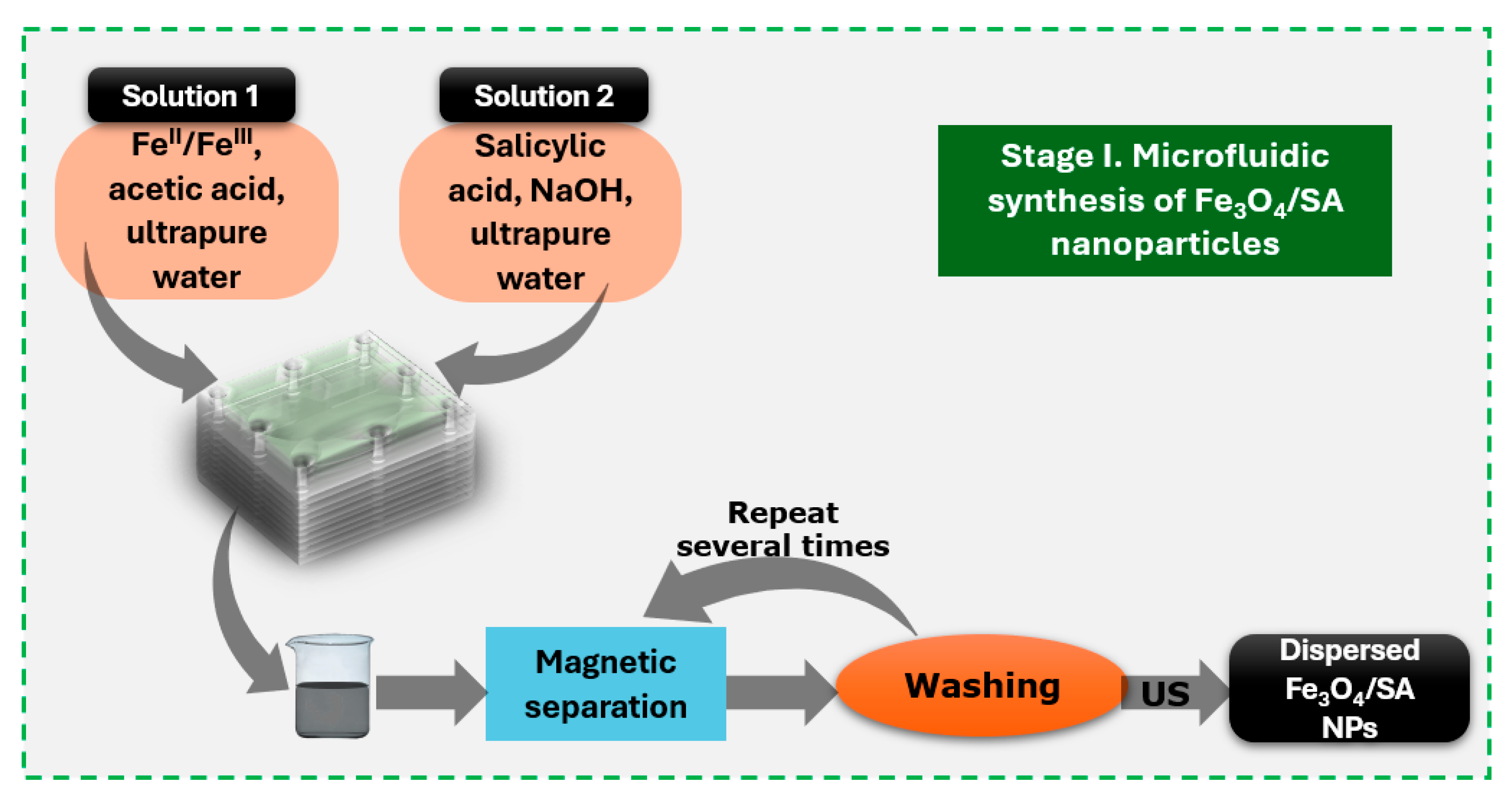
2.2. Salicylic Acid-Surface Modified Nanoparticles Synthesis

To obtain functionalized iron oxide nanoparticles (Fe3O4/SA), two separate precursor solutions were prepared. Solution 1 consisted of FeCl3 and FeSO4·7H2O dissolved in ultrapure water at a molar ratio of Fe3⁺:Fe2⁺ = 2:1. Solution 2 was prepared by dissolving 2% sodium hydroxide and 1% salicylic acid in 900 mL of ultrapure water. Initially, the microfluidic platform was filled with ultrapure water and degassed to remove any trapped air within the channels. Subsequently, the NaOH/salicylic acid solution (Solution 2) was introduced to condition the system, followed by the injection of the iron precursor solution (Solution 1) using a conventional osmosis pump. This sequential flow enabled controlled mixing and the insitu formation of salicylic acid-functionalized Fe3O4 nanoparticles within the microchannels. The resulting reaction product was magnetically separated and thoroughly washed with ultrapure water to remove residual reagents. The nanoparticles were then redispersed in ultrapure water via ultrasonication. A schematic overview of the synthesis process is provided in Figure 1 [17].

2.3. Hybrid Iron Oxide Nanoparticles

Two types of hybrid iron oxide nanoparticles—Fe3O4/SA–Ag and Fe3O4/SA–CuO—were synthesized by introducing silver nitrate and copper sulfate, respectively, into separate aqueous dispersions of preformed Fe3O4/SA nanoparticles (Figure 2). The amounts of silver and copper precursors were calculated to correspond to a 1:4 mass ratio relative to the iron oxide content, as the secondary surface modification was carried out through a physical adsorption and precipitation process.
For Fe3O4/SA–Ag, we used a simplified and modified silver nanoparticle synthesis protocol adapted from [48]. A secondary solution was prepared by dissolving 20 g of glucose and 5% (w/v) sodium hydroxide in ultrapure water, adjusted to a final volume of 120 mL. This reducing solution was continuously and slowly dripped into the silver precursor dispersion under stirring. The reaction mixture reached a final pH of approximately 12.5. The resulting Fe3O4/SA–Ag nanoparticles were magnetically separated, thoroughly washed with ultrapure water, and redispersed via ultrasonication.
For Fe3O4/SA–CuO, we used a simplified and modified copper oxide nanoparticle synthesis method adapted from [49]. A third solution consisting of 5% (w/v) sodium hydroxide in 200 mL of ultrapure water was gradually added to the Fe3O4/SA–Cu2⁺ dispersion under controlled stirring. The pH of the reaction mixture was maintained around 11.5. The resulting Fe3O4/SA–CuO nanoparticles were magnetically collected, rinsed multiple times with ultrapure water, and dispersed using ultrasonication.

2.4. Silica-Based Aerogel Thin Coatings

Coating fabrication was performed following the flowchart outlined in Figure 3. In more detail, two precursor solutions were prepared and combined to obtain a silica-based aerogel matrix without iron oxide nanoparticles. Silica-based aerogel was prepared using a simplified and adapted protocol based on the methods described in [50,51,52].
Solution A was prepared by dissolving 90 g sodium trisilicate and 3 g sodium hydroxide in 400 mL of ultrapure water, yielding a basic silicate solution with a pH of approximately 11.5–12. Solution B consisted of 600 mg alginic acid, 400 mg hexadecyltrimethylammonium bromide (CTAB), and 10 g ammonium chloride, dissolved in 400 mL of ultrapure water, with a pH of approximately 6.5–7.5.
These two solutions were mixed in a 1:1 volumetric ratio (Solution B:Solution A) and homogenized by ultrasonication for 60 s. Separately, a third solution (Solution C) was prepared by dissolving 20 g of calcium chloride and 14 mL of acetic acid in ultrapure water to a final volume of 400 mL, with a pH of approximately 4.5–5. A volume of 5 mL of this calcium-containing solution was then added to the silicate system to initiate crosslinking and gelation.
The final aerogel coatings were fabricated using the dip-coating method onto clean rectangular glass substrates. The coated surfaces were then dried by supercritical CO2 extraction to preserve the porous aerogel structure.
For hybrid coatings containing nanoparticles (Fe3O4/SA, Fe3O4/SA–Ag, or Fe3O4/SA–CuO), the only modification to the procedure was the addition of the respective nanoparticle dispersion (200 mg Fe3O4/SA dispersed in 15 mL ultrapure water) into Solution B before mixing with Solution A. This enabled uniform incorporation of the hybrid nanomaterials into the silica aerogel matrix during coating formation.

2.5. Nanoparticle Characterization

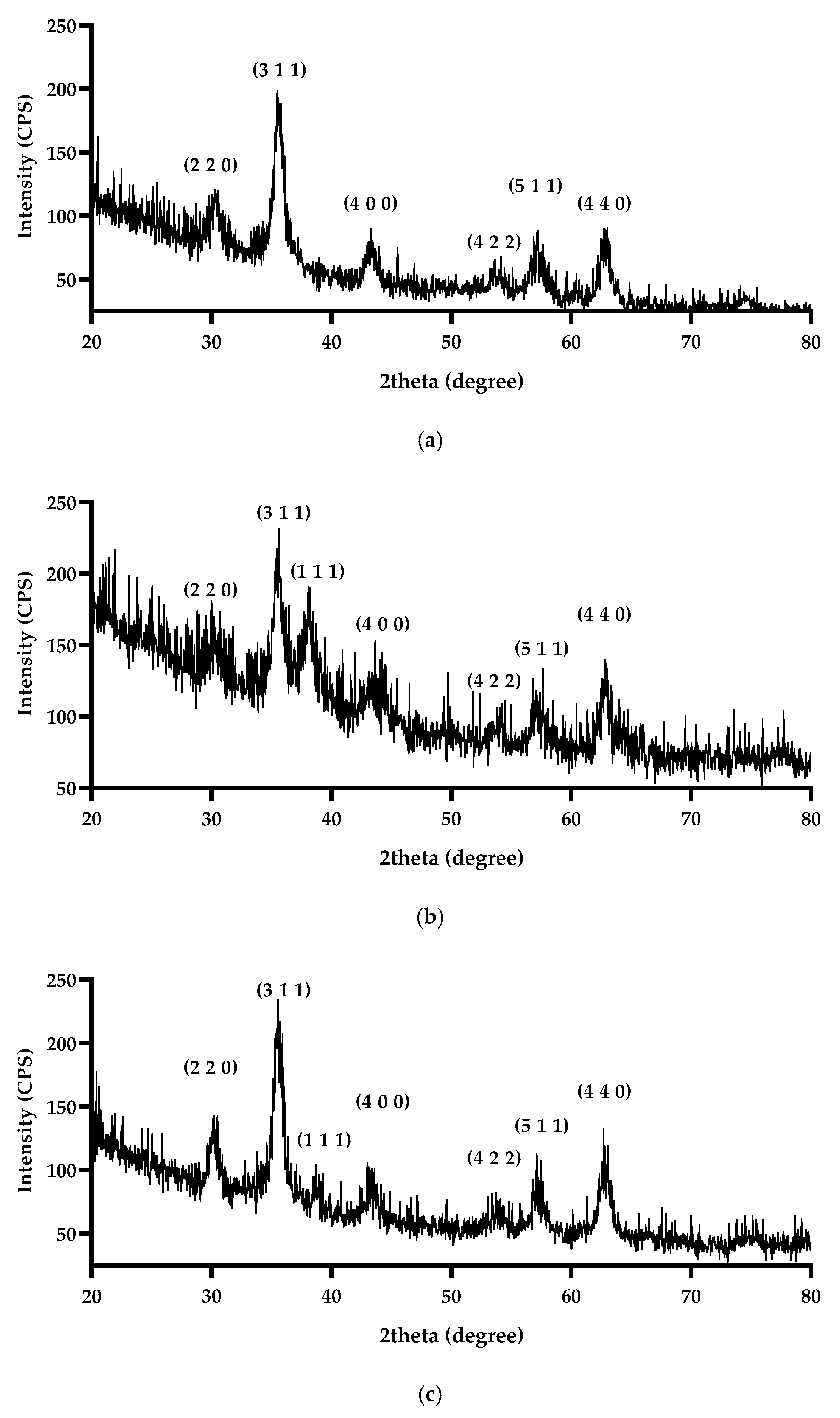
2.5.1. X-Ray Diffraction

An X-ray diffraction (XRD) methodology was used to investigate the nanoparticles’ crystal structure, phase composition, and degree of crystallinity. The analysis was conducted using a PANalytical Empyrean model diffractometer (PANalytical, Almelo, The Netherlands), which was equipped with a 2xGe 220 hybrid monochromator on the incident side and a parallel plate collimator coupled to a PIXcel 3D detector on the diffracted side. Grazing incidence X-ray diffraction measurements were carried out at ambient temperature with an incidence angle (ω) of 0.5° and a range of Bragg angles (2θ) from 27° to 80°, utilizing Cu Kα radiation with a wavelength (λ) of 1.5406 Å, under conditions of 40 mA and 45 kV.

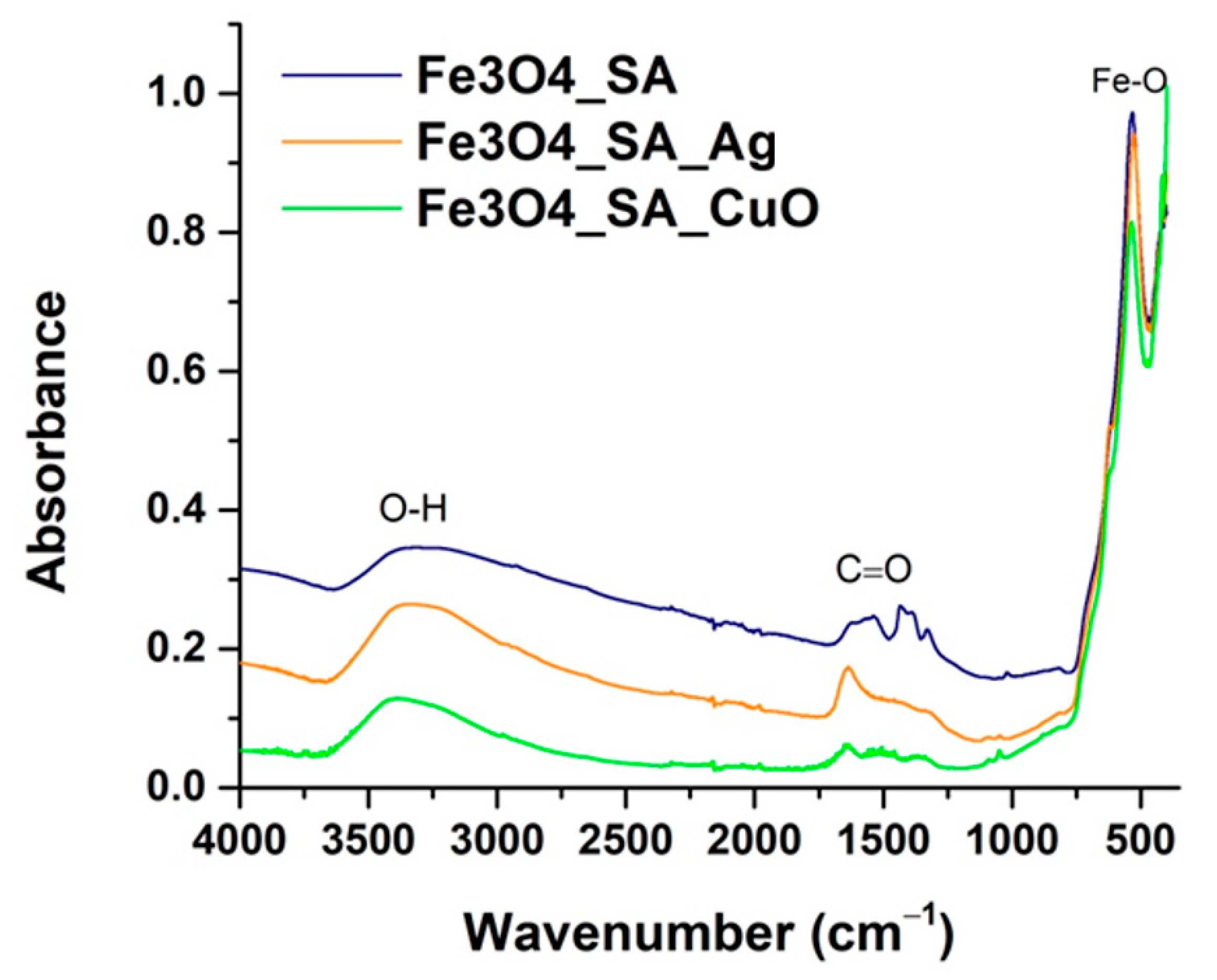
2.5.2. Fourier Transform Infrared Spectroscopy

The IR bands were identified via Fourier Transform Infrared (FT-IR) spectroscopy. For this analysis, a Thermo iN10-MX FTIR spectrometer, acquired from Thermo Fisher Scientific (Waltham, MA, USA), was employed. Spectra were collected within the wavenumber range of 4000–400 cm−1.

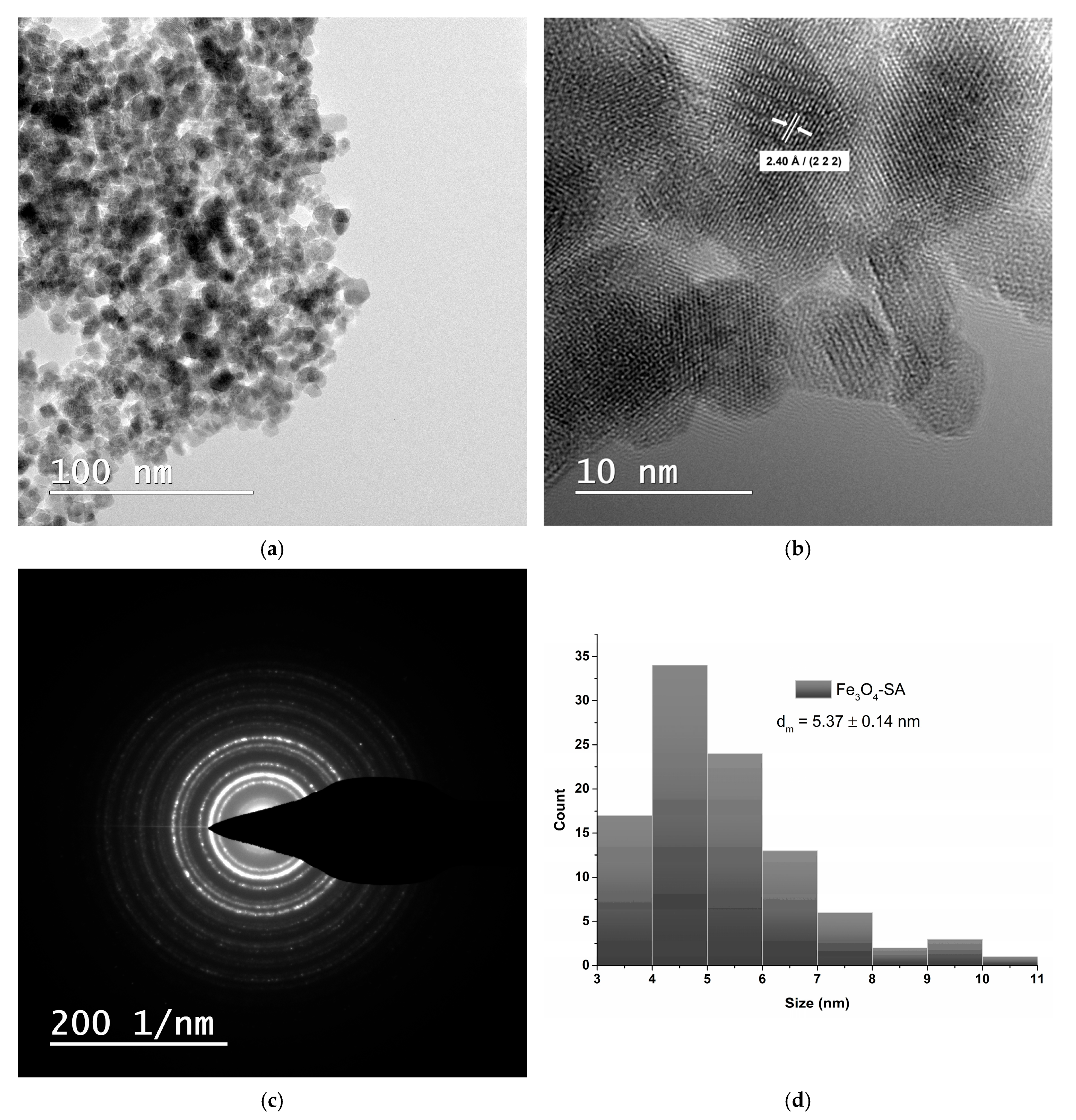
2.5.3. Transmission Electron Microscopy and Selected Area Electron Diffraction

High-resolution transmission electron microscopy (TEM) micrographs were obtained using the Thermo Fisher Scientific 80–200 Titan Themis transmission electron microscope (Hillsboro, OR, USA). The microscope operates at 200 kV in transmission mode, achieving line and point resolutions of 2 Å and 1 Å, respectively. Additionally, crystallographic information was acquired through the selected area electron diffraction (SAED) module, which is integrated into the TEM system.

2.5.4. Cell Viability and Proliferation of Hybrid Iron Oxide Nanoparticles

The cytotoxicity of iron oxide and hybrid iron oxide nanoparticles was tested using two cell lines: renal epithelial (VERO) and human keratinocyte (HaCat) cell lines. The cells were kept in Dulbecco’s Modified Eagle’s Medium (DMEM) for the duration of the experiment, with 1% penicillin-streptomycin mix and 10% fetal bovine serum (FBS) added as supplements. Following enzymatic-chemical trypsin/EDTA treatment to detach the cells from the culture surface, the cells were subsequently seeded into sterile 96-well culture plates at an initial density of 1 × 10⁴ cells/well. After optical microscopy confirmed that the cells had adhered to the culture surface for 24 h, the culture medium was removed and replaced with different concentrations (1 mg/mL, 200 μg/mL, 100 μg/mL, 40 μg/mL, 10 μg/mL, and 1 μg/mL) of the evaluated nanoparticles.
Using an MTT assay, cell viability and proliferation were evaluated using an MTT reagent (i.e., 3-(4,5-dimethylthiazol-2-yl)-2,5-diphenyltetrazolium bromide) (Sigma Aldrich, Merck Group, Darmstadt, Germany). Following a 24 h treatment period, the culture medium was extracted from the cell monolayers and substituted with an MTT solution (1 mg/mL), which was made by dissolving MTT in DMEM without FBS. After the samples had been incubated for four hours at 37 °C, the MTT solution was removed from the cell layers, and the formazan crystals that had formed were dissolved in DMSO. The FlexStation III multimodal plate reader (Molecular Devices, San Jose, CA, USA) was used to measure the optical density of the resultant solution for each sample in triplicate at a wavelength of 550 nm. GraphPad Prism 6 software was used to process the data statistically, utilizing ANOVA (Bonferroni correction). The statistical significance criterion was set at p < 0.05, and the data were displayed as the mean of three biological replicates ± the standard deviation.

2.5.5. Antimicrobial Assay

The minimum inhibitory concentration (MIC) was determined using the standard broth microdilution technique in sterile 96-well microtiter plates. The test was conducted in Triptycase Soy Broth (TSB) or simple nutrient broth, with a final volume of 150 µL per well. Serial two-fold (binary) dilutions of each nanoparticle suspension or test compound were prepared directly in the wells. After completing the dilution series, each well was inoculated with 15 µL of bacterial suspension adjusted to a 0.5 McFarland standard (approximately 1.5 × 10⁸ CFU/mL).
The bacterial strains tested were Staphylococcus aureus (Gram-positive) and Escherichia coli (Gram-negative). To rule out solvent-induced inhibition, solvent controls were prepared in parallel using the same dilution steps applied to the solvent (e.g., DMSO) used to disperse the nanoparticles. Two additional controls were included: a microbial growth control (medium with bacterial suspension but no treatment) and a sterility control (medium only, without bacterial inoculation).
Plates were incubated at 37 °C for 24 h in a humidified chamber. After incubation, bacterial growth was assessed macroscopically and confirmed by spectrophotometric reading at 600 nm. For optical measurement, 100 µL of each sample was transferred into a new sterile 96-well plate before reading. The MIC was defined as the lowest concentration of the tested compound at which no visible bacterial growth or increase in absorbance was observed, compared to the growth control.

2.6. Coatings Characterization

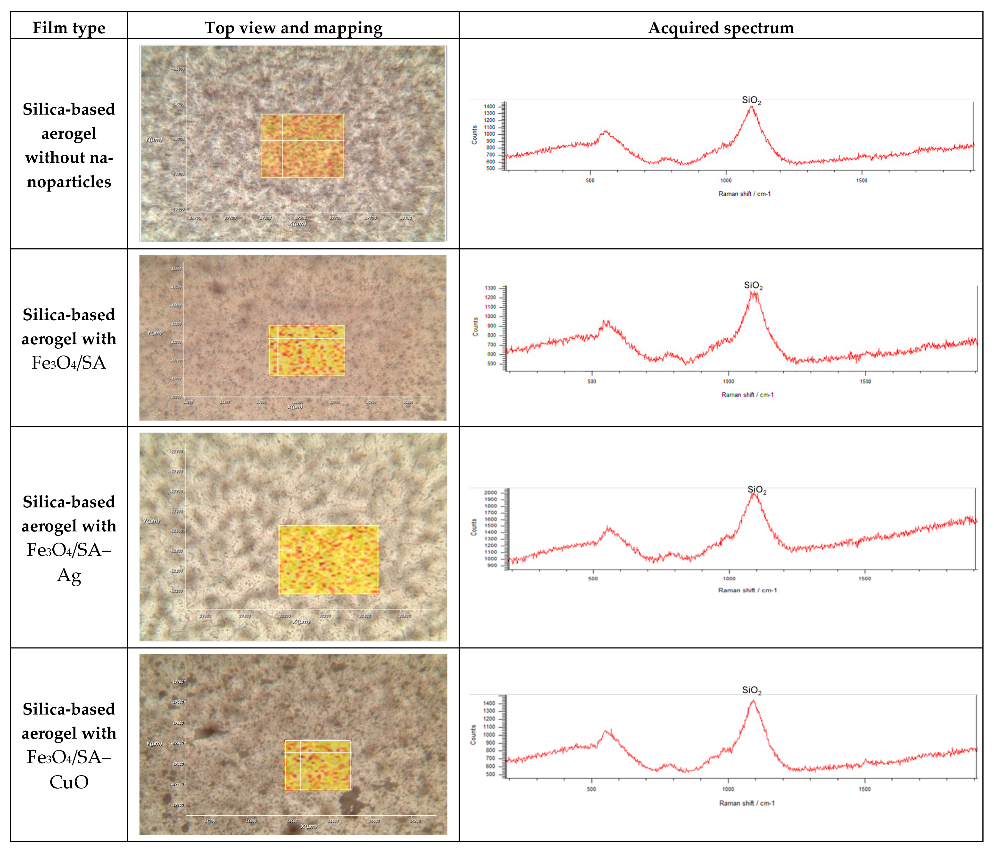
2.6.1. Raman Microscopy and Spectroscopy

RAMAN confocal microscopy (Renishaw inVia Raman microscope, Wotton-under-Edge, UK) analysis was performed with a spatial resolution of 0.25–1 µm to obtain information about the distribution of silica-based aerogel coatings. Spectra were acquired using a 532 nm wavelength laser at a laser intensity of 50%. A total of 40 scans were executed for each sample between 200 and 1900 cm−1.

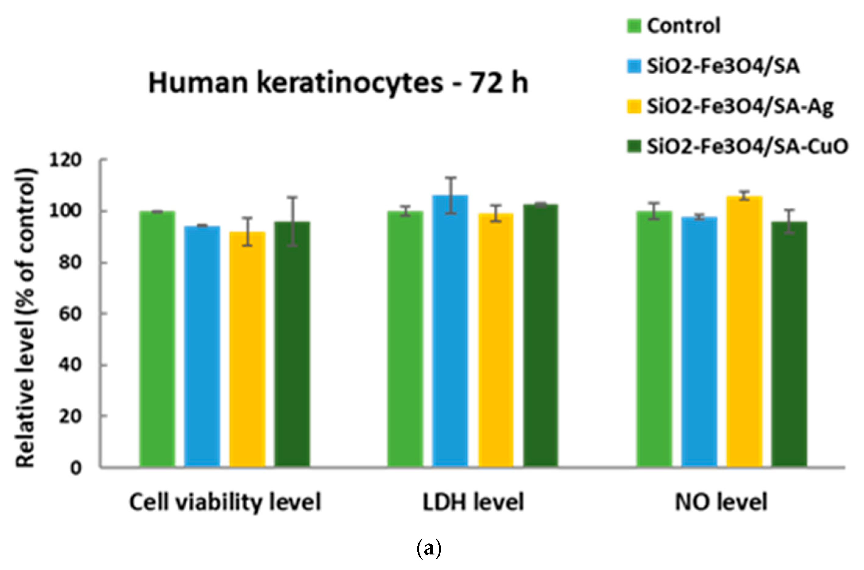
2.6.2. In Vitro Cytotoxicity Screening Protocol of Hybrid Iron Oxide Coatings

HaCaT keratinocytes were seeded on UV-sterilized samples represented by coated (SiO2-Fe3O4/SA, SiO2-Fe3O4/SA-Ag and SiO2-Fe3O4/SA-CuO) and uncoated glass slides (which served as controls) at a density of 105 cells/cm2 and left to grow for 72 h.
MTT assay (represented by incubation for 2 h with 1 mg/mL MTT solution and solubilization of formazan crystals with 2-propanol) was used to assess the cell viability at 595 nm (FlexStation, Molecular Device, USA). The inflammation status was investigated by Griess assay (0.1% naphthylethylenediamine dihydrochloride, 1% sulphanilamide in 5% H3PO4) which measured the level of nitric oxide (NO) in the culture supernatants at 550 nm. In addition, cell membrane damage was evaluated by lactate dehydrogenase (LDH) level kit from Roche Diagnostics (Mannheim, Germany) at 490 nm. Statistical evaluation was performed by one-way ANOVA with Bonferroni post-hoc test (GraphPad Prism 6) on triplicate experiments (p < 0.05 was considered significant).
Morphology of actin filaments was observed on Olympus IX71 microscope (Olympus, Tokyo, Japan) after the staining with Alexa Fluor 488-conjugated phalloidin (Invitrogen, Waltham, MA, USA) for 30 min at room temperature the cells previously fixed in 70% ethanol for 15 min and permeabilized with 0.1% Triton X-100.

2.7. In Vivo Experimental Model

The experimental protocol was applied according to European Council Directive No. 86/609/24 November 1986, the European Convention on the Protection of Vertebrate Animals (2005), and Romanian Law No. 43/2014 regarding the protection of animals used for scientific purposes. The study was approved by the Ethics Committee of the University of Medicine and Pharmacy of Craiova, Romania (Approval No. 54/19 May 2016).
Three-month-old BALB/c male mice were used for the Fe3O4/SA sample and for the control. The administration was carried out under general anesthesia. The mice were intraperitoneally inoculated in the groin area with 200 μL of a 1 mg/mL dispersion of Fe3O4/SA nanostructures obtained in ultrapure water, previously sterilized by UV irradiation for 30 min. Control mice were intraperitoneally inoculated in the groin area with 200 μL of saline.
During the examination period, the mice were housed in the Animal Care Unit of the University of Medicine and Pharmacy of Craiova, maintained in standard conditions (22 ± 2 °C, 55 ± 10% humidity, a 12–hour light–dark cycle, water, and food ad libitum).
Seven days and 14 days after the beginning of the experiment, the animals were euthanized, under general anesthesia, for the sampling of internal organs (brain, myocardium, liver, pancreas, lung, kidney, and spleen). Directly after the sampling, the biological material was washed in phosphate-buffered saline (PBS) to remove blood. Then, the internal organs were fixed in 10% neutral buffered formalin for 72 h at room temperature and processed for the routine histological paraffin embedding technique.
For the histological study, 4 μm-thick serial sections were cut on a MICROM HM355s rotary microtome (MICROM International GmbH, Walldorf, Germany) equipped with a waterfall-based section transfer system (STS, MICROM). The cross sections were placed on histological slides treated with poly-L-Lysine (Sigma-Aldrich, Munich, Germany). After Hematoxylin–Eosin (HE) classical staining, cross sections were evaluated and photographed using a Nikon Eclipse 55i light microscope equipped with a Nikon DS–Fi1 CCD high definition video camera (Nikon Instruments, Apidrag, Romania). Images were captured, stored, and analyzed using Image ProPlus 7 AMS software (Media Cybernetics Inc., Marlow, Buckinghamshire, UK).

3. Results

3.1. X-Ray Diffraction

The crystalline structure and phase composition of the synthesized nanomaterials were evaluated using X-ray diffraction (Figure 4). For all three samples—Fe3O4/SA, Fe3O4/SA–Ag, and Fe3O4/SA–CuO—a series of characteristic diffraction peaks were observed at 2θ values corresponding to the (220), (311), (400), (422), (511), and (440) planes. These reflections are in good agreement with the standard pattern for Fe3O4 with a face-centered cubic inverse spinel structure (JCPDS Card No. 19-0629) [53], confirming the successful formation and preservation of the Fe3O4 core across all samples. The absence of impurity peaks such as those from γ-Fe2O3 or hematite indicates the phase purity of the Fe3O4 core after functionalization.
In the Fe3O4/SA–Ag sample, an additional diffraction peak was observed at 2θ ≈ 38.1°, which corresponds to the (111) plane of silver nanoparticles, consistent with the face-centered cubic structure (JCPDS Card No. 04-0783) [54].
Similarly, the Fe3O4/SA–CuO sample exhibited an additional peak around 2θ ≈ 38.7°, which can be attributed to the (111) reflection of monoclinic CuO (JCPDS Card No. 45-0937) [55].

3.2. Fourier Transform Infrared Spectroscopy

The chemical structure and surface functionalization of the synthesized nanomaterials were investigated by FT-IR spectroscopy, and the spectra of Fe3O4/SA, Fe3O4/SA–Ag, and Fe3O4/SA–CuO are shown in Figure 5.
All three samples exhibit a prominent absorption band in the low-wavenumber region near 550–580 cm−1, corresponding to the Fe–O stretching vibration, which is a characteristic fingerprint of the spinel structure of Fe3O4 nanoparticles. This confirms the presence of the magnetite phase across all hybrid iron oxide samples.
In the Fe3O4/SA spectrum, broad absorption around 3400 cm−1 is attributed to O–H stretching vibrations, indicative of surface-bound hydroxyl groups and/or adsorbed water. Additionally, the clear presence of a band near 1700 cm−1 is assigned to C=O stretching from the carboxylic acid group of SA, confirming the successful functionalization of Fe3O4 with the organic molecule. A weaker band around 2900 cm−1, corresponding to C–H stretching vibrations, further supports the incorporation of the aromatic SA moiety on the nanoparticle surface.
Compared to Fe3O4/SA, both Fe3O4/SA–Ag and Fe3O4/SA–CuO display a noticeable decrease in signal intensity within the molecular fingerprint region, particularly around 1700–1300 cm−1, where C=O stretching and other aromatic vibrations associated with SA are typically observed. This spectral attenuation is attributed to the presence of Ag and CuO nanostructures on the nanoparticle surface, which likely interfere with the vibrational response of the salicylic acid layer. Residual fingerprint features confirm that salicylic acid remains on the surface, with modified vibrations indicating successful hybridization without complete displacement of the organic layer.

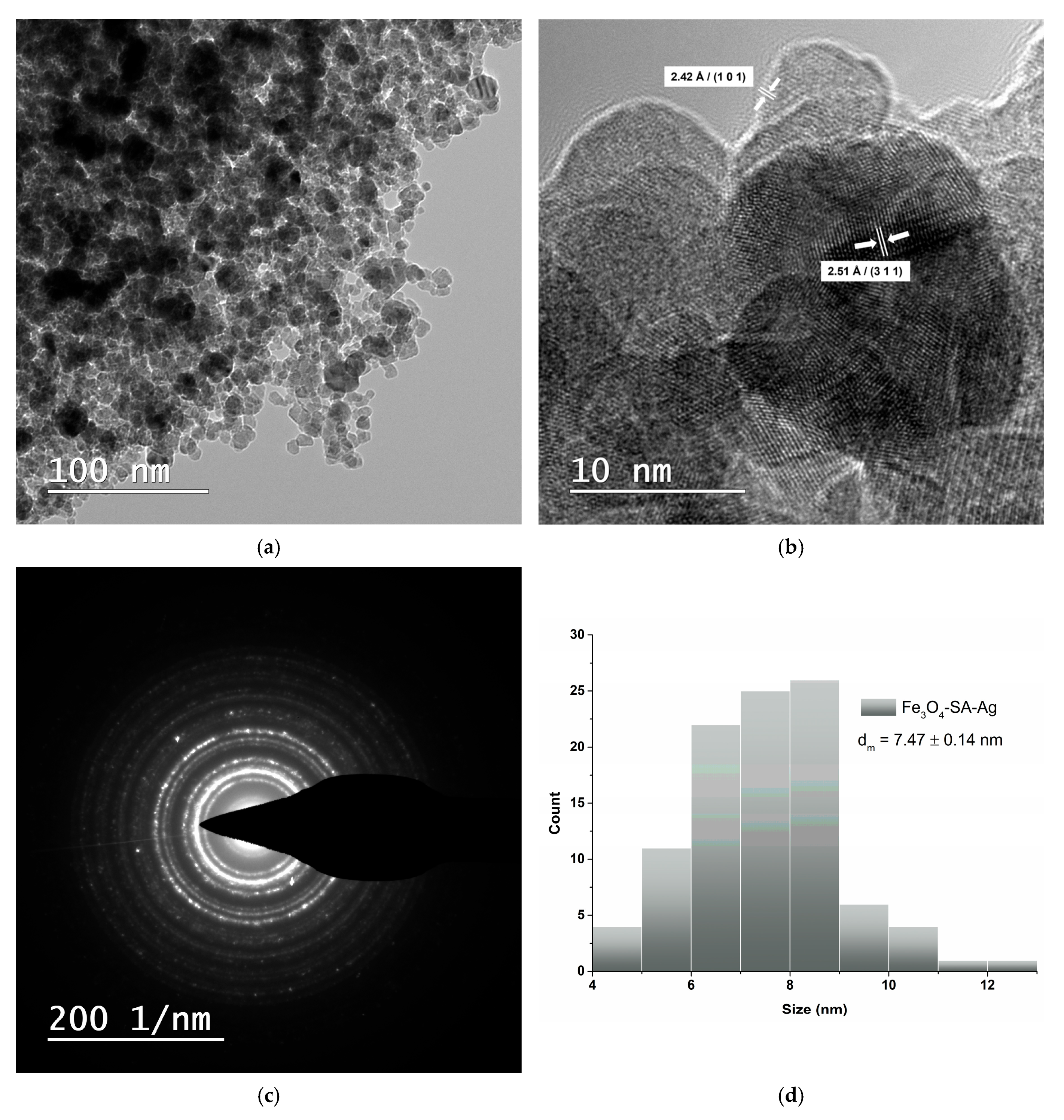
3.3. Transmission Electron Microscopy and Selected Area Electron Diffraction

TEM analysis was used to evaluate the morphology, crystallinity, and size of Fe3O4/SA nanoparticles. The bright field TEM image (Figure 6a) shows that the nanoparticles are roughly spherical, uniformly dispersed, and tend to form loosely packed aggregates, likely due to magnetic interactions. No large-scale agglomeration is observed, indicating good colloidal stability likely conferred by the salicylic acid surface coating. HR-TEM (Figure 6b) reveals the crystalline nature of the particles, with visible lattice fringes. The measured interplanar spacing of 2.40 Å corresponds to the (222) plane of the cubic spinel Fe3O4 structure, in agreement with XRD results and standard crystallographic data (JCPDS No. 19-0629) [53]. The SAED pattern (Figure 6c) shows concentric diffraction rings, confirming the polycrystalline nature of the Fe3O4/SA nanoparticles and supporting the well-defined magnetite structure observed in the XRD analysis. The size distribution histogram (Figure 6d), derived from TEM image analysis, shows that most particles fall within the 3–7 nm range, with a calculated mean diameter (dm) of 5.37 ± 0.14 nm.
The TEM images reveal that the Fe3O4/SA–Ag nanoparticles retain a nearly spherical morphology with moderate aggregation, similar to the Fe3O4/SA (Figure 7a). High-resolution TEM shows well-defined lattice fringes with interplanar spacings of 2.51 Å and 2.42 Å, corresponding to the (311) plane of Fe3O4 and the (101) plane of Ag, respectively (Figure 7b). The average particle size increased to 7.47 ± 0.14 nm (Figure 7d), compared to Fe3O4/SA, reflecting the addition of the Ag. The SAED pattern (Figure 7c) exhibits bright, well-defined rings indicative of a highly crystalline, polycrystalline structure, consistent with the coexistence of both Fe3O4 and Ag phases and correlating well with the XRD results.
The Fe3O4/SA–CuO nanoparticles display a quasi-spherical morphology with moderate clustering (Figure 8a). High-resolution TEM (Figure 8b) reveals well-defined lattice planes with measured interplanar spacings of 2.51 Å and 2.10 Å, corresponding to the (311) plane of Fe3O4 and the (400) plane of monoclinic CuO, respectively. The average particle size is 8.66 ± 0.16 nm (Figure 8d) while the SAED pattern (Figure 8c) presents well-defined, continuous rings, indicative of a polycrystalline structure, consistent with the co-presence of Fe3O4 and CuO phases and correlating well with the XRD results.

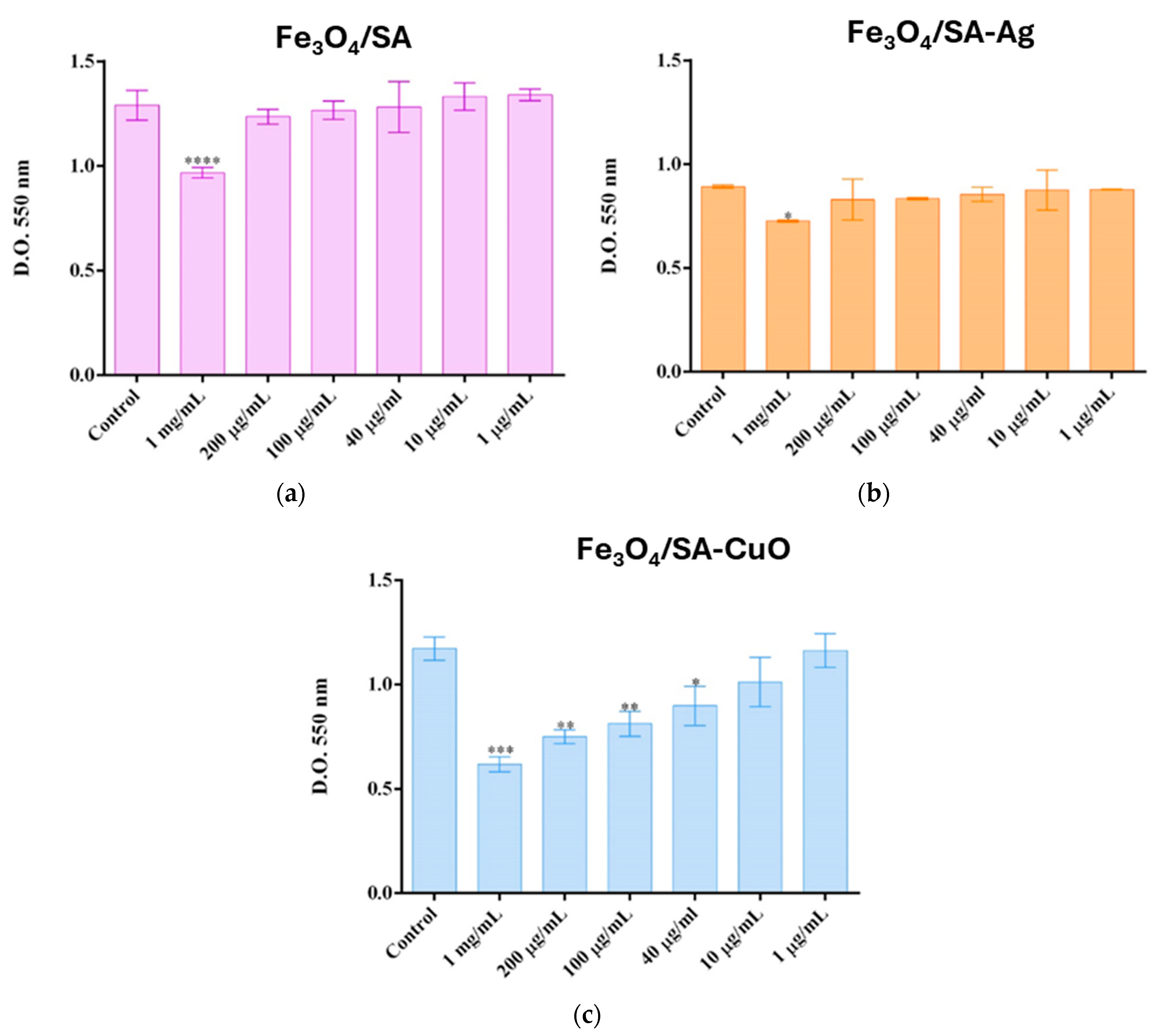
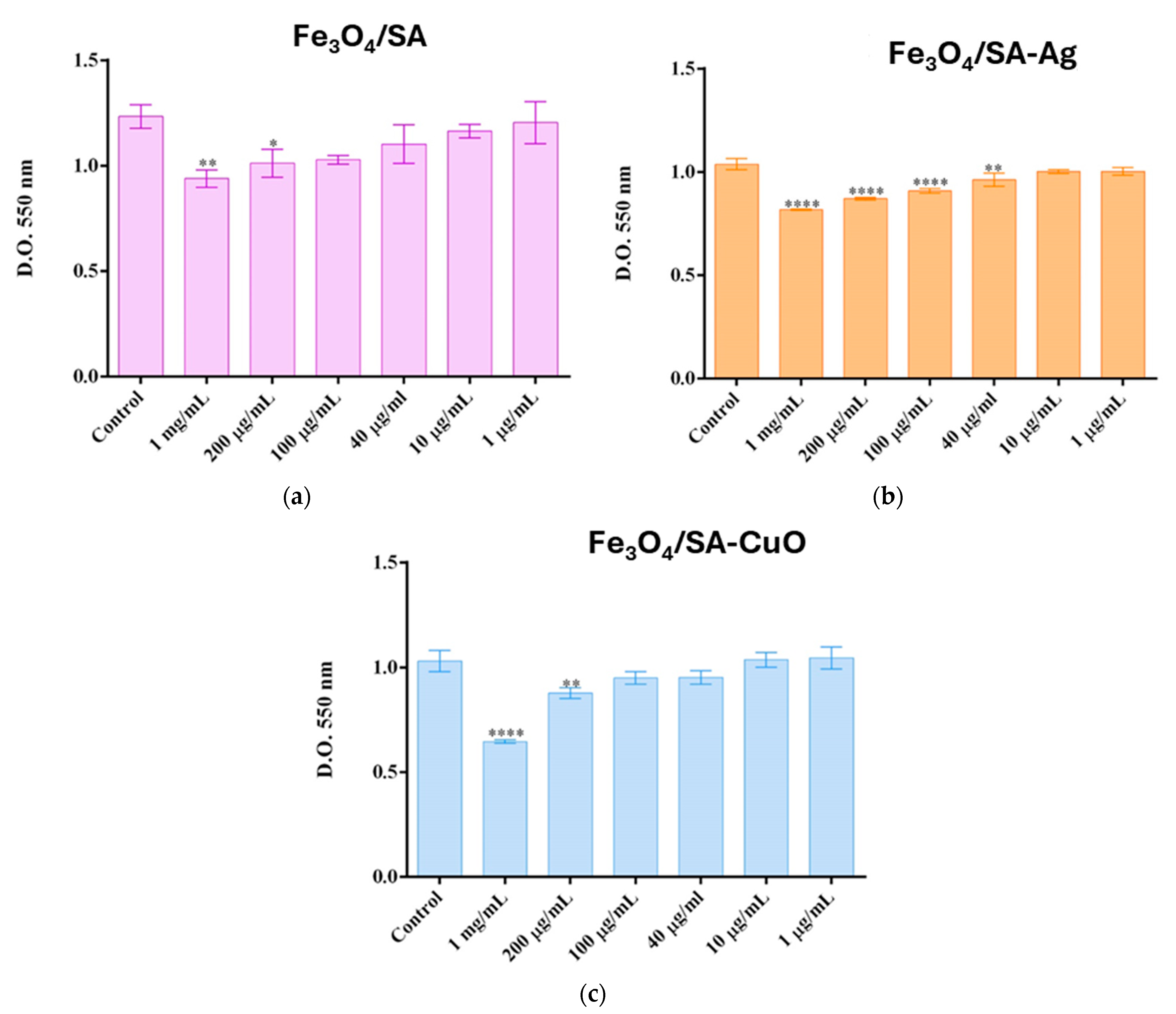
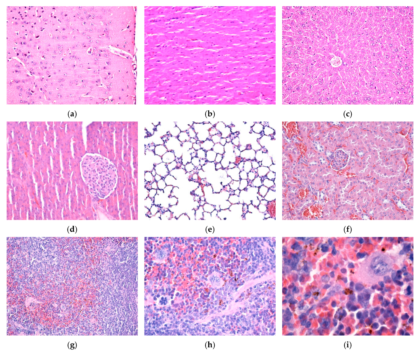
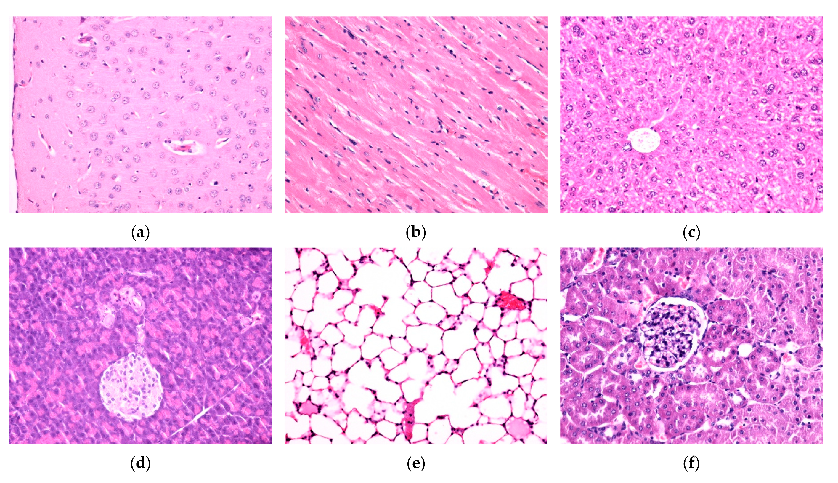
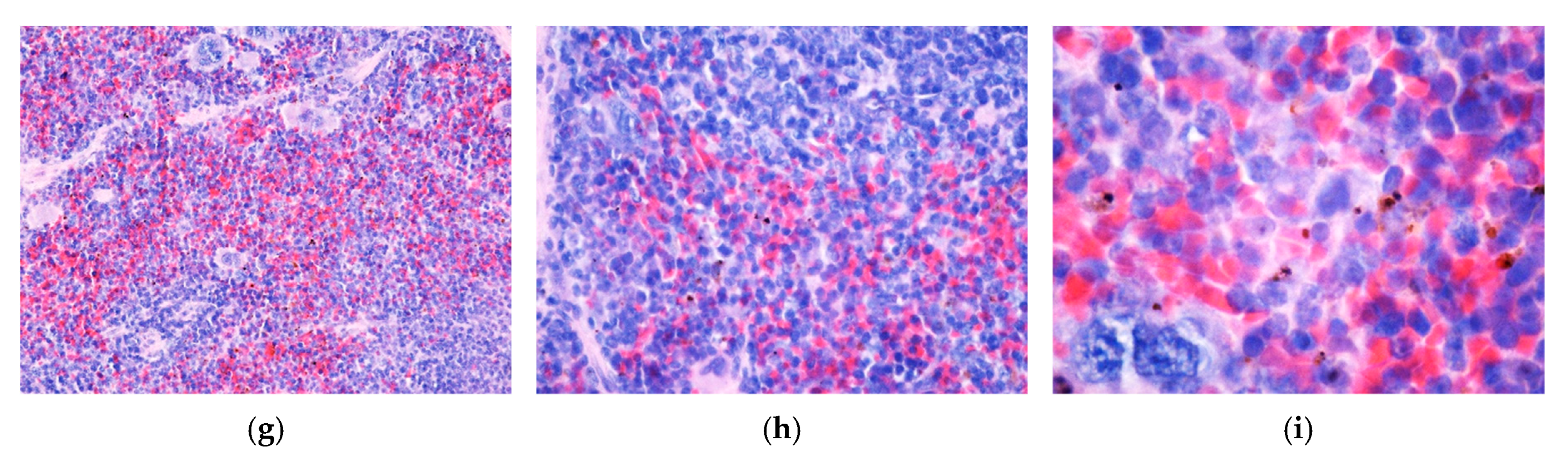
3.4. In Vitro and In Vivo Biocompatibility of Hybrid Iron Oxide Nanoparticles

The cytotoxicity assessment on HaCaT (Figure 9) and VERO (Figure 10) cell lines demonstrated that each nanoparticle type—Fe3O4/SA, Fe3O4/SA–Ag, and Fe3O4/SA–CuO—exhibits a distinct toxicity profile, with biocompatibility dependent on both concentration and cell type.
On the HaCaT line, Fe3O4/SA nanoparticles were biocompatible at all concentrations ≤ 200 µg/mL, with a significant decrease in viability observed only at 1 mg/mL. Similarly, Fe3O4/SA–Ag did not induce cytotoxic effects up to 200 µg/mL, suggesting that silver surface modification does not compromise biocompatibility in keratinocytes. In contrast, Fe3O4/SA–CuO showed higher toxicity, with a statistically significant reduction in viability starting from 40 µg/mL, indicating a more reactive surface and potential oxidative stress response.
In the more sensitive VERO cell line, Fe3O4/SA reduced viability at concentrations ≥ 200 µg/mL, while Fe3O4/SA–Ag induced cytotoxicity starting from 40 µg/mL, pointing to a stronger response to silver in renal epithelial cells. Interestingly, Fe3O4/SA–CuO was better tolerated by VERO cells, with no significant cytotoxicity observed below 200 µg/mL, despite its higher toxicity in HaCaT cells.
These results show that the cytotoxic effect is both dose- and cell type-dependent, and that each nanomaterial presents a different safety threshold. Fe3O4/SA and Fe3O4/SA–Ag demonstrate good biocompatibility at concentrations up to 200 µg/mL, particularly for skin-contact applications, while CuO-functionalized systems require careful dose consideration, especially when applied to keratinocyte-rich tissues.
Fe3O4/SA nanostructures were not detected in the brain, myocardium, liver, pancreas, lung, and kidney, neither 7 days (Figure 11a–f) nor at 14 days (Figure 12a–f) after intravenous injection. No histopathological alterations of the nervous tissue could be observed, suggesting that NPs do not cross the blood–brain barrier (Figure 11a and Figure 12a).
In the spleen, 7 days and 14 days after intravenous injection, Fe3O4/SA nanostructures were only evident in the red pulp. At 14 days, NPs were detected in higher concentration than in the sample collected at 7 days. In the white pulp, NPs were absent. However, hypertrophy of the white pulp was observed because nanoparticles stimulated the formation of macrophages with multi-lobular nuclei. In the red pulp, NPs were evident in the cells of the macrophage system, both in the Billroth cords and in the sinusoidal capillaries. The NPs appeared as granular, agglomerated, spherical structures of variable size, with a diameter of up to 3 μm, of brownish-blackish color. The density of the NPs varied from one cell to another, some cells presenting a greater amount of endocytosed NPs (Figure 11g–i and Figure 12g–i).

3.5. Antibacterial Characterization

The antimicrobial activity of Fe3O4-based nanostructures was evaluated against Staphylococcus aureus and Escherichia coli by determining their minimum inhibitory concentrations, as shown in Figure 13. All nanocomposites displayed dose-dependent antibacterial effects, with significant variation based on both the type of surface functionalization and the bacterial strain.
For S. aureus, Fe3O4/SA exhibited modest antimicrobial activity, with an MIC of approximately 250 µg/mL, likely due to mild ROS generation by Fe3O4 and the inherent antibacterial properties of salicylic acid. Functionalization with Ag or CuO significantly enhanced efficacy, with Fe3O4/SA–Ag and Fe3O4/SA–CuO demonstrating MIC values of approximately 18 µg/mL and 38 µg/mL, respectively.
In the case of E. coli, Fe3O4/SA showed minimal inhibitory effect, with a much higher MIC of approximately 500 µg/mL, underscoring its limited action on Gram-negative bacteria. By contrast, Fe3O4/SA–Ag exhibited the strongest antibacterial effect, with an MIC of only 8 µg/mL, while Fe3O4/SA–CuO required a higher MIC of about 52 µg/mL to inhibit growth. These findings suggest that silver-functionalized systems are particularly effective against Gram-negative pathogens, likely due to their ability to penetrate the outer membrane and disrupt intracellular targets.
Overall, Fe3O4/SA–Ag demonstrates the most potent and broad-spectrum antimicrobial profile, while Fe3O4/SA–CuO shows preferential activity toward S. aureus, reflecting known differences in bacterial wall composition and susceptibility.

3.6. Silica-Aerogel-Based Coatings Characterization

Raman spectroscopy and mapping were used to evaluate the structural features and homogeneity of the silica-based coatings. All four samples—SiO2, SiO2/Fe3O4/SA, SiO2/Fe3O4/SA–Ag, and SiO2/Fe3O4/SA–CuO—displayed similar Raman spectral profiles (Figure 14).
The Raman spectra are characterized by a strong and broad peak centered around ~1100 cm−1, which is attributed to the Si–O–Si asymmetric stretching vibration, confirming the presence of amorphous silica (SiO2) as the dominant matrix. A secondary, broader feature near ~550 cm−1 corresponds to Si–O bending modes, further validating the silica network. No significant shifts or new peaks were observed after embedding Fe3O4, Ag, or CuO components, suggesting that the incorporation of nanoparticles does not disrupt the silica framework at the molecular level.
The corresponding Raman image maps confirm the homogeneous distribution of the silica matrix across the analyzed surface area. The uniform Raman signal across the scanned area confirms consistent material dispersion and coating, indicating that the silica aerogel structure remains intact and continuous after nanoparticle incorporation.

3.7. In Vitro Behaviour of Human Keratinocytes on Thin Coatings

In vitro measurements on keratinocytes (Figure 15a) revealed cell viability levels higher than 90% of the control for all tested coatings, demonstrating their good biocompatibility. The 8% decrease in the case of SiO2-Fe3O4/SA-Ag could be correlated with the 6% increase in NO production after 72 h, suggesting a slight inflammatory potential, most probably due to silver ions, as previously described for Ag nanoparticles on human primary keratinocytes [56]. However, no cell membrane damage was recorded for both SiO2-Fe3O4/SA-Ag and SiO2-Fe3O4/SA-CuO samples, the levels being near the control ones. In addition, these results are in accordance with previous data reporting low toxicity of CuO-based nanostructures on human keratinocytes [57].
Furthermore, the exploration of F-actin organization in human keratinocytes (Figure 15b) showed the formation of a robust network of actin stress fibers along with the presence of adhesion zippers (arrows), which serve as intermediates in the maturation of adherens junctions, bringing adjacent membranes together and maintaining epithelial integrity. Actin filaments were concentrated and formed a honeycomb of cortical actin belts, which are essential in achieving stable cell contacts. The formation of radial actin fibers is an indicator of a good model for cell stratification, dependent upon calcium-induced intercellular contacts [58]. This was probably supported in our study by the biocompatible coatings of SiO2-Fe3O4/SA-Ag and SiO2-Fe3O4/SA-CuO.
Taken together, our results suggest that the tested samples support human keratinocytes’ viability and adhesion, offering promising applications for skin regeneration and wound healing.

4. Discussion

The present study follows our previous work [17], which successfully demonstrated the rapid, microfluidic-assisted synthesis of iron oxide nanoparticles functionalized with salicylic acid. Compared to traditional co-precipitation methods, the microfluidic platform ensures superior control over particle size, monodispersity, and reaction kinetics, allowing for reproducible synthesis of uniform nanoparticles suitable for biomedical applications [59,60,61]. In the current study, we advanced this platform by first synthesizing Fe3O4 nanoparticles via microfluidics and functionalizing them in situ with salicylic acid, followed by distinct post-synthetic surface decoration with either Ag or CuO nanostructures. Through this multi-step process, we obtained two hybrid nanomaterials—Fe3O4/SA-Ag and Fe3O4/SA-CuO—designed to enhance antimicrobial efficacy while improving biocompatibility compared to bulk Ag and CuO. While Ag and CuO nanoparticles are widely recognized for their strong antimicrobial activity [62,63], their cytotoxicity remains a major concern [64,65,66,67]. AgNPs can induce mitochondrial dysfunction, ROS overproduction, and DNA fragmentation in mammalian cells at doses as low as 10–25 µg/mL [68,69]. CuO nanoparticles, though less commonly used, have been shown to cause even more pronounced toxicity through Fenton-like reactions, triggering intracellular oxidative stress and membrane lipid peroxidation [70,71]. These dose-dependent effects limit their standalone use in biomedical coatings.
To overcome the limitations associated with the cytotoxicity of Ag and CuO nanoparticles, we prepared hybrid Fe3O4-based nanoparticles by functionalizing the iron oxide core with salicylic acid, followed by decoration with Ag and CuO. This configuration minimizes metal ion leaching and reduces direct nanoparticle–cell interaction from the Ag and CuO components, thus mitigating their cytotoxicity. Our cytotoxicity results confirmed that Fe3O4/SA–Ag and Fe3O4/SA–CuO nanocomposites exhibited improved biocompatibility compared to bulk Ag and CuO nanoparticles, with no significant reduction in HaCaT cell viability at concentrations up to 200 µg/mL, and variable tolerance in VERO cells, depending on the formulation. These findings are consistent with previous reports on hybrid systems designed to reduce ion release and nanoparticle–cell contact [72,73]. The cytotoxicity profile was cell type-dependent: HaCaT keratinocytes showed higher tolerance to Fe3O4/SA–Ag, while VERO cells were more sensitive, particularly to Ag-functionalized coatings. This highlights the importance of cell-specific responses when evaluating nanoparticle safety and underscores the need for application-specific material optimization.
Additionally, in vivo biodistribution studies confirmed the absence of Fe3O4/SA accumulation in vital organs such as the brain, liver, and kidney, supporting the systemic safety of these nanostructures. Their selective localization in the spleen, particularly within macrophages in the red pulp, suggests clearance through the reticuloendothelial system (RES) without triggering tissue damage. This distribution pattern aligns with previous studies reporting that iron oxide nanoparticles tend to accumulate in the spleen and liver following intravenous injection, depending on their size, coating, and surface charge [74,75,76].
Importantly, the lack of brain accumulation observed in our study further supports the notion that Fe3O4/SA nanoparticles do not cross the blood–brain barrier, which is consistent with findings from [77,78] and confirms their suitability for systemic applications where central nervous system exposure is undesirable. Compared to some earlier reports where uncoated or unstable iron oxide nanoparticles induced hepatic or renal retention and oxidative stress, our results indicate that salicylic acid surface functionalization may improve biodistribution profiles and minimize off-target organ accumulation.
From an antimicrobial perspective, the hybrid nanomaterials displayed differential but effective antibacterial activity against both Staphylococcus aureus and Escherichia coli, with Fe3O4/SA-CuO exhibiting the strongest inhibition, followed by Fe3O4/SA-Ag, while Fe3O4/SA alone showed only mild activity, primarily against S. aureus. The limited activity of Fe3O4/SA may be attributed to modest ROS generation by Fe3O4 and the intrinsic antimicrobial properties of salicylic acid, which can interfere with bacterial membrane integrity and biofilm formation.
The enhanced efficacy of the Ag- and CuO-functionalized systems can be attributed to the distinct antimicrobial mechanisms of Ag⁺ and Cu2⁺ ions. In S. aureus, both ions interfere with peptidoglycan synthesis, membrane stability, and inhibit thiol-dependent enzymes involved in energy metabolism [47,79]. In E. coli, their small size and membrane-penetrating ability allow them to breach the outer membrane, resulting in intracellular ROS production, protein oxidation, and DNA damage [45,80].
To translate the bioactivity and stability of the Fe3O4-based nanocomposites into practical biomedical coatings, we further integrated the system into a silica aerogel matrix—a platform known for its versatility in biomedical applications. The incorporation of a silica aerogel network provided several advantages. Silica aerogels are valued for their ultrahigh porosity, biocompatibility, and tunable surface chemistry, which support both cell compatibility and drug-loading capacity [81,82]. In our system, the aerogel served as a stabilizing scaffold, preventing nanoparticle agglomeration and ensuring uniform distribution across the coating surface. This homogeneous dispersion enhances antimicrobial surface contact while also contributing to mechanical durability, chemical stability, and reduced cytotoxicity [83,84]. Additionally, the inert and non-immunogenic nature of the silica matrix may help minimize inflammatory responses, supporting its application in sensitive environments such as wound dressings, implant surfaces, or diagnostic interfaces [85,86]. The modularity of the aerogel system also enables future integration of additional therapeutic agents, growth factors, or responsive layers, allowing for multifunctional and customizable surface designs tailored for advanced biomedical use [87,88].

5. Conclusions

In this study, we successfully synthesized Fe3O4 nanoparticles functionalized with salicylic acid using a microfluidic platform, followed by post-synthetic surface modification with either Ag or CuO to obtain hybrid nanosystems. This two-step approach led to the development of hybrid nanosystems—Fe3O4/SA–Ag and Fe3O4/SA–CuO—designed to enhance antimicrobial activity while reducing the cytotoxic effects commonly associated with bulk Ag and CuO nanoparticles. In vitro cytotoxicity assays demonstrated a cell type-dependent response: Fe3O4/SA–Ag was well tolerated by HaCaT keratinocytes at concentrations up to 200 µg/mL, while Fe3O4/SA–CuO induced greater sensitivity in this cell line but showed improved compatibility in VERO epithelial cells. Antibacterial testing revealed that Fe3O4/SA–CuO displayed the most potent activity against both S. aureus and E. coli, consistent with its higher potential for ROS generation and membrane disruption. In vivo studies showed no accumulation of Fe3O4/SA nanostructures in major organs and no associated tissue damage, with selective localization in the spleen indicating safe clearance via the reticuloendothelial system. The silica aerogel matrix enhanced nanoparticle stability, dispersion, and biocompatibility, supporting the development of effective antimicrobial coatings. These results highlight the potential of Fe3O4-based hybrids in biomedical applications, with future work focused on wound healing models and multifunctional therapeutic integration.

Author Contributions

Data curation, D.-A.M., A.-G.N., L.M., G.D.M., I.C.V., T.H., D.E.M., A.M.G., and A.A.; Formal analysis, D.-A.M., D.-I.T., A.-G.N., G.D.M., A.C.B., B.Ș.V., A.H., I.C.V., M.S.S., and A.M.G.; Investigation, D.-A.M., D.-I.T., A.-G.N., L.M., G.D.M., A.C.B., B.Ș.V., A.H., M.S.S., T.H., D.E.M., and A.A.; Methodology, T.H., D.E.M., and A.M.G.; Validation, L.M., A.H., I.C.V., M.S.S., T.H., A.M.G., and A.A.; Writing—original draft, D.-A.M., D.-I.T., A.-G.N., L.M., G.D.M., A.C.B., B.Ș.V., A.H., I.C.V., M.S.S., T.H., D.E.M., A.M.G., and A.A.; Writing—review and editing, A.-G.N., T.H., D.E.M., A.M.G., and A.A. All authors have read and agreed to the published version of the manuscript.

Funding

The authors acknowledge the financial support from the European Union (NextGenerationEU) through PNRR.C9-I8: Aerogel-based magnetic nanocomposites for water decontamination (CF 231/29.11.2022). The content of this material does not necessarily represent the official position of the European Union or of the Government of Romania.

Data Availability Statement

Data are contained within the article.

Acknowledgments

The infrastructure of the National Center for Micro and Nanomaterials—“Installations and Strategic Objectives of National Interest” Program within the National University of Science and Technology Politehnica Bucharest is highly acknowledged.

Conflicts of Interest

The authors declare no conflicts of interest.

References

  1. Gupta, R.K.; Gawad, F.A.E.; Ali, E.A.E.; Karunanithi, S.; Yugiani, P.; Srivastav, P.P. Nanotechnology: Current applications and future scope in food packaging systems. Meas. Food 2024, 13, 100131. [Google Scholar] [CrossRef]
  2. Malik, S.; Muhammad, K.; Waheed, Y. Emerging Applications of Nanotechnology in Healthcare and Medicine. Molecules 2023, 28, 6624. [Google Scholar] [CrossRef] [PubMed]
  3. Girardet, T.; Venturini, P.; Martinez, H.; Dupin, J.-C.; Cleymand, F.; Fleutot, S. Spinel Magnetic Iron Oxide Nanoparticles: Properties, Synthesis and Washing Methods. Appl. Sci. 2022, 12, 8127. [Google Scholar] [CrossRef]
  4. Saleh, H.M.; Hassan, A.I. Synthesis and Characterization of Nanomaterials for Application in Cost-Effective Electrochemical Devices. Sustainability 2023, 15, 10891. [Google Scholar] [CrossRef]
  5. Li, G.; Wang, C.; Jin, B.; Sun, T.; Sun, K.; Wang, S.; Fan, Z. Advances in smart nanotechnology-supported photodynamic therapy for cancer. Cell Death Discov. 2024, 10, 466. [Google Scholar] [CrossRef]
  6. Gimondi, S.; Ferreira, H.; Reis, R.L.; Neves, N.M. Microfluidic Devices: A Tool for Nanoparticle Synthesis and Performance Evaluation. ACS Nano 2023, 17, 14205–14228. [Google Scholar] [CrossRef]
  7. Yao, F.; Zhu, P.; Chen, J.; Li, S.; Sun, B.; Li, Y.; Zou, M.; Qi, X.; Liang, P.; Chen, Q. Synthesis of nanoparticles via microfluidic devices and integrated applications. Microchim. Acta 2023, 190, 256. [Google Scholar] [CrossRef]
  8. Bezelya, A.; Küçüktürkmen, B.; Bozkır, A. Microfluidic Devices for Precision Nanoparticle Production. Micro 2023, 3, 822–866. [Google Scholar] [CrossRef]
  9. Roy, S.; Kumar, R.; Acooli, A.; Roy, S.; Chatterjee, A.; Chattaraj, S.; Nayak, J.; Jeon, B.-H.; Basu, A.; Banerjee, S.; et al. Transforming Nanomaterial Synthesis through Advanced Microfluidic Approaches: A Review on Accessing Unrestricted Possibilities. J. Compos. Sci. 2024, 8, 386. [Google Scholar] [CrossRef]
  10. Liu, S.; Zhang, C.; Wang, Y.; Wei, X. Synthesis of energetic materials by microfluidics. Def. Technol. 2025, 44, 306–319. [Google Scholar] [CrossRef]
  11. Popa, M.L.; Preda, M.D.; Neacșu, I.A.; Grumezescu, A.M.; Ginghină, O. Traditional vs. Microfluidic Synthesis of ZnO Nanoparticles. Int. J. Mol. Sci. 2023, 24, 1875. [Google Scholar] [CrossRef]
  12. Zhang, H.; Yang, J.; Sun, R.; Han, S.; Yang, Z.; Teng, L. Microfluidics for nano-drug delivery systems: From fundamentals to industrialization. Acta Pharm. Sin. B 2023, 13, 3277–3299. [Google Scholar] [CrossRef] [PubMed]
  13. Jiang, K.; Zhang, L.; Bao, G. Magnetic iron oxide nanoparticles for biomedical applications. Curr. Opin. Biomed. Eng. 2021, 20, 100330. [Google Scholar] [CrossRef] [PubMed]
  14. Stiufiuc, G.F.; Stiufiuc, R.I. Magnetic Nanoparticles: Synthesis, Characterization, and Their Use in Biomedical Field. Appl. Sci. 2024, 14, 1623. [Google Scholar] [CrossRef]
  15. Montiel Schneider, M.G.; Martín, M.J.; Otarola, J.; Vakarelska, E.; Simeonov, V.; Lassalle, V.; Nedyalkova, M. Biomedical Applications of Iron Oxide Nanoparticles: Current Insights Progress and Perspectives. Pharmaceutics 2022, 14, 204. [Google Scholar] [CrossRef]
  16. Wu, K.; Wang, J.-P.; Natekar, N.A.; Ciannella, S.; González-Fernández, C.; Gomez-Pastora, J.; Bao, Y.; Liu, J.; Liang, S.; Wu, X.; et al. Roadmap on magnetic nanoparticles in nanomedicine. Nanotechnology 2024, 36, 042003. [Google Scholar] [CrossRef]
  17. Niculescu, A.-G.; Munteanu, O.M.; Bîrcă, A.C.; Moroșan, A.; Purcăreanu, B.; Vasile, B.Ș.; Istrati, D.; Mihaiescu, D.E.; Hadibarata, T.; Grumezescu, A.M. New 3D Vortex Microfluidic System Tested for Magnetic Core-Shell Fe3O4-SA Nanoparticle Synthesis. Nanomaterials 2024, 14, 902. [Google Scholar] [CrossRef]
  18. Nowak-Jary, J.; Machnicka, B. Comprehensive Analysis of the Potential Toxicity of Magnetic Iron Oxide Nanoparticles for Medical Applications: Cellular Mechanisms and Systemic Effects. Int. J. Mol. Sci. 2024, 25, 12013. [Google Scholar] [CrossRef]
  19. Ansari, K.; Ahmad, R.; Tanweer, M.S.; Azam, I. Magnetic Iron Oxide Nanoparticles as a Tool for the Advancement of Biomedical and Environmental Application: A Review. Biomed. Mater. Devices 2024, 2, 139–157. [Google Scholar] [CrossRef]
  20. Nedylakova, M.; Medinger, J.; Mirabello, G.; Lattuada, M. Iron oxide magnetic aggregates: Aspects of synthesis, computational approaches and applications. Adv. Colloid Interface Sci. 2024, 323, 103056. [Google Scholar] [CrossRef]
  21. Niculescu, A.-G.; Chircov, C.; Grumezescu, A.M. Magnetite nanoparticles: Synthesis methods—A comparative review. Methods 2022, 199, 16–27. [Google Scholar] [CrossRef] [PubMed]
  22. Upadhyay, J.; Singh, A. Lignin Derived Biofabrication of Magnetic Iron Nanoparticles from Moringa oleifera. Lett. Appl. NanoBioSci. 2024, 13, 104. [Google Scholar]
  23. Ezealigo, U.S.; Ezealigo, B.N.; Aisida, S.O.; Ezema, F.I. Iron oxide nanoparticles in biological systems: Antibacterial and toxicology perspective. JCIS Open 2021, 4, 100027. [Google Scholar] [CrossRef]
  24. Solanki, R.; Makwana, N.; Kumar, R.; Joshi, M.; Patel, A.; Bhatia, D.; Sahoo, D.K. Nanomedicines as a cutting-edge solution to combat antimicrobial resistance. RSC Adv. 2024, 14, 33568–33586. [Google Scholar] [CrossRef]
  25. Roca, A.G.; Lopez-Barbera, J.F.; Lafuente, A.; Özel, F.; Fantechi, E.; Muro-Cruces, J.; Hémadi, M.; Sepulveda, B.; Nogues, J. Iron oxide nanoparticles (Fe3O4, γ-Fe2O3 and FeO) as photothermal heat mediators in the first, second and third biological windows. Phys. Rep. 2023, 1043, 1–35. [Google Scholar] [CrossRef]
  26. Niculescu, A.-G.; Moroșan, A.; Bîrcă, A.C.; Gherasim, O.; Oprea, O.C.; Vasile, B.Ș.; Purcăreanu, B.; Mihaiescu, D.E.; Rădulescu, M.; Grumezescu, A.M. Microwave-Assisted Silanization of Magnetite Nanoparticles Pre-Synthesized by a 3D Microfluidic Platform. Nanomaterials 2023, 13, 2795. [Google Scholar] [CrossRef]
  27. Abdussalam-Mohammed, W.; Abraheem, M.S.; Mezoughi, A.B.; Mohamed, L.; Alwahsh, M.A.A. Comparative Analysis of Novel Iron Oxide Nanoparticles Synthesized by Different Approaches with Evaluation of Their Antibacterial Activities. Biointerface Res. Appl. Chem. 2022, 13, 317. [Google Scholar]
  28. Fadeev, M.; Kozlovskiy, A.; Korolkov, I.; Egizbek, K.; Nazarova, A.; Chudoba, D.; Rusakov, V.; Zdorovets, M. Iron oxide @ gold nanoparticles: Synthesis, properties and potential use as anode materials for lithium-ion batteries. Colloids Surf. A Physicochem. Eng. Asp. 2020, 603, 125178. [Google Scholar] [CrossRef]
  29. Ismail, A.M.; Tiama, T.M.; Farghaly, A.; Elhaes, H.; Ibrahim, M.A. Assessment of the functionalization of chitosan/iron oxide nanoparticles. Biointerface Res. Appl. Chem 2023, 13, 582. [Google Scholar]
  30. Albukhaty, S.; Sulaiman, G.M.; Al-Karagoly, H.; Mohammed, H.A.; Hassan, A.S.; Alshammari, A.A.A.; Ahmad, A.M.; Madhi, R.; Almalki, F.A.; Khashan, K.S.; et al. Iron oxide nanoparticles: The versatility of the magnetic and functionalized nanomaterials in targeting drugs, and gene deliveries with effectual magnetofection. J. Drug Deliv. Sci. Technol. 2024, 99, 105838. [Google Scholar] [CrossRef]
  31. Dharmalingam, N.; Arumugasamy, V.; Mariappan, R. Amoxicillin Encapsulated Magnetite Nanomaterial for Antibiotic Drug Delivery and Deoxyribonucleic Acid Interaction. Lett. Appl. NanoBioSci. 2024, 13, 102. [Google Scholar]
  32. Salehirozveh, M.; Dehghani, P.; Mijakovic, I. Synthesis, Functionalization, and Biomedical Applications of Iron Oxide Nanoparticles (IONPs). J. Funct. Biomater. 2024, 15, 340. [Google Scholar] [CrossRef] [PubMed]
  33. Koo, T.M.; Fu, H.E.; Moon, J.H.; Oh, E.; Kim, Y.; Ko, M.J.; Kim, Y.K. Porous Iron Oxide Core–Gold Satellite Nanocomposite: A Cost-Effective and Recyclable Solution for Photocatalytic Wastewater Treatment. Small Sci. 2024, 4, 2300266. [Google Scholar] [CrossRef] [PubMed]
  34. Sodipo, B.K.; Aziz, A.A. Recent advances in synthesis and surface modification of superparamagnetic iron oxide nanoparticles with silica. J. Magn. Magn. Mater. 2016, 416, 275–291. [Google Scholar] [CrossRef]
  35. AlQurashi, D.M.; AlQurashi, T.F.; Alam, R.I.; Shaikh, S.; Tarkistani, M.A.M. Advanced Nanoparticles in Combating Antibiotic Resistance: Current Innovations and Future Directions. J. Nanotheranostics 2025, 6, 9. [Google Scholar] [CrossRef]
  36. Kaymaz, S.V.; Nobar, H.M.; Sarıgül, H.; Soylukan, C.; Akyüz, L.; Yüce, M. Nanomaterial surface modification toolkit: Principles, components, recipes, and applications. Adv. Colloid Interface Sci. 2023, 322, 103035. [Google Scholar] [CrossRef]
  37. Chandorkar, Y.; Valeske, M.; Kolrosova, B.; Elbs-Glatz, Y.; Zuber, F.; Schoeller, J.; Kummer, N.; Ren, Q.; Rottmar, M.; Maniura-Weber, K. Bioactive Salicylic Acid Containing Coating for Dental Implants to Combat Infection and Inflammation. Adv. Mater. Interfaces 2024, 11, 2300750. [Google Scholar] [CrossRef]
  38. Sykes, E.M.E.; White, D.; McLaughlin, S.; Kumar, A. Salicylic acids and pathogenic bacteria: New perspectives on an old compound. Can. J. Microbiol. 2023, 70, 1–14. [Google Scholar] [CrossRef]
  39. Ewonkem, M.B.; Deussom, P.M.; Mbock, M.A.; Tiakouang, E.N.; Toze, A.F.A.; Wansi, D.J. Antibacterial, antifungal activities and toxicity of new synthetic fatty acid salicylate esters. Med. Chem. Res. 2023, 32, 736–748. [Google Scholar] [CrossRef]
  40. Covelli, V.; Cozzolino, A.; Rizzo, P.; Rodriquez, M.; Vestuto, V.; Bertamino, A.; Daniel, C.; Guerra, G. Salicylic Acid Release from Syndiotactic Polystyrene Staple Fibers. Molecules 2023, 28, 5095. [Google Scholar] [CrossRef]
  41. Ren, G.; Hu, D.; Cheng, E.W.C.; Vargas-Reus, M.A.; Reip, P.; Allaker, R.P. Characterisation of copper oxide nanoparticles for antimicrobial applications. Int. J. Antimicrob. Agents 2009, 33, 587–590. [Google Scholar] [CrossRef] [PubMed]
  42. Rai, M.; Yadav, A.; Gade, A. Silver nanoparticles as a new generation of antimicrobials. Biotechnol. Adv. 2009, 27, 76–83. [Google Scholar] [CrossRef] [PubMed]
  43. Parvin, N.; Joo, S.W.; Mandal, T.K. Nanomaterial-Based Strategies to Combat Antibiotic Resistance: Mechanisms and Applications. Antibiotics 2025, 14, 207. [Google Scholar] [CrossRef] [PubMed]
  44. Mandal, T.K. Nanomaterial-Enhanced Hybrid Disinfection: A Solution to Combat Multidrug-Resistant Bacteria and Antibiotic Resistance Genes in Wastewater. Nanomaterials 2024, 14, 1847. [Google Scholar] [CrossRef]
  45. Morones, J.R.; Elechiguerra, J.L.; Camacho, A.; Holt, K.; Kouri, J.B.; Ramírez, J.T.; Yacaman, M.J. The bactericidal effect of silver nanoparticles. Nanotechnology 2005, 16, 2346–2353. [Google Scholar] [CrossRef]
  46. Furno, F.; Morley, K.S.; Wong, B.; Sharp, B.L.; Arnold, P.L.; Howdle, S.M.; Bayston, R.; Brown, P.D.; Winship, P.D.; Reid, H.J. Silver nanoparticles and polymeric medical devices: A new approach to prevention of infection? J. Antimicrob. Chemother. 2004, 54, 1019–1024. [Google Scholar] [CrossRef]
  47. Lemire, J.A.; Harrison, J.J.; Turner, R.J. Antimicrobial activity of metals: Mechanisms, molecular targets and applications. Nat. Rev. Microbiol. 2013, 11, 371–384. [Google Scholar] [CrossRef]
  48. Nie, P.; Zhao, Y.; Xu, H. Synthesis, applications, toxicity and toxicity mechanisms of silver nanoparticles: A review. Ecotoxicol. Environ. Saf. 2023, 253, 114636. [Google Scholar] [CrossRef]
  49. Saleem, M.H.; Ejaz, U.; Vithanage, M.; Bolan, N.; Siddique, K.H.M. Synthesis, characterization, and advanced sustainable applications of copper oxide nanoparticles: A review. Clean Technol. Environ. Policy 2024, 1–26. [Google Scholar] [CrossRef]
  50. Meti, P.; Wang, Q.; Mahadik, D.B.; Lee, K.-Y.; Gong, Y.-D.; Park, H.-H. Evolutionary Progress of Silica Aerogels and Their Classification Based on Composition: An Overview. Nanomaterials 2023, 13, 1498. [Google Scholar] [CrossRef]
  51. Menshutina, N.; Tsygankov, P.; Ivanov, S. Synthesis and Properties of Silica and Alginate Hybrid Aerogel Particles with Embedded Carbon Nanotubes (CNTs) for Selective Sorption. Materials 2018, 12, 52. [Google Scholar] [CrossRef] [PubMed]
  52. Vareda, J.P.; Maximiano, P.; Cunha, L.P.; Ferreira, A.F.; Simões, P.N.; Durães, L. Effect of different types of surfactants on the microstructure of methyltrimethoxysilane-derived silica aerogels: A combined experimental and computational approach. J. Colloid Interface Sci. 2018, 512, 64–76. [Google Scholar] [CrossRef] [PubMed]
  53. Yang, Z.; Qian, K.; Lv, J.; Yan, W.; Liu, J.; Ai, J.; Zhang, Y.; Guo, T.; Zhou, X.; Xu, S.; et al. Encapsulation of Fe3O4 Nanoparticles into N, S co-Doped Graphene Sheets with Greatly Enhanced Electrochemical Performance. Sci. Rep. 2016, 6, 27957. [Google Scholar] [CrossRef]
  54. Lanje, A.S.; Sharma, S.J.; Pode, R.B. Synthesis of silver nanoparticles: A safer alternative to conventional antimicrobial and antibacterial agents. J. Chem. Pharm. Res. 2010, 2, 478–483. [Google Scholar]
  55. Jia, B.; Qin, M.; Zhang, Z.; Cao, Z.; Wu, H.; Chen, P.; Zhang, L.; Lu, X.; Qu, X. The formation of CuO porous mesocrystal ellipsoids via tuning the oriented attachment mechanism. CrystEngComm 2016, 18, 1376–1383. [Google Scholar] [CrossRef]
  56. Samberg, M.E.; Oldenburg, S.J.; Monteiro-Riviere, N.A. Evaluation of silver nanoparticle toxicity in skin in vivo and keratinocytes in vitro. Environ. Health Perspect. 2010, 118, 407–413. [Google Scholar] [CrossRef]
  57. Vihodceva, S.; Šutka, A.; Iesalnieks, M.; Orlova, L.; Pludonis, A.; Otsus, M.; Sihtmäe, M.; Vija, H.; Nefedova, A.; Ivask, A.; et al. Emerging investigator series: CeO2/CuO nanostructured composite with enhanced antimicrobial properties and low cytotoxicity to human keratinocytes in vitro. Environ. Sci. Nano 2025, 12, 276–291. [Google Scholar] [CrossRef]
  58. Vaezi, A.; Bauer, C.; Vasioukhin, V.; Fuchs, E. Actin cable dynamics and Rho/Rock orchestrate a polarized cytoskeletal architecture in the early steps of assembling a stratified epithelium. Dev. Cell 2002, 3, 367–381. [Google Scholar] [CrossRef]
  59. Liu, Y.; Yang, G.; Hui, Y.; Ranaweera, S.; Zhao, C.-X. Microfluidic Nanoparticles for Drug Delivery. Small 2022, 18, 2106580. [Google Scholar] [CrossRef]
  60. Shepherd, S.J.; Issadore, D.; Mitchell, M.J. Microfluidic formulation of nanoparticles for biomedical applications. Biomaterials 2021, 274, 120826. [Google Scholar] [CrossRef]
  61. Niculescu, A.-G.; Chircov, C.; Bîrcă, A.C.; Grumezescu, A.M. Nanomaterials Synthesis through Microfluidic Methods: An Updated Overview. Nanomaterials 2021, 11, 864. [Google Scholar] [CrossRef] [PubMed]
  62. Pavan Kumar, M.A.; Suresh, D.; Sneharani, A.H. Eco-friendly Ag-CuO nanoparticles for antidiabetic, antimicrobial, anti-cancer, platelet aggregation inducing, antioxidant and photocatalytic applications. Results Chem. 2024, 7, 101391. [Google Scholar] [CrossRef]
  63. Fan, X.; Yahia, L.; Sacher, E. Antimicrobial Properties of the Ag, Cu Nanoparticle System. Biology 2021, 10, 137. [Google Scholar] [CrossRef]
  64. Moschini, E.; Colombo, G.; Chirico, G.; Capitani, G.; Dalle-Donne, I.; Mantecca, P. Biological mechanism of cell oxidative stress and death during short-term exposure to nano CuO. Sci. Rep. 2023, 13, 2326. [Google Scholar] [CrossRef] [PubMed]
  65. Semisch, A.; Ohle, J.; Witt, B.; Hartwig, A. Cytotoxicity and genotoxicity of nano-and microparticulate copper oxide: Role of solubility and intracellular bioavailability. Part. Fibre Toxicol. 2014, 11, 10. [Google Scholar] [CrossRef]
  66. Akter, M.; Sikder, M.T.; Rahman, M.M.; Ullah, A.; Hossain, K.F.B.; Banik, S.; Hosokawa, T.; Saito, T.; Kurasaki, M. A systematic review on silver nanoparticles-induced cytotoxicity: Physicochemical properties and perspectives. J. Adv. Res. 2018, 9, 1–16. [Google Scholar] [CrossRef]
  67. Liao, C.; Li, Y.; Tjong, S.C. Bactericidal and Cytotoxic Properties of Silver Nanoparticles. Int. J. Mol. Sci. 2019, 20, 449. [Google Scholar] [CrossRef]
  68. Franci, G.; Falanga, A.; Galdiero, S.; Palomba, L.; Rai, M.; Morelli, G.; Galdiero, M. Silver nanoparticles as potential antibacterial agents. Molecules 2015, 20, 8856–8874. [Google Scholar] [CrossRef]
  69. Kim, S.; Choi, J.E.; Choi, J.; Chung, K.H.; Park, K.; Yi, J.; Ryu, D.Y. Oxidative stress-dependent toxicity of silver nanoparticles in human hepatoma cells. Toxicol. Vitr. 2009, 23, 1076–1084. [Google Scholar] [CrossRef]
  70. Karlsson, H.L.; Cronholm, P.; Gustafsson, J.; Möller, L. Copper Oxide Nanoparticles Are Highly Toxic: A Comparison between Metal Oxide Nanoparticles and Carbon Nanotubes. Chem. Res. Toxicol. 2008, 21, 1726–1732. [Google Scholar] [CrossRef]
  71. Studer, A.M.; Limbach, L.K.; Van Duc, L.; Krumeich, F.; Athanassiou, E.K.; Gerber, L.C.; Moch, H.; Stark, W.J. Nanoparticle cytotoxicity depends on intracellular solubility: Comparison of stabilized copper metal and degradable copper oxide nanoparticles. Toxicol. Lett. 2010, 197, 169–174. [Google Scholar] [CrossRef] [PubMed]
  72. Sharma, V.K.; Yngard, R.A.; Lin, Y. Silver nanoparticles: Green synthesis and their antimicrobial activities. Adv. Colloid Interface Sci. 2009, 145, 83–96. [Google Scholar] [CrossRef] [PubMed]
  73. Ogunsona, E.O.; Muthuraj, R.; Ojogbo, E.; Valerio, O.; Mekonnen, T.H. Engineered nanomaterials for antimicrobial applications: A review. Appl. Mater. Today 2020, 18, 100473. [Google Scholar] [CrossRef]
  74. Mahmoudi, M.; Sahraian, M.A.; Shokrgozar, M.A.; Laurent, S. Superparamagnetic iron oxide nanoparticles: Promises for diagnosis and treatment of multiple sclerosis. ACS Chem. Neurosci. 2011, 2, 118–140. [Google Scholar] [CrossRef]
  75. Bourrinet, P.; Bengele, H.H.; Bonnemain, B.; Dencausse, A.; Idee, J.M.; Jacobs, P.M.; Lewis, J.M. Preclinical safety and pharmacokinetic profile of ferumoxtran-10, an ultrasmall superparamagnetic iron oxide magnetic resonance contrast agent. Investig. Radiol. 2006, 41, 313–324. [Google Scholar] [CrossRef]
  76. Wang, Y.X.; Hussain, S.M.; Krestin, G.P. Superparamagnetic iron oxide contrast agents: Physicochemical characteristics and applications in MR imaging. Eur. Radiol. 2001, 11, 2319–2331. [Google Scholar] [CrossRef]
  77. Wu, L.; Wen, W.; Wang, X.; Huang, D.; Cao, J.; Qi, X.; Shen, S. Ultrasmall iron oxide nanoparticles cause significant toxicity by specifically inducing acute oxidative stress to multiple organs. Part. Fibre Toxicol. 2022, 19, 24. [Google Scholar] [CrossRef]
  78. Wahajuddin; Arora, S. Superparamagnetic iron oxide nanoparticles: Magnetic nanoplatforms as drug carriers. Int. J. Nanomed. 2012, 7, 3445–3471. [Google Scholar] [CrossRef]
  79. Dakal, T.C.; Kumar, A.; Majumdar, R.S.; Yadav, V. Mechanistic Basis of Antimicrobial Actions of Silver Nanoparticles. Front. Microbiol. 2016, 7, 1831. [Google Scholar] [CrossRef]
  80. Azam, A.; Ahmed, A.S.; Oves, M.; Khan, M.S.; Habib, S.S.; Memic, A. Antimicrobial activity of metal oxide nanoparticles against Gram-positive and Gram-negative bacteria: A comparative study. Int. J. Nanomed. 2012, 7, 6003–6009. [Google Scholar] [CrossRef]
  81. Liu, H.; Xing, F.; Yu, P.; Zhe, M.; Shakya, S.; Liu, M.; Xiang, Z.; Duan, X.; Ritz, U. Multifunctional aerogel: A unique and advanced biomaterial for tissue regeneration and repair. Mater. Des. 2024, 243, 113091. [Google Scholar] [CrossRef]
  82. Firoozi, A.A.; Firoozi, A.A.; El-Abbasy, A.A.; Aati, K. Enhanced perspectives on silica aerogels: Novel synthesis methods and emerging engineering applications. Results Eng. 2025, 25, 103615. [Google Scholar] [CrossRef]
  83. Pierre, A.C.; Pajonk, G.M. Chemistry of Aerogels and Their Applications. Chem. Rev. 2002, 102, 4243–4266. [Google Scholar] [CrossRef] [PubMed]
  84. Karamikamkar, S.; Yalcintas, E.P.; Haghniaz, R.; de Barros, N.R.; Mecwan, M.; Nasiri, R.; Davoodi, E.; Nasrollahi, F.; Erdem, A.; Kang, H.; et al. Aerogel-Based Biomaterials for Biomedical Applications: From Fabrication Methods to Disease-Targeting Applications. Adv. Sci. 2023, 10, 2204681. [Google Scholar] [CrossRef]
  85. Lee, S.-H.; Yoo, S.; Kim, S.H.; Kim, Y.-M.; Han, S.I.; Lee, H. Nature-inspired surface modification strategies for implantable devices. Mater. Today Bio 2025, 31, 101615. [Google Scholar] [CrossRef]
  86. Vallet-Regí, M.; Schüth, F.; Lozano, D.; Colilla, M.; Manzano, M. Engineering mesoporous silica nanoparticles for drug delivery: Where are we after two decades? Chem. Soc. Rev. 2022, 51, 5365–5451. [Google Scholar] [CrossRef]
  87. García-González, C.A.; Sosnik, A.; Kalmár, J.; De Marco, I.; Erkey, C.; Concheiro, A.; Alvarez-Lorenzo, C. Aerogels in drug delivery: From design to application. J. Control. Release 2021, 332, 40–63. [Google Scholar] [CrossRef]
  88. Li, C.; Guihua, Z.; Liangliang, L.; Tingting, W.; Samuel, B.; Sandra, G.; Jianting, B.; Malfait, W.J.; Shanyu, Z.; Ostrikov, K. Silica aerogels: From materials research to industrial applications. Int. Mater. Rev. 2023, 68, 862–900. [Google Scholar] [CrossRef]
Figure 1. Schematic representation of microfluidic one-shell nanoparticle synthesis.
Nanomaterials 15 00637 g001
Figure 2. Schematic representation of multiple-shell iron oxide nanoparticle synthesis.
Nanomaterials 15 00637 g002
Figure 3. Schematic representation of silica aerogel-based nanostructured thin coatings fabrication.
Nanomaterials 15 00637 g003
Figure 4. XRD analysis for (a) Fe3O4/SA; (b) Fe3O4/SA-Ag; (c) Fe3O4/SA-CuO powders.
Nanomaterials 15 00637 g004
Figure 5. FT-IR spectra of Fe3O4/SA, Fe3O4/SA-Ag, Fe3O4/SA-CuO nanoparticles.
Nanomaterials 15 00637 g005
Figure 6. TEM analysis of Fe3O4/SA nanoparticles: (a) brightfield TEM micrograph, (b) HR-TEM micrograph, (c) SAED analysis, and (d) size distribution histogram.
Nanomaterials 15 00637 g006
Figure 7. TEM analysis of Fe3O4/SA-Ag nanoparticles: (a) brightfield TEM micrograph, (b) HR-TEM micrograph, (c) SAED analysis, and (d) size distribution histogram.
Nanomaterials 15 00637 g007
Figure 8. TEM analysis of Fe3O4/SA-CuO nanoparticles: (a) brightfield TEM micrograph, (b) HR-TEM micrograph, (c) SAED analysis, and (d) size distribution histogram.
Nanomaterials 15 00637 g008
Figure 9. Cell viability of HaCat 24 h after treatment with (a) Fe3O4/SA; (b) Fe3O4/SA-Ag, and (c) Fe3O4/SA-CuO nanoparticles (* p ≤ 0.05, ** p ≤ 0.01, *** p ≤ 0.001, **** p ≤ 0.0001 sample vs. control).
Nanomaterials 15 00637 g009
Figure 10. Cell viability of VERO cells 24 h after treatment with (a) Fe3O4/SA; (b) Fe3O4/SA-Ag, and (c) Fe3O4/SA-CuO nanoparticles (* p ≤ 0.05, ** p ≤ 0.01, **** p ≤ 0.0001 sample vs. control).
Nanomaterials 15 00637 g010
Figure 11. Cross-section through (a) brain, (b) myocardium, (c) liver, (d) pancreas, (e) lung, (f) kidney, and (gi) spleen from sample mice injected with Fe3O4/SA nanostructures and harvested 7 days after intravenous administration. Normal morphology (af). Fe3O4/SA nanostructures revealed only in the red pulp of the spleen (gi). Hematoxylin–Eosin staining: (ag) 200×, (h) 400×, and (i) 1000× magnification.
Nanomaterials 15 00637 g011
Figure 12. Cross-section through (a) brain, (b) myocardium, (c) liver, (d) pancreas, (e) lung, (f) kidney, and (gi) spleen from sample mice injected with Fe3O4/SA nanostructures and harvested 14 days after intravenous administration. Normal morphology (af). Fe3O4/SA nanostructures revealed only in the red pulp of the spleen (gi). Hematoxylin–Eosin staining: (ag) 200×, (h) 400×, and (i) 1000× magnification.
Nanomaterials 15 00637 g012aNanomaterials 15 00637 g012b
Figure 13. MIC values of Fe3O4/SA–Ag and Fe3O4/SA–CuO nanocomposites against S. aureus and E. coli.
Nanomaterials 15 00637 g013
Figure 14. Raman analysis results for the evaluated aerogel-based thin coatings.
Nanomaterials 15 00637 g014
Figure 15. (a) In vitro behavior of human keratinocytes after 72 h of cell growth on the surfaces of SiO2-Fe3O4/SA, SiO2-Fe3O4/SA-Ag, and SiO2-Fe3O4/SA-CuO, measured by cell viability, LDH release, and NO production; and (b) phase-contrast images. Results are represented relative to control (uncoated glass slides), being means ± standard deviation (n = 3). Cell adherence to these coatings was revealed by F-actin staining (green) with phalloidin-FITC dye (nuclei counterstained in blue with DAPI). Note the adhesion zippers indicated by white arrows. Images were captured with 40× objective. Scale bar is 20 µm for all images.
Nanomaterials 15 00637 g015aNanomaterials 15 00637 g015b
Disclaimer/Publisher’s Note: The statements, opinions and data contained in all publications are solely those of the individual author(s) and contributor(s) and not of MDPI and/or the editor(s). MDPI and/or the editor(s) disclaim responsibility for any injury to people or property resulting from any ideas, methods, instructions or products referred to in the content.

