Article

Electrochemical Sensing of Dopamine Using Polypyrrole/Molybdenum Oxide Bilayer-Modified ITO Electrode

by
Nadiyah Alahmadi
1 and
Waleed Ahmed El-Said
1,2,*
1
Department of Chemistry, College of Science, University of Jeddah, Jeddah 21959, Saudi Arabia
2
Chemistry Department, Faculty of Science, Assiut University, Assiut 71516, Egypt
*
Author to whom correspondence should be addressed.
Biosensors 2023, 13(6), 578; https://doi.org/10.3390/bios13060578
Submission received: 8 March 2023 / Revised: 16 May 2023 / Accepted: 23 May 2023 / Published: 26 May 2023
(This article belongs to the Special Issue Current Trends in Polymer-Based Biosensors)

Abstract

:
The electrochemical sensing of biomarkers has attracted more and more attention due to the advantages of electrochemical biosensors, including their ease of use, excellent accuracy, and small analyte volumes. Thus, the electrochemical sensing of biomarkers has a potential application in early disease diagnosis diagnosis. Dopamine neurotransmitters have a vital role in the transmission of nerve impulses. Here, the fabrication of a polypyrrole/molybdenum dioxide nanoparticle (MoO3 NP)-modified ITO electrode based on a hydrothermal technique followed by electrochemical polymerization is reported. Several techniques were used to investigate the developed electrode’s structure, morphology, and physical characteristics, including SEM, FTIR, EDX, N2 adsorption, and Raman spectroscopy. The results imply the formation of tiny MoO3 NPs with an average diameter of 29.01 nm. The developed electrode was used to determine low concentrations of dopamine neurotransmitters based on cyclic voltammetry and square wave voltammetry techniques. Furthermore, the developed electrode was used for monitoring dopamine in a human serum sample. The LOD for detecting dopamine by using MoO3 NPs/ITO electrodes based on the SWV technique was around 2.2 nmol L−1.

1. Introduction

The real-time detection of biomarkers with high sensitivity and accuracy could play a potential role in the clinical diagnostics of several diseases. Biomarkers include genes/gene products, enzymes, proteins, antigens, cells, hormones, etc. The fast and accurate monitoring of the concentrations of the biomarkers inside the human body, as well as in the human fluids, has great significance in the investigation of several human physiological functions besides diagnosis and disease inhibition [1]. Recently, several biomarkers were used for detecting different types of cancer, neural diseases, metabolic diseases, etc. [2,3,4,5,6,7,8,9].
The dopamine (DA) neurotransmitter is considered to be a biomarker for several diseases, including Parkinson’s disease, Huntington’s disease, and schizophrenia [10,11,12,13]. Several techniques were applied to detect DA, including electrochemical [14,15,16,17,18], optical [19,20,21], spectroscopic [21,22,23], and chromatographical [24,25,26,27,28,29,30] techniques. Although spectroscopic-based techniques have high sensitivity, they are time-consuming and need expensive equipment. The electrochemical techniques were used for monitoring the neurotransmitters due to their advantages, which include their fast response, ease of use, and high sensitivity [14]. The main challenge for developing an electrochemical DA sensor is its capability to detect DA in complicated matrices in the presence of ascorbic acid (AA) and uric acid, which are the main interferences. Therefore, several modified electrodes were reported for detecting DA at very low concentrations in biological samples containing different interferences. Label-based electrochemical sensors of biomarkers are distinguished from other techniques by their high selectivity and sensitivity. On the other hand, label-free electrochemical sensors of biomarkers could solve the main drawbacks of label-based electrochemical sensors, including the high cost and low stability. Hence, several label-free electrochemical sensors were reported to enhance sensitivity and be more cost-effective.
Nanomaterials were used to modify several electrodes and enhance their conductivity, biocompatibility, stability, and sensitivity [31,32,33,34]. Gold nanoparticle (Au NP)-modified electrodes were also used for enhancing DA sensing [35]. CuO NP- [36], Ag-doped CuO NP- [37], Co3O4/CuO nanocage- [38], and g-C3N4/CuO nanocomposite-based [39] sensors were used for monitoring the DA level. Ionic liquid-supported Ni–metal–organic framework (MOF)-modified GCE electrodes were used for the detection of DA with a limit of detection (LOD) of 60 nmol L−1 [40]. Furthermore, Ag NPs anchored onto a CuO porous nanobelt-modified ITO electrode were utilized for DA determination and showed a linear range for the detection of DA ranging from 0.04 to 10 μM, with a LOD of 7.0 nM. Furthermore, the practicality of the developed sensor has been evaluated by analyzing DA in human serum samples [37]. An Au-SiO2 nanocomposite-modified GCE-based DA biosensor was reported based on the differential pulse voltammetry (DPV) technique. The large surface area and high conductivity of the Au-SiO2/GCE resulted in a high electrocatalytic response towards DA. The modified electrode displayed a linear relationship within a range from 10 μM to 500 μM, with a LOD of 1.98 μM [41]. A Fe2O3 NP-modified microelectrode was used for in vitro and in vivo DA detection based on the fast-scan cyclic voltammetry (CV) technique. The fabricated microelectrode showed a LOD of 8.76 nM. Furthermore, this microelectrode was used for monitoring DA in a freely moving mouse [42]. CuO and NiO NP-modified graphite electrodes were used for the detection of DA in some biological samples [43]. Carbon NPs (CNPs) functionalized with sulfonic groups encapsulated in silica matrix-modified rotating ring disc electrodes (RRDE) were reported for the detection of serotonin and DA at very low concentrations [44].
Conducting polymers are widely utilized for developing several electrochemical sensors because of their higher conductivity, doping and de-doping processes, cost-effectiveness, biocompatibility, and multifunctional features [45,46,47,48,49,50,51,52,53,54,55,56]. A polymer-incorporated nanomaterial could increase the strength of the electrochemical transmission nanocomposites based on metal oxide/CuO [57]. Several polymers, including PEG (polyethylene glycol), PVP (polyvinylpyrrolidone), PVA (poly(vinyl alcohol)), PAA (poly(acrylic acid)), and polyaniline (PANI) [58,59,60,61,62], were used in the fabrication of a biosensor. Polypyrrole (PPy) is potentially one of the most conductive polymers due to its high conductivity and biocompatibility [63]. Metal oxides/polymer composites could induce new features besides the characteristics of the conducting polymer and the metal oxides [64,65,66]. The use of polymer/metal oxide composites for the modification of traditional electrodes could enhance the electrode’s sensitivity, stability, and repeatability. A PPy–mesoporous silica-modified gold electrode was used for the electrochemical monitoring of DA based on CV and square-wave voltammetry (SWV) techniques. A linear range for the determination of the DA concentration over 10 µM to 1.2 mM with a LOD of 2.5 µM was observed [67].
A CuO/PVA-modified GCE-based electrochemical sensor was reported for the detection of DA; it showed high sensitivity with a LOD of 0.017 μM [68]. Molecularly imprinted polymer membranes of graphene oxide and PPy-modified micropipette tip carbon paste electrode have been used for detecting DA within a range from 6.4 × 10⁻⁸ to 2 × 10⁻⁴ M, with a LOD of 1 × 10⁻⁸ M [69]. Furthermore, a smartphone-based electrochemical DA sensor was reported. The poly(3,4-ethylenedioxythiophene)/chitosan/graphene/screen-printed electrode was used to measure DA; it showed high sensitivity with a LOD of 0.29 µM [70]. Another nanocomposite composed of reduced graphene oxide/multi-walled carbon nanotubes/PPy was reported for sensing DA. The use of this nanocomposite showed a fast response and high sensitivity over a range from 25 to 1000 nM, and it had a LOD of 2.3 nM [71]. Moreover, cobalt ferrite NP- and manganese ferrite NP-modified graphite electrodes were reported to simultaneously detect paracetamol and DA, with LODs of 350 nM and 400 nM, respectively [72].
This paper reported on the fabrication of MoO3 NPs/ITO electrodes based on the hydrothermal technique. The MoO3 NPs/ITO electrode was used for detecting different concentrations of DA based on the CV and SWV techniques. The MoO3 NPs/ITO electrode showed a very low LOD of the DA neurotransmitter. Furthermore, the MoO3 NPs/ITO electrode was used for monitoring DA in a biological sample.

2. Materials and Methods

2.1. Materials

Ammonium heptamolybdate (99.5% (NH4)6Mo7O24), pyrrole, ethylene glycol (99.5% (CH₂OH)₂), dopamine hydrochloride, AA, human serum, K2Fe(CN)6, K3Fe(CN)6, and phosphate-buffered saline (PBS, pH 7.4) were obtained from Sigma-Aldrich, USA. All solutions used in the experiment were prepared using deionized water (DIW). The solvents utilized in this study were of analytical grade.

2.2. Fabrication of MoO3 NP-Modified ITO Electrodes

A solution of one g of (NH4)6Mo7O24 in a mixture of DIW and ethylene glycol (90:10 v/v) was made by continuous stirring for about 30 min at normal temperature. Then, the solution was transferred into an autoclave that contained ITO substrate (1 cm × 2 cm) and heated in an oven for 36 h at 180 °C. The obtained molybdenum oxide NP-modified ITO electrode was rinsed with DIW and ethanol. Then, it was dried in an oven at a temperature of 80 °C for 6 h.

2.3. Fabrication of Polypyrrole/MoO3 NP Bilayer-Modified ITO Electrodes

Different thickness layers of PPy were deposited onto the MoO3 NP-modified ITO electrode based on the electrochemical polymerization process. An aqueous solution of 100 µmol L−1 of the pyrrole containing 0.1 mol L−1 HCl was used as a polymerization bath. Then, the MoO3 NPs/ITO substrate (1 cm × 2 cm) was immersed in the pyrrole solution (1cm × 2cm). The polymerization was performed based on the CV technique within a potential window from −0.6 V to 1.0 V at a scan rate of 100 mV s−1. Different numbers of cycles were applied to control the thickness of the polymer layer. The obtained PPy/molybdenum oxide NP-modified ITO electrode was cleaned in DIW and dried in an oven at 80 °C for 6 h.

2.4. Analyses

The FTIR spectrum of the manufactured PPy/MoO3 NPs was recorded with a Pye-Unicam Sp-883 spectrophotometer from PerkinElmer (Waltham, MA, USA). Scanning electron microscope (SEM) pictures were acquired (Quanta 250 FEG, FEI, Thermo Fisher Scientific, Hillsboro, OR, USA). The BET measurements were evaluated by nitrogen adsorption using a BELSORP MIN-II analyzer (MicrotracBEL Corp., Osaka, Japan); the measurement was performed at 77 K. The TGA/DTA studies were collected using a Shimadzu DT = TG-50 thermogravimeter, under a N2 environment (Shimadzu, Kyoto, Japan) (Shimadzu, Kyoto, Japan). The Raman spectra were recorded by using a Bruker Senterra Raman microscope (Bruker Optics Inc., Germany) with 785 nm excitation, 1200 rulings mm−1 holographic grating, and a charge-coupled device (CCD) detector. The accumulation time was 5 s with a power of 50 mW.

2.5. Electrochemical Measurements

A homemade three-electrode electrochemical cell was used for all the electrochemical measurements. The PPy/molybdenum oxide NP-modified ITO electrode (1 cm × 2 cm) was used as the working electrode; the active surface area of the working electrode was 1 cm2. Furthermore, a platinum wire was used as the auxiliary electrode, and Ag/AgCl (Metrohm, 3 mol L−1 of KCl) was used as the reference electrode. Different concentrations of DA were determined using these three electrodes. CV and SWV were used for detecting DA neurotransmitters in buffer solution as well as in biological samples. All the electrochemical measurements were performed in PBS buffer (pH, 7.4)

3. Results

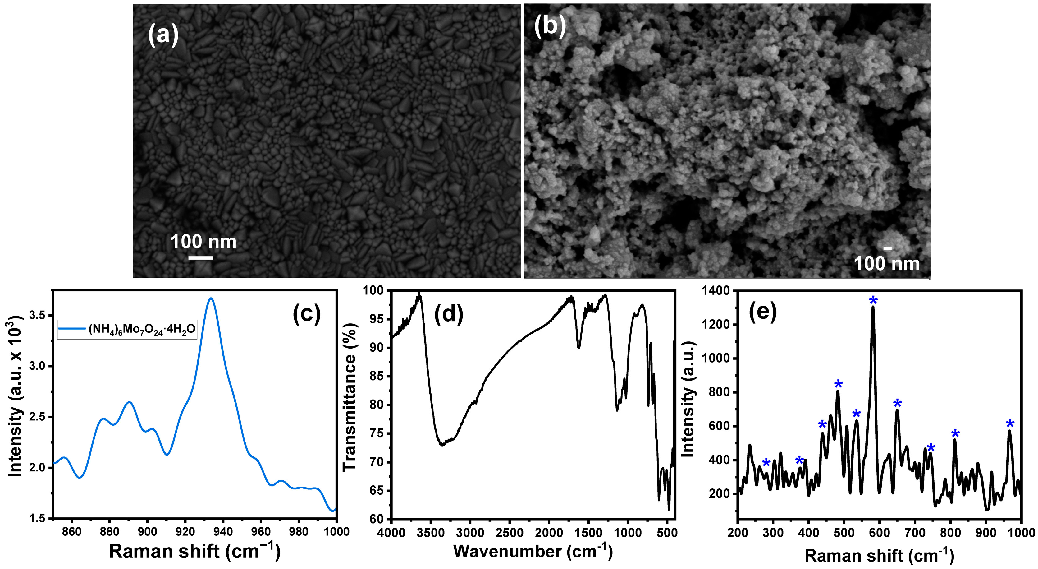
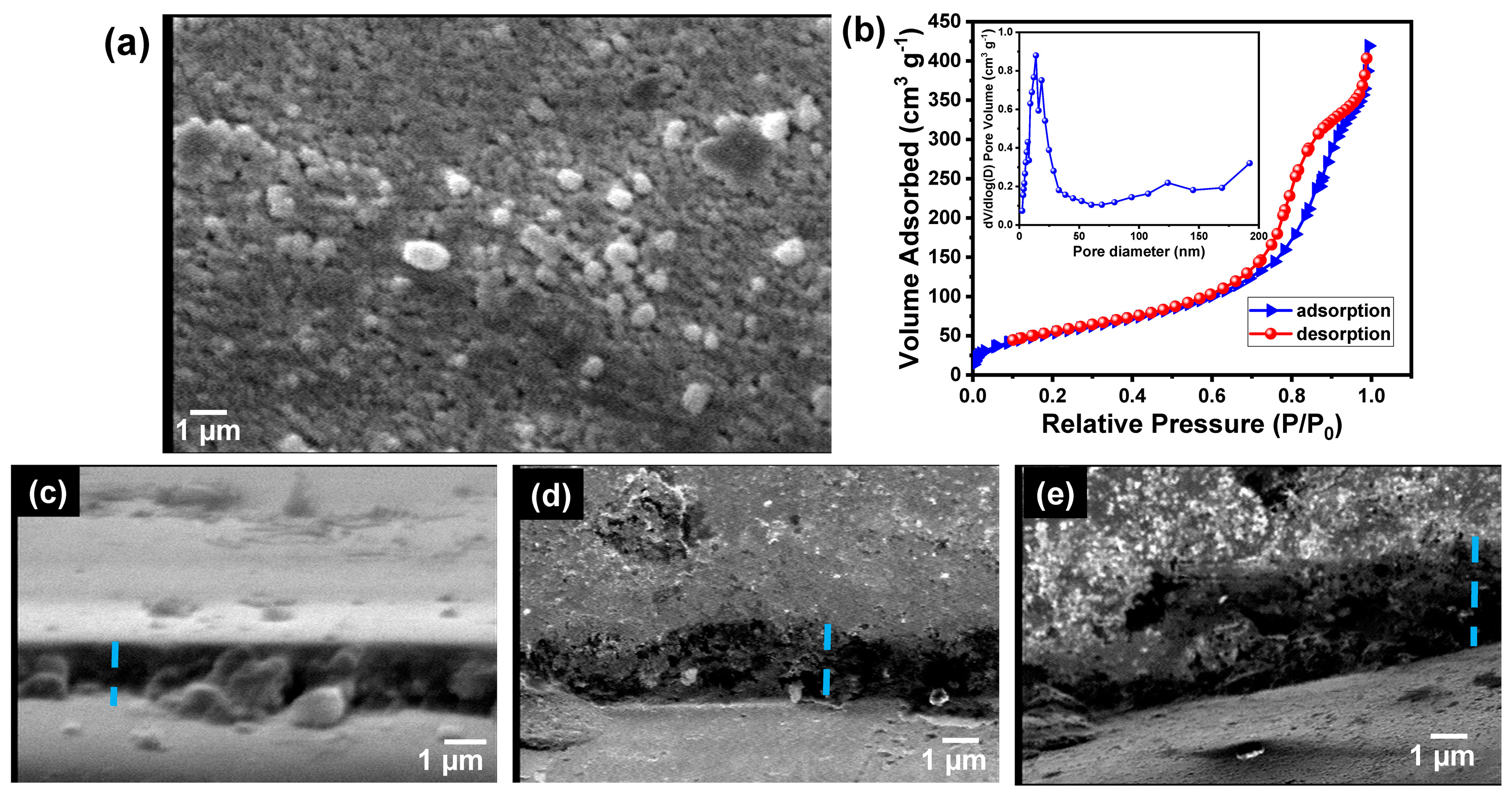
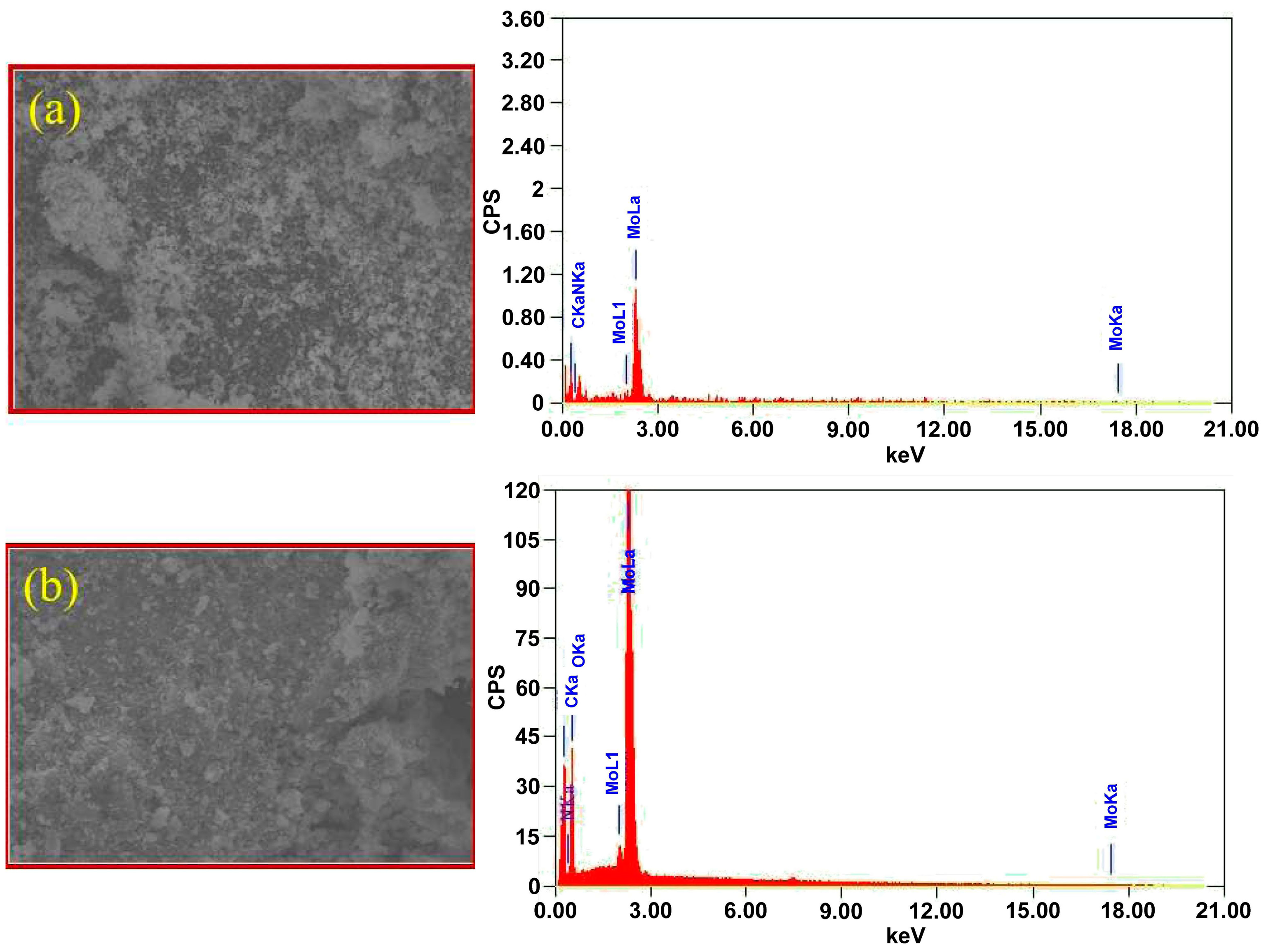
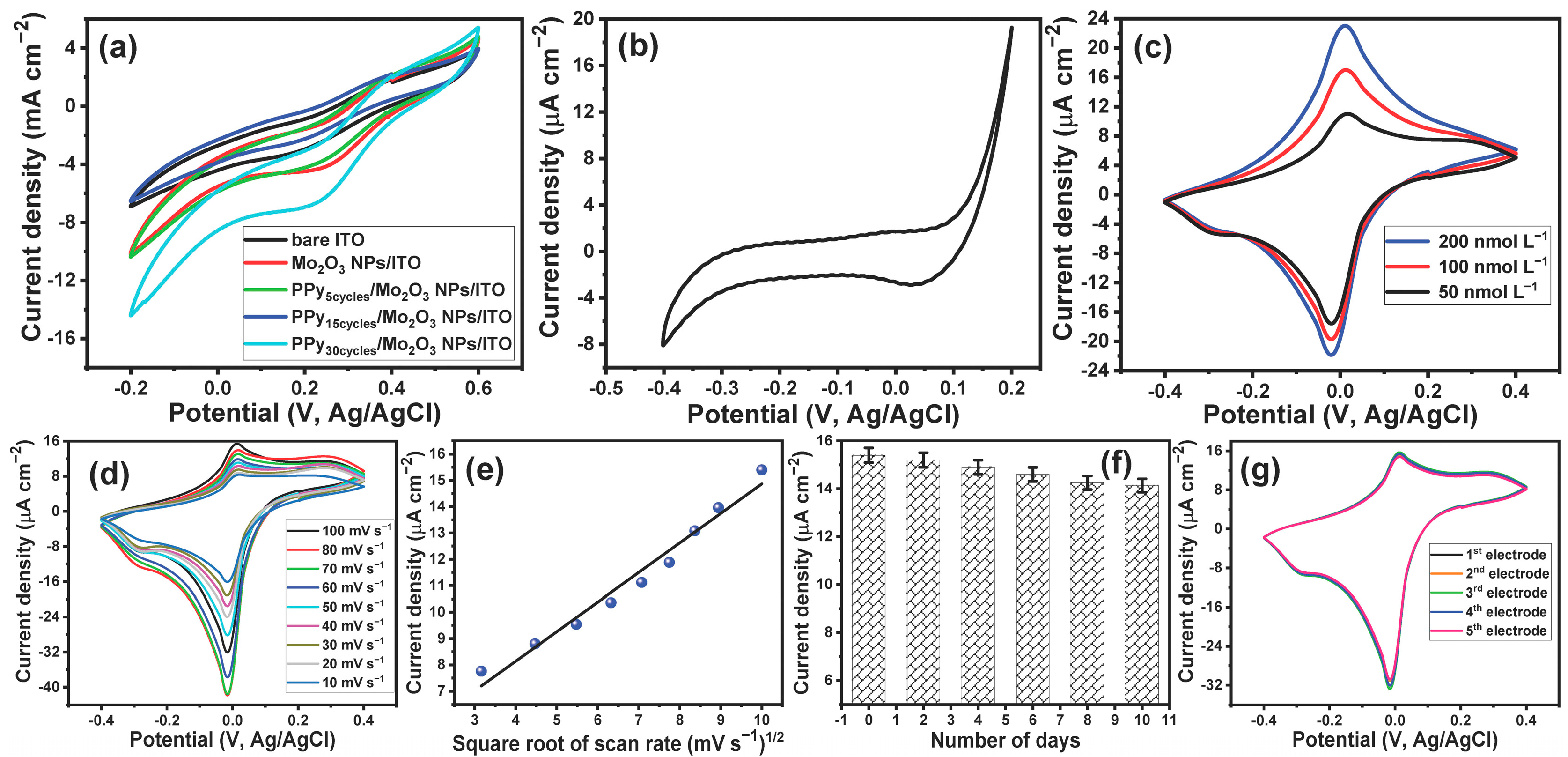
3.1. Synthesis and Characterization of PPy/MoO3 Bilayer-Modified ITO Electrodes

The formation of MoO3 NPs and their PPy/MoO3 bilayer-modified ITO electrodes were analyzed using different techniques, including FTIR, SEM, EDX, Raman spectroscopy, and N2 adsorption. Figure 1a shows the SEM image of the bare ITO electrode and demonstrates the characteristic morphology of the ITO substrate. The morphology of the MoO3 NP-modified ITO electrodes was also investigated by the SEM micrographs. The SEM image of the MoO3 NPs/ITO electrode shows the formation of tiny nanospheres (Figure 1b). The average particle size of the fabricated MoO3 NPs/ITO electrode was calculated using ImageJ (IJ153) software, in which 30 particles were selected and their diameter was studied. The results showed the formation of nanospheres of an average diameter of 29.01 ± 12.47 nm. The distribution of the particle sizes is shown in the supplementary data (Figure S1a,b), which indicates that the particle size range was within the range from 5.59 nm to 54.85 nm.
The Raman spectrum of ammonium molybdate is shown in Figure 1c, which shows a set of peaks at Raman shifts of 856, 876, 890, and 933.5 cm−1, which are in good agreement with the previously reported data [73]. Figure 1d illustrates the FTIR spectrum of the prepared MoO3 NPs. The FTIR spectrum shows two bands at 1040 and 1110 cm−1, which could be attributed to the Mo=O stretching and Mo-O-Mo vibration modes [74,75]. The strong and sharp band at around 1620 cm−1 and the broadband centered at 3434 cm−1 are attributed to the stretching absorption of the water molecules and the hydroxyl groups [74]. Furthermore, the shoulder peak at 1400 cm−1 is related to the Mo−OH bond vibration [75,76]. Moreover, the spectrum shows a band at about 681 cm−1 (asymmetric stretching vibrations of the O−Mo−O bonds) and a band at 741 cm−1 (stretching vibration of the Mo−O bond) [77,78].
The chemical composition of the MoO3 NPs/ITO electrode was also investigated by Raman spectroscopy. Figure 1e displays the Raman spectrum of the MoO3 NPs/ITO electrode, which showed many characteristic bands of MoO3 NPs. The Raman bands at 580.5 and 742.5 cm−1 were assigned to the stretching vibrations of the Mo–O (I) and Mo–O (II) groups, respectively [53]. Furthermore, the Raman band at 279 cm−1 O=M=O was assigned to the wagging vibration, while the Raman bands at 650.5 cm−1, 812.5 cm−1, and 966.5 cm−1 corresponded to the stretching vibration of O–M–O, M=O, and M=O, respectively [79,80,81,82].
A layer of PPy was fabricated onto the MoO3 NPs/ITO surface based on the electrochemical polymerization process. The polymerization process was performed based on the CV technique. The thickness of the polymer layer was controlled based on the number of cycles during the polymerization. The CV for the MoO3/ITO electrode in 0.1 M HCl in the absence of pyrrole is represented in Figure 2a, which shows a background response without any redox peaks. Figure 2b–d shows the cyclic voltammograms of the electrochemical polymerization of pyrrole after 5, 15, and 30 cycles. The CV response for the first cycle showed redox potential peaks at 0.38 V and 0.43 V. The intensity couples of the redox current peaks were increased with the increasing number of cycles. Moreover, the redox potential peaks were shifted to 0.15 V and 0.27 V. In addition, there was the appearance of new redox potential peaks at 0.47 V and 0.55 V. This behavior is characteristic of the polymerization process.
Figure 3a shows the SEM image of the PPy/MoO3 bilayer-modified ITO electrode. The micrograph shows the formation of larger particles compared with those in the case of the MoO3 NP-modified ITO electrode. The average particle size of the fabricated PPy/MoO3 bilayer/ITO electrode was also calculated using ImageJ (IJ153) software, in which 30 particles were selected and their diameters were studied. The results showed the formation of nanospheres of an average diameter of 250.69 ± 98.7 nm. The distribution of the particle sizes is shown in the supplementary data, which indicates that the particle size range was within the range from 64.21 nm to 431.10 nm.
The BET method was used to measure the surface area of the synthesized PPy/MoO3 bilayer. The formed PPy/MoO3 bilayer powder was obtained by scratching the formed layer and using it for analysis. The sample was first heated at 100 °C for 1 h to remove the physio-adsorbed water molecules. Then, the nitrogen adsorption/desorption isotherm against relative pressure was obtained at 77 K. Figure 3b demonstrates the nitrogen adsorption/desorption isotherm, which showed a type IV isotherm (H1) with a significant hysteresis loop. The results indicated the mesoporous trait of the PPy/MoO3 nanostructure. The BET surface area, pore volume, and BJH pore diameter were found to be 227.54 m2/g, 0.62 cm3/g, and 13.91 nm, respectively. The results demonstrated a high surface area compared to the reported polymers and bilayer-based polymers.
The thicknesses of the formed PPy/MoO3 bilayer-modified ITO electrodes were investigated by using the SEM. Figure 3c–e shows the cross-section SEM images of the different PPy/MoO3 bilayer-modified ITO electrodes, which formed at different numbers of cycles. The results indicate that the PPy/MoO3 bilayer’s thickness was increased with increasing cycles. The average thicknesses of the different layers were found to be 0.9, 1.4, and 1.8 µm, corresponding to the polymerization for 5, 15, and 30 cycles.
Finally, the EDX was used to quantitatively analyze the elemental composition of the fabricated PPy/MoO3 bilayer. Figure 4 shows the EDX of the MoO3 NPs and PPy/MoO3 bilayer and confirms that the as-prepared material consists of MoO3 together with C, N, and O only. Thus, the formed material did not contain any other materials.

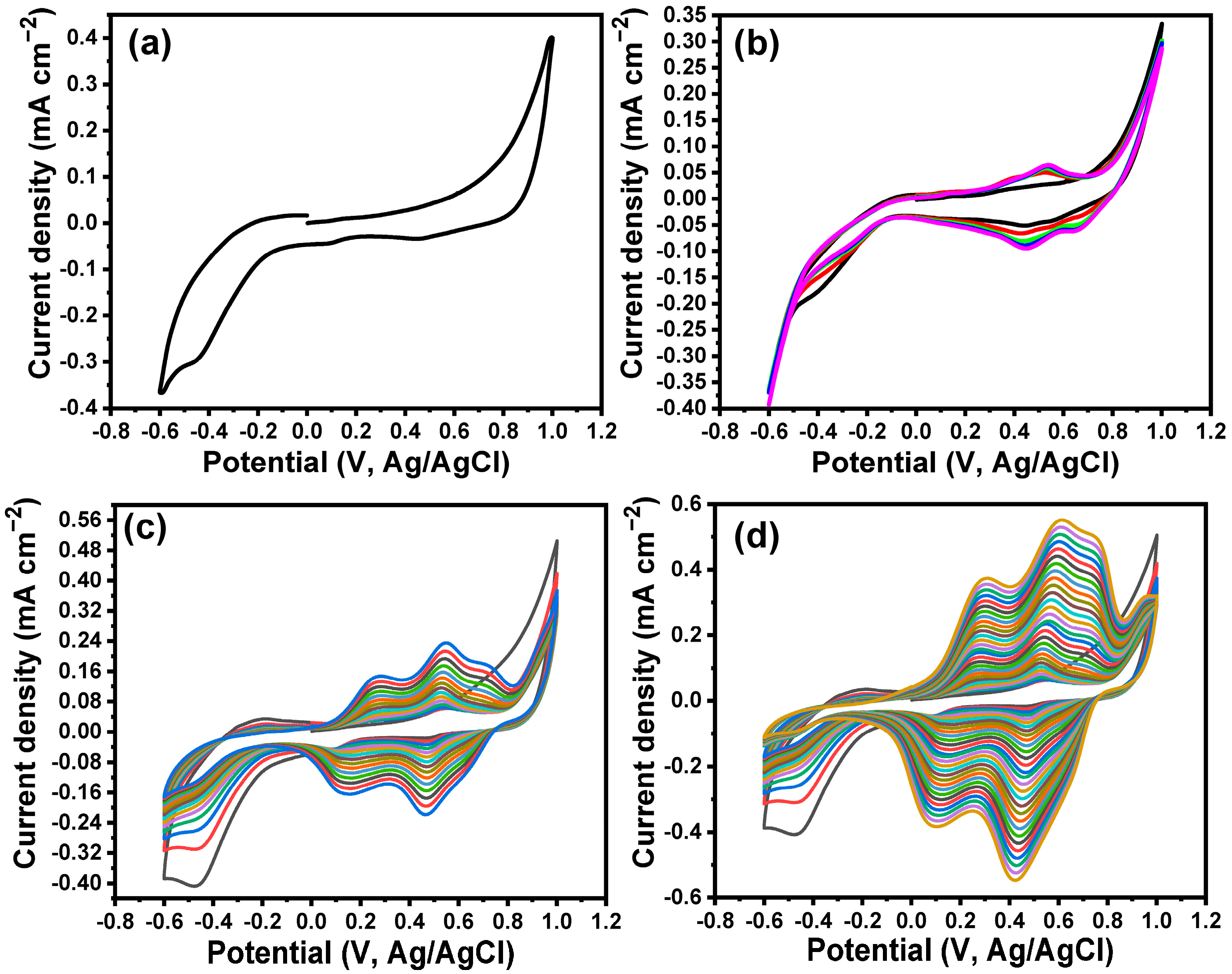
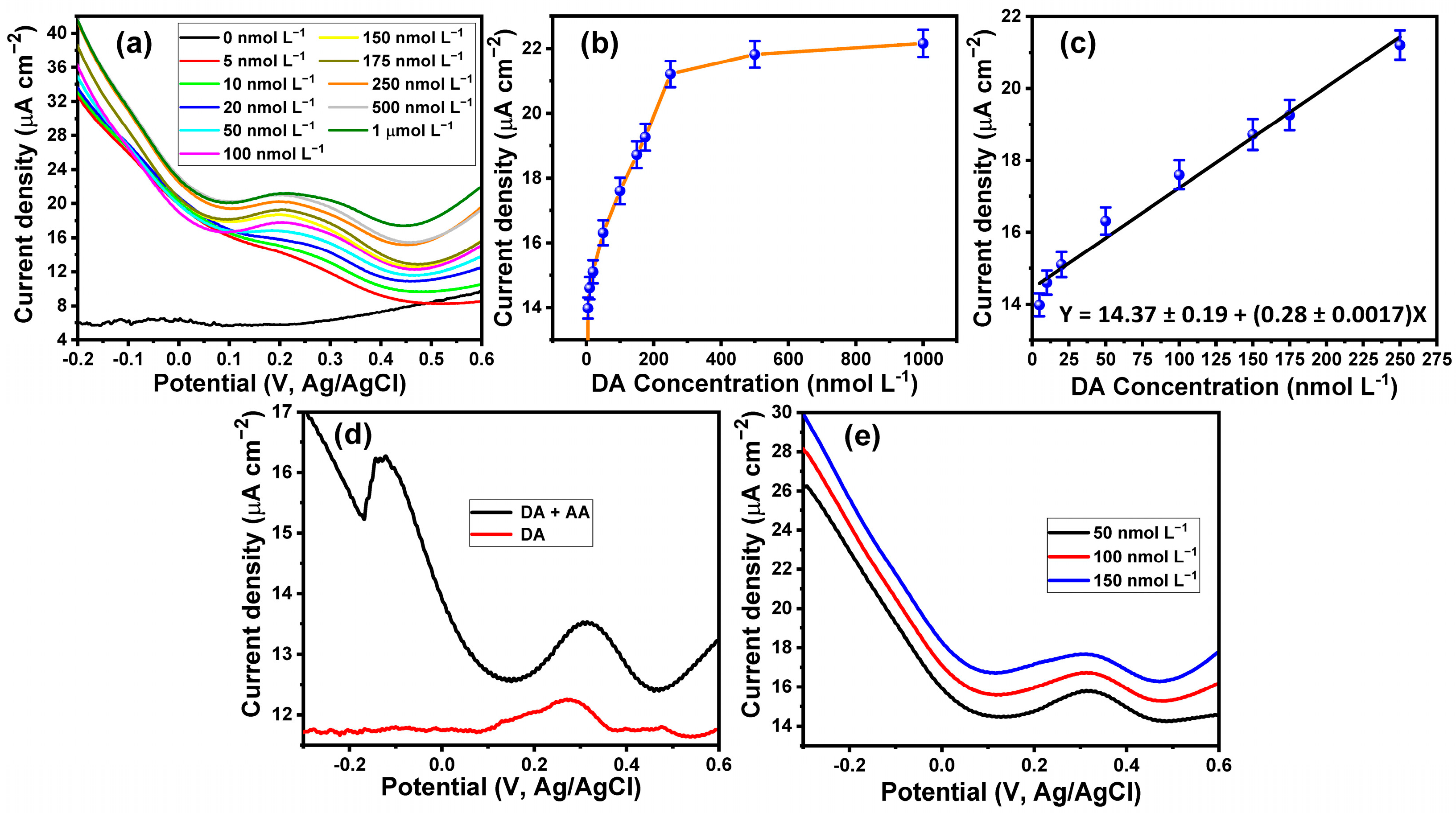
3.2. Electrochemical Detection of DA, Durability, and Reproducibility of the Fabricated Sensor

In the beginning, the effect of the MoO3 NPs and the PPy/MoO3 bilayer layer of different thicknesses on the conductivity of the ITO electrode was investigated. The electrochemical conductivity of all the modified electrodes was investigated in the presence of 5 mmol L−1 of [Fe(CN)6]3−/4− in PBS (pH 7.4) and their electrochemical responses were compared to that of the naked ITO electrode. Figure 5a (black curve) demonstrates the CV response of the bare ITO electrode in the presence of 5 mmol L−1 of [Fe(CN)6]3−/4−; it shows a reduction peak at 0.186 V and an oxidation peak at 0.386 V. These redox potential peaks were shifted to become more positive upon the modification of the ITO electrode with MoO3 NPs, as shown in Figure 5a (red curve). The CV behavior of the 5 mmol L−1 of [Fe(CN)6]3−/4− at the MoO3 NPs/ITO electrode demonstrated the appearance of a reduction potential peak at 0.25 V and an anodic potential peak at 0.39 V. It is noteworthy that the intensities of the redox current peaks were increased after modification of the ITO electrode with MoO3 NPs. Furthermore, the difference between the oxidation and reduction potential peaks was decreased upon the modification of the bare ITO electrode, which indicated the enhancement of the electron transfer rate at the modified electrode compared with the bare ITO electrode. Moreover, the deposition of a 0.9 µm layer of PPy onto the MoO3 NPs/ITO electrode showed almost the same current redox potential peaks as those of the MoO3/ITO electrode, but it resulted in the enhancement of the electrical conductivity of the MoO3 NPs/ITO electrode by about 23% (Figure 5a (green curve)). On the other hand, increasing the thickness of the PPy layer to about 1.4 µm resulted in more enhancement of the electrical conductivity of the modified electrode (Figure 5a (blue curve)), while the deposition of a PPy layer of a thickness of about 1.8 µm showed the same redox potential peaks but reduced the intensity of the redox current peaks (Figure 5a (cyan curve)). The above results confirmed that the modification of the bare ITO electrode with the PPy/MoO3 bilayer resulted in the enhancement of the conductivity and revisability of the electrode. Furthermore, the highest conductivity corresponded to the modified electrode with a 1.4 µm PPy/MoO3 bilayer; thus, this modified electrode was utilized for all the electrochemical measurements.
The PPy/MoO3 bilayer-modified ITO electrode was used for determining DA neurotransmitters dissolved in PBS buffer (pH, 7.4). Figure 5b illustrates the CV response of the PPy/MoO3 bilayer/ITO electrode in PBS (pH, 7.4); it shows a background voltammogram in which no redox peaks can be observed. Furthermore, the CV response of the PPy/MoO3 bilayer-modified ITO electrode towards the three different concentrations of DA (50, 100, and 200 nmol L−1) is represented in Figure 5c. The voltammograms showed a revisable redox behavior with a cathodic peak at −0.02 V and an anodic peak at 0.01 V. These results indicated the capability of the PPy/MoO3 bilayer-modified ITO electrode to detect low concentrations of DA neurotransmitters. The effect of scan rate on the electrochemical response of the DA neurotransmitters at the PPy/MoO3 bilayer/ITO electrode was studied. Figure 5d shows the cyclic voltammograms of 100 nmol L−1 of DA at the PPy/MoO3 bilayer-modified ITO electrode under different scan rates from 10 mVs−1 to 100 mVs−1. The voltammograms showed an increase in the oxidation current peak with the increase in the scan rate. Figure 5e shows the relationship between the square root of the scan rate and the intensity of the oxidation current peaks; the relationship is demonstrated by a linear curve over the scan rate range from 10 to 100 mVs−1. The direct proportion between the square root of the scan rate and the intensity of the oxidation current peaks indicated that the oxidation of DA was diffusion controlled. The durability of the fabricated sensor over 10 days was investigated (Figure 5f). The results demonstrated that the prepared sensors had over 91% of their efficiency after 10 days of use. The reproducibility of the fabricated sensor was evaluated by comparing the responses of five electrodes toward 100 nmol L−1 of DA (Figure 5g). The responses indicated that the fabricated sensors had almost the same electrochemical conductivity, with a slight variation.

3.3. Sensitivity, Specificity of the Electrochemical DA Sensor, and the Interference Effects

The sensitivity of the developed electrode toward the detection of DA was tested based on the SWV technique. Figure 6a shows the SWV voltammograms of a wide range of DA concentrations from 5 nmol L−1 to 1 µmol L−1. The SWVs showed an oxidation potential peak at about 0.27 V. Furthermore, Figure 6b shows the relationship between the DA concentration and the corresponding oxidation current peak. The results indicated that the oxidation current peak was increased with the increase of the concentration of DA until the concentration of 250 nmol L−1. Then, the curve reached saturation at concentrations over 250 nmol L−1, with a small increase in the current peak with a large increase in DA concentration. The linear relationship between the DA concentration and the oxidation current peak could be observed within the range from 5 nmol L−1 to 250 nmol L−1 (Figure 6c). The LOD of the developed electrode was calculated according to LOD = 3.3SteDev/slope. The LOD of the DA at the PPy/MoO3 bilayer-modified ITO electrode based on the SWV technique was found to be about 2.2 nmol L−1. The limit of quantification (LOQ) was also calculated as LOQ = 10SteDev/slope, and it was found to be 6.66 nmol L−1. The LOD of the fabricated sensor was lower than some of the previously reported sensors, as shown in Table 1.
Ascorbic acid is one of the interferences that are present in the blood, and it is oxidized at the same potential as the DA at most traditional electrodes [83]. Figure 6d shows the SWV voltammogram of a mixture of 10 µmol L−1 of AA and 50 nmol L−1 DA compared with that of DA alone. The voltammogram of the AA and DA mixture showed an oxidation potential peak at 0.31 V corresponding to the oxidation of DA and another oxidation potential peak at −0.13 V attributed to the oxidation of AA. Thus, the developed sensor could detect DA in the presence of other interferences. Moreover, three DA concentrations (20, 50, and 100 nmol L−1) in human serum were detected, as shown in Figure 6e. The SWV voltammogram of DA in human serum indicated that the oxidation potential peak was shifted to a more positive potential, at about 0.32 V. The recovery of the 20, 50, and 100 nmol L−1 DA in human serum was found at 101.5 ± 0.4, 100.6 ± 0.6, and 99.6 ± 0.4, respectively.

4. Conclusions

The PPy/MoO3 bilayer-modified ITO electrode was synthesized based on a two-step method, which involved the deposition of MoO3 NPs based on the hydrothermal technique and the formation of the polymer layer using the electrochemical polymerization technique. FTIR, SEM, EDX, Raman spectroscopy, and N2 adsorption–desorption measurements were used to fully characterize the fabricated electrode. The modified electrode was utilized to determine very low concentrations of DA in PBS as well as in the presence of other interferences such as AA and human serum. The modified electrode had a LOD of the DA based on the SWV technique of about 2.2 nmol L−1. Although the fabricated sensor was fabricated based on cheap materials, it had a lower LOD than some of the recently reported DA sensors. Our results confirmed the capability of the modified electrode to detect very low concentrations of DA in human serum; thus, it is a potential sensor for detecting DA in real samples.

Supplementary Materials

The following supporting information can be downloaded at: https://www.mdpi.com/article/10.3390/bios13060578/s1, Figure S1: Distribution of the particle sizes of (a) MoO3 NPs/ITO, and (b) PPy/MoO3 NPs/ITO.

Author Contributions

Conceptualization, W.A.E.-S.; methodology, software, validation, formal analysis, investigation, W.A.E.-S. and N.A.; resources, data curation, writing—original draft preparation, writing—review and editing, W.A.E.-S. and N.A. All authors have read and agreed to the published version of the manuscript.

Funding

University of Jeddah, Jeddah, Saudi Arabia, under grant No. (UJ-21-DR-114).

Institutional Review Board Statement

Not applicable.

Informed Consent Statement

Not applicable.

Data Availability Statement

The data presented in this study are available on request from the corresponding author.

Acknowledgments

This work was funded by the University of Jeddah, Jeddah, Saudi Arabia, under grant No. (UJ-21-DR-114). The authors, therefore, acknowledge with thanks the University of Jeddah technical and financial support.

Conflicts of Interest

The authors declare no conflict of interest.

References

  1. Zhang, C.; Du, X. Electrochemical Sensors Based on Carbon Nanomaterial Used in Diagnosing Metabolic Disease. Front. Chem. 2020, 8, 651. [Google Scholar] [CrossRef]
  2. Topkaya, S.N.; Azimzadeh, M.; Ozsoz, M. Electrochemical Biosensors for Cancer Biomarkers Detection: Recent Advances and Challenges. Electroanalysis 2016, 28, 1402–1419. [Google Scholar] [CrossRef]
  3. Guinovart, T.; Valdés-Ramírez, G.; Windmiller, J.R.; Andrade, F.J.; Wang, J. Bandage-Based Wearable Potentiometric Sensor for Monitoring Wound pH. Electroanalysis 2014, 26, 1345–1353. [Google Scholar] [CrossRef]
  4. Nocke, A.; Schröter, A.; Cherif, C.; Gerlach, G. Miniaturized Textile-based Multi-layer pH-sensor for Wound Monitoring Applications. Autex Res. J. 2012, 12, 20–22. [Google Scholar] [CrossRef]
  5. Elkhawaga, A.A.; Khalifa, M.M.; El-badawy, O.H.B.; Hassan, M.A.; El-Said, W. Rapid and highly sensitive detection of pyocyanin biomarker in different Pseudomonas aeruginosa infections using gold nanoparticles modified sensor. PLoS ONE 2019, 14, e0216438. [Google Scholar] [CrossRef] [PubMed]
  6. Khalifa, M.M.; Elkhawaga, A.A.; Hassan, M.A.; Zahran, A.M.; Fathalla, A.M.; El-Said, W.A.; El-Badawy, O. Highly specific Electrochemical Sensing of Pseudomonas aeruginosa in patients suffering from corneal ulcers: A comparative study. Sci. Rep. 2019, 9, 18320. [Google Scholar] [CrossRef]
  7. Martín, A.; Kim, J.; Kurniawan, J.F.; Sempionatto, J.R.; Moreto, J.R.; Tang, G.; Campbell, A.S.; Shin, A.; Lee, M.Y.; Liu, X.; et al. Epidermal Microfluidic Electrochemical Detection System: Enhanced Sweat Sampling and Metabolite Detection. ACS Sens. 2017, 2, 1860–1868. [Google Scholar] [CrossRef] [PubMed]
  8. Sekretaryova, A.N.; Eriksson, M.; Turner, A.P. Bioelectrocatalytic Systems for Health Applications. Biotechnol. Adv. 2016, 34, 177–197. [Google Scholar] [CrossRef]
  9. Ashley, B.K.; Brown, M.S.; Park, Y.; Kuan, S.; Koh, A. Skininspired, Open Mesh Electrochemical Sensors for Lactate and Oxygen Monitoring. Biosens. Bioelectron. 2019, 132, 343–351. [Google Scholar] [CrossRef]
  10. Schapira, A.H. Dopamine agonists and neuroprotection in Parkinson’s disease. Eur. J. Neurol. 2002, 9, 7–14. [Google Scholar]
  11. Ali, S.R.; Ma, Y.; Parajuli, R.R.; Balogun, Y.; Lai, W.Y.C.; He, H. A Nonoxidative Sensor Based on a Self-Doped Polyaniline/Carbon Nanotube Composite for Sensitive and Selective Detection of the Neurotransmitter Dopamine. Anal. Chem. 2007, 79, 2583–2587. [Google Scholar] [CrossRef]
  12. Damier, P.; Hirsch, E.C.; Agid, Y.; Graybiel, A.M. The substantia nigra of the human brain. II. Patterns of loss of dopamine-containing neurons in Parkinson’s disease. Brain 1999, 122, 1437. [Google Scholar] [CrossRef]
  13. Ismail, W.Z.W.; Liu, G.; Zhang, K.; Goldys, E.M.; Dawes, M.J. Dopamine sensing and measurement using threshold and spectral measurements in random lasers. Opt. Express 2016, 24, A85–A94. [Google Scholar] [CrossRef] [PubMed]
  14. Kim, Y.-R.; Bong, S.; Kang, Y.-J.; Yang, Y.; Mahajan, R.K.; Kim, J.S.; Kim, H. Electrochemical detection of dopamine in the presence of ascorbic acid using graphene modified electrodes. Biosens. Bioelectron. 2010, 25, 2366–2369. [Google Scholar] [CrossRef]
  15. Mo, J.W.; Ogorevc, B. Simultaneous measurement of dopamine and ascorbate at their physiological levels using voltammetric microprobe based on overoxidized poly(1,2-phenylenediamine)-coated carbon fiber. Anal. Chem. 2001, 73, 1196–1202. [Google Scholar] [CrossRef] [PubMed]
  16. Kushwaha, C.S.; Shukla, S.K. Electrochemical sensing of paracetamol using iron oxide encapsulated in chitosan-grafted-polyaniline. ACS Appl. Polym. Mater 2020, 2, 2252–2259. [Google Scholar] [CrossRef]
  17. Iqbal, K.; Ishaq, M.A.; Ahmad, A.; Ali, M.D.; Zeeshan, T.; Tahir, W.; Aslam, A.; Amami, M.; ben Farhat, I.; ben Ahmed, S.; et al. Fabrication and characterizations of hybrid materials based on polyaniline, metal oxide, and graphene nano-platelets for supercapacitor electrodes. Inorg. Chem. Commun. 2020, 137, 109201. [Google Scholar] [CrossRef]
  18. Dakshayini, B.S.; Reddy, K.R.; Mishra, A.; Shetti, N.P.; Malode, S.J.; Basu, S.; Naveen, S.; Anjanapura, V.R. Role of conducting polymer and metal oxide-based hybrids for applications in ampereometric sensors and biosensors. Microchem. J. 2019, 147, 7–24. [Google Scholar] [CrossRef]
  19. Shang, F.; Zhou, L.; Mahmoud, K.A.; Hrapovic, S.; Liu, Y.; Moynihan, H.A.; Glennon, J.D.; Luong, J.H. Selective nanomolar detection of dopamine using a boron-doped diamond electrode modified with an electropolymerized sulfobutylether-β-cyclodextrin-doped poly (N-acetyltyramine) and polypyrrole composite film. Anal. Chem. 2009, 81, 4089. [Google Scholar] [CrossRef]
  20. Zhang, T.; Xu, H.; Wang, H.; Zhu, J.; Zhai, Y.; Bai, X.; Dong, B.; Song, H. Green fluorescent organic nanoparticles based on carbon dots and self-polymerized dopamine for cell imaging. RSC Adv. 2017, 7, 28987–28993. [Google Scholar] [CrossRef]
  21. Dijkstra, R.J.; Scheenen, W.J.; Dam, N.; Roubos, E.W.; Meulen, J.J.T. Monitoring neurotransmitter release using surface-enhanced Raman spectroscopy. J. Neurosci. Methods 2007, 159, 43. [Google Scholar] [CrossRef]
  22. Abbaspour, A.; Khajehzadeh, A.; Ghaffarinejad, A. A simple and cost-effective method, as an appropriate alternative for visible spectrophotometry: Development of a dopamine biosensor. Analyst 2009, 134, 1692–1698. [Google Scholar] [CrossRef]
  23. McGashen, M.L.; Davis, K.L.; Morris, M.D. Surface-enhanced Raman scattering of dopamine at polymer-coated silver electrodes. Anal. Chem. 1990, 62, 846–849. [Google Scholar] [CrossRef] [PubMed]
  24. Li, N.; Guo, J.; Liu, B.; Yu, Y.; Cui, H.; Mao, L.; Lin, Y. Determination of monoamine neurotransmitters and their metabolites in a mouse brain microdialysate by coupling high-performance liquid chromatography with gold nanoparticle-initiated chemiluminescence. Anal. Chim. Acta 2009, 645, 48–55. [Google Scholar] [CrossRef]
  25. Syslova, K.; Rambousek, L.; Kuzma, M.; Najmanova, V.; Bubenikova-Valesova, V.; Slamberova, R.; Kacer, P.J. Monitoring of dopamine and its metabolites in brain microdialysates: Method combining freeze-drying with liquid chromatography–tandem mass spectrometry. J. Chromatogr. A 2011, 1218, 3382–3391. [Google Scholar] [CrossRef]
  26. Wang, H.Y.; Hui, Q.S.; Xu, L.X.; Jiang, J.G.; Sun, Y. Fluorimetric determination of dopamine in pharmaceutical products and urine using ethylene diamine as the fluorigenic reagent. Anal. Chim. Acta 2003, 497, 93–99. [Google Scholar] [CrossRef]
  27. Mei, E.; Yue-Ting, F.; Jiang-Yan-Ping, S.J. Simultaneous determination of noradrenaline and dopamine in Portulaca oleracea L. by capillary zone electrophoresis. Sep. Sci. 2005, 28, 360–364. [Google Scholar]
  28. Wei, W.; Wang, H.J.; Jiang, C.Q. Spectrofluorimetric determination of dopamine using 1,5-bis (4,6-dicholro-1,3,5-triazinylamino) naphthalene. J. Biol. Chem. Lumin. 2007, 22, 581. [Google Scholar] [CrossRef]
  29. Wen, J.; Zhou, L.; Jin, L.; Cao, X.; Ye, B.C. Overoxidized polypyrrole/multi-walled carbon nanotubes composite modified electrode for in vivo liquid chromatography–electrochemical detection of dopamine. J. Chromatogr. B 2009, 877, 1793. [Google Scholar] [CrossRef]
  30. Huang, J.; Liu, Y.; Hou, H.; You, T. An electrochemical ascorbic acid sensor based on palladium nanoparticles supported on graphene oxide. Biosens. Bioelectron. 2008, 24, 518. [Google Scholar]
  31. Khan, F.; Akhtar, N.; Jalal, N.; Hussain, I.; Szmigielski, R.; Hayat, M.Q.; Ahmad, H.B.; El-Said, W.A.; Yang, M.; Janjua, H.A. Carbon-dot wrapped ZnO nanoparticle-based photoelectrochemical sensor for selective monitoring of H2O2 released from cancer cells. Microchim. Acta 2019, 186, 127. [Google Scholar] [CrossRef] [PubMed]
  32. El-Said, W.A.; Kim, T.-H.; Chung, Y.-H.; Choi, J.-W. Fabrication of new single cell chip to monitor intracellular and extracellular redox state based on spectroelectrochemical method. Biomaterials 2015, 40, 80–87. [Google Scholar] [CrossRef] [PubMed]
  33. Kim, T.-H.; El-Said, W.A.; Choi, J.-W. Highly sensitive electrochemical detection of potential cytotoxicity of CdSe/ZnS quantum dots using neural cell chip. Biosens. Bioelectron. 2012, 32, 266–272. [Google Scholar] [CrossRef] [PubMed]
  34. Fouad, D.M.; El-Said, W.A. Selective Electrochemical Detection of Epinephrine Using Gold Nanoporous Film. J. Nanomater. 2016, 2016, 6194230. [Google Scholar] [CrossRef]
  35. Ouellette, M.; Mathault, J.; Niyonambaza, S.D.; Miled, A.; Boisselier, E. Electrochemical Detection of Dopamine Based on Functionalized Electrodes. Coatings 2019, 9, 496. [Google Scholar] [CrossRef]
  36. Reddy, S.; Swamy, B.E.K.; Jayadevappa, H. CuO nanoparticle sensor for the electrochemical determination of dopamine. Electrochim. Acta 2012, 61, 78–86. [Google Scholar] [CrossRef]
  37. Li, Y.Y.; Kang, P.; Wang, S.Q.; Liu, Z.G.; Li, Y.X.; Guo, Z. Ag nanoparticles anchored onto porous CuO nanobelts for the ultrasensitive electrochemical detection of dopamine in human serum. Sens. Actuators B Chem. 2021, 327, 128878. [Google Scholar] [CrossRef]
  38. Zhuang, Y.; Zhang, X.; Chen, Q.; Li, S.; Cao, H.; Huang, Y. Co3O4/CuO hollow nanocage hybrids with high oxidase-like activity for biosensing of dopamine. Mater. Sci. Eng. C 2019, 94, 858–866. [Google Scholar] [CrossRef]
  39. Zou, J.; Wu, S.; Liu, Y.; Sun, Y.; Cao, Y.; Hsu, J.-P.; Wee, A.T.S.; Jiang, J. An ultrasensitive electrochemical sensor based on 2D g-C3N4/CuO nanocomposites for dopamine detection. Carbon 2018, 130, 652–663. [Google Scholar] [CrossRef]
  40. Huang, Z.; Zhang, L.; Cao, P.; Wang, N.; Lin, M. Electrochemical sensing of dopamine using a Ni-based metal-organic framework modified electrode. Ionics 2021, 27, 1339–1345. [Google Scholar] [CrossRef]
  41. Immanuel, S.; Aparna, T.K.; Sivasubramanian, R. A facile preparation of Au—SiO2 nanocomposite for simultaneous electrochemical detection of dopamine and uric acid. Surf. Interfaces 2019, 14, 82–91. [Google Scholar] [CrossRef]
  42. Sato, N.; Ohta, Y.; Haruta, M.; Takehara, H.; Tashiro, H.; Sasagawa, K.; Jongprateep, O.; Ohta, J. Electrochemical activities of Fe2O3-modified microelectrode for dopamine detection using fast-scan cyclic voltammetry. AIP Adv. 2023, 13, 025026. [Google Scholar] [CrossRef]
  43. Swamy, B.K.; Shiprath, K.; Ratnam, K.V.; Manjunatha, H.; Janardan, S.; Ratnamala, A.; Naidu, K.C.B.; Ramesh, S.; Babu, K.S. Electrochemical Detection of Dopamine and Tyrosine using Metal oxide (MO, M=Cu and Ni) Modified Graphite Electrode: A Comparative Study. Biointerface Res. Appl. Chem. 2020, 10, 6460–6473. [Google Scholar]
  44. Kundys-Siedlecka, M.; Bączyńska, E.; Jönsson-Niedziółka, M. Electrochemical Detection of Dopamine and Serotonin in the Presence of Interferences in a Rotating Droplet System. Anal. Chem. 2019, 91, 10908–10913. [Google Scholar] [CrossRef] [PubMed]
  45. Alsulami, Q.A.; Hussein, M.A.; Alsheheri, S.Z.; Elshehy, E.A.; El-Said, W.A. Unexpected ultrafast and high adsorption performance of Ag(I) and Hg(II) ions from multiple aqueous solutions using microporous functional silica-polymer sponge-like composite. J. Mater. Res. Technol. 2022, 17, 2000–2013. [Google Scholar] [CrossRef]
  46. El-Said, W.A.; Choi, J.-H.; Hajjar, D.; Makki, A.A.; Choi, J.-W. Fabrication of Hollow Nanocones Membrane with an Extraordinary Surface Area as CO2 Sucker. Polymers 2022, 14, 183. [Google Scholar] [CrossRef]
  47. El-Said, W.A.; Nasr, O.; Soliman, A.; Elshehy, E.A.; Khan, Z.A.; Abdel-Wadood, F.K. Fabrication of polypyrrole/Au nanoflowers modified gold electrode for highly sensitive sensing of paracetamol in pharmaceutical formulation. Appl. Surf. Sci. Adv. 2021, 4, 100065. [Google Scholar] [CrossRef]
  48. El-Said, W.A.; Abdelshakour, M.; Choi, J.-H.; Choi, J.-W. Application of Conducting Polymer Nanostructures to Electrochemical Biosensors. Molecules 2020, 25, 307. [Google Scholar] [CrossRef]
  49. Choi, J.H.; El-Said, W.A.; Choi, J.-W. Highly sensitive surface-enhanced Raman spectroscopy (SERS) platform using core/double shell (Ag/polymer/Ag) nanohorn for proteolytic biosensor. Appl. Surf. Sci. 2020, 506, 144669. [Google Scholar] [CrossRef]
  50. El-Said, W.A.; Alshitari, W.; Choi, J.-W. Controlled fabrication of gold nanobipyramids/polypyrrole for shell-isolated nanoparticle-enhanced Raman spectroscopy to detect γ-aminobutyric acid, pectrochim. Acta A Mol. Biomol. Spectrosc. 2019, 229, 117890. [Google Scholar] [CrossRef]
  51. El-Said, W.A.; Choi, J.-W. Electrochemical biosensor consisted of conducting polymer layer on gold nanodots patterned indium tin oxide electrode for rapid and simultaneous determination of purine bases. Electrochim. Acta 2014, 2014, 51–57. [Google Scholar] [CrossRef]
  52. El-Said, W.A.; Yea, C.-H.; Jung, M.; Kim, H.; Choi, J.-W. Analysis of effect of nanoporous alumina substrate coated with polypyrrole nanowire on cell morphology based on AFM topography. Ultramicroscopy 2010, 110, 676–681. [Google Scholar] [CrossRef] [PubMed]
  53. El-Said, W.A.; Yea, C.-H.; Choi, J.-W.; Kwon, I.-K. Ultrathin polyaniline film coated on an indium-tin oxide cell-based chip for study of anticancer effect. Thin Solid Films 2009, 518, 661–667. [Google Scholar] [CrossRef]
  54. Gu, Z.J.; Ye, J.R.; Song, W.; Shen, Q. Synthesis of polyaniline nanotubes with controlled rectangular or square pore shape. Mater. Lett. 2014, 121, 12–14. [Google Scholar] [CrossRef]
  55. Rezaee, O.; Chenari, H.M.; Ghodsi, F.E.; Ziyadi, H. Preparation of PVA nanofibers containing tungsten oxide nanoparticle by electrospinning and consideration of their structural properties and photocatalytic activity. J. Alloy Compd. 2017, 690, 864–872. [Google Scholar] [CrossRef]
  56. Jan, T.; Iqbal, J.; Farooq, U.; Gul, A.; Abbasi, R.; Ahmad, I.; Malik, M. Structural, Raman and optical characteristics of Sn doped CuO nanostructures: A novel anticancer agent. Ceram. Int. 2015, 41, 13074–13079. [Google Scholar] [CrossRef]
  57. Li, B.; Zhou, Y.; Wu, W.; Liu, M.; Mei, S.; Zhou, Y.; Jing, T. Highly selective and sensitive determination of dopamine by the novel molecularly imprinted poly(nicotinamide)/CuO nanoparticles modified electrode. Biosens. Bioelectron. 2015, 67, 121–128. [Google Scholar] [CrossRef]
  58. Kohler, N.; Sun, C.; Fichtenholtz, A.; Gunn, J.; Fang, C.; Zhang, M.Q. Methotrexate-immobilized poly (ethylene glycol) magnetic nanoparticles for MR imaging and drug delivery. Small 2006, 2, 785–792. [Google Scholar] [CrossRef]
  59. Lee, H.Y.; Lee, S.H.; Xu, C.; Xie, J.; Lee, J.-H.; Wu, B.; Koh, A.L.; Wang, X.; Sinclair, R.; Wang, S.X.; et al. Synthesis and characterization of PVP-coated large core iron oxide nanoparticles as an MRI contrast agent. Nanotechnology 2018, 19, 165101. [Google Scholar] [CrossRef]
  60. Hariharan, R.; Senthilkumar, S.; Suganthi, A.; Rajarajan, M. Photodynamic action of curcumin derived polymer modified ZnO nanocomposites. Mater. Res. Bull. 2005, 94, 454–459. [Google Scholar] [CrossRef]
  61. Mo, C.Y.; Wang, M.; You, Y.R.; Zhu, Z.Y.C.; Hu, Y. A novel ultraviolet-irradiation route to CdS nanocrystallites with different morphologies. Mater. Res. Bull. 2001, 36, 2277–2282. [Google Scholar] [CrossRef]
  62. Azharudeen, A.M.; Karthiga, R.; Rajarajan, M.; Suganthi, A. Fabrication, characterization of polyaniline intercalated NiO nanocomposites and application in the development of nonenzymatic glucose biosensor. Arab. J. Chem. 2020, 13, 4053–4064. [Google Scholar] [CrossRef]
  63. Sen, T.; Mishra, S.; Shimpi, N.G. Synthesis and sensing applications of polyaniline nanocomposites: A review. RSC Adv. 2016, 6, 42196–42222. [Google Scholar] [CrossRef]
  64. Zhu, S.; Wei, W.; Chen, X.; Jiang, M.; Zhou, Z. Hybrid structure of polyaniline/ZnO nanograss and its application in dye-sensitized solar cell with performance improvement. J. Solid State Chem. 2012, 190, 174–179. [Google Scholar] [CrossRef]
  65. Yadav, M.; Singh, G.; Lata, S. Revisiting some recently developed conducting polymer@metal oxide nanostructures for electrochemical sensing of vital biomolecules: A review. J. Anal. Test. 2022, 6, 274–295. [Google Scholar] [CrossRef]
  66. Ghosh, M.M.; Yuan, J.R. Adsorption of inorganic arsenic and organosenicals on hydrous oxides. Environ. Prog. 1987, 6, 150–157. [Google Scholar] [CrossRef]
  67. Zablocka, I.; Wysocka-Zolopa, M.; Winkler, K. Electrochemical Detection of Dopamine at a Gold Electrode Modified with a Polypyrrole–Mesoporous Silica Molecular Sieves (MCM-48) Film. Int. J. Mol. Sci. 2019, 20, 111. [Google Scholar] [CrossRef]
  68. Azharudeen, A.M.; Roy, A.; Karthiga, R.; Prabhu, S.A.; Prakash, M.G.A.; Badhusha, M.I.; Ali, H.; Katubi, K.M.; Islam, M.R. Ultrasensitive and Selective Electrochemical Detection of Dopamine Based on CuO/PVA Nanocomposite-Modified GC Electrode. Int. J. Photoenergy 2022, 2022, 8755464. [Google Scholar] [CrossRef]
  69. Wang, Y.; Tang, J.; Xiang, L. Detection of dopamine by a minimizedelectrochemical sensor using a graphene oxide molecularly imprinted polymer modified micropipette tip shaped graphite electrode. J. Chem. Res. 2021, 45, 702–707. [Google Scholar] [CrossRef]
  70. Shen, X.; Ju, F.; Li, G.; Ma, L. Smartphone-Based Electrochemical Potentiostat Detection System Using PEDOT:PSS/Chitosan/Graphene Modified Screen-Printed Electrodes for Dopamine Detection. Sensors 2020, 20, 2781. [Google Scholar] [CrossRef]
  71. Kathiresan, V.; Thirumalai, D.; Rajarathinam, T.; Yeom, M.; Lee, J.; Kim, S.; Yoon, J.-H.; Chang, S.-C. A simple one-step electrochemical deposition of bioinspired nanocomposite for the non-enzymatic detection of dopamine. J. Anal. Sci. Technol. 2021, 12, 5. [Google Scholar] [CrossRef]
  72. Kumar, Y.; Pramanik, P.; Das, D.K. Electrochemical detection of paracetamol and dopamine molecules using nano-particles of cobalt ferrite and manganese ferrite modified with graphite. Heliyon 2019, 5, e02031. [Google Scholar] [CrossRef] [PubMed]
  73. Han, X.-J.; Sun, P.-X.; Pan, W.-J.; Chen, L.-J.; Chen, Z.-J.; Zhang, Q.; Wang, J.; Yang, C. Porous multi-layer MoO2/β-MnO2 composite cathode for phosphorylated glucose fuel cell. J. Solid State Electrochem. 2021, 25, 1861–1869. [Google Scholar] [CrossRef]
  74. Anilkumar, K.; Parveen, A.; Badiger, G.R.; Ambika Prasad, M. Effect of molybdenum trioxide (MoO3) on the electrical conductivity of polyaniline. Physica B 2009, 404, 1664–1667. [Google Scholar] [CrossRef]
  75. Morajkar, S.M.; Srinivasan, B.R. Convenient synthesis and Raman spectral characterization of diammonium monomolybdate(VI). Indian J. Chem. 2020, 59, 1760–1767. [Google Scholar]
  76. Dukstiene, N.; Tatariskinaite, L.; Andrulevicius, M. Characterization of electrochemically deposited thin Mo-O-C-Se film layers. Mater. Sci. Poland 2010, 28, 93–103. [Google Scholar]
  77. Baran, E.J.; Mormann, T.J.; Grocke, H.J.; Jeitschko, W. Infrared and Raman spectra of Hg2MoO4 and Hg2WO4. J. Raman Spectrosc. 2001, 32, 395. [Google Scholar] [CrossRef]
  78. Xu, G.; Xu, G.; Chang, W.; Wang, H. Fabrication of microsphere-like nano-MoO2 modified with silane coupling agent KH550. Mater. Res. Express 2019, 6, 11508. [Google Scholar] [CrossRef]
  79. Narro-García, R.; Méndez, N.; Apátiga, L.M.; Flores-De los Ríos, J.P.; Nava-Dino, C.G.; Quintero-Torres, R. Effect of the Substrate Temperature on the Structural and Morphological Properties of MoO2 Thin Films Obtained by Pulsed Injection MOCVD. Int. J. Electrochem. Sci. 2017, 12, 3907–3915. [Google Scholar] [CrossRef]
  80. Camacho-López, M.A.; Escobar-Alarcón, L.; Picquart, M.; Arroyo, R.; Córdoba, G.; Haro-Poniatowski, E. Micro-Raman study of the m-MoO2 to α-MoO3 transformation induced by cw-laser irradiation. Opt. Mater. 2011, 33, 480–484. [Google Scholar] [CrossRef]
  81. Kumari, L.; Ma, Y.-R.; Tsai, C.-C.; Lin, Y.-W.; Wu, S.Y.; Cheng, K.-W.; Liou, Y. X-ray diffraction and Raman scattering studies on large-area array and nanobranched structure of 1D MoO2 nanorods. Nanotechnology 2007, 18, 115717. [Google Scholar] [CrossRef]
  82. Lai, K.; Yuan, K.; Ye, Q.; Chen, A.; Chen, D.; Chen, D.; Gu, C. Constructing the Mo2C@MoOx Heterostructure for Improved SERS Application. Biosensors 2022, 12, 50. [Google Scholar] [CrossRef] [PubMed]
  83. El-Said, W.A.; Lee, J.-H.; Oh, B.-K.; Choi, J.-W. 3-D nanoporous gold thin film for the simultaneous electrochemical determination of dopamine and ascorbic acid. Electrochem. Commun. 2010, 12, 1756–1759. [Google Scholar] [CrossRef]
Figure 1. (a) The SEM image of the ITO electrode; (b) the SEM image of the MoO3 NP-modified ITO electrode; (c) Raman spectrum of ammonium molybdate; (d) FTIR spectrum of the MoO3 NPs; and (e) Raman spectrum of MoO3 NP-modified ITO.
Biosensors 13 00578 g001
Figure 2. (a) CV of MoO3 NPs/ITO electrode in 0.1 M HCl in the absence of pyrrole, CV voltammograms of the electrochemical polymerization of pyrrole onto MoO3 NPs/ITO electrode at (b) 5 cycles, (c) 15 cycles, and (d) 30 cycles.
Biosensors 13 00578 g002
Figure 3. (a) SEM image of the top view of PPy/MoO3 bilayer-modified ITO; (b) N2 adsorption/desorption isotherm of PPy/MoO3 bilayer; (ce) SEM images of the cross-sections of the PPy/MoO3 bilayer/ITO electrodes, which formed after different cycle numbers.
Biosensors 13 00578 g003
Figure 4. EDX analysis of (a) MoO3 NPs and (b) PPy/MoO3 bilayer.
Biosensors 13 00578 g004
Figure 5. (a) CV voltammograms of different electrodes in the presence of 5 mmol L−1 of [Fe(CN)6]3−/4− electrolyte, scan rate 100 mV/s vs. Ag/AgCl; (b) CV voltammogram of PPy/MoO3 bilayer/ITO electrode in presence of PBS (pH 7.4), scan rate 100 mV/s vs. Ag/AgCl; (c) CV voltammograms of PPy/MoO3 bilayer/ITO electrode in presence of different concentrations of DA in PBS (pH 7.4), scan rate 100 mV/s vs. Ag/AgCl; (d) CV voltammograms of PPy/MoO3 bilayer/ITO electrode in presence of 100 nmol L−1 of DA at different scan rates from 10 mVs−1 to 100 mVs−1; (e) the relationship between the square root of the scan rate and the oxidation current peak; (f) the durability of the fabricated sensor; and (g) the reproducibility of the fabricated sensor.
Biosensors 13 00578 g005
Figure 6. (a) SWV voltammograms of different concentrations of DA within a range from 5 nmol L−1 to 1000 nmol L−1; (b,c) calibration plot between the concentrations of DA and the oxidation current peak; (d) SWV voltammogram of a mixture of AA and DA; and (e) SWV voltammograms of different concentrations of DA in human serum.
Biosensors 13 00578 g006
Table 1. A comparison of the performance of some previously reported DA sensors.
Modified ElectrodeMethodLODReference
Screen-printed electrodeDPV0.29 μmol L−1[68]
GO/MWCNTs/PPyAmperometry2.3 nmol L−1[69]
Ag/CuO PNBsCV7 nmol L−1[37]
Au—SiO2CV1.98 μmol L−1[40]
Fe2O3-modified microelectrodeFast-scan CV8.76 nmol L−1[41]
PVP-GR/GCECV2 nmol L−1[40]
Ppy/MoO3/ITOSWV2.2 nmol L−1Present work
Disclaimer/Publisher’s Note: The statements, opinions and data contained in all publications are solely those of the individual author(s) and contributor(s) and not of MDPI and/or the editor(s). MDPI and/or the editor(s) disclaim responsibility for any injury to people or property resulting from any ideas, methods, instructions or products referred to in the content.

