Abstract
Escherichia coli (E. coli) O157:H7 is a major foodborne and waterborne pathogen that can threaten human health. Due to its high toxicity at low concentrations, it is crucial to establish a time-saving and highly sensitive in situ detection method. Herein, we developed a rapid, ultrasensitive, and visualized method for detecting E. coli O157:H7 based on a combination of Recombinase-Aided Amplification (RAA) and CRISPR/Cas12a technology. The CRISPR/Cas12a-based system was pre-amplified using the RAA method, which showed high sensitivity and enabled detecting as low as ~1 CFU/mL (fluorescence method) and 1 × 10² CFU/mL (lateral flow assay) of E. coli O157:H7, which was much lower than the detection limit of the traditional real-time PCR technology (103 CFU/mL) and ELISA (104~107 CFU/mL). In addition, we demonstrated that this method still has good applicability in practical samples by simulating the detection in real milk and drinking water samples. Importantly, our RAA-CRISPR/Cas12a detection system could complete the overall process (including extraction, amplification, and detection) within 55 min under optimized conditions, which is faster than most other reported sensors, which take several hours to several days. The signal readout could also be visualized by fluorescence generated with a handheld UV lamp or a naked-eye-detected lateral flow assay depending on the DNA reporters used. Because of the advantages of being fast, having high sensitivity, and not requiring sophisticated equipment, this method has a promising application prospect for in situ detection of trace amounts of pathogens.
1. Introduction
Since being identified as a human pathogen in 1982, the zoonotic life-threatening Shiga-toxin-producing E. coli has become a serious public health concern [1,2]. Among the known serotypes of E. coli O157:H7 is a notorious pathogen confirmed in outbreaks of illness in many countries such as Canada, the United Kingdom, Ireland, the United States, and China [1]. As a foodborne and waterborne pathogen, E. coli O157:H7 is transmitted to humans primarily through contaminated food and water and direct contact with infected humans or animals [3,4,5]. Its infection can cause bloody diarrhea, hemorrhagic colitis, and hemolytic uremic syndrome [4,5,6,7]. It is estimated that more than two million E. coli O157:H7 infections occur worldwide each year. A low dose (10–100 cells) of E. coli O157:H7 infection can lead to disease or even death, especially in immunocompromised individuals. Furthermore, a long detection time of E. coli O157:H7 may result in antibiotic use without bacterial testing, potentially leading to the growth of antibiotic resistance [8,9,10]. Therefore, an ultrasensitive, rapid, and specific detection method is particularly necessary to identify E. coli O157:H7 in food and drinking water.
Currently, detection methods for E. coli O157:H7 include both traditional culture-based methods, immunoassay approaches, and molecular detection methods [3,11,12,13,14,15]. The traditional culture-based method mainly involves bacterial culture, colony morphology observation, and special biochemical characteristics’ measurement. Although this method is stable and reliable, the process is relatively tedious, time-consuming (~7 days for identification), and has a heavy workload, which makes rapid prevention and control of pathogenic bacteria difficult. The immunological detection methods include the enzyme-linked immunosorbent assay (ELISA), the Immunomagnetic separation (IMS), the immunofluorescence test (IFT), and immunochromatographic test (ICT) [16,17,18,19]. However, they may have several drawbacks such as high costs and low sensitivity. To address these issues, nucleic-acid-based methods, which have high specificity and a short operation time, have been exploited in recent years [20,21,22,23,24]. Representative methods include polymerase chain reaction (PCR), quantitative real-time PCR (qPCR), multiplex PCR, and gene chips [20,21,22,23,24]. However, these methods may require special instruments, complex steps, and well-trained technicians, which further result in practical inconvenience. Meanwhile, the inability to perform diagnostic point-of-care testing (POCT) hinders their extensive application [20]. RAA is a novel method that works under isothermal conditions, which shows tremendous potential for applications in the molecular diagnosis of POCT. Compared to traditional PCR, it has the advantages of a fast reaction time, no requirement of special equipment, and an optimal working temperature of 37 °C. This enables it to better meet the requirements of rapid and simple on-site pathogen detection. However, false-positive results sometimes can be generated due to cross-contamination and non-specific amplification, which hinder its wide application.
Recently, clustered regularly interspaced short palindromic repeats (CRISPR) have attracted extensive attention due to their potential application in gene editing, nucleic acid testing, transcriptional regulation, and gene therapy [25,26,27,28,29,30]. The CRISPR/Cas system, consisting of CRISPR and Cas proteins, was first discovered in prokaryotes as an immune defense system against the invasion of foreign viruses, phages, plasmids, and other genetic elements [31]. Furthermore, the proteins Cas12, Cas13, and Cas14 have sequence-specific recognition, endonuclease activity, and target-activated trans-cleavage activity guided by CRISPR RNA (crRNA). After specifically recognizing the target sequence, the Cas-crRNA complex can activate trans-cleavage activity to randomly degrade ssDNA or ssRNA, which can realize the tasks of recognition and signal transduction simultaneously. Among the many CRISPR/Cas systems, the CRISPR/Cas12a system has precise DNA targeting and cutting functions, which can be easily combined with various nucleic acid amplification technologies [32,33]. Several isothermal amplification technologies (IATs), such as loop-mediated isothermal amplification (LAMP) and recombinase polymerase amplification (RPA), were combined with CRISPR/Cas12a to detect pathogens [34,35,36,37,38,39]. For instance, following the outbreak of the SARS-CoV-2 pandemic, the LAMP assay was combined with the CRISPR/Cas system for sensitive detection of SARS-CoV-2 infection. Through combining a gold-nanoparticle-based visual assay with the LAMP-CRISPR/Cas12a system, our group further realized high-throughput visual detection of SARS-CoV-2 [40]. Pei et al. also developed a novel high-throughput RPA-CRISPR/Cas12a method for sensitively monitoring pathogenic Staphylococcus aureus in the water environment. These studies suggested that the combination of IATs and the CRISPR/Cas12a system enable improving the sensitivity and specificity, which is particularly suitable for the development of highly sensitive POCT technology.
In the present study, we aimed to achieve ultrasensitive and rapid detection of E. coli O157:H7 using the CRISPR/Cas12a system combined with RAA, using the O157 antigen gene cluster rfbE as the target. In the established system, the detection process could be completed within 51–55 min by optimizing the whole processes including DNA extraction, RAA amplification, and the CRISPR/Cas cutting step. Test strips and ultraviolet lamps were used to visualize the results directly. Since the technology does not require complex equipment, the cost was greatly reduced. Moreover, the RAA amplification technique combined with CRISPR/Cas12a technology greatly improved the sensitivity of the assay. This study provides a rapid, ultrasensitive, and non-quantitative method for in situ detection of pathogenic microorganisms existing in the food and water environments.
2. Materials and Methods
2.1. Materials and Apparatus
The Ezup Column Bacterial Genomic DNA Extraction Kit was purchased from Sangon Biotech Co., Ltd. (Shanghai, China). The RAA nucleic Acid Amplification Kit was bought from Jiangsu Qitian Gene Biotechnology Co., Ltd. (Jiangsu, China). Oligonucleotides were synthesized by Sangon Biotech Co., Ltd. (Shanghai, China). The oligonucleotide sequences used in this study are listed in Table S1. Cas12a was purchased from New England Biolabs. The Cas12/13 special nucleic acid test strip and 1 × Buffer were purchased from Guangzhou Bio-lifesci Co., Ltd. (Guangzhou, China). DEPC water (DNase-/RNase-free) and the Ribonuclease Inhibitor (RNase Inhibitor) were obtained from Beyotime Institute of Biotechnology (Shanghai, China). Tris-saturated Phenol was purchased from Beijing Solarbio Science & Technology Co., Ltd. (Beijing, China). Gelstain was bought from TransGen Biotech Co., Ltd. (Beijing, China). All reagents were used as received and without further purification.
The oligonucleotide annealing process and RAA reaction were carried out by T100 Thermal Cycler PCR (from Bio-Rad, Tokyo, Japan). Gel imaging was performed using the Tanon 1200 imaging system. The fluorescence spectra were measured by the LightCycler96 qPCR (from Roche, Basel, Switzerland).
2.2. Bacterial Strains and Culture Conditions
The bacterial strains used in this study are listed in Table S2. All strains were cultured in a culture bottle containing Luria–Bertani (LB) broth and kept in a constant temperature incubator at 37 °C.
2.3. Bacterial DNA Extraction
The DNA of all bacterial strains was extracted using the Ezup Column Bacterial Genomic DNA Extraction Kit according to the manufacturer’s instructions. The extracted DNA was stored at −20 °C or immediately used for the experiments.
2.4. Isothermal Amplification
Isothermal amplification reactions were performed according to the instructions of the RAA nucleic Acid Amplification Kit. Each reaction contained one tube of reaction unit, 25 μL of Buffer V, 2 μL of the forward and reverse primers (10 μM), 2.5 μL of magnesium acetate (MgAc), and the DNA template. Purified water was added to bring the total volume of the mixture to 50 μL. The mixture reacted at 37 °C for 20 min. After the reaction, 50 μL of phenol/chloroform (1:1) was added to each reaction tube. The amplified products were detected by 1.5–2.0% agarose gel electrophoresis, stained with Gelstain, and visualized under an automatic gel imaging system. The primer sequences for specific rfbE were designed based on the published work [33].
2.5. CRISPR/Cas12a Fluorescence Assay
In the CRISPR/Cas12a detection system, 400 nM Cas12a (Cpf1), 300 nM crRNA, and 1× Buffer were mixed and incubated for 5 min on ice. Then, 1 μL of the RNase Inhibitor (40 U/μL) and 400 nM ssDNA-FQ (FAM-ssDNA-BHQ1) reporter were added. Finally, 1 μL of amplified product was added. The 20 μL reaction system was placed at 37 °C for 40 min in a LightCycler96 machine, with fluorescence measurements taken every 1 min (λex: 492 nm; λem: 518 nm). The sequences of crRNA and reporters are listed in Table S1.
2.6. CRISPR/Cas12a-LFA Assay
The nucleic acid test strip consisted of an absorption pad, an interpretation area, and a binding pad. Anti-FAM antibody-conjugated Au NPs were embedded in the binding pad; streptavidin was embedded in the C line, and the secondary antibody was embedded in the T line in the interpretation area. In the LFA assay, reporter ssDNA probes were labeled with FAM and biotin at the 5′ and 3′ ends, respectively. The Cas12a-mediated cleavage assay contained 400 nM Cas12a, 300 nM crRNA, 400 nM ssDNA-FB reporter, and a certain amount of Buffer with 1 μL of substrate dsDNA in a 20 μL reaction volume. After reaction at 37 °C for 40 min, ultrapure water was added to bring the volume of the reaction to 50 μL. A test strip was placed into the solution for signal readout. The results can be directly visualized or photographed within 10 min based on the color of the test strip. The purified water was used as a negative control for the experiment.
2.7. Sensitivity and Specificity Tests
The optimized CRISPR/Cas12a system and isothermal amplification time were used to evaluate the sensitivity and specificity of the system. Then, 10-fold serial dilutions of E. coli O157:H7 bacterial concentrations were used for sensitivity detection (the initial concentration of the bacterial solution was 7 × 10⁶ CFU/mL), and DNase-/RNase-free DEPC water was used as a non-template control (NTC). All experiments were performed in triplicate.
Additionally, one strain of target bacteria and six other strains of non-target bacteria were selected to validate the specificity of RAA-CRISPR/Cas12a (Table S2). To save reagents, the CRISPR /Cas12a reaction system was halved for detection. All experiments were repeated three times.
2.8. Detection of E. coli O157:H7 in Milk and Drinking Water
Skim milk and drinking water were used to evaluate the applicability of the system. The samples (including drinking water and skim milk) were purchased from a local market in Guangzhou without further treatment. E. coli O157:H7 was spiked in skim milk and drinking water, respectively, to achieve concentrations ranging from 100 to 106 CFU/mL using 10-fold serial dilutions (the initial concentration of the bacterial solution was 7 × 10⁶ CFU/mL). DEPC water was used as a negative control. After that, DNA extraction and RAA amplification were performed following the methods described above. The resulting solution was further used for the CRISPR/Cas12a system. To save reagents, the CRISPR /Cas12a reaction system was halved for detection. All experiments were repeated three times.
2.9. Optimizing the Time Required for the Entire Detection Process
The bacteria DNA extraction, amplification, CRISPR/Cas12a cutting, and detection process were performed as described above, but with shorter reaction times of 25, 10, 15, and 5 min for each step.
3. Results and Discussion
3.1. CRISPR/Cas12a-Based E. coli O157:H7 Genomic DNA Detection System
An RAA-CRISPR/Cas12a detecting system, which consisted of the Cas12a protein, specific crRNAs, ssDNA probes, and target dsDNA, was successfully established for rapid, specific, and sensitive detection of E. coli O157:H7. Utilizing the programmability of the CRISPR/Cas12a system, crRNA is designed according to the principle of base pairing with the target sequence, which is partially complementary to the target sequence. Combining the nucleic acid amplification method with CRISPR/Cas12a, the detection sensitivity of the target nucleic acid was doubled (Figure 1A). First, Cas12a and crRNA were incubated to form functional complexes. After that, the crRNA guided the Cas12a nuclease to specifically recognize and cleave the target dsDNA, and then, the enzyme’s trans collateral activity was immediately activated to randomly shear and degrade the ssDNA probe. Based on this principle, the fluorescence-quenched ssDNA probe was used as a fluorescence reporter. After the DNase activity of the Cas12a-crRNA complex was activated, the fluorescence reporter was simultaneously cleaved, leading to the restoring of the fluorescence signal, which can be used for target detection. Besides, a lateral flow readout based on a fluorescein (FAM) and biotin dual-modified ssDNA reporter was also constructed because of their low cost and ease of operation. With this type of ssDNA reporter, test results can be read out directly according to the color of the strip (Figure 1B). As shown in Figure S1, the strip was composed of a binding pad, an interpretation area, and an absorbing pad. The binding pad was coated with anti-FAM antibody-conjugated Au NPs, which can specifically bind FAM and exhibit the band signal on the strip. Streptavidin and the secondary antibody were coated in the C and T lines of the interpretation area, respectively. The cleaved ssDNA reporter by CRISPR/Cas12a can migrate along the strip until it is captured by the secondary antibody, which leads to a red T line. The un-cleaved reporter can bind to the anti-FAM antibody-conjugated Au NPs and further be captured by the streptavidin to help verify the accuracy of the test results (Figure S1). Notably, the entire strip test took less than 10 min.
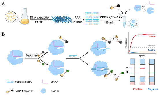
Figure 1.
Schematic illustration of detecting the E. coli O157:H7 using the RAA-CRISPR/Cas12a system. (A) Workflow for CRISPR/Cas12a-mediated detection of E. coli O157:H7. It included genomic DNA extraction, RAA preamplification, and Cas12a/crRNA cleavage assay. The times required for each step are based on the Kit’s instructions or previous studies. (B) The detection principle and signal readout process of CRISPR/Cas12a. The nucleic acid test strip comprises an absorption pad, an interpretation area, and a binding pad (Figure S1). The binding pad contains embedded Au NPs; the C line contains embedded streptavidin; the T line in the interpretation area contains embedded secondary antibody for signal readout. A red band appearing on the T line can be judged as a positive result.
3.2. Optimization of the RAA-CRISPR/Cas12a Reaction System
The feasibility of using the CRISPR/Cas12a system for bacterial target sequence detection was evaluated (Figure 2A). The Escherichia coli rfbE gene was selected as the target gene. To improve the sensitivity and specificity of the method, we investigated the optimal concentration of the crRNA, Cas12a, and ssDNA reporter in the reaction system, as the cleavage activity was directly related to their concentration. The concentration of target DNA used in the experiment was ~5 nM. In the 20 μL reaction system, the fluorescence value reached the highest when the concentrations of the Cas12a and crRNA were 400 nM and 300 nM, respectively (Figure 2B,C). The higher amount of the crRNA or Cas12a probably affected the efficiency of the trans-cleavage of CRISPR/Cas12a [41,42]. The concentration of the ssDNA reporter was also critical in the CRISPR/Cas12a detection system. As shown in Figure 2D, we found that, with the increase of the probes’ concentration, the fluorescence intensity also increased. In order to better distinguish negative samples without wasting reagents, 400 nM ssDNA reporter was used for the subsequent experiment. To understand the effect of the component ratios on the assay sensitivity, different Cas12a/crRNA ratios were used in the CRISPR/Cas12a assay. It was found that the optimal Cas12a/crRNA ratio was 4:3, which also agreed with previous studies (Figure S2). A suitable amplification time of genomic DNA is an important parameter for CRISPR/Cas12a analysis. Therefore, different reaction times were used for the target DNA amplification, and the optimal amplification time found was 20 min (Figure S3).
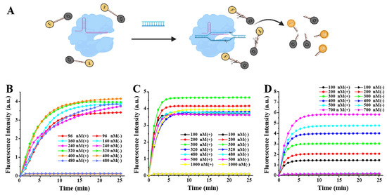
Figure 2.
Optimization of the RAA-CRISPR/Cas12a reaction system. (A) A schematic illustration of the RAA-CRISPR/Cas12a-FQ detector. Real-time fluorescence detection assay using the CRISPR/Cas12a-FQ detector with different concentrations of (B) Cas12a (crRNA 480 nM and ssDNA-FQ reporter 400 nM), (C) crRNA (Cas12a 400 nM and ssDNA-FQ reporter 400 nM), and (D) ssDNA-FQ reporter (Cas12a 400 nM and crRNA 480 nM).
3.3. Detection Sensitivity of the RAA-CRISPR/Cas12a System
Under the optimized reaction conditions, the detection sensitivity of the system toward the E. coli O157:H7 samples was estimated. As shown in Figure 3, compared with the negative control, the fluorescence intensity increased significantly with E. coli O157:H7 ranging from 100 CFU/mL to 106 CFU/mL. This agreed well with the gel electrophoresis pattern of the target DNA amplified by RAA (Figure S4). These results demonstrated that the RAA-CRISPR/Cas12a method developed here had high detecting sensitivity. Even samples with a concentration of ~1 CFU/mL produced significant signals, which is much lower than the detection limit of the traditional real-time PCR technology (103 CFU/mL), ELISA (104~107 CFU/mL), and some recently developed methods (Table S3) [43,44]. The excellent performance of this method can be attributed to the combination of nucleic acid amplification technology and the CRISPR/Cas12a system and the optimized reaction conditions, which enabled specific identification of the target genes and further efficient amplification of the signal. Although the experimental results did not show a good linear relationship between the E. coli O157:H7 concentration and fluorescence intensity, it can be very useful for the identification of trace E. coli O157:H7 infection. This phenomenon may be attributed to the fact that the developed system involves RAA amplification and CRISPR/Cas12a cutting. Achieving a linear response requires that the product generated through RAA amplification is proportional to the target concentration and that a linear correlation exists between the RAA product and the fluorescence signal from the CRISPR/Cas12a step. This can be difficult to achieve, and a similar non-linear relationship has been observed in some reported sensors that combine DNA amplification and the CRISPR/Cas system [40,45]. For naked-eye observation, the CRISPR/Cas12a-LFA assay was carried out using the FAM and biotin dual-modified ssDNA reporter. Significantly, a flow strip can generate a visible signal when the concentration of E. coli O157:H7 is as low as 1 × 10² CFU/mL (Figure 3C). This indicates that the RAA-CRISPR/Cas12a method can realize rapid visual detection with high sensitivity.
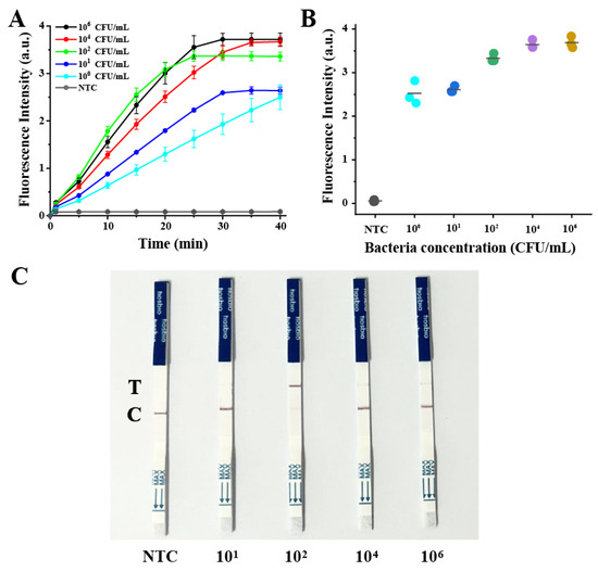
Figure 3.
Evaluation of the detection sensitivity of the RAA-CRISPR/Cas12a system. (A) Real-time fluorescence detection assay using the CRISPR/Cas12a-FQ detector. (B) Fluorescence generated with the different concentrations of E. coli O157:H7. (C) Results of LFAs treated with different concentrations of E. coli O157:H7. NTC: no-template control.
3.4. Specificity of the CRISPR/Cas12a System for Detecting E. coli O157:H7
To verify the specificity of the CRISPR/Cas12a system, five different bacterial strains (E. coli, S. aureus, P. aeruginosa, S. typhimurium, L. monocytogenes) were selected, and the fluorescence signal was recorded. Compared with the target bacterial strain, the other non-target bacterial strains exhibited a low fluorescence signal, which was consistent with the results of gel electrophoresis (Figure 4 and Figure S5). The results confirmed the high specificity and good selectivity of this method. It should be noted that the specificity could be attributed to the recognition function of the Cas12a/crRNA complex.
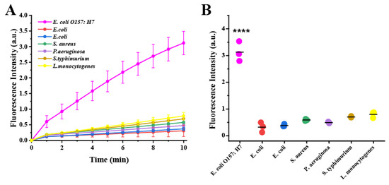
Figure 4.
Specificity of the CRISPR/Cas12a system for detecting E. coli O157:H7. (A) Real-time fluorescence detection assay using the CRISPR/Cas12a-FQ detector for different bacterial strains. (B) Fluorescence generated from different bacterial strains. Numbers 1–7 represent E. coli O157:H7 (CICC 24187), E. coli (ATCC 25922), E. coli (ATCCBAA2452), S. aureus (CICC 10306), P. aeruginosa (CICC 21636), S. typhimurium (CICC 22956), and L. monocytogenes (CICC 21635), respectively. Significant differences between groups were tested with the independent samples t-test (**** represents p < 0.0001).
3.5. The Application Potential of the RAA-CRISPR/Cas12a System
Since the presented method demonstrated high sensitivity and specificity, the potential application of the approach in real samples including skim milk and drinking water was further investigated. The standard addition of different concentrations of E. coli O157:H7 (104, 102, 101, and 100 CFU/mL) in practical samples was carried out, followed by sample detection with the RAA-CRISPR/Cas12a method. As shown in Figure 5 and Table S4, the method displayed good detecting sensitivity in both the milk and drinking water samples, demonstrating the applicability of the method.
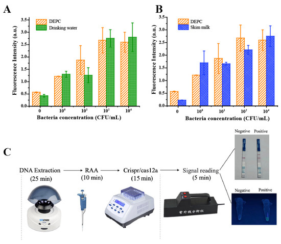
Figure 5.
The application potential of the RAA-CRISPR/Cas12a system. The fluorescence signal against varying E. coli O157:H7 concentrations in drinking water (A) and skim milk (B). The fluorescence intensity of E. coli O157:H7 in DEPC water (DNase-/RNase-free) was also recorded for comparison. (C) The schematic of the time cost of the developed system for visualized detection.
In order to achieve rapid, sensitive, and simple in situ detection of E. coli O157:H7, the entire detection process was optimized (Figure 5C). The three processes, including DNA extraction, RAA amplification, and CRISPR reaction can be performed with just two simple, portable instruments: a handheld centrifuge and a metal bath. In terms of signal reading, the test results can be visualized with a handheld UV analyzer and test strip by adding different ssDNA reporters, which allows the results to be displayed more intuitively without complicated instruments and data processing. The cost coming from the strips can be solved by mass production. Thus, we expect future tests to cost only a few dollars. Besides, on the premise of being distinguishable from the negative control, the duration of the entire test process can be shortened to less than 55 min, which improved the efficiency of the test. Our proposed method provides a rapid, simple in situ assay for the detection of E. coli O157:H7 without quantification.
4. Conclusions
In this study, a rapid and ultrasensitive method for the detection of E. coli O157:H7 was developed based on the RAA-CRISPR/Cas12a system. By combining the CRISPR/Cas12a system with isothermal amplification techniques, E. coli O157:H7 concentrations as low as ~1 CFU/mL (fluorescence method) and 1 × 10² CFU/mL (lateral flow assay) can still be detected with significant accuracy, which was superior to most previously reported methods. Moreover, this method achieved high specificity among different strains and showed good performance in practical sample analysis. Importantly, we optimized the whole reaction process, allowing the detection process to be completed within 55 min, which was faster than most other reported sensors, which take several hours to several days. Signal visualization could be achieved by a handheld UV lamp or lateral flow strips, which facilitates the in situ detection of E. coli O157:H7 without complex instruments. Overall, our study provides a simple and facile method for the evaluation of E. coli O157:H7 contamination, which can also be used for the detection of other pathogens. This method has broad application prospects in the fields of food safety, clinical diagnosis, and environmental science.
Supplementary Materials
The following Supporting Information can be downloaded at: https://www.mdpi.com/article/10.3390/bios13060659/s1, Figure S1: Principle of visual detection with LFA; Table S1: Sequences used in this study; Figure S2: Effects of different Cas12a/crRNA ratios on fluorescence intensity; Table S2: Verification of CRISPR/Cas12a specificity; Figure S3: Effect of different amplification times of RAA on fluorescence intensity; Table S3: Detection limit of E. coli O157:H7 with different methods [32,33,43,46,47,48,49]; Figure S4: The gel electrophoresis result of RAA for different bacterial concentrations; Table S4: Detection of E. coli O157:H7 in milk and drinking water samples using fluorescence signal; Figure S5: The gel electrophoresis result of RAA for different bacterial strains.
Author Contributions
Conceptualization, L.Z. (Lishan Zhu), J.W. and L.Z. (Li Zhou); formal analysis, Z.L.; funding acquisition, J.W. and L.Z. (Li Zhou); investigation, Y.X. and Z.C.; methodology, L.Z. (Lishan Zhu); supervision, J.W. and L.Z. (Li Zhou); validation, Z.L., Y.X. and Z.C.; writing—original draft, L.Z. (Lishan Zhu); writing—review and editing, J.W. and L.Z. (Li Zhou). All authors have read and agreed to the published version of the manuscript.
Funding
This work was supported by the introduced innovative R&D team project under the “The Pearl River Talent Recruitment Program” of Guangdong Province (2019ZT08L387), the National Natural Science Foundation of China (21906033 and 22036002), the National Key Research and Development Program of China (2021YFF0703300), and Funding by Science and Technology Projects of Guangzhou (202102020570, 202201020392).
Institutional Review Board Statement
Not applicable.
Informed Consent Statement
Not applicable.
Data Availability Statement
The experimental data are contained within the article.
Conflicts of Interest
The authors declare no conflict of interest.
References
- Majowicz, S.E.; Scallan, E.; Jones-Bitton, A.; Sargeant, J.M.; Stapleton, J.; Angulo, F.J.; Yeung, D.H.; Kirk, M.D. Global Incidence of Human Shiga Toxin-Producing Escherichia coli Infections and Deaths: A Systematic Review and Knowledge Synthesis. Foodborne Pathog. Dis. 2014, 11, 447–455. [Google Scholar] [CrossRef] [PubMed]
- Allison, S.E.; Silphaduang, U.; Mascarenhas, M.; Konczy, P.; Quan, Q.; Karmali, M.; Coombes, B.K. Novel Repressor of Escherichia coli O157:H7 Motility Encoded in the Putative Fimbrial Cluster OI-1. J. Bacteriol. 2012, 194, 5343–5352. [Google Scholar] [CrossRef] [PubMed]
- Bian, X.; Jing, F.; Li, G.; Fan, X.; Jia, C.; Zhou, H.; Jin, Q.; Zhao, J. A Microfluidic Droplet Digital PCR for Simultaneous Detection of Pathogenic Escherichia coli O157 and Listeria Monocytogenes. Biosens. Bioelectron. 2015, 74, 770–777. [Google Scholar] [CrossRef] [PubMed]
- Chekabab, S.M.; Paquin-Veillette, J.; Dozois, C.M.; Harel, J. The Ecological Habitat and Transmission of Escherichia coli O157:H7. FEMS Microbiol. Lett. 2013, 341, 1–12. [Google Scholar] [CrossRef] [PubMed]
- Mead, P.S.; Griffin, P.M. Escherichia coli O157:H7. Lancet 1998, 352, 1207–1212. [Google Scholar] [CrossRef] [PubMed]
- Kaper, J.B.; Nataro, J.P.; Mobley, H.L.T. Pathogenic Escherichia coli. Nat. Rev. Microbiol. 2004, 2, 123–140. [Google Scholar] [CrossRef]
- Nataro, J.P.; Kaper, J.B. Diarrheagenic Escherichia coli. Clin. Microbiol. Rev. 1998, 11, 142–201. [Google Scholar] [CrossRef]
- Brunetti, G.; Conteduca, D.; Armenise, M.; Ciminelli, C. Novel Micro-Nano Optoelectronic Biosensor for Label-Free Real-Time Biofilm Monitoring. Biosensors 2021, 11, 361. [Google Scholar] [CrossRef] [PubMed]
- Di Toma, A.; Brunetti, G.; Chiriacò, M.S.; Ferrara, F.; Ciminelli, C. A Novel Hybrid Platform for Live/Dead Bacteria Accurate Sorting by On-Chip DEP Device. Int. J. Mol. Sci. 2023, 24, 7077. [Google Scholar] [CrossRef] [PubMed]
- Leva-Bueno, J.; Peyman, S.A.; Millner, P.A. A Review on Impedimetric Immunosensors for Pathogen and Biomarker Detection. Med. Microbiol. Immunol. 2020, 209, 343–362. [Google Scholar] [CrossRef] [PubMed]
- Pugia, M.; Bose, T.; Tjioe, M.; Frabutt, D.; Baird, Z.; Cao, Z.; Vorsilak, A.; McLuckey, I.; Barron, M.R.; Barron, M.; et al. Multiplexed Signal Ion Emission Reactive Release Amplification (SIERRA) Assay for the Culture-Free Detection of Gram-Negative and Gram-Positive Bacteria and Antimicrobial Resistance Genes. Anal. Chem. 2021, 93, 6604–6612. [Google Scholar] [CrossRef] [PubMed]
- Adams, A.N.; Clark, M.F. Characteristics of the Microplate Method of Enzyme-Linked Immunosorbent Assay for the Detection of Plant Viruses. J. Gen. Virol. 1977, 34, 475–483. [Google Scholar]
- Klein, D. Quantification Using Real-Time PCR Technology: Applications and Limitations. Trends Mol. Med. 2002, 8, 257–260. [Google Scholar] [CrossRef] [PubMed]
- Lei, S.; Gu, X.; Zhong, Q.; Duan, L.; Zhou, A. Absolute Quantification of Vibrio Parahaemolyticus by Multiplex Droplet Digital PCR for Simultaneous Detection of Tlh, Tdh and UreR Based on Single Intact Cell. Food Control 2020, 114, 107207. [Google Scholar] [CrossRef]
- March, S.B.; Ratnam, S. Sorbitol-MacConkey Medium for Detection of Escherichia coli O157:H7 Associated with Hemorrhagic Colitis. J. Clin. Microbiol. 1986, 23, 869–872. [Google Scholar] [CrossRef] [PubMed]
- Boonham, N.; Kreuze, J.; Winter, S.; van der Vlugt, R.; Bergervoet, J.; Tomlinson, J.; Mumford, R. Methods in Virus Diagnostics: From ELISA to next Generation Sequencing. Virus Res. 2014, 186, 20–31. [Google Scholar] [CrossRef] [PubMed]
- Lim, M.-C.; Lee, G.-H.; Huynh, D.T.N.; Hong, C.-E.; Park, S.-Y.; Jung, J.-Y.; Park, C.-S.; Ko, S.; Kim, Y.-R. Biological Preparation of Highly Effective Immunomagnetic Beads for the Separation, Concentration, and Detection of Pathogenic Bacteria in Milk. Colloids Surf. B Biointerfaces 2016, 145, 854–861. [Google Scholar] [CrossRef]
- Yu, J.; Su, J.; Zhang, J.; Wei, X.; Guo, A. CdTe/CdS Quantum Dot-Labeled Fluorescent Immunochromatography Test Strips for Rapid Detection of Escherichia coli O157:H7. RSC Adv. 2017, 7, 17819–17823. [Google Scholar] [CrossRef]
- Huang, Z.; Peng, J.; Han, J.; Zhang, G.; Huang, Y.; Duan, M.; Liu, D.; Xiong, Y.; Xia, S.; Lai, W. A Novel Method Based on Fluorescent Magnetic Nanobeads for Rapid Detection of Escherichia coli O157:H7. Food Chem. 2019, 276, 333–341. [Google Scholar] [CrossRef]
- Brakstad, O.G.; Aasbakk, K.; Maeland, J.A. Detection of Staphylococcus Aureus by Polymerase Chain Reaction Amplification of the Nuc Gene. J. Clin. Microbiol. 1992, 30, 1654–1660. [Google Scholar] [CrossRef]
- Macori, G.; McCarthy, S.C.; Burgess, C.M.; Fanning, S.; Duffy, G. A Quantitative Real Time PCR Assay to Detect and Enumerate Escherichia coli O157 and O26 Serogroups in Sheep Recto-Anal Swabs. J. Microbiol. Methods 2019, 165, 105703. [Google Scholar] [CrossRef] [PubMed]
- Fu, Z.; Rogelj, S.; Kieft, T.L. Rapid Detection of Escherichia coli O157:H7 by Immunomagnetic Separation and Real-Time PCR. Int. J. Food Microbiol. 2005, 99, 47–57. [Google Scholar] [CrossRef]
- Jofré, A.; Martin, B.; Garriga, M.; Hugas, M.; Pla, M.; Rodríguez-Lázaro, D.; Aymerich, T. Simultaneous Detection of Listeria Monocytogenes and Salmonella by Multiplex PCR in Cooked Ham. Food Microbiol. 2005, 22, 109–115. [Google Scholar] [CrossRef]
- Donhauser, S.C.; Niessner, R.; Seidel, M. Quantification of E. coli DNA on a Flow-through Chemiluminescence Microarray Readout System after PCR Amplification. Anal. Sci. 2009, 25, 669–674. [Google Scholar] [CrossRef]
- Doudna, J.A.; Charpentier, E. The New Frontier of Genome Engineering with CRISPR-Cas9. Science 2014, 346, 1258096. [Google Scholar] [CrossRef]
- Knott, G.J.; Doudna, J.A. CRISPR-Cas Guides the Future of Genetic Engineering. Science 2018, 361, 866–869. [Google Scholar] [CrossRef] [PubMed]
- Samanta, D.; Ebrahimi, S.B.; Ramani, N.; Mirkin, C.A. Enhancing CRISPR-Cas-Mediated Detection of Nucleic Acid and Non-Nucleic Acid Targets Using Enzyme-Labeled Reporters. J. Am. Chem. Soc. 2022, 144, 16310–16315. [Google Scholar] [CrossRef] [PubMed]
- Bonini, A.; Poma, N.; Vivaldi, F.; Biagini, D.; Bottai, D.; Tavanti, A.; Di Francesco, F. A Label-Free Impedance Biosensing Assay Based on CRISPR/Cas12a Collateral Activity for Bacterial DNA Detection. J. Pharm. Biomed. Anal. 2021, 204, 114268. [Google Scholar] [CrossRef] [PubMed]
- Sun, X.; Wang, Y.; Zhang, L.; Liu, S.; Zhang, M.; Wang, J.; Ning, B.; Peng, Y.; He, J.; Hu, Y.; et al. CRISPR-Cas9 Triggered Two-Step Isothermal Amplification Method for E. coli O157:H7 Detection Based on a Metal–Organic Framework Platform. Anal. Chem. 2020, 92, 3032–3041. [Google Scholar] [CrossRef] [PubMed]
- Kadam, U.S.; Cho, Y.; Park, T.Y.; Hong, J.C. Aptamer-Based CRISPR-Cas Powered Diagnostics of Diverse Biomarkers and Small Molecule Targets. Appl. Biol. Chem. 2023, 66, 13. [Google Scholar] [CrossRef] [PubMed]
- Barrangou, R.; Fremaux, C.; Deveau, H.; Richards, M.; Boyaval, P.; Moineau, S.; Romero, D.A.; Horvath, P. CRISPR Provides Acquired Resistance Against Viruses in Prokaryotes. Science 2007, 315, 1709–1712. [Google Scholar] [CrossRef]
- Wang, Y.; Ke, Y.; Liu, W.; Sun, Y.; Ding, X. A One-Pot Toolbox Based on Cas12a/CrRNA Enables Rapid Foodborne Pathogen Detection at Attomolar Level. ACS Sens. 2020, 5, 1427–1435. [Google Scholar] [CrossRef]
- Wang, S.; Fan, Y.; Feng, Z.; Song, M.; Li, Q.; Jiang, B.; Qin, F.; Liu, H.; Lan, L.; Yang, M. Rapid Nucleic Acid Detection of Escherichia coli O157:H7 Based on CRISPR/Cas12a System. Food Control 2021, 130, 108194. [Google Scholar] [CrossRef]
- Liu, L.; Duan, J.-J.; Wei, X.-Y.; Hu, H.; Wang, Y.-B.; Jia, P.-P.; Pei, D.-S. Generation and Application of a Novel High-Throughput Detection Based on RPA-CRISPR Technique to Sensitively Monitor Pathogenic Microorganisms in the Environment. Sci. Total Environ. 2022, 838, 156048. [Google Scholar] [CrossRef]
- Li, F.; Ye, Q.; Chen, M.; Xiang, X.; Zhang, J.; Pang, R.; Xue, L.; Wang, J.; Gu, Q.; Lei, T.; et al. Cas12aFDet: A CRISPR/Cas12a-Based Fluorescence Platform for Sensitive and Specific Detection of Listeria Monocytogenes Serotype 4c. Anal. Chim. Acta 2021, 1151, 338248. [Google Scholar] [CrossRef]
- Feng, W.; Newbigging, A.M.; Tao, J.; Cao, Y.; Peng, H.; Le, C.; Wu, J.; Pang, B.; Li, J.; Tyrrell, D.L.; et al. CRISPR Technology Incorporating Amplification Strategies: Molecular Assays for Nucleic Acids, Proteins, and Small Molecules. Chem. Sci. 2021, 12, 4683–4698. [Google Scholar] [CrossRef] [PubMed]
- Zhang, M.; Wang, H.; Wang, H.; Wang, F.; Li, Z. CRISPR/Cas12a-Assisted Ligation-Initiated Loop-Mediated Isothermal Amplification (CAL-LAMP) for Highly Specific Detection of MicroRNAs. Anal. Chem. 2021, 93, 7942–7948. [Google Scholar] [CrossRef] [PubMed]
- Yang, B.; Shi, Z.; Ma, Y.; Wang, L.; Cao, L.; Luo, J.; Wan, Y.; Song, R.; Yan, Y.; Yuan, K.; et al. LAMP Assay Coupled with CRISPR/Cas12a System for Portable Detection of African Swine Fever Virus. Transbound. Emerg. Dis. 2022, 69, e216–e223. [Google Scholar] [CrossRef] [PubMed]
- Chang, W.; Liu, W.; Liu, Y.; Zhan, F.; Chen, H.; Lei, H.; Liu, Y. Colorimetric Detection of Nucleic Acid Sequences in Plant Pathogens Based on CRISPR/Cas9 Triggered Signal Amplification. Microchim. Acta 2019, 186, 243. [Google Scholar] [CrossRef]
- Zhang, Y.; Chen, M.; Liu, C.; Chen, J.; Luo, X.; Xue, Y.; Liang, Q.; Zhou, L.; Tao, Y.; Li, M.; et al. Sensitive and Rapid On-Site Detection of SARS-CoV-2 Using a Gold Nanoparticle-Based High-Throughput Platform Coupled with CRISPR/Cas12-Assisted RT-LAMP. Sens. Actuators B Chem. 2021, 345, 130411. [Google Scholar] [CrossRef]
- Xiang, X.; Li, F.; Ye, Q.; Shang, Y.; Chen, M.; Zhang, J.; Zhou, B.; Suo, H.; Ding, Y.; Wu, Q. High-throughput Microfluidic Strategy Based on RAA-CRISPR/Cas13a Dual Signal Amplification for Accurate Identification of Pathogenic Listeria. Sens. Actuators B Chem. 2022, 358, 131517. [Google Scholar] [CrossRef]
- Liu, L.; Zhao, G.; Li, X.; Xu, Z.; Lei, H.; Shen, X. Development of Rapid and Easy Detection of Salmonella in Food Matrics Using RPA-CRISPR/Cas12a Method. LWT Food Sci. Technol. 2022, 162, 113443. [Google Scholar] [CrossRef]
- Ibekwe, A.M.; Watt, P.M.; Grieve, C.M.; Sharma, V.K.; Lyons, S.R. Multiplex Fluorogenic Real-Time PCR for Detection and Quantification of Escherichia coli O157:H7 in Dairy Wastewater Wetlands. Appl. Environ. Microbiol. 2002, 68, 4853–4862. [Google Scholar] [CrossRef] [PubMed]
- Ibekwe, A.M.; Grieve, C.M. Detection and Quantification of Escherichia coli O157:H7 in Environmental Samples by Real-Time PCR. J. Appl. Microbiol. 2003, 94, 421–431. [Google Scholar] [CrossRef] [PubMed]
- Zhou, R.; Li, Y.; Dong, T.; Tang, Y.; Li, F. A Sequence-Specific Plasmonic Loop-Mediated Isothermal Amplification Assay with Orthogonal Color Readouts Enabled by CRISPR Cas12a. Chem. Commun. 2020, 56, 3536–3538. [Google Scholar] [CrossRef] [PubMed]
- Feng, M.; Yong, Q.; Wang, W.; Kuang, H.; Wang, L.; Xu, C. Development of a Monoclonal Antibody-Based ELISA to Detect Escherichia coli O157:H7. Food Agric. Immunol. 2013, 24, 481–487. [Google Scholar] [CrossRef]
- Pang, B.; Zhao, C.; Li, L.; Song, X.; Xu, K.; Wang, J.; Liu, Y.; Fu, K.; Bao, H.; Song, D.; et al. Development of a Low-Cost Paper-Based ELISA Method for Rapid Escherichia coli O157:H7 Detection. Anal. Biochem. 2018, 542, 58–62. [Google Scholar] [CrossRef]
- Shen, Z.; Hou, N.; Jin, M.; Qiu, Z.; Wang, J.; Zhang, B.; Wang, X.; Wang, J.; Zhou, D.; Li, J. A Novel Enzyme-Linked Immunosorbent Assay for Detection of Escherichia coli O157:H7 Using Immunomagnetic and Beacon Gold Nanoparticles. Gut Pathog. 2014, 6, 14. [Google Scholar] [CrossRef]
- Jung, B.Y.; Jung, S.C.; Kweon, C.H. Development of a Rapid Immunochromatographic Strip for Detection of Escherichia coli O157. J. Food Prot. 2005, 68, 2140–2143. [Google Scholar] [CrossRef]


Disclaimer/Publisher’s Note: The statements, opinions and data contained in all publications are solely those of the individual author(s) and contributor(s) and not of MDPI and/or the editor(s). MDPI and/or the editor(s) disclaim responsibility for any injury to people or property resulting from any ideas, methods, instructions or products referred to in the content. |
© 2023 by the authors. Licensee MDPI, Basel, Switzerland. This article is an open access article distributed under the terms and conditions of the Creative Commons Attribution (CC BY) license (https://creativecommons.org/licenses/by/4.0/).