Analytical Determination of Serotonin Exocytosis in Human Platelets with BDD-on-Quartz MEA Devices

 , ,
, ,  , , and
Abstract
:1. Introduction
2. Materials and Methods
2.1. Solutions Used, Unless Specified (in mM), pH Adjusted with NaOH
2.2. Human Platelets Preparation
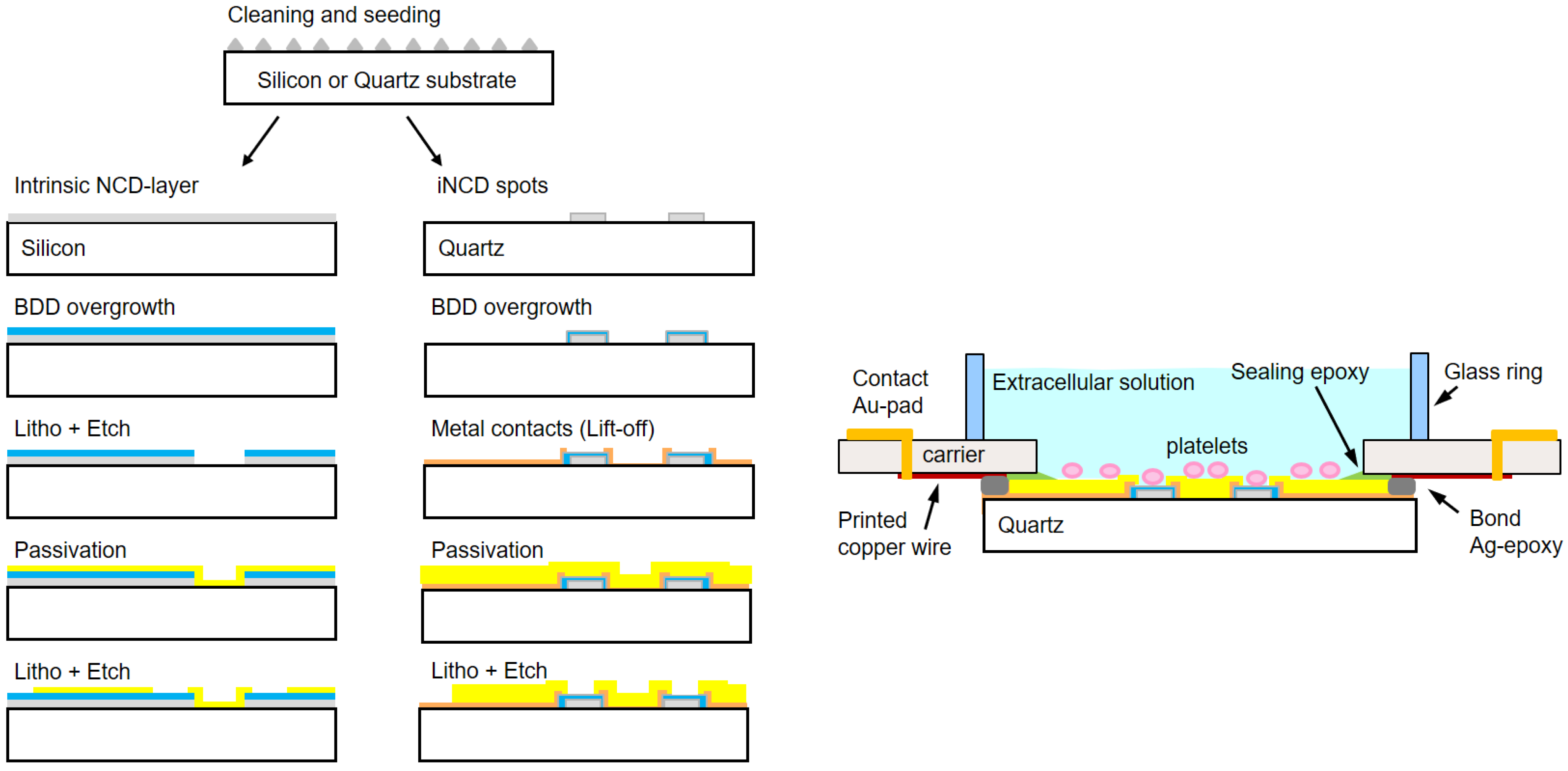
2.3. BDD-on-Quartz MEA
- Clean and then spin-coat the wafer with a NanoAmando seeding solution (New Metals and Chemicals, Tokyo, Japan).
- Grow a 50 nm thin intrinsic nanocrystalline diamond (iNCD) layer by microwave plasma chemical vapor deposition (MWCVD) at a power of 2200 W in an H2 atmosphere at a gas flow of 400 sccm, with 1.5% of CH4, at a temperature of 800 °C and pressure of 30 Torr. This growth step has a duration of 10 min.
- Create the wafer by lifting off a titanium hard mask with a pattern of the 60 µm spots.
- Etch the unprotected iNCD by reactive ion etching (RIE) in an Ar-O2 atmosphere. This leaves the ‘footprint’ for growing the diamond spots of the microelectrodes.
- Grow the iNCD spots resulting from the previous step up to a thickness of 1 µm. This growth is carried out with the same modality of step 2 but for a duration of 190 min.
- Overgrow the iNCD-spots with ~350 nm BDD in a doping-dedicated MWCVD-reactor. Parameters are the same as above for steps 2 and 5, but the process duration is 70 min in this case. Doping is provided by boron wires (Goodfellow, Bad Nauheim, Germany) introduced into the plasma.
- Create the wafer by lifting off the metal ring contacts and wires out of 100 nm titanium and 50 nm gold.
- Passivate the wafer with a polyimide-based photoresist (Durimide® 7505, Fujifilm, Tokyo, Japan) and transfer the pattern of the openings of electrodes and contact pads by lithography.
- Dice the wafer, bind the chips onto appropriate polychlorinated biphenyl carriers (PCB carriers), and glue a 10 mm wide and 4 mm thick glass ring to provide a ~200 µL incubation volume.
2.4. BDD-on-Quartz MEA Recording System
2.5. Amperometric Data Analysis
2.6. Microscopy Observation
2.7. Statistics
3. Results
3.1. Characterization and Comparison of BDD-on-Quartz MEAs and BDD-on-Silicon MEAs
3.2. Characterization of Serotonin Exocytosis of Human Platelets Using the BDD-on-Quartz MEA
4. Discussion
4.1. Different Types of Recorded Spikes
4.2. Brief Comparison of the Three BDD MEAs
4.2.1. BDD-on-Glass MEAs
4.2.2. BDD-on-Silicon MEAs
4.2.3. BDD-on-Quartz MEAs
5. Conclusions
Supplementary Materials
Author Contributions
Funding
Institutional Review Board Statement
Informed Consent Statement
Data Availability Statement
Acknowledgments
Conflicts of Interest
References
- Wightman, R.M.; Haynes, C.L. Synaptic vesicles really do kiss and run. Nat. Neurosci. 2004, 7, 321–322. [Google Scholar] [CrossRef]
- Li, X.; Majdi, S.; Dunevall, J.; Fathali, H.; Ewing, A.G. Quantitative Measurement of Transmitters in Individual Vesicles in the Cytoplasm of Single Cells with Nanotip Electrodes. Angew. Chem. Int. Ed. Engl. 2015, 127, 12146–12150. [Google Scholar] [CrossRef]
- Ren, L.; Mellander, L.J.; Keighron, J.; Cans, A.-S.; Kurczy, M.E.; Svir, I.; Oleinick, A.; Amatore, C.; Ewing, A.G. The evidence for open and closed exocytosis as the primary release mechanism. Q. Rev. Biophys. 2016, 49, e12. [Google Scholar] [CrossRef]
- Wang, Y.; Ewing, A. Electrochemical Quantification of Neurotransmitters in Single Live Cell Vesicles Shows Exocytosis is Predominantly Partial. ChemBioChem 2021, 22, 807–813. [Google Scholar] [CrossRef] [PubMed]
- Carlsson, A. Perspectives on the Discovery of Central Monoaminergic Neurotransmission. Annu. Rev. Neurosci. 1987, 10, 19–40. [Google Scholar] [CrossRef] [PubMed]
- Greengard, P. The Neurobiology of Slow Synaptic Transmission. Science 2001, 294, 1024–1030. [Google Scholar] [CrossRef]
- Engle, K.; Zhou, M.; Wang, J. Identification and Characterization of a Novel Monoamine Transporter in the Human Brain. J. Biol. Chem. 2004, 279, 50042–50049. [Google Scholar] [CrossRef]
- Capellino, S.; Claus, M.; Watzl, C. Regulation of natural killer cell activity by glucocorticoids, serotonin, dopamine, and epinephrine. Cell. Mol. Immunol. 2020, 17, 705–711. [Google Scholar] [CrossRef]
- Jiang, Y.; Zou, D.; Li, Y.; Gu, S.; Dong, J.; Ma, X.; Xu, S.; Wang, F.; Huang, J.H. Monoamine Neurotransmitters Control Basic Emotions and Affect Major Depressive Disorders. Pharmaceuticals 2022, 15, 1203. [Google Scholar] [CrossRef]
- Moura, C.; Vale, N. The Role of Dopamine in Repurposing Drugs for Oncology. Biomedicines 2023, 11, 1917. [Google Scholar] [CrossRef]
- Leszczyszyn, D.J.; Jankowski, J.A.; Viveros, O.H.; Diliberto, E.J.; Near, J.A.; Wightman, R.M. Nicotinic receptor-mediated catecholamine secretion from individual chromaffin cells. Chemical evidence for exocytosis. J. Biol. Chem. 1990, 265, 14736–14737. [Google Scholar] [CrossRef]
- Leszczyszyn, D.J.; Jankowski, J.A.; Viveros, O.H.; Diliberto, E.J.; Near, J.A.; Wightman, R.M. Secretion of catecholamines from individual adrenal medullary chromaffin cells. J. Neurochem. 1991, 56, 1855–1863. [Google Scholar] [CrossRef]
- Wightman, R.M.; Jankowski, J.A.; Kennedy, R.T.; Kawagoe, K.T.; Schroeder, T.J.; Leszczyszyn, D.J.; Near, J.A.; Diliberto, E.J.; Viveros, O.H. Temporally resolved catecholamine spikes correspond to single vesicle release from individual chromaffin cells. Proc. Natl. Acad. Sci. USA 1991, 88, 10754–10758. [Google Scholar] [CrossRef] [PubMed]
- Lemaître, F.; Collignon, M.G.; Amatore, C. Recent advances in Electrochemical Detection of Exocytosis. Electrochim. Acta 2014, 140, 457–466. [Google Scholar] [CrossRef]
- Li, Y.-T.; Zhang, S.-H.; Wang, L.; Xiao, R.-R.; Liu, W.; Zhang, X.-W.; Zhou, Z.; Amatore, C.; Huang, W.-H. Nanoelectrode for Amperometric Monitoring of Individual Vesicular Exocytosis Inside Single Synapses. Angew. Chemie Int. Ed. 2014, 53, 12456–12460. [Google Scholar] [CrossRef] [PubMed]
- Montenegro, P.; Pueyo, M.; Lorenzo, J.N.; Villar-Martinez, M.D.; Alayón, A.; Carrillo, F.; Borges, R. A Secretory Vesicle Failure in Parkinson’s Disease Occurs in Human Platelets. Ann. Neurol. 2020, 91, 697–703. [Google Scholar] [CrossRef]
- Brito, R.G.; Montenegro, P.; Méndez, A.; Carabelli, V.; Tomagra, G.; Shabgahi, R.E.; Pasquarelli, A.; Borges, R. Multielectrode Arrays as a Means to Study Exocytosis in Human Platelets. Biosensors 2023, 13, 86. [Google Scholar] [CrossRef]
- Ge, S.; Wittenberg, N.J.; Haynes, C.L. Quantitative and Real-Time Detection of Secretion of Chemical Messengers from Individual Platelets. Biochemistry 2008, 47, 7020–7024. [Google Scholar] [CrossRef] [PubMed]
- Ge, S.; White, J.G.; Haynes, C.L. Quantal Release of Serotonin from Platelets. Anal. Chem. 2009, 81, 2935–2943. [Google Scholar] [CrossRef]
- Ge, S.; Woo, E.; Haynes, C.L. Quantal Regulation and Exocytosis of Platelet Dense-Body Granules. Biophys. J. 2011, 101, 2351–2359. [Google Scholar] [CrossRef]
- Ge, S.; Woo, E.; White, J.G.; Haynes, C.L. Electrochemical measurement of endogenous serotonin release from human blood platelets. Anal. Chem. 2011, 83, 2598–2604. [Google Scholar] [CrossRef]
- Gao, Z.; Carabelli, V.; Carbone, E.; Colombo, E.; Dipalo, M.; Manfredotti, C.; Pasquarelli, A.; Feneberg, M.; Thonke, K.; Vittone, E.; et al. Transparent microelectrode array in diamond technology. J. Micro-Nano Mech. 2011, 6, 33–37. [Google Scholar] [CrossRef]
- Kiran, R.; Rousseau, L.; Lissorgues, G.; Scorsone, E.; Bongrain, A.; Yvert, B.; Picaud, S.; Mailley, P.; Bergonzo, P. Multichannel Boron Doped Nanocrystalline Diamond Ultramicroelectrode Arrays: Design, Fabrication and Characterization. Sensors 2012, 12, 7669–7681. [Google Scholar] [CrossRef] [PubMed]
- Gillis, K.D.; Liu, X.A.; Marcantoni, A.; Carabelli, V. Electrochemical measurement of quantal exocytosis using microchips. Pflugers Arch. 2018, 470, 97–112. [Google Scholar] [CrossRef] [PubMed]
- Purcell, E.K.; Becker, M.F.; Guo, Y.; Hara, S.A.; Ludwig, K.A.; McKinney, C.J.; Monroe, E.M.; Rechenberg, R.; Rusinek, C.A.; Saxena, A.; et al. Next-Generation Diamond Electrodes for Neurochemical Sensing: Challenges and Opportunities. Micromachines 2021, 12, 128. [Google Scholar] [CrossRef]
- Picollo, F.; Battiato, A.; Carbone, E.; Croin, L.; Enrico, E.; Forneris, J.; Gosso, S.; Olivero, P.; Pasquarelli, A.; Carabelli, V. Development and Characterization of a Diamond-Insulated Graphitic Multi Electrode Array Realized with Ion Beam Lithography. Sensors 2015, 15, 515–528. [Google Scholar] [CrossRef]
- Granado, T.C.; Neusser, G.; Kranz, C.; Filho, J.B.D.; Carabelli, V.; Carbone, E.; Pasquarelli, A. Progress in transparent diamond microelectrode arrays. Phys. Status Solidi A 2015, 212, 2445–2453. [Google Scholar] [CrossRef]
- Watt, F.; Bettiol, A.A.; Van Kan, J.A.; Teo, E.J.; Breese, M.B.H. Ion Beam Lithography and Nanofabrication: A Review. Int. J. Nanosci. 2005, 04, 269–286. [Google Scholar] [CrossRef]
- Sharma, E.; Rathi, R.; Misharwal, J.; Sinhmar, B.; Kumari, S.; Dalal, J.; Kumar, A. Evolution in Lithography Techniques: Microlithography to Nanolithography. Nanomaterials 2022, 12, 2754. [Google Scholar] [CrossRef] [PubMed]
- Picollo, F.; Battiato, A.; Bernardi, E.; Boarino, L.; Enrico, E.; Forneris, J.; Gatto Monticone, D.; Olivero, P. Realization of a diamond based high density multi electrode array by means of deep ion beam lithography. Nucl. Instrum. Methods Phys. Res. Sect. B Beam Interact. Mater. At. 2015, 348, 199–202. [Google Scholar] [CrossRef]
- Wigström, J.; Dunevall, J.; Najafinobar, N.; Lovrić, J.; Wang, J.; Ewing, A.G.; Cans, A.-S. Lithographic Microfabrication of a 16-Electrode Array on a Probe Tip for High Spatial Resolution Electrochemical Localization of Exocytosis. Anal. Chem. 2016, 88, 2080–2087. [Google Scholar] [CrossRef] [PubMed]
- Picollo, F.; Battiato, A.; Boarino, L.; Tchernij, S.D.; Enrico, E.; Forneris, J.; Gilardino, A.; Jakšić, M.; Sardi, F.; Skukan, N.; et al. Fabrication of monolithic microfluidic channels in diamond with ion beam lithography. Nucl. Instrum. Methods Phys. Res. Sect. B Beam Interact. Mater. At. 2017, 404, 193–197. [Google Scholar] [CrossRef]
- Tomagra, G.; Franchino, C.; Carbone, E.; Marcantoni, A.; Pasquarelli, A.; Picollo, F.; Carabelli, V. Methodologies for Detecting Quantal Exocytosis in Adrenal Chromaffin Cells through Diamond-Based MEAs. In Chromaffin Cells, 1st ed.; Borges, R., Ed.; Methods in Molecular Biology; Humana: New York, NY, USA, 2023; Volume 2565, pp. 213–221. [Google Scholar] [CrossRef]
- Pippione, G.; Olivero, P.; Fischer, M.; Schreck, M.; Pasquarelli, A. Characterization of CVD heavily B-doped diamond thin films for multi electrode array biosensors. Phys. Status Solidi A 2017, 214, 1700223. [Google Scholar] [CrossRef]
- Segura, F.; Brioso, M.A.; Gomez, J.F.; Machado, J.D.; Borges, R. Automatic Analysis for Amperometrical Recordings of Exocytosis. J. Neurosci. Methods 2000, 103, 151–156. [Google Scholar] [CrossRef] [PubMed]
- Finnegan, J.M.; Pihel, K.; Cahill, P.S.; Huang, L.; Zerby, S.E.; Ewing, A.G.; Kennedy, R.T.; Wightman, R.M. Vesicular quantal size measured by amperometry at chromaffin, mast, pheochromocytoma and pancreatic beta-cells. J. Neurochem. 1996, 66, 1914–1923. [Google Scholar] [CrossRef] [PubMed]
- Hochstetler, S.E.; Puopolo, M.; Gustincich, S.; Raviola, E.; Wightman, R.M. Real-Time Amperometric Measurements of Zeptomole Quantities of Dopamine Released from Neurons. Anal. Chem. 2000, 72, 489–496. [Google Scholar] [CrossRef]
- Elhamdani, A.; Palfrey, H.C.; Artalejo, C.R. Quantal Size Is Dependent on Stimulation Frequency and Calcium Entry in Calf Chromaffin Cells. Neuron 2001, 31, 819–830. [Google Scholar] [CrossRef] [PubMed]
- Qin, N.; Chen, Z.; Xue, R. A two-subpopulation model that reflects heterogeneity of large dense core vesicles in exocytosis. Cell Cycle 2022, 21, 531–546. [Google Scholar] [CrossRef] [PubMed]
- Ramachandran, S.B.; Gillis, K.D. A matched-filter algorithm to detect amperometric spikes resulting from quantal secretion. J. Neurosci. Methods 2018, 293, 338–346. [Google Scholar] [CrossRef]
- Ramachandran, S.B.; Gillis, K.D. Estimating amperometric spike parameters resulting from quantal exocytosis using curve fitting seeded by a matched-filter algorithm. J. Neurosci. Methods 2019, 311, 360–368. [Google Scholar] [CrossRef]
- Keighron, J.D.; Wang, Y.; Cans, A.-S. Electrochemistry of Single-Vesicle Events. Annu. Rev. Anal. Chem. 2020, 13, 159–181. [Google Scholar] [CrossRef]
- Hatamie, A.; He, X.; Zhang, X.-W.; Oomen, P.E.; Ewing, A.G. Advances in nano/microscale electrochemical sensors and biosensors for analysis of single vesicles, a key nanoscale organelle in cellular communication. Biosens. Bioelectron. 2023, 220, 114899. [Google Scholar] [CrossRef]
- Huang, M.; Delacruz, J.B.; Ruelas, J.C.; Rathore, S.S.; Lindau, M. Surface-modified CMOS IC electrochemical sensor array targeting single chromaffin cells for highly parallel amperometry measurements. Pflug. Arch.—Eur. J. Physiol. 2018, 470, 113–123. [Google Scholar] [CrossRef] [PubMed]
- de Diego, A.M.G.; Ortega-Cruz, D.; García, A.G. Disruption of Exocytosis in Sympathoadrenal Chromaffin Cells from Mouse Models of Neurodegenerative Diseases. Int. J. Mol. Sci. 2020, 21, 1946. [Google Scholar] [CrossRef] [PubMed]
- Sombers, L.A.; Hanchar, H.J.; Colliver, T.L.; Wittenberg, N.; Cans, A.; Arbault, S.; Amatore, C.; Ewing, A.G. The effects of vesicular volume on secretion through the fusion pore in Exocytotic release from PC12 cells. J. Neurosci. 2004, 24, 303–309. [Google Scholar] [CrossRef] [PubMed]
- Gu, C.; Ewing, A.G. Simultaneous detection of vesicular content and exocytotic release with two electrodes in and at a single cell. Chem. Sci. 2021, 12, 7393–7400. [Google Scholar] [CrossRef] [PubMed]
- de Toledo, G.Á.; Montes, M.A.; Montenegro, P.; Borges, R. Phases of the exocytotic fusion pore. FEBS Lett. 2018, 592, 3532–3541. [Google Scholar] [CrossRef]
- Jackson, M.B.; Hsiao, Y.-T.; Chang, C.-W. Fusion pore expansion and contraction during catecholamine release from endocrine cells. Biophys. J. 2020, 119, 219–231. [Google Scholar] [CrossRef] [PubMed]
- Machado, J.D.; Segura, F.; Brioso, M.A.; Borges, R. Nitric oxide modulates a late step of exocytosis. J. Biol. Chem. 2000, 275, 20274–20279. [Google Scholar] [CrossRef] [PubMed]
- Leiter, O.; Walker, T.L. Platelets: The missing link between the blood and brain? Prog. Neurobiol. 2019, 183, 101695. [Google Scholar] [CrossRef]
- Canobbio, I. Blood platelets: Circulating mirrors of neurons? Res. Pract. Thromb. Haemost. 2019, 3, 564–565. [Google Scholar] [CrossRef] [PubMed]
- Izzi, B.; Tirozzi, A.; Cerletti, C.; Donati, M.B.; de Gaetano, G.; Hoylaerts, M.F.; Iacoviello, L.; Gialluisi, A. Beyond Haemostasis and Thrombosis: Platelets in Depression and Its Co-Morbidities. Int. J. Mol. Sci. 2020, 21, 8817. [Google Scholar] [CrossRef] [PubMed]
- Tirozzi, A.; Izzi, B.; Noro, F.; Marotta, A.; Gianfagna, F.; Hoylaerts, M.F.; Cerletti, C.; Donati, M.B.; De Gaetano, G.; Iacoviello, L.; et al. Assessing genetic overlap between platelet parameters and neurodegenerative disorders. Front. Immunol. 2020, 11, 2127. [Google Scholar] [CrossRef] [PubMed]
- Canobbio, I.; Barbieri, S.S. Are platelets more than a model of brain neurons? Bleeding Thromb. Vasc. Biol. 2022, 1, 29. [Google Scholar] [CrossRef]
- Burnouf, T.; Walker, T.L. The multifaceted role of platelets in mediating brain function. Blood 2022, 140, 815–827. [Google Scholar] [CrossRef]
- Wang, Y.; Liu, S.; Wang, H.; Zhao, Y.; Zhang, X.-D. Neuron devices: Emerging prospects in neural interfaces and recognition. Microsyst. Nanoeng. 2022, 8, 128. [Google Scholar] [CrossRef]
- Liu, Y.; Xu, S.; Yang, Y.; Zhang, K.; He, E.; Liang, W.; Luo, J.; Wu, Y.; Cai, X. Nanomaterial-based microelectrode arrays for in vitro bidirectional brain–computer interfaces: A review. Microsyst. Nanoeng. 2023, 9, 13. [Google Scholar] [CrossRef]
- Everts, P.; Onishi, K.; Jayaram, P.; Lana, J.F.; Mautner, K. Platelet-Rich Plasma: New Performance Understandings and Therapeutic Considerations in 2020. Int. J. Mol. Sci. 2020, 21, 7794. [Google Scholar] [CrossRef]
- Nebie, O.; Carvalho, K.; Barro, L.; Delila, L.; Faivre, E.; Renn, T.-Y.; Chou, M.-L.; Wu, Y.-W.; Nyam-Erdene, A.; Chou, S.-Y.; et al. Human platelet lysate biotherapy for traumatic brain injury: Preclinical assessment. Brain 2021, 144, 3142–3158. [Google Scholar] [CrossRef] [PubMed]
- Nebie, O.; Buée, L.; Blum, D.; Burnouf, T. Can the administration of platelet lysates to the brain help treat neurological disorders? Cell Mol. Life Sci. 2022, 79, 379. [Google Scholar] [CrossRef] [PubMed]
- Burnouf, T.; Chou, M.-L.; Lundy, D.J.; Chuang, E.-Y.; Tseng, C.-L.; Goubran, H. Expanding applications of allogeneic platelets, platelet lysates, and platelet extracellular vesicles in cell therapy, regenerative medicine, and targeted drug delivery. J. Biomed. Sci. 2023, 30, 79. [Google Scholar] [CrossRef] [PubMed]


| MEA Material | BDD-on-Glass | BDD-on-Silicon | BDD-on-Quartz |
|---|---|---|---|
| Substrate wafer | AF32Eco 1 | Silicon | Amorphous quartz |
| Microelectrodes | BDD | BDD | BDD |
| Connecting wires | BDD | BDD | Titanium/Gold |
| Passivation | Stack of SiO/SiN | Stack of SiO/SiN | Polyimide |
| Bonds | Silver-epoxy | Silver-epoxy | Silver-epoxy |
| Carrier | PCB | PCB | PCB |
| MEA | Imax (pA) | Q (pC) | t1/2 (ms) | m (nA/s) |
|---|---|---|---|---|
| Basal | 6.59 ± 0.35 | 0.17 ± 0.02 | 17.3 ± 1.25 | 1.43 ± 0.07 |
| 5-HT loaded | 9.70 ± 0.71 ** | 0.30 ± 0.02 *** | 28.9 ± 1.22 ** | 1.76 ± 0.14 n.s. |
Disclaimer/Publisher’s Note: The statements, opinions and data contained in all publications are solely those of the individual author(s) and contributor(s) and not of MDPI and/or the editor(s). MDPI and/or the editor(s) disclaim responsibility for any injury to people or property resulting from any ideas, methods, instructions or products referred to in the content. |
© 2024 by the authors. Licensee MDPI, Basel, Switzerland. This article is an open access article distributed under the terms and conditions of the Creative Commons Attribution (CC BY) license (https://creativecommons.org/licenses/by/4.0/).
Share and Cite
González Brito, R.; Montenegro, P.; Méndez, A.; Shabgahi, R.E.; Pasquarelli, A.; Borges, R. Analytical Determination of Serotonin Exocytosis in Human Platelets with BDD-on-Quartz MEA Devices. Biosensors 2024, 14, 75. https://doi.org/10.3390/bios14020075
González Brito R, Montenegro P, Méndez A, Shabgahi RE, Pasquarelli A, Borges R. Analytical Determination of Serotonin Exocytosis in Human Platelets with BDD-on-Quartz MEA Devices. Biosensors. 2024; 14(2):75. https://doi.org/10.3390/bios14020075
Chicago/Turabian StyleGonzález Brito, Rosalía, Pablo Montenegro, Alicia Méndez, Ramtin E. Shabgahi, Alberto Pasquarelli, and Ricardo Borges. 2024. "Analytical Determination of Serotonin Exocytosis in Human Platelets with BDD-on-Quartz MEA Devices" Biosensors 14, no. 2: 75. https://doi.org/10.3390/bios14020075