Review

Fluorogenic RNA-Based Biosensors of Small Molecules: Current Developments, Uses, and Perspectives

Université de Strasbourg, CNRS, Architecture et Réactivité de l’ARN, UPR 9002, F-67000 Strasbourg, France
*
Author to whom correspondence should be addressed.
Biosensors 2024, 14(8), 376; https://doi.org/10.3390/bios14080376
Submission received: 5 June 2024 / Revised: 14 July 2024 / Accepted: 19 July 2024 / Published: 1 August 2024

Abstract

:
Small molecules are highly relevant targets for detection and quantification. They are also used to diagnose and monitor the progression of disease and infectious processes and track the presence of contaminants. Fluorogenic RNA-based biosensors (FRBs) represent an appealing solution to the problem of detecting these targets. They combine the portability of molecular systems with the sensitivity and multiplexing capacity of fluorescence, as well as the exquisite ligand selectivity of RNA aptamers. In this review, we first present the different sensing and reporting aptamer modules currently available to design an FRB, together with the main methodologies used to discover modules with new specificities. We next introduce and discuss how both modules can be functionally connected prior to exploring the main applications for which FRB have been used. Finally, we conclude by discussing how using alternative nucleotide chemistries may improve FRB properties and further widen their application scope.

1. Introduction

Small molecules are defined as compounds having molecular weights lower than 1000 daltons. Although they only partially fit this definition, ions will also be considered herein, as they share functional importance and properties with small molecules. This very large and diverse class of compounds can be of natural (biological or abiological) or synthetic origin and comprises families of molecules as diverse as vitamins, metabolites, neurotransmitters, hormones, sugars, antibiotics, toxins, synthetic drugs, pesticides, and industrial wastes, just as a few examples. Being able to detect and quantify these small molecules paves the way to a tremendous number of applications in diagnostics, infectiology, food safety control, and in the tracking of environmental pollutants [1,2]. Conventional analytical chemistry usually tackles this problem using chromatographic and mass spectroscopic characterizations, often performed in tandem [3].
Despite their high sensitivities and applicability to a wide range of targets, these technologies require invasive sample preparation; they are expensive and use pieces of equipment that are difficult to move. This lack of portability makes them poorly compatible with on-site and point-of-care applications. Yet, several of these limitations can be overcome by biosensing. A biosensor is a device that is made of at least two components, a sensor and a reporter, that should be functionally connected. The biosensor includes at least one biological component (e.g., enzyme, antibody, or nucleic acid) that reports on the presence of a target molecule by the emission of a measurable signal (Figure 1a) [4]. In some cases, a biosensor is a whole reprogrammed cell, but this situation will not be further discussed in this article and the reader is redirected to a recent review on this topic [5].
The first biosensor was introduced in 1962 by Clark and Lyons to sense glucose using an enzyme (i.e., glucose oxidase) functionally linked to a pH-sensitive electrode by the proton exchange resulting from the reaction [6]. This first prototype later led to the development of a plethora of electrochemical biosensors [7]. With ever-improving electronics and fabrication techniques, such biosensors have now reached a great degree of miniaturization [8]. However, as they rely on the enzymatic conversion of the target analyte, their use is restricted to small molecules for which a catalyst exists. This approach can, nevertheless, be adapted to an ELISA-like format if a pair of target-specific and -compatible antibodies (one immobilized antibody displayed at the surface of the sensor to capture the analyte and a second coupled to a reporting enzyme) is available, which remains a significant bottleneck.
Besides electrochemistry, fluorescence is likely the other most-used reporting strategy, as it offers several advantages. First, the use of proper filter sets and state-of-the-art detectors enables the setup of highly sensitive assays. Second, as fluorescent reporters are excited and emit fluorescence in well-defined spectral windows, multiplexed assays can easily be devised. Finally, noninvasive fluorescent assays can be set up and data collected directly from living cells while preserving their integrity using genetically encoded biosensors directly synthesized by the cell itself. The discoveries of the green fluorescent protein (GFP) [9] and its derivatives enabled the development of many protein-based fluorescent biosensors using a fluorescent protein (FP) as a reporting module [10,11]. The sensing module can be taken from the vast collection of proteins (e.g., transcription factors, ligand receptors, and transporters) known to be able to specifically interact with the target molecule [12,13]. This has enabled the creation of various fusion proteins emitting fluorescence in response to small-molecule detection. Comprehensive reviews on this topic can be found elsewhere [14,15,16,17]. Yet their development is often long and tedious, and the isolation of a new sensing domain (e.g., by reprogramming the specificity of an existing one) remains a rather complex task.
Nucleic acids represent an attractive alternative to proteins. The capacities of ribonucleic acids (RNAs) to specifically interact with small molecules have been known for a long time [18]. On the one hand, many organisms evolved to produce antibiotics specifically targeting bacterial ribosomal RNA [19]. On the other hand, bacteria also acquired RNA motifs, called riboswitches, able to specifically sense small molecules and adapt gene expressions accordingly. Besides these natural motifs, synthetic RNA-sensing modules can also be identified de novo, improved, or even reprogrammed using in vitro selection and evolution technologies that will be depicted in more detail below. Upon obtention, these sensing modules can be converted to biosensors using reporting strategies exploiting electrochemistry [20], nanoparticle-selective aggregation [21], or fluorescently labelled oligonucleotides [22,23,24]. Whereas these technologies are well suited for in vitro target detection, they are more limited for applications in living cells and organisms. This challenge can be partially overcome using a genetically encodable RNA-based sensor. However, in contrast to proteins, no naturally fluorescent RNA has been discovered so far, leading to significant efforts in developing approaches aimed at converting native ligand-specific structure-switching nucleic acids to fluorescent reporters. Such sensors can first be obtained by staining a sensing RNA module with a fluorogenic intercalating dye to turn it fluorescent [25]. The binding of the target small molecule then induces dye displacement, quenching, or sometimes recruitment, followed by variations in absorbance or fluorescence (Figure 1b). Although functional and suited for in vitro applications, the use of such assays in living cells is strongly challenged by the lack of specificity of the fluorogen and the elevated cellular contents in competitive nucleic acids. This limitation can, however, be overcome using light-up RNA aptamers, i.e., small single-stranded nucleic acids adopting a three-dimensional structure, allowing them to specifically interact with and activate the emission capacity of a fluorogen. These light-up aptamers can be converted into target-specific biosensors by coupling them with a target-sensing aptamer (Figure 1c). In this short review, we will first present how both reporting and sensing aptamer modules can be isolated, prior to introducing the main strategies to functionally associate them into efficient fluorogenic RNA-based biosensors (FRBs). After having explored some applications made possible by these FRBs, we will conclude by discussing how using alternative chemistries may further expand the future application range of these biosensors.

2. Reporting and Sensing Modules

As stated above, an FRB is typically made of a target sensor functionally connected to a reporting module. Although both modules are expected to specifically bind a small molecule (the target or the fluorogen), the way they are obtained may significantly differ.

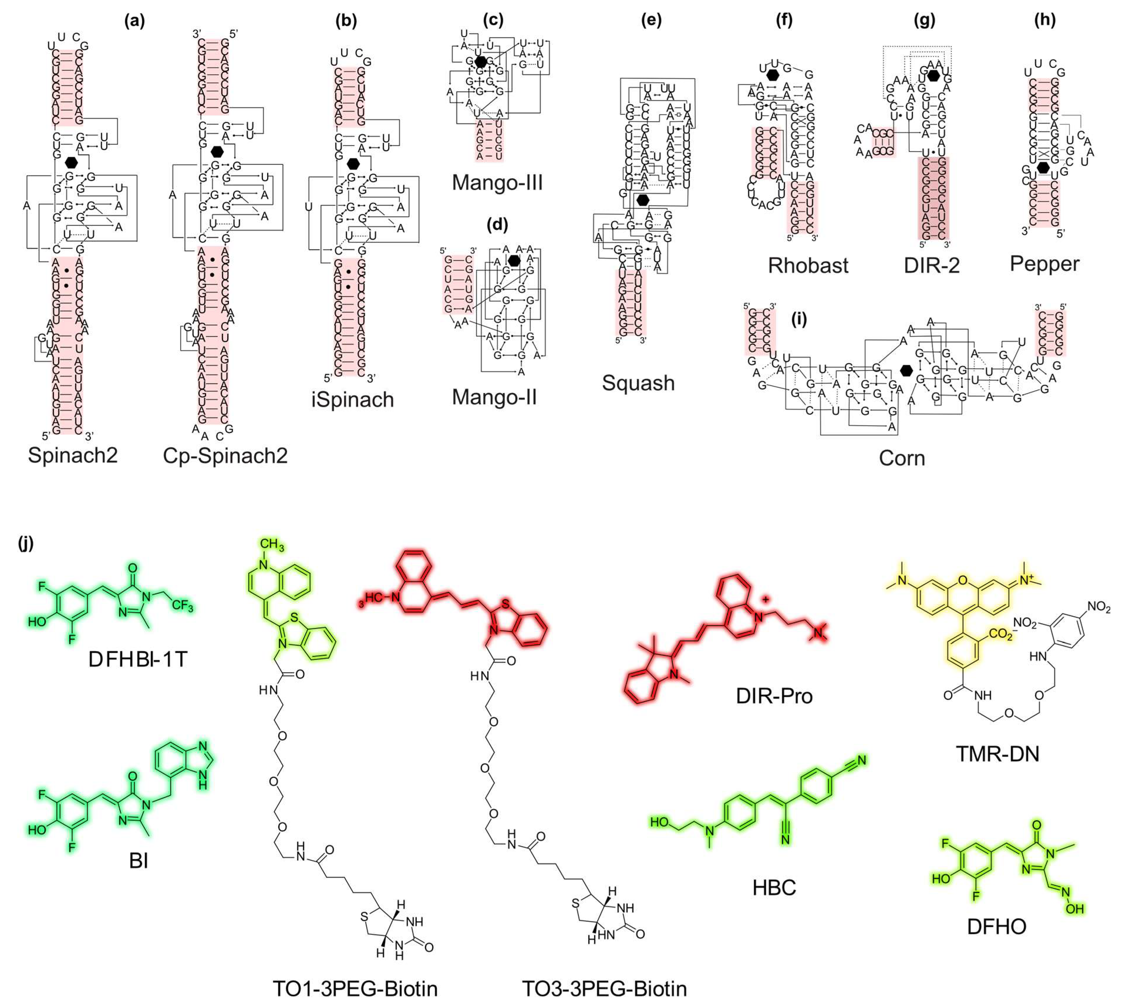
2.1. Light-Up Aptamers Discovery and Properties

A wide variety of light-up aptamer/fluorogen pairs has been developed over the past decade with colors spanning the entire visible spectrum (Table 1), some systems displaying great color flexibility [26,27,28]. Their deep description and characterization can be found in several recent excellent reviews on the topic [29,30,31,32,33,34].
Most of these synthetic aptamers were originally isolated using systematic evolution of ligands by exponential enrichment (SELEX) [68,69]. This in vitro selection approach consists of isolating nucleic acids contained in a large library (up to 1016 variants) for their ability to specifically bind their fluorogen immobilized on a solid phase (Figure 2a). These libraries are frequently made of a 30-40-nucleotide-long randomized stretch interrupted by a stem-loop structure, as the presence of such an internal prefolded element was found to drastically increase the success rate of SELEX targeting small molecules [70]. Whereas this approach is highly effective for selecting binders, it does not allow any selection pressure to be applied for their capacity to trigger fluorescence emission. Therefore, following the SELEX step, enriched libraries are usually cloned and some constructs are functionally tested either in vitro or upon expression in living cells (mostly in bacteria) later tested in microtiter plates [28,49,60,63]. Upon incubation with the target fluorogen, cells displaying the best fluorescence signal can be analyzed and sorted by FACS [36,38,67]. Alternatively, genes contained in the libraries can be isolated at the surface of beads [70], of microparticles (Figure 2b) [71,72], or within water-in-oil microfluidic droplets (Figure 2c) [47,51,56] where they are amplified and expressed. In each case, fluorogen addition turns the particle (compartment) fluorescent according to the functionality of the encoded aptamer, making it possible to sort the particles of interest by FACS or the droplets using a fluorescence-activated droplet sorter [73]. Finally, the overall selection/screening process can be characterized a posteriori using high-throughput sequencing and variants of interest identified through bioinformatics [74].
The crystal structure of most light-up aptamer/fluorogen pairs has now been solved (Table 1), providing extremely valuable information on the recognition mechanism between the aptamer and its cognate fluorogen, but this may also guide the development of an improved version of the fluorogen [39]. The 17 structures currently available (Table 1) highlight some general features shared by all or part of these aptamers. In every case, the fluorogen binding pocket is made by an extended planar platform made of a G-quadruplex (Spinach, Broccoli, Mango, Corn, Beetroot, and Peach), a base quadruple (RhoBAST), or a base triple (DIR-2 and Pepper). Most of the time (except for Mango, Mango-II, and Peach), the fluorogen is accommodated either in a binding pocket or at the interface of two aptamers (Corn and Beetroot) that maintains the fluorogen in a planar conformation while restraining solvent accessibility. The fluorogen-binding pocket either lies at the apex of the structure (Mangos, DIR-2, and RhoBAST) or sits in its middle. Moreover, at least one (Mangos, Corn, and Beetroot) and up to five paired regions (o-Coral) surround this binding site and maintain its structure. Interestingly, some of those stems represent a possible entry point in view of future engineering (Figure 3). To date, 40% of these light-up aptamers have been successfully converted into biosensors of small molecules (see Section 4).

2.2. Choice and Development of Sensing Aptamers

The sensing module is the second key module of an FRB. Whereas, in rare cases, introducing simple mismatches in a helix is sufficient to sense a metal ion (e.g., C–C mismatch signals the presence of silver by the formation of a C-Ag-C metallobase pair) [76,77], the specific detection of a small molecule usually requires a whole module. Moreover, to yield an efficient biosensor, target-binding should trigger a structural change in the sensing aptamer that will be transmitted to a functionally connected light-up aptamer module. A natural target-specific aptamer can be either directly used or reprogrammed toward a new specificity. Finally, structure-switching aptamers can be developed de novo using dedicated methods.

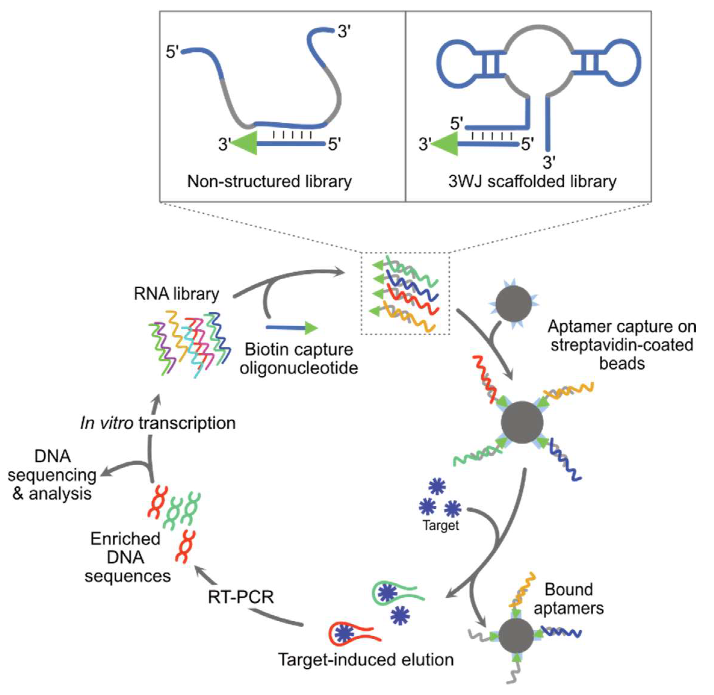
2.2.1. Repurposing Natural Structure-Switching Aptamers

Structure-switching aptamers are naturally found embedded within riboswitches, a class of cis-regulatory RNA mostly found in the 5′ untranslated region of bacterial genes and operons and that control the expression of downstream genes at a transcriptional and/or at translational level (see [78] for a recent comprehensive review on the topic). Aptamers of natural origin arise in intracellular mediums with particular physicochemical conditions, a crowding induced by the presence of numerous macromolecules and a large number of competing small molecules. Consequently, these aptamers are well adapted to work in cellular environments, with good folding efficiencies, high specificities, and physiologically relevant affinities for their target.
The crystal structure of many of these aptamers has been solved, helping to guide their engineering. Yet, of the over 55 classes of riboswitch currently known (responding to ions, vitamins, purines, and nucleotide-derived cofactors), only ~20% entered in the design of an FRB, among which those responding to cyclic dinucleotides and S-adenosyl-methionine (SAM) have been particularly used. Therefore, riboswitches represent a rich reservoir of aptamers for developing new FRBs, especially when considering that the ligands of ~100 orphan riboswitches remain to be identified [78,79,80]. Despite this impressive source of natural aptamers, they are limited to the recognition of molecules that can be found in cells, which represents a serious bottleneck and justifies the efforts undertaken to identify synthetic structure-switching aptamers.

2.2.2. Reprogramming the Specificity of Natural Structure-Switching Aptamers

Riboswitches have evolved over a long period of time to acquire their structure-switching properties. Therefore, they are appealing scaffolds for the isolation of structure-switching aptamers with new specificity. This can be achieved by introducing mutations into the ligand-binding region while preserving the overall aptamer structure. In the simplest case, the specificity of an aptamer whose crystal structure is known can be reprogrammed by the rational mutation of residues essential for ligand recognition. One of the best-known examples is the reprogramming of the xpt-pbuX aptamer [81]. Indeed, while the specificity of this aptamer was driven by the formation of a Watson Crick base pair between the target guanine and the C74 nucleobase, this aptamer was reprogrammed toward adenine only by mutating this position to U74 [82]. The in-depth study of the structure of the different classes of riboswitches and their mode of interaction with their target also led to the development of a biosensor based on the GEMM-II riboswitch scaffold capable of recognizing 2′,3′-cGAMP [83]. However, the possibility of performing such rational design remains marginal and deeper reshaping of the ligand-binding site is often required.
Porter and colleagues nicely demonstrated that the three-way junction (3WJ) motif found in several natural RNAs can provide a robust structural scaffold for the search of synthetic structure-switching aptamer [84]. The authors first randomized every unpaired residue of the core junction as well as the first proximal base pair of each stem of two riboswitches (guanine-specific xpt-pbuX aptamer of Bacillus subtilis and cyclic di-GMP-specific Vc2 riboswitch of Vibrio cholerae) and one ribozyme (Schistosoma mansoni hammerhead ribozyme) prior to subjecting the libraries to several rounds of SELEX. Applying moderate selection stringency and using a high specificity reverse transcriptase enabled the identification of structure-switching aptamers specific to 5-hydroxy-L-tryptophan or to 3,4-dihydroxy-L-phenylalanine. Interestingly, although aptamers isolated from each library differ in terms of sequence and framework, they all functionally converge in terms of affinity and specificity, and they preserved the overall structural organization of their parental molecule.
The concept of reprogramming robust riboswitch-derived scaffolds was deeper explored by the Jaffrey lab using the adenine-specific aptamer module from the add riboswitch of Vibrio vulnificus [66]. Not only was the ligand-binding pocket (the residues of the junctions as well as the first proximal base pairs of each stem) of this 3WJ-containing aptamer randomized but the use of a “Sprouts & Clips” solid-phase synthesis strategy enabled its size to be varied. Upon several rounds of SELEX and FACS sorting, the authors were able to isolate Squash, a light-up aptamer characterized by very high folding efficiency, likely inherited from the parental scaffold, allowing it to outperform its functional counterparts (i.e., Broccoli and Corn isolated by conventional SELEX using a pre-folded but unscaffolded library).

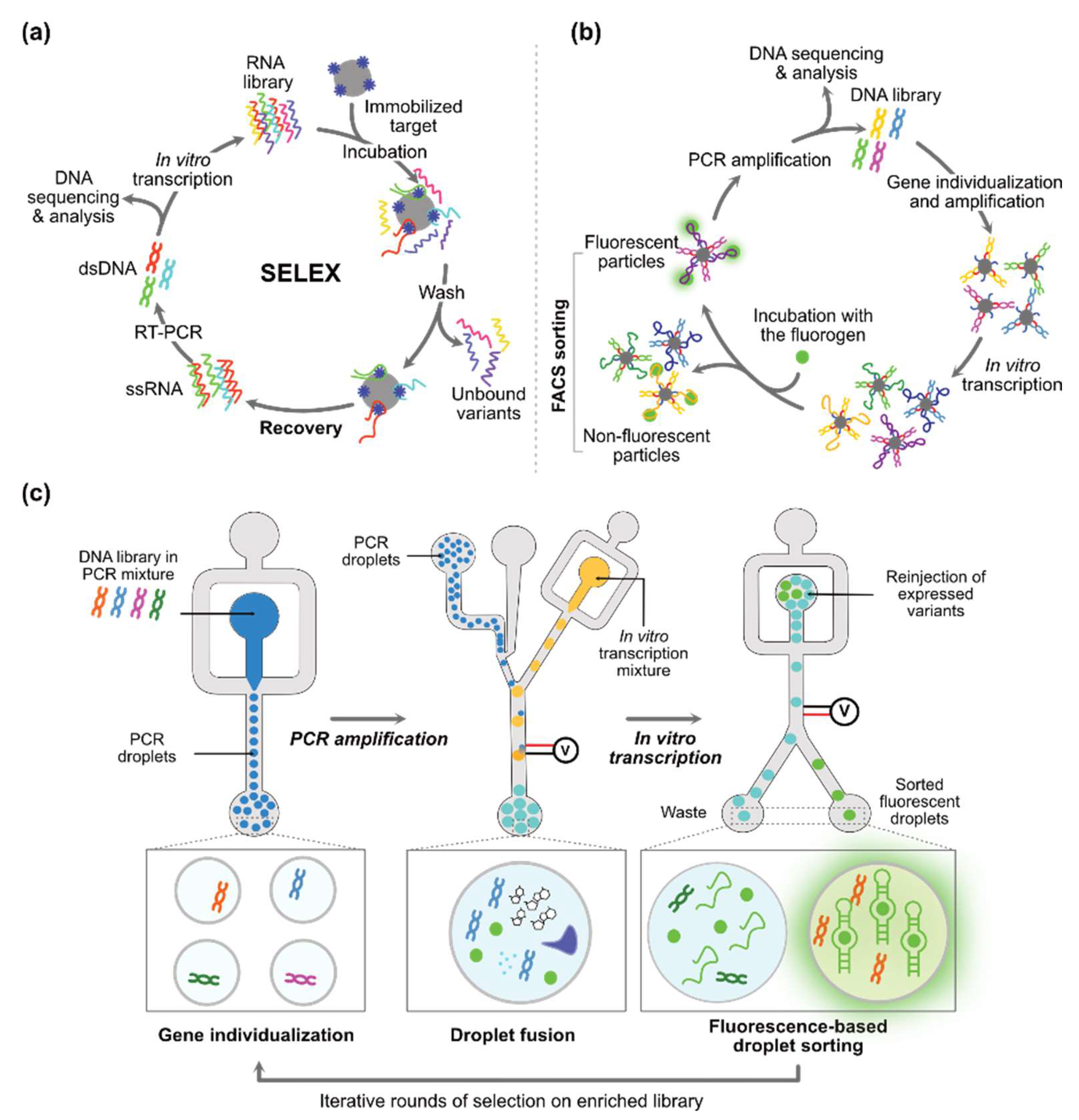
2.2.3. De Novo Discovery of Synthetic Structure-Switching Aptamers

Most of the aptamers identified by classical SELEX are not expected to have an efficient structure-switching capacity, despite this being a critical feature for the development of an FRB. However, the isolation of such molecules could be favored using Capture-SELEX. In this approach, the oligonucleotide library is first immobilized on a solid phase (beads) via a capture oligonucleotide (Figure 4) [85]. After stringent washing steps, the aptamers are eluted by addition of the free target. Advantageously, the use of the target in its native form is expected to both simplify the overall process and increase the likelihood of isolating an efficient aptamer. While Capture-SELEX is mainly used to identify DNA aptamers, the Suess group adapted the procedure to RNA chemistry to successfully isolate sensors of rATP [86], paromomycin [87], tobramycin [88], and levofloxacin [89]. Although functional, the conversion of these aptamers into FRBs may, however, be challenged by the internal location of the docking sequence (Figure 4), i.e., where the impact of structure switching is expected to be the highest. This limitation was recently partly overcome by the Mayer lab who introduced a modular approach in which aptamers isolated from Capture-SELEX can be directly coupled with a light-up module, allowing a TPP-specific FRB to be identified [90]. Yet, this biosensor displays a limited response amplitude, suggesting a suboptimal folding.
An appealing strategy for the de novo discovery of efficient structure-switching aptamers exploits a variant of Capture-SELEX in which a 3WJ-scaffolded mutant library is docked at the level of a P1 stem that can directly serve as an entry point for future engineering (Figure 4) [91]. Mutant libraries of the guanine-specific aptamer xpt-pbux from Bacillus subtilis allowed the identification of new quinine-, caffeine-, and guanine-specific aptamers functional both in vitro and in vivo, likely thanks to their 3WJ scaffold.

3. Functional Connection of the Sensing with the Reporting Modules

Beside the individual properties of the sensing and the reporting modules, the performance of a biosensor also strongly relies on the quality of their functional connection. Indeed, an optimal FRB biosensor of small molecules should exhibit minimal fluorescence in the absence of the target molecule, while emitting the highest possible fluorescence in its presence. Sensing and reporting modules can first be functionally coupled by protein-based molecular machineries [92,93]. For instance, the sensing aptamer can be used in its natural context (i.e., embedded within a riboswitch) to control the transcription of a DNA fragment templating a reporting light-up aptamer [94,95]. This strategy comes with some signal amplification (several light-up aptamers can be produced from a single template). A much higher amplification was achieved in the SPRINT technology [96], which combines the use of riboswitch-mediated transcription control and Cas13a collateral cleaving activity, reported using dually labelled substrate RNA oligonucleotides. To the best of our knowledge, this detection strategy has never been applied to sense small molecules using light-up aptamers as reporters. However, Cas-based methodologies were developed to report on the presence of target RNA [97,98] or even DNA [99] by the synthesis [98,99] or degradation of a light-up aptamer [97]. Despite the substantial gain of sensitivity offered by these protein-mediated approaches, they cannot easily be implemented within living cells and they do not allow fluctuation of small molecule content to be monitored in a dynamic manner throughout an experiment. Nevertheless, both limitations can be overcome using FRB at the cost of a lower sensitivity. Upon target ligand binding to an FRB, the allosteric control event can be exerted in two manners: (i) a direct allosteric control mediated by the stabilization of a dynamic stem or (ii) a structural reshaping through a strand displacement mechanism (Figure 5).

3.1. Direct Allosteric Control

A-type helix (also called paired region, or P in short) is the most common, simple, and predictable structural RNA motif, making it an appealing entry point for engineering purposes. Consequently, most FRBs were obtained by identifying a helix on each module to connect (one near the ligand-binding site and the second near the fluorogen-binding site; see possible entry points of light-up aptamers on Figure 3) and merging them in a single stem (Figure 5a). Although candidate helices can be chosen from 2D structural models, the knowledge of the crystal structure of a module greatly facilitates the task. Moreover, further flexibility in the choice of the entry points can be obtained by preparing circularly permuted mutants (i.e., by connecting the 5′ and 3′ ends of the RNA by a tetraloop prior to generating new 5′ and 3′ ends by cleaving a closing loop) [75]. The length (typically one to four base pairs) and sequence of this connecting stem, also known as transducer or communication module (CM), is then adjusted to transiently destabilize the fluorogen-binding pocket (Figure 5a). Therefore, in the ligand-free state, the fluorogen-binding site is expected to be destabilized, which prevents interaction with the fluorogen and keeps the FRB in a dark state. However, the presence of the target ligand induces a structural change in the sensing module that stabilizes the communication module and restores the fluorogen-binding capacity of the reporting module, ultimately leading to fluorescence emission. Overall, this strategy yields FRBs displaying a good dynamic range and a rapid and reversible response enabling dynamic monitoring of target ligand concentration, many of them having been functionally validated in living cells (see Section 4).
Most of the time, the CM is designed in a trial-and-error manner where a few sequences are designed considering thermodynamic criteria prior to being individually tested, with the best-performing one being selected. Nevertheless, the required dynamic behavior remains difficult to predict in silico and these modules are often sub-optimal. As an alternative, a library of randomized communication modules can be prepared and the best candidates are isolated by Capture-SELEX [85] or particle display [72]. Our group also introduced a microfluidic-assisted pipeline in which every variant of the library is individually amplified, expressed, and evaluated in water-in-oil droplets through positive and negative rounds of screening (Figure 5b). Subsequently, the whole process is monitored by high-throughput sequencing and bioinformatics [100]. Such deep analyses of large sequence sets enable the rapid isolation of optimal communication modules, even when only a very low number of solutions exist in the library [101].

3.2. Strand Displacement-Mediated Control

FRBs can also be obtained by replacing the regulation platform of a natural riboswitch by a light-up aptamer module [102,103]. Then, sequence variations are introduced into the sensing and/or the light-up modules to generate a transducer sequence in the former that invades and destabilizes the latter (Figure 5c). The binding of the target ligand to the sensing module triggers the displacement of the invading strand and its relocation within the sensing aptamer module, ultimately leading to the restoration of a functional light-up module and fluorescence emission. Using a similar concept, the Mayer lab introduced a strategy in which aptamers, isolated by SELEX [104] or Capture-SELEX [90], are connected to a DFHBI-binding module (Baby-Spinach or Broccoli) via a transducing invader strand that disrupts the folding of the light-up aptamer. Several of these riboswitch-like FRBs have been tested and found to work in living bacteria or mammalian cells to report on metabolite concentrations (see Section 4). Although not demonstrated in living cells, structural reshaping of the light-up aptamer module can also be mediated in trans by the binding of a trigger nucleic acid (e.g., via the formation of a kissing complex) having its annealing capacity under the allosteric control of a small molecule [105].

3.3. Mixed Control

Interestingly, both types of control (direct allosteric and strand displacement-mediated) can be elegantly combined by the addition of a ribozyme module [106]. Building on early work by the Breaker lab, which demonstrated the possibility to control self-cleaving ribozymes with small molecules [107], three-party architectures can be designed in which a controlled ribozyme sequesters a key region of the light-up module, maintaining the FRB in a dark state (Figure 5d) [106]. The binding of a target ligand triggers RNA self-cleavage, which, in turn, releases a functional light-up aptamer. Interestingly, the small molecule somehow acts like a catalyst since, upon release from an activated FRB, its interaction with another FRB leads to a new trigger event. This enables a signal amplification that improves the detection limit of the FRB. Even higher sensitivity can be achieved with CHARGE, which combines this strategy with the catalytic hairpin assembly amplification cascade [108]. Despite being highly sensitive, ribozyme-based FRBs suffer from the unidirectionality of their sensing, making them unable to report on the dynamics of the cellular content of the target small molecule.

4. Applications of FRBs

Dozens of FRBs (Table 2) have been developed with applications both in living cells and in the extracellular space. Depending on their final use, FRBs can either be directly used or combined with additional modules endowing them with additional properties.

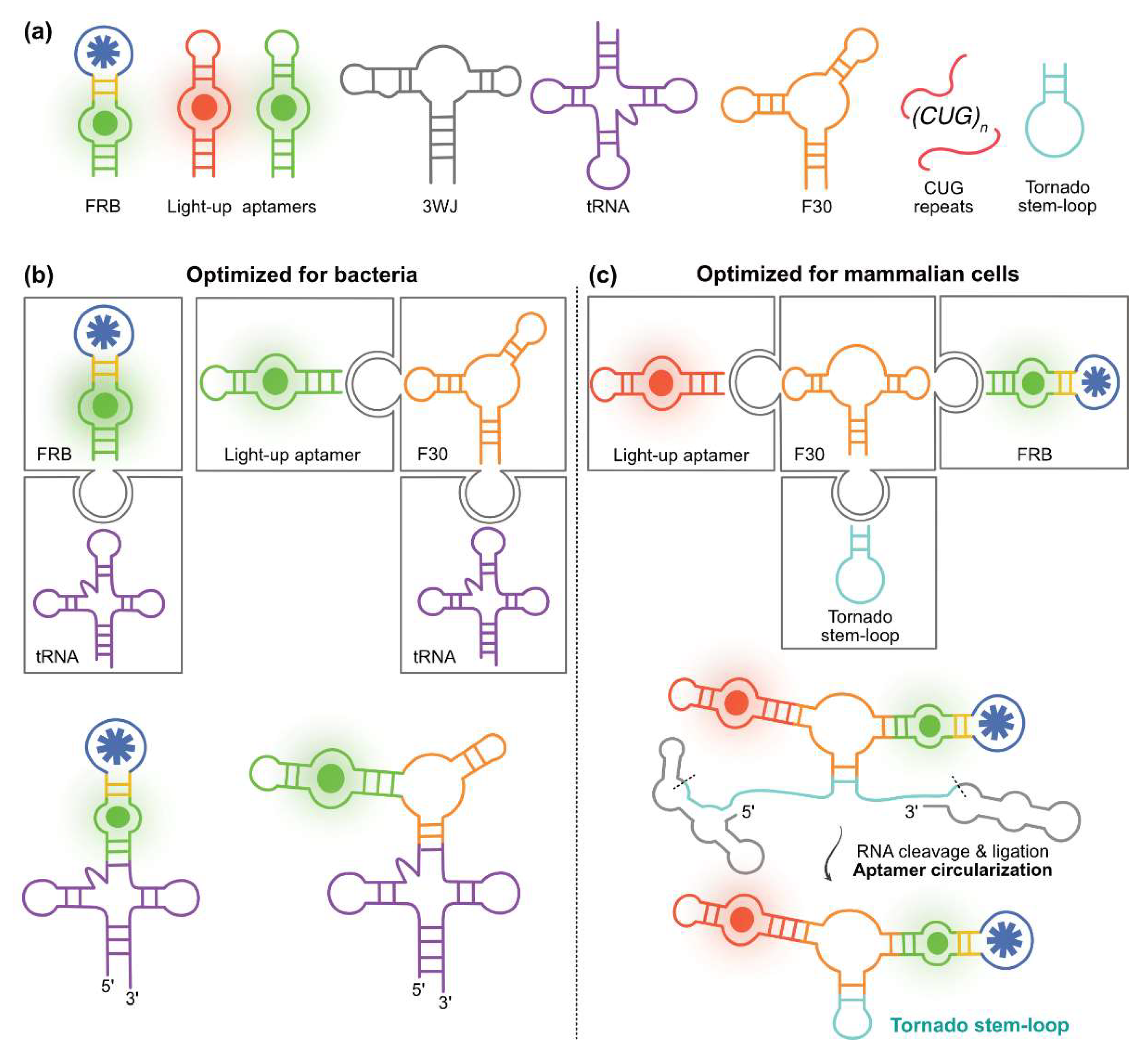
4.1. Exploiting RNA Modularity to Extend FRBs Properties

A strong advantage of nucleic acids is their modular organization allowing biosensors to be tailored to specific applications by adding a dedicated module (Figure 6a). For instance, the efficiency of an FRB expressed in living bacteria can be increased by inserting the construct into the anticodon loop of a tRNA-derived scaffold [133]. Not only can the strong folding capacity of the tRNA module assist that of the inserted FRB but the robust tRNA moiety also shields the construct from bacterial RNases activity. Nevertheless, it was later found that this scaffold only partially preserved light-up aptamers and FRBs integrity in bacteria and that better protection can be obtained with the F29 motif (taken form Phi29 phage genomic RNA) or its mutant derivative F30, the latter being particularly well suited for expression in mammalian cells [134]. While efficient in protecting RNAs expressed in bacteria, the effect of these scaffolds was more limited in mammalian systems where a strong exonuclease activity challenges the constructs. Longer term stability can, however, be achieved by adding the so-called Tornado module, which circularizes the construct by exploiting the tandem activity of two ribozymes surrounding the construct and generating an anticodon-like stem-loop that is closed by the ubiquitous endogenous RtcB ligase (Figure 6c) [135]. Finally, although no use of FRB has yet been described in plant cells, their future development would likely benefit from the use of 3WJ, an engineered version of F30 showing superior scaffolding capacity in plant cells [136]. RNA stability can also be enhanced in living cells by appending specific trinucleotide repeats to induce the formation of RNA condensates through phase separation [137]. Accordingly, the addition of a module made of 31 to 121 CUG repeats to light-up aptamers and FRBs drives the formation of fluorogenic aptamer-based RNA condensates (FLARE and FLARE sensors) in living cells (bacteria and mammalian cells) displaying superior resistance to nuclease, thermostability, photostability, and brightness [138].
Thanks to their three-way junction organizations, the F30 and 3WJ motifs can also be used as an assembly platform to which up to three modules can be connected while remaining structurally insulated (Figure 6b,c). This makes possible the design of ratiometric biosensors containing an internal reference, i.e., an FRB fused to a light-up aptamer emitting fluorescence at a different wavelength, as pioneered by the You lab [139]. Fusing red-emitting DNB-based FRBs (sensing tetracycline or c-di-GMP) to the green-emitting light-up aptamer Broccoli through an F30 scaffold yielded FRBs displaying superior accuracy when expressed in living bacteria. Indeed, these self-calibrating biosensors are not affected by the variability in RNA concentration and cellular distribution that may occur within the cell and between cells. Interestingly, a recent report from the Hammond lab suggests that inserting such constructs within a tRNA scaffold (Figure 6b, construct shown on the left part) could even further increase their stability in various bacterial types [140]. Ratiometric FRBs were also produced and validated in mammalian cells by fusing either a Squash-based FRB or a Pepper-based FRB in the F30 scaffold, respectively, with Broccoli or RhoBAST light-up aptamers. The addition of the Tornado components to these constructs (Figure 6c, construct shown on the right part) enabled their stabilization in living cells and allowed exquisite monitoring of target metabolites in living cells.
RNA (or DNA) origami represents another appealing way of developing FRBs. Indeed, this nanotechnology allows nucleic acid modules to be precisely arranged in space and applied to a wide range of functions [141]. For instance, the Apta-FRET sensor uses donor–acceptor systems based on Mango and Spinach aptamers whose relative orientation and distance can be modulated upon recognition of a target small molecule [131]. Inserting an SAM-sensing aptamer in this construct converted it into a FRET-based SAM reporter of superior accuracy, as FRET probes are also inherently self-calibrating. The use of resonance energy transfer was recently extended to bioluminescence by exchanging the donor light-up aptamer for NanoLuc luciferase [142]. As before, these genetically encodable BRET sensors are self-calibrating, with the additional advantage of internally producing exciting photons through luciferase activity. Moreover, placing the light-up module under the allosteric control of an SAM or of a ppGpp-specific sensing aptamer yielded the corresponding FRB.

4.2. Intracellular In Vivo FRB Applications

Most FRBs have been developed to sense natural small molecules (Table 2) and monitor the effect of mutations or drug treatment on their intracellular concentration, often with single-cell resolution. The choice of the target was mainly driven by the availability of sensing aptamers either taken from natural riboswitches (e.g., TPP, SAM, ppGpp, and c-di-GMP) or less frequently made of a synthetic one (e.g., tetracycline, theophylline, and 1,6-fructose bisphosphate). Whereas many studies consisted of proof-of-concept experiments (i.e., demonstrating the functionality of the FRB in the complex cellular environment), some reports went a step further and shed more light on FRB potential. Among them, cyclic dinucleotide-specific and SAM-specific FRBs were likely the most exploited (Table 2).
Cyclic dinucleotides are intracellular signaling molecules ubiquitous in all domains of life. Produced by bacteria in response to environmental changes, their synthesis may lead to gene expression reprogramming both in Gram-positive and Gram-negative bacteria [116]. These metabolites are involved in stress responses (especially surface sensing) and biofilm production. Therefore, monitoring their concentration in various cell types and conditions relevant to microbiota (i.e., in aerobic and anaerobic conditions) is important to properly understand biological mechanisms at work [118]. In this context, FRBs are well suited since natural riboswitches responding to different cyclic dinucleotide molecules have been identified and light-up aptamers are not sensitive to oxygen level (conversely, to fluorescent proteins that require oxygen to maturate their fluorophore). The Hammond lab has been particularly active in this field [143] by developing several cyclic dinucleotide-specific FRBs allowing sensing of these metabolites in vitro but also in vivo by live-cell imaging [116] and flow cytometry [118]. After introducing c-di-AMP [115] and c-di-GMP-specific FRBs [114,118], the same group prepared an FRB containing the sensing aptamer of a bioinformatically predicted GEMM-I riboswitch and identified 3′-3′-cGAMP as its natural ligand [113,144]. They also rationally reprogrammed the specificity of this FRB towards 2′-3′-cGAMP [83]. These FRBs were next used to identify Hypr GGDEF and the phosphodiesterase HD GYP as the enzymes, respectively, responsible for 3′,3′-cGAMP synthesis [144] and degradation [145]. In addition to its use in bacteria, the 2′-3′-cGAMP-specific FRB was also applied in mammalian cell lysates to characterize the STING (interferon response stimulator) pathway initiated by the cyclic GMP-AMP synthase (cGAS) activity upon binding dsDNA present in the cytosol [83].
S-adenosylmethionine (SAM) is an essential metabolite acting as a universal methyl donor group. As such, it is the cofactor of methyl transferases, a class of enzymes central to epigenetic regulation and associated disorders since they catalyze the methylation of DNA [146], RNA [147], and proteins [148], especially histones. Therefore, monitoring SAM metabolism is of prime importance to properly understand epigenetics and assist the development of drugs targeting related pathways. The design of SAM-specific FRB was greatly eased by the identification of six classes of SAM-specific riboswitches throughout the living kingdoms, the crystal structure of a representative of each class being available [78,149]. Consequently, many SAM-specific FRBs have been developed, most of which are intensiometric (Table 2). Yet, most of these sensors were made of a light-up module (e.g., Broccoli and Corn) comprising a G-quartet core, the stability of which has been questioned in mammalian cells [150]. Although this first generation of SAM-specific FRBs provided a rough evaluation of SAM levels in bacterial and mammalian cells, it could monitor the fluctuation of SAM concentrations in response to a treatment by cycloleucine (an inhibitor of SAM biosynthesis). A significant gain in measurement accuracy was then achieved with the development of ratiometric SAM sensors. Indeed, the use of a SAM-Squash/Broccoli ratiometric sensor allowed precise monitoring of the impact of SAM biosynthesis inhibition but also the impact of culturing conditions on this metabolism with a resolution good enough to discriminate different types of cell response within the same population [66]. Similarly, a SAM-Pepper/RhoBAST ratiometric sensor, an FRB only made of G4-free aptamers (Figure 7a), could be used not only to precisely evaluate the effect of both cycloleucine and SAHA (a drug stimulating SAM synthesis) with single-cell resolution but also to precisely determine the IC50 value of a new drug inhibiting SAM biosynthesis [76]. Such molecular imaging devices could therefore be used in future screening campaigns aimed at discovering new compounds that modulate cell content in SAM to identify drugs for the treatment of epigenetic-related diseases. Moreover, these FRBs could also find application in diagnostics and disease progression monitoring (see below).

4.3. Using FRB as Extracellular In Vitro Reporters

Whereas most FRBs have been used to monitor intracellular concentrations of small molecules, there are also many other applications for these molecules in the extracellular space. The ease of producing FRBs in large scale by in vitro transcription stimulated the set-up of several in vitro FBR-based assays to specifically detect target small molecules.
By coupling an S-adenosyl homocysteine (SAH)-specific sensing aptamer to a circularly permuted version of the light-up aptamer Spinach2, the Hammond lab devised a fluorescent high-throughput compatible assay for enzyme activities involved in SAH metabolism [126]. This FRB enabled, for instance, the characterization of inhibitors of methyl transferases. This strategy therefore holds great promise for the discovery of new drugs targeting SAM/SAH metabolism, especially for therapies targeting epigenetic-related diseases. Moreover, together with the SAM-specific FRBs described above, this SAH-specific FRB could find application in diagnostics and disease progression monitoring, as variations in the concentration of both metabolites in patient fluids and tissues have been associated with a wide range of pathologies [151].
FRB were also developed and used to sense metabolites secreted by yeast [123]. In RNA aptamer in droplets (RAPID) technology, libraries of mutant yeast are encapsulated in microfluidic droplets together with a Spinach-derived FRB specific to the target secreted molecule (i.e., tyrosine, tryptophan, or phenylalanine) and DFHBI-1T. Upon incubation, droplets containing the best secreting cells turn the most fluorescent and are specifically sorted from the bulk. Following a similar concept, we recently developed FluorMango (Figure 7b), the first fluoride-specific FRB [101,152]. To do so, we inserted the sequence of the light-up aptamer Mango-III in the P2 helix of the sensor aptamer domain of the crcB riboswitch from Thermotoga petrophila. We then used our µIVC-seq screening pipeline (Figure 2c) to identify a handful of communication modules, one of which conferred an impressive turn-on efficiency to FluorMango. The encapsulation of bacteria with the fluorinated substrate, FluorMango, and the fluorogen TO1-Biotin then allowed the detection of defluorinase activity from live bacteria with single-cell resolution.
These different studies illustrate the broad application scope of FRB for the detection of small molecules. Whereas incremental technical progress has improved the performance and stability of FRB in living cells, there is still room for improvement for extracellular applications, especially in complex media.

5. Exploring Alternative Chemistries

The great ligand selectivity of FRBs makes them highly valuable tools for small molecule detection out of the cell, with potential applications in diagnostics, microorganism screening, and enzyme and drug discovery. Yet, exploiting the full potential of these biosensors requires them to resist adverse conditions met in the medium. An important limitation of FRBs comes from the inherent instability of their RNA backbone prone to 2′OH-mediated cleavage mainly through nuclease activities. Biosensors can be protected by confining them within lipid particles permeable to the target small molecule but preventing nuclease entry and unwanted release of the biosensors from the vesicles [153]. Although, so far, not tested with FRBs, this strategy may be restricted to the detection of target molecules that are small and hydrophobic enough to cross the membrane. Nuclease activity can also be inactivated through protease treatment. For instance, detection of bacterial fluoride release requires FluorMango-based assay to be performed in the presence of proteinase K [101]. RNA can also be stabilized by adapting the composition of the reaction mixture. Ionic liquid like choline dihydrogen phosphate was shown to preserve malachite-green-specific aptamer light-up function while protecting it from nuclease-mediated degradation. In addition, other organic solvents were shown to protect RNA from degradation [154,155]. Yet, these different treatments may likely interfere with the assay or downstream steps, making more appealing the development of FRBs made of nuclease-resistant chemistry.
The use of DNA-based biosensors represents a first attractive solution, as these molecules are cheap to produce in large quantities by solid-phase chemical synthesis, they are less susceptible to enzymatic and hydrolysis degradation than RNA, and SELEX procedures are well established to identify DNA aptamers. Whereas a handful of light-up DNA aptamers have been described (Table 3), only a very few were further engineered. To our knowledge, the dapoxyl-activating aptamer DAP-10-42 is the only light-up DNA [156] that was engineered into a fluorogenic serotonin-specific biosensor (SERblas) and used as a point-of-care device to detect serotonin in complex media [157]. The light-up aptamer Lettuce was selected for its capacity to activate DFHBI-1T fluorescence emission through what could be the same mechanism as its RNA counterparts Spinach and Broccoli [158]. Although not converted into a small molecule sensor, a split version of Lettuce was engineered to report on the presence of SARS-CoV2 RNA. However, despite their specificity and increased stability, DNA aptamers hardly match RNA aptamers in terms of structural complexity and flexibility [159], which are crucial for the development of specific and dynamic biosensing of small molecules. Moreover, the lack of natural DNA riboswitch further restricts the source of possible target-specific sensing modules and scaffolds.
A large set of synthetic nucleotide chemistries were developed for therapeutic nucleic acids, but they remain poorly explored in the field of fluorogenic biosensing [160,161,162]. For instance, RNA aptamers can be stabilized by modifying the 2′OH group (e.g., 2′fluoro or 2′-O-methyl groups) of their sugar moiety and converting them into xenobiotic nucleic acids (XNA). This allowed, for instance, the preparation of 2′F-Spinach [163] and 2′F-Broccoli [164] aptamers in which all pyrimidines were exchanged for their 2′-fluoro counterparts, resulting in increased stability to degradation. Yet, as this could be expected, introducing such modifications affected aptamers’ properties. While it was beneficial for the capacity of 2′F-Spinach to report on the presence of Pb2+ [163] (the G-quartet of Spinach has been shown to sense Pb2+ by itself [165]), the same modification introduced in Broccoli led to decreased affinity of the aptamer for K+, enabling a potassium sensor to be developed [164]. Resistance to nuclease can also be obtained using enantiomers, i.e., L-RNAs (Spiegelmers) that cannot be processed by natural nucleases [166], as demonstrated by the conversion of Mango-III aptamer into its L counterpart [167]. The light-up aptamer Spinach was also converted into an L-Spinach-based FRB for guanine detection based on the design of the guanine-Spinach FRB developed in 2012 by the Jaffrey Lab as a proof of concept [168]. Even more impressive modifications, consisting of synthetic nucleobases collectively known as Hachimoji, can be specifically introduced into light-up aptamers [169] and used to improve the folding performance of FRBs [170].
However, not all nucleotide modifications are well tolerated by light-up aptamers and FRBs. For instance, the Jaffrey lab showed that modification of nucleobases can significantly drop the function of Broccoli light-up aptamer [171]. Although this adverse effect was used to design a sensor of demethylase activity, most of the time, the introduction of such modifications compromises the function of light-up aptamers and FRBs. Yet, the possibility of enzymatically incorporating these modifications during aptamer synthesis [161] enables in vitro selection and molecular evolution of modified nucleic acids to be undertaken [172,173] and possibly to be applied to the isolation of new generations of light-up aptamers and FRBs with advanced properties.
Table 3. Light-up aptamers and FRBs of small molecules made of alternative chemistries.
Aptamer ChemistryFluorogenEvaluated as FRB of Small MoleculeTarget Small MoleculeOther TargetRef.
AO-bindingDNAAuramine OYesSerotonin [157]
AptII-Mini 3-4 DNAHoechstNo [174]
BBR4S3DNABerberineNo [175]
CV-30SDNACrystal VioletNo [176]
DAP-10-42DNADapoxylYesATP [156,177]
DIR 2-1DNADIRNo [178]
GuanidineRNA/HachimojiDFHBI-TYesGuanidine [170]
L-Mango-IIIL-RNATO1-BiotinNo MicroRNA-155[167]
L-SpinachL-RNADFHBIYesGuanine [168]
LettuceDNADFHBI-TNo SARS-CoV2 RNA[158]
Spinach-HachimojiDNA/HachimojiDFHBI-TNo [169]
2′F-BroccoliRNA/2′ FluoropyrimidineDFHBI-TYesK+ [164]
2′F-SpinachRNA/2′ FluoropyrimidineDFHBI-TYesPb2+ [163]

6. Conclusions

This article aimed to review the main strategies used to develop fluorogenic RNA-based biosensors (FRBs) by exploiting the great diversity of natural and synthetic aptamers available. The performance of light-up aptamers and their fluorogens has constantly improved over the years and delivered a new generation of reporting systems (i.e., Pepper, RhoBAST, Squash, and Okra) endowed with all the key requirements expected for efficient reporters: (i) high brightness, (ii) high affinity, (iii) absence of G-quartet, (iv) good folding efficiency, and (v) good cell permeability of a nontoxic fluorogen.
Most FRBs used so far exploited natural riboswitch-derived aptamers, since they evolved to display both the expected structure-switching capacity and high ligand selectivity. Only a small fraction of the 55 known riboswitch families currently known have been used so far. Theoretical calculations based on bioinformatics suggest that nearly 20% of the total riboswitch diversity is still left to be discovered [78].
In addition to this natural reservoir of sensing aptamers, the latest developments have identified general scaffolds like three-way junction motifs to be better suited for specificity reprogramming than more simple, diverse but less constrained libraries. Indeed, imposing a structural context prone to structure switching efficiently restricts the sequence space to the region driving the specificity.
Optimal communication modules functionally linking sensing and reporting modules can now be identified using approaches able to evaluate large sequence libraries (>65,000 sequence permutations). Among them, we believe that the µIVC-seq pipeline represents an important advance [179]. Equally important is the modular nature of nucleic acid that supports the design of FRBs and allows additional functionalities (e.g., nuclease resistance in cells, controlled aggregation, and accurate ratiometric quantification) to the constructs, enabling the development of sensitive and accurate genetically encoded biosensors of small molecules.
The first generation of FRB was mainly restricted to in vitro applications and expression in bacteria in which RNA degradation may be significantly inhibited by inserting the construct into a tRNA scaffold. As described in this review, these first constructs opened new perspectives in drug discovery by enabling the design of enzymatic activity (e.g., demethylase and methyl transferase) or riboswitching (e.g., TPP-responsive riboswitch) reporters suited to the search for specific modulators (i.e., inhibitors, agonists, or antagonists) by high-throughput screening of chemical libraries. Moreover, expressing FRBs in bacteria also shed new light on bacteria biology, as exemplified above by the sensing of cyclic dinucleotides [143]. Indeed, the use of FRB enabled a new riboswitch and several enzymes involved in cyclic dinucleotide metabolism to be identified, each representing a potential target for an antibacterial compound, since cyclic dinucleotides are key mediators of bacterial stress adaptation [180]. Sensing these metabolites in eukaryotic cells would also likely help in addressing complex questions such as the evolution of antiviral immunity [181]. More generally, FRB-mediated sensing of small molecules in mammalian cells may also have a deep impact on the understanding on cell metabolism and assist in the discovery of new drugs, as illustrated by the case of SAM-specific FRBs introduced above.
Although, to our knowledge, no FBR as defined in this review has yet been exploited in a multicellular organism, the functionality of many FRBs in cultured cells together with that established for light-up aptamers like Mango in C. elegans [49] and Broccoli in plants [136] supports the feasibility of such application. In such a case, the design of FRBs activating near infrared-emitting fluorogen (e.g., TO3-Biotin, BC, SiR-PEG3-NH2, or DIR-Pro) will likely be instrumental to enable both the efficient excitation of the fluorogen at tissue-penetrating wavelengths and the collection of emitted fluorescence (while limiting the adverse effect of the natural green background autofluorescence of living systems).
Another important future development axis of FRBs will concern their use for extracellular applications, especially in vitro diagnostics, drug screening, and enzyme discovery. For these different applications, the fluorogenic nature of FRBs and their derivative is an important feature by limiting the appearance of false positive signals. Indeed, most DNA probes currently used in fluorescent assays exploit the quenching of fluorescent dyes [182]. Therefore, the possible degradation of the probe by an adverse nuclease activity would lead to the release of the fluorophore and a false positive signal. Conversely, the uncontrolled degradation of the fluorogenic probe keeps it dark, with no signal emitted.
Whereas the first generation of RNA-based biosensors now established the advantages of sensing small molecules using nucleic-acid-based biosensors in terms of substrate selectivity, development time, and production cost, we anticipate that important advances could arise from the use of alternative nucleotide chemistries. Although only a few developments have been attempted in this direction, it makes no doubt that the adaptation of FRBs to alternative chemistries will endow them with exceptional stability, for instance, in human fluids (e.g., urine, blood, and saliva) and extended capacity to detect new targets currently difficult to sense. For instance, the introduction of hydrophobic modifications on nucleobases (like those found on SOMAmers and that enabled the development of aptamers specific to nearly any protein [183]) may increase the potential of FRBs in the detection of hydrophobic compounds. Moreover, adapting FRBs and their derivatives to work in nonconventional solvents may also open up new analytical strategies that are likely to be explored in the near future.

Author Contributions

Conceptualization, M.R., J.K. and C.H.; writing—review and editing, M.R., J.K. and C.H.; visualization, J.K. and C.H.; funding acquisition, M.R. All authors have read and agreed to the published version of the manuscript.

Funding

This research was funded by Agence Nationale de la Recherche (Amplisense project ANR-21-CE42-0019) and Fondation pour la Recherche Médicale (PhD grant funding to CH). Performed within Interdisciplinary Thematic Institute “IMCBio, as part of the ITI 2021–2028 program of the University of Strasbourg, CNRS and Inserm, this work was supported by IdEx Unistra (ANR-10-IDEX-0002) and by SFRI-STRAT’US project and EUR IMCBio (ANR-17-EURE-0023) under the framework of the French Investments for the Future Program. This work was also supported by the “Centre National de la Recherche Scientifique” (CNRS) and the “Université de Strasbourg”, from whom it received support from its Initiative of Excellence (IdEx).

Acknowledgments

We thank Redmond Smyth and Magali Frugier for their help in reviewing the manuscript.

Conflicts of Interest

The authors declare no conflicts of interest.

Abbreviations

2′FY2′ fluorinated pyrimidine
3WJ3-way junction
5-HT5-hydroxytryptamine
5-HTP5-hydroxytryptophan
ADPAdenosine diphosphate
AMPAdenosine monophosphate
AOAuramine O
apta-FRETaptamer-based Förster resonance energy transfer
ATPAdenosine triphosphate
BCBenzopyrylium Coumarin
BFRBlue fluorescent RNA
BRETBioluminescence Resonance Energy Transfer
c-di-GMPcyclic-dimeric guanosine monophosphate
c-AMPcyclic adenosine monophosphate
CblCobalamin
c-diAcyclic di-AMP
cGAMPcyclic GMP-AMP
cGAScyclic GMP-AMP synthase
CHARGECatalytic Hairpin Assembly RNA circuit that is Genetically Encoded
CMCommunication Module
CVCrystal violet
DAPDapoxyl DNA aptamer
DFAMF3,5-difluoro-4-hydroxybenzylidene imidazolinone-2-acrylate methyl
DFHBI3,5-difluoro-4-hydroxybenzylidene imidazolinone
DFHBI1-T3,5-difluoro-4-hydroxybenzylidene-1-trifluoroethyl-imidazolinone
DFHO3,5-difluoro-4-hydroxybenzylidene-imidazolinone-2-oxime
DIRDimethylindole red
DMHBI3,5-dimethoxy-4-hydroxybenzylidene imidazolone
DNADeoxyribonucleic acid
DNBDinitroaniline-binding aptamer
dsDNAdouble stranded DNA
FACSFluorescence-activated Cell Sorting
FLAREFluorogenic Aptamer-based RNA condensates
FMNFlavin mononucleotide
FPFluorescent protein
FRBFluorogenic RNA-based biosensor
FRETFluorescence Resonance Energy Transfer
GEMMGenes for the Environment, for Membranes and for Motility
GFPGreen Fluorescent Protein
GRAPGene-linked RNA aptamer particle
GTPGuanosine 5′-triphosphate
HBC4-((2-hydroxyethyl)(methyl)amino)-benzylidene)-cyanophenylacetonitrile
IC50Half-maximal inhibitory concentration
IVTIn vitro transcription
MGMalachite green
DIR-2Dimethylindole red 2
PCRPolymerase chain reaction
ppGppGuanosine tetraphosphate
RAPIDRNA Aptamer In Droplets
RhoBASTRhodamine Binding Aptamer for Super-resolution Imaging Techniques
RNARibonucleic acid
SAHS-Adenosyl-L-homocysteine
SAHASuberoylanilide hydroxamic acid
SAMS-adenosylmethionine
SARS-CoV-2Severe acute respiratory syndrome coronavirus 2
SELEXSystematic Evolution of Ligands by EXponential enrichment
SerBLASSerotonin-responsive binary light-up aptameric sensor
SiRASilicon Rhodamine-Binding Aptamer
SR-DNSulforhodamine B-dinitroaniline
SRBSulforhodamine B
TMRTetramethylrhodamine
TMR-DNTetramethylrhodamine dinitroaniline
TOThiazole orange
TORNADOTwister-optimized RNA for durable overexpression
TPPThiamine pyrophosphate
tRNAtransfert RNA
µIVCMicrofluidic-assisted in vitro compartmentalization
XNAXeno nucleic acid
xpt-pbuXXanthine phosphoribosyltransferase- xanthine-specific purine permease

References

  1. Huang, C.-W.; Lin, C.; Nguyen, M.K.; Hussain, A.; Bui, X.-T.; Ngo, H.H. A Review of Biosensor for Environmental Monitoring: Principle, Application, and Corresponding Achievement of Sustainable Development Goals. Bioengineered 2023, 14, 58–80. [Google Scholar] [CrossRef] [PubMed]
  2. Meliana, C.; Liu, J.; Show, P.L.; Low, S.S. Biosensor in Smart Food Traceability System for Food Safety and Security. Bioengineered 2024, 15, 2310908. [Google Scholar] [CrossRef]
  3. Visconti, G.; Boccard, J.; Feinberg, M.; Rudaz, S. From Fundamentals in Calibration to Modern Methodologies: A Tutorial for Small Molecules Quantification in Liquid Chromatography–Mass Spectrometry Bioanalysis. Anal. Chim. Acta 2023, 1240, 340711. [Google Scholar] [CrossRef]
  4. Malik, P.; Katyal, V.; Malik, V.; Asatkar, A.; Inwati, G.; Mukherjee, T.K. Nanobiosensors: Concepts and Variations. ISRN Nanomater. 2013, 2013, 1–9. [Google Scholar] [CrossRef]
  5. Mao, Y.; Huang, C.; Zhou, X.; Han, R.; Deng, Y.; Zhou, S. Genetically Encoded Biosensor Engineering for Application in Directed Evolution. J. Microbiol. Biotechnol. 2023, 33, 1257–1267. [Google Scholar] [CrossRef]
  6. Clark, L.C.; Lyons, C. Electrode Systems for Continuous Monitoring in Cardiovascular Surgery. Ann. N. Y. Acad. Sci. 1962, 102, 29–45. [Google Scholar] [CrossRef]
  7. Wang, J. Electrochemical Glucose Biosensors. Chem. Rev. 2008, 108, 814–825. [Google Scholar] [CrossRef]
  8. Koyappayil, A.; Lee, M.-H. Ultrasensitive Materials for Electrochemical Biosensor Labels. Sensors 2020, 21, 89. [Google Scholar] [CrossRef]
  9. Shimomura, O.; Johnson, F.H.; Saiga, Y. Extraction, Purification and Properties of Aequorin, a Bioluminescent Protein from the Luminous Hydromedusan, Aequorea. J. Cell. Comp. Physiol. 1962, 59, 223–239. [Google Scholar] [CrossRef]
  10. Tamura, T.; Hamachi, I. Recent Progress in Design of Protein-Based Fluorescent Biosensors and Their Cellular Applications. ACS Chem. Biol. 2014, 9, 2708–2717. [Google Scholar] [CrossRef]
  11. Nasu, Y.; Shen, Y.; Kramer, L.; Campbell, R.E. Structure- and Mechanism-Guided Design of Single Fluorescent Protein-Based Biosensors. Nat. Chem. Biol. 2021, 17, 509–518. [Google Scholar] [CrossRef] [PubMed]
  12. Ding, N.; Zhou, S.; Deng, Y. Transcription-Factor-Based Biosensor Engineering for Applications in Synthetic Biology. ACS Synth. Biol. 2021, 10, 911–922. [Google Scholar] [CrossRef] [PubMed]
  13. Funk, M.A.; Leitner, J.; Gerner, M.C.; Hammerler, J.M.; Salzer, B.; Lehner, M.; Battin, C.; Gumpelmair, S.; Stiasny, K.; Grabmeier-Pfistershammer, K.; et al. Interrogating Ligand-Receptor Interactions Using Highly Sensitive Cellular Biosensors. Nat. Commun. 2023, 14, 7804. [Google Scholar] [CrossRef] [PubMed]
  14. Reja, S.I.; Minoshima, M.; Hori, Y.; Kikuchi, K. Recent Advancements of Fluorescent Biosensors Using Semisynthetic Probes. Biosens. Bioelectron. 2024, 247, 115862. [Google Scholar] [CrossRef] [PubMed]
  15. Broch, F.; Gautier, A. Illuminating Cellular Biochemistry: Fluorogenic Chemogenetic Biosensors for Biological Imaging. ChemPlusChem 2020, 85, 1487–1497. [Google Scholar] [CrossRef] [PubMed]
  16. Gautier, A. Fluorescence-Activating and Absorption-Shifting Tags for Advanced Imaging and Biosensing. Acc. Chem. Res. 2022, 55, 3125–3135. [Google Scholar] [CrossRef] [PubMed]
  17. Broch, F.; El Hajji, L.; Pietrancosta, N.; Gautier, A. Engineering of Tunable Allosteric-like Fluorogenic Protein Sensors. ACS Sens. 2023, 8, 3933–3942. [Google Scholar] [CrossRef] [PubMed]
  18. Childs-Disney, J.L.; Yang, X.; Gibaut, Q.M.R.; Tong, Y.; Batey, R.T.; Disney, M.D. Targeting RNA Structures with Small Molecules. Nat. Rev. Drug Discov. 2022, 21, 736–762. [Google Scholar] [CrossRef] [PubMed]
  19. Lin, J.; Zhou, D.; Steitz, T.A.; Polikanov, Y.S.; Gagnon, M.G. Ribosome-Targeting Antibiotics: Modes of Action, Mechanisms of Resistance, and Implications for Drug Design. Annu. Rev. Biochem. 2018, 87, 451–478. [Google Scholar] [CrossRef]
  20. Mikaeeli Kangarshahi, B.; Naghib, S.M.; Rabiee, N. DNA/RNA-Based Electrochemical Nanobiosensors for Early Detection of Cancers. Crit. Rev. Clin. Lab. Sci. 2024, 1–23. [Google Scholar] [CrossRef]
  21. Xu, R.; Ouyang, L.; Chen, H.; Zhang, G.; Zhe, J. Recent Advances in Biomolecular Detection Based on Aptamers and Nanoparticles. Biosensors 2023, 13, 474. [Google Scholar] [CrossRef]
  22. Cai, R.; Yin, F.; Zhang, Z.; Tian, Y.; Zhou, N. Functional Chimera Aptamer and Molecular Beacon Based Fluorescent Detection of Staphylococcus Aureus with Strand Displacement-Target Recycling Amplification. Anal. Chim. Acta 2019, 1075, 128–136. [Google Scholar] [CrossRef] [PubMed]
  23. Cheng, L.; Ma, D.; Zhang, J.; Kang, X.; Wu, Y.; Zhao, Y.; Yi, L.; Xi, Z. Development of a Universal RNA Dual-Terminal Labeling Method for Sensing RNA-Ligand Interactions. CCS Chem. 2022, 5, 221–233. [Google Scholar] [CrossRef]
  24. Klöcker, N.; Weissenboeck, F.P.; Rentmeister, A. Covalent Labeling of Nucleic Acids. Chem. Soc. Rev. 2020, 49, 8749–8773. [Google Scholar] [CrossRef] [PubMed]
  25. Wicks, S.L.; Hargrove, A.E. Fluorescent Indicator Displacement Assays to Identify and Characterize Small Molecule Interactions with RNA. Methods 2019, 167, 3–14. [Google Scholar] [CrossRef]
  26. Braselmann, E.; Wierzba, A.J.; Polaski, J.T.; Chromiński, M.; Holmes, Z.E.; Hung, S.-T.; Batan, D.; Wheeler, J.R.; Parker, R.; Jimenez, R.; et al. A Multicolor Riboswitch-Based Platform for Imaging of RNA in Live Mammalian Cells. Nat. Chem. Biol. 2018, 14, 964–971. [Google Scholar] [CrossRef]
  27. Sunbul, M.; Jäschke, A. SRB-2: A Promiscuous Rainbow Aptamer for Live-Cell RNA Imaging. Nucleic Acids Res. 2018, 46, e110. [Google Scholar] [CrossRef]
  28. Chen, X.; Zhang, D.; Su, N.; Bao, B.; Xie, X.; Zuo, F.; Yang, L.; Wang, H.; Jiang, L.; Lin, Q.; et al. Visualizing RNA Dynamics in Live Cells with Bright and Stable Fluorescent RNAs. Nat. Biotechnol. 2019, 37, 1287–1293. [Google Scholar] [CrossRef]
  29. Neubacher, S.; Hennig, S. RNA Structure and Cellular Applications of Fluorescent Light-Up Aptamers. Angew. Chem. Int. Ed. 2019, 58, 1266–1279. [Google Scholar] [CrossRef]
  30. Swetha, P.; Fan, Z.; Wang, F.; Jiang, J.-H. Genetically Encoded Light-up RNA Aptamers and Their Applications for Imaging and Biosensing. J. Mater. Chem. B 2020, 8, 3382–3392. [Google Scholar] [CrossRef]
  31. Braselmann, E.; Rathbun, C.; Richards, E.M.; Palmer, A.E. Illuminating RNA Biology: Tools for Imaging RNA in Live Mammalian Cells. Cell Chem. Biol. 2020, 27, 891–903. [Google Scholar] [CrossRef] [PubMed]
  32. Ryckelynck, M. Development and Applications of Fluorogen/Light-Up RNA Aptamer Pairs for RNA Detection and More. In RNA Tagging; Heinlein, M., Ed.; Methods in Molecular Biology; Springer US: New York, NY, USA, 2020; Volume 2166, pp. 73–102. ISBN 978-1-07-160711-4. [Google Scholar]
  33. Lu, X.; Kong, K.Y.S.; Unrau, P.J. Harmonizing the Growing Fluorogenic RNA Aptamer Toolbox for RNA Detection and Imaging. Chem. Soc. Rev. 2023, 52, 4071–4098. [Google Scholar] [CrossRef] [PubMed]
  34. Zhou, H.; Zhang, S. Recent Development of Fluorescent Light-Up RNA Aptamers. Crit. Rev. Anal. Chem. 2022, 52, 1644–1661. [Google Scholar] [CrossRef] [PubMed]
  35. Zhang, J.; Wang, L.; Jäschke, A.; Sunbul, M. A Color-Shifting Near-Infrared Fluorescent Aptamer–Fluorophore Module for Live-Cell RNA Imaging. Angew. Chem. Int. Ed. 2021, 60, 21441–21448. [Google Scholar] [CrossRef] [PubMed]
  36. Wu, J.; Svensen, N.; Song, W.; Kim, H.; Zhang, S.; Li, X.; Jaffrey, S.R. Self-Assembly of Intracellular Multivalent RNA Complexes Using Dimeric Corn and Beetroot Aptamers. J. Am. Chem. Soc. 2022, 144, 5471–5477. [Google Scholar] [CrossRef] [PubMed]
  37. Passalacqua, L.F.M.; Starich, M.R.; Link, K.A.; Wu, J.; Knutson, J.R.; Tjandra, N.; Jaffrey, S.R.; Ferré-D’Amaré, A.R. Co-Crystal Structures of the Fluorogenic Aptamer Beetroot Show That Close Homology May Not Predict Similar RNA Architecture. Nat. Commun. 2023, 14, 2969. [Google Scholar] [CrossRef]
  38. Filonov, G.S.; Moon, J.D.; Svensen, N.; Jaffrey, S.R. Broccoli: Rapid Selection of an RNA Mimic of Green Fluorescent Protein by Fluorescence-Based Selection and Directed Evolution. J. Am. Chem. Soc. 2014, 136, 16299–16308. [Google Scholar] [CrossRef] [PubMed]
  39. Li, X.; Kim, H.; Litke, J.L.; Wu, J.; Jaffrey, S.R. Fluorophore-Promoted RNA Folding and Photostability Enables Imaging of Single Broccoli-Tagged mRNAs in Live Mammalian Cells. Angew. Chem. Int. Ed. 2020, 59, 4511–4518. [Google Scholar] [CrossRef]
  40. Steinmetzger, C.; Palanisamy, N.; Gore, K.R.; Höbartner, C. A Multicolor Large Stokes Shift Fluorogen-Activating RNA Aptamer with Cationic Chromophores. Chem.–Eur. J. 2019, 25, 1931–1935. [Google Scholar] [CrossRef]
  41. Mieczkowski, M.; Steinmetzger, C.; Bessi, I.; Lenz, A.-K.; Schmiedel, A.; Holzapfel, M.; Lambert, C.; Pena, V.; Höbartner, C. Large Stokes Shift Fluorescence Activation in an RNA Aptamer by Intermolecular Proton Transfer to Guanine. Nat. Commun. 2021, 12, 3549. [Google Scholar] [CrossRef]
  42. Song, W.; Filonov, G.S.; Kim, H.; Hirsch, M.; Li, X.; Moon, J.D.; Jaffrey, S.R. Imaging RNA Polymerase III Transcription Using a Photostable RNA–Fluorophore Complex. Nat. Chem. Biol. 2017, 13, 1187–1194. [Google Scholar] [CrossRef] [PubMed]
  43. Warner, K.D.; Sjekloća, L.; Song, W.; Filonov, G.S.; Jaffrey, S.R.; Ferré-D’Amaré, A.R. A Homodimer Interface without Base Pairs in an RNA Mimic of Red Fluorescent Protein. Nat. Chem. Biol. 2017, 13, 1195–1201. [Google Scholar] [CrossRef] [PubMed]
  44. Tan, X.; Constantin, T.P.; Sloane, K.L.; Waggoner, A.S.; Bruchez, M.P.; Armitage, B.A. Fluoromodules Consisting of a Promiscuous RNA Aptamer and Red or Blue Fluorogenic Cyanine Dyes: Selection, Characterization, and Bioimaging. J. Am. Chem. Soc. 2017, 139, 9001–9009. [Google Scholar] [CrossRef] [PubMed]
  45. Shelke, S.A.; Shao, Y.; Laski, A.; Koirala, D.; Weissman, B.P.; Fuller, J.R.; Tan, X.; Constantin, T.P.; Waggoner, A.S.; Bruchez, M.P.; et al. Structural Basis for Activation of Fluorogenic Dyes by an RNA Aptamer Lacking a G-Quadruplex Motif. Nat. Commun. 2018, 9, 4542. [Google Scholar] [CrossRef] [PubMed]
  46. Arora, A.; Sunbul, M.; Jäschke, A. Dual-Colour Imaging of RNAs Using Quencher- and Fluorophore-Binding Aptamers. Nucleic Acids Res. 2015, 43, e144. [Google Scholar] [CrossRef] [PubMed]
  47. Autour, A.; Westhof, E.; Ryckelynck, M. iSpinach: A Fluorogenic RNA Aptamer Optimized for in Vitro Applications. Nucleic Acids Res. 2016, 44, 2491–2500. [Google Scholar] [CrossRef]
  48. Fernandez-Millan, P.; Autour, A.; Ennifar, E.; Westhof, E.; Ryckelynck, M. Crystal Structure and Fluorescence Properties of the iSpinach Aptamer in Complex with DFHBI. RNA 2017, 23, 1788–1795. [Google Scholar] [CrossRef] [PubMed]
  49. Dolgosheina, E.V.; Jeng, S.C.Y.; Panchapakesan, S.S.S.; Cojocaru, R.; Chen, P.S.K.; Wilson, P.D.; Hawkins, N.; Wiggins, P.A.; Unrau, P.J. RNA Mango Aptamer-Fluorophore: A Bright, High-Affinity Complex for RNA Labeling and Tracking. ACS Chem. Biol. 2014, 9, 2412–2420. [Google Scholar] [CrossRef] [PubMed]
  50. Trachman, R.J.; Demeshkina, N.A.; Lau, M.W.L.; Panchapakesan, S.S.S.; Jeng, S.C.Y.; Unrau, P.J.; Ferré-D’Amaré, A.R. Structural Basis for High-Affinity Fluorophore Binding and Activation by RNA Mango. Nat. Chem. Biol. 2017, 13, 807–813. [Google Scholar] [CrossRef]
  51. Autour, A.; Jeng, S.C.Y.; Cawte, A.D.; Abdolahzadeh, A.; Galli, A.; Panchapakesan, S.S.S.; Rueda, D.; Ryckelynck, M.; Unrau, P.J. Fluorogenic RNA Mango Aptamers for Imaging Small Non-Coding RNAs in Mammalian Cells. Nat. Commun. 2018, 9, 656. [Google Scholar] [CrossRef]
  52. Trachman, R.J.; Abdolahzadeh, A.; Andreoni, A.; Cojocaru, R.; Knutson, J.R.; Ryckelynck, M.; Unrau, P.J.; Ferré-D’Amaré, A.R. Crystal Structures of the Mango-II RNA Aptamer Reveal Heterogeneous Fluorophore Binding and Guide Engineering of Variants with Improved Selectivity and Brightness. Biochemistry 2018, 57, 3544–3548. [Google Scholar] [CrossRef] [PubMed]
  53. Trachman, R.J.; Autour, A.; Jeng, S.C.Y.; Abdolahzadeh, A.; Andreoni, A.; Cojocaru, R.; Garipov, R.; Dolgosheina, E.V.; Knutson, J.R.; Ryckelynck, M.; et al. Structure and Functional Reselection of the Mango-III Fluorogenic RNA Aptamer. Nat. Chem. Biol. 2019, 15, 472–479. [Google Scholar] [CrossRef] [PubMed]
  54. Grate, D.; Wilson, C. Laser-Mediated, Site-Specific Inactivation of RNA Transcripts. Proc. Natl. Acad. Sci. USA 1999, 96, 6131–6136. [Google Scholar] [CrossRef] [PubMed]
  55. Baugh, C.; Grate, D.; Wilson, C. A Crystal Structure of the Malachite Green Aptamer. J. Mol. Biol. 2000, 301, 117–128. [Google Scholar] [CrossRef] [PubMed]
  56. Bouhedda, F.; Fam, K.T.; Collot, M.; Autour, A.; Marzi, S.; Klymchenko, A.; Ryckelynck, M. A Dimerization-Based Fluorogenic Dye-Aptamer Module for RNA Imaging in Live Cells. Nat. Chem. Biol. 2020, 16, 69–76. [Google Scholar] [CrossRef] [PubMed]
  57. Zuo, F.; Jiang, L.; Su, N.; Zhang, Y.; Bao, B.; Wang, L.; Shi, Y.; Yang, H.; Huang, X.; Li, R.; et al. Imaging the Dynamics of Messenger RNA with a Bright and Stable Green Fluorescent RNA. Nat. Chem. Biol. 2024, 1–10. [Google Scholar] [CrossRef] [PubMed]
  58. Kong, K.Y.S.; Jeng, S.C.Y.; Rayyan, B.; Unrau, P.J. RNA Peach and Mango: Orthogonal Two-Color Fluorogenic Aptamers Distinguish Nearly Identical Ligands. RNA 2021, 27, 604–615. [Google Scholar] [CrossRef] [PubMed]
  59. Huang, K.; Chen, X.; Li, C.; Song, Q.; Li, H.; Zhu, L.; Yang, Y.; Ren, A. Structure-Based Investigation of Fluorogenic Pepper Aptamer. Nat. Chem. Biol. 2021, 17, 1289–1295. [Google Scholar] [CrossRef] [PubMed]
  60. Sunbul, M.; Lackner, J.; Martin, A.; Englert, D.; Hacene, B.; Grün, F.; Nienhaus, K.; Nienhaus, G.U.; Jäschke, A. Super-Resolution RNA Imaging Using a Rhodamine-Binding Aptamer with Fast Exchange Kinetics. Nat. Biotechnol. 2021, 39, 686–690. [Google Scholar] [CrossRef]
  61. Zhang, Y.; Xu, Z.; Xiao, Y.; Jiang, H.; Zuo, X.; Li, X.; Fang, X. Structural Mechanisms for Binding and Activation of a Contact-Quenched Fluorophore by RhoBAST. Nat. Commun. 2024, 15, 4206. [Google Scholar] [CrossRef]
  62. Wirth, R.; Gao, P.; Nienhaus, G.U.; Sunbul, M.; Jäschke, A. SiRA: A Silicon Rhodamine-Binding Aptamer for Live-Cell Super-Resolution RNA Imaging. J. Am. Chem. Soc. 2019, 141, 7562–7571. [Google Scholar] [CrossRef] [PubMed]
  63. Paige, J.S.; Wu, K.Y.; Jaffrey, S.R. RNA Mimics of Green Fluorescent Protein. Science 2011, 333, 642–646. [Google Scholar] [CrossRef] [PubMed]
  64. Huang, H.; Suslov, N.B.; Li, N.-S.; Shelke, S.A.; Evans, M.E.; Koldobskaya, Y.; Rice, P.A.; Piccirilli, J.A. A G-Quadruplex–Containing RNA Activates Fluorescence in a GFP-like Fluorophore. Nat. Chem. Biol. 2014, 10, 686–691. [Google Scholar] [CrossRef] [PubMed]
  65. Strack, R.L.; Disney, M.D.; Jaffrey, S.R. A Superfolding Spinach2 Reveals the Dynamic Nature of Trinucleotide Repeat–Containing RNA. Nat. Methods 2013, 10, 1219–1224. [Google Scholar] [CrossRef] [PubMed]
  66. Dey, S.K.; Filonov, G.S.; Olarerin-George, A.O.; Jackson, B.T.; Finley, L.W.S.; Jaffrey, S.R. Repurposing an Adenine Riboswitch into a Fluorogenic Imaging and Sensing Tag. Nat. Chem. Biol. 2022, 18, 180–190. [Google Scholar] [CrossRef] [PubMed]
  67. Truong, L.; Kooshapur, H.; Dey, S.K.; Li, X.; Tjandra, N.; Jaffrey, S.R.; Ferré-D’Amaré, A.R. The Fluorescent Aptamer Squash Extensively Repurposes the Adenine Riboswitch Fold. Nat. Chem. Biol. 2022, 18, 191–198. [Google Scholar] [CrossRef] [PubMed]
  68. Ellington, A.D.; Szostak, J.W. In Vitro Selection of RNA Molecules That Bind Specific Ligands. Nature 1990, 346, 818–822. [Google Scholar] [CrossRef] [PubMed]
  69. Tuerk, C.; Gold, L. Systematic Evolution of Ligands by Exponential Enrichment: RNA Ligands to Bacteriophage T4 DNA Polymerase. Science 1990, 249, 505–510. [Google Scholar] [CrossRef] [PubMed]
  70. Davis, J.H.; Szostak, J.W. Isolation of High-Affinity GTP Aptamers from Partially Structured RNA Libraries. Proc. Natl. Acad. Sci. USA 2002, 99, 11616–11621. [Google Scholar] [CrossRef]
  71. Endoh, T.; Ohyama, T.; Sugimoto, N. RNA-Capturing Microsphere Particles (R-CAMPs) for Optimization of Functional Aptamers. Small 2019, 15, 1805062. [Google Scholar] [CrossRef]
  72. Endoh, T.; Sugimoto, N. Signaling Aptamer Optimization through Selection Using RNA-Capturing Microsphere Particles. Anal. Chem. 2020, 92, 7955–7963. [Google Scholar] [CrossRef] [PubMed]
  73. Bart, J.; Tiggelaar, R.; Yang, M.; Schlautmann, S.; Zuilhof, H.; Gardeniers, H. Room-Temperature Intermediate Layer Bonding for Microfluidic Devices. Lab. Chip 2009, 9, 3481. [Google Scholar] [CrossRef] [PubMed]
  74. Bouhedda, F.; Cubi, R.; Baudrey, S.; Ryckelynck, M. μIVC-Seq: A Method for Ultrahigh-Throughput Development and Functional Characterization of Small RNAs. In Small Non-Coding RNAs; Rederstorff, M., Ed.; Methods in Molecular Biology; Springer US: New York, NY, USA, 2021; Volume 2300, pp. 203–237. ISBN 978-1-07-161385-6. [Google Scholar]
  75. Truong, J.; Hsieh, Y.-F.; Truong, L.; Jia, G.; Hammond, M.C. Designing Fluorescent Biosensors Using Circular Permutations of Riboswitches. Methods 2018, 143, 102–109. [Google Scholar] [CrossRef] [PubMed]
  76. Chen, Z.; Chen, W.; Reheman, Z.; Jiang, H.; Wu, J.; Li, X. Genetically Encoded RNA-Based Sensors with Pepper Fluorogenic Aptamer. Nucleic Acids Res. 2023, 51, 8322–8336. [Google Scholar] [CrossRef] [PubMed]
  77. Yu, Q.; Shi, J.; Mudiyanselage, A.P.K.K.K.; Wu, R.; Zhao, B.; Zhou, M.; You, M. Genetically Encoded RNA-Based Sensors for Intracellular Imaging of Silver Ions. Chem. Commun. 2019, 55, 707–710. [Google Scholar] [CrossRef] [PubMed]
  78. Kavita, K.; Breaker, R.R. Discovering Riboswitches: The Past and the Future. Trends Biochem. Sci. 2023, 48, 119–141. [Google Scholar] [CrossRef] [PubMed]
  79. Greenlee, E.B.; Stav, S.; Atilho, R.M.; Brewer, K.I.; Harris, K.A.; Malkowski, S.N.; Mirihana Arachchilage, G.; Perkins, K.R.; Sherlock, M.E.; Breaker, R.R. Challenges of Ligand Identification for the Second Wave of Orphan Riboswitch Candidates. RNA Biol. 2018, 15, 377–390. [Google Scholar] [CrossRef] [PubMed]
  80. Weinberg, Z.; Lünse, C.E.; Corbino, K.A.; Ames, T.D.; Nelson, J.W.; Roth, A.; Perkins, K.R.; Sherlock, M.E.; Breaker, R.R. Detection of 224 Candidate Structured RNAs by Comparative Analysis of Specific Subsets of Intergenic Regions. Nucleic Acids Res. 2017, 45, 10811–10823. [Google Scholar] [CrossRef] [PubMed]
  81. Porter, E.B.; Marcano-Velázquez, J.G.; Batey, R.T. The Purine Riboswitch as a Model System for Exploring RNA Biology and Chemistry. Biochim. Biophys. Acta BBA-Gene Regul. Mech. 2014, 1839, 919–930. [Google Scholar] [CrossRef]
  82. Mandal, M.; Breaker, R.R. Adenine Riboswitches and Gene Activation by Disruption of a Transcription Terminator. Nat. Struct. Mol. Biol. 2004, 11, 29–35. [Google Scholar] [CrossRef]
  83. Bose, D.; Su, Y.; Marcus, A.; Raulet, D.H.; Hammond, M.C. An RNA-Based Fluorescent Biosensor for High-Throughput Analysis of the cGAS-cGAMP-STING Pathway. Cell Chem. Biol. 2016, 23, 1539–1549. [Google Scholar] [CrossRef] [PubMed]
  84. Porter, E.B.; Polaski, J.T.; Morck, M.M.; Batey, R.T. Recurrent RNA Motifs as Scaffolds for Genetically Encodable Small-Molecule Biosensors. Nat. Chem. Biol. 2017, 13, 295–301. [Google Scholar] [CrossRef] [PubMed]
  85. Lam, S.Y.; Lau, H.L.; Kwok, C.K. Capture-SELEX: Selection Strategy, Aptamer Identification, and Biosensing Application. Biosensors 2022, 12, 1142. [Google Scholar] [CrossRef]
  86. Boussebayle, A.; Groher, F.; Suess, B. RNA-Based Capture-SELEX for the Selection of Small Molecule-Binding Aptamers. Methods 2019, 161, 10–15. [Google Scholar] [CrossRef] [PubMed]
  87. Boussebayle, A.; Torka, D.; Ollivaud, S.; Braun, J.; Bofill-Bosch, C.; Dombrowski, M.; Groher, F.; Hamacher, K.; Suess, B. Next-Level Riboswitch Development—Implementation of Capture-SELEX Facilitates Identification of a New Synthetic Riboswitch. Nucleic Acids Res. 2019, 47, 4883–4895. [Google Scholar] [CrossRef]
  88. Kraus, L.; Duchardt-Ferner, E.; Bräuchle, E.; Fürbacher, S.; Kelvin, D.; Marx, H.; Boussebayle, A.; Maurer, L.-M.; Bofill-Bosch, C.; Wöhnert, J.; et al. Development of a Novel Tobramycin Dependent Riboswitch. Nucleic Acids Res. 2023, 51, 11375–11385. [Google Scholar] [CrossRef] [PubMed]
  89. Kramat, J.; Kraus, L.; Gunawan, V.J.; Smyej, E.; Froehlich, P.; Weber, T.E.; Spiehl, D.; Koeppl, H.; Blaeser, A.; Suess, B. Sensing Levofloxacin with an RNA Aptamer as a Bioreceptor. Biosensors 2024, 14, 56. [Google Scholar] [CrossRef]
  90. Legen, T.; Mayer, G. Modular Approach for Rapid Identification of RNA-Based Sensors. ACS Sens. 2024, 9, 753–758. [Google Scholar] [CrossRef]
  91. Mohsen, M.G.; Midy, M.K.; Balaji, A.; Breaker, R.R. Exploiting Natural Riboswitches for Aptamer Engineering and Validation. Nucleic Acids Res. 2023, 51, 966–981. [Google Scholar] [CrossRef]
  92. Citartan, M. The Dynamicity of Light-up Aptamers in One-Pot in Vitro Diagnostic Assays. Analyst 2021, 147, 10–21. [Google Scholar] [CrossRef]
  93. Li, Y.; Arce, A.; Lucci, T.; Rasmussen, R.A.; Lucks, J.B. Dynamic RNA Synthetic Biology: New Principles, Practices and Potential. RNA Biol. 2023, 20, 817–829. [Google Scholar] [CrossRef] [PubMed]
  94. Jung, J.K.; Alam, K.K.; Verosloff, M.S.; Capdevila, D.A.; Desmau, M.; Clauer, P.R.; Lee, J.W.; Nguyen, P.Q.; Pastén, P.A.; Matiasek, S.J.; et al. Cell-Free Biosensors for Rapid Detection of Water Contaminants. Nat. Biotechnol. 2020, 38, 1451–1459. [Google Scholar] [CrossRef] [PubMed]
  95. Thavarajah, W.; Silverman, A.D.; Verosloff, M.S.; Kelley-Loughnane, N.; Jewett, M.C.; Lucks, J.B. Point-of-Use Detection of Environmental Fluoride via a Cell-Free Riboswitch-Based Biosensor. ACS Synth. Biol. 2020, 9, 10–18. [Google Scholar] [CrossRef] [PubMed]
  96. Iwasaki, R.S.; Batey, R.T. SPRINT: A Cas13a-Based Platform for Detection of Small Molecules. Nucleic Acids Res. 2020, 48, e101. [Google Scholar] [CrossRef] [PubMed]
  97. Zhang, T.; Zhou, W.; Lin, X.; Khan, M.R.; Deng, S.; Zhou, M.; He, G.; Wu, C.; Deng, R.; He, Q. Light-up RNA Aptamer Signaling-CRISPR-Cas13a-Based Mix-and-Read Assays for Profiling Viable Pathogenic Bacteria. Biosens. Bioelectron. 2021, 176, 112906. [Google Scholar] [CrossRef] [PubMed]
  98. Wang, Y.; Xue, T.; Wang, M.; Ledesma-Amaro, R.; Lu, Y.; Hu, X.; Zhang, T.; Yang, M.; Li, Y.; Xiang, J.; et al. CRISPR-Cas13a Cascade-Based Viral RNA Assay for Detecting SARS-CoV-2 and Its Mutations in Clinical Samples. Sens. Actuators B Chem. 2022, 362, 131765. [Google Scholar] [CrossRef] [PubMed]
  99. Song, J.; Song, Y.; Jang, H.; Moon, J.; Kang, H.; Huh, Y.-M.; Son, H.Y.; Rho, H.W.; Park, M.; Lim, E.-K.; et al. Elution-Free DNA Detection Using CRISPR/Cas9-Mediated Light-up Aptamer Transcription: Toward All-in-One DNA Purification and Detection Tube. Biosens. Bioelectron. 2023, 225, 115085. [Google Scholar] [CrossRef] [PubMed]
  100. Autour, A.; Bouhedda, F.; Cubi, R.; Ryckelynck, M. Optimization of Fluorogenic RNA-Based Biosensors Using Droplet-Based Microfluidic Ultrahigh-Throughput Screening. Methods 2019, 161, 46–53. [Google Scholar] [CrossRef] [PubMed]
  101. Husser, C.; Vuilleumier, S.; Ryckelynck, M. FluorMango, an RNA-Based Fluorogenic Biosensor for the Direct and Specific Detection of Fluoride. Small 2023, 19, 2205232. [Google Scholar] [CrossRef]
  102. You, M.; Litke, J.L.; Jaffrey, S.R. Imaging Metabolite Dynamics in Living Cells Using a Spinach-Based Riboswitch. Proc. Natl. Acad. Sci. 2015, 112, E256–E265. [Google Scholar] [CrossRef]
  103. Wang, T.; Simmel, F.C. Switchable Fluorescent Light-Up Aptamers Based on Riboswitch Architectures. Angew. Chem. Int. Ed. 2023, 62, e202302858. [Google Scholar] [CrossRef] [PubMed]
  104. Geraci, I.; Autour, A.; Pietruschka, G.; Shiian, A.; Borisova, M.; Mayer, C.; Ryckelynck, M.; Mayer, G. Fluorogenic RNA-Based Biosensor to Sense the Glycolytic Flux in Mammalian Cells. ACS Chem. Biol. 2022, 17, 1164–1173. [Google Scholar] [CrossRef] [PubMed]
  105. Sett, A.; Zara, L.; Dausse, E.; Toulmé, J.-J. A Malachite Green Light-up Aptasensor for the Detection of Theophylline. Talanta 2021, 232, 122417. [Google Scholar] [CrossRef]
  106. You, M.; Litke, J.L.; Wu, R.; Jaffrey, S.R. Detection of Low-Abundance Metabolites in Live Cells Using an RNA Integrator. Cell Chem. Biol. 2019, 26, 471–481.e3. [Google Scholar] [CrossRef] [PubMed]
  107. Tang, J.; Breaker, R.R. Rational Design of Allosteric Ribozymes. Chem. Biol. 1997, 4, 453–459. [Google Scholar] [CrossRef] [PubMed]
  108. Karunanayake Mudiyanselage, A.P.K.K.; Yu, Q.; Leon-Duque, M.A.; Zhao, B.; Wu, R.; You, M. Genetically Encoded Catalytic Hairpin Assembly for Sensitive RNA Imaging in Live Cells. J. Am. Chem. Soc. 2018, 140, 8739–8745. [Google Scholar] [CrossRef]
  109. Wu, R.; Karunanayake Mudiyanselage, A.P.K.K.; Shafiei, F.; Zhao, B.; Bagheri, Y.; Yu, Q.; McAuliffe, K.; Ren, K.; You, M. Genetically Encoded Ratiometric RNA-Based Sensors for Quantitative Imaging of Small Molecules in Living Cells. Angew. Chem. Int. Ed. 2019, 58, 18271–18275. [Google Scholar] [CrossRef]
  110. Paige, J.S.; Nguyen-Duc, T.; Song, W.; Jaffrey, S.R. Fluorescence Imaging of Cellular Metabolites with RNA. Science 2012, 335, 1194. [Google Scholar] [CrossRef]
  111. Furutani, C.; Shinomiya, K.; Aoyama, Y.; Yamada, K.; Sando, S. Modular Blue Fluorescent RNA Sensors for Label-Free Detection of Target Molecules. Mol. Biosyst. 2010, 6, 1569. [Google Scholar] [CrossRef]
  112. Stojanovic, M.N.; Kolpashchikov, D.M. Modular Aptameric Sensors. J. Am. Chem. Soc. 2004, 126, 9266–9270. [Google Scholar] [CrossRef]
  113. Kellenberger, C.A.; Wilson, S.C.; Hickey, S.F.; Gonzalez, T.L.; Su, Y.; Hallberg, Z.F.; Brewer, T.F.; Iavarone, A.T.; Carlson, H.K.; Hsieh, Y.-F.; et al. GEMM-I Riboswitches from Geobacter Sense the Bacterial Second Messenger Cyclic AMP-GMP. Proc. Natl. Acad. Sci. USA 2015, 112, 5383–5388. [Google Scholar] [CrossRef] [PubMed]
  114. Kellenberger, C.A.; Wilson, S.C.; Sales-Lee, J.; Hammond, M.C. RNA-Based Fluorescent Biosensors for Live Cell Imaging of Second Messengers Cyclic Di-GMP and Cyclic AMP-GMP. J. Am. Chem. Soc. 2013, 135, 4906–4909. [Google Scholar] [CrossRef] [PubMed]
  115. Mukkayyan, N.; Poon, R.; Sander, P.N.; Lai, L.-Y.; Zubair-Nizami, Z.; Hammond, M.C.; Chatterjee, S.S. In Vivo Detection of Cyclic-Di-AMP in Staphylococcus Aureus. ACS Omega 2022, 7, 32749–32753. [Google Scholar] [CrossRef] [PubMed]
  116. Kellenberger, C.A.; Chen, C.; Whiteley, A.T.; Portnoy, D.A.; Hammond, M.C. RNA-Based Fluorescent Biosensors for Live Cell Imaging of Second Messenger Cyclic Di-AMP. J. Am. Chem. Soc. 2015, 137, 6432–6435. [Google Scholar] [CrossRef] [PubMed]
  117. Nakayama, S.; Luo, Y.; Zhou, J.; Dayie, T.K.; Sintim, H.O. Nanomolar Fluorescent Detection of C-Di-GMP Using a Modular Aptamer Strategy. Chem. Commun. 2012, 48, 9059–9061. [Google Scholar] [CrossRef] [PubMed]
  118. Wang, X.C.; Wilson, S.C.; Hammond, M.C. Next-Generation RNA-Based Fluorescent Biosensors Enable Anaerobic Detection of Cyclic Di-GMP. Nucleic Acids Res. 2016, 44, e139. [Google Scholar] [CrossRef]
  119. Ketterer, S.; Gladis, L.; Kozica, A.; Meier, M. Engineering and Characterization of Fluorogenic Glycine Riboswitches. Nucleic Acids Res. 2016, 44, 5983–5992. [Google Scholar] [CrossRef] [PubMed]
  120. Moon, J.D.; Wu, J.; Dey, S.K.; Litke, J.L.; Li, X.; Kim, H.; Jaffrey, S.R. Naturally Occurring Three-Way Junctions Can Be Repurposed as Genetically Encoded RNA-Based Sensors. Cell Chem. Biol. 2021, 28, 1569–1580.e4. [Google Scholar] [CrossRef] [PubMed]
  121. Allchin, E.R.; Rosch, J.C.; Stoneman, A.D.; Kim, H.; Lippmann, E.S. Generalized Strategy for Engineering Mammalian Cell-Compatible RNA-Based Biosensors from Random Sequence Libraries. ACS Sens. 2023, 8, 2079–2086. [Google Scholar] [CrossRef]
  122. Xu, J.; Cotruvo, J.A., Jr. The czcD (NiCo) Riboswitch Responds to Iron(II). Biochemistry 2020, 59, 1508–1516. [Google Scholar] [CrossRef]
  123. Abatemarco, J.; Sarhan, M.F.; Wagner, J.M.; Lin, J.-L.; Liu, L.; Hassouneh, W.; Yuan, S.-F.; Alper, H.S.; Abate, A.R. RNA-Aptamers-in-Droplets (RAPID) High-Throughput Screening for Secretory Phenotypes. Nat. Commun. 2017, 8, 332. [Google Scholar] [CrossRef] [PubMed]
  124. Sun, Z.; Wu, R.; Zhao, B.; Zeinert, R.; Chien, P.; You, M. Live-Cell Imaging of Guanosine Tetra- and Pentaphosphate (p)ppGpp with RNA-Based Fluorescent Sensors. Angew. Chem. Int. Ed. Engl. 2021, 60, 24070–24074. [Google Scholar] [CrossRef] [PubMed]
  125. Shafiei, F.; McAuliffe, K.; Bagheri, Y.; Sun, Z.; Yu, Q.; Wu, R.; You, M. Paper-Based Fluorogenic RNA Aptamer Sensors for Label-Free Detection of Small Molecules. Anal. Methods 2020, 12, 2674–2681. [Google Scholar] [CrossRef] [PubMed]
  126. Su, Y.; Hickey, S.F.; Keyser, S.G.L.; Hammond, M.C. In Vitro and In Vivo Enzyme Activity Screening via RNA-Based Fluorescent Biosensors for S-Adenosyl-l-Homocysteine (SAH). J. Am. Chem. Soc. 2016, 138, 7040–7047. [Google Scholar] [CrossRef] [PubMed]
  127. Kim, H.; Jaffrey, S.R. A Fluorogenic RNA-Based Sensor Activated by Metabolite-Induced RNA Dimerization. Cell Chem. Biol. 2019, 26, 1725–1731. [Google Scholar] [CrossRef] [PubMed]
  128. Li, X.; Mo, L.; Litke, J.L.; Dey, S.K.; Suter, S.R.; Jaffrey, S.R. Imaging Intracellular S-Adenosyl Methionine Dynamics in Live Mammalian Cells with a Genetically Encoded Red Fluorescent RNA-Based Sensor. J. Am. Chem. Soc. 2020, 142, 14117–14124. [Google Scholar] [CrossRef] [PubMed]
  129. Fang, M.; Li, H.; Xie, X.; Wang, H.; Jiang, Y.; Li, T.; Zhang, B.; Jiang, X.; Cao, Y.; Zhang, R.; et al. Imaging Intracellular Metabolite and Protein Changes in Live Mammalian Cells with Bright Fluorescent RNA-Based Genetically Encoded Sensors. Biosens. Bioelectron. 2023, 235, 115411. [Google Scholar] [CrossRef] [PubMed]
  130. Endoh, T.; Tan, J.-H.; Chen, S.-B.; Sugimoto, N. Cladogenetic Orthogonal Light-Up Aptamers for Simultaneous Detection of Multiple Small Molecules in Cells. Anal. Chem. 2022, 95, 976–985. [Google Scholar] [CrossRef] [PubMed]
  131. Jepsen, M.D.E.; Sparvath, S.M.; Nielsen, T.B.; Langvad, A.H.; Grossi, G.; Gothelf, K.V.; Andersen, E.S. Development of a Genetically Encodable FRET System Using Fluorescent RNA Aptamers. Nat. Commun. 2018, 9, 18. [Google Scholar] [CrossRef]
  132. Yuan, S.-F.; Alper, H.S. Improving Spinach2-and Broccoli-Based Biosensors for Single and Double Analytes. Biotechnol. Notes 2020, 1, 2–8. [Google Scholar] [CrossRef]
  133. Ponchon, L.; Dardel, F. Recombinant RNA Technology: The tRNA Scaffold. Nat. Methods 2007, 4, 571–576. [Google Scholar] [CrossRef] [PubMed]
  134. Filonov, G.S.; Kam, C.W.; Song, W.; Jaffrey, S.R. In-Gel Imaging of RNA Processing Using Broccoli Reveals Optimal Aptamer Expression Strategies. Chem. Biol. 2015, 22, 649–660. [Google Scholar] [CrossRef] [PubMed]
  135. Litke, J.L.; Jaffrey, S.R. Highly Efficient Expression of Circular RNA Aptamers in Cells Using Autocatalytic Transcripts. Nat. Biotechnol. 2019, 37, 667–675. [Google Scholar] [CrossRef] [PubMed]
  136. Bai, J.; Luo, Y.; Wang, X.; Li, S.; Luo, M.; Yin, M.; Zuo, Y.; Li, G.; Yao, J.; Yang, H.; et al. A Protein-Independent Fluorescent RNA Aptamer Reporter System for Plant Genetic Engineering. Nat. Commun. 2020, 11, 3847. [Google Scholar] [CrossRef] [PubMed]
  137. Poudyal, R.R.; Sieg, J.P.; Portz, B.; Keating, C.D.; Bevilacqua, P.C. RNA Sequence and Structure Control Assembly and Function of RNA Condensates. RNA N. Y. N. 2021, 27, 1589–1601. [Google Scholar] [CrossRef] [PubMed]
  138. Ji, R.; Wang, L.; Shang, Y.; Du, S.; Xiao, Y.; Dong, W.; Cui, L.; Gao, R.; Ren, K. RNA Condensate as a Versatile Platform for Improving Fluorogenic RNA Aptamer Properties and Cell Imaging. J. Am. Chem. Soc. 2024, 146, 4402–4411. [Google Scholar] [CrossRef] [PubMed]
  139. Wu, R.; Karunanayake Mudiyanselage, A.P.K.K.; Ren, K.; Sun, Z.; Tian, Q.; Zhao, B.; Bagheri, Y.; Lutati, D.; Keshri, P.; You, M. Ratiometric Fluorogenic RNA-Based Sensors for Imaging Live-Cell Dynamics of Small Molecules. ACS Appl. Bio Mater. 2020, 3, 2633–2642. [Google Scholar] [CrossRef] [PubMed]
  140. Mumbleau, M.M.; Chevance, F.; Hughes, K.; Hammond, M.C. Investigating the Effect of RNA Scaffolds on the Multicolor Fluorogenic Aptamer Pepper in Different Bacterial Species. ACS Synth. Biol. 2024, 13, 1093–1099. [Google Scholar] [CrossRef] [PubMed]
  141. Poppleton, E.; Urbanek, N.; Chakraborty, T.; Griffo, A.; Monari, L.; Göpfrich, K. RNA Origami: Design, Simulation and Application. RNA Biol. 2023, 20, 510–524. [Google Scholar] [CrossRef]
  142. Mi, L.; Yu, Q.; Karunanayake Mudiyanselage, A.P.K.K.; Wu, R.; Sun, Z.; Zheng, R.; Ren, K.; You, M. Genetically Encoded RNA-Based Bioluminescence Resonance Energy Transfer (BRET) Sensors. ACS Sens. 2023, 8, 308–316. [Google Scholar] [CrossRef]
  143. Su, Y.; Hammond, M.C. RNA-Based Fluorescent Biosensors for Live Cell Imaging of Small Molecules and RNAs. Curr. Opin. Biotechnol. 2020, 63, 157–166. [Google Scholar] [CrossRef] [PubMed]
  144. Ren, A.; Wang, X.C.; Kellenberger, C.A.; Rajashankar, K.R.; Jones, R.A.; Hammond, M.C.; Patel, D.J. Structural Basis for Molecular Discrimination by a 3′,3′-cGAMP Sensing Riboswitch. Cell Rep. 2015, 11, 1–12. [Google Scholar] [CrossRef] [PubMed]
  145. Wright, T.A.; Jiang, L.; Park, J.J.; Anderson, W.A.; Chen, G.; Hallberg, Z.F.; Nan, B.; Hammond, M.C. Second Messengers and Divergent HD-GYP Phosphodiesterases Regulate 3’,3’-cGAMP Signaling. Mol. Microbiol. 2020, 113, 222–236. [Google Scholar] [CrossRef]
  146. Greenberg, M.V.C.; Bourc’his, D. The Diverse Roles of DNA Methylation in Mammalian Development and Disease. Nat. Rev. Mol. Cell Biol. 2019, 20, 590–607. [Google Scholar] [CrossRef]
  147. Zaccara, S.; Ries, R.J.; Jaffrey, S.R. Reading, Writing and Erasing mRNA Methylation. Nat. Rev. Mol. Cell Biol. 2019, 20, 608–624. [Google Scholar] [CrossRef] [PubMed]
  148. Wu, Q.; Schapira, M.; Arrowsmith, C.H.; Barsyte-Lovejoy, D. Protein Arginine Methylation: From Enigmatic Functions to Therapeutic Targeting. Nat. Rev. Drug Discov. 2021, 20, 509–530. [Google Scholar] [CrossRef]
  149. Zheng, L.; Song, Q.; Xu, X.; Shen, X.; Li, C.; Li, H.; Chen, H.; Ren, A. Structure-Based Insights into Recognition and Regulation of SAM-Sensing Riboswitches. Sci. China Life Sci. 2023, 66, 31–50. [Google Scholar] [CrossRef]
  150. Guo, J.U.; Bartel, D.P. RNA G-Quadruplexes Are Globally Unfolded in Eukaryotic Cells and Depleted in Bacteria. Science 2016, 353, aaf5371. [Google Scholar] [CrossRef]
  151. Hao, X.; Huang, Y.; Qiu, M.; Yin, C.; Ren, H.; Gan, H.; Li, H.; Zhou, Y.; Xia, J.; Li, W.; et al. Immunoassay of S-Adenosylmethionine and S-Adenosylhomocysteine: The Methylation Index as a Biomarker for Disease and Health Status. BMC Res. Notes 2016, 9, 498. [Google Scholar] [CrossRef]
  152. Khodr, R.; Husser, C.; Ryckelynck, M. Direct Fluoride Monitoring Using a Fluorogenic RNA-Based Biosensor. In Methods in Enzymology; Elsevier: Amsterdam, The Netherlands, 2024; Volume 696, pp. 85–107. ISBN 978-0-443-23643-3. [Google Scholar]
  153. Boyd, M.A.; Thavarajah, W.; Lucks, J.B.; Kamat, N.P. Robust and Tunable Performance of a Cell-Free Biosensor Encapsulated in Lipid Vesicles. Sci. Adv. 2023, 9, eadd6605. [Google Scholar] [CrossRef]
  154. Chang, T.; Li, G.; Chang, D.; Amini, R.; Zhu, X.; Zhao, T.; Gu, J.; Li, Z.; Li, Y. An RNA-Cleaving DNAzyme That Requires an Organic Solvent to Function. Angew. Chem. Int. Ed. 2023, 62, e202310941. [Google Scholar] [CrossRef]
  155. Nakano, S.-I.; Sugimoto, N. The Structural Stability and Catalytic Activity of DNA and RNA Oligonucleotides in the Presence of Organic Solvents. Biophys. Rev. 2016, 8, 11–23. [Google Scholar] [CrossRef] [PubMed]
  156. Kato, T.; Shimada, I.; Kimura, R.; Hyuga, M. Light-up Fluorophore–DNA Aptamer Pair for Label-Free Turn-on Aptamer Sensors. Chem. Commun. 2016, 52, 4041–4044. [Google Scholar] [CrossRef]
  157. Song, G.; Zou, Z.; Feng, A.; Li, C.; Liang, M.; Wang, F.; Liu, X. Light-Up Aptameric Sensor of Serotonin for Point-of-Care Use. Anal. Chem. 2023, 95, 9076–9082. [Google Scholar] [CrossRef]
  158. VarnBuhler, B.S.; Moon, J.; Dey, S.K.; Wu, J.; Jaffrey, S.R. Detection of SARS-CoV-2 RNA Using a DNA Aptamer Mimic of Green Fluorescent Protein. ACS Chem. Biol. 2022, 17, 840–853. [Google Scholar] [CrossRef]
  159. Egli, M.; Manoharan, M. Chemistry, Structure and Function of Approved Oligonucleotide Therapeutics. Nucleic Acids Res. 2023, 51, 2529–2573. [Google Scholar] [CrossRef] [PubMed]
  160. Kang, B.; Park, S.V.; Oh, S.S. Ionic Liquid-Caged Nucleic Acids Enable Active Folding-Based Molecular Recognition with Hydrolysis Resistance. Nucleic Acids Res. 2023, 52, gkad1093. [Google Scholar] [CrossRef]
  161. Hollenstein, M. Enzymatic Synthesis of Base-Modified Nucleic Acids; Springer Nature: Singapore, 2023. [Google Scholar]
  162. Chan, K.Y.; Kinghorn, A.B.; Hollenstein, M.; Tanner, J.A. Chemical Modifications for a Next Generation of Nucleic Acid Aptamers. Chembiochem Eur. J. Chem. Biol. 2022, 23, e202200006. [Google Scholar] [CrossRef]
  163. Savage, J.C.; Shinde, P.; Bächinger, H.P.; Davare, M.A.; Shinde, U. A Ribose Modification of Spinach Aptamer Accelerates Lead(ii) Cation Association in Vitro. Chem. Commun. 2019, 55, 5882–5885. [Google Scholar] [CrossRef]
  164. Savage, J.C.; Shinde, P.; Yao, Y.; Davare, M.A.; Shinde, U. A Broccoli Aptamer Chimera Yields a Fluorescent K + Sensor Spanning Physiological Concentrations. Chem. Commun. 2021, 57, 1344–1347. [Google Scholar] [CrossRef]
  165. DasGupta, S.; Shelke, S.A.; Li, N.; Piccirilli, J.A. Spinach RNA Aptamer Detects Lead(ii) with High Selectivity. Chem. Commun. 2015, 51, 9034–9037. [Google Scholar] [CrossRef]
  166. Klußmann, S.; Nolte, A.; Bald, R.; Erdmann, V.A.; Fürste, J.P. Mirror-Image RNA That Binds D-Adenosine. Nat. Biotechnol. 1996, 14, 1112–1115. [Google Scholar] [CrossRef]
  167. Zhong, W.; Sczepanski, J.T. A Mirror Image Fluorogenic Aptamer Sensor for Live-Cell Imaging of MicroRNAs. ACS Sens. 2019, 4, 566–570. [Google Scholar] [CrossRef]
  168. Xu, Y.; Zhu, T.F. Mirror-Image T7 Transcription of Chirally Inverted Ribosomal and Functional RNAs. Science 2022, 378, 405–412. [Google Scholar] [CrossRef]
  169. Hoshika, S.; Leal, N.A.; Kim, M.-J.; Kim, M.-S.; Karalkar, N.B.; Kim, H.-J.; Bates, A.M.; Watkins, N.E.; SantaLucia, H.A.; Meyer, A.J.; et al. Hachimoji DNA and RNA: A Genetic System with Eight Building Blocks. Science 2019, 363, 884–887. [Google Scholar] [CrossRef]
  170. Manna, S.; Kimoto, M.; Truong, J.; Bommisetti, P.; Peitz, A.; Hirao, I.; Hammond, M.C. Systematic Mutation and Unnatural Base Pair Incorporation Improves Riboswitch-Based Biosensor Response Time. ACS Sens. 2023, 8, 4468–4472. [Google Scholar] [CrossRef]
  171. Svensen, N.; Jaffrey, S.R. Fluorescent RNA Aptamers as a Tool to Study RNA-Modifying Enzymes. Cell Chem. Biol. 2016, 23, 415–425. [Google Scholar] [CrossRef]
  172. Ji, D.; Feng, H.; Liew, S.W.; Kwok, C.K. Modified Nucleic Acid Aptamers: Development, Characterization, and Biological Applications. Trends Biotechnol. 2023, 41, 1360–1384. [Google Scholar] [CrossRef]
  173. Dunn, M.R.; Chaput, J.C. An In Vitro Selection Protocol for Threose Nucleic Acid (TNA) Using DNA Display. Curr. Protoc. Nucleic Acid. Chem. 2014, 57, 9.8.1–9.8.19. [Google Scholar] [CrossRef]
  174. Sando, S.; Narita, A.; Aoyama, Y. Light-Up Hoechst–DNA Aptamer Pair: Generation of an Aptamer-Selective Fluorophore from a Conventional DNA-Staining Dye. Available online: https://chemistry-europe.onlinelibrary.wiley.com/doi/10.1002/cbic.200700325 (accessed on 4 June 2024).
  175. Wang, J.; Zhang, Y.; Wang, H.; Chen, Y.; Xu, L.; Gao, T.; Pei, R. Selection and Analysis of DNA Aptamers to Berberine to Develop a Label-Free Light-up Fluorescent Probe. New J. Chem. 2016, 40, 9768–9773. [Google Scholar] [CrossRef]
  176. Chen, Y.; Wang, J.; Zhang, Y.; Xu, L.; Gao, T.; Wang, B.; Pei, R. Selection and Characterization of a DNA Aptamer to Crystal Violet. Photochem. Photobiol. Sci. 2018, 17, 800–806. [Google Scholar] [CrossRef] [PubMed]
  177. Connelly, R.P.; Madalozzo, P.F.; Mordeson, J.E.; Pratt, A.D.; Gerasimova, Y.V. Promiscuous Dye Binding by a Light-up Aptamer: Application for Label-Free Multi-Wavelength Biosensing. Chem. Commun. 2021, 57, 3672–3675. [Google Scholar] [CrossRef] [PubMed]
  178. Wang, H.; Wang, J.; Wang, Q.; Chen, X.; Liu, M.; Chen, H.; Pei, R. Selection and Characterization of Dimethylindole Red DNA Aptamers for the Development of Light-up Fluorescent Probes. Talanta 2017, 168, 217–221. [Google Scholar] [CrossRef] [PubMed]
  179. Husser, C.; Baudrey, S.; Ryckelynck, M. High-Throughput Development and Optimization of RNA-Based Fluorogenic Biosensors of Small Molecules Using Droplet-Based Microfluidics. Methods Mol. Biol. Clifton NJ 2023, 2570, 243–269. [Google Scholar] [CrossRef]
  180. Mudgal, S.; Manikandan, K.; Mukherjee, A.; Krishnan, A.; Sinha, K.M. Cyclic Di-AMP: Small Molecule with Big Roles in Bacteria. Microb. Pathog. 2021, 161, 105264. [Google Scholar] [CrossRef] [PubMed]
  181. Imler, J.-L.; Cai, H.; Meignin, C.; Martins, N. Evolutionary Immunology to Explore Original Antiviral Strategies. Philos. Trans. R. Soc. B Biol. Sci. 2024, 379, 20230068. [Google Scholar] [CrossRef] [PubMed]
  182. Rangel, A.E.; Hariri, A.A.; Eisenstein, M.; Soh, H.T. Engineering Aptamer Switches for Multifunctional Stimulus-Responsive Nanosystems. Adv. Mater. 2020, 32, 2003704. [Google Scholar] [CrossRef]
  183. Kraemer, S.; Vaught, J.D.; Bock, C.; Gold, L.; Katilius, E.; Keeney, T.R.; Kim, N.; Saccomano, N.A.; Wilcox, S.K.; Zichi, D.; et al. From SOMAmer-Based Biomarker Discovery to Diagnostic and Clinical Applications: A SOMAmer-Based, Streamlined Multiplex Proteomic Assay. PLoS ONE 2011, 6, e26332. [Google Scholar] [CrossRef]
Figure 1. Principle of fluorogenic biosensors for small molecules. (a) General working principle of a fluorogenic biosensor. In absence of the ligand, the biosensor is in a dark conformation preventing fluorescence emission. In the presence of the ligand, the biosensor undergoes a structural rearrangement leading to fluorescence emission. (b) Working principle of a biosensor exploiting intercalating dye displacement. The sensing nucleic acid is turned fluorescent upon staining by a nonspecific fluorogenic intercalating dye. The recognition of the target small molecule leads to dye displacement and concomitant fluorescence reduction. (c) Nucleic-acid-based fluorogenic biosensor. The molecule is made of a light-up aptamer (green) functionally connected to a sensing aptamer (blue). In absence of target ligand, the structure of the light-up aptamer is destabilized, and its fluorescence capacity is abrogated. However, the binding of the target ligand to the sensing aptamer, induces a structure switching that the transducer module (yellow) transmits to the light-up aptamer. The latter is then stabilized; it recovers its capacity to bind its fluorogen and forms a fluorescent complex. Arrows symbolize molecular recognition events between RNA and target ligand or specific fluorogen.
Biosensors 14 00376 g001
Figure 2. Main approaches to isolate light-up aptamers. (a) Systematic evolution of ligands by exponential enrichment (SELEX). During this in vitro selection process, an oligonucleotide (DNA/RNA/XNA) library is generated and incubated with beads coated with the aptamer ligand. Aptamers that remain attached to the beads after multiple washing steps are recovered. Each round of selection allows the gradual enrichment of aptamer based on their affinity for the target. (b) Fluorescence-activated cell sorting (FACS)-based selection of aptamers displayed on beads and particles. DNA genes contained in the library are individualized and amplified, at the surface of the particles, followed by in vitro transcription and incubation with the fluorogen. FACS is used to sort particles coated with the aptamer of interest (i.e., the most fluorescent particles). Aptamers are therefore directly selected for their ability to activate the fluorogen fluorescence. (c) Microfluidic-assisted in vitro compartmentalization. The starting DNA library is encapsulated into water-in-oil droplets (2.5 pL) using a droplet generator microfluidic chip. The emulsion is collected, and DNA contained in each droplet is amplified by PCR. Droplets are then fused with a droplet containing all necessary reagents for in vitro transcription as well as the fluorogen (green dot). Droplets are finally reinjected into a sorting device, where fluorescence emission is measured, and the droplets are sorted accordingly.
Biosensors 14 00376 g002
Figure 3. Structural organization of the principal light-up aptamers and their cognate fluorogens. The sequence and two-dimensional structure of the light-up aptamer is represented according to crystal structures and to Leontis–Westhof representation. The fluorogen is represented by a black dot. Possible entry points usable to design a fluorogenic RNA-based biosensor are highlighted in red. (a) Spinach2 aptamer [65] and its circular permuted version (Cp-Spinach2) [75], (b) iSpinach aptamer [48], (c) Mango-III aptamer [53], (d) Mango-II aptamer [52], (e) Squash aptamer [67], (f) RhoBAST aptamer [61], (g) DIR2s aptamer [45], (h) Pepper aptamer [59], and (i) Corn aptamer [43]. (j) Chemical structure of the main fluorogens specifically activated by a light-up RNA aptamer. The fluorogenic part of the molecule is shadowed in a color matching its emission wavelength. Aptamer/fluorogen compatible pairs are listed in Table 1.
Biosensors 14 00376 g003
Figure 4. Capture-SELEX. Nucleic acid (DNA/RNA) libraries are incubated with a complementary biotinylated capture-oligonucleotide and incubated with streptavidin-coated beads. The library can be either unstructured (left scheme in the box) or 3WJ-scaffolded (right scheme in the box). The library is immobilized on the beads through the capture-oligonucleotide. Upon extensive washing, the addition of the target molecule induces a structure-switching event that undocks the variants of interest. The latter are then recovered and used for subsequent rounds of selection and/or sequence identification.
Biosensors 14 00376 g004
Figure 5. Main design strategies to generate fluorogenic RNA-based biosensors. (a) Direct allosteric control: the sensor aptamer (in blue) is directly connected to the light-up aptamer (in green) by merging essential stems through a communication module (CM in orange). The latter transiently destabilizes the light-up aptamer in absence of the ligand but enables its stabilization upon ligand binding, leading to the restoration of fluorogen binding capacity and to fluorescence emission. (b) Microfluidic-assisted screening used to select FRBs with optimal communication module. The same microfluidic-assisted pipeline described on Figure 2c is used but iterative rounds of positive and negative selections are performed. During a positive selection, the droplets containing a light-up/fluorogen fluorescence signal in the presence of the ligand (over stabilized variants or optimized FRB) are sorted and the dark droplets (destabilized variants) are discarded. Then, negative selection is performed in absence of ligand and droplets remaining dark (expected FRB) are sorted, while fluorescent droplets (ligand-independent over stabilized variant) are discarded. (c) Strand displacement-mediated control. The sensing aptamer (in blue) is connected to the light-up aptamer (in green) replacing the natural riboswitch regulation platform. Transducer sequence (in orange) is designed so that a strand invades and destabilizes the light-up aptamer in absence of ligand. The binding of the ligand triggers the displacement of the invading strand and leads to refolding of the light-up and the fluorogen fluorescence activation. (d) Mixed control. The sensing aptamer (in blue) is directly connected to a ribozyme (in brown) through a CM (in orange). One strand of the light-up aptamer (in green) is sequestered by the ribozyme, preventing its folding and abrogating its fluorogenic capacity. Upon recognition of the ligand, the ribozyme self-cleaves and releases the light-up strand that refolds and activates the fluorescence of the fluorogen. Arrows symbolize molecular recognition between the RNA and the target ligand.
Biosensors 14 00376 g005
Figure 6. Exploiting RNA modularity. (a) Schemes of different additional modules that can be linked to an FRB to increase their accuracy and their stability in living cells. The Tornado stem-loop is shown as expected upon sequence processing. (b) FRB-derived constructs optimized biosensing in bacteria and (c) in mammalian cells. The modular architecture of the constructs is shown in the upper part. The FRB or the light-up aptamer are connected to other modules via their available stems, depending on the application. The illustration shows only a few possible constructions, highlighting the modularity and variety available for adapting in vivo expression. For example, adding a light-up aptamer allows the construction of a ratiometric biosensor in mammalian cells (right). The Tornado system is represented in a stepwise manner (bottom, right). A linear construct is first generated by transcription. Then, ribozymes surrounding the FRB construct self-cleave and release an anticodon-like structure later recognized by a cellular ligase that catalyzes the circularization of the construct. This circularization event is symbolized by the arrow.
Biosensors 14 00376 g006
Figure 7. Examples of FRBs applications. (a) SAM-Pepper/RhoBAST ratiometric sensor (adapted from [76]). The SAM sensor (blue) exerts an allosteric control on the folding of the light-up Pepper (green). The FRB is inserted into an F30 scaffold (orange) together with the light-up RhoBAST (red). In the absence of SAM, the SAM-Pepper aptamer remains unfolded and dark. Meanwhile, RhoBAST is properly folded and able to activate the fluorescence of TMR-DN (RhoBAST’s cognate fluorogen). In the presence of SAM, the SAM-Pepper aptamer is folded and Pepper activates HBC fluorescence. Correcting the Pepper/HBC fluorescence by that of RhoBAST/TMR-DN allows for normalizing SAM-related fluorescence by the amount of produced biosensor, therefore reducing the impact of cell-to-cell expression variability. The recognition event between the target molecule and the RNA is symbolized by the arrow. (b) FluorMango: a fluoride-specific FRB (adapted from [101]). The secondary structure of the biosensor is shown (left), with the sensing aptamer module from crcB riboswitch (in blue) fused to Mango-III light-up aptamer (in green) via a communication module (in orange) identified upon µIVC-seq screening. The addition of the FRB to a mixture containing fluoroacetate as fluorinated substrate and various bacterial strains enables monitoring defluorination activity in real time (right). A defluorinating strain (i.e., Caballeronia sp. S22) can easily be discriminated from a non-defluorinating one (i.e., Pseudomonas fluorescens). Note that a small background is caused by the free fluoride brought by the fluoroacetate substrate.
Biosensors 14 00376 g007
Table 1. Principal light-up RNA aptamers and their cognate fluorogen.
AptamersFluorogenComplexRef.
NameG4#P aNameEx/EmKDTurn-OnBrightness cPDBD/S d
BeCAno2BC-6478/686230 nM51n.d.n.d.[35]
Beetrootyes1/2 bDFAME514/619460 nM4038258EYU[36,37]
Broccoliyes2DFHBI-1T470/505305 nM80027,800n.d.[38]
BI470/50551 nM110022,500n.d.[39]
Chiliyes2DMHBI+413/54263 nMn.d.84007OAW[40,41]
DMHBO+456/59212 nMn.d.21007OAX
Cornyes1/2 bDFHO505/54570 nM41772505BJO[42,43]
DIR-2no4DIR-Pro600/658252 nM2054,0006DB8[44,45]
DNBn.d.2TMR-DN555/582350 nM7342,430n.d.[46]
SR-DN572/591800 nM5649,240n.d.
iSpinachyes2DFHBI442/503920 nMn.d.n.d.5OB3[47,48]
Mangoyes1TO1-B510/5354 nM130011,0005V3F[49,50]
Mango-IIyes1TO1-B510/5350.7 nM165017,0006C63[51,52]
Mango-IIIyes1TO1-B510/5355.6 nM440043,0006E8S[51,53]
MGAno2MG350/630117 nM236028,0001F1T[54,55]
o-Coralno5Gemini-561580/59673 nM1382,000n.d.[56]
Okran.d.2ACE468/5051.2 nM66422,320n.d.[57]
Peachyes1TO3-B637/65819.6 nM5017,800n.d.[58]
Pepperno2HBC530485/5303.5 nM359543,0987EOK[28,59]
HBC620577/6206.1 nM12,600
RhoBASTno4TMR-DN564/59015.1 nM2655,0008JY0[60,61]
Riboglowno4Cbl-5xPEG-Atto590594/62234 nM537,200n.d.[26]
Cbl-Cy5646/6623 nM2.765,750
SiRAno3SiR-PEG3-NH2649/662430 nM784,280n.d.[62]
Spinachyes2DFHBI452/496562 nM200017,5004KZD[63,64]
Spinach2yes2DFHBI454/498430 nM77029,100n.d.[65]
Squashno3DFHO505/54553 nM55014,7607KVU[66,67]
a #P: number of paired regions involved in the structural stabilization of the fluorogen-binding site. b Works as a homodimer. c Brightness was calculated by multiplying the absorption coefficient of the complex by its quantum yield. d The first reference discloses (D) the system, while the others introduce its 3D structure (S).
Table 2. FRB of small molecules and their applications.
TargetLight-Up 1 (Fluorogen)Light-Up 2 (Fluorogen) aSensing Aptamer TypeFunctional Connection TypeConnection DesignApplicationsRef.
AdenineSpinach
(DFHBI)
Purine riboswitchStrand displacementTrial-ErrorProof-of-concept[102]
DNB
(SR-DN)
Broccoli
(DFHBI-1T)
Purine riboswitchAllostericTrial-ErrorLive-cell ratiometric imaging[109]
Spinach
(DFHBI)
SyntheticAllostericTrial-ErrorProof-of-concept[110]
Adenosine 5-diphosphate (ADP)Spinach
(DFHBI)
SyntheticAllostericTrial-ErrorLive-cell imaging[110]
BFR
(Hoechst 1C)
SyntheticStrand displacementTrial-ErrorProof-of-concept[111]
Adenosine triphosphate (ATP)MGA
(MG)
SyntheticAllostericTrial-ErrorProof-of-concept[112]
Ag2+Pepper
(HBC530)
CC mismatchAllostericRational designLive-cell entry imaging[76]
Broccoli
(DFHBI-1T)
CC mismatchAllostericRational designLive-cell entry imaging[77]
Cyclic AMP (cAMP)Broccoli
(DFHBI-1T)
SyntheticRibozymeTrial-ErrorProof-of-concept[106]
Cyclic AMP-GMP (3′-5′ cGAMP)Spinach
(DFHBI)
GEMM-IbAllostericTrial-ErrorLive-cell imaging and riboswitch discovery[113]
Cyclic AMP-GMP (c-AMP-GMP)Spinach
(DFHBI)
G20A GEMM-IAllostericTrial-ErrorLive-cell imaging and in vitro enzymatic assay[114]
Cyclic di-AMP (cdiA)Spinach
(DFHBI-1T)
yuaAAllostericTrial-ErrorLive-cell imaging[115]
Spinach2
(DFHBI)
yuaAAllostericTrial-ErrorLive-cell imaging and cytometry profiling[116]
Cyclic di-GMP (c-di-GMP)Spinach
(DFHBI)
Vc2AllostericTrial-ErrorProof-of-concept[117]
Spinach
(DFHBI)
GEMM-IAllostericTrial-ErrorLive-cell imaging[114]
DNB
(SR-DN)
Broccoli
(DFHBI-1T)
GEMM-IAllostericTrial-ErrorLive-cell ratiometric imaging[109]
Spinach2
(DFHBI/DFHBI-1T)
GEMM-IAllostericTrial-ErrorLive-cell imaging under aerobic and anaerobic conditions[118]
Broccoli
(DFHBI-1T)
GEMM-IRibozymeTrial-ErrorProof-of-concept[106]
FMNMGA
(MG)
SyntheticAllostericTrial-ErrorProof-of-concept[112]
GlycineSpinach
(DFHBI-1T)
Tandem glycine aptamer (B. subtillis)Strand displacementScreeningThermodynamic characterization of a riboswitch[119]
GuanineSpinach
(DFHBI)
SyntheticAllostericTrial-ErrorProof-of-concept[110]
Spinach
(DFHBI)
Purine riboswitchStrand DisplacementTrial-ErrorLive-cell imaging[102]
Broccoli
(DFHBI-1T)
Purine riboswitchAllostericTrial-ErrorProof-of-concept[120]
Pepper
(HBC)
Purine riboswitchAllostericTrial-ErrorLive-cell entry imaging[76]
Guanosine-5′-triphosphate (GTP)Spinach
(DFHBI)
SyntheticAllostericTrial-ErrorProof-of-concept[110]
L-DopaBroccoli
(DFHBI)
SyntheticAllostericScreeningProof-of-concept[121]
L-Dopa/DopamineBroccoli
(DFHBI-1T)
SyntheticAllostericTrial-ErrorProof-of-concept
post SELEX screening of aptamer
[84]
Nickel (Ni2+), Cobalt (Co2+), Iron (Fe2+)Spinach2
(DFHBI-1T)
NiCo, czcDAllostericTrial-ErrorDetermine riboswitch ligand specificity[122]
PhenylalanineSpinach2
(DFHBI or DFHBI-1T)
SyntheticAllostericTrial-ErrorScreening of secreting microbes[123]
ppGppBroccoli
(DFHBI-1T)
(p)ppGpp riboswitchAllostericTrial-ErrorLive-cell imaging[124]
Broccoli
(DFHBI-1T)
(p)ppGpp riboswitchAllostericTrial-ErrorPaper-based biosensing[125]
S-adenosyl-homocysteine (SAH)Spinach2
(DFHBI)
SAH riboswitchAllostericTrial-ErrorLive-cell imaging/In vitro methyltransferase assay[126]
S-adenosylmethionine (SAM)Spinach
(DFHBI)
SAM-I riboswitchStrand displacementTrial-ErrorLive-cell imaging[102]
Spinach
(DFHBI)
SyntheticAllostericTrial-ErrorLive-cell imaging[110]
Corn
(DFHO)
SAM-III circular permutedAllostericTrial-ErrorLive-cell imaging and drug effect monitoring[127]
Red Broccoli
(OBI)
SAM riboswitchAllostericTrial-ErrorLive-cell imaging[128]
Pepper
(HBC530 or HBC620)
SAM-III riboswitchAllostericTrial-ErrorLive-cell imaging and drug effect monitoring[129]
Spinach
(DFHBI-1T)
SAM-III riboswitchAllostericScreeningProof-of-concept[72]
Cladogenetic B2
(Isa-5a)
Cladogenetic G2
(DFHBI-1T)
SAM-III riboswitchAllostericScreening from [72]Live-cell imaging[130]
Squash
(DFHO)
Broccoli
(BI)
SAM-III riboswitchAllostericSelectionAccurate detection and drug effect monitoring[66]
Pepper
(HBC530)
RhoBAST
(TMR-DN)
SAM-III riboswitchAllostericTrial-ErrorAccurate detection and drug effect monitoring[76]
Corn
(DFHO)
SAM-III riboswitchAllostericTrial-ErrorLive-cell imaging and drug effect monitoring[120]
Spinach
(DFHBI)
SAM-I riboswitchStrand DisplacementTrial-ErrorLive-cell imaging[102]
Spinach
(DFHBI-1T)
Mango
(YO-3)
SAM-III riboswitchFRETRational designProof-of-concept[131]
Spinach
(DFHBI-1T)
SAM-III riboswitchAllostericScreeningAccurate detection and drug effect monitoring[72]
TetracyclineDNB
(SR-DN)
Broccoli
(DFHBI-1T)
SyntheticAllostericTrial-ErrorLive-cell ratiometric imaging[109]
Pepper
(HBC530)
SyntheticAllostericTrial-ErrorLive-cell entry imaging[76]
Brocolli
(DFHBI-1T)
SyntheticAllostericTrial-ErrorPaper-based biosensing[125]
TheophyllineMGA
(MG)
SyntheticAllostericTrial-ErrorProof-of-concept[112]
iSpinach
(DFHBI-1T)
SyntheticAllostericScreeningProof-of-concept[100]
Cladogenetic B2
(Isa-5a)
Cladogenetic G2
(DFHBI-1T)
SyntheticAllostericScreening from [72]Live-cell imaging[130]
Broccoli
(DFHBI-1T)
SyntheticRibozymeTrial-ErrorLive-cell imaging and cytometry profiling[106]
MGA
(MG)
SyntheticKissingRational designProof-of-concept[105]
TPPSpinach
(DFHBI)
TPP riboswitchStrand DisplacementTrial-ErrorLive-cell imaging and drug screening[102]
Broccoli
(DFHBI-1T)
TPP riboswitchRibozymeTrial-ErrorProof-of-concept[106]
Broccoli
(DFHBI-1T)
Red Broccoli
(OBI)
SyntheticStrand displacementSelectionProof-of-concept[90]
TryptophaneSpinach2
(DFHBI or DFHBI-1T)
SyntheticAllostericTrial-ErrorScreening of secreting microbes[123]
TyrosineBroccoli or Spinach2
(DFHBI-1T)
SyntheticAllostericTrial-ErrorProof-of-concept[132]
Spinach2
(DFHBI or DFHBI-1T)
SyntheticAllostericTrial-ErrorScreening of secreting microbes[123]
2′,3′-cGAMPSpinach2
(DFHBI-1T)
G103A GEMM-IIAllostericTrial-ErrorLive-cell imaging and characterization of cGAS-cGAMP-STING pathway[83]
5-HTPBroccolli
(DFHBI-1T)
SyntheticStrand displacementTrial-ErrorProof-of-concept[84]
5-HTP/SerotonineBroccoli
(DFHBI-1T)
SyntheticAllostericTrial-ErrorLive-cell imaging[120]
a When a ratiometric sensor was used.
Disclaimer/Publisher’s Note: The statements, opinions and data contained in all publications are solely those of the individual author(s) and contributor(s) and not of MDPI and/or the editor(s). MDPI and/or the editor(s) disclaim responsibility for any injury to people or property resulting from any ideas, methods, instructions or products referred to in the content.

