
Anti-Tuberculosis Activity of Three Carbapenems, Clofazimine and Nitazoxanide Using a Novel Ex Vivo Phenotypic Drug Susceptibility Model of Human Tuberculosis

 , and
, and 
Abstract
:1. Introduction
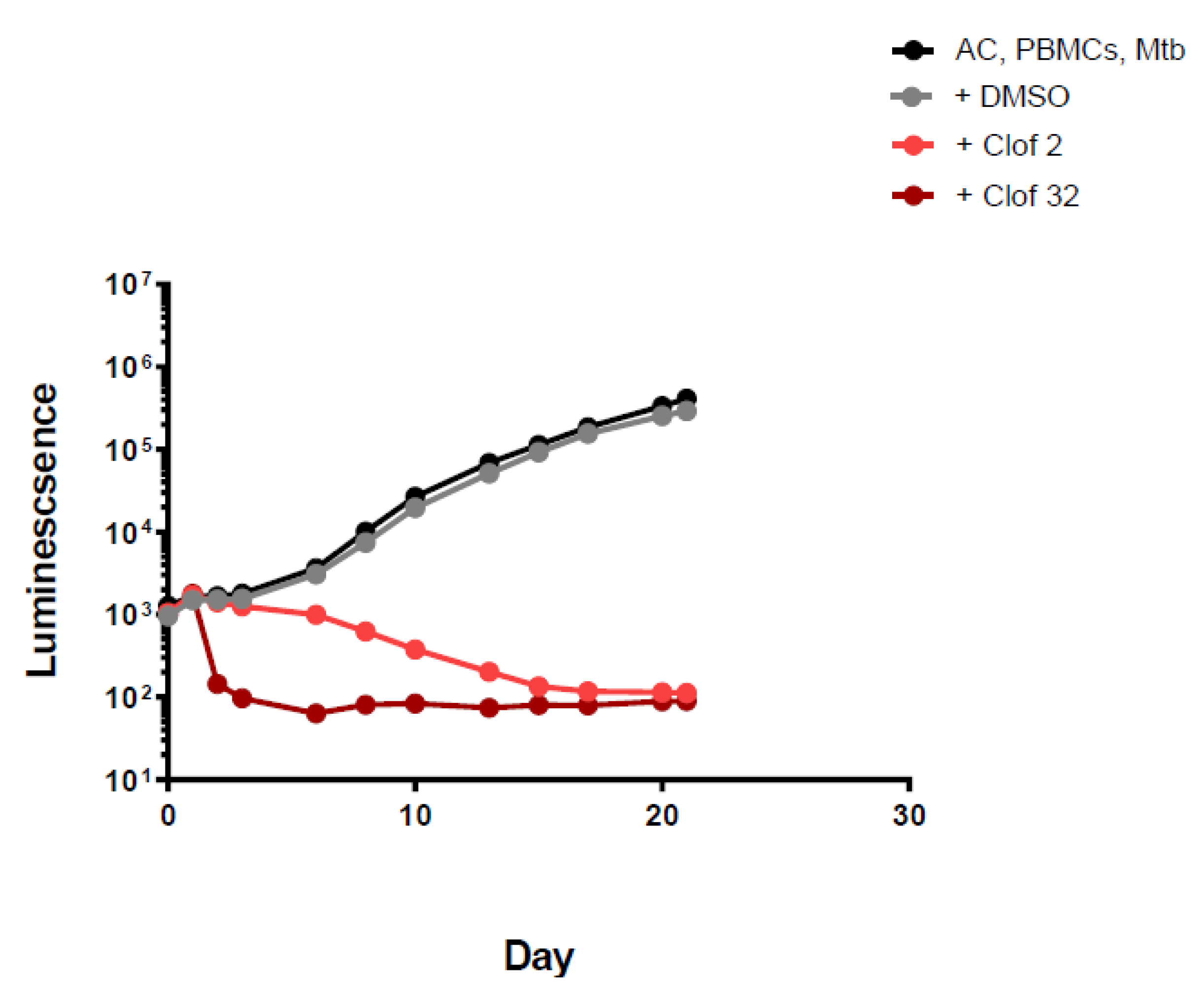
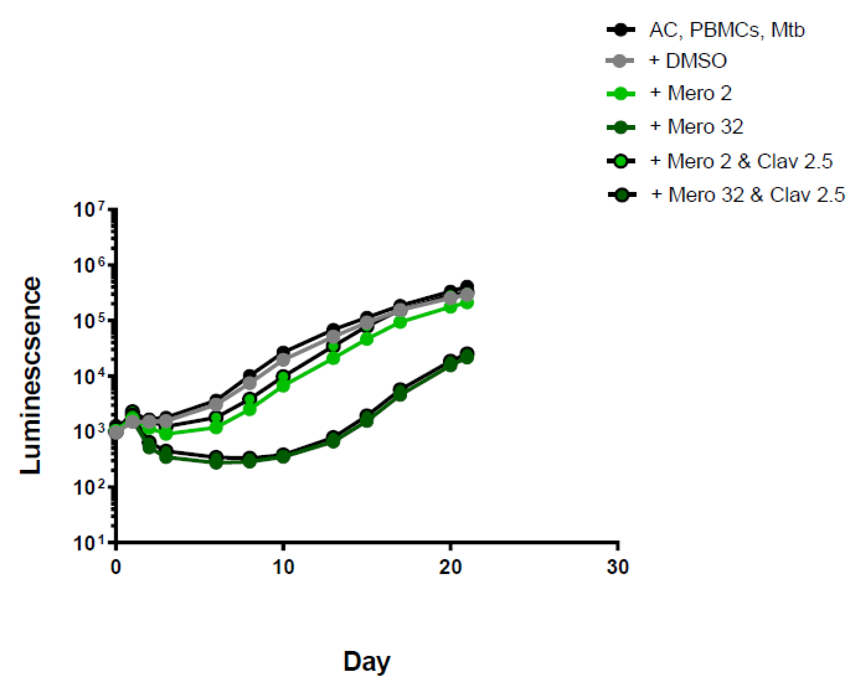
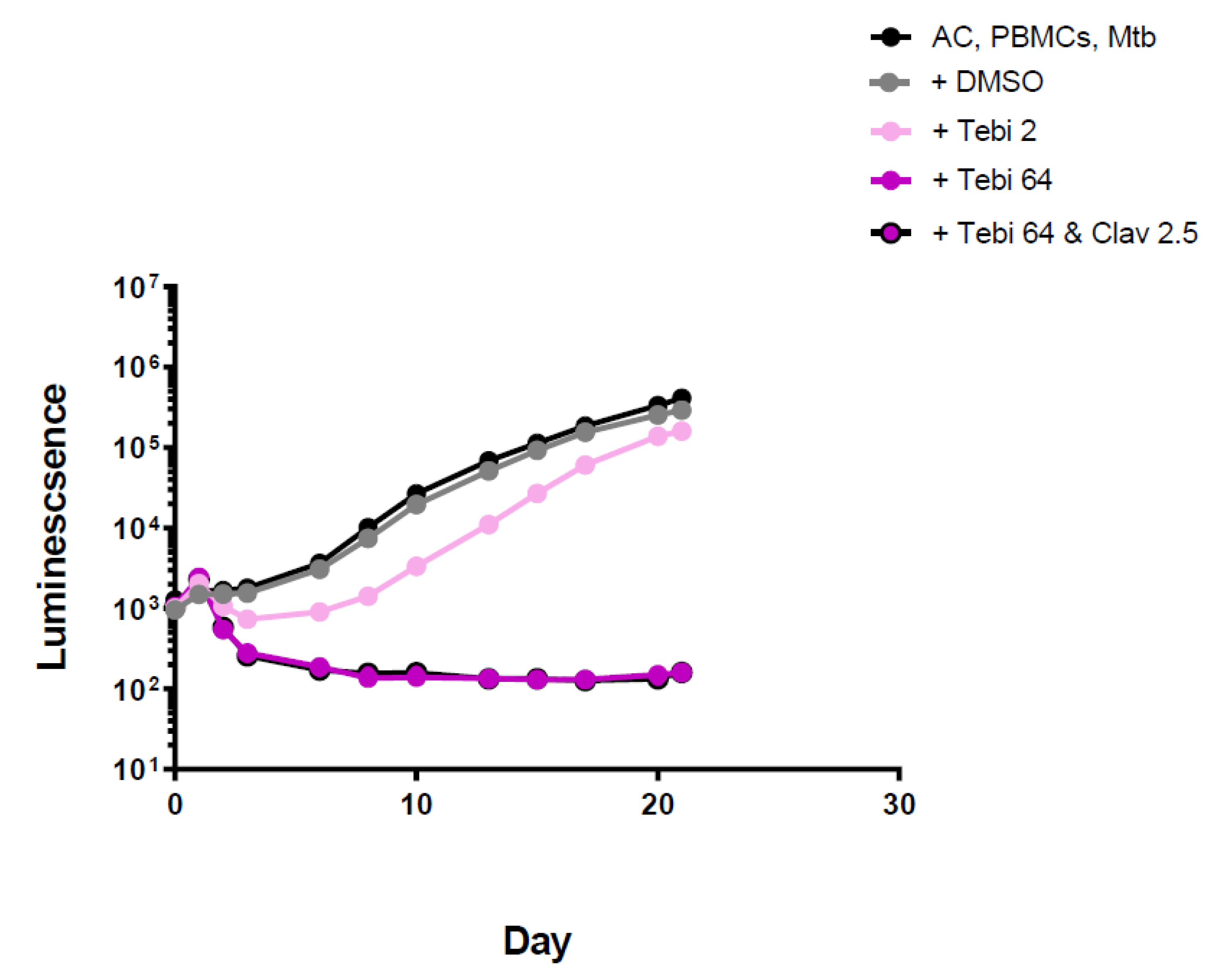
2. Results
3. Discussion
4. Materials and Methods
4.1. Peripheral Blood Mononuclear Cells (PBMCs)
4.2. Bacterial Culture
4.3. PBMCs Encapsulation
4.4. Drugs and Concentration Tested
5. Conclusions
Supplementary Materials
Author Contributions
Funding
Institutional Review Board Statement
Informed Consent Statement
Data Availability Statement
Conflicts of Interest
References
- World Health Organization. Global Tuberculosis Report 2019; World Health Organization: Geneva, Switzerland, 2019. [Google Scholar]
- Sharma, D.; Dhuriya, Y.K.; Deo, N.; Bisht, D. Repurposing and Revival of the Drugs: A New Approach to Combat the Drug Resistant Tuberculosis. Front. Microbiol. 2017, 8, 2452. [Google Scholar] [CrossRef] [PubMed]
- Tezera, L.T.; Bielecka, M.K.; Chancellor, A.; Reichmann, M.T.; Basim, A.l.; Shammari, B.A.; Brace, P.; Batty, A.; Tocheva, A.; Jogai, S.; et al. Dissection of the host-pathogen interaction in human tuberculosis using a bioengineered 3-dimensional model. eLife 2017, 6, e21283. [Google Scholar] [CrossRef] [PubMed]
- Bielecka, M.K.; Tezera, L.B.; Zmijan, R.; Drobniewski, F.; Zhang, X.; Jayasinghe, S.; Elkington, P. A Bioengineered Three Dimensional Cell Culture Platform Integrated with Microfluidics To Address Antimicrobial Resistance in Tuberculosis. mBio 2017, 8, e02073-16. [Google Scholar] [CrossRef] [PubMed]
- Tezera, L.B.; Bielecka, M.K.; Elkington, P.T. Bioelectrospray Methodology for Dissection of the Host-pathogen Interaction in Human Tuberculosis. Bio-Protocol 2017, 7, e2418. [Google Scholar] [CrossRef] [PubMed]
- Moodley, R.; Godec, T.R. Short-course treatment for multidrug-resistant tuberculosis: The STREAM trials. Eur. Respir. Rev. 2016, 25, 29–35. [Google Scholar] [CrossRef]
- World Health Organization. Rapid Communication: Key Changes to Treatment of Multidrug- and Rifampicin-Resistant Tuberculosis (Mdr/Rr-Tb); World Health Organization: Geneva, Switzerland, 2018. [Google Scholar]
- Chambers, H.F.; Turner, J.; Schecter, G.F.; Kawamura, M.; Hopewell, P.C. Imipenem for treatment of tuberculosis in mice and humans. Antimicrob. Agents Chemother. 2005, 49, 2816–2821. [Google Scholar] [CrossRef]
- De Carvalho, L.P.S.; Lin, G.; Jiang, X.; Nathan, C. Nitazoxanide Kills Replicating and Nonreplicating Mycobacterium tuberculosis and Evades Resistance. J. Med. Chem. 2009, 52, 5789–5792. [Google Scholar] [CrossRef]
- Shigyo, K.; Ocheretina, O.; Merveille, Y.M.; Johnson, W.D.; Pape, J.W.; Nathan, C.F.; Fitzgerald, D.W. Efficacy of nitazoxanide against clinical isolates of Mycobacterium tuberculosis. Antimicrob. Agents Chemother. 2013, 57, 2834–2837. [Google Scholar] [CrossRef]
- Castillo-Salazar, M.; Sánchez-Muñoz, F.; Springall Del Villar, R.; Navarrete-Vázquez, G.; Hernández-Diazcouder, A.; Mojica-Cardoso, C.; García-Jiménez, S.; Toledano-Jaimes, C.; Bernal-Fernández, G. Nitazoxanide Exerts Immunomodulatory Effects on Peripheral Blood Mononuclear Cells from Type 2 Diabetes Patients. Biomolecules 2021, 11, 1817. [Google Scholar] [CrossRef]
- Yuan, S.; Yin, X.; Meng, X.; Chan, J.F.-W.; Ye, Z.-W.; Riva, L.; Pache, L.; Chan, C.C.-Y.; Lai, P.-M.; Chan, C.C.-S.; et al. Clofazimine broadly inhibits coronaviruses including SARS-CoV-2. Nature 2021, 593, 418–423. [Google Scholar] [CrossRef]
- Saravanan, P.; Dusthackeer, V.N.A.; Rajmani, R.S.; Mahizhaveni, B.; Nirmal, C.R.; Rajadas, S.E.; Bhardwaj, N.; Ponnuraja, C.; Bhaskar, A.; Hemanthkumar, A.K.; et al. Discovery of a highly potent novel rifampicin analog by preparing a hybrid of the precursors of the antibiotic drugs rifampicin and clofazimine. Sci. Rep. 2021, 11, 1029. [Google Scholar] [CrossRef] [PubMed]
- Memar, M.Y.; Yekani, M.; Ghanbari, H.; Shahi, S.; Sharifi, S.; Maleki Dizaj, S. Biocompatibility, cytotoxicity and antibacterial effects of meropenem-loaded mesoporous silica nanoparticles against carbapenem-resistant Enterobacteriaceae. Artif. Cells Nanomed. Biotechnol. 2020, 48, 1354–1361. [Google Scholar] [CrossRef]
- Noufflard, H.; Berteaux, S. Antituberculous activity of compound B-663. Ann. Inst. Pasteur. 1958, 95, 449–455. [Google Scholar]
- Dey, T.; Brigden, G.; Cox, H.; Shubber, Z.; Cooke, G.; Ford, N. Outcomes of clofazimine for the treatment of drug-resistant tuberculosis: A systematic review and meta-analysis. J. Antimicrob. Chemother. 2012, 68, 284–293. [Google Scholar] [CrossRef] [PubMed]
- Nunn, A.J.; Phillips, P.P.J.; Meredith, S.K.; Chiang, C.-Y.; Conradie, F.; Dalai, D.; Van Deun, A.; Dat, P.-T.; Lan, N.; Master, I.; et al. A Trial of a Shorter Regimen for Rifampin-Resistant Tuberculosis. N. Engl. J. Med. 2019, 380, 1201–1213. [Google Scholar] [CrossRef]
- Aung, K.J.; Van Deun, A.; Declercq, E.; Sarker, M.R.; Das, P.K.; Hossain, M.A.; Rieder, H.L. Successful ‘9-month Bangladesh regimen’ for multidrug-resistant tuberculosis among over 500 consecutive patients. Int. J. Tuberc. Lung. Dis. 2014, 18, 1180–1187. [Google Scholar] [CrossRef]
- Kaniga, K.; Cirillo, D.M.; Hoffner, S.; Ismail, N.A.; Kaur, D.; Lounis, N.; Metchock, B.; Pfyffer, G.E.; Venter, A. A Multilaboratory, Multicountry Study To Determine MIC Quality Control Ranges for Phenotypic Drug Susceptibility Testing of Selected First-Line Antituberculosis Drugs, Second-Line Injectables, Fluoroquinolones, Clofazimine, and Linezolid. J. Clin. Microbiol. 2016, 54, 2963–2968. [Google Scholar] [CrossRef]
- Pang, Y.; Zong, Z.; Huo, F.; Jing, W.; Ma, Y.; Dong, L.; Li, Y.; Zhao, L.; Fu, Y.; Huang, H. Drug Susceptibility of Bedaquiline, Delamanid, Linezolid, Clofazimine, Moxifloxacin, and Gatifloxacin against Extensively Drug-Resistant Tuberculosis in Beijing, China. Antimicrob. Agents Chemother. 2017, 61, e00900-17. [Google Scholar] [CrossRef]
- Maurer, F.P.; Shubladze, N.; Kalmambetova, G.; Felker, I.; Kuchukhidze, G.; Koser, C.U.; Cirillo, D.M.; Drobniewski, F.; Yedilbayev, A.; Ehsani, S.; et al. Diagnostic Capacities for Multidrug-Resistant Tuberculosis in the World Health Organization European Region: Action is Needed by all Member States. J. Mol. Diagn. 2022, in press. [Google Scholar] [CrossRef]
- Broekhuysen, J.; Stockis, A.; Lins, R.L.; De Graeve, J.; Rossignol, J.F. Nitazoxanide: Pharmacokinetics and metabolism in man. Int. J. Clin. Pharmacol. Ther. 2000, 38, 387–394. [Google Scholar] [CrossRef]
- Sotgiu, G.; D′ambrosio, L.; Centis, R.; Tiberi, S.; Esposito, S.; Dore, S.; Spanevello, A.; MigliorI, G.B. Carbapenems to Treat Multidrug and Extensively Drug-Resistant Tuberculosis: A Systematic Review. Int. J. Mol. Sci. 2016, 17, 373. [Google Scholar] [CrossRef] [PubMed]
- Gonzalo, X.; Satta, G.; Ortiz Canseco, J.; Mchugh, T.D.; Drobniewski, F. Ertapenem and Faropenem against Mycobacterium tuberculosis: In vitro testing and comparison by macro and microdilution. BMC Microbiol. 2020, 20, 271. [Google Scholar] [CrossRef] [PubMed]
- Gonzalo, X.; Drobniewski, F. Is there a place for β-lactams in the treatment of multidrug-resistant/extensively drug-resistant tuberculosis? Synergy between meropenem and amoxicillin/clavulanate. J. Antimicrob. Chemother. 2012, 68, 366–369. [Google Scholar] [CrossRef] [PubMed]
- Guo, Z.Y.; Zhao, W.J.; Zheng, M.Q.; Liu, S.; Yan, C.X.; Li, P.; Xu, S.F. Activities of Biapenem against Mycobacterium tuberculosis in Macrophages and Mice. Biomed. Environ. Sci. 2019, 32, 235–241. [Google Scholar]
- Van Rijn, S.P.; Zuur, M.A.; Anthony, R.; Wilffert, B.; Van Altena, R.; Akkerman, O.W.; De Lange, W.C.M.; Van Der Werf, T.S.; Kosterink, J.G.W.; AlffenaaR, J.-W.C. Evaluation of Carbapenems for Treatment of Multi- and Extensively Drug-Resistant Mycobacterium tuberculosis. Antimicrob. Agents Chemother. 2019, 63, e01489-18. [Google Scholar] [CrossRef] [PubMed]
- Reichmann, M.T.; Tezera, L.B.; Vallejo, A.F.; Vukmirovic, M.; Xiao, R.; Reynolds, J.; Jogai, S.; Wilson, S.; Marshall, B.; Jones, M.G.; et al. Integrated transcriptomic analysis of human tuberculosis granulomas and a biomimetic model identifies therapeutic targets. J. Clin. Invest. 2021, 131, e148136. [Google Scholar] [CrossRef]
- Tezera, L.B.; Bielecka, M.K.; Ogongo, P.; Walker, N.F.; Ellis, M.; Garay-Baquero, D.J.; Thomas, K.; Reichmann, M.T.; Johnston, D.A.; Wilkinson, K.A.; et al. Anti-PD-1 immunotherapy leads to tuberculosis reactivation via dysregulation of TNF-α. eLife 2020, 9, e52668. [Google Scholar] [CrossRef]
- Andreu, N.; Zelmer, A.; Fletcher, T.; Elkington, P.T.; Ward, T.H.; Ripoli, J.; Parish, T.; Bancroft, G.J.; Schaible, U.; Robertson, B.D.; et al. Optimisation of Bioluminescent Reporters for Use with Mycobacteria. PLoS ONE 2010, 5, e10777. [Google Scholar] [CrossRef]
- Kothekar, A.; Divatia, J.V.; Myatra, S.N.; Gota, V. Response to: 500 mg as bolus followed by an extended infusion of 1500 mg of meropenem every 8 h failed to achieve in one-third of the patients an optimal PK/PD against nonresistant strains of these organisms: Is CRRT responsible for this situation? Ann. Intensive Care 2020, 10, 164. [Google Scholar] [CrossRef]
- Li, Z.; Su, M.; Cheng, W.; Xia, J.; Liu, S.; Liu, R.; Sun, S.; Feng, L.; Zhu, X.; Zhang, X.; et al. Pharmacokinetics, Urinary Excretion, and Pharmaco-Metabolomic Study of Tebipenem Pivoxil Granules After Single Escalating Oral Dose in Healthy Chinese Volunteers. Front. Pharmacol. 2021, 12, 696165. [Google Scholar] [CrossRef]
- Gettig, J.P.; Crank, C.W.; Philbrick, A.H. Faropenem medoxomil. Ann. Pharmacother. 2008, 42, 80–89. [Google Scholar] [CrossRef] [PubMed]
- Abdelwahab, M.T.; Wasserman, S.; Brust, J.C.; Gandi, N.R.; Meintjes, G.; Everitt, D.; Diacon, A.; Dawson, R.; Wiesner, L.; Svensson, E.M.; et al. Clofazimine pharmacokinetics in patients with TB: Dosing implications. J. Antimicrob. Chemother. 2020, 75, 3269–3277. [Google Scholar] [CrossRef] [PubMed]
- Imani, S.; Buscher, H.; Marriott, D.; Gentili, S.; Sandaradura, I. Too much of a good thing: A retrospective study of β-lactam concentration-toxicity relationships. J. Antimicrob. Chemother. 2017, 72, 2891–2897. [Google Scholar] [CrossRef]
- Stockis, A.; Deroubaix, X.; Lins, R.; Jeanbaptise, B.; Calderon, P.; Rossignol, J.F. Pharmacokinetics of nitazoxanide after single oral dose administration in 6 healthy volunteers. Int. J. Clin. Pharmacol. Ther. 1996, 34, 349–351. [Google Scholar] [PubMed]


Publisher’s Note: MDPI stays neutral with regard to jurisdictional claims in published maps and institutional affiliations. |
© 2022 by the authors. Licensee MDPI, Basel, Switzerland. This article is an open access article distributed under the terms and conditions of the Creative Commons Attribution (CC BY) license (https://creativecommons.org/licenses/by/4.0/).
Share and Cite
Gonzalo, X.; Bielecka, M.K.; Tezera, L.; Elkington, P.; Drobniewski, F. Anti-Tuberculosis Activity of Three Carbapenems, Clofazimine and Nitazoxanide Using a Novel Ex Vivo Phenotypic Drug Susceptibility Model of Human Tuberculosis. Antibiotics 2022, 11, 1274. https://doi.org/10.3390/antibiotics11101274
Gonzalo X, Bielecka MK, Tezera L, Elkington P, Drobniewski F. Anti-Tuberculosis Activity of Three Carbapenems, Clofazimine and Nitazoxanide Using a Novel Ex Vivo Phenotypic Drug Susceptibility Model of Human Tuberculosis. Antibiotics. 2022; 11(10):1274. https://doi.org/10.3390/antibiotics11101274
Chicago/Turabian StyleGonzalo, Ximena, Magdalena K. Bielecka, Liku Tezera, Paul Elkington, and Francis Drobniewski. 2022. "Anti-Tuberculosis Activity of Three Carbapenems, Clofazimine and Nitazoxanide Using a Novel Ex Vivo Phenotypic Drug Susceptibility Model of Human Tuberculosis" Antibiotics 11, no. 10: 1274. https://doi.org/10.3390/antibiotics11101274
APA StyleGonzalo, X., Bielecka, M. K., Tezera, L., Elkington, P., & Drobniewski, F. (2022). Anti-Tuberculosis Activity of Three Carbapenems, Clofazimine and Nitazoxanide Using a Novel Ex Vivo Phenotypic Drug Susceptibility Model of Human Tuberculosis. Antibiotics, 11(10), 1274. https://doi.org/10.3390/antibiotics11101274