Case Report

Antimicrobial Treatment on a Catheter-Related Bloodstream Infection (CRBSI) Case Due to Transition of a Multi-Drug-Resistant Ralstonia mannitolilytica from Commensal to Pathogen during Hospitalization

1
College of Light Industry and Food Science, Guangdong Provincial Key Laboratory of Science and Technology of Lingnan Special Food Science and Technology, Innovation Research Institute of Modern Agricultural Engineering, Zhongkai University of Agriculture and Engineering, Guangzhou 510225, China
2
Key Laboratory of Green Processing and Intelligent Manufacturing of Lingnan Specialty Food, Zhongkai University of Agriculture and Engineering, Guangzhou 510225, China
3
Department of Clinical Pharmacy and Translational Science, University of Tennessee Health Science Center, Memphis, TN 38163, USA
4
Department of Laboratory Medicine, The First Affiliated Hospital of Guangzhou Medical University, Guangzhou Medical University, Guangzhou 510120, China
5
Centre for Translational Medicine, The First Affiliated Hospital of Guangzhou Medical University, Guangzhou Medical University, Guangzhou 510120, China
6
School of Food Science and Engineering, Guangdong Province Key Laboratory for Green Processing of Natural Products and Product Safety, Engineering Research Center of Starch and Vegetable Protein Processing Ministry of Education, South China University of Technology, Guangzhou 510640, China
7
Department of Laboratory Medicine, The Second Affiliated Hospital of Shantou University Medical College, Shantou 515041, China
*
Authors to whom correspondence should be addressed.
These authors contributed equally to this work.
Antibiotics 2022, 11(10), 1376; https://doi.org/10.3390/antibiotics11101376
Submission received: 18 August 2022 / Revised: 4 October 2022 / Accepted: 7 October 2022 / Published: 8 October 2022
(This article belongs to the Collection Antimicrobial Resistance and Anti-Biofilms)

Abstract

:
Despite its commonly overlooked role as a commensal, Ralstonia mannitolilytica becomes an emerging global opportunistic human pathogen and a causative agent of various infections and diseases. In respiratory illnesses, including cystic fibrosis and chronic obstructive pulmonary disease (COPD), R. mannitolilytica is also identified presumably as colonizer. In this study, one distinctive clone of R. mannitolilytica was firstly identified as colonizer for the first 20 days during hospitalization of a patient. It was then identified as a causative agent for catheter-related bloodstream infection with negative identification after effective treatment, verifying its transition from commensal to pathogen. In conclusion, we provide convincing evidence that during hospitalization of a patient, R. mannitolilytica transitioned from commensal to pathogen in the respiratory tract leading to catheter-related bloodstream infection (CRBSI).

1. Introduction

As a worldwide public health challenge [1], chronic obstructive pulmonary disease (COPD) ranks the third leading cause of death and overall prevalence ranges from 8.6% to 13.6% in China [2,3]. COPD patients present an altered airway microbiome, with Ralstonia mannitolilytica identified at a significantly higher rate comparing with a healthy population [4]. Reservoirs of R. mannitolilytica in the hospital environment include water [5], saline solutions [6] and oxygen delivery devices [7]. Despite its commonly overlooked role as a commensal, Ralstonia mannitolilytica becomes an emerging global opportunistic human pathogen and a causative agent of various infections and diseases, including bacteremia [8,9,10], meningitis [11], sepsis [12], peritonitis [13], osteomyelitis [14], hemoperitoneum [14] and urinary tract infection [7]. Furthermore, R. mannitolilytica is also identified in respiratory illnesses such as cystic fibrosis and COPD, presumably as a colonizer [15,16,17,18]. From the first report in 2011, R. mannitolilytica isolate G100 was sampled from the sputum of a COPD patient on admission, and the patient was treated with piperacillin/tazobactam [16]. Despite negative identification of R. mannitolilytica thereafter, the patient continued to suffer from respiratory failure and eventually died, suggesting this microorganism was a benign colonizer [16,19,20,21]. Such a finding is in accordance with a recent comprehensive study on airway microbiomes, where the presence of R. mannitolilytica was occasionally found colonizing COPD patients and indicating its potential role in recurrent infections and diseases [4,22].
For the first time, we report a catheter-related bloodstream infection (CRBSI) case caused by R. mannitolilytica in the respiratory tract of a hospitalized COPD patient.

2. Results and Discussion

2.1. Antimicrobial Agent Treatment

2.1.1. Phase I: From Admission to Day 3

On admission, the patient was diagnosed with acute exacerbations COPD (AECOPD), severe pneumonia and type II respiratory failure, with symptoms of fever and dyspnea on day 1 and high fever and dyspnea on day 2. On day 1, sputum culture identified Acinetobacter baumannii, Chryseobacterium meningosepticum and R. mannitolilytica, with negative culture from a blood sample. During day 1 to 3, antimicrobial treatment had been conducted using meropenem, vancomycin, caspofungin and voriconazole, with peripherally inserted central venous catheters (PICC) used from day 1 onwards.

2.1.2. Phase II: From Day 4 to 18

Despite antimicrobial treatment during day 1 to 3, the patient failed to recover and a blood test on day 4 revealed an uncontrolled infection accompanied by dyspnea and wheezing. In detail, he had suffered from low fever and wheezing on day 4; low fever, dyspnea and wheezing on day 5; and fever and wheezing on day 6. Consequently, the patient was treated with different antimicrobial therapy (Table 1). Nevertheless, he suffered from intermittent fever. Despite negative blood culture, sputum culture on Day 6 identified A. baumannii, C. meningosepticum and R. mannitolilytica (Guangzhou-RMAS10/11, and on day 10, only R. mannitolilytica was identified), which are common colonizers in the respiratory tract. As antimicrobial treatment was concerned, the patient had been treated with meropenem, vancomycin, caspofungin and amphotericin B during day 4 to 7; meropenem, linezolid, caspofungin and amphotericin B during day 8 and 9; piperacillin/tazobactam, linezolid, caspofungin and amphotericin B during day 10 to 13; cefoperazone/sulbactam, rifampin, vancomycin and amphotericin B during day 14 to 16; and cefoperazone/sulbactam, meropenem, rifampin, vancomycin and caspofungin during day 17 and 18.

2.1.3. Phase III: From Day 19 to 27

On day 19, the patient presented with fever, shivering and dyspnea, and on day 20, his condition deteriorated to high fever, severe dyspnea and septic shock. Remarkably, R. mannitolilytica had been identified from both sputum and blood samples (Guangzhou-RMAB10/11/12, three isolates, one from the catheter of PICC and two from peripheral blood) on day 19, validating the occurrence of catheter-related bloodstream infection (CRBSI). According to its antimicrobial profile, ciprofloxacin was used from day 21 on. The patient showed decreased body temperature and improvement in dyspnea, with symptoms of low fever and relieved dyspnea during day 21 to 25, then no fever or dyspnea on day 26 and 27. In addition, a blood test on day 26 revealed normal white blood cell count and serum procalcitonin (PCT), with no positive identification of R. mannitolilytica from either blood or sputum samples. Also, PICC was removed on day 27 after the result was reported.

2.1.4. Phase IV: After Day 27 to Discharging

Two months later, this patient presented cough with thick yellow sputum and occasional wheezing. On day 82, the sputum culture identified Pseudomonas aeruginosa, Achromobacter xylosoxidans and A. baumannii, and blood culture identified Enterococcus faecium and A. baumannii. Consequently, routine treatment for these symptoms was further conducted and the patient was eventually discharged on day 132.

2.2. Bacterial Identification

Seven R. mannitolilytica isolates were sampled, including four strains (Guangzhou-RMAS10/11/12/13) from sputum and three strains (Guangzhou-RMAB10/11/12) from blood. In detail, R. mannitolilytica Guangzhou-RMAS10, Guangzhou-RMAS11, Guangzhou-RMAS12 and Guangzhou-RMAS13 had been recovered from sputum samples on day 1, day 6, day 10 and day 19, respectively. R. mannitolilytica Guangzhou-RMAB10, Guangzhou-RMAB11 and Guangzhou-RMAB12 had been obtained from blood samples on day 19. According to the results of 16S rRNA sequencing, compared with all known sequences in GenBank by BLASTn, the sequencing results of all R. mannitolilytica strains were found to be identical and clustered at 100% sequence similarity with the R. mannitolilytica strain AU255 (AY043379) and AU428 (AY043378) from a cystic fibrosis patient in the United States, and strains LMG 6866 (NR_025385) from cases of nosocomial recurrent meningitis in Belgium.

2.3. Antimicrobial Susceptibility Testing

Antimicrobial susceptibility testing (AST) using Vitek2TM Automated System had been performed on seven R. mannitolilytica strains [23]. All R. mannitolilytica strains shared distinctive antimicrobial resistance profiles, and exhibited resistance to 14 antimicrobial agents, including ampicillin/sulbactam, aztreonam, cefazolin, cefepime, cefoperazone/sulbactam, cefotaxime, cefotetan, cefuroxime, gentamicin, imipenem, meropenem, piperacillin, piperacillin/tazobactam and tobramycin, with only a few susceptible exceptions such as ceftriaxone, ciprofloxacin and levofloxacin. The AST results had been used to determine the antimicrobial treatment on R. mannitolilytica strains.

2.4. Clonal Relatedness of R. mannitolilytica Strains

According to the results from randomly amplified polymorphic DNA polymerase chain reaction (RAPD-PCR), the acquired indistinguishable fingerprinting patterns had suggested all seven R. mannitolilytica belonged to the same genotype and thus were clonally related (patterns from representative strains shown in Figure 1). As further confirmed by genomic comparison, their identical genomes with similarity higher than 99.99% had convincingly verified that all R. mannitolilytica strains originated from a distinct clone (the genome sequences of the distinct R. mannitolilytica strain are deposited under accession number CP049132 for chromosome 1 and CP049133 for chromosome 2). In connection with the sampling background, the colonizing R. mannitolilytica strain had highly likely induced invasive infection in the respiratory tract and subsequently caused bacteremia in the patient, which explained the identification of clonally related bacteria from sputum and blood culture [24].

2.5. Extensive Surveillance on R. mannitolilytica Strains

In addition, from an extensive surveillance during 2014 (including the duration of hospitalization), the R. mannitolilytica strain was rarely identified as an infectious agent for other patients, nor was it routinely isolated from drinking water, laboratory-based water, saline solutions used for patient care or catheters [25,26,27].

2.6. Transition from Commensal to Pathogen

In this case, rare identification of R. mannitolilytica from patients or environmental samples, no recent hospitalization history for the patient, as well as the immediate detection of R. mannitolilytica on admission ruled out the likelihood of nosocomial infection or contamination and, thus, highly suggests a community origin of this microorganism. During hospitalization, one identical R. mannitolilytica strain was found colonizing the patient’s respiratory tract despite broad antimicrobial therapy (during admission to day 20, shown by four isolates independently sampled from sputum at different time points), and further caused CRBSI (shown by three isolates separately sampled on day 19 including one isolate sampled from the catheter). In addition, the specific treatment of R. mannitolilytica (since day 21) correlated with the improvement of symptoms and negative identification of R. mannitolilytica (on day 26 and thereafter).
R. mannitolilytica has been implicated in acute exacerbation of chronic obstructive pulmonary disease (AECOPD); however, it is still controversial if the bacterium is the causative agent [4,16]. Mostly importantly, this study reported R. mannitolilytica bacteremia in a patient diagnosed with AECOPD, confirming its pathogenic role in this disease. Important evidence for this confirmation and verification were as follows: Firstly, for a patient diagnosed with AECOPD, R. mannitolilytica strain Guangzhou-RMAS10 was independently isolated from sputum samples (with identical strains) on day 6, but negative for blood samples. Secondly, R. mannitolilytica strain Guangzhou-RMAB10 was independently isolated from all three pairs of blood culture bottles on Day 20. Thirdly, R. mannitolilytica strains Guangzhou-RMAS10 and Guangzhou-RMAB10 were identical according to the antimicrobial profile, RAPD and genomic sequences. The above evidence strongly indicates the opportunistic pathogen R. mannitolilytica induced invasive infection in the respiratory tract (isolates from sputum) and subsequently caused bacteremia (isolates from blood) for this patient. Fourthly, no R. mannitolilytica infection for other patients had been reported in the First Affiliated Hospital of Guangzhou Medical University (FAHGMU) in the whole year of 2014 (this bacterium is extremely rare to be reported), and also, during hospitalization of the patient, none of R. mannitolilytica were isolated from drinking water, laboratory-based water, saline solutions used for patient care, or catheters. The above evidence and observation highly suggested the unlikelihood of nosocomial infection or contamination as the infection cause. Fifthly, after the confirmation of R. mannitolilytica on day 20, the antimicrobial therapy was changed to ciprofloxacin after day 22. Shortly after this, significant improvement from the blood test and negative identification of R. mannitolilytica (on day 25 and thereafter) were found. The above evidence presented strong evidence that R. mannitolilytica caused bacteremia during the therapy of AECOPD of a patient, as well as its pathogenic role in this disease. In combination with the above evidence, the R. mannitolilytica bacteremia in a patient diagnosed with AECOPD, as well as its pathogenicity, is convincing.

3. Materials and Methods

3.1. Clinical Samples and Bacterial Strains

A total of 16 strains were isolated during the hospitalization of a patient, including 7 Acinetobacter baumannii and R. mannitolilytica (4 from sputum and 3 from blood samples, sampling on day 1 and 6, and on day 82 (June 22) from both sputum and blood), 2 Chryseobacterium meningosepticum (sampling on day 1 and 6), 1 Pseudomonas aeruginosa (sampling on day 82 (22 June) from sputum), 1 Achromobacter xylosoxidans (sampling on day 82 (22 June) from sputum), and 1 Enterococcus faecium (sampling on day 82 (22 June) from blood) strains.

3.2. Clinical Case

On 2 April 2014, a 72-year-old male patient presenting with fever and dyspnea was admitted to the First Affiliated Hospital of Guangzhou Medical University (FAHGMU) in Guangzhou, China. With a COPD history dating back to 2004, this patient was treated to relieve symptoms of dyspnea. On admission, the patient was diagnosed with acute exacerbations COPD (AECOPD), severe pneumonia and type II respiratory failure, with symptoms of fever and dyspnea on day 1 and high fever and dyspnea on day 2.
Concerning the clinical history, in January of 2014, the patient was diagnosed with pulmonary aspergillosis and was treated with voriconazole during hospitalization. On 26 February 2014, the patient was hospitalized again due to gastrointestinal bleeding and cured by an antiacid drug. Considering the pulmonary aspergillosis was not cured, the patient was successively treated with voriconazole, mezlocillin sodium/sulbactam sodium, and meropenem in combination with continuous positive airway pressure. On 14 March 2014, the patient was transferred to ICU and successively treated with biapenem, vancomycin hydrochloride, voriconazole and moxifloxacin hydrochloride.

3.3. Species Identification

For all samples, bacterial identification was performed with a Vitek2TM Automated System (bioMerieux, Saint-Louis, MO, USA). In addition, all R. mannitolilytica isolates were further assessed by sequencing the 16S rRNA gene [28] using universal primers 27F and 1492R to amplify and then sequence the 16S rRNA gene, and the obtained sequences were compared to all known sequences in GenBank by BLASTn against the Nr database.

3.4. Antimicrobial Susceptibility Testing

Antimicrobial susceptibility testing (AST) was performed with a Vitek2TM Automated System [23]. For R. mannitolilytica isolates, 17 antibiotics were used including ampicillin/sulbactam, aztreonam, cefazolin, cefepime, cefoperazone/sulbactam, cefotaxime, cefotetan, ceftriaxone, cefuroxime, ciprofloxacin, gentamicin, imipenem, levofloxacin, meropenem, piperacillin, Piperacillin/Tazobactam and tobramycin (BBI Solutions, Crumlin, UK).

3.5. Analysis on Clonal Relatedness of R. mannitolilytica Strains

Random amplification of polymorphic DNA (RAPD) PCR was applied to characterize the genetic relatedness of all R. mannitolilytica strains with different origin. The RAPD primer (RM270: 5′–TGC GCG CGG G–3′) was used for random amplification as described previously for Ralstonia spp. [29]. Obtaining identical DNA fingerprinting from all 7 R. mannitolilytica strains, additionally, genomic sequencing by Illumina HiSeq 2500 (Illumina, San Diego, CA, USA) and genome comparison were further performed for confirmation [30,31].

3.6. Extensive Surveillance on R. mannitolilytica Strains

Extensive surveillance on R. mannitolilytica was conducted at the same medical setting (FAHGMU) during the whole year of hospitalization (2014).

4. Conclusions

In conclusion, we provide convincing evidence that during hospitalization of a patient, R. mannitolilytica transitioned from commensal to pathogen in the respiratory tract leading to CRBSI.

Author Contributions

Conceptualization, L.Y.; Data curation, J.L.; Formal analysis, J.L.; Funding acquisition, Z.X.; Investigation, J.L. and D.F.; Methodology, L.Y. and H.Y.; Project administration, Z.X.; Resources, L.Y.; Software, H.Y.; Supervision, D.C.; Validation, D.C.; Visualization, D.F.; Writing—original draft, J.L.; Writing—review & editing, B.M.P. All authors have read and agreed to the published version of the manuscript.

Funding

This work was supported by the Guangdong Major Project of Basic and Applied Basic Research (2020B0301030005), Guangdong International S&T Cooperation Programme (2021A0505030007), Guangdong Provincial Key Laboratory of Lingnan Specialty Food Science and Technology (No. 2021B1212040013), Young S&T Talent Training Program of Guangdong Provincial Association for S&T, China (SKXRC202207), Young Talent Support Project of Guangzhou Association for Science and Technology (QT20220101076), Collaborative grant with AEIC (KEO-2019-0624-001-1), the 111 Project (B17018).

Institutional Review Board Statement

Not applicable.

Informed Consent Statement

The research has been approved by the medical ethics committee of the First Affiliated Hospital of Guangzhou Medical University.

Data Availability Statement

Accession number: CP049132 and CP049133.

Acknowledgments

We express high gratitude to the staff in the First Affiliated Hospital of Guangzhou Medical University (FAHGMU) in Guangzhou representative of Southern China for collecting the R. mannitolilytica isolates.

Conflicts of Interest

The authors declare no conflict of interest.

References

  1. Adeloye, D.; Song, P.; Zhu, Y.; Campbell, H.; Sheikh, A.; Rudan, I. Global, regional, and national prevalence of, and risk factors for, chronic obstructive pulmonary disease (COPD) in 2019: A systematic review and modelling analysis. Lancet Respir. Med. 2022, 10, 447–458. [Google Scholar] [CrossRef]
  2. Fang, L.; Gao, P.; Bao, H.; Tang, X.; Wang, B.; Feng, Y.; Cong, S.; Juan, J.; Fan, J.; Lu, K.; et al. Chronic obstructive pulmonary disease in China: A nationwide prevalence study. Lancet Respir. Med. 2018, 6, 421–430. [Google Scholar] [CrossRef]
  3. Wang, C.; Xu, J.; Yang, L.; Xu, Y.; Zhang, X.; Bai, C.; Kang, J.; Ran, P.; Shen, H.; Wen, F.; et al. Prevalence and risk factors of chronic obstructive pulmonary disease in China (the China Pulmonary Health [CPH] study): A national cross-sectional study. Lancet 2018, 391, 1706–1717. [Google Scholar] [CrossRef]
  4. Wang, Z.; Liu, H.; Wang, F.; Yang, Y.; Wang, X.; Chen, B.; Stampfli, M.R.; Zhou, H.; Shu, W.; Brightling, C.E.; et al. A refined view of airway microbiome in chronic obstructive pulmonary disease at species and strain-levels. Front. Microbiol. 2020, 30(11), 1758. [Google Scholar] [CrossRef] [PubMed]
  5. Baird, R.M.; Elhag, K.M.; Shaw, E.J. Pseudomonas Thomasii in a hospital distilled-water supply. J. Med. Microbiol. 1976, 9, 493–495. [Google Scholar] [CrossRef] [PubMed]
  6. Pan, H.J.; Teng, L.-J.; Tzeng, M.S.; Chang, S.-C.; Ho, S.W.; Luh, K.T.; Hsieh, W.C. Identification and typing of Pseudomonas pickettii during an episode of nosocomial outbreak. Zhonghua Minguo Wei Sheng Wu Ji Mian Yi Xue Za Zhi 1992, 25, 115–123. [Google Scholar] [PubMed]
  7. Jhung, M.A.; Sunenshine, R.H.; Noble-Wang, J.; Coffin, S.E.; John, K.S.; Lewis, F.M.; Jensen, B.; Peterson, A.; LiPuma, J.; Arduino, M.J.; et al. A National Outbreak of Ralstonia mannitolilytica Associated With Use of a Contaminated Oxygen-Delivery Device Among Pediatric Patients. Pediatrics 2007, 119, 1061–1068. [Google Scholar] [CrossRef] [Green Version]
  8. Boattini, M.; Bianco, G.; Biancone, L.; Cavallo, R.; Costa, C. Ralstonia mannitolilytica bacteraemia: A case report and literature review. Infez. Med. 2018, 26, 374–378. [Google Scholar]
  9. Basso, M.; Venditti, C.; Raponi, G.; Navazio, A.S.; Alessandri, F.; Giombini, E.; Nisii, C.; Di Caro, A.; Venditti, M. A case of persistent bacteraemia by Ralstonia mannitolilytica and Ralstonia pickettii in an intensive care unit. Infect. Drug Resist. 2019, 12, 2391. [Google Scholar] [CrossRef] [PubMed] [Green Version]
  10. De Souza, D.C.; Palmeiro, J.K.; Maestri, A.C.; Cogo, L.L.; Rauen, C.H.; Graaf, M.E.; Grein, F.L.; Nogueira, K.D.S. Ralstonia mannitolilytica bacteremia in a neonatal intensive care unit. Rev. da Soc. Bras. de Med. Trop. 2018, 51, 709–711. [Google Scholar] [CrossRef] [PubMed]
  11. Vaneechoutte, M.; De Baere, T.; Wauters, G.; Steyaert, S.; Claeys, G.; Vogelaers, D.; Verschraegen, G. One Case Each of Recurrent Meningitis and Hemoperitoneum Infection with Ralstonia mannitolilytica. J. Clin. Microbiol. 2001, 39, 4588–4590. [Google Scholar] [CrossRef]
  12. Owusu, M.; Acheampong, G.; Annan, A.; Marfo, K.S.; Osei, I.; Amuasi, J.; Sarpong, N.; Im, J.; Mogeni, D.; Chiang, H.-Y.; et al. Ralstonia mannitolilytica sepsis: A case report. J. Med. Case Rep. 2019, 13, 318. [Google Scholar] [CrossRef] [Green Version]
  13. Dotis, J.; Printza, N.; Orfanou, A.; Papathanasiou, E.; Papachristou, F. Peritonitis due to Ralstonia mannitolilytica in a pediatric peritoneal dialysis patient. New Microbiol. 2012, 35, 503–506. [Google Scholar] [PubMed]
  14. Ryan, M.P.; Adley, C.C. Ralstonia spp.: Emerging global opportunistic pathogens. Eur. J. Clin. Microbiol. Infect. Dis. 2013, 33, 291–304. [Google Scholar] [CrossRef] [PubMed] [Green Version]
  15. Coenye, T.; Vandamme, P.; LiPuma, J.J. Infection by Ralstonia Species in Cystic Fibrosis Patients: Identification of R. pickettii and R. mannitolilytica by Polymerase Chain Reaction. Emerg. Infect. Dis. 2002, 8, 692–696. [Google Scholar] [CrossRef] [PubMed]
  16. Zong, Z.Y.; Peng, C.H. Ralstonia mannitolilytica and COPD: A case report. Eur. Respir. J. 2011, 38, 1482–1483. [Google Scholar] [CrossRef] [PubMed] [Green Version]
  17. Coman, I.; Bilodeau, L.; Lavoie, A.; Carricart, M.; Tremblay, F.; Zlosnik, J.; Berthiaume, Y. Ralstonia mannitolilytica in cystic fibrosis: A new predictor of worse outcomes. Respir. Med. Case Rep. 2016, 20, 48–50. [Google Scholar] [CrossRef] [PubMed]
  18. Mukhopadhyay, C.; Bhargava, A.; Ayyagari, A. Ralstonia mannitolilytica infection in renal transplant recipient: First report. Indian J. Med. Microbiol. 2003, 21, 284–286. [Google Scholar] [CrossRef]
  19. Shankar, M.; Rampure, S.; Siddini, V.; Ballal, H. Outbreak of Ralstonia mannitolilytica in hemodialysis unit: A case series. Indian J. Nephrol. 2018, 28, 323–326. [Google Scholar] [CrossRef] [PubMed]
  20. Gröbner, S.; Heeg, P.; Autenrieth, I.B.; Schulte, B. Monoclonal outbreak of catheter-related bacteraemia by Ralstonia mannitolilytica on two haemato-oncology wards. J. Infect. 2007, 55, 539–544. [Google Scholar] [CrossRef]
  21. Said, M.; Dangor, Y.; Van Hougenhouck-Tulleken, W.; Mbelle, N.; Strydom, K.-A.; Ismail, F. First outbreak of Ralstonia mannitolilytica bacteraemia in patients undergoing haemodialysis at a tertiary hospital in Pretoria, South Africa. Int. J. Infect. Dis. 2018, 73, 93. [Google Scholar] [CrossRef]
  22. Saiman, L. Providing a Safety Net for Children: Investigating a Multistate Outbreak of Ralstonia mannitolilytica Related to a Contaminated Reusable Device. Pediatrics 2007, 119, 1207–1209. [Google Scholar] [CrossRef] [PubMed]
  23. Bayot, M.L.; Bragg, B.N. Antimicrobial Susceptibility Testing; StatPearls Publishing: Treasure Island, FL, USA, 2022. [Google Scholar]
  24. Vaneechoutte, M.; Kampfer, P.; De Baere, T.; Falsen, E.; Verschraegen, G. Wautersia gen. nov., a novel genus accommodating the phylogenetic lineage including Ralstonia eutropha and related species, and proposal of Ralstonia [Pseudomonas] syzygii (Roberts et al. 1990) comb. nov. Int. J. Syst. Evol. Microbiol. 2004, 54 Pt 2, 317–327. [Google Scholar] [CrossRef]
  25. Daroy, M.L.G.; Lopez, J.S.; Torres, B.C.L.; Loy, M.J.; Tuaño, P.M.C.; Matias, R.R. Identification of unknown ocular pathogens in clinically suspected eye infections using ribosomal RNA gene sequence analysis. Clin. Microbiol. Infect. 2011, 17, 776–779. [Google Scholar] [CrossRef] [Green Version]
  26. Ferro, P.; Vaz-Moreira, I.; Manaia, C.M. Association between gentamicin resistance and stress tolerance in water isolates of Ralstonia pickettii and R. mannitolilytica. Folia Microbiol. 2019, 64, 63–72. [Google Scholar] [CrossRef] [Green Version]
  27. Lim, C.T.S.; Lee, S.E. A rare case of Ralstonia mannitolilytica infection in an end stage renal patient on maintenance dialysis during municipal water contamination. Pak. J. Med. Sci. 2017, 33, 1047–1049. [Google Scholar] [CrossRef] [PubMed]
  28. Stackebrant, E.; Goodfellow, M.; Lane, D.J. 16S/23S rRNA sequencing. In Nucleic Acid Tech-Niques in Bacterial Systematics; Stackebrant, E., Goodfellow., M., Eds.; John Wiley & Sons: New York, NY, USA, 1991; pp. 115–175. [Google Scholar]
  29. Daxboeck, F.; Stadler, M.; Assadian, O.; Marko, E.; Hirschl, A.M.; Koller, W. Characterization of clinically isolated Ralstonia mannitolilytica strains using random amplification of polymorphic DNA (RAPD) typing and antimicrobial sensitivity, and comparison of the classification efficacy of phenotypic and genotypic assays. J. Med. Microbiol. 2005, 54, 55–61. [Google Scholar] [CrossRef] [Green Version]
  30. Coenye, T.; Spilker, T.; Reik, R.; Vandamme, P.; LiPuma, J.J. Use of PCR Analyses To Define the Distribution of Ralstonia Species Recovered from Patients with Cystic Fibrosis. J. Clin. Microbiol. 2005, 43, 3463–3466. [Google Scholar] [CrossRef] [Green Version]
  31. Suzuki, M.; Nishio, H.; Asagoe, K.; Kida, K.; Suzuki, S.; Matsui, M.; Shibayama, K. Genome Sequence of a Carbapenem-Resistant Strain of Ralstonia mannitolilytica. Genome Announc. 2015, 3, e00405-15. [Google Scholar] [CrossRef] [PubMed]
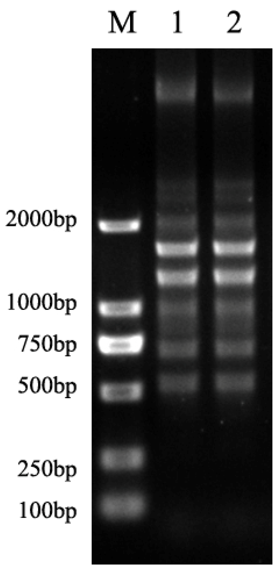
Figure 1. Fingerprinting patterns of two representative strains.
Antibiotics 11 01376 g001
Table 1. Clinical data of the patient.
Detection
Admission TimeTemperature (°C)WBC
(×109 L)
Neutrophils (%)Blood Gas: pH/HCO3 * (mmol/L)
/SpO2 * /PaCO2 * (mm Hg)
/PaO2 * (mm Hg)
PCT * (ng/mL)HCCT *
Phase ID138.316.6 77.47.42/24.5 /98%/51.2/90.60.45 RLLLA *
D238.8–4020.690.1
D337.519.894.1
Phase IID4 18.384.77.435/29.1/96.3%/42.8/85.6
D5 15.782.9
D6 15.682.37.504/26.5/98.8%/33.9/1594.08 RLLLA
D737.6–38.014.681.9
D838.8
D938.4
D1036.7–38.3
D1138.0
D1238.014.381.2
D1337.7
D1438.0
D1538.3
D1637.9
D1738.0
D1836.916.090.7
Phase IIID1937.0–38.5 7.346/24.7/98.1%/47.1/152
D2039.4
D2137.913.7 75.5
D2237.0–37.914.188.9
D2338.3
D2437.2–38.3
D2538.0
D2637.111.789.07.365/25.3/98.5%/47.2/1190.80 Not done
D2737.29.682.7
* HCCT: High-resolution chest computed tomography. * RLLLA: Right lung lower lobe cavity. * PCT: procalcitonin. * HCO3: bicarbonate. * SpO2: Saturation of peripheral oxygen. * PaCO2: Partial pressure of carbon dioxide. * PaO2: Partial pressure of oxygen. Sampling on D1, D6, D10, D19 and D26, and results obtained on D3, D8, D12, D20 and D27, respectively. For blood samples, 3 pairs of aerobic and anaerobic blood culture bottles were collected at different fever spikes, including 1 pair drawn through the catheter and another 2 pairs drawn by bilateral peripheral venipuncture.
Publisher’s Note: MDPI stays neutral with regard to jurisdictional claims in published maps and institutional affiliations.