Article

Establishment of Epidemiological Cut-Off Values and the Distribution of Resistance Genes in Aeromonas hydrophila and Aeromonas veronii Isolated from Aquatic Animals

1
Aquaculture Industry Research Division, East Sea Fisheries Research Institute, National Institute of Fisheries Science, Gangneung 25435, Korea
2
Pathology Research Division, National Institute of Fisheries Science, Busan 46083, Korea
*
Author to whom correspondence should be addressed.
Antibiotics 2022, 11(3), 343; https://doi.org/10.3390/antibiotics11030343
Submission received: 13 January 2022 / Revised: 24 February 2022 / Accepted: 25 February 2022 / Published: 5 March 2022

Abstract

:
The emergence of antimicrobial-resistant bacteria is an enormous challenge to public health. Aeromonas hydrophila and Aeromonas veronii are opportunistic pathogens in fish. They exert tremendous adverse effects on aquaculture production, owing to their acquired antibiotic resistance. A few Clinical and Laboratory Standards Institute (CLSI) epidemiological cut-off values (ECVs) against Aeromonas spp. are available. We evaluated antimicrobial susceptibility by establishing 8 ECVs using two analytical methods, normalized resistance interpretation and ECOFFinder. We detected antimicrobial resistance genes in two motile Aeromonas spp. isolated from aquatic animals. Results showed that 89.2% of A. hydrophila and 75.8% of A. veronii isolates were non-wild types according to the oxytetracycline ECVCLSI and ECVNRI, respectively. The antimicrobial resistance genes included tetA, tetB, tetD, tetE, cat, floR, qnrA, qnrB, qnrS, strA-strB, and aac(6′)-1b. The most common tet gene in Aeromonas spp. isolates was tetE, followed by tetA. Some strains carried more than one tet gene, with tetAtetD and tetAtetE found in A. hydrophila; however, tetB was not detected in any of the strains. Furthermore, 18.6% of A. hydrophila and 24.2% of A. veronii isolates showed presumptive multidrug-resistant phenotypes. The emergence of multidrug resistance among aquatic aeromonads suggests the spread of drug resistance and difficult to treat bacterial infections.

1. Introduction

The genus Aeromonas comprises 36 species representing ubiquitous bacteria isolated from food, animal, and aquatic environments [1]. Among the salmonids, the genus Aeromonas is an enteric pathogen, which causes haemorrhagic septicaemia, fin rot, and soft-tissue rot companied by high mortality [2,3]. Aeromonas spp. produce a variety of toxins, including hemolysins, aerolysins, and cytotonic enterotoxins, which cause diarrhea, enteritis, and dysentery [4,5]. Aeromonas spp. are opportunistic bacteria commonly present in freshwater and marine environments, with Aeromonas salmonicida subsp. salmonicida, Aeromonas hydrophila, and Aeromonas veronii identified as causative agents of hemorrhagic skin ulcers and furunculosis in Nile tilapia, common carp, and channel catfish [1,6,7,8,9]. Pathogenic Aeromonas spp. kills 80–100% of commercial carp within 1–2 weeks, resulting in the deterioration of production quality in fisheries [10]. The resulting unfavorable conditions, such as hypoxia or nitrogen-waste accumulation, induce a significant reduction in immune response leading to increased risk of pathogen translocation, infection, and disease [11]. β-lactam-, aminoglycoside-, and quinolone-resistant strains of Aeromonas spp. have been isolated from water and fish worldwide [12,13,14]. Resistant strains have been isolated even from heavily polluted water; they harbor multiple resistant plasmids [15]. Aeromonas spp. can receive and deliver a set of gene-associated plasmids, integrons, and transposons [16]. These mobile elements are important for the delivery of genetic material and can specifically encode antimicrobial resistance. Aeromonas spp. resistant to several antimicrobials raises the issue of the One Health concept, which involves transmission of resistant pathogens to humans who share an aquatic source through the food chain or direct contact. Therefore, it is necessary to monitor the emergence of antimicrobial resistance in Aeromonas spp. to guide clinical treatment.
There is no effective vaccination against Aeromonas spp., because of the presence of various serotypes. Most infections caused by Aeromonas are treated using antimicrobial therapy. Another challenge in treating Aeromonas infections is the absence of Clinical and Laboratory Standards Institute (CLSI) antimicrobial breakpoints and susceptibility test protocols against Aeromonas spp., except those established for A. salmonicida [17]. Recently, the CLSI guideline (VET 04) updated the epidemiological cut-off values (ECVs) for the isolates of A. salmonicida, A. hydrophila, Flavobacterium columnare, and F. psychrophilum [18]. The ECV for A. salmonicida was established more than 10 years ago, and the isolates used to establish the ECV were not from fish that were part of a clinical field trial. The antimicrobial susceptibility of Aeromonas isolates have been extensively studied [19,20]; however, there are only a few studies, which determined the ECVs of Aeromonas spp. isolates from rivers and fish [21,22]. The antimicrobial susceptibilities of motile Aeromonas spp. isolates were determined by applying the florfenicol, tetracycline, and sulphonamide ECVs [23]. Therefore, it is necessary to evaluate antimicrobial-sensitivity data and ascertain the latest ECVs and resistance genes for pathogenic aquatic aeromonads sampled from the aquaculture field.
In this study, we determined the minimum inhibitory concentration (MIC) distributions, ECVs, and resistance genes for two representatives motile Aeromonas spp. (A. hydrophila and A. veronii) to demonstrate the possible hazards of excessive antimicrobial use in aquaculture, for both humans and animals.

2. Results

2.1. Antimicrobial Susceptibility

Distribution of the MICs for eight antimicrobial agents and the corresponding MIC50 and MIC90 against A. hydrophila and A. veronii were evaluated (Table 1 and Table 2). The MICs obtained for Aeromonas spp. isolates ranged from 0.25–64 µg mL−1 for doxycycline, 0.03–32 µg mL−1 for enrofloxacin, and 0.03–64 µg mL−1 for erythromycin and florfenicol. Among the antimicrobials, oxytetracycline had the highest MICs at >256 µg mL−1 for four A. hydrophila isolates and one A. veronii isolate. In A. hydrophila, differences between the MIC50 and MIC90 for flumequine, neomycin, and oxytetracycline were within two dilution steps; for florfenicol and enrofloxacin, five and six dilution steps, respectively. In A. veronii, differences between the MIC50 and MIC90 for gentamicin, neomycin, and oxytetracycline were within one dilution step; for florfenicol and flumequine, five and six dilution steps, respectively.

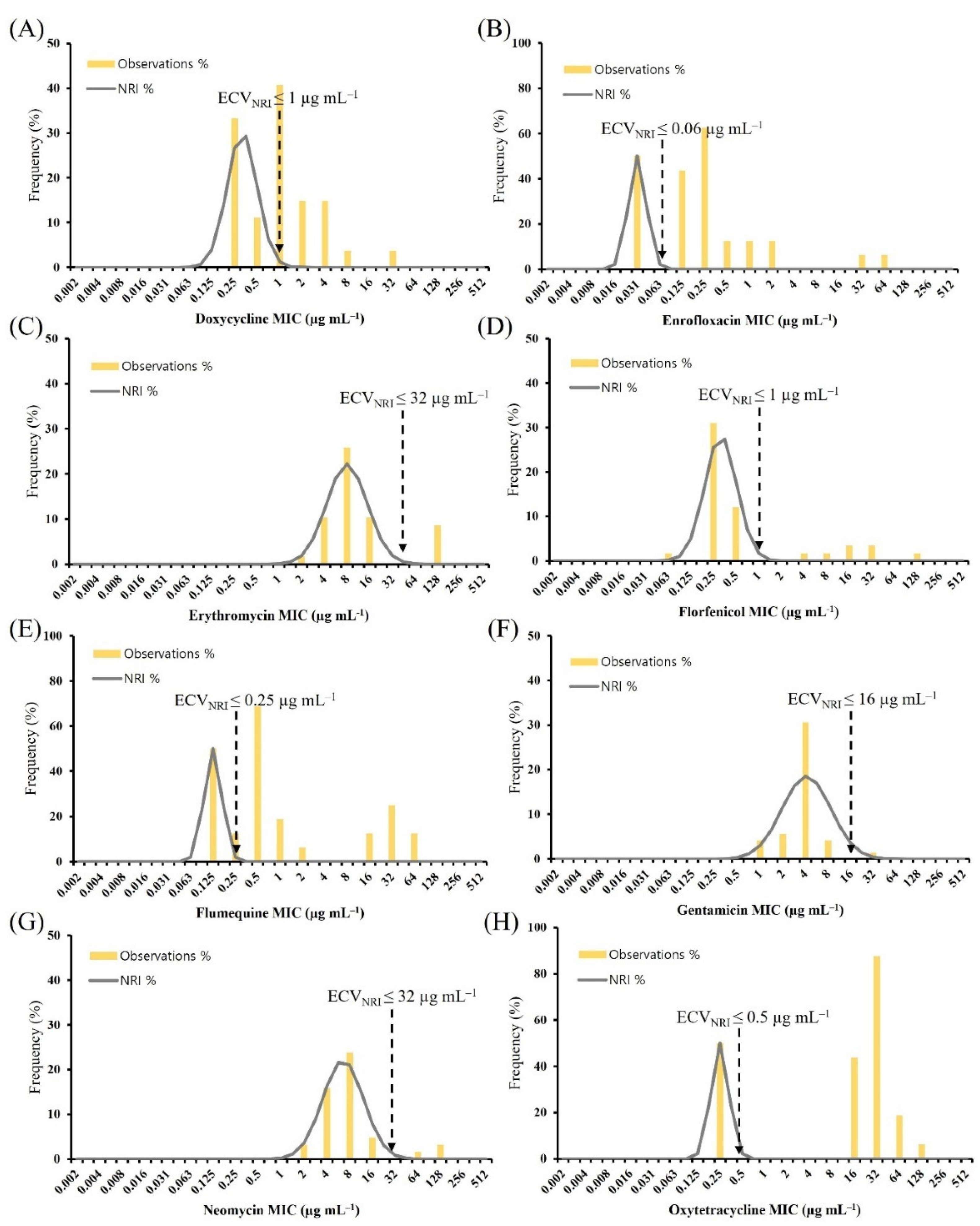
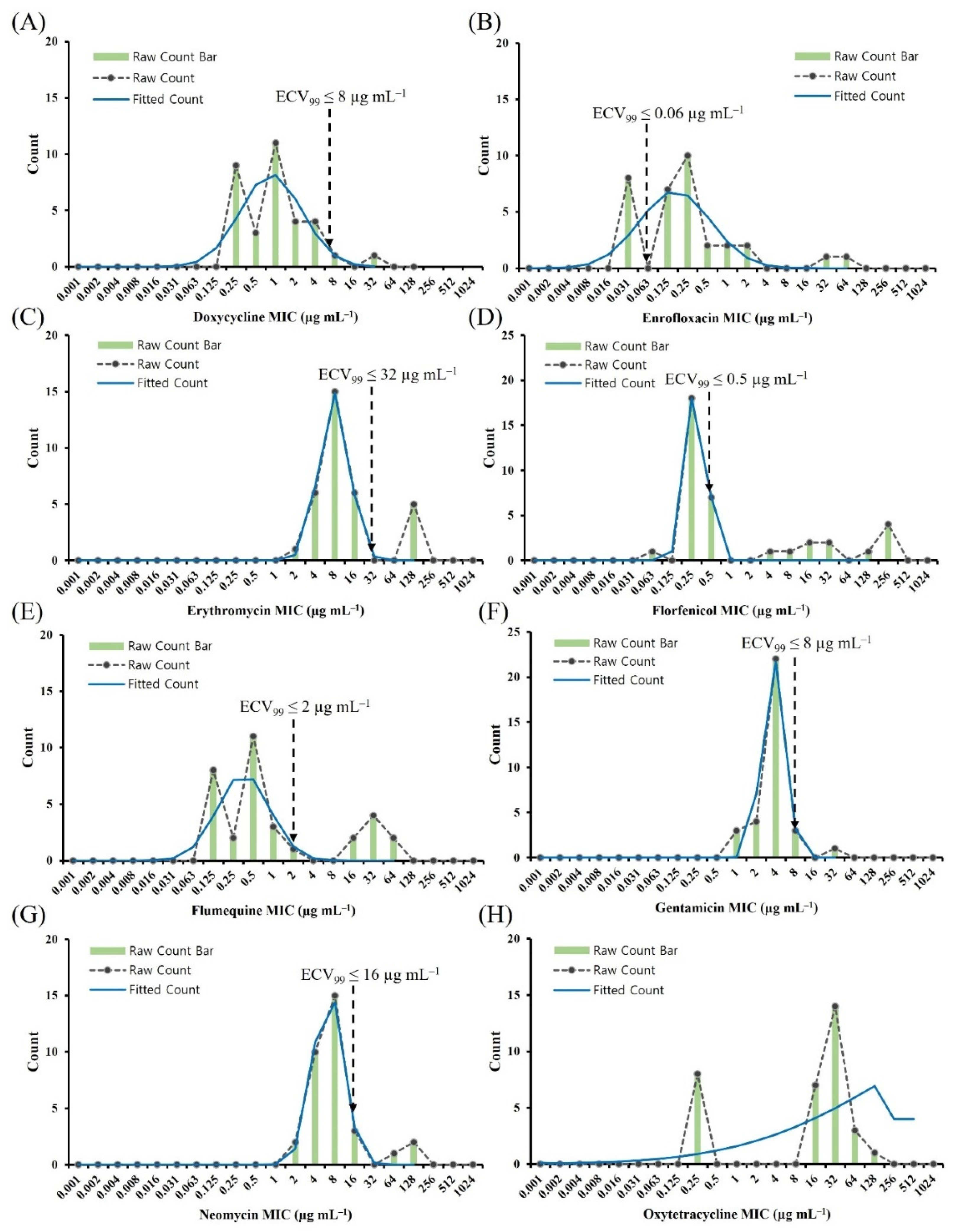
2.2. ECV Establishment Using Two Analytical Methods

We aimed to establish the ECVs for doxycycline, enrofloxacin, erythromycin, florfenicol, flumequine, gentamicin, neomycin, and oxytetracycline by testing 43 A. hydrophila and 33 A. veronii isolates from various diseased aquatic animals using the normalized resistance interpretation (NRI) and ECOFFinder methods. Figure 1 shows the histogram of MICs for eight antimicrobial agents against A. hydrophila using the NRI method. Based on the MIC distributions, the ECVNRI for doxycycline was 2 µg mL−1. This categorized 23 (53.5%) isolates as non-wild type (NWT); they exhibited reduced susceptibility. The ECVNRI values for erythromycin and florfenicol were 64 µg mL−1 and 1 µg mL −1, which categorized 23 (53.5%) isolates and 24 (55.8%) isolates as NWT, respectively. The ECVNRI values for enrofloxacin and flumequine were 32 µg mL−1 and 64 µg mL−1, respectively; however, the standard deviation values of 1.2 log2 indicated inadequate precision. The NRI calculations did not generate results for oxytetracycline.
Figure 2 shows the histogram of MICs for eight antimicrobial agents and the 99.0% ECV (ECV99), which was calculated using ECOFFinder software. The ECV99 value for doxycycline was 128 µg mL−1, indicating that no isolates could be considered NWT. The ECV99 value for enrofloxacin and gentamicin was 16 µg mL−1, which categorized 11 (25.6%) and six (14.0%) isolates as NWT, respectively. However, ECOFFinder failed to provide ECV99 values for four antimicrobial agents (erythromycin, flumequine, neomycin, and oxytetracycline) revealing the lack of a normal distribution; this complicated the interpretation of the MIC distributions.
Figure 3 shows the histogram of MIC for eight antimicrobial agents against A. veronii using the NRI method. The ECVNRI values for doxycycline and enrofloxacin were 1 µg mL−1 and 0.06 µg mL−1, which categorized 10 (30.3%) and 25 (75.8%) isolates as NWT, respectively.
Figure 4 shows the histogram of MICs for eight antimicrobial agents and the ECV99, The ECV99 values for florfenicol and flumequine were 0.5 µg mL−1 and 2 µg mL−1, which categorized seven (21.2%) and eight (24.2%) isolates as NWT, respectively. The ECV99 values for gentamicin and neomycin were 8 µg mL−1 and 16 µg mL−1, respectively.

2.3. Comparison of the ECVCLSI, ECVNRI, and ECV99

We compared the ECVs of eight antimicrobial agents for A. hydrophila and A. veronii isolates using the CLSI, NRI, and ECOFFinder methods. There is no breakpoint for the two Aeromonas spp. isolates; however, recently, the CLSI provided six ECVs for A. hydrophila [18]. The ECVCLSI and ECVNRI for erythromycin against A. hydrophila, was 64 µg mL−1 (Table 3). Additionally, the ECVNRI and ECV99 for gentamicin was 16 µg mL−1, which was two-fold higher than ECVCLSI. The ECV99 for enrofloxacin was 16 µg mL−1, which was more than nine dilution steps from the ECVCLSI. Among the ECVs for the eight antimicrobials, the ECV for florfenicol was optimal, showing the least 1-fold dilution between ECVCLSI and ECVNRI or ECV99. We calculated values for flumequine and neomycin using only the NRI method. The CLSI has not provided the breakpoint or ECVs for A. veronii. The ECVNRI and ECV99 values for enrofloxacin (0.06 µg mL−1) and erythromycin (32 µg mL−1) were the same (Table 4), whereas the ECVNRI values for florfenicol, gentamicin, and neomycin were one-fold higher than the ECV99 values. Oxytetracycline was evaluated using only the NRI method with 0.5 µg mL−1 as the ECVNRI value.

2.4. Presumptive Multidrug-Resistant (pMDR) Aeromonas spp. Isolates

A total of 18.6% (n = 8) of the isolates presented a pMDR phenotype, suggesting that multiple antimicrobial resistance is a common phenomenon in A. hydrophila (Table 5). All isolates from Anguilla japonica, Silurus asotus, Salmo salar, and Misgurnus mizolepis were resistant to three or more classes of antimicrobials. One isolate was resistant to seven antimicrobial agents, and five isolates were resistant to six agents. Additionally, 24.2% (n = 8) of A. veronii isolates presented the pMDR phenotype, and were highly resistant to enrofloxacin, florfenicol, and oxytetracycline. None of the isolates were resistant to all the eight antimicrobial agents.

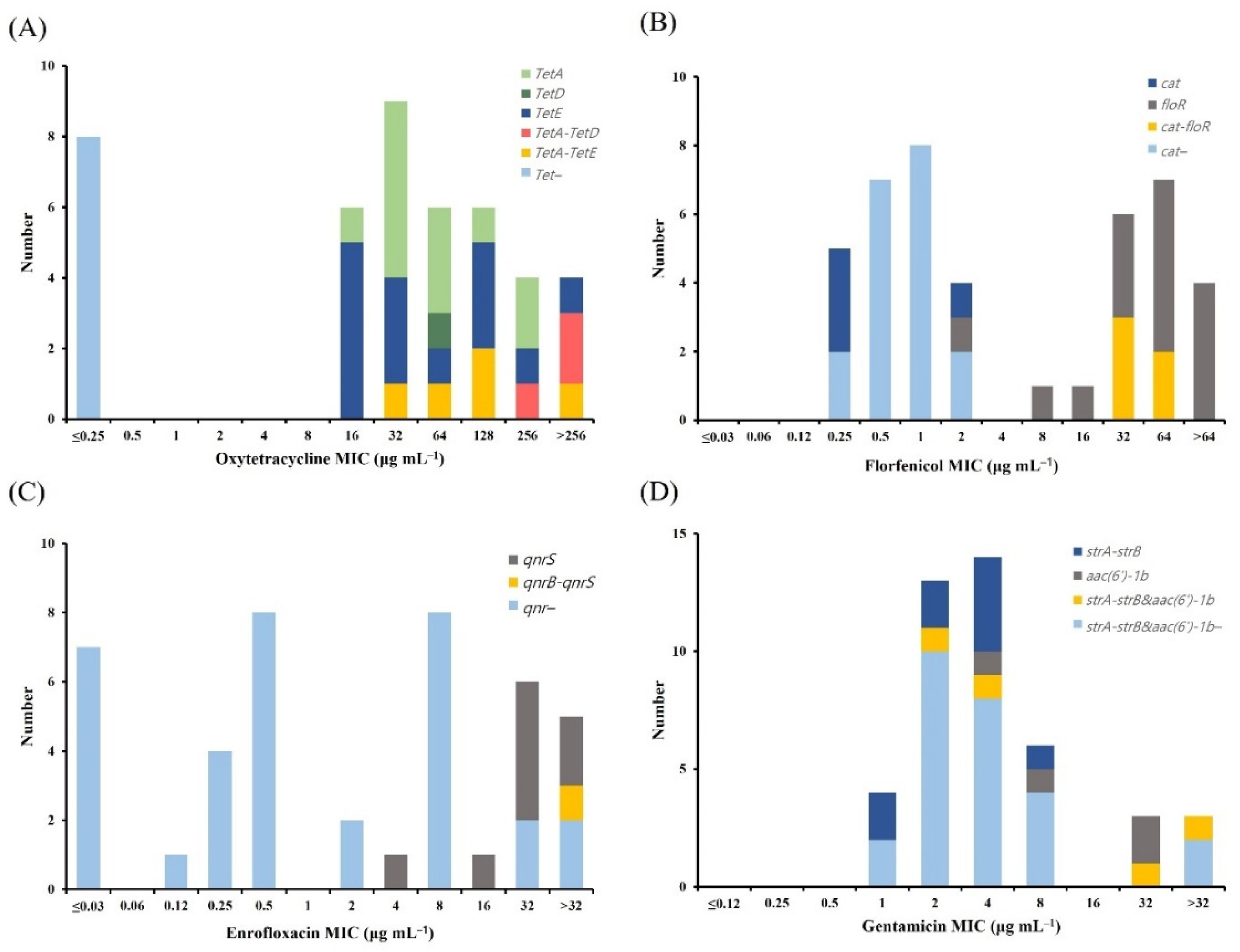
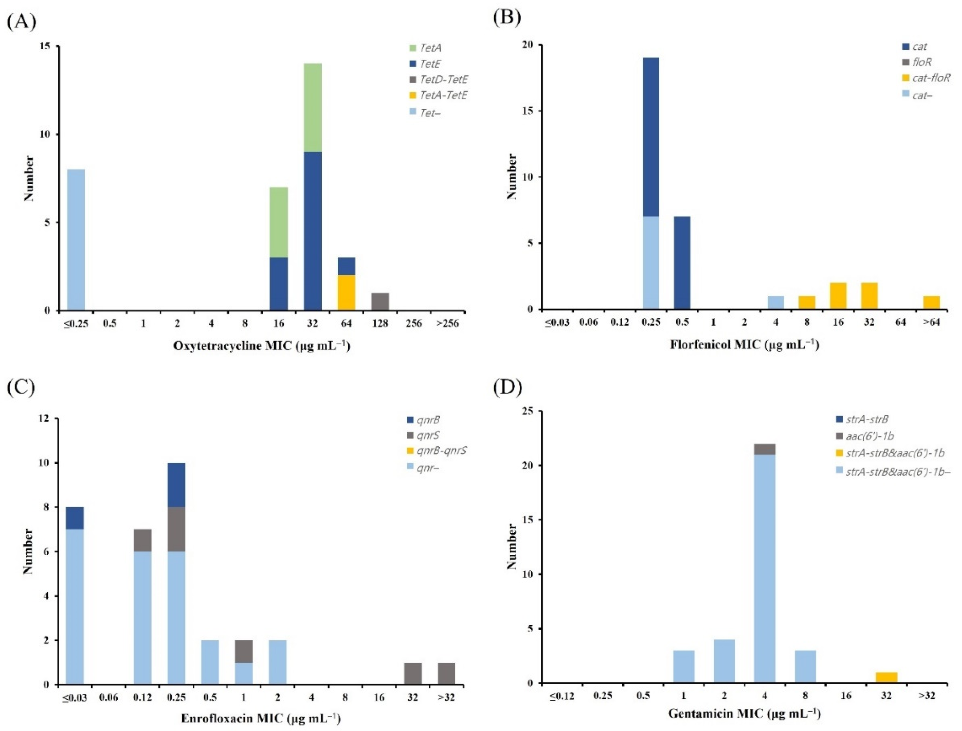
2.5. Distribution of Antimicrobial Resistance Genes (ARGs)

We analyzed four tet genes (tetA, tetB, tetD, and tetE) encoding proteins involved in tetracycline efflux (Figure 5). In A. hydrophila, all the tet-positive isolates (35 isolates) were oxytetracycline NWT at ECVCLSI (Figure 5A). The most common tet gene was tetE, which was found in 14 (40%) NWT isolates, followed by tetA, which was found in 12 (34.3%) NWT isolates. Some of the isolates carried more than one tet gene, with tetAtetD (three isolates) and tetAtetE (five isolates) related to the oxytetracycline MICs ranging from 32 µg mL−1 to 256 µg mL−1 and demonstrating high resistance to oxytetracycline. The tetB gene was not detected in any of the strains. We analyzed the four tet genes in A. veronii (Figure 6). In A. veronii, all the tet-positive isolates (25 isolates) were oxytetracycline NWT at ECVNRI (Figure 6A), and the most common tet gene was tetE, which was found in 13 (52%) of the NWT isolates. Additionally, A. veronii isolates with MICs of 64 µg mL−1 (two strains) and 128 µg mL−1 (one strain) carried two tet genes, (tetAtetE and tetDtetE, respectively). The tetB gene was not detected in any of the strains.
Florfenicol NWT in A. hydrophila and A. veronii isolates was examined to determine the presence of the resistance genes for chloramphenicol acetyltransferase (cat) and florfenicol resistance (floR). In A. hydrophila, 79.2% (19/24) of the ARG-positive isolates were florfenicol NWT at ECV99 (Figure 5B), with cat and floR detected in 0% (0/19) and 73.7% (14/19) of the NWT isolates, respectively. Moreover, five isolates with MICs of 32 µg mL−1 and 64 µg mL−1 carried both the resistance genes (catfloR). We detected no resistance genes in 19 isolates among all the strains. In A. veronii, 24.0% (6/25) of the florfenicol NWT at ECV99 were ARG-positive isolates (Figure 6B); however, 73.1% (19/26) of florfenicol WT carried the cat gene. Furthermore, six A. veronii isolates with MICs ranging from 8 µg mL−1 to 32 µg mL−1 and >64 µg mL−1 carried two resistant genes (catfloR).
We tested A. hydrophila and A. veronii enrofloxacin NWT isolates for the three resistance genes, qnrA, qnrB, and qnrS. In A. hydrophila, 77.8% (7/9) of the ARG-positive isolates were enrofloxacin NWT at ECV99 (Figure 5C), with qnrS detected in 54.5% (6/11) of the NWT isolates; only one at MIC >32 µg mL−1 harbored more than one type of qnr gene. In A. veronii, 88.9% (8/9) of the ARG-positive isolates were enrofloxacin NWT at ECV99 (Figure 6C), with qnrB and qnrS detected in 8% (2/25) and 24% (6/25) of the NWT isolates, respectively. Furthermore, we did not detect the qnrA gene in any of the A. hydrophila or A. veronii strains.
We tested A. hydrophila and A. veronii gentamicin NWT isolates for the two resistance genes strA-strB and aac(6′)-1b. In A. hydrophila, 23.5% (4/17) of the ARG-positive isolates were gentamicin NWT at ECV99 (Figure 5D); however, 24.3% of the gentamicin WT 37 isolates carried the strA-strB gene. Among the NWT isolates, 66.7% harbored more than one resistance gene [e.g., strA-strBaac(6′)-1b]. In A. veronii, there were no ARGs in the 31 isolates from the different strains (Figure 6D); however, one isolate with an MIC of 32 µg mL−1 harbored two resistant genes [strA-strBaac(6′)-1b]. Table 6 summarizes the ARG distributions in the A. hydrophila and A. veronii isolates.

2.6. Quality Control (QC)

Eight antimicrobial agents of QC MICs for Escherichia coli ATCC 25922, Aeromonas salmonicida subsp. salmonicida ATCC 33658, and Enterococcus faecalis ATCC 29212 were within the acceptable range (94.3 to 100%) for the standard broth-microdilution method, as stipulated by the CLSI documents, M45, M7, and VET04 [18,24,25]. The results for doxycycline and neomycin against A. salmonicida ATCC 33658 were excluded from the QC, because of the lack of an established acceptable range in CLSI document VET04. Table S1 shows the MICs for the QC strains.

3. Discussion

The development of multiple antibiotic resistance strains of A. hydrophila and A. veronii in recent years is a serious public health concern, because of the possibility of their transmission from infected fish or water sources to humans and the subsequent infections [26]. In this study, we established eight ECVs for 43 A. hydrophila and 33 A. veronii isolates from aquatic animals and evaluated their ARG distributions. Some ECVCLSI values were suggested for A. hydrophila [18]. The lack of clinical breakpoints or guidelines to interpret ECVs for A. veronii prompted the use of two methods for determining ECVs and interpreting the antimicrobial susceptibility of A. hydrophila and A. veronii.
Three antimicrobials (doxycycline, enrofloxacin, and oxytetracycline) exhibited bimodal MIC distributions, which revealed two clearly distinct populations of A. hydrophila and A. veronii. Based on these distributions, the calculated MIC50 (4 µg mL−1) for gentamicin against A. hydrophila and A. veronii was higher than 1 µg mL−1. This is in line with that reported for 138 Aeromonas spp. isolates recovered from European rivers [27]. The MIC50 and MIC90 values for oxytetracycline were 34.97 µg mL−1 and 149.26 µg mL−1, respectively, for 64 pathogenic Aeromonas strains isolated from ornamental fish [28]. Similarly, the MIC50 values were ≤2 µg mL−1 for florfenicol, 8 µg mL−1 for oxytetracycline, and 0.5 µg mL−1 for ciprofloxacin for 72 aeromonads isolated from koi carp [29]. These findings suggested that the isolates obtained 10 years ago were more susceptible to these drugs.
Tetracycline classes, including oxytetracycline and doxycycline, are broad-spectrum agents extensively used to treat bacterial infections and prevent infections in aquaculture. However, oxytetracycline is poorly absorbed in the fish gut; therefore, it must be administered at high doses [30]. This study showed that 89.2% of A. hydrophila could be categorized as NWT upon applying an oxytetracycline ECVCLSI of 0.25 µg mL−1; 75.8% of A. veronii were determined as NWT upon applying an oxytetracycline ECVNRI of 0.5 µg mL−1. This confirmed the high resistance rate in Aeromonas spp. However, 33 Aeromonas isolates (14.2%) recovered from 16 rivers were considered NWT for tetracycline (23), and 39 Aeromonas isolates (40.6%) from different fish species with reduced susceptibility to tetracycline were classified as NWT [23]. Additionally, A. hydrophila isolates from tilapia, carp, and channel catfish were more susceptible to doxycycline than to oxytetracycline [31]. Aeromonas spp. easily develop single or multiple antibiotic resistance phenotypes and are generally resistant to tetracyclines, quinolones, and β-lactams [5,32]. Moreover, tetracycline-resistant Aeromonas isolates are observed in wastewater discharge, lakes, and carp ponds [32,33,34,35]. In this study, we found that 62.8% of A. hydrophila isolates and 75.8% of A. veronii NWT isolates harbored tetA, tetD, tetE, or more than one tet gene, indicating that the WT isolates did not possess any tet genes. Aeromonas spp. isolates predominantly carried tetE, followed by tetA. However, 37% of A. veronii isolates recovered from channel catfish carried tetE, and 3.8% of isolates carried tetA [36]. Furthermore, A. hydrophila isolates showing oxytetracycline MICs ranging from 32–256 µg mL−1 harbored more than one tet gene (tetA-tetE and tetA-tetD), indicating that the degree of oxytetracycline resistance was associated with the number and type of tet genes present. E. coli isolates harboring tetA and tetB or tetA and tetC exhibited high MICs for tetracycline (256 µg mL−1) or oxytetracycline (512 µg mL−1) [37]. The ECVCLSI for A. hydrophila and ECVNRI for A. veronii might account for the correlations between the NWT isolates and the distribution of resistance genes.
In Korea, florfenicol is approved for use against bacterial diseases in Oncorhynchus mykiss, A. japonica, and Seriola quinqueradiata [38]. The ECVNRI for florfenicol is 1 µg mL−1 for A. hydrophila (55.8%) and A. veronii (21.2%), which were categorized as NWT with reduced susceptibility. However, 2.1% isolates of Aeromonas spp. are NWT considering the ECVNRI (2 µg mL−1) [21], and 25.5% are NWT considering the ECVNRI (4 µg mL−1) [23]. The high frequency of NWT isolates from Korea could be associated with the excessive use of antimicrobial agents in aquaculture; the recorded florfenicol sales was approximately six tons in 2019 [39]. Additionally, we detected cat and floR in A. hydrophila and A. veronii NWT isolates; both genes are associated with high MICs. A total of 7.5% A. veronii isolates harbored floR, which conferred resistance to florfenicol [36]. A resistance cassette, carrying the floR gene in A. salmonicida enables mobilization [40]. The first floR-containing plasmid was discovered in Aeromonas bestiarum [41]. Interestingly, the presence of cat was related to a low MIC for florfenicol (0.25 or 0.5 µg mL−1). These results indicated a higher correlation between the presence of floR and NWT categorization, compared to that with the presence of cat.
Enrofloxacin is a member of the fluoroquinolone family of antibiotics and exhibits strong bactericidal activity against aerobic and facultative anaerobic bacteria [42]. For A. hydrophila, the ECVCLSI of 0.03 µg mL−1 was lower than the ECV99 of 16 µg mL−1, indicating that lowering the ECV would increase the likelihood of identifying resistance genes or mutants while increasing the risk of misclassifying the number of WT isolates. Based on our findings, an ECVCLSI of 0.03 µg mL−1 would misclassify 58.1% of NWT (25 isolates), compared to an ECV99 of 16 µg mL−1. We mostly detected qnrS in A. hydrophila and A. veronii NWT isolates; therefore, ECVs should be established in detail based on the ARG distributions. qnrS was the most prevalent, with its presence in 68% of aeromonad isolates that demonstrated high levels of resistance to nalidixic acid and ciprofloxacin; no amplicon was detected for qnrA [43]. The detection of the factors enabling plasmid-mediated quinolone resistance indicated that the complex Aeromonas mobilome increases the possibility of horizontal gene transfer, including that of qnrS and qnrB.
Erythromycin is not approved for use in the USA; however, Aeromonas strains highly resistant to erythromycin have been isolated from foreign countries [44]. Additionally, Aeromonas spp. are resistant to penicillin, cephalosporins, vancomycin, and erythromycin [45,46]. In this study, 53.5% and 15.2% of A. hydrophila and A. veronii, respectively, were categorized as NWT upon application of the erythromycin ECVNRI. Similarly, 50% and 53% of aeromonads isolated from lakes and chickens, respectively, showed resistance to erythromycin [47,48]. Furthermore, harboring macrolide MacB ABC transporter genes confers erythromycin resistance; the MacA gene regulates the drug-binding and ATPase activity of MacB [49]. We did not investigate the distribution of macrolide resistance genes; further studies are required to elucidate the cause underlying the acquisition of erythromycin resistance, owing to the high prevalence of erythromycin NWT Aeromonas spp. isolates.
The results showed that 3% of A. veronii was classified as NWT upon application of the gentamicin ECVNRI and ECV99. Consistent with these findings, 2% of Aeromonas spp. exhibited gentamicin resistance; however, no A. veronii isolate was resistant to gentamicin [50]. We did not detect any aminoglycoside- resistance genes among the 31 A. veronii isolates (94%). However, in an earlier report, all Aeromonas spp. isolates recovered from marketed cockles harbored aac(6′)-1b, with strA-strB found in 41% of the isolates [43]. The recommended first-line therapeutic options for Aeromonas infections are aminoglycosides and fluoroquinolones. We identified gentamicin as an aquatic medicine that can be inoculated orally to prevent Aeromonas infection. Its appropriate use could potentially prevent the emergence of new resistant strains.
The resistance phenotypes varied among isolates. The pMDR of A. hydrophila, which was resistant to three or more classes of antimicrobials, was 18.6%; this was lower than that observed in a previous study conducted on tilapia where 64% of isolates were resistant to six to eight drugs [31] and that in 95 motile pMDR aeromonads isolated from freshwater [46]. Additionally, multi-antibiotic resistant Aeromonas spp. isolates harbored a tripartite AheABC efflux pump, and the use of phenylalanine–arginine–β–naphthylamide contributed to intrinsic resistance [51]. Among the Aeromonas spp. isolates identified as pMDR, the most common resistance was against oxytetracycline (100%). Oxytetracycline is among the most commonly used antibiotics in humans and animals, and these results are consistent with those of a previous study [52]. The distribution of strains resistant to oxytetracycline has increased with the global use of antibiotics; the emergence of pMDR strains complicates the selection of available therapeutics.
This study provides eight putative ECVs for classifying WT and NWT isolates; however, the findings should not be used as Aeromonas-pathogen-treatment guidelines. These ECVs were derived from one laboratory; therefore, it is essential to evaluate different sources and a large number of isolates for reliably establishing ECVs for each Aeromonas strain [53]. The results from this study can be used as a foundation to establish clinical breakpoints for each Aeromonas strain. Additionally, it is necessary to study the NWT bacterial transcriptome and the mechanism of antibiotic resistance transmission between humans and fish to determine the cause of resistance acquisition.

4. Materials and Methods

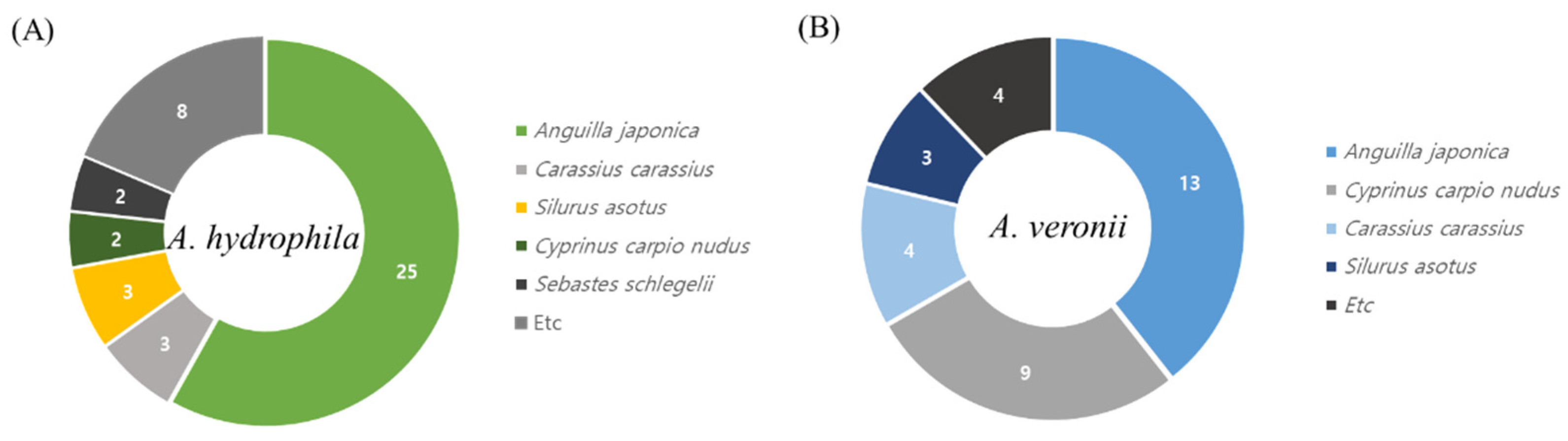
4.1. Collection and Isolation of Aeromonas spp.

Aeromonas spp. isolates were collected between 2008 and 2020 from eight Korean provinces (Chungbuk, Chungnam, Gyeongbuk, Gyeongnam, Gyeonggi, Jeonbuk, Jeonnam, and Gangwon), with 43 A. hydrophila isolates recovered from A. japonica (n = 25), Carassius carassius (n = 3), S. asotus (n = 3), Cyprinus carpio nudus (n = 2), Sebastes schlegelii (n = 2), and others (n = 8); and 33 A. veronii isolates recovered from A. japonica (n = 13), C. carpio nudus (n = 9), C. carassius (n = 4), S. asotus (n = 3), and others (n = 4) (Figure 7). The bacterial strains are listed in Tables S2 and S3. The fish species were sampled from among seemingly healthy, clinical–subclinical, and moribund fish that differed by the year and region of collection. Samples were taken from the lesions, kidneys, and spleens of fish. All experiments were performed in accordance with Directive 2010/63/EU of the European Parliament and the Council (22 September 2010) on the protection of animals used for scientific purposes. Aeromonas spp. isolates were grown on Aeromonas agar (MB cells, Los Angeles, CA, USA), incubated at 37 °C for 24 h. Presumptive aeromonad colonies showing typical dark-green opaque color with a dark center were chosen and subjected to molecular identification. Genomic DNA was extracted from a single colony using a QIAmp DNA blood mini kit (Qiagen, Milan, Italy), according to the manufacturer instructions. DNA concentration and purity were quantified using a Nano Drop R 2000 spectrophotometer (Thermo Fisher Scientific, Waltham, MA, USA) DNA was stored at −80 °C until use. Aeromonas spp. isolates were stored at −80 °C in tryptic soy broth (Merck, Kenilworth, NJ, USA) supplemented with 20% glycerol until further use.

4.2. Molecular Identification

Bacterial identities were confirmed using PCR with two different primer sets for amplification and sequencing of 16S rRNA and gyrB. The 16s rRNA gene (1361 bp) was amplified and sequenced using specific primers (27F: 5′-AGA GTT TGA TCM TGG CTC AG-3′ and 1387R: 5′-GGG CGG WGT GTA CAA GGC-3′). gyrB (904 bp) was used as the housekeeping gene to further identify species (gyrB 3F: 5′-TCC GGC GGT CTG CAC GGC GT-3′ and gyrB 14R: 5′-TTG TTC GGG TTG TAC TCG TC-3′) [54]. The PCR reaction mix at 50 µL contained 5 µL of 10× Ex Taq buffer, 4 µL dNTP mixture (2.5 mM each), 10 pmol of each primer, 0.25 µL Ex Taq DNA polymerase (Takara, Shiga, Japan), 10 ng DNA template, and sterile purified water. The reaction conditions were as follows: initial denaturation at 95 °C for 3 min, followed by 30 cycles of denaturation at 98 °C for 10 s, 55 °C for 30 s, and extension at 72 °C for 30 s, with a final extension at 72 °C for 7 min. The PCR products were confirmed through sequence analyses (Bionics, Seoul, Korea); the strains were verified based on the reference sequences accessed from GenBank (https://www.ncbi.nlm.nih.gov/genbank/) (accessed on: 5 May 2021).

4.3. Antimicrobial Susceptibility Test

Antimicrobial susceptibility tests were performed according to the broth microdilution method described in the CLSI guidelines VET04 [17,18]. The antimicrobial agents for Aeromonas spp. isolates are licensed and commonly used for aquatic animals in Korea [38]. The MICs of 43 A. hydrophila and 33 A. veronii isolates were tested using Sensititre CAMPY2 and KRAQ1 plates (Trek Diagnostics System, Cleveland, OH, USA). MICs for erythromycin (0.03–64 mg L−1), florfenicol (0.03–64 mg L−1), and gentamicin (0.12–32 mg L−1) were tested using CAMPY2; and those for doxycycline (0.25–64 mg L−1), enrofloxacin (0.03–32 mg L−1), flumequine (0.12–128 mg L−1), neomycin (0.5–64 mg L−1), and oxytetracycline (0.25–256 mg L−1) were tested using KRAQ1. Isolates were cultured on tryptic soy agar for 24 h at 28 °C, after which a suspension was prepared in sterile saline solution, adjusted to 0.5 McFarland standard, and diluted to reach a final inoculum concentration of 5 × 10⁵ CFU/mL using a Nephelometer® (V3011, Thermo Scientific, Roskilde, Denmark)) to standardize inoculum density/turbidity. Microplates were incubated at 28 °C for 24 h for A. hydrophila and A. veronii. MICs were defined as the lowest drug concentrations that inhibited growth, compared to that in the drug-free growth control. E. coli ATCC 25922, A. salmonicida subsp. salmonicida ATCC 33658, and E. faecalis ATCC 29212 were included in the susceptibility test as QC strains. Recently, additional MICs of ECVs were made available for A. hydrophila in the updated CLSI guidelines [18]. We compared the A. hydrophila and A. veronii isolates among WT and NWT populations, according to the CLSI guidelines and the provisional ECVs proposed in this study.

4.4. Determination of Provisional ECVs

ECVs were calculated using two methods: NRI [55] and ECOFFinder [56]. The NRI method is a fully automatic and freely available Excel spreadsheet calculator (last updated in 2019; http://www.bioscand.se/nri) (accessed on: 3 May 2021). The ECOFFinder method (v.2.1; last updated in 2020) is available from the EUCAST website (https://www.eucast.org/mic_distributions_and_ecoffs) (accessed on: 3 May 2021). In this study, ECV determination was based on the distribution of antimicrobial MICs for each drug against A. hydrophila and A. veronii. ECV allows isolates to be categorized as WT at ≤x mg L−1 and NWT as >x mg L−1. A 99.0% cut-off was applied, which means that approximately 99.0% of the WT MIC distribution was less than the identified ECV. pMDR was defined as resistance to more than three antimicrobial agents, classes, or subclasses of antimicrobial categories [57]. The number of pMDR Aeromonas was determined for eight antimicrobial agents (doxycycline, enrofloxacin, erythromycin, florfenicol, flumequine, gentamicin, neomycin, and oxytetracycline) in the clinical samples.

4.5. Terminology

When referring to the categorization of isolates based on their susceptibility, we followed the recommendations, which suggested that when isolates are categorized by applying ECVs, the terms “sensitive” and “resistant” should not be used [58]. WT is defined, for a fully susceptible species, as the absence of acquired- and mutational-resistance mechanisms to the drug, and NWT is defined as the reduced susceptibility to the presence of an acquired- or mutational- resistance mechanism to the drug. However, when referring to studies that used the term “resistant”, we did not change their terminology. The CLSI uses the abbreviation “ECV” for epidemiological cut-off values, whereas EUCAST uses the ECOFF. This study used “ECV” to prevent confusion when comparing the ECOFF values using the two analytical methods.

4.6. Analysis of ARGs

We tested 43 A. hydrophila and 33 A. veronii isolates for the presence of ARGs, including tetA, tetB, tetD, and tetE for tetracycline; cat and floR for phenicol; qnr-type pentapeptide proteins encoded by qnrA, qnrB, and qnrS for quinolone; and strA-strB and aac(6′)-1b for aminoglycosides (Table 7). The primers used to detect these genes were selected from previous studies. The PCR cycling conditions were as follows: 94 °C for 5 min, followed by 35 cycles of 95 °C for 30 s, annealing for 30 s at different temperatures, 72 °C for 30 s, and 72 °C for 5 min. The PCR products were separated using electrophoresis on a 1% agarose gel and purified for sequencing. Sequence identities were confirmed using the sequence information in the NCBI database (on https://www.ncbi.nlm.nih.gov/) (accessed date: 22 June 2021).

5. Conclusions

This is the first study to establish ECVNRI and ECV99 values for eight antimicrobials against 43 A. hydrophila and 33 A. veronii isolates recovered from aquatic animals in Korea and to detect ARGs in Aeromonas strains. A total of 89.2% A. hydrophila isolates and 75.8% A. veronii isolates were classified as NWT against oxytetracycline; they harbored tet genes; Aeromonas spp. isolates predominantly carried tetE, followed by tetA. Additionally, the distribution of floR and qnrS was prevalent in NWT isolates, whereas no aac(6′)-1b or strA-strB was detected in the 31 A. veronii isolates. The emergence of antibiotic-resistant strains of Aeromonas spp. reduces the choice of currently available therapeutic agents and it could lead to prolonged Aeromonas infections. Therefore, these results can potentially help aquaculture managers and researchers alleviate Aeromonas infections in aquaculture systems and raise awareness of the appropriate use of antimicrobials in aquaculture. Furthermore, these findings encourage the application of vaccination or herbal therapy, to reduce antibiotic resistance and public health problems.

Supplementary Materials

The following supporting information can be downloaded at: https://www.mdpi.com/article/10.3390/antibiotics11030343/s1, Table S1: CLSI-approved broth microdilution MIC QC ranges determined for eight antimicrobial agents against selected reference strains; Table S2: Isolate year, fish species, disease outbreak, isolation source, and geographical location of the 43 A. hydrophila strains; Table S3: Isolate year, fish species, disease outbreak, isolation source, and geographical location of the 33 A. veronii strains.

Author Contributions

Conceptualization, S.-J.W.; methodology, S.-J.W. and M.-G.J.; software, M.-G.J. and M.-Y.D.; validation, M.-G.J. and M.-Y.D.; formal analysis, S.-J.W. and M.-G.J.; investigation, S.-J.W. and M.-Y.D.; data curation, S.-J.W., M.-S.K., S.-D.H. and W.-J.K.; writing—original draft preparation, S.-J.W.; writing—review and editing, S.-J.W., M.-S.K., S.-D.H. and W.-J.K.; project administration, M.-S.K. All authors have read and agreed to the published version of the manuscript.

Funding

This research was supported by a grant from the National Institute of Fisheries Science (grant No. R2022076).

Institutional Review Board Statement

Not applicable.

Informed Consent Statement

Not applicable.

Data Availability Statement

The data that support the findings of this study are available upon request from the corresponding author.

Conflicts of Interest

The authors declare no conflict of interest.

References

  1. Khor, W.C.; Puah, S.M.; Koh, T.H.; Tan, J.A.M.A.; Puthucheary, S.D.; Chua, K.H. Comparison of clinical isolates of Aeromonas from Singapore and Malaysia with regard to molecular identification, virulence, and antimicrobial profiles. Microb. Drug Resist. 2018, 24, 469–478. [Google Scholar] [CrossRef]
  2. Fečkaninová, A.; Koščová, J.; Mudroňová, D.; Popelka, P.; Toropilova, J. The use of probiotic bacteria against Aeromonas infections in salmonid aquaculture. Aquaculture 2017, 469, 42. [Google Scholar] [CrossRef]
  3. Mazumder, A.; Choudhury, H.; Dey, A.; Sarma, D. Isolation and characterization of two virulent Aeromonads associated with haemorrhagic septicaemia and tail-rot disease in farmed climbing perch Anabas testudineus. Sci. Rep. 2021, 11, 5826. [Google Scholar] [CrossRef]
  4. Sen, K.; Rodgers, M. Distribution of six virulence factors in Aeromonas species isolated from US drinking water utilities: A PCR identification. J. Appl. Microbiol. 2004, 97, 1077–1086. [Google Scholar] [CrossRef] [Green Version]
  5. Janda, J.M.; Abbott, S.L. The genus Aeromonas: Taxonomy, pathogenicity, and infection. Clin. Microbiol. Rev. 2010, 23, 35–73. [Google Scholar] [CrossRef] [Green Version]
  6. Abd-El-Rhman, A.M. Antagonism of Aeromonas hydrophila by propolis and its effect on the performance of Nile tilapia, Oreochromis niloticus. Fish. Shellfish Immunol. 2009, 27, 454–459. [Google Scholar] [CrossRef] [PubMed]
  7. Falco, A.; Frost, P.; Miest, J.; Pionnier, N.; Irnazarow, I.; Hoole, D. Reduced inflammatory response to Aeromonas salmonicida infection in common carp (Cyprinus carpio L.) fed with β-glucan supplements. Fish. Shellfish Immunol. 2012, 32, 1051–1057. [Google Scholar] [CrossRef] [PubMed]
  8. Vanden Bergh, P.; Frey, J. Aeromonas salmonicida subsp. salmonicida in the light of its type-three secretion system. Microb. Biotechnol. 2014, 7, 381–400. [Google Scholar] [CrossRef] [PubMed] [Green Version]
  9. Wang, R.; Hu, X.; Lü, A.; Liu, R.; Sun, J.; Sung, Y.Y.; Song, Y. Transcriptome analysis in the skin of Carassius auratus challenged with Aeromonas hydrophila. Fish. Shellfish Immunol. 2019, 94, 510–516. [Google Scholar] [CrossRef]
  10. Mulyani, Y.; Aryantha, I.N.P.; Suhandono, S.; Pancoro, A. Intestinal bacteria of common carp (Cyprinus carpio L.) as a biological control agent for Aeromonas. J. Pure Appl. Microbiol. 2018, 12, 601–610. [Google Scholar] [CrossRef]
  11. Rasmussen-Ivey, C.R.; Figueras, M.J.; McGarey, D.; Liles, M.R. Virulence factors of Aeromonas hydrophila: In the wake of reclassification. Front. Microbiol. 2016, 7, 1337. [Google Scholar] [CrossRef] [PubMed]
  12. Ramalivhana, J.N.; Obi, C.L.; Moyo, S.R. Prevalence of extended-spectrum β-lactamases producing Aeromonas hydrophila isolated from stool samples collected in the Limpopo province, South Africa. Afr. J. Microbiol. Res. 2010, 4, 1203–1208. [Google Scholar] [CrossRef]
  13. Chenia, H.Y. Prevalence and characterization of plasmid-mediated quinolone resistance genes in Aeromonas spp. isolated from South African freshwater fish. Int. J. Food Microbiol. 2016, 231, 26–32. [Google Scholar] [CrossRef] [PubMed]
  14. Hossain, S.; De Silva, B.C.J.; Wimalasena, S.H.M.P.; Pathirana, H.N.K.S.; Dahanayake, P.S.; Heo, G.J. Distribution of antimicrobial resistance genes and class 1 integron gene cassette arrays in motile Aeromonas spp. isolated from goldfish (Carassius auratus). Microb. Drug Resist. 2018, 24, 1217–1225. [Google Scholar] [CrossRef]
  15. Aravena-Román, M.; Inglis, T.J.; Henderson, B.; Riley, T.V.; Chang, B.J. Antimicrobial susceptibilities of Aeromonas strains isolated from clinical and environmental sources to 26 antimicrobial agents. Antimicrob. Agents Chemother. 2012, 56, 1110–1112. [Google Scholar] [CrossRef] [Green Version]
  16. Boerlin, P.; Reid-Smith, R.J. Antimicrobial resistance: Its emergence and transmission. Anim. Health Res. Rev. 2008, 9, 115–126. [Google Scholar] [CrossRef]
  17. Clinical and Laboratory Standards Institute (CLSI). Performance Standards for Antimicrobial Susceptibility Testing of Bacteria Isolated from Aquatic Animals; VET03/VET04-S2.; CLSI: Wayne, PA, USA, 2014; pp. 1–42. [Google Scholar]
  18. Clinical and Laboratory Standards Institute (CLSI). Performance Standards for Antimicrobial Susceptibility Testing of Bacteria Isolated from Aquatic Animals, 3rd ed.; VET04.; CLSI: Wayne, PA, USA, 2020; pp. 1–88. [Google Scholar]
  19. Yao, Z.; Sun, L.; Wang, Y.; Lin, L.; Guo, Z.; Li, D.; Lin, W.; Lin, X. Quantitative proteomics reveals antibiotics resistance function of outer membrane proteins in Aeromonas hydrophila. Front. Cell. Infect. Microbiol. 2018, 8, 390. [Google Scholar] [CrossRef] [Green Version]
  20. Li, Z.; Wang, Y.; Li, X.; Lin, Z.; Lin, Y.; Srinivasan, R.; Lin, X. The characteristics of antibiotic resistance and phenotypes in 29 outer-membrane protein mutant strains in Aeromonas hydrophila. Environ. Microbiol. 2019, 21, 4614–4628. [Google Scholar] [CrossRef]
  21. Baron, S.; Granier, S.A.; Larvor, E.; Jouy, E.; Cineux, M.; Wilhelm, A.; Gassilloud, B.; Bouquin, S.L.; Kempf, I.; Chauvin, C. Aeromonas Diversity and Antimicrobial Susceptibility in Freshwater—An Attempt to Set Generic Epidemiological Cut-Off Values. Front. Microbiol. 2017, 8, 503. [Google Scholar] [CrossRef] [Green Version]
  22. Hayatgheib, N.; Calvez, S.; Fournel, C.; Pineau, L.; Pouliquen, H.; Moreau, E. Antimicrobial Susceptibility Profiles and Resistance Genes in Genus Aeromonas spp. Isolated from the Environment and Rainbow Trout of Two Fish Farms in France. Microorganisms 2021, 9, 1201. [Google Scholar] [CrossRef]
  23. Duman, M.; Saticioglu, I.B.; Altun, S. The determination of antimicrobial susceptibility by MIC and epidemiological cut-off values and the detection of resistance genes in Aeromonas species isolated from cultured fish. Lett. Appl. Microbiol. 2020, 71, 531–541. [Google Scholar] [CrossRef] [PubMed]
  24. Clinical and Laboratory Standards Institute (CLSI). Methods for Antimicrobial Dilution and Disk Susceptibility Testing of Infrequently Isolated or Fastidious Bacteria, 3rd ed.; CLSI guideline M45.; CLSI: Wayne, PA, USA, 2016; pp. 1–19. [Google Scholar]
  25. Clinical and Laboratory Standards Institute (CLSI). Methods for Dilution Antimicrobial Susceptibility Tests for Bacteria That Grow Aerobically, 11th ed.; CLSI standard M07; CLSI: Wayne, PA, USA, 2018; pp. 1–13. [Google Scholar]
  26. Pessoa, R.B.G.; de Oliveira, W.F.; Marques, D.S.C.; dos Santos Correia, M.T.; de Carvalho, E.V.M.M.; Coelho, L.C.B.B. The genus Aeromonas: A general approach. Microb. Pathog. 2019, 130, 81–94. [Google Scholar] [CrossRef] [PubMed]
  27. Goñi-Urriza, M.; Pineau, L.; Capdepuy, M.; Roques, C.; Caumette, P.; Quentin, C. Antimicrobial resistance of mesophilic Aeromonas spp. isolated from two European rivers. J. Antimicrob. Chemother. 2000, 46, 297–301. [Google Scholar] [CrossRef] [Green Version]
  28. Saengsitthisak, B.; Chaisri, W.; Punyapornwithaya, V.; Mektrirat, R.; Klayraung, S.; Bernard, J.K.; Pikulkaew, S. Occurrence and antimicrobial susceptibility profiles of multidrug-resistant aeromonads isolated from freshwater ornamental fish in Chiang Mai province. Pathogens 2020, 9, 973. [Google Scholar] [CrossRef] [PubMed]
  29. Čížek, A.; Dolejská, M.; Sochorová, R.; Strachotová, K.; Piačková, V.; Veselý, T. Antimicrobial resistance and its genetic determinants in aeromonads isolated in ornamental (koi) carp (Cyprinus carpio koi) and common carp (Cyprinus carpio). Vet. Microbiol. 2010, 142, 435–439. [Google Scholar] [CrossRef] [PubMed]
  30. Limbu, S.M.; Zhou, L.; Sun, S.X.; Zhang, M.L.; Du, Z.Y. Chronic exposure to low environmental concentrations and legal aquaculture doses of antibiotics cause systemic adverse effects in Nile tilapia and provoke differential human health risk. Environ. Int. 2018, 115, 205–219. [Google Scholar] [CrossRef] [PubMed]
  31. Nhinh, D.T.; Le, D.V.; Van, K.V.; Huong Giang, N.T.; Dang, L.T.; Hoai, T.D. Prevalence, Virulence Gene Distribution and Alarming the Multidrug Resistance of Aeromonas hydrophila Associated with Disease Outbreaks in Freshwater Aquaculture. Antibiotics 2021, 10, 532. [Google Scholar] [CrossRef]
  32. Piotrowska, M.; Przygodzińska, D.; Matyjewicz, K.; Popowska, M. Occurrence and variety of β-lactamase genes among Aeromonas spp. isolated from urban wastewater treatment plant. Front. Microbiol. 2017, 8, 863. [Google Scholar] [CrossRef] [Green Version]
  33. Goñi-Urriza, M.; Capdepuy, M.; Arpin, C.; Raymond, N.; Caumette, P.; Quentin, C. Impact of an urban effluent on antibiotic resistance of riverine Enterobacteriaceae and Aeromonas spp. Appl. Environ. Microbiol. 66, 125–132. [CrossRef] [Green Version]
  34. Skwor, T.; Shinko, J.; Augustyniak, A.; Gee, C.; Andraso, G. Aeromonas hydrophila and Aeromonas veronii predominate among potentially pathogenic ciprofloxacin-and tetracycline-resistant Aeromonas isolates from Lake Erie. Appl. Environ. Microbiol. 2014, 80, 841–848. [Google Scholar] [CrossRef] [Green Version]
  35. Zdanowicz, M.; Mudryk, Z.J.; Perliński, P. Abundance and antibiotic resistance of Aeromonas isolated from the water of three carp ponds. Vet. Res. Commun. 2020, 44, 9–18. [Google Scholar] [CrossRef] [PubMed] [Green Version]
  36. Tekedar, H.C.; Arick, M.A.; Hsu, C.Y.; Thrash, A.; Blom, J.; Lawrence, M.L.; Abdelhamed, H. Identification of antimicrobial resistance determinants in Aeromonas veronii strain MS-17-88 recovered from channel catfish (Ictalurus punctatus). Front. Cell. Infect. Microbiol. 2020, 10, 348. [Google Scholar] [CrossRef] [PubMed]
  37. Shin, S.W.; Shin, M.K.; Jung, M.; Belaynehe, K.M.; Yoo, H.S. Prevalence of antimicrobial resistance and transfer of tetracycline resistance genes in Escherichia coli isolates from beef cattle. Appl. Environ. Microbiol. 2015, 81, 5560–5566. [Google Scholar] [CrossRef] [PubMed] [Green Version]
  38. National Institute of Food and Drug Safety Evaluation (NIFDS). National Antimicrobial Resistance Surveillance on the Domestic and Imported Meat and Fishery Products; NIFDS: Cheongju, Korea, 2019; pp. 1–161. [Google Scholar]
  39. National Institute of Fisheries Science (NIFS). Aquatic Medicine Catalog; NIFS: Busan, Korea, 2020; pp. 12–102. [Google Scholar]
  40. McIntosh, D.; Cunningham, M.; Ji, B.; Fekete, F.A.; Parry, E.M.; Clark, S.E.; Zalinger, Z.B.; Gilg, I.C.; Danner, R.D.; Johnson, K.A.; et al. Transferable, multiple antibiotic and mercury resistance in Atlantic Canadian isolates of Aeromonas salmonicida subsp. salmonicida is associated with carriage of an IncA/C plasmid similar to the Salmonella enterica plasmid pSN254. J. Antimicrob. Chemother. 2008, 61, 1221–1228. [Google Scholar] [CrossRef] [PubMed] [Green Version]
  41. Gordon, L.; Cloeckaert, A.; Doublet, B.; Schwarz, S.; Bouju-Albert, A.; Ganière, J.P.; Le Bris, H.; Le Flèche-Matéos, A.; Giraud, E. Complete sequence of the floR-carrying multiresistance plasmid pAB5S9 from freshwater Aeromonas bestiarum. J. Antimicrob. Chemother. 2008, 62, 65–71. [Google Scholar] [CrossRef] [Green Version]
  42. Sezer, A.D.; Akbuğa, J.; Baş, A.L. In vitro evaluation of enrofloxacin-loaded MLV liposomes. Drug Deliv. 2007, 14, 47–53. [Google Scholar] [CrossRef]
  43. Dahanayake, P.S.; Hossain, S.; Wickramanayake, M.V.K.S.; Heo, G.J. Prevalence of virulence and antimicrobial resistance genes in Aeromonas species isolated from marketed cockles (Tegillarca granosa) in Korea. Lett. Appl. Microbiol. 2020, 71, 94–101. [Google Scholar] [CrossRef]
  44. Schmidt, A.S.; Bruun, M.S.; Dalsgaard, I.; Larsen, J.L. Incidence, distribution, and spread of tetracycline resistance determinants and integron-associated antibiotic resistance genes among motile aeromonads from a fish farming environment. Appl. Environ. Microbiol. 2001, 67, 5675–5682. [Google Scholar] [CrossRef] [PubMed] [Green Version]
  45. Koksal, F.; Oguzkurt, N.; Samastı, M.; Altas, K. Prevalence and antimicrobial resistance patterns of Aeromonas strains isolated from drinking water samples in Istanbul, Turkey. Chemotherapy 2007, 53, 30–35. [Google Scholar] [CrossRef]
  46. Borella, L.; Salogni, C.; Vitale, N.; Scali, F.; Moretti, V.M.; Pasquali, P.; Alborali, G.L. Motile aeromonads from farmed and wild freshwater fish in northern Italy: An evaluation of antimicrobial activity and multidrug resistance during 2013 and 2016. Acta Vet. Scand. 2020, 62, 1–8. [Google Scholar] [CrossRef]
  47. Igbinosa, I.H. Antibiogram profiling and pathogenic status of Aeromonas species recovered from Chicken. Saudi J. Biol. Sci. 2014, 21, 481–485. [Google Scholar] [CrossRef] [PubMed] [Green Version]
  48. Rawal, I.; Joshi, H.; Chaudhary, L. Isolation, identification, and antibiotics resistance of Aeromonas spp. from lakes of Udaipur (Rajasthan), India. Asian J. Pharm. 2016, 10, 132–136. [Google Scholar] [CrossRef]
  49. Lin, H.T.; Bavro, V.N.; Barrera, N.P.; Frankish, H.M.; Velamakanni, S.; van Veen, H.W.; Robinson, C.; Borges-Walmsley, M.I.; Walmsley, A.R. MacB ABC transporter is a dimer whose ATPase activity and macrolide-binding capacity are regulated by the membrane fusion protein MacA. J. Biol. Chem. 2009, 284, 1145–1154. [Google Scholar] [CrossRef] [Green Version]
  50. Azzam-Sayuti, M.; Ina-Salwany, M.Y.; Zamri-Saad, M.; Yusof, M.T.; Annas, S.; Najihah, M.Y.; Liles, M.R.; Monir, M.S.; Zaidi, Z.; Amal, M.N.A. The prevalence, putative virulence genes and antibiotic resistance profiles of Aeromonas spp. isolated from cultured freshwater fishes in peninsular Malaysia. Aquaculture 2021, 540, 736719. [Google Scholar] [CrossRef]
  51. Hernould, M.; Gagné, S.; Fournier, M.; Quentin, C.; Arpin, C. Role of the AheABC efflux pump in Aeromonas hydrophila intrinsic multidrug resistance. Antimicrob. Agents Chemother. 2008, 52, 1559–1563. [Google Scholar] [CrossRef] [PubMed] [Green Version]
  52. Yu, J.; Ramanathan, S.; Chen, L.; Zeng, F.; Li, X.; Zhao, Y.; Lin, L.; Monaghan, S.J.; Lin, X.; Pang, H. Comparative transcriptomic analysis reveals the molecular mechanisms related to oxytetracycline-resistance in strains of Aeromonas hydrophila. Aquac. Rep. 2021, 21, 100812. [Google Scholar] [CrossRef]
  53. Smith, P.; Kronvall, G. How many strains are required to set an epidemiological cut-off value for MIC values determined for bacteria isolated from aquatic animals? Aquac. Int. 2015, 23, 465–470. [Google Scholar] [CrossRef]
  54. Yanez, M.A.; Catalán, V.; Apráiz, D.; Figueras, M.J.; Martínez-Murcia, A.J. Phylogenetic analysis of members of the genus Aeromonas based on gyrB gene sequences. Int. J. Syst. Evol. Microbiol. 2003, 53, 875–883. [Google Scholar] [CrossRef] [Green Version]
  55. Kronvall, G. Normalized resistance interpretation as a tool for establishing epidemiological MIC susceptibility breakpoints. J. Clin. Microbiol. 2010, 48, 4445–4452. [Google Scholar] [CrossRef] [PubMed] [Green Version]
  56. Turnidge, J.; Kahlmeter, G.; Kronvall, G. Statistical characterization of bacterial wild-type MIC value distributions and the determination of epidemiological cut-off values. Clin. Microbiol. Infect. 2006, 12, 418–425. [Google Scholar] [CrossRef] [PubMed]
  57. Magiorakos, A.P.; Srinivasan, A.; Carey, R.B.; Carmeli, Y.; Falagas, M.E.; Giske, C.G.; Harbarth, S.; Hindler, J.F.; Kahlmeter, G.; Olsson-Liljequist, B.; et al. Multidrug-resistant, extensively drug-resistant and pandrug-resistant bacteria: An international expert proposal for interim standard definitions for acquired resistance. Clin. Microbiol. Infect. 2012, 18, 268–281. [Google Scholar] [CrossRef] [PubMed] [Green Version]
  58. Silley, P. Susceptibility testing methods, resistance and breakpoints: What do these terms really mean? OIE Rev. Sci. Tech. 2012, 31, 33–41. [Google Scholar] [CrossRef] [PubMed]
  59. Jun, L.J.; Jeong, J.B.; Huh, M.D.; Chung, J.K.; Choi, D.L.; Lee, C.H.; Jeong, H.D. Detection of tetracycline-resistance determinants by multiplex polymerase chain reaction in Edwardsiella tarda isolated from fish farms in Korea. Aquaculture 2004, 240, 89–100. [Google Scholar] [CrossRef]
  60. Akinbowale, O.L.; Peng, H.; Barton, M.D. Diversity of tetracycline resistance genes in bacteria from aquaculture sources in Australia. J. Appl. Microbiol. 2007, 103, 2016–2025. [Google Scholar] [CrossRef]
  61. Cattoir, V.; Poirel, L.; Rotimi, V.; Soussy, C.J.; Nordmann, P. Multiplex PCR for detection of plasmid-mediated quinolone resistance qnr genes in ESBL-producing enterobacterial isolates. J. Antimicrob. Chemother. 2007, 60, 394–397. [Google Scholar] [CrossRef] [Green Version]
  62. Sunde, M.; Norström, M. The genetic background for streptomycin resistance in Escherichia coli influences the distribution of MICs. J. Antimicrob. Chemother. 2005, 56, 87–90. [Google Scholar] [CrossRef] [Green Version]
  63. Park, C.H.; Robicsek, A.; Jacoby, G.A.; Sahm, D.; Hooper, D.C. Prevalence in the United States of aac(6′)-Ib-cr encoding a ciprofloxacin-modifying enzyme. Antimicrob. Agents Chemother. 2006, 50, 3953–3955. [Google Scholar] [CrossRef] [Green Version]
Figure 1. Distribution of MICs for Aeromonas hydrophila. MICs for A. hydrophila (n = 43) were determined using the broth microdilution method for (A) doxycycline, (B) enrofloxacin, (C) erythromycin, (D) florfenicol, (E) flumequine, (F) gentamicin, (G) neomycin, and (H) oxytetracycline. Gray line indicates the NRI-derived normal distribution of WT isolates. Yellow vertical lines indicate the ECVs calculated from the data. Vertical black dashed lines indicate the ECVNRI determined in this study. The standard deviations for enrofloxacin and flumequine were >1.2 log2 (*). Oxytetracycline did not allow for ECVNRI calculation. ECV, epidemiological cut-off value; MIC, minimum inhibitory concentration; NRI, normalized resistance interpretation; WT, wild type.
Antibiotics 11 00343 g001
Figure 2. Distribution of MICs for Aeromonas hydrophila. MICs for A. hydrophila (n = 43) were determined using the broth microdilution method for (A) doxycycline, (B) enrofloxacin, (C) erythromycin, (D) florfenicol, (E) flumequine, (F) gentamicin, (G) neomycin, and (H) oxytetracycline. The blue raw-count bar and red dashed raw-count line indicate the observed number of isolates at each MIC, with the green fitted line of the MIC distribution modeled by ECOFFinder to include 99.0% of the WT isolates below the ECV. Vertical black dashed lines indicates the ECV99 determined in this study. Erythromycin, flumequine, neomycin, and oxytetracycline did not allow for ECV99 calculation. ECV, epidemiological cut-off value; MIC, minimum inhibitory concentration; WT, wild type.
Antibiotics 11 00343 g002
Figure 3. Distribution of MICs for Aeromonas veronii. MICs for A. veronii (n = 33) were determined using the broth microdilution method for (A) doxycycline, (B) enrofloxacin, (C) erythromycin, (D) florfenicol, (E) flumequine, (F) gentamicin, (G) neomycin, and (H) oxytetracycline. Gray lines indicate the NRI-derived normal distribution of WT isolates. Yellow vertical lines indicate the ECVs calculated from the data. Vertical black dashed lines indicate the ECVNRI determined in this study. The standard deviations for eight antimicrobials were below 1.2 log2. ECV, epidemiological cut-off value; MIC, minimum inhibitory concentration; NRI, normalized resistance interpretation; WT, wild type.
Antibiotics 11 00343 g003
Figure 4. Distribution of MICs for Aeromonas veronii. MICs for A. veronii (n = 33) were determined using the broth microdilution method for (A) doxycycline, (B) enrofloxacin, (C) erythromycin, (D) florfenicol, (E) flumequine, (F) gentamicin, (G) neomycin, and (H) oxytetracycline. The blue raw- count bar and red dashed raw-count line depict the observed number of isolates at each MIC, with the green fitted line of the MIC distribution modeled by ECOFFinder to include 99.0% of the WT isolates below the ECV. Vertical black dashed lines indicate the ECV99 determined in this study. Oxytetracycline did not allow for ECV99 calculation. ECV, epidemiological cut-off value; MIC, minimum inhibitory concentration; WT, wild type.
Antibiotics 11 00343 g004
Figure 5. Distribution of ARGs among 43 Aeromonas hydrophila isolates. (A) Tetracycline-resistant genes (tetA, tetB, tetD, and tetE), (B) florfenicol-resistant genes (cat and floR), (C) quinolone-resistant genes (qnrA, qnrB, and qnrS), and (D) aminoglycoside-resistant genes (strA-strB and aac(6′)-1b). ARG, antimicrobial resistance gene.
Antibiotics 11 00343 g005
Figure 6. Distribution of ARGs among 33 Aeromonas veronii isolates. (A) Tetracycline-resistant genes (tetA, tetB, tetD, and tetE), (B) florfenicol-resistant genes (cat and floR), (C) quinolone-resistant genes (qnrA, qnrB, and qnrS), and (D) aminoglycoside-resistant genes (strA-strB and aac(6′)-1b). ARG, antimicrobial resistance gene.
Antibiotics 11 00343 g006
Figure 7. Isolated strains used in this study. These included (A) 43 Aeromonas hydrophila and (B) 33 Aeromonas veronii strains isolated from various aquatic animals from Korea.
Antibiotics 11 00343 g007
Table 1. MIC distribution of antimicrobial agents in 43 Aeromonas hydrophila isolates obtained from aquatic animals in Korea.
AntimicrobialsNo. of Isolates with MIC a (µg mL−1) MIC50MIC90
0.030.060.120.250.51248163264128256512
Doxycycline 8354693221 432
Enrofloxacin701488210165 132<
Erythromycin00000001484323 64<64<
Florfenicol0005775011557 264<
Flumequine 701220257674 32128
Gentamicin 000413146033 432
Neomycin 00310433317 3264<
Oxytetracycline 80000069664464256
a MIC, minimum inhibitory concentration. White fields represent the range of the dilutions tested.
Table 2. MIC distribution of antimicrobial agents in 33 Aeromonas veronii isolates obtained from aquatic animals in Korea.
AntimicrobialsNo. of Isolates with MIC a (µg mL−1) MIC50MIC90
0.030.060.120.250.51248163264128256512
Doxycycline 93114410100 14
Enrofloxacin8071022200011 0.252
Erythromycin000000016156005 864<
Florfenicol01018700112201 0.2516
Flumequine 8211310024200 0.516
Gentamicin 00034223010 48
Neomycin 00210153012 816
Oxytetracycline 80000071431003264
a MIC, minimum inhibitory concentration. White fields represent the range of the dilutions tested.
Table 3. Comparison of the ECVs of eight antimicrobial agents for Aeromonas hydrophila isolates based on the CLSI, NRI, and ECOFFinder methods.
SpeciesAntimicrobialECVCLSI
(µg mL−1)
WT(%)NWT(%)ECVNRI
(µg mL−1)
WT(%)NWT(%)ECV99
(µg mL−1)
WT(%)NWT(%)
A. hydrophilaDoxycyclineND--246.553.5128100.00.0
Enrofloxacin0.0316.383.732 #88.411.61674.425.6
Erythromycin6446.553.56446.553.5ND--
Florfenicol255.844.2144.255.8455.844.2
FlumequineND--64 #74.425.6ND--
Gentamicin472.127.91686.014.01686.014.0
NeomycinND--1646.553.5ND--
Oxytetracycline0.2518.662.8ND--ND--
# Standard deviation >1.2 log2. CLSI, Clinical and Laboratory Standards Institute; ECV, epidemiological cut-off value; ND, not possible to determine the ECV; NWT, non-wild type; WT, wild type.
Table 4. Comparison of the ECVs of eight antimicrobial agents for Aeromonas veronii isolates based on the CLSI, NRI, and ECOFFinder methods.
SpeciesAntimicrobialECVCLSI
(µg mL−1)
WT(%)NWT(%)ECVNRI
(µg mL−1)
WT(%)NWT(%)ECV99
(µg mL−1)
WT(%)NWT(%)
A. veroniiDoxycyclineND--169.730.3897.03.0
EnrofloxacinND--0.0624.275.80.0624.275.8
ErythromycinND--3284.815.23284.815.2
FlorfenicolND--178.821.20.578.821.2
FlumequineND--0.2530.369.7275.824.2
GentamicinND--1697.03.0897.03.0
NeomycinND--3290.99.11690.99.1
OxytetracyclineND--0.524.275.8ND--
CLSI, Clinical and Laboratory Standards Institute; ECV, epidemiological cut-off value; ND, not possible to determine the ECV; NWT, non-wild type; WT, wild type.
Table 5. pMDR profiles of Aeromonas hydrophila and Aeromonas veronii isolates collected from aquatic animals.
StrainIsolate No.HostYearPhenotype
A. hydrophila20FBAer0358Anguilla japonica2020E, Er, F, Fl, G, N, O
20FBAer0371Anguilla japonica2020Er, F, Fl, G, N, O
20FBAer0351Anguilla japonica2020E, F, G, O
19FBAHy0001Silurus asotus2019E, Er, F, Fl, N, O
18FBAHy0001Silurus asotus2018E, Er, F, Fl, N, O
18FBAhy0003Anguilla japonica2018E, Er, F, Fl, N, O
17FBAHy0013Salmo salar2017E, Er, F, Fl, N, O
17FBAHy0006Misgurnus mizolepis2017F, G, N, O
A. veronii20FBAer0306Anguilla japonica2020E, F, G, N, O
20FBAer0374Oncorhynchus mykiss2020E, Er, N, O
21FBAer0172Cyprinus carpio nudus2018E, Er, F, Fl, N, O
21FBAer0163Cyprinus carpio nudus2018E, F, Fl, O
21FBAer0164Carassius carassius2018E, F, Fl, O
21FBAer0171Cyprinus carpio nudus2018E, F, O
FP3978Cyprinus carpio nudus2010D, E, Er, Fl, O
FP3973Cyprinus carpio nudus2010E, F, O
D, doxycycline; E, enrofloxacin; Er, erythromycin; F, florfenicol; Fl, flumequine; G, gentamicin; N, neomycin; O, oxytetracycline; pMDR, presumptive multidrug-resistant.
Table 6. ARG distribution in Aeromonas hydrophila and Aeromonas veronii.
TetracyclineFlorfenicolQuinoloneAminoglycoside
tetAtetBtetDtetEOthers *catfloROthers *qnrAqnrBqnrSOthers *strA-strBaac(6′)-1bOthers *
A. hydrophila12-11484155--81944
A. veronii9-013319-6-36--11
* Indicates the presence of >1 ARG: tetAtetD, tetAtetE, tetDtetE; catfloR; qnrBqnrS; strA-strBaac(6’)-1b.
Table 7. PCR primers to detect ARGs.
ClassPrimerSequence (5′–3′)AT * (°C)Size(bp)Reference
TetracyclinetetA-FGCG CTN TAT GCG TTG ATG CA53387[59]
tetA-RACA GCC CGT CAG GAA ATT
tetB-FCTC AGT ATT CCA AGC CTT TG58400[60]
tetB-RCTA AGC ACT TGT CTC CTG TT
tetD-FGCG CTN TAT GCG TTG ATG CA50484[59]
tetD-RCCA GAG GTT TAA GCA GTG T
tetE-FGCG CTN TAT GCG TTG ATG CA50246[59]
tetE-RATG TGT CCT GGA TTC CT
Phenicolcat-FAGC GCA ACG TCC TCT ATC AC55378This study
(PMU05929.1)
cat-RTGT CGT CGT CAA AGC GGT AG
floR-FGCC CGC TAT GAT CCA ACT CA55289This study
(QEV84023.1)
floR-RAAG GCC GTA GAT GAC GAC AC
QuinoloneqnrA-FAGA GGA TTT CTC ACG CCA GG56580[61]
qnrA-RTGC CAG GCA CAG ATC TTG AC
qnrB-FGAT CGT GAA AGC CAG AAA GG53496[61]
qnrB-RACG ATG CCT GGT AGT TGT CC
qnrS-FGCA AGT TCA TTG AAC AGG GT56428[61]
qnrS-RTCT AAA CCG TCG AGT TCG GCG
AminoglycosidestrA-strB-FTAT CTG CGA TTG GAC CCT CTG55538[62]
strA-strB-RCAT TGC TCA TCA TTT GAT CGG CT
aac(6′)-1b-FTTG CGA TGC TCT ATG AGT GGC TA55482[63]
aac(6′)-1b-RCTC GAA TGC CTG GCG TGT TT
* AT; annealing temperature.
Publisher’s Note: MDPI stays neutral with regard to jurisdictional claims in published maps and institutional affiliations.

