Review

Bacterial Co-Infection in Patients with COVID-19 Hospitalized (ICU and Not ICU): Review and Meta-Analysis

by
Adailton P. Santos
1,
Lucas C. Gonçalves
1,
Ana C. C. Oliveira
1,
Pedro H. P. Queiroz
1,
Célia R. M. Ito
2,
Mônica O. Santos
1 and
Lilian C. Carneiro
2,*
1
Medicine College, Federal University of Goiás, 235 Street, Goiânia 74690-900, Brazil
2
Institute of Tropical Pathology and Public Health, Federal University of Goiás, 235 Street, Goiânia 74605-050, Brazil
*
Author to whom correspondence should be addressed.
Antibiotics 2022, 11(7), 894; https://doi.org/10.3390/antibiotics11070894
Submission received: 19 May 2022 / Revised: 14 June 2022 / Accepted: 22 June 2022 / Published: 4 July 2022
(This article belongs to the Section Antibiotics Use and Antimicrobial Stewardship)

Abstract

:
The prevalence of patients hospitalized in ICUs with COVID-19 and co-infected by pathogenic bacteria is relevant in this study, considering the integrality of treatment. This systematic review assesses the prevalence of co-infection in patients admitted to ICUs with SARS-CoV-2 infection, using the PRISMA guidelines. We examined the results of the PubMed, Embase, and SciELO databases, searching for published English literature from December 2019 to December 2021. A total of 542 rec ords were identified, but only 38 were eligible and, and of these only 10 were included. The tabulated studies represented a sample group of 1394 co-infected patients. In total, 35%/138 of the patients were co-infected with Enterobacter spp., 27% (17/63) were co-infected with methicillin-sensitive Staphylococ cus aureus, 21% (84/404) were co-infected with Klebsiella spp., 16% (47/678) of patients were co-infected with coagulase-negative Staphylococcus, 13% (10/80) co-infected with Escherichia coli (ESBL), and 3% (30/1030) of patients were co-infected with Pseudomonas aeruginosa. The most common co-infections were related to blood flow; although in the urinary and respiratory tracts of patients Streptococcus pneumoniae was found in 57% (12/21) of patients, coagulase negative Staphylococcus in 44% (7/16) of patients, and Escherichia coli was found in 37% (11/29) of patients. The present research demonstrated that co-infections caused by bacteria in patients with COVID-19 are a concern.

1. Introduction

Respiratory viral and bacterial infections contribute substantially to the global burden of morbidity and mortality. Such simultaneous infections with the flu virus or bacteria that cause pneumonia, tend to make the patient’s condition critical [1,2]. Although, critically ill patients rapidly develop acute respiratory distress syndrome and sepsis, leading to death from multiple organ failure. The main symptoms of COVID-19 are fever, fatigue, and dry cough. However, most patients have a good prognosis [3,4,5,6].
Bacterial co-infections associated with other coronaviruses, such as SARS-CoV-1 and MERS-CoV, have been reported in association with pandemic viruses at rates of 20–30%, respectively [2,7]. Bacterial co-infection is directly linked to increased morbidity and mortality from viral respiratory infections. Hospital admissions increase the risk of healthcare-associated infections (HCAI) which makes the disease more aggressive and difficult to treat, as well as inducing life-threatening complications and increasing the consumption of antibiotics [8,9].
Super infections and co-infections are commonly found in many respiratory diseases; viral infectious diseases and bacterial co-infections may be the cause of the increased mortality rate in patients infected with any viral infection [10,11,12,13]. Co-infection associated with viral pneumonia is the main cause of mortality and can considerably inhibit the host’s immune system, which decreases the pharmacological response and makes the prognosis of the disease harmful [14,15]. SARS-CoV-2 is a newly emerged pathogen that causes pneumonia with the possibility of worsening to hypoxic-type respiratory failure, organ failure, and acute kidney injury followed by myocarditis and thromboembolism. SARS-Cov-2 (COVID-19) leaves the body vulnerable to bacterial infections; however, this co-infection mechanism is not well understood but represents a threat to the respiratory epithelium favoring bacteremia [16,17,18,19,20,21]. A study carried out with ICU (Intensive Care Unit) patients in 88 countries showed that those patients who received at least one antibiotic during acute hospitalization, of these, more than half developed a secondary bacterial infection, requiring antibiotic therapy [22]. In China, 95% of patients and in the United Kingdom 80% of patients received antibiotics [23].
Antimicrobial resistance is seen as a major threat to public health, as well as to the economy and health security at the local and international levels. It is estimated that due to its spread across countries and continents the bacterial resistance increase will cause 10 million deaths annually by the year 2050 [24,25]. Relevant advances have been achieved and determined by the national AMR programme which is guided by the WHO Global Laboratory AMR Surveillance System (GLASS) in Uganda. Using the WHONET software [26], ARM data management was installed at the surveillance sites with trained personnel to guarantee the quality of the data. Six major pathogens that cause resistance-related deaths (Escherichia coli, followed by Staphylococcus aureus, Klebsiella pneumoniae, Streptococcus pneumoniae, Acinetobacter baumannii, and Pseudomonas aeruginosa) were responsible for 929,000 deaths from ADR and 3.57 million (2.62–4.78) ADR-related deaths in 2019 [27,28]. Secondary infections predominantly involve a specific group of bacterial pathogens such as S. aureus, Staphylococcus pneumoniae, Streptococcus pyogenes, and Haemophilus influenzae [29,30,31,32,33,34,35].
However, there are many uncertainties regarding the impact of bacterial co-infections during the pandemic, especially in intensive care settings, that need to be evaluated for the sake of global health. The objective of this work was to determine the prevalence of bacterial co-infection in hospitalized patients with COVID-19 and to correlate the pathogenic bacteria that cause co-infection.

2. Results

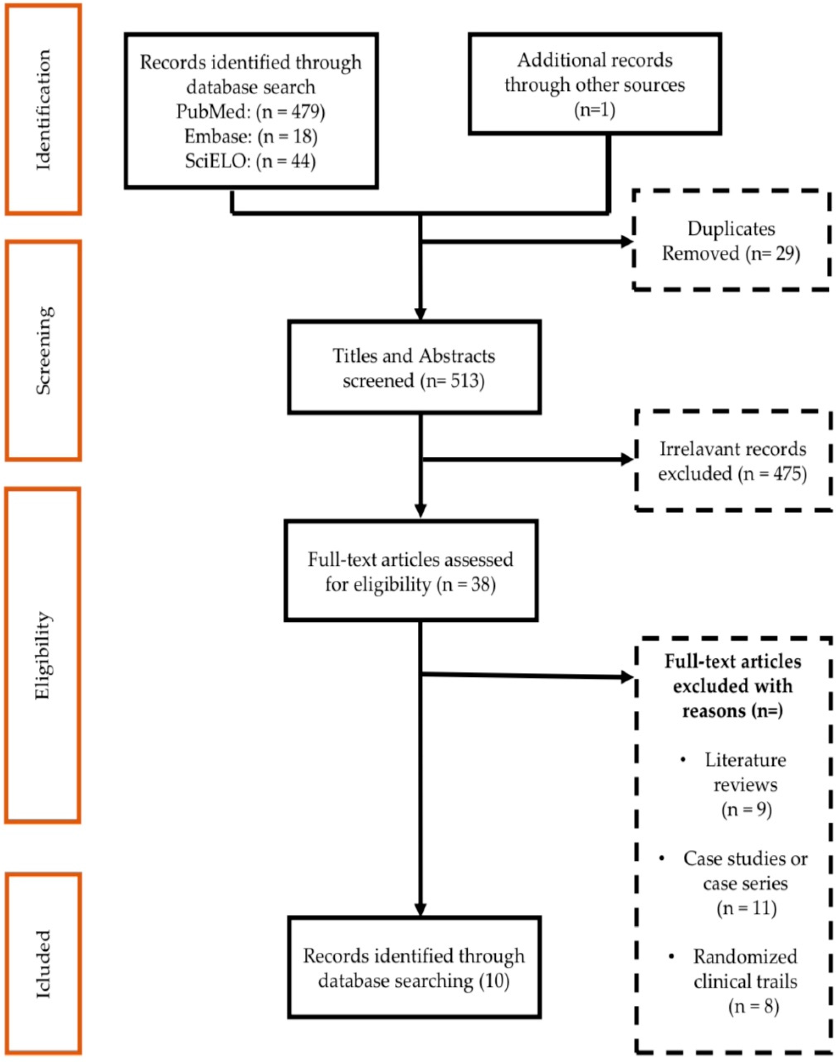
2.1. Result of the Study Identification Process

A total of 542 records were identified from databases and manual searches. After remov ing duplicates (n = 29), 513 studies were screened by titles and abstracts, and 38 selected articles were eligible for full-text review. In 38 full-text studies, 10 were included in the review. Figure 1 provides details of the records excluded at each stage of the screening process.

2.2. Overall Co-Infections Result

The results of 10 articles were tabulated, resulting in a sample group of 1394 studies and 56% of the participants had bacterial co-infections. Among the species that stood out in this research, Staphylococcus aureus sensitive to methicillin was one of them with 27% (17/63) co-infections with 0.27 proportion (0.16–0.42; I2 = 65%). Another 16% (47/678) with 0.16 proportion (0.03–0.50; I2 = 97%) were co-infected with coagulase-negative Staphylococcus spp.
Another very important pathogen in terms of nosocomial/hospital infections and relevant in our research was Klebsiella. spp., with 21% (84/404) and 0.21 proportion (0.04–0.48; I2 = 96% and p < 0.001); Klebsiella oxytoca and K. pneumoniae were the second most expressive with 10% (9.9/99) co-infections and 0.10 proportion (0.03–0.30; I2 = 83%) and 3% (28/935), 0.03 (0.03–0.30; I2 = 97%), respectively.
The species Enterobacter complex proved to be very significant in this research with 42% (28/69) co-infections and 0.42 proportion (0.31–0.54); followed by 35% (48/138) infection caused by Enterobacter spp., with 0.35 proportion (0.25–0.46). Another species that proved to be important in the results of our study was Escherichia coli and extended-spectrum beta-lactamase-producing Echerichia coli (ESBL), both with 13% (10/80) co-infected patients in the respective 0.13 proportions (0.09–0.18; I2 = 63%; p < 0.02) and 13% (88/683) 0.13 (0.07–0.22; I2 = 15%; p < 0.28).
Other pathogens such as Haemophilus influenza and Proteus mirabilis showed 12% (39/330) co-infection and 0.12 proportions (0.03–0.40) and 11% (4/37) co-infection with 0.11 proportion (0.04–0.25). Given its biological importance, the species Pseudomonas aeruginosa appeared with 3% (30/1030) and 0.03 proportion (0.01–0.12) of co-infected patients in this study. Other species were also analyzed, as shown in Figure 2.
Some species did not show statistical significance for co-infection, such as Sphingobacterium multivorum and Enterobacter cloacae.

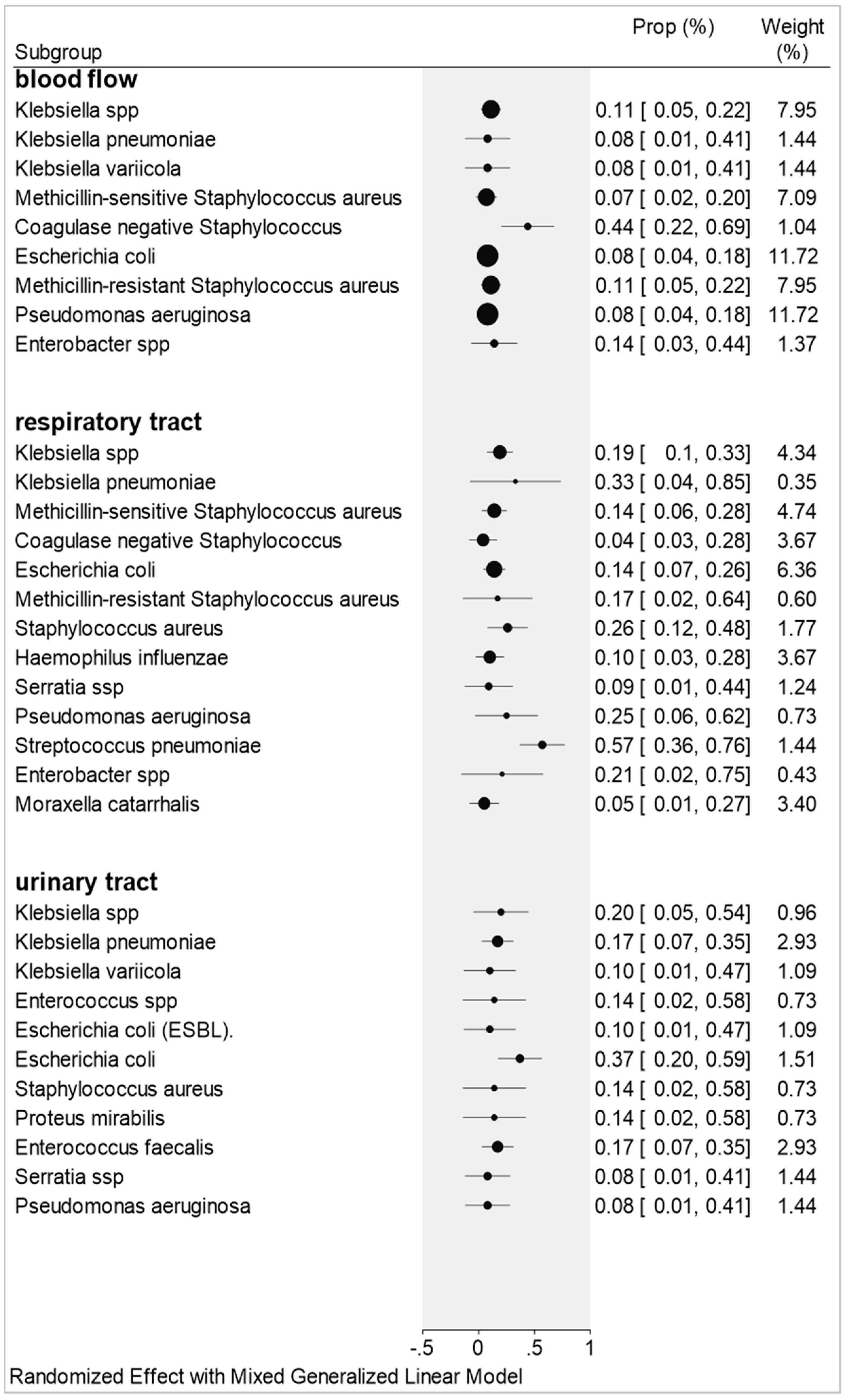
2.3. Co-Infections by Blood Flow, Urinary Tract, and Respiratory Tract Samples

The results for co-infections taking into account the biological materials from blood flow, urinary tract, and respiratory tract showed great relevance. This study describes the prevalence and characteristics of bacterial co-infection in patients with COVID-19 in the Intensive Care Unit (ICU) and hospitalized (not ICU).

2.3.1. Blood Flow

The most common bacterial co-infections isolated from blood cultures include Coagulase-negative Staphylococcus with 44% (7/16) of patients with proportion 0.44 (0.22–0.68), Enterobacter spp. presented 14%/68 of patients with proportion 0.14 (0.03–0.44; I2 = 86%). Other data from other microorganisms can be seen in Figure 3.

2.3.2. Respiratory Tract

The main results in relation to the respiratory tract were: Streptococcus pneumoniae presented in 57% (12/21) of patients and proportion 0.57 (0.36–0.76); Klebsiella pneumoniae with 33% (1/3) of patients with proportion 0.33 (0.04–0.85); Pseudomonas aeruginosa with 25% (16/64) of patients and proportion 0.25 (0.06–0.62; I2 = 42%). Other species were also important in this study: Staphylocuccus aureus accounted for 26% (18/71) of patients and proportion 0.26 (0.12–0.48; I2 = 51%); Enterobacter spp., with 21% (9/46) of patients and proportion 0.21 (0.02–0.75; I2 = 83%); Kle spp., for 9 (19%)/47 patients and proportion 0.19 (0.10–0.33; I2 = 56%); methicillin-resistant Staphylococcus aureus by 17% (8/51) and proportion 0.17 (0.02–0.64; I2 = 89%). Figure 3, shows the bacterial species and their proportions in relation to the respiratory tract.

2.3.3. Urinary Tract

The pathogens found and relevant related to patients from urinary tract samples were: Echerichia coli presented in 37% (11/29) of patients with proportion 0.37 (0.20–0.59; I2 = 42%); Klebsiella spp. with 20% (2/10) of patients and proportion 0.20 (0.05–0.54); Klebsiella pneumoniae and Enterococcus faecalis, both with 17% (5/29) of patients and proportion 0.17 (0.07–0.35; I2 = 0%.); Staphylococcus aureus and Proteus mirabilis, both with 14% (1/7) of patients and proportion 0.14 (0.02–0.58); Klebsiella variicola and Echerichia coli (ESBL), both with 10% (1/10) of patients and proportion 0.10 (0.01–0.47). Figure 3 shows the bacterial species and their proportions in relation to the urinary tract.

3. Discussion

It is well known that seasonal viral respiratory tract infections are directly linked to an increased risk of bacterial co-infection. One of the main causes of mortality in previous influenza pandemics is bacterial infections. An assessment of bacterial respiratory tract co-infections in patients with COVID-19 admitted to the ICU also had secondary infections with Acinetobacter baumannii [36,37].
Chen et al. (2020) reported that this same microorganism infected by SARS-CoV-2 was significant among the species found in sputum material [38]. In another study focused on the same species, it was responsible for 20% of co-infections in ICU patients [39]. In our results, this bacterium was not important, but we know the importance of this bacterial species in the biological part regarding antimicrobial resistance; it is considered the first of the three main species related to antibiotic failure.
Several studies showed that bacterial co-infections in COVID-19 pneumonia are still emerging, but an association was made among the detection of bacterial pathogens with disease severity in COVID-19 patients. The most commonly identified co-infected bacteria include Acinotobacter baumannii, Klebsiella pneumoniae, Legionella pneumophila, and Streptococcus pneumoniae [40,41,42]. Such studies corroborate with our results, regarding the biological importance of these microorganisms in relation to antimicrobials, demonstrating the relevance of the study in relation to co-infections related to patients with COVID-19.
According to Martinez-Guerra and collaborators, 69 patients were studied, and it was found that the most frequent cause of healthcare-associated pneumonia in hospitalized patients was Enterobacteriaceae with 69.6% (48/69), followed by Gram-negative non-fermenting bacilli with 26.1% (18/69). The same authors studied 35 patients with bloodstream infections and found 40% (14/35) of samples positive for coagulase-negative Staphylococcus [43]. In our study, 16% (50/678) of patients were positive for coagulase-negative Staphylococcus for blood flow samples.

3.1. Blood Flow

A study was conducted with 92 adult patients, ICU admitted with acute respiratory failure, manifesting SARS-CoV-2 pneumonia; in total, (28%) 26 patients were co-infected with pathogenic bacteria on ICU admission; from these patients, 32 different bacteria were isolated from cultures and (31%) 10 of methicillin-sensitive Staphylococcus, 22% (7/32) Haemophilus influenza, and 19% (6/32) Streptococcus pneumoniae were found [44].
The prevalence of viral or bacterial co-infections in patients admitted to the ICU, for acute respiratory failure related to SARS-CoV-2 pneumonia is poorly studied [45,46]. One study reported 41% (7/17) co-infection among patients admitted to an ICU of a US hospital [47]. A cross-sectional study with 12.46% (5/43) positive cultures in blood samples and endotracheal aspirate samples were obtained and, Klebsiella spp. 25.59% (11/43), Staphylococcus aureus (MSSA) 20.93%, Escherichia coli 18.6% (8/43), Staphylococcus aureus (MRSA), 13.95% (6/43), Enterobacter spp. 11.63% (5/43), and Pseudomonas aeruginosa 9.30% (4/43) were isolated [48].
A study on central-line-associated bloodstream infection and catheter-associated urinary tract infection was carried out in 78 hospitals, considering the period prior to the COVID-19 pandemic and during the COVID-19 pandemic. It was found that the hospitals with COVID-19 patients had in one month 2.38 times higher co-infection, with <5% prevalence during the pandemic period (p = 0.004), when compared with a hospital with no COVID-19 patients. Coagulase-negative Staphylococcus-associated bloodstream infection had an increase of 130%/1000 days (p < 0.001) [49].
In our study, results similar to those of the described authors were found, with Klebsiella species 11%, proportion 0.11 (0.05–0.22); Escherichia coli 8% (6/71), proportion 0.08 (0.01–0.41); Enterobacter species 14% (10/68), proportion 0.14 (0.03–0.44); Pseudomonas aeruginosa 8% (6/71), proportion 0.08 (0.04–0.18); and 44% (7/16) for coagulase-negative Staphylococcus aureus with proportion 0.44 (0.22–0.69).

3.2. Urinary Tract

A retrospective observational study aimed to assess the mortality rate of 87 patients with COVID-19 from bacterial co-infections. The most common microorganisms were Escherichia coli 28.4% (27/87) and Enterococcus faecalis 26.3% (25/87) [50]. Our study corroborates this research, because we found co-infections for the same species Escherichia coli 37% (11/29), 0.37 [0.20–0.59]; Enterococcus faecalis 17% (5/29), 0.17 [0.07–0.35].
Another study carried out in Lahore (Pakistan), involving 130 pediatric ICU patients, isolated the pathogenic bacteria Escherichia coli, with 48% (63/130), and 28% (37/130) for Klebsiella pneumoniae [51]. A study with 242 patients positive for COVID-19 isolated 19% (46/242) bacterial co-infections. The most frequent was genitourinary representing 57% (138/242) of all co-infections and the most common organism was Escherichia coli, 26% (63/242) [52]. Our study corroborates with the two last studies and confirms the presence of the same organisms in urinary tract co-infections with 20% (2/10), 0.20 [0.05–0.54] for Klebsiella species, and 37% (11/29), 0.37 [0.20–0. 59] for Escherichia coli.

3.3. Respiratory Tract

The prevalence for COVID-19 patients with respiratory co-infections is not well elucidated. Patients who trigger a more critical degree of the disease and need to be transferred to the Intensive Care Units (ICUs) end up needing the help of an intubation endotracheal tube after at least 48 h of mechanical ventilation and when undergoing this procedure, are susceptible to acquiring microbial pathogens. Due to high demand in the pandemic period, many hygiene protocols are not followed correctly, or are not performed, causing bacterial infections. Among people who are in the ICU with COVID-19, about 30% acquire secondary bacterial infections due to the procedure of intubation. Case studies show the presence of COVID-19 co-infections, including influenza in adults [53,54,55,56,57,58,59,60,61,62,63,64,65,66,67,68,69]. It has been proven that co-infections by Staphylococcus aureus or other bacteria during COVID-19 disease impair the innate and adaptive defenses of the host, temporarily compromising the physical and immunological barrier, causing secondary bacterial pneumonia, raising the severity and deaths in healthy people with pre-existing comorbidities [70].
In total, 836 patients have been studied with SARS-CoV-2 in respiratory culture samples and positivity was found in 13.3% (112/836) of samples and 112 positivity results, in 34.8% (39/112) was identified bacterial pathogens. The respiratory bacterial co-infections findings were Staphylococcus aureus 4% (4/24), as a community-acquired pathogen [50]. A retrospective cohort study analyzed 91.8% (236/243) of patients and they had bacterial co-infections with Streptococcus pneumoniae 59.5% (153/236), Klebsiella pneumoniae 55.6% (143/236), and Haemophilus influenzae 40.1% (103/236) [71].
Another retrospective cohort study analyzed 989 patients and showed 3.1% (31/989) and 3.74% (37/989) co-infections. Two of these co-infections were with different bacteria: Streptococcus pneumoniae in co-infection with Moraxella catarrhalis and Staphylococcus aureus in co-infection with Haemophilus influenzae [72]. In our study, the most relevant species was Streptococcus pneumoniae with 57% (12/21) and proportion 0.57 (0.36–0.76) of co-infec tion and even though Klebsiella pneumoniae has biological relevance in relation to antimicrobial resistance, which has proved to be statistically irrelevant in this research.
In a retrospective analysis of 78 patients with COVID-19, 14.1% (11/78) were coinfected with respiratory pathogens that cause lung lesions, the most relevant being Mycoplassma pneumoniae 45.5% (5/11) [73]. In our study, the results corroborate this finding, only for Legionella pneumophila that presented 9% (1/11), a proportion of 0.09 (0.01–0.44) of co-infection that presented itself in a general way and not by blood flow or urinary and respiratory tracts.
A retrospective study analyzed 140 samples from the respiratory tract of ICU patients, positive for COVID-19, and detected 46% (23/50) positive for Enterobacterales, especially Klebsiella spp., 22% (11/50) [74]. It is important to emphasize that this bacterial genus is among the three main genera responsible for antimicrobial failure and in our study it was shown to be expressive in a general and specific way in ICU patients.

4. Materials and Methods

This systematic review was conducted based on the PRISMA (Figure 1) guidelines for reviews, to determine the proportions of co-infections in patients admitted to ICUs with COVID-19.

4.1. Study Identification

This research is characterized as a meta-analysis, with a search in the following databases: PubMed, Embase, and SciELO from December 2019 to December 2021. The search for references was performed using words found on the Health Sciences Descriptors website (DeCS/MeSH), considering: “Corona virus”, “SARS-CoV-2”, “COVID-19”, “bacterial infection”, “cross sectional”, and “co-infection”. The descriptors were associated with the Boolean operators AND, OR, and NOT. In PubMed, 479 references were found, in Embase 18, and in SciELO 44, totaling 541 publications; duplicate articles (n = 29) were later discarded and articles that did not meet the inclusion criteria (475), totaling 38 references used in this research. Out of 38 studies, 10 were selected for meta-analysis, following the Reporting Items for Systematic Reviews and Meta-Analyses (Figure 1) [75]. The evaluation process of the data extracted for meta-analysis was carried out by two statistical reviewers.

4.2. Inclusion and Exclusion Criteria

Inclusion criteria were: (1) retrospective, prospective, and cross-sectional studies of humans with laboratory-confirmed SARS-CoV-2 co-infection, clinical features, and (2) outcomes with ICU patients with co-infection. SARS-CoV-2 infections in blood culture, urinary tract, and respiratory tract.
Exclusion criteria were: (1) articles that did not report data on the number of patients with co-infection; (2) articles with less than 18 patients (defined as case series) and/or case reports; and (3) randomized controlled trials, reviews, editorials, animal studies, letters, and conference abstracts.
After identifying the studies that met all the inclusion and exclusion criteria, the full texts were reviewed for final inclusion in this research. Disagreements were independently reviewed by a researcher who did not participate in the screening.

4.3. Data Extraction

The authors independently extracted data from the included articles using a data collection form. Information was collected according to demographic variables: author and year of publication; country of study and healthcare environment (hospital ICU, non-ICU). This study considered and extracted data from cross-sectional studies, retrospective and prospective cohorts in order to assess the proportions of co-infections (Table 1).

4.4. Data Analysis

The primary outcome measure was the prevalence of co-infection among patients with COVID-19. Articles were classified by country to compare geographic prevalence. It was stratified by patients with co-infection and the co-infection identified with COVID-19 ICU patients was analyzed. The combination of data was performed using the proportion test, considering the generalized linear mixed model (GLMM), associated with logistic transformation (PLOGIT). Using this method, the proportion of patients with co-infection was determined and which was the most prevalent was established.
Heterogeneity was determined by the Higgins and Thompson test (I2). When the I2 presented a result equal to or greater than 50%, it was considered a randomized effect; on the other hand, when the I2 presented a result below 50%, the effect was considered fixed. For the significance of the results, the limit of 5% was considered. Statistical analyses were performed using RStudio® 4.0.2 software [76] and Stata 16.0. [77].

5. Conclusions

We report that the highest rates of bacterial co-infection were for Enterobacter complex 42% (29/69), proportion 0.42 (0.31–0.54) and Staphylococcus aureus 27% (17/63), proportion 0.27 (0.16–0.42). Regarding ICU patients on admission, the co-infection positivity in blood flow was (44%), proportion 0.44 (0.22–0.42) for coagulase-negative Staphylococcus; in the respiratory tract, Streptococcus pneumoniae was found in 57% (12/21), proportion 0.57 (0.36–0.76) of the samples and in the urinary tract 37% (11/29), proportion 0.37 (0.20–0.59) of Escherichia coli were isolated. It is important to note that the species Acinetobacter baumanii, Klebsiella pneumoniae, and Pseudomonas aeruginosa are biologically relevant in this study, due to their history of resistance to antimicrobials and nosocomial/hospital infections.

Author Contributions

Bibliographic review and discussion A.P.S.; systematic review C.R.M.I. and M.O.S.; tabulation of data A.C.C.O. and P.H.P.Q.; statistical analysis and methodology, L.C.G.; statistical analysis, analysis and design of the methodology and relevant corrections L.C.C. All authors have read and agreed to the published version of the manuscript.

Funding

FAPEG: Research Support Foundation of the State of Goiás.

Data Availability Statement

The data are contained in the article.

Conflicts of Interest

The authors declare no conflict of interest.

References

  1. Mina, M.; Klugman, K.P. The role of influenza in the severity and transmission of respiratory bacterial disease. Lancet Respir. Med. 2014, 2, 750–763. [Google Scholar] [CrossRef] [Green Version]
  2. Bashir, A.; Abdullah, M.S.; Momin, N.R.; Chong, P.L.; Asli, R.; Ivan, B.M.; Chong, V.H. Prevalence of primary bacterial co-infections among patients with COVID-19 in Brunei Darussalam. West. Pac. Surveill. Response J. 2021, 12, 65–70. [Google Scholar] [CrossRef] [PubMed]
  3. Benvenuto, D.; Giovanetti, M.; Ciccozzi, A.; Spoto, S.; Angeletti, S.; Ciccozzi, M. The 2019-new coronavirus epidemic: Evidence for virus evolution. J. Med. Virol. 2020, 92, 455–459. [Google Scholar] [CrossRef] [PubMed] [Green Version]
  4. Guan, W.J.; Ni, Z.Y.; Hu, Y.; Liang, W.H.; Ou, C.Q.; He, J.X.; Liu, L.; Shan, H.; Lei, C.L.; Hui, D.S.C.; et al. Clinical characteristics of coronavirus disease 2019 in China. N. Engl. J. Med. 2020, 382, 1708–1720. [Google Scholar] [CrossRef] [PubMed]
  5. Wang, D.; Hu, B.; Hu, C.; Zhu, F.; Liu, X.; Zhang, J.; Wang, B.; Xiang, H.; Cheng, Z.; Xiong, Y.; et al. Clinical characteristics of 138 hospitalized patients with 2019 novel coronavirus-infected pneumonia in Wuhan, China. JAMA 2020, 323, 1061–1069. [Google Scholar] [CrossRef]
  6. Richardson, S.; Hirsch, J.S.; Narasimhan, M.; Crawford, J.M.; McGinn, T.; Davidson, K.W. The Northwell COVID-19 Research Consortium. Presenting characteristics, comorbidities, and outcomes among 5700 patients hospitalized with COVID-19 in the New York City area. JAMA 2020, 323, 2052–2059. [Google Scholar] [CrossRef]
  7. Joseph, C.; Togawa, Y.; Shindo, N. Bacterial and viral infections associated with influenza. Influenza Other Respir. Viruses 2013, 7, 105–113. [Google Scholar] [CrossRef] [Green Version]
  8. Cavalcante, G.A. COINFECÇÃO BACTERIANA EM PACIENTES COM COVID-19. Rev. Multidiscip. Em Saúde 2021, 2, 14. [Google Scholar] [CrossRef]
  9. Langford, B.J.; So, M.; Raybardhan, S.; Leung, V.; Westwood, D.; MacFadden, D.R.; Soucy, J.P.R.; Daneman, N. Bacterial co-infection and secondary infection in patients with COVID-19: A living rapid review and meta-analysis. Clin. Microbiol. Infect. 2020, 26, 1622–1629. [Google Scholar] [CrossRef]
  10. Mirzaei, R.; Goodarzi, P.; Asadi, M.; Soltani, A.; Aljanabi, H.A.A.; Jeda, A.S.; Dashtbin, S.; Jalalifar, S.; Mohammadzadeh, R.; Teimoori, A.; et al. Bacterial co-infections with SARS-CoV-2. IUBMB Life 2020, 72, 2097–2111. [Google Scholar] [CrossRef]
  11. Paget, C.; Trottein, F. Mechanisms of bacterial superinfection post-influenza: A role for unconventional T cells. Front. Immunol. 2019, 10, 336. [Google Scholar] [CrossRef] [PubMed]
  12. Jia, L.; Xie, K.; Zhao, J.; Cao, D.; Liang, Y.; Hou, X.; Wang, L.; Li, Z. Mechanisms of severe mortality-associated bacterial co-infections following influenza virus infection. Front. Cell. Infect. Microbiol. 2017, 7, 338. [Google Scholar] [CrossRef] [PubMed] [Green Version]
  13. Quah, J.; Jiang, B.; Tan, P.C.; Siau, C.; Tan, T.Y. Impact of microbial Aetiology on mortality in severe community-acquired pneumonia. BMC Infect. Dis. 2018, 18, 451. [Google Scholar] [CrossRef] [PubMed]
  14. Li, X.; Zhou, X. Co-infection of tuberculosis and parasitic diseases in humans: A systematic review. Parasites Vectors 2013, 6, 79. [Google Scholar] [CrossRef] [Green Version]
  15. Guo, L.; Wei, D.; Zhang, X.; Wu, Y.; Li, Q.; Zhou, M.; Qu, J. Clinical features predicting mortality risk in patients with viral pneumonia: The MuLBSTA Score. Front. Microbiol. 2019, 10, 2752. [Google Scholar] [CrossRef] [Green Version]
  16. Wu, Z.; McGoogan, J.M. Characteristics of and Important Lessons From the Coronavirus Disease 2019 (COVID-19) Outbreak in China: Summary of a Report of 72 314 Cases From the Chinese Center for Disease Control and Prevention. JAMA 2020, 323, 1239–1242. [Google Scholar] [CrossRef] [PubMed]
  17. Hirsch, J.S.; Ng, J.H.; Ross, D.W.; Sharma, P.; Shah, H.H.; Barnett, R.L.; Hazzan, A.D.; Fishbane, S.; Jhaveri, K.D.; on behalf of the Northwell COVID-19 Research Consortium and the Northwell Nephrology COVID-19 Research Consortium. Acute kidney injury in patients hospitalized with COVID-19. Kidney Int. 2020, 98, 209–218. [Google Scholar] [CrossRef] [PubMed]
  18. Siripanthong, B.; Nazarian, S.; Muser, D.; Deo, R.; Santangeli, P.; Khanji, M.Y.; Cooper, L.T., Jr.; Chahal, C.A.A. Recognizing COVID-19-related myocarditis: The possible pathophysiology and proposed guideline for diagnosis and management. Heart Rhythm 2020, 17, 1463–1471. [Google Scholar] [CrossRef]
  19. Abou-Ismail, M.Y.; Diamond, A.; Kapoor, S.; Arafah, Y.; Nayak, L. The hypercoagulable state in COVID-19: Incidence, pathophysiology, and management. Thromb. Res. 2020, 194, 101–115. [Google Scholar] [CrossRef]
  20. Morris, D.E.; Cleary, D.W.; Clarke, S.C. Secondary Bacterial Infections Associated with Influenza Pandemics. Front. Microbiol. 2017, 8, 1041. [Google Scholar] [CrossRef] [Green Version]
  21. Prasso, J.E.; Deng, J.C. Post viral Complications: Bacterial Pneumonia. Clin. Chest Med. 2017, 38, 127–138. [Google Scholar] [CrossRef]
  22. Wang, R.; Kong, L.; Xu, Q.; Yang, P.; Wang, X.; Chen, N.; Li, L.; Jiang, S.; Lu, X. On-ward participation of clinical pharmacists in a Chinese intensive care unit for patients with COVID-19: A retrospective, observational study. Res. Soc. Adm. Pharm. 2021, 17, 1853–1858. [Google Scholar] [CrossRef] [PubMed]
  23. Zhou, F.; Yu, T.; Du, R.; Fan, G.; Liu, Y.; Liu, Z.; Xiang, J.; Wang, Y.; Song, B.; Gu, X.; et al. Clinical course and risk factors for mortality of adult inpatients with COVID-19 in Wuhan, China: A retrospective cohort study. Lancet 2020, 395, 1054–1062. [Google Scholar] [CrossRef]
  24. Lima-Setta, F.; Magalhães-Barbosa, M.C.; Rodrigues-Santos, G.; Figueiredo, E.A.N.; Jacques, M.L.; Zeitel, R.S.; Sapolnik, R.; Borges, C.T.S.; Lanziotti, V.S.; Castro, R.E.V.; et al. Multisystem inflammatory syndrome in children (MIS-C) during SARS-CoV-2 pandemic in Brazil: A multicenter, prospective cohort study. J. Pediatr. 2021, 9, 354–361. [Google Scholar] [CrossRef] [PubMed]
  25. Chen, L.F.; Chopra, T.; Kaye, K.S. Pathogens resistant to antibacterial agents. Infect. Dis. Clin. 2009, 23, 817–845. [Google Scholar] [CrossRef] [PubMed]
  26. Vatopoulos, A.C.; Kalapothaki, V.; Legakis, N.J. An electronic network for the surveillance of antimicrobial resistance in bacterial nosocomial isolates in Greece. The greek network for the surveillance of antimicrobial resistance. Bull. World Health Organ 1999, 77, 595–601. [Google Scholar]
  27. Murray, C.J.L.; Ikuta, K.S.; Sharara, F.; Swetschinski, L.; Aguilar, G.R.; Gray, A.; Han, C.; Bisignano, C.; Rao, P.; Wool, E.; et al. Global burden of bacterial antimicrobial resistance in 2019: A systematic analysis. Lancet 2022, 399, 629–655. [Google Scholar] [CrossRef]
  28. World Health Organization (WHO). Global Antimicrobial Resistance Surveillance System (GLASS) Report: Early Implementation 2020. Available online: https://www.who.int/publications/i/item/9789240005587 (accessed on 20 April 2021).
  29. Smith, A.M.; McCullers, J.A. Secondary bacterial infections in influenza virus infection pathogenesis. In Influenza Pathogenesis and Control—Volume I; Springer: Berlin/Heidelberg, Germany, 2014; Volume 385, pp. 327–356. [Google Scholar] [CrossRef]
  30. Abbasi, S.; Pendergrass, L.B.; Leggiadro, R.J. Influenza complicated by Moraxella catarrhalis bacteremia. Pediatr. Infect. Dis. J. 1994, 13, 937–938. [Google Scholar] [CrossRef]
  31. Jacobs, J.H.; Viboud, C.; Tchetgen, E.T.; Schwartz, J.; Steiner, C.; Simonsen, L.; Lipsitch, M. The association of meningococcal disease with influenza in the United States, 1989–2009. PLoS ONE 2014, 9, e107486. [Google Scholar] [CrossRef]
  32. Mulcahy, M.E.; McLoughlin, R.M. Staphylococcus aureus and influenza A virus: Partners in coinfection. mBio 2016, 7, e02068-16. [Google Scholar] [CrossRef]
  33. Rizzo, C.; Caporali, M.G.; Rota, M.C. Pandemic influenza and pneumonia due to Legionella pneumophila: A frequently underestimated coinfection. Clin. Infect. Dis. 2010, 51, 115. [Google Scholar] [CrossRef] [PubMed] [Green Version]
  34. Chertow, D.S.; Memoli, M.J. Bacterial coinfection in influenza: A grand rounds review. JAMA 2013, 309, 275–282. [Google Scholar] [CrossRef] [PubMed]
  35. Dela Cruz, C.S.; Wunderink, R.G. Respiratory Viral and Atypical Pneumonias. Clin. Chest Med. 2017, 38, xiii–xiv. [Google Scholar] [CrossRef] [PubMed]
  36. Yang, X.; Yu, Y.; Xu, J.; Shu, H.; Xia, J.; Liu, H.; Wu, Y.; Zhang, L.; Yu, Z.; Fang, M.; et al. Clinical course and outcomes of critically ill patients with SARS-CoV-2 pneumonia in Wuhan, China: A single-centered, retrospective, observational study. Lancet Respir. Med. 2020, 8, 475–481. [Google Scholar] [CrossRef] [Green Version]
  37. Lansbury, L.; Lim, B.; Baskaran, V.; Lim, W.S. Co-infections in people with COVID-19: A systematic review and meta-analysis. J. Infect. 2020, 81, 266–275. [Google Scholar] [CrossRef]
  38. Chen, T.; Wu, D.; Chen, H.; Yan, W.; Yang, D.; Chen, G.; Ma, K.; Xu, D.; Yu, H.; Wang, H.; et al. Clinical characteristics of 113 deceased patients with coronavirus disease 2019: Retrospective study. BMJ 2020, 368, m1091. [Google Scholar] [CrossRef] [Green Version]
  39. Wang, L.; Amin, A.K.; Khanna, P.; Aali, A.; McGregor, A.; Bassett, P.; Rao, G.G. An observational cohort study of bacterial co-infection and implications for empirical antibiotic therapy in patients presenting with COVID-19 to hospitals in North West London. J. Antimicrob. Chemother. 2021, 76, 796–803. [Google Scholar] [CrossRef]
  40. Hoque, M.N.; Rahman, M.S.; Ahmed, R.; Hossain, M.S.; Islam, S.; Islam, T.; Hossain, M.A.; Siddiki, A.Z. Diversity and Genomic Determinants of the Microbiomes Associated with COVID-19 and Non-COVID respiratory diseases. Resp. Dis. 2020, 23, 101200. [Google Scholar] [CrossRef]
  41. Khatiwada, S.; Subedi, A. Lung microbiome and coronavirus disease 2019 (COVID-19): Possible link and implications. Hum. Microbiome J. 2020, 17, 100073. [Google Scholar] [CrossRef]
  42. Peddu, V.; Shean, R.C.; Xie, H.; Shrestha, L.; Perchetti, G.A.; Minot, S.S.; Roychoudhury, P.; Huang, M.-L.; Nalla, A.; Reddy, S.B.; et al. Metagenomic analysis reveals clinical SARS-CoV-2 infection and bacterial or viral superinfection and colonization. Clin. Chem. 2020, 66, 966–972. [Google Scholar] [CrossRef]
  43. Martinez-Guerra, B.A.; Gonzalez-Lara, M.F.; Leon-Cividanes, N.A.; Tamez-Torres, K.M.; Roman-Montes, C.M.; Rajme-Lopez, S.; Villalobos-Zapata, G.I.; Lopez-Garcia, N.I.; Martínez-Gamboa, A.; Sifuentes-Osornio, J.; et al. Antimicrobial Resistance Patternsand Antibiotic Use during Hospital Conversion in the COVID-19 Pandemic. Antibiotics 2021, 10, 182. [Google Scholar] [CrossRef] [PubMed]
  44. Contou, D.; Claudinon, A.; Pajot, O.; Micaëlo, M.; Longuet, F.P.; Dubert, M.; Cally, R.; Logre, E.; Fraissé, M.; Mentec, H.; et al. Bacterial and viral co-infections in patients with severe SARS-CoV-2 pneumonia admitted to a French ICU. Ann. Intensive Care 2020, 10, 119. [Google Scholar] [CrossRef] [PubMed]
  45. Huttner, B.; Catho, G.; Pano-Pardo, J.R.; Pulcini, C.; Schouten, J. COVID-19: Don’t neglect antimicrobial stewardship principles! Clin. Microbiol. Infect. 2020, 26, 808–810. [Google Scholar] [CrossRef]
  46. Cox, M.J.; Loman, N.; Bogaert, D.; O’Grady, J. Co-infections: Potentially lethal and unexplored in COVID-19. Lancet Microbe 2020, 1, e110. [Google Scholar] [CrossRef]
  47. Mahmoudi, H. Bacterial co-infections and antibiotic resistance in patients with COVID-19. GMS Hyg. Infect. Control. 2020, 15, Doc35. [Google Scholar] [CrossRef] [PubMed]
  48. Hughes, S.; Troise, O.; Donaldson, H.; Mughal, N.; Moore, L.S.P. Bacterial and fungal coinfection among hospitalized patients with COVID-19: A retrospective cohort study in a UK secondary-care setting. Clin. Microbiol. Infect. 2020, 26, 1395–1399. [Google Scholar] [CrossRef] [PubMed]
  49. Fakih, M.G.; Bufalino, A.; Sturm, L.; Huang, R.H.; Ottenbacher, A.; Saake, K.; Winegar, A.; Fogel, R.; Cacchione, J. Coronavirus disease 2019 (COVID-19) pandemic, central-line-associated bloodstream infection (CLABSI), and catheter-associated urinary tract infection (CAUTI): The urgent need to refocus on hardwiring prevention efforts. Infect. Control. Hosp. Epidemiol. 2022, 43, 26–31. [Google Scholar] [CrossRef]
  50. Díaz, P.B.; Guedez, L.G.V.; García, C.P.M.; Jiménez, G.M.; García, B.S.; Gómez-Gil, M.M.R. Urinary Tract Infections in Hospitalized COVID-19 Patients, What’s Up, Doc? J. Clin. Med. 2022, 11, 1815. [Google Scholar] [CrossRef]
  51. Iqbal, Z.; Mumtaz, M.Z.; Malik, A. Extensive drug-resistance in strains of Escherichia coli and Klebsiella pneumoniae isolated from paediatric urinary tract infections. J. Taibah Univ. Med. Sci. 2021, 16, 565–574. [Google Scholar] [CrossRef]
  52. Goncalves, A.; Lo, K.B.; Wattoo, A.; Salacup, G.; Pelayo, J.; DeJoy, R.; Bhargav, R.; Gul, F.; Peterson, E.; Albano, J.; et al. Bacterial infections and patterns of antibiotic use in patients with COVID-19. J. Med. Virol. 2021, 93, 1489–1495. [Google Scholar] [CrossRef]
  53. Azekawa, S.; Namkoong, H.; Mitamura, K.; Kawaoka, Y.; Saito, F. Co-infection with SARS-CoV-2 and influenza A virus. IDCases 2020, 20, e00775. [Google Scholar] [CrossRef] [PubMed]
  54. Blasco, M.L.; Buesa, J.; Colomina, J.; Forner, M.J.; Galindo, M.J.; Navarro, J.; Noceda, J.; Redón, J.; Signes-Costa, J.; Navarro, D. Co-detection of respiratory pathogens in patients hospitalized with Coronavirus viral disease-2019 pneumonia. J. Med. Virol. 2020, 92, 1799–1801. [Google Scholar] [CrossRef] [PubMed]
  55. Cuadrado-Payan, E.; Montagud-Marraha, E.; Torres-Elorza, M.; Bodro, M.; Blasco, M.; Poch, E.; Soriano, A.; Piñeiro, G.J. SARS-CoV-2 and influenza virus co-infection. Lancet 2020, 395, e84. [Google Scholar] [CrossRef]
  56. Ding, Q.; Lu, P.; Fan, Y.; Xia, Y.; Liu, M. The clinical characteristics of pneumonia patients coinfected with 2019 noval coronavirus and influenza virus in Wuhan, China. J. Med. Virol. 2020, 92, 1549–1555. [Google Scholar] [CrossRef] [PubMed]
  57. Fan, B.; Lim, K.; Chong, V.; Chan, S.; Ong, K.; Kuperan, P. COVID-19 and mycoplasma pneumoniae coinfection. Am. J. Hematol. 2020, 95, 723–724. [Google Scholar] [CrossRef]
  58. Khodamoradi, Z.; Moghadami, M.; Lotfi, M. Co-infection of coronavirus disease 2019 and influenza A: A report from Iran. Arch. Iran. Med. 2020, 23, 239–243. [Google Scholar] [CrossRef] [Green Version]
  59. Konala, V.; Adapa, S.; Gayam, V.; Naramala, S.; Daggubati, S.; Kammari, C.; Chenna, A. Co-infection with Influenza A and COVID-19. Eur. J. Case Rep. Intern. Med. 2020, 7, 001656. [Google Scholar] [CrossRef]
  60. Lin, D.; Liu, L.; Zhang, M.; Hu, Y.; Yang, Q.; Guo, J.; Guo, Y.; Dai, Y.; Xu, Y.; Cai, Y.; et al. Co-infections of SARS-CoV-2 with multiple common respiratory pathogens in infected patients. Sci. China Life Sci. 2020, 63, 606–609. [Google Scholar] [CrossRef] [Green Version]
  61. Luyt, C.E.; He’kimian, G.; Koulenti, D.; Chastre, J. Microbial cause of ICU-acquired pneumonia: Hospital-acquired pneumonia versus ventilator-associated pneumonia. Curr. Opin. Crit. Care 2018, 24, 332–338. [Google Scholar] [CrossRef]
  62. Motta, J.; Gomez, C. Adenovirus and novel coronavirus (SARS-CoV2) coinfection: A case report. IDCases 2020, 22, e00936. [Google Scholar] [CrossRef]
  63. Bardi, T.; Pintado, V.; Gomez-Rojo, M.; Escudero-Sanchez, R.; Lopez, A.; Diez-Remesal, Y.; Castro, N.M.; Ruiz-Garbajosa, P.; Pestaña, D. Nosocomial infections associated to COVID-19 in the intensive care unit: Clinical characteristics and outcome. Eur. J. Clin. Microbiol. Infect. Dis. 2021, 40, 495–502. [Google Scholar] [CrossRef] [PubMed]
  64. Ozaras, R.; Cirpin, R.; Duran, A.; Duman, H.; Arslan, O.; Bakcan, Y.; Kaya, M.; Mutlu, H.; Isayeva, L.; Kebanlı, F.; et al. Influenza and COVID-19 coinfection: Report of six cases and review of the literature. J. Med. Virol. 2020, 92, 2657–2665. [Google Scholar] [CrossRef] [PubMed]
  65. Touzard-Romo, F.; Tape, C.; Lonks, J. Co-infection with SARS-CoV-2 and human metapneumovirus. R. I. Med. J. 2020, 103, 75–76. Available online: http://rimed.org/rimedicaljournal/2020/03/2020-03-75-touzard-romo.pdf (accessed on 21 November 2021).
  66. Wee, L.; Ko, K.K.K.; Ho, W.Q.; Kwek, G.T.C.; Tan, T.T.; Wijaya, L. Community-acquired viral respiratory infections amongst hospitalized inpatients during a COVID-19 outbreak in Singapore: Co-infection and clinical outcomes. J. Clin. Virol. 2020, 128, 104436. [Google Scholar] [CrossRef]
  67. Wu, X.; Cai, T.; Huang, X.; Yu, X.; Zhao, L.; Wang, F.; Li, Q.; Gu, S.; Xu, T.; Li, Y.; et al. Co-infection with SARS-CoV-2 and influenza A virus in patient with pneumonia, China. Emerg. Infect. Dis. 2020, 6, 1324–1326. [Google Scholar] [CrossRef]
  68. Xie, Y.; Bowe, B.; Maddukuri, G.; Al-Aly, Z. Comparative evaluation of clinical manifestations and risk of death in patients admitted to hospital with COVID-19 and seasonal influenza: A cohort study. BMJ 2020, 371, m4677. [Google Scholar] [CrossRef]
  69. Ginsburg, A.S.; Klugman, K.P. COVID-19 pneumonia and the appropriate use of antibiotics. Lancet Glob. Health 2020, 8, e1453–e1454. [Google Scholar] [CrossRef]
  70. Lai, C.C.; Wang, C.Y.; Hsueh, P.R. Co-infections among patients with COVID-19: The need for combination therapy with non-anti-SARS-CoV-2 agents? J. Microbiol. Immunol. Infect. 2020, 53, 505–512. [Google Scholar] [CrossRef]
  71. Zhu, X.; Ge, Y.; Wu, T.; Zhao, K.; Chen, Y.; Wu, B.; Zhu, F.; Zhu, B.; Cui, L. Co-infection with respiratory pathogens among COVID-2019 cases. Virus Res. 2020, 285, 198005. [Google Scholar] [CrossRef]
  72. Garcia-Vidal, C.; Sanjuan, G.; Moreno-García, E.; Puerta-Alcalde, P.; Garcia-Pouton, N.; Chumbita, M.; Fernandez-Pittol, M.; Pitart, C.; Inciarte, A.; Bodro, M.; et al. COVID-19 Researchers Group. Incidence of co-infections and superinfections in hospitalized patients with COVID-19: A retrospective cohort study. Clin. Microbiol. Infect. 2021, 27, 83–88. [Google Scholar] [CrossRef]
  73. Man-Ling, T.; Li, Y.Q.; Chen, X.; Lin, H.; Jiang, Z.C.; Gu, D.L.; Tang, C.X.; Xie, Z.Q. Co-Infection with Common Respiratory Pathogens and SARS-CoV-2 in Patients with COVID-19 Pneumonia and Laboratory Biochemistry Findings: A Retrospective Cross-Sectional Study of 78 Patients from a Single Center in China. Med. Sci. Monit. 2021, 3, e929783. [Google Scholar] [CrossRef]
  74. Rothe, K.; Feihl, S.; Schneider, J.; Wallnöfer, F.; Wurst, M.; Lukas, M.; Treiber, M.; Lahmer, T.; Heim, M.; Dommasch, M.; et al. Rates of bacterial co-infections and antimicrobial use in COVID-19 patients: A retrospective cohort study in light of antibiotic stewardship. Eur. J. Clin. Microbiol. Infect. Dis. 2021, 40, 859–869. [Google Scholar] [CrossRef] [PubMed]
  75. Shamseer, L.; Moher, D.; Clarke, M.; Ghersi, D.; Liberati, A.; Petticrew, M.; Shekelle, P.; Stewart, L.A.; PRISMA-P Group. Preferred reporting items for systematic review and meta-analysis protocols (PRISMA-P) 2015: Elaboration and explanation. BMJ 2015, 350, g7647. [Google Scholar] [CrossRef] [PubMed] [Green Version]
  76. RStudio Team RStudio: Integrated Development for R. RStudio. Version 4.0.2, PBC, Boston, USA 2020. Available online: http://www.rstudio.com (accessed on 7 January 2022).
  77. StataCorp LLC. Stata Statistical Software. Version 16.0, College Station, Washington, DC, USA 2021. Available online: https://filecr.com/windows/stata-mp (accessed on 10 January 2022).
Figure 1. PRISMA schematic selection process of the included studies at each stage of the screening process.
Antibiotics 11 00894 g001
Figure 2. Forest plot graph: showing the subgroup (genus and bacterial species) and their respective proportions by co-infection.
Antibiotics 11 00894 g002
Figure 3. Bacterial species and their proportions in relation to blood flow, respiratory tract, and urinary tract.
Antibiotics 11 00894 g003
Table 1. Research selected for meta-analysis.
Studies Selected for Meta-Analysis
AuthorYearCountriesType of Study
Wang et al. [40]2021LondonRetrospective observational study
Martinez-Guerra et al. [45]2021MexicoProspective cohort study
Contou et al. [46]2020FranceRetrospective study
Hughes et al. [49]2020LondonRetrospective observational analysis
Mahmoudi [50]2020IranCross-sectional study
Neto et al. [54]2020USARetrospective analysis
Zhu et al. [73]2020ChinaRetrospective study
Garcia-Vidal et al. [74]2021SpainRetrospective cohort study
Man-Ling et al. [75]2021ChinaRetrospective analysis
Rothe et al. [26]2021GermanyRetrospective cohort study
Publisher’s Note: MDPI stays neutral with regard to jurisdictional claims in published maps and institutional affiliations.

