Article

Clostridioides difficile in South American Camelids in Germany: First Insights into Molecular and Genetic Characteristics and Antimicrobial Resistance

by
Ines Dost
1,*,
Mostafa Abdel-Glil
1,
Gernot Schmoock
1,
Christian Menge
2,
Christian Berens
2,
Belén González-Santamarina
2,†,
Elisabeth Wiegand
2,‡,
Heinrich Neubauer
1,
Stefan Schwarz
3,4 and
Christian Seyboldt
1
1
Institute of Bacterial Infections and Zoonoses, Friedrich-Loeffler-Institut, Federal Research Institute for Animal Health, Naumburger Straße 96a, 07743 Jena, Germany
2
Institute of Molecular Pathogenesis, Friedrich-Loeffler-Institut, Federal Research Institute for Animal Health, Naumburger Straße 96a, 07743 Jena, Germany
3
Institute of Microbiology and Epizootics, Centre for Infection Medicine, Department of Veterinary Medicine, Freie Universität Berlin, 14163 Berlin, Germany
4
Veterinary Centre for Resistance Research (TZR), Freie Universität Berlin, 14163 Berlin, Germany
*
Author to whom correspondence should be addressed.
Current address: Department of Anesthesiology and Intensive Care Medicine, University Hospital Jena, Am Klinikum 1, 07747 Jena, Germany.
Current address: Tiergarten Delitzsch, Markt 3, 04509 Delitzsch, Germany.
Antibiotics 2023, 12(1), 86; https://doi.org/10.3390/antibiotics12010086
Submission received: 1 December 2022 / Revised: 22 December 2022 / Accepted: 23 December 2022 / Published: 4 January 2023

Abstract

:
Little is known about zoonotic pathogens and their antimicrobial resistance in South American camelids (SAC) in Germany including Clostridioides (C.) difficile. The aim of this study was to investigate prevalence, molecular characteristics and antimicrobial resistance of C. difficile in SAC. Composite SAC faecal samples were collected in 43 husbandries in Central Germany and cultured for C. difficile. Toxinotyping and ribotyping was done by PCR. Whole genome sequencing was performed with Illumina® Miseq™. The genomes were screened for antimicrobial resistance determinants. Genetic relatedness of the isolates was investigated using core genome multi locus sequence typing (cgMLST) and single nucleotide polymorphism analysis. Antimicrobial susceptibility testing was done using the Etest® method. Eight C. difficile isolates were recovered from seven farms. The isolates belonged to different PCR ribotypes. All isolates were toxinogenic. cgMLST revealed a cluster containing isolates recovered from different farms. Seven isolates showed similar resistance gene patterns. Different phenotypic resistance patterns were found. Agreement between phenotypic and genotypic resistance was identified only in some cases. Consequently, SAC may act as a reservoir for C. difficile. Thus, SAC may pose a risk regarding zoonotic transmission of toxinogenic, potentially human-pathogenic and resistant C. difficile isolates.

1. Introduction

The Gram-positive, anaerobic, spore-forming bacterium Clostridioides (C.) difficile is a well-known enteric pathogen. It can cause diseases in humans and animals ranging from mild diarrhoea to sudden death, but asymptomatic carriage is also possible [1,2].
C. difficile infection (CDI) was initially recognized as a mainly nosocomial disease in humans, but the number of community-associated C. difficile infections (CA-CDI) has risen in the last years representing approximately 33 to 41% of the CDI cases in the USA and possibly up to 25% in Europe [3,4,5]. In many CA-CDI cases, well-known traditional risk factors, such as antimicrobial treatment or contact with health-care settings, are absent [5].
C. difficile has been isolated from many animal species and environmental niches [1,6]. Although a zoonotic transmission has not been experimentally proven so far, genetically indistinguishable C. difficile isolates found in pigs and humans strongly suggest zoonotic transmission [7,8].
A risk for successful CDI treatment is the emergence of antimicrobial resistance. C. difficile isolates show resistance to various antimicrobial agents and can serve as antimicrobial resistance reservoir for other Gram-positive bacteria due to many mobile genetic elements in their genome [9,10].
During the last years, the number of South American camelid (SAC) husbandries in Germany has steadily increased. According to a survey conducted among German SAC owners (255 participants), more than every second owner (55.1%) started keeping these animals from 2014 to 2019 [11]. In August 2020, 12,458 SAC were registered on the website of the “Alpaka Zucht Verband Deutschland e.V.” (Alpaca Breeding Association Germany) [11], this number doubled to approximately 25,000 animals in October 2022 (https://www.azvd.de/; accessed on 11 October 2022). Llamas and alpacas, mostly kept for hobby purposes, are used for wool production, breeding, trekking tours, animal assisted therapy or landscape conservation [11,12]. This increasing popularity and their frequent close contact with humans urge a risk analysis on potential zoonotic pathogens.
The aim of this study was to investigate the prevalence, molecular characteristics and antimicrobial resistances of C. difficile recovered from SAC.

2. Results

2.1. Prevalence and Molecular Characteristics

Eight samples from seven husbandries were positive for C. difficile (typical phenotype and confirmation by PCR of cdd3) resulting in an overall prevalence of 16% (7/43) at the husbandry-level. All isolates were obtained only from enrichment cultures and belonged to five different PCR ribotypes (RT): RT 002/2 (3/8), RT 015 (2/8), RT 029 (1/8), RT 078 (1/8) and RT AI-75 (1/8). All eight isolates were positive for the toxin genes tcdA and tcdB. Only the isolate 19S0136 (RT 078) was positive for the binary toxin genes cdtA and cdtB (Table 1).

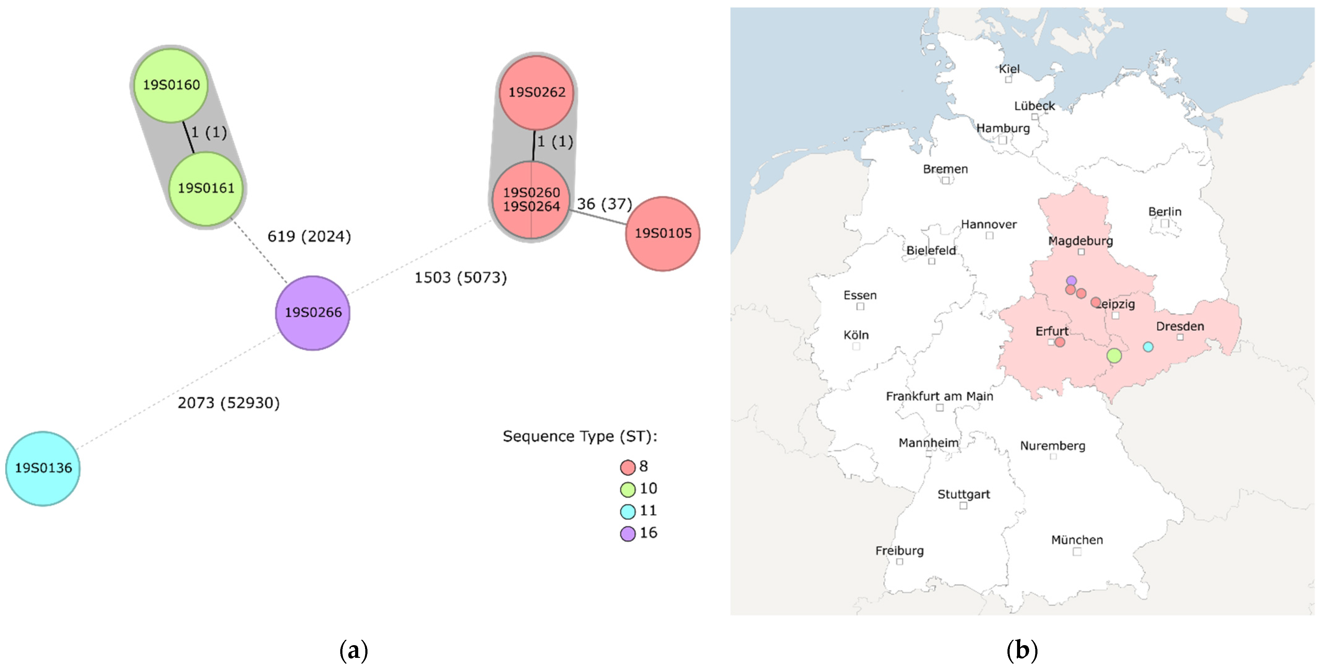
2.2. Whole Genome Sequencing (WGS)

Quality control values collected during WGS data analysis by WGSBAC revealed that all isolates belonged to C. difficile [13]. The genome assembly sizes ranged from 4 to 4.5 Mbp and theoretical coverages from 58- to 98-fold. Values for the average nucleotide identity (ANI) between all genomes were above 96%.
The analysis of single nucleotide polymorphisms (SNPs) is shown in Table 2. The isolates 19S0260 and 19S0262 were identical (no SNP). They possessed five SNPs difference to 19S0264, and 19S0105, another sequence type (ST) 8 isolate, showed 116—121 SNPs to the RT 002/2 isolates (19S0260, 19S0262, 19S0264). The two RT 015 (ST 10) isolates 19S0160 and 19S0161 varied by six SNPs. 19S0136 (RT 078) was the most distant isolate with more than 91,000 SNPs difference to every other isolate.
A total of 2147 core genes were compared by core genome multi locus sequence typing (cgMLST) [14]. The cgMLST revealed four different clusters in accordance with the ST with a cluster type threshold of three differing alleles [15]. According to cgMLST, the RT 002/2 isolates 19S0260 and 19S0264 were identical and revealed one allele difference to 19S0262. 19S0160 and 19S0161 (both RT 015) also differed in one allele (Figure 1).
Seven isolates showed a similar content of resistance genes, including blaCDD-1, vanZ1 and the vanG gene cluster, consisting of the regulatory genes vanR and vanS, as well as the effector genes vanG and vanT. The isolate 19S0136 (RT 078) possessed aadE, blaCDD-1, tet(40), tet(M) and vanZ1 (Table 3). In addition, this isolate showed nucleotide changes in the genes gyrA and gyrB, which resulted in several amino acid substitutions in the corresponding proteins GyrA and GyrB, some of them previously reported to confer fluoroquinolone resistance [10]. In GyrA, two amino acid substitutions were found (Lys-413-Asn and Thr-82-Val), whereas three substitutions were present in GyrB (Gln-160-His, Ser-416-Ala and Ser-366-Val). The substitution Val-130-Ile in GyrB was found in both RT 015-isolates, 19S0160 and 19S0161 (Table 4).

2.3. Antimicrobial Susceptibility Testing (AST)

The AST results are summarised in Table 5. All isolates were susceptible to metronidazole (MEZ), vancomycin (VAN), meropenem (MEP) and amoxicillin-clavulanate (AMC). Seven isolates were susceptible to moxifloxacin (MOX), tetracycline (TET) and classified as wild type (WT) with respect to erythromycin (ERY). In contrast, isolate 19S0136 was resistant to MOX and TET and classified as ERY-non wild type (NWT). Four isolates were classified as resistant to penicillin (PEN) and ampicillin (AMP), two isolates were classified as intermediate for both antimicrobial agents (19S0160 and 19S0264). Isolate 19S0136 was resistant to PEN and intermediate for AMP, while isolate 19S0161 was resistant to AMP and intermediate for PEN. For chloramphenicol (CHL), seven isolates were classified as susceptible, while isolate 19S0105 was classified as CHL-intermediate. Seven isolates were resistant to clindamycin (CLI), 19S0136 was classified as CLI-susceptible. All isolates were classified as NWT for ciprofloxacin (CIP) with minimum inhibitory concentration (MIC) values of >32 mg/L. For linezolid (LZD), five isolates had MIC values of 3 mg/L, whereas isolates 19S0161, 19S0160 and 19S0266 showed MIC values of 4 mg/L, 6 mg/L and 8 mg/L, respectively.

3. Discussion

3.1. Clostridioides difficile and (South American) Camelids in Literature—A Short Review

As only a few samples were investigated in this study, a literature search was performed to gain a broader overview of C. difficile in SAC.
For this, the literature database PubMed was searched with the term ((Clostridium difficile) OR (Clostridioides difficile)) AND ((camel*) OR (llama) OR (alpaca)). A total of 24 results were obtained (22 December 2022). Of these 24 results, 21 articles had to be excluded as they investigated another topic. Mostly, they were related to single-domain camelid antibodies as a therapeutic option for CDI (13 articles). Three articles investigated C. difficile and its prevalence in camels [21,22,23]. C. difficile was found in two of these studies, none of these research articles described C. difficile in SAC.
In one study from Saudi Arabia, camel minced meat was investigated. C. difficile was found in 4% (4/100) of the samples. All isolates were positive for toxin B, three isolates were positive for toxin A. AST was performed by broth microdilution for seven different antimicrobial agents (ceftriaxone, TET, CLI, MEZ, PEN, MOX and AMC). The isolates showed different antimicrobial resistance patterns (TET, CLI + TET, ceftriaxone + TET, PEN + ceftriaxone + MOX) [21].
In another study, C. difficile isolates were found in camel faeces (in 1 of 25 samples; the isolate belonged to RT IR43) and on a postevisceration camel carcass (25 carcasses were tested; the isolate belonged to RT IR48) in Iran. No further characterisation of these isolates was performed [22].
The third study investigated 124 camel meat samples, all of them were negative for C. difficile [23].
Next to these articles, a fourth article was found, which investigated different samples for C. difficile including one llama faecal sample. This sample was negative for C. difficile [24].
The lack of investigations of C. difficile in camelids shows the importance of this study and that more research is needed regarding C. difficile in SAC. This is the first study to report the presence of C. difficile in SAC.

3.2. Prevalence of Clostridioides difficile in German South American Camelids

Comparing the prevalence of 16% at the husbandry-level to the prevalence of C. difficile of other herbivore livestock-farms, this prevalence is similar to that for calves (19.8% of 101 Italian farms) but lower than that of Slovenian dairy-farms with an average of 39.8% [25,26]. Taking these results into consideration, C. difficile seems to be as present in SAC-husbandries as in other herbivore livestock-husbandries, but with an apparently lower prevalence.
All eight isolates were cultivated from enrichment cultures, the bacterial or spore load of the samples was presumably not sufficiently high for detection in direct cultures. This is not surprising considering that pooled faecal samples of animals of presumably different age classes were investigated. The prevalence of C. difficile is known to be age-related and is higher in younger individuals in many animal species and in humans [1,5,27]. The calculation of a prevalence at the animal-level for these samples is not possible, because composite faecal samples were used which had been collected in a previous study [12]. However, considering that 9% (8/94) of the samples were tested positive with a low bacterial (or spore) load, the obtained results are comparable to the prevalence in small ruminants (2–9.5%) and adult horses (0–8.4%), which are probably the most comparable animal species regarding husbandry conditions and human—animal interactions [1].

3.3. Isolates of Different Toxinogenic Ribotypes Resistant to Antimicrobial Agents Were Detected

The five RTs found in this study have been detected in humans before [28,29], and as the corresponding isolates are positive for the toxin genes tcdA and tcdB, they may be capable of causing disease [30].
Overall, results obtained by cgMLST (Figure 1) correspond with results obtained by SNPs analysis (Table 2). The number of SNPs reflect clusters and differing alleles revealed by cgMLST with a close relatedness of isolates belonging to the same ST (0 to 121 SNPs). According to both methods, the most distant isolate was 19S0136 (RT 078).
With 19S0136 belonging to RT 078, a hypervirulent and multidrug-resistant isolate was found in our study [2,31]. RT 078 is livestock-associated and potentially zoonotic [1,7]. Our results from the AST (Table 5) and resistome data (Table 3 and Table 4) are in line with those of other studies, since resistance to TET, MOX and ERY is common in ST 11 and RT 078 isolates, respectively, as well as the presence of antimicrobial resistance genes for aminoglycosides and TET [31]. We could not identify the genetic basis for the high ERY MIC value as erm genes or other known macrolide resistance genes were not detected in the isolates studied. However, the absence of erm genes in ERY-resistant isolates is a common finding in C. difficile [10].
Three RT 002/2 isolates (19S0260, 19S0262, 19S0264) of ST 8 were found in different husbandries from Saxony-Anhalt. RT 002/2 is a sublineage of the emerging RT 002, which has been found in humans, animals and the environment [2]. Isolates of this RT can cause severe CDI and are associated with various antimicrobial resistances, e.g., resistances to fluoroquinolones and CLI [2]. For CIP and CLI, the three RT 002/2 isolates showed resistance; for MOX, the isolates were classified as susceptible. The RT 002/2 isolates found in this study seem to be closely related as revealed by cgMSLT as well as by SNP analysis. Interestingly, the classification as identical isolates differed for the two methods of analysis as depicted by the results for 19S0260, 19S0262 and 19S0264 in Figure 1 and Table 2. This can be explained by different approaches of the methods: with the SNP analysis, we looked for nucleotide substitutions, while insertions and deletions were ignored. SNPs analysis strongly depends on the reference sequence used as only similar regions are mapped and divergent parts of the genome are excluded from the analysis. It is possible that, using another reference strain, other regions would be in- or excluded from the analysis and slightly different results would be obtained as a consequence. In general, the closer related the reference strain is to the tested isolates, the less sequence elements are excluded from the analysis [32]. In contrast, cgMLST is a gene-by-gene approach. Predefined genes are selected and compared. The advantage of this method is the standardisation of nomenclature, thereby rendering a comparison of isolate data between laboratories much easier. In contrast to SNPs analysis, cgMLST ignores variations in the intergenic regions [33], and collapses genetic variations of genes into allelic numbers. Thus, cgMLST is considered less discriminatory in comparison to the core genome SNP (cgSNP) analysis methods. In our study, the well-characterized C. difficile strain 630 (GenBank accession numbers NC_009089 and NC_008226) served as reference genome for SNP calling. This genome was the first closed genome of the species and has been used as a seed genome for the development of the C. difficile cgMLST scheme [9,34]. In summary, the cgMLST based analysis supplemented with cgSNP calling and phylogenetic analysis are considered complementary methods for better delineation of the genomic relatedness of the isolates. Both methods resulted in similar genetic relatedness for the isolates investigated in this study. Comparing the results obtained by cgMLST with the results obtained by SNPs analysis, both methods are on a par and give comparable results.
Taking into consideration that the RT 002/2 isolates are closely related but originated from different husbandries, a common source and epidemiological connection seems possible. Perhaps the corresponding animals had direct contact, originated from the same breeding farm, received feed from the same source or had contact with the same persons. Unfortunately, this background information was not available due to the fact that the samples were collected in a previous study [12].
A fourth isolate belonging to ST 8 and RT AI-75 is 19S0105. This isolate showed similar results in AST compared to the RT 002/2 isolates, except for CHL. For CHL, 19S0105 was the only isolate classified as intermediate. Recently, two human RT AI-75 isolates found in Italy were classified as multidrug-resistant [28]. RT AI-75 has rarely been detected. For a better risk assessment of this RT, more data is needed.
A prevalent RT in Europe is RT 015 which is linked to CA-CDI and to animals [2]. The two isolates 19S0160 and 19S0161 found in this study belong to RT 015. They differed in one allele according to cgMLST, and six polymorphisms were detected in the SNPs analysis. This suggested a very close or clonal relatedness, given that allelic differences up to three in cgMLST can be regarded as clonal [15]. Interestingly, these isolates exhibited the same amino acid substitution Val-130-Ile in GyrB, which was not observed in any of the other isolates of this study. However, this substitution had also been identified in another study: a RT 015 isolate with an additional substitution in GyrA and two isolates (RT 020 and RT 011) harboured the substitution Val-130-Ile [35]. Like these RT 015 isolates, the isolates in the study of Mac Aogáin et al. showed MIC values > 32 mg/L for CIP and low MIC values for MOX [35]. However, it is uncertain whether the substitution Val-130-Ile confers a CIP-resistance phenotype, as all of our isolates showed MIC values > 32 mg/L for CIP regardless of their respective amino acid substitutions in Gyr proteins. The high MIC values for CIP of our isolates are in line with previous results, as CIP is one of the antimicrobial agents with the highest resistance rates measured for C. difficile [10]. In this regard, antimicrobial characteristics of C. difficile isolates from SAC correspond to C. difficile isolates from other sources.
19S0266 belonged to RT 029. It showed slightly higher MIC values for penicillins than the other isolates. RT 029 is not as prevalent as RT 078 or RT 015 and has been detected in rodents from Dutch farms [36,37,38]. It is possible that this RT is present in farm- or stable-surroundings and that it has been transmitted either from the environment with vermin as potential vector or from humans to SAC.
In all isolates, the chromosomal class D β-lactamase gene blaCDD-1 was found which is common for C. difficile [10,39]. As in this study, reduced susceptibility for penicillins was observed in other studies (MIC ranges for AMP ≤ 0.5–4 mg/L and for PEN 0.5–2 mg/L) [25,40,41,42]. For AMC, an aminopenicillin combined with a β-lactamase inhibitor, and the carbapenem MEP, all isolates were classified as susceptible. In other studies, similar results were obtained with only a few isolates reported to be resistant [25,41,42,43].
In seven of the eight isolates, a vanG gene cluster (vanG, vanR, vanS and vanT) was found. All SAC isolates were susceptible to VAN (MIC range 0.75–1 mg/L). In previous studies, this cluster was detected in 85% (35/41) of clinical human isolates belonging to different RTs and in 69% (57/83) of isolates representing the main linages of C. difficile, respectively [44,45], but it did not confer resistance to VAN [45,46]. VAN MIC values of these SAC isolates underscore those findings showing that this cluster could be widely distributed in animal and human C. difficile isolates.
In summary, these SAC C. difficile isolates showed comparable antimicrobial resistance profiles as did isolates from other sources. Therefore, they may pose a similar risk to other putative C. difficile infection sources regarding the transmission of antimicrobial resistant isolates belonging to toxinogenic RTs.

3.4. South American Camelids and Antimicrobial Agents

The use of antimicrobial agents in SAC in Germany is restricted because the German animal health law lists camelids as livestock [47]. Therefore, only substances listed in the European Commission Regulation (EU) No 37/2010 for foodstuffs of animal origin can be used—including the following (antimicrobial) agents tested: AMP, PEN, AMC (clavulanic acid only bovine and porcine species), ERY and TET [48]. Exemptions can be made for “hobby-animals” which are not classified as food-producing animals. For these single cases, other substances can be used—but it is unknown how often this option is used for SAC. Next to legal limitations of antimicrobial use in SAC, there are no authorised antimicrobial drugs for these species in Germany. As a consequence, drugs with authorisation for small ruminants are reallocated and used [49].
It seems unlikely that the SAC C. difficile isolates developed resistance in their SAC hosts as the detected resistance patterns are in agreement with antimicrobial resistance characteristics of C. difficile in general [10], and particularly for the multidrug resistant 19S0136 isolate belonging to livestock-associated RT 078 [1,31]. Therefore, a transmission of already antimicrobial resistant C. difficile isolates to SAC is assumed.

3.5. Further Investigation Is Needed

With this investigation, first insights have been gained regarding C. difficile in SAC. However, there are still many open questions.
First, the question of how these SAC became infected or why these samples carried C. difficile isolates arises. One possibility could be a zoonotic transfer from humans to the SAC during direct contact while handling the animals. As mentioned above, all RTs detected in this study have been found in humans before [28,29], and zoonotic transmission has already been discussed in other studies [7,8]. Next to this transmission route, an infection or transmission via other livestock could have taken place. In total, 53.3% (136/255) of SAC owners also keep other animal species and 6.7% (17/255) even in direct contact with SAC [11]. Mostly, poultry is kept together with SAC [11]. C. difficile prevalences in healthy chickens range from 0–62% with a high isolate diversity [1]. Another possible infection source could have been the environment, the feed or vermin as C. difficile has been detected in water samples, compost, soil, sediment, vegetables or rodents and insectivores [5,6,38,41]. As composite faecal samples were collected from the pen floor or the meadow ground, a contamination of the samples, e.g., via airborne dispersal or vectors could have taken place [31]. However, this seems unlikely as highly similar RT 002/2 isolates (19S0260, 19S0262, 19S0264) were detected in samples obtained from different husbandries.
Another open question is whether C. difficile can cause disease in SAC. Due to pooled samples and the fact that no data concerning health or diarrhoeal status of the animals were available, it is not possible to give a statement about occurrence of clinical disease and economic relevance of C. difficile in SAC. Diarrhoea and colics can be induced by C. difficile [1]. These symptoms were reported by Neubert et al. to have been observed by 36.9% (94/255) and 15.3% (39/255) of SAC owners, respectively [11].
As only farms in Central Germany were sampled, this study can just give first insights in the prevalence and characteristics of C. difficile in SAC. Reported prevalence of C. difficile as well as the RT distribution in different geographical regions can be highly variable [1,2,5,31]. Therefore, further research is important—in Germany, but also in other regions.
Taking all findings together, toxinogenic, potentially human-pathogenic and antimicrobial resistant C. difficile isolates have been detected in SAC. Thus, it should be considered to implement a monitoring system for SAC—not only for C. difficile but also for other pathogenic and zoonotic bacteria and for antimicrobial resistance. This has been already recommended in a previous investigation of antimicrobial resistant Escherichia coli from SAC [50].

4. Materials and Methods

4.1. Sampling

In a pilot study on the prevalence of zoonotic and epizootic bacteria and antimicrobial-resistant Escherichia coli in SAC, 94 pooled faecal SAC samples were collected from 43 alpaca-/llama-husbandries in the German federal states of Saxony, Saxony-Anhalt and Thuringia between May and September 2019 [12]. The faecal samples were collected from the pen floor or from the meadow ground and were not allocated to individual animals. The number of samples per farm ranged from one to seven. In 41 (out of the 43) sampled farms, the number of animals (llamas and/or alpacas) ranged from 2 to 150, while data were not available for two farms.

4.2. Isolation of C. difficile

Two methods of isolating C. difficile were performed involving direct plating and pre-enrichment. For the enrichment, approximately 0.5 g of each faecal sample was inoculated in 10 mL C. difficile broth supplemented with CDMN selective supplement (Oxoid, Wesel, Germany) and 0.1% sodium-taurocholate. Aliquots of 100 µL of this mixture were plated onto CDMN agar (Clostridium difficile Agar Base, Oxoid, Wesel, Germany) and on ChromID C. difficile agar (bioMérieux, Nürtingen, Germany).
Plates and enrichment cultures were incubated under anaerobic conditions at 37 °C, plates for 2 to 3 days and enrichment cultures for 7 to 10 days. For spore selection, 1 mL of the enrichment culture was mixed with 1 mL of ethanol and incubated for 30 min to 1 h at room temperature. The mixture was centrifuged at 5.000× g for 10 min, the supernatant removed and the pellet resuspended in 200 µL NaCl (0.85%). Aliquots of 100 µL were plated onto CDMN and on ChromID C. difficile plates and incubated anaerobically at 37 °C for 2 to 3 days.
One morphologically suspicious colony (CDMN: irregular margins, branched and off-white colour; CDIF: irregular margins, grey or black colour) per sample was selected and cultivated. For species confirmation, DNA isolation of blood agar cultures of 48 h was performed with the DNeasy Blood and Tissue kit (Qiagen, Hilden, Germany), followed by a PCR of the cdd3 gene with primers Tim 6 (5′-TCCAATATAATAAATTAGCATTCCA-3′) and Struppi 6 (5′-GGCTATTACACGTAATCCAGATA-3′) [51]. PCR was performed as described in Table 6.

4.3. Molecular Characterisation

PCR amplification of toxin genes was performed to detect the toxin genes tcdA, tcdB, cdtA and cdtB (Table 6), using the following primer pairs. The primers NK 2 (5′-CCCAATAGAAGATTCAATATTAAGCTT-3′) and NK 3 (5′-GGAAGAAAAGAACTTCTGGCTCACTCAGGT-3′) served to amplify the non-repetitive region of tcdA, NK 9 (5′-CCACCAGCTGCAGCCATA-3′) and NK 11 (5′-TGATGCTAATAATGAATCTAAAATGGTAAC-3′) the repetitive region of tcdA. For tcdB, the primer pair NK 104 (5′-GTGTAGCAATGAAAGTCCAAGTTTACGC-3′) and NK 105 (5′-CACTTAGCTCTTTGATTGCTGCACCT-3′) was used [52,53]. For the binary toxin, primers cdtApos (5′-TGAACCTGGAAAAGGTGATG-3′) and cdtArev (5′-AGGATTATTTACTGGACCATTTG-3′) were used to amplify cdtA; cdtBpos (5′-CTTAATGCAAGTAAATACTGAG-3′) and cdtBrev (5′-AACGGATCTCTTGCTTCAGTC-3′) for cdtB [54].
PCR ribotyping was performed with capillary gel electrophoresis according to a modified version of the protocol of Indra et al. [55]. Primers 16S 6FAM (5′-6FAM-GTGCGGCTGGATCACCTCCT-3′) and 23S (5′-PET-CCCTGCACCCTTAATAACTTGACC-3′) were used. DNA was diluted to 1 ng/µL and 1 µL was added to 24 µL of a PCR-mix containing 0.2 µL (5 U/µL) DreamTaq DNA polymerase (Thermo Fisher Scientific, Waltham, MA USA), 1 µL of each primer (working dilution of 10 pmol/µL), 1 µL of 10 mM dNTP-Mix (Carl Roth, Karlsruhe, Germany), 1 µL MgCl2 (25 mM) (Qiagen, Hilden, Germany), 2.5 µL DreamTaq buffer (Thermo Fisher Scientific, Waltham, MA USA) and 17.3 µL water.
PCR was performed as follows: 95 °C (2 min) for initial denaturation; 30 cycles of 95 °C (30 s) for denaturation, 52 °C (30 s) for annealing, 72 °C (2 min) for elongation; 72 °C (8 min) for the final extension. PCR products were diluted 1:75 with water and ribotyping was performed by capillary electrophoresis followed by a web-based analysis. The fragment separation was performed with a SeqStudio Genetic Analyzer, SeqStudio™ Cartridge v2 (POP-1 polymer, four capillaries, 28 cm capillary length) and GeneScan™ 600 LIZ™ Size Standard v2.0. The conditions of separation were defined via FragAnalysis Run Module. The fragment length results were converted into a suitable data format and analysed with the publicly accessible WEBRIBO database to assign a ribotype [56].

4.4. Whole Genome Sequencing

WGS was performed with the Illumina® Miseq™ platform. Bioinformatic analysis of WGS data was performed with the pipeline WGSBAC (version 2.1.0) with following modules [13]: FastQC (v. 0.11.9) for quality control [57], shovill (v. 1.0.4) for de-novo genome assembly [58], QUAST (v. 5.0.2) for quality assessment of the genome assemblies [59], prokka (v. 1.14.6) for annotation [60,61], kraken2 (v. 2.1.2) for species confirmation [62], and snippy (v. 4.6.0) for SNP analysis [63]. The genome of C. difficile strain 630 (GenBank accession numbers NC_009089 and NC_008226) was used as reference. All tools were used with default settings.
cgMLST analysis was done with Ridom SeqSphere+ ™(v. 8.2.0) [16]. Antimicrobial resistance genes were searched using BLASTn as implemented in ABRicate v0.8 using the Bacterial Antimicrobial Resistance Reference Gene Database (NCBI BioProject PRJNA313047) [64].
Geneious Prime® 2021.0.1 was used for detection of amino acid substitutions in GyrA and GyrB. Briefly, sequences of both genes were mapped to gyrA and gyrB of reference strain C. difficile 630 (GenBank accession number NZ_009089). Variations and SNPs were searched for, and amino acid changes in the deduced corresponding proteins were documented.

4.5. Antimicrobial Susceptibility Testing

For each isolate, the MIC for AMP, PEN, AMC, MEP, TET, CHL, ERY, CLI, MOX, CIP, MEZ, VAN and LZD was determined by Etest® (bioMérieux, Nürtingen, Germany). For quality control, Clostridioides (Clostridium) difficile ATCC® 700057 was included. As there are no quality control ranges provided by CLSI for CHL and TET for C. difficile ATCC® 700057, Bacterioides fragilis ATCC® 25285 was used additionally as quality control for these antimicrobial agents [19]. For ERY and CIP, no quality control ranges were available.
Etest® was performed following the instructions for anaerobes (manual available as package insert online [17]) with the following adaptation: the inoculum was prepared by inoculating colonies of a 24 h old lawn culture grown on a Schaedler agar plate (Oxoid, Wesel, Germany) in 0.5 mL NaCl (0.85%).
For the classification of the results as susceptible/intermediate/resistant respectively WT or NWT (see Table 5), the MICs obtained were rounded up to the next two-fold standard dilution if they were in between two steps as indicated by the Etest® instructions [17].
The breakpoints used according to CLSI and EUCAST as well as the epidemiological cut-off values (ECOFF) from EUCAST can be found in Table 5 [18,19,20]. As there is no official breakpoint nor an ECOFF for LZD, no classification in susceptible/resistant or WT/NWT was performed.

5. Conclusions

Isolation of C. difficile is described for the first time in the environment of South American camelids. The isolates were assigned to different RTs, all previously detected in humans. All isolates are toxinogenic and, therefore, are potentially pathogenic for humans. A hypervirulent, multidrug-resistant RT 078 isolate was isolated. All isolates showed resistance to antimicrobial agents or were classified as NWT. As the bacterial load of these samples was low, C. difficile carriage by SAC does not appear to pose a high risk for conferring CA-CDI. Nevertheless, SAC must not be ignored as a reservoir and potential source for transmission of C. difficile contributing to the complex epidemiology of these zoonotic bacteria.

Author Contributions

Conceptualization, I.D., H.N. and C.S.; data curation, I.D., M.A.-G. and G.S.; funding acquisition, C.S.; investigation, I.D.; methodology, M.A.-G. and G.S.; project administration, I.D., H.N. and C.S.; resources, C.M., C.B., B.G.-S. and E.W.; software, M.A.-G.; supervision, H.N., S.S. and C.S.; validation, I.D., M.A.-G., G.S. and C.S.; visualization, I.D. and M.A.-G.; writing —original draft, I.D., M.A.-G. and C.S.; writing—review and editing, I.D., M.A.-G., G.S., C.M., C.B., B.G.-S., E.W., H.N., S.S. and C.S. All authors have read and agreed to the published version of the manuscript.

Funding

This work as well as I.D. and B.G.-S. were supported by funding from the European Union’s Horizon 2020 Research and Innovation programme under grant agreement No 773830: One Health European Joint Programme, Project FED-AMR (I.D.).

Institutional Review Board Statement

Not applicable.

Informed Consent Statement

Not applicable.

Data Availability Statement

Publicly available datasets were analysed in this study. This data can be found here: https://www.ncbi.nlm.nih.gov/bioproject/902649 (accessed on 1 December 2022). BioSample accession numbers: SAMN31765301 (19S0105), SAMN31765302 (19S0136), SAMN31765303 (19S0160), SAMN31765304 (19S0161), SAMN31765305 (19S0260), SAMN31765306 (19S0262), SAMN31765307 (19S0264), SAMN31765308 (19S0266).

Acknowledgments

We thank Sandra Pfeifer, Annegret Jahn and Helke Mayer for the invaluable technical support.

Conflicts of Interest

The authors declare no conflict of interest.

References

  1. Weese, J.S. Clostridium (Clostridioides) difficile in animals. J. Vet. Diagn. Investig. 2020, 32, 213–221. [Google Scholar] [CrossRef] [PubMed]
  2. Martínez-Meléndez, A.; Morfin-Otero, R.; Villarreal-Treviño, L.; Baines, S.D.; Camacho-Ortíz, A.; Garza-González, E. Molecular epidemiology of predominant and emerging Clostridioides difficile ribotypes. J. Microbiol. Methods 2020, 175, 105974. [Google Scholar] [CrossRef] [PubMed]
  3. European Centre for Disease Prevention and Control. Healthcare-associated infections: Clostridium difficile infections. In ECDC. Annual Epidemiological Report for 2016; ECDC: Stockholm, Sweden, 2018. [Google Scholar]
  4. Ofori, E.; Ramai, D.; Dhawan, M.; Mustafa, F.; Gasperino, J.; Reddy, M. Community-acquired Clostridium difficile: Epidemiology, ribotype, risk factors, hospital and intensive care unit outcomes, and current and emerging therapies. J. Hosp. Infect. 2018, 99, 436–442. [Google Scholar] [CrossRef] [PubMed]
  5. Lim, S.C.; Knight, D.R.; Riley, T.V. Clostridium difficile and One Health. Clin. Microbiol. Infect. 2020, 26, 857–863. [Google Scholar] [CrossRef] [PubMed]
  6. Diaz, C.R.; Seyboldt, C.; Rupnik, M. Non-human C. difficile Reservoirs and Sources: Animals, Food, Environment. Adv. Exp. Med. Biol. 2018, 1050, 227–243. [Google Scholar] [CrossRef]
  7. Moloney, G.; Eyre, D.W.; Mac Aogáin, M.; McElroy, M.C.; Vaughan, A.; Peto, T.E.A.; Crook, D.W.; Rogers, T.R. Human and Porcine Transmission of Clostridioides difficile Ribotype 078, Europe. Emerg. Infect. Dis. 2021, 27, 2294–2300. [Google Scholar] [CrossRef]
  8. Knetsch, C.W.; Connor, T.R.; Mutreja, A.; van Dorp, S.M.; Sanders, I.M.; Browne, H.P.; Harris, D.; Lipman, L.; Keessen, E.C.; Corver, J.; et al. Whole genome sequencing reveals potential spread of Clostridium difficile between humans and farm animals in the Netherlands, 2002 to 2011. Eurosurveillance 2014, 19, 20954. [Google Scholar] [CrossRef] [Green Version]
  9. Sebaihia, M.; Wren, B.W.; Mullany, P.; Fairweather, N.F.; Minton, N.; Stabler, R.; Thomson, N.R.; Roberts, A.P.; Cerdeño-Tárraga, A.M.; Wang, H.; et al. The multidrug-resistant human pathogen Clostridium difficile has a highly mobile, mosaic genome. Nat. Genet. 2006, 38, 779–786. [Google Scholar] [CrossRef]
  10. O’Grady, K.; Knight, D.R.; Riley, T.V. Antimicrobial resistance in Clostridioides difficile. Eur. J. Clin. Microbiol. Infect. Dis. 2021, 40, 2459–2478. [Google Scholar] [CrossRef]
  11. Neubert, S.; von Altrock, A.; Wendt, M.; Wagener, M.G. Llama and Alpaca Management in Germany-Results of an Online Survey among Owners on Farm Structure, Health Problems and Self-Reflection. Animals 2021, 11, 102. [Google Scholar] [CrossRef]
  12. González-Santamarina, B.; Schnee, C.; Köhler, H.; Weber, M.; Methner, U.; Seyboldt, C.; Berens, C.; Menge, C. Survey on shedding of selected pathogenic, zoonotic or antimicrobial resistant bacteria by South American camelids in Central Germany. Berl. Munch. Tierarztl. Wochenschr. 2022, 135, 1–16. [Google Scholar] [CrossRef]
  13. García-Soto, S.; Abdel-Glil, M.Y.; Tomaso, H.; Linde, J.; Methner, U. Emergence of Multidrug-Resistant Salmonella enterica Subspecies enterica Serovar Infantis of Multilocus Sequence Type 2283 in German Broiler Farms. Front. Microbiol. 2020, 11, 1741. [Google Scholar] [CrossRef] [PubMed]
  14. Clostridioides Difficile cgMLST. Available online: https://www.cgmlst.org/ncs/schema/12556067/ (accessed on 22 August 2022).
  15. Baktash, A.; Corver, J.; Harmanus, C.; Smits, W.K.; Fawley, W.; Wilcox, M.H.; Kumar, N.; Eyre, D.W.; Indra, A.; Mellmann, A.; et al. Comparison of Whole-Genome Sequence-Based Methods and PCR Ribotyping for Subtyping of Clostridioides difficile. J. Clin. Microbiol. 2022, 60, e0173721. [Google Scholar] [CrossRef] [PubMed]
  16. Jünemann, S.; Sedlazeck, F.J.; Prior, K.; Albersmeier, A.; John, U.; Kalinowski, J.; Mellmann, A.; Goesmann, A.; von Haeseler, A.; Stoye, J.; et al. Updating benchtop sequencing performance comparison. Nat. Biotechnol. 2013, 31, 294–296. [Google Scholar] [CrossRef] [Green Version]
  17. BIOMÉRIEUX Resource Center. Available online: https://resourcecenter.biomerieux.com/ (accessed on 2 January 2023).
  18. The European Committee on Antimicrobial Susceptibility Testing. Breakpoint Tables for Interpretation of MICs and Zone Diameters. Version 12.0. 2022. Available online: http://www.eucast.org (accessed on 2 January 2023).
  19. Clinical and Laboratory Standards Institute (CLSI). Performance Standards for Antimicrobial Susceptibility Testing, 32nd ed.; CLSI Supplement M100; Clinical and Laboratory Standards Institute: Wayne, PA, USA, 2022; ISBN 978-1-68440-135-2. [Google Scholar]
  20. European Committee on Antimicrobial Susceptibility Testing. Data from the EUCAST MIC Distribution Website. Available online: https://mic.eucast.org/ (accessed on 18 February 2022).
  21. Fayez, M.; El-Ghareeb, W.R.; Elmoslemany, A.; Alsunaini, S.J.; Alkafafy, M.; Alzahrani, O.M.; Mahmoud, S.F.; Elsohaby, I. Genotyping and Antimicrobial Susceptibility of Clostridium perfringens and Clostridioides difficile in Camel Minced Meat. Pathogens 2021, 10, 1640. [Google Scholar] [CrossRef]
  22. Esfandiari, Z.; Weese, J.S.; Ezzatpanah, H.; Chamani, M.; Shoaei, P.; Yaran, M.; Ataei, B.; Maracy, M.R.; Ansariyan, A.; Ebrahimi, F.; et al. Isolation and characterization of Clostridium difficile in farm animals from slaughterhouse to retail stage in Isfahan, Iran. Foodborne Pathog. Dis. 2015, 12, 864–866. [Google Scholar] [CrossRef]
  23. Rahimi, E.; Jalali, M.; Weese, J.S. Prevalence of Clostridium difficile in raw beef, cow, sheep, goat, camel and buffalo meat in Iran. BMC Public Health 2014, 14, 119. [Google Scholar] [CrossRef] [Green Version]
  24. McNamara, S.E.; Abdujamilova, N.; Somsel, P.; Gordoncillo, M.J.; DeDecker, J.M.; Bartlett, P.C. Carriage of Clostridium difficile and other enteric pathogens among a 4-H avocational cohort. Zoonoses Public Health 2011, 58, 192–199. [Google Scholar] [CrossRef]
  25. Blasi, F.; Lovito, C.; Albini, E.; Bano, L.; Dalmonte, G.; Drigo, I.; Maresca, C.; Massacci, F.R.; Orsini, S.; Primavilla, S.; et al. Clostridioides difficile in Calves in Central Italy: Prevalence, Molecular Typing, Antimicrobial Susceptibility and Association with Antibiotic Administration. Animals 2021, 11, 515. [Google Scholar] [CrossRef]
  26. Bandelj, P.; Blagus, R.; Briski, F.; Frlic, O.; Rataj, A.V.; Rupnik, M.; Ocepek, M.; Vengust, M. Identification of risk factors influencing Clostridium difficile prevalence in middle-size dairy farms. Vet. Res. 2016, 47, 41. [Google Scholar] [CrossRef]
  27. Knight, D.R.; Riley, T.V. Prevalence of gastrointestinal Clostridium difficile carriage in Australian sheep and lambs. Appl. Environ. Microbiol. 2013, 79, 5689–5692. [Google Scholar] [CrossRef] [PubMed] [Green Version]
  28. Barbanti, F.; Spigaglia, P. Microbiological characteristics of human and animal isolates of Clostridioides difficile in Italy: Results of the Istituto Superiore di Sanità in the years 2006–2016. Anaerobe 2020, 61, 102136. [Google Scholar] [CrossRef] [PubMed]
  29. Piepenbrock, E.; Stelzer, Y.; Berger, F.; Jazmati, N. Changes in Clostridium (Clostridioides) difficile PCR-Ribotype Distribution and Antimicrobial Resistance in a German Tertiary Care Hospital Over the Last 10 Years. Curr. Microbiol. 2019, 76, 520–526. [Google Scholar] [CrossRef] [PubMed]
  30. Brown, A.W.W.; Wilson, R.B. Clostridium difficile colitis and zoonotic origins-a narrative review. Gastroenterol. Rep. 2018, 6, 157–166. [Google Scholar] [CrossRef] [PubMed] [Green Version]
  31. Knight, D.R.; Riley, T.V. Genomic Delineation of Zoonotic Origins of Clostridium difficile. Front. Public Health 2019, 7, 164. [Google Scholar] [CrossRef] [Green Version]
  32. Carriço, J.A.; Rossi, M.; Moran-Gilad, J.; van Domselaar, G.; Ramirez, M. A primer on microbial bioinformatics for nonbioinformaticians. Clin. Microbiol. Infect. 2018, 24, 342–349. [Google Scholar] [CrossRef] [Green Version]
  33. Quainoo, S.; Coolen, J.P.M.; van Hijum, S.A.F.T.; Huynen, M.A.; Melchers, W.J.G.; van Schaik, W.; Wertheim, H.F.L. Whole-Genome Sequencing of Bacterial Pathogens: The Future of Nosocomial Outbreak Analysis. Clin. Microbiol. Rev. 2017, 30, 1015–1063. [Google Scholar] [CrossRef] [Green Version]
  34. Bletz, S.; Janezic, S.; Harmsen, D.; Rupnik, M.; Mellmann, A. Defining and Evaluating a Core Genome Multilocus Sequence Typing Scheme for Genome-Wide Typing of Clostridium difficile. J. Clin. Microbiol. 2018, 56, e01987-17. [Google Scholar] [CrossRef] [Green Version]
  35. Mac Aogáin, M.; Kilkenny, S.; Walsh, C.; Lindsay, S.; Moloney, G.; Morris, T.; Jones, S.; Rogers, T.R. Identification of a novel mutation at the primary dimer interface of GyrA conferring fluoroquinolone resistance in Clostridium difficile. J. Glob. Antimicrob. Resist. 2015, 3, 295–299. [Google Scholar] [CrossRef]
  36. Abdrabou, A.M.M.; Ul Habib Bajwa, Z.; Halfmann, A.; Mellmann, A.; Nimmesgern, A.; Margardt, L.; Bischoff, M.; von Müller, L.; Gärtner, B.; Berger, F.K. Molecular epidemiology and antimicrobial resistance of Clostridioides difficile in Germany, 2014–2019. Int. J. Med. Microbiol. 2021, 311, 151507. [Google Scholar] [CrossRef]
  37. Freeman, J.; Vernon, J.; Pilling, S.; Morris, K.; Nicholson, S.; Shearman, S.; Longshaw, C.; Wilcox, M.H. The ClosER study: Results from a three-year pan-European longitudinal surveillance of antibiotic resistance among prevalent Clostridium difficile ribotypes, 2011–2014. Clin. Microbiol. Infect. 2018, 24, 724–731. [Google Scholar] [CrossRef] [PubMed] [Green Version]
  38. Krijger, I.M.; Meerburg, B.G.; Harmanus, C.; Burt, S.A. Clostridium difficile in wild rodents and insectivores in the Netherlands. Lett. Appl. Microbiol. 2019, 69, 35–40. [Google Scholar] [CrossRef] [PubMed] [Green Version]
  39. Toth, M.; Stewart, N.K.; Smith, C.; Vakulenko, S.B. Intrinsic Class D β-Lactamases of Clostridium difficile. mBio 2018, 9, e01803-18. [Google Scholar] [CrossRef] [PubMed] [Green Version]
  40. Avberšek, J.; Pirš, T.; Pate, M.; Rupnik, M.; Ocepek, M. Clostridium difficile in goats and sheep in Slovenia: Characterisation of strains and evidence of age-related shedding. Anaerobe 2014, 28, 163–167. [Google Scholar] [CrossRef] [PubMed]
  41. Rivas, L.; Dupont, P.-Y.; Gilpin, B.J.; Cornelius, A.J. Isolation and characterization of Clostridium difficile from a small survey of wastewater, food and animals in New Zealand. Lett. Appl. Microbiol. 2020, 70, 29–35. [Google Scholar] [CrossRef]
  42. Zhang, L.-J.; Yang, L.; Gu, X.-X.; Chen, P.-X.; Fu, J.-L.; Jiang, H.-X. The first isolation of Clostridium difficile RT078/ST11 from pigs in China. PLoS ONE 2019, 14, e0212965. [Google Scholar] [CrossRef] [Green Version]
  43. Sholeh, M.; Krutova, M.; Forouzesh, M.; Mironov, S.; Sadeghifard, N.; Molaeipour, L.; Maleki, A.; Kouhsari, E. Antimicrobial resistance in Clostridioides (Clostridium) difficile derived from humans: A systematic review and meta-analysis. Antimicrob. Resist. Infect. Control 2020, 9, 158. [Google Scholar] [CrossRef]
  44. Ammam, F.; Marvaud, J.-C.; Lambert, T. Distribution of the vanG-like gene cluster in Clostridium difficile clinical isolates. Can. J. Microbiol. 2012, 58, 547–551. [Google Scholar] [CrossRef]
  45. Peltier, J.; Courtin, P.; El Meouche, I.; Catel-Ferreira, M.; Chapot-Chartier, M.-P.; Lemée, L.; Pons, J.-L. Genomic and expression analysis of the vanG-like gene cluster of Clostridium difficile. Microbiology 2013, 159, 1510–1520. [Google Scholar] [CrossRef] [Green Version]
  46. Ammam, F.; Meziane-Cherif, D.; Mengin-Lecreulx, D.; Blanot, D.; Patin, D.; Boneca, I.G.; Courvalin, P.; Lambert, T.; Candela, T. The functional vanGCd cluster of Clostridium difficile does not confer vancomycin resistance. Mol. Microbiol. 2013, 89, 612–625. [Google Scholar] [CrossRef]
  47. Gesetz zur Vorbeugung vor und Bekämpfung von Tierseuchen (Tiergesundheitsgesetz—TierGesG), § 2, 4. Available online: https://www.gesetze-im-internet.de/tiergesg/__2.html (accessed on 2 January 2023).
  48. Commission Regulation (EU) No 37/2010 of 22 December 2009 on Pharmacologically Active Substances and Their Classification Regarding Maximum Residue Limits in Foodstuffs of Animal Origin. Available online: https://eur-lex.europa.eu/legal-content/EN/TXT/?uri=CELEX%3A32010R0037 (accessed on 2 January 2023).
  49. Emmerich, I.U.; Ganter, M.; Wittek, T. Dosierungsvorschläge für Arzneimittel bei Kleinen Wiederkäuern und Neuweltkameliden: MemoVet, 2., Vollst. Aktualisierte und Überarb. Aufl.; Schattauer: Stuttgart, Germany, 2016; ISBN 978-3-7945-3168-4. [Google Scholar]
  50. González-Santamarina, B.; Weber, M.; Menge, C.; Berens, C. Comparative Genomic analysis of antimicrobial-resistant Escherichia coli from South American Camelids in Central Germany. Microorganisms 2022, 10, 1697. [Google Scholar] [CrossRef] [PubMed]
  51. Zidaric, V.; Zemljic, M.; Janezic, S.; Kocuvan, A.; Rupnik, M. High diversity of Clostridium difficile genotypes isolated from a single poultry farm producing replacement laying hens. Anaerobe 2008, 14, 325–327. [Google Scholar] [CrossRef] [PubMed]
  52. Kato, H.; Kato, N.; Watanabe, K.; Iwai, N.; Nakamura, H.; Yamamoto, T.; Suzuki, K.; Kim, S.M.; Chong, Y.; Wasito, E.B. Identification of toxin A-negative, toxin B-positive Clostridium difficile by PCR. J. Clin. Microbiol. 1998, 36, 2178–2182. [Google Scholar] [CrossRef] [PubMed] [Green Version]
  53. Kato, N.; Ou, C.Y.; Kato, H.; Bartley, S.L.; Brown, V.K.; Dowell, V.R.; Ueno, K. Identification of toxigenic Clostridium difficile by the polymerase chain reaction. J. Clin. Microbiol. 1991, 29, 33–37. [Google Scholar] [CrossRef] [Green Version]
  54. Stubbs, S.; Rupnik, M.; Gibert, M.; Brazier, J.; Duerden, B.; Popoff, M. Production of actin-specific ADP-ribosyltransferase (binary toxin) by strains of Clostridium difficile. FEMS Microbiol. Lett. 2000, 186, 307–312. [Google Scholar] [CrossRef]
  55. Indra, A.; Huhulescu, S.; Schneeweis, M.; Hasenberger, P.; Kernbichler, S.; Fiedler, A.; Wewalka, G.; Allerberger, F.; Kuijper, E.J. Characterization of Clostridium difficile isolates using capillary gel electrophoresis-based PCR ribotyping. J. Med. Microbiol. 2008, 57, 1377–1382. [Google Scholar] [CrossRef] [Green Version]
  56. WEBRIBO. Available online: https://webribo.ages.at/ (accessed on 2 January 2023).
  57. FastQC. Available online: https://www.bioinformatics.babraham.ac.uk/projects/fastqc/ (accessed on 25 August 2022).
  58. Shovill. Available online: https://github.com/tseemann/shovill (accessed on 25 August 2022).
  59. Mikheenko, A.; Prjibelski, A.; Saveliev, V.; Antipov, D.; Gurevich, A. Versatile genome assembly evaluation with QUAST-LG. Bioinformatics 2018, 34, i142–i150. [Google Scholar] [CrossRef]
  60. Prokka. Available online: https://github.com/tseemann/prokka (accessed on 25 August 2022).
  61. Seemann, T. Prokka: Rapid prokaryotic genome annotation. Bioinformatics 2014, 30, 2068–2069. [Google Scholar] [CrossRef] [Green Version]
  62. Wood, D.E.; Lu, J.; Langmead, B. Improved metagenomic analysis with Kraken 2. Genome Biol. 2019, 20, 257. [Google Scholar] [CrossRef]
  63. Snippy. Available online: https://github.com/tseemann/snippy (accessed on 25 August 2022).
  64. Seemann, T. Abricate, Github. Available online: https://github.com/tseemann/abricate (accessed on 17 October 2022).
Figure 1. (a) Results of core genome multi locus sequence typing (cgMLST) of C. difficile isolates. Numbers next to the branches indicate numbers of allelic differences in the core genome. In brackets, the number of SNPs within the core genome is indicated. cgMLST classified the eight isolates into five genetic groups. Three isolates were singletons, while the other five isolates were grouped into two clusters (marked in grey) with two and three isolates, respectively. Each node represents a cgMLST sequence type (cgST) which was coloured according to their ST (classical MLST) classification. (b) Geographic origin (according to postal code) of the corresponding samples to the isolates. 19S0160 and 19S0161 (green; RT 015) originated from the same husbandry. RT 002/2 isolates (19S0260, 19S0162, 19S0264; red) originated from husbandries in Saxony-Anhalt which were close to each other, as did 19S0266 (purple; RT 029). 19S0136 (blue; RT 078) originated from a husbandry in Saxony. Figures were created with Ridom™ SeqSphere+ (v. 8.2.0) [16].
Antibiotics 12 00086 g001
Table 1. Origin and molecular characteristics of isolates found in South American camelids (SAC).
IsolateFarm-IDFederal StateRibotype (RT)WEBRIBO-IDSequence Type (ST)Toxin AToxin BBinary Toxin
tcdAtcdBcdtA/cdtB
19S0105008ThuringiaAI-75PR282248++−/−
19S0136016Saxony078PR2822511+++/+
19S0160018Saxony015PR2635010++−/−
19S0161018Saxony015PR2823510++−/−
19S0260039Saxony-Anhalt002/2PR282268++−/−
19S0262040Saxony-Anhalt002/2PR282268++−/−
19S0264041Saxony-Anhalt002/2PR263528++−/−
19S0266042Saxony-Anhalt029PR2823716++−/−
Table 2. Pairwise single nucleotide polymorphisms (SNPs) between the eight Clostridioides (C.) difficile isolates.
Isolate19S010519S013619S016019S016119S026019S026219S026419S0266CD630
19S01050919949286928611611612193069147
19S013691994091623916239199991999919969171492383
19S016092869162306931093109307395510527
19S016192869162360931093109307395310531
19S0260116919999310931000593309173
19S0262116919999310931000593309173
19S0264121919969307930755093279168
19S026693069171439553953933093309327010484
CD6309147923831052710531917391739168104840
CD630: C. difficile strain 630 (GenBank accession numbers NC_009089 and NC_008226), reference strain.
Table 3. Resistance genes identified in the C. difficile isolates from SAC.
IsolateResistance Genes
19S0105blaCDD-1; vanG; vanR; vanS; vanT; vanZ1
19S0136aadE; blaCDD-1; tet(40); tet(M); vanZ1
19S0160blaCDD-1; vanG; vanR; vanS; vanT; vanZ1
19S0161blaCDD-1; vanG; vanR; vanS; vanT; vanZ1
19S0260blaCDD-1; vanG; vanR; vanS; vanT; vanZ1
19S0262blaCDD-1; vanG; vanR; vanS; vanT; vanZ1
19S0264blaCDD-1; vanG; vanR; vanS; vanT; vanZ1
19S0266blaCDD-1; vanG; vanR; vanS; vanT; vanZ1
Table 4. Amino acid substitutions in gyrase proteins in C. difficile isolates from SAC.
IsolateProteinAmino Acid Substitutions
19S0136GyrAThr-82-Val *
Lys-413-Asn
GyrBGln-160-His
Ser-366 #-Val
Ser-416-Ala *
19S0160GyrBVal-130-Ile
19S0161GyrBVal-130-Ile
* Amino acid substitutions that were reported to confer fluoroquinolone resistance [10]. # Another amino acid substitution at this position (Ser-366-Ala) was reported to confer fluoroquinolone resistance [10].
Table 5. Results of antimicrobial susceptibility testing (minimum inhibitory concentration, in mg/L) for C. difficile isolates from SAC.
Isolate (RT)AntimicrobialsMEZVANMOXCIPAMPPENAMCMEPERYCLICHLTETLZD
BP/ECOFF (mg/L)>2 a>2 a≤2/≥8 b32 c≤0.5/≥2 b≤0.5/≥2 b≤4/2/≥16/8 b≤4/≥16 b4 c≤2/≥8 b≤8/≥32 b≤4/≥16 b-
19S0105 (RT AI-75)0.190.751.5>321.51.50.751.528120.383
19S0136 (RT 078)0.1250.75>32>320.751.50.380.75>25626163
19S0160 (RT 015)0.380.751.5>321111.521260.386
19S0161 (RT 015)0.1250.751>321.510.751.521260.384
19S0260 (RT 002/2)0.3811.5>321.51.50.51.52880.193
19S0262 (RT 002/2)0.380.751.5>321.51.50.751.52880.253
19S0264 (RT 002/2)0.380.751.5>32110.5121280.0943
19S0266 (RT 029)0.3811>32340.751.52680.58
RT: Ribotype, BP: Breakpoints, ECOFF: Epidemiological cut-off values. MEZ: metronidazole, VAN: vancomycin, MOX: moxifloxacin, CIP: ciprofloxacin, AMP: ampicillin, PEN: penicillin, AMC: amoxicillin-clavulanate, MEP: meropenem, ERY: erythromycin, CLI: clindamycin, CHL: chloramphenicol, TET: tetracycline, LZD: linezolid. Etest® provides a continuous scale for the determination of MIC values. Therefore, results can be obtained that are in-between the conventional two-fold dilutions. For interpretation, results were rounded to the next two-fold standard dilution if they were in between two steps as indicated by the instructions for Etest® [17]: green: susceptible or wild type, yellow: intermediate, red: resistant or non wild type. a Breakpoints according to EUCAST [18]. b Breakpoints according to CLSI; first values indicate breakpoints for susceptible isolates, second values indicate breakpoints for resistant isolates [19]. c Epidemiological cut-off values from EUCAST [20].
Table 6. PCR-protocols for C. difficile species confirmation and toxin gene detection.
PCR-MIX and -ProgrammeTest ComponentsWorking Concentrationcdd3tcdA—Non-Repetitive RegiontcdA—Repetitive RegiontcdBcdtAcdtB
PCR-MixDNAAs obtained by DNA isolation2 µL2 µL2 µL2 µL2 µL2 µL
DreamTaq Buffer
(Thermo Fisher Scientific, Waltham, MA USA)
10 x2.5 µL2.5 µL2.5 µL2.5 µL2.5 µL2.5 µL
dNTP-Mix
(Carl Roth, Karlsruhe, Germany)
10 mM0.5 µL1 µL0.5 µL1 µL0.5 µL0.5 µL
Primer 1 a10 pmol/µL0.5 µL1 µL1 µL1 µL1 µL1 µL
Primer 2 a10 pmol/µL0.5 µL1 µL1 µL1 µL1 µL1 µL
DreamTaq DNA Polymerase
(Thermo Fisher Scientific, Waltham, MA USA)
5 U/µL0.1 µL0.2 µL0.1 µL0.2 µL0.1 µL0.1 µL
MgCl2
(Qiagen, Hilden, Germany)
25 mM1 µL1 µL1 µL1 µL1 µL1 µL
H2O 17.9 µL16.3 µL16.9 µL16.3 µL16.9 µL16.9 µL
PCR-ProgrammeCycles353535353535
Initial Denaturation2 min, 95 °C2 min, 95 °C2 min, 95 °C2 min, 95 °C2 min, 95 °C2 min, 95 °C
Denaturation30 s, 95 °C30 s, 95 °C30 s, 95 °C30 s, 95 °C30 s, 95 °C30 s, 95 °C
Annealing45 s, 50 °C30 s, 55 °C30 s, 62 °C30 s, 55 °C45 s, 52 °C45 s, 52 °C
Elongation1 min, 72 °C30 s, 72 °C1,5 min 72 °C30 s, 72 °C40 s, 72 °C40 s, 72 °C
Finale Extension8 min, 72 °C5 min, 72 °C8 min, 72 °C5 min, 72 °C8 min, 72 °C8 min, 72 °C
a The primers used are mentioned in the text.
Disclaimer/Publisher’s Note: The statements, opinions and data contained in all publications are solely those of the individual author(s) and contributor(s) and not of MDPI and/or the editor(s). MDPI and/or the editor(s) disclaim responsibility for any injury to people or property resulting from any ideas, methods, instructions or products referred to in the content.

