Article

Genotypic Shift and Diversification of MRSA Blood Stream Isolates in a University Hospital Setting: Evidence from a 12-Year Observational Study

1
Faculty of Pharmaceutical Sciences, Fukuoka University, Fukuoka 814-0180, Japan
2
Department of Pharmacy, Fukuoka University Chikushi Hospital, Fukuoka 818-8502, Japan
3
Department of Pharmacy, Fukuoka University Hospital, Fukuoka 814-0180, Japan
4
Department of Emergency and Critical Care Medicine, Faculty of Medicine, Fukuoka University, Fukuoka 814-0180, Japan
5
Department of Oncology, Hematology, and Infectious Diseases, Fukuoka University Hospital, Fukuoka 814-0180, Japan
6
Department of Infection Control, Fukuoka University Hospital, Fukuoka 814-0180, Japan
*
Author to whom correspondence should be addressed.
Antibiotics 2024, 13(7), 670; https://doi.org/10.3390/antibiotics13070670
Submission received: 29 June 2024 / Revised: 13 July 2024 / Accepted: 17 July 2024 / Published: 19 July 2024

Abstract

:
There have been few reports regarding the long-term trends in the genotypes of methicillin-resistant Staphylococcus aureus (MRSA) bloodstream isolates. Therefore, this study was performed to investigate the longitudinal trends in the genotypes of MRSA bloodstream isolates obtained from hospitalized patients during a 12-year study period from 2010 to 2021 at a tertiary care university hospital. Over the 12-year period from 2010 to 2021, we conducted a genetic investigation focusing on 245 MRSA strains isolated from the blood of hospitalized patients. The genotypes of the MRSA bloodstream isolates were determined by Staphylococcal Cassette Chromosome mec (SCCmec) typing, accessory gene regulator (agr) typing, PCR-based ORF typing (POT), and multilocus sequence typing (MLST). Strains with the same POT type detected in two or more isolates were designated as epidemic clones, while strains without a common POT type were classified as sporadic clones. Until 2015, isolates with SCCmec II/agr II were prevalent, but isolates with SCCmec IV/agr III increased from 2016. A total of 128 strains (52%) were identified as epidemic clones, while 117 strains (48%) were classified as sporadic clones. The detection rate of sporadic clones increased significantly since 2016 (p < 0.05). The epidemic clones were classified into three clusters, with MRSA of clonal complex (CC) 1 being prominent after 2016. This study showed that the genotypes of MRSA bloodstream isolates underwent a shift from SCCmec II/agr II type to SCCmec IV/agr III type, with a notable increase in MRSA of CC1, after 2016. There was a significant increase in the proportion of sporadic strains among the isolates, suggesting the diversification of genotypes.
Keywords:
MRSA; BSI; SCCmec; agr; POT; MLST

1. Introduction

The drug-resistant bacterium methicillin-resistant Staphylococcus aureus (MRSA) is a major pathogen in nosocomial infections in healthcare facilities. Bloodstream infections with MRSA, such as bacteremia and infective endocarditis, are severe conditions, constituting critical illnesses with high mortality rates [1].
From the late 1970s to the late 1980s, MRSA in Japanese healthcare facilities was primarily associated with the detection of Staphylococcal Cassette Chromosome mec (SCCmec) types I and IV [2,3]. However, from around the 1990s to the early 2000s, a multidrug-resistant MRSA strain characterized by the sequence type (ST)5-SCCmec type II became widespread in countries such as the USA, South Korea, and Japan [4,5,6].
Since the late 1990s, reports of fatalities resulting from community-acquired (CA)-MRSA infections have emerged [7], focusing attention on CA-MRSA characterized by SCCmec type IV [8,9]. In addition, from the 2000s, the USA300 strain carrying high-pathogenicity factors, such as Panton–Valentine leukocidin (PVL) and arginine catabolic mobile element (ACME), and exhibiting SCCmec type IV, which had been prevalent in the community, was also detected in healthcare facilities, particularly in the USA [10,11]. This strain led to severe infections, including necrotizing fasciitis [12].
In Japanese healthcare facilities, SCCmec type IV MRSA has been increasing in both non-blood isolates and blood isolates since the mid-2010s, indicating a shift in genotypic pattern [13,14,15,16]. With the increasing detection of CA-MRSA strains in healthcare settings, it has become important to analyze the longitudinal trends in the genotypes of MRSA from the perspectives of infection control and antimicrobial therapy.
The investigation of genotypes in MRSA blood isolates has been reported both domestically in Japan and internationally, but these studies focused predominantly on analyzing SCCmec types and sequence types in multilocus sequence typing (MLST) analysis [17,18,19,20,21,22,23,24,25,26,27,28]. There have been very few reports comprehensively examining longitudinal trends over periods of more than a decade for multiple genotypes, including SCCmec and accessory gene regulator (agr) typing. Previously, we investigated the genotypes of 137 MRSA bloodstream isolates from Fukuoka University Hospital over the 18-year period from 1987 to 2004 [29]. We reported a transition from SCCmec type IV/accessory gene regulator (agr) type III to SCCmec type II/agr type II during this period. The present study was performed to elucidate the longitudinal molecular epidemiological changes in MRSA blood isolates detected at our institution from 2010 to 2021. We conducted investigations using molecular epidemiological analysis methods, such as SCCmec typing, agr typing, PCR-based ORF typing (POT), and MLST. In addition, we explored the associations between antibiotic susceptibility patterns and various genotypes.

2. Results

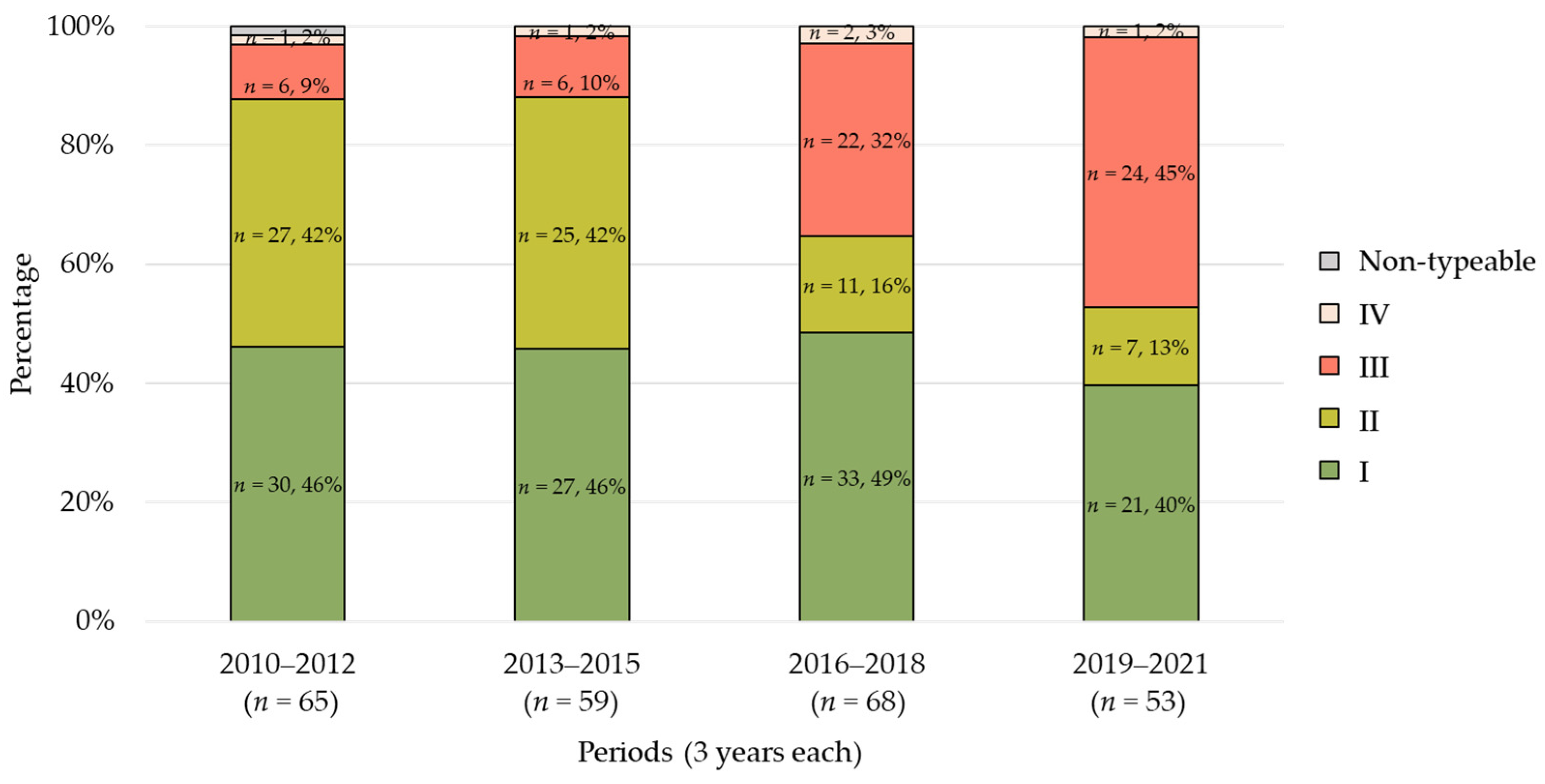
2.1. Trends in SCCmec Types

The trends of changes in SCCmec types over the years during the study period are shown in Figure 1. In the first period, type II (32%) was the most prevalent, followed by type IV (31%) and type I (23%). In the second period, type I (32%) was predominant, followed by type II (29%) and type IV (24%). However, type IV exhibited a significant increasing trend in prevalence from the third period (p < 0.01). In the fourth period, type IV (79%) was detected most frequently, followed by type I (6%) and type II (6%).

2.2. Trends in Agr Types

The trends of changes in agr types over the years are shown in Figure 2. In both the first and second periods, type I (both 46%) was detected most frequently, followed by type II (both 42%) and type III (9% and 10%, respectively). However, type III showed a significant increasing trend from the third period (p < 0.01). In the fourth period, type III (45%) was the most prevalent, followed by type I (40%) and type II (13%), establishing the dominance of type III.

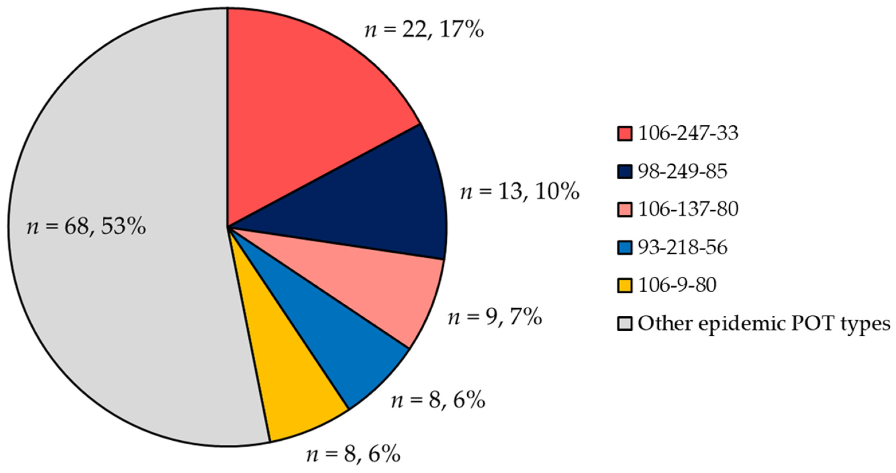
2.3. POT Type Distribution

Among the 245 MRSA bloodstream isolates, there were 143 distinct POT types, with 26 identified as epidemic clones (n = 128, 52%) and 117 as sporadic clones (48%). Among the epidemic clones, the top five POT types accounted for 45% (Figure 3). The genotypes (SCCmec/agr) of these POT types were as follows. POT type 106-247-33 (n = 22) were all identified as SCCmec type IV/agr type III. POT type 98-249-85 (n = 13) except for one isolate were SCCmec type I/agr type I. POT types 106-137-80 (n = 9) and 106-9-80 (n = 8) were all identified as SCCmec type IV/agr type I. POT type 93-218-56 (n = 8) were all SCCmec type II/agr type II (Table S1).

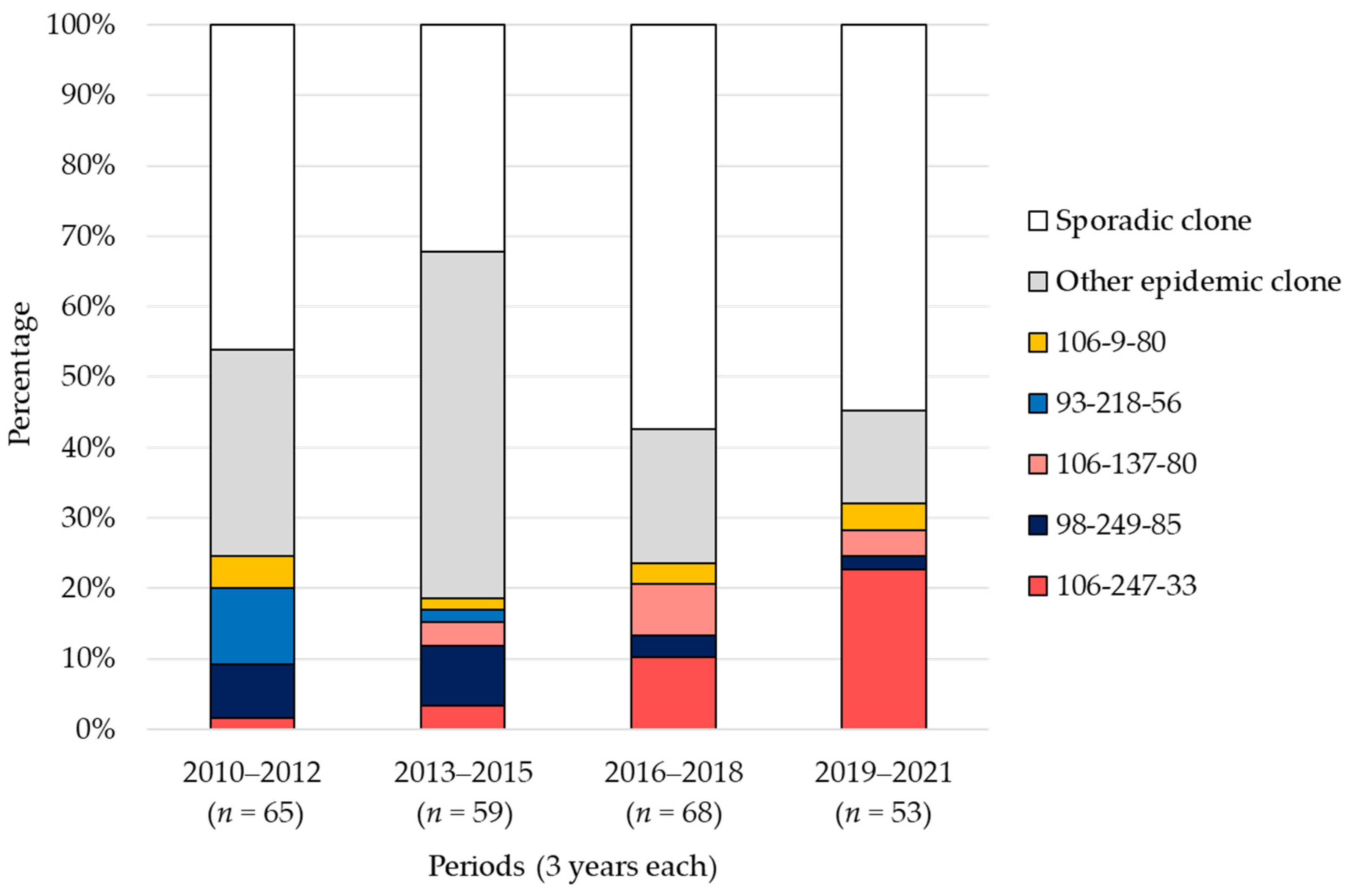
2.4. Trends in POT Types

Figure 4 presents the longitudinal trends of the top five major epidemic clones classified by POT type, along with other epidemic clones and sporadic clones. POT type 93-218-56, characterized by SCCmec type II/agr type II, was detected at a rate of 11% in the first period and was a major strain. However, in the second period, the detection rate dropped to 2%, and it was not detected in the third and fourth periods. On the other hand, POT type 106-247-33, characterized by SCCmec type IV/agr type III, had detection rates of 2% and 3% in the first and second periods, respectively. However, it increased to 10% in the third period and rose further to 23% in the fourth period.
The detection rate of sporadic clones increased significantly in the third and fourth periods compared to the first and second periods [38% (49/124) vs. 56% (68/121), respectively; p = 0.013].

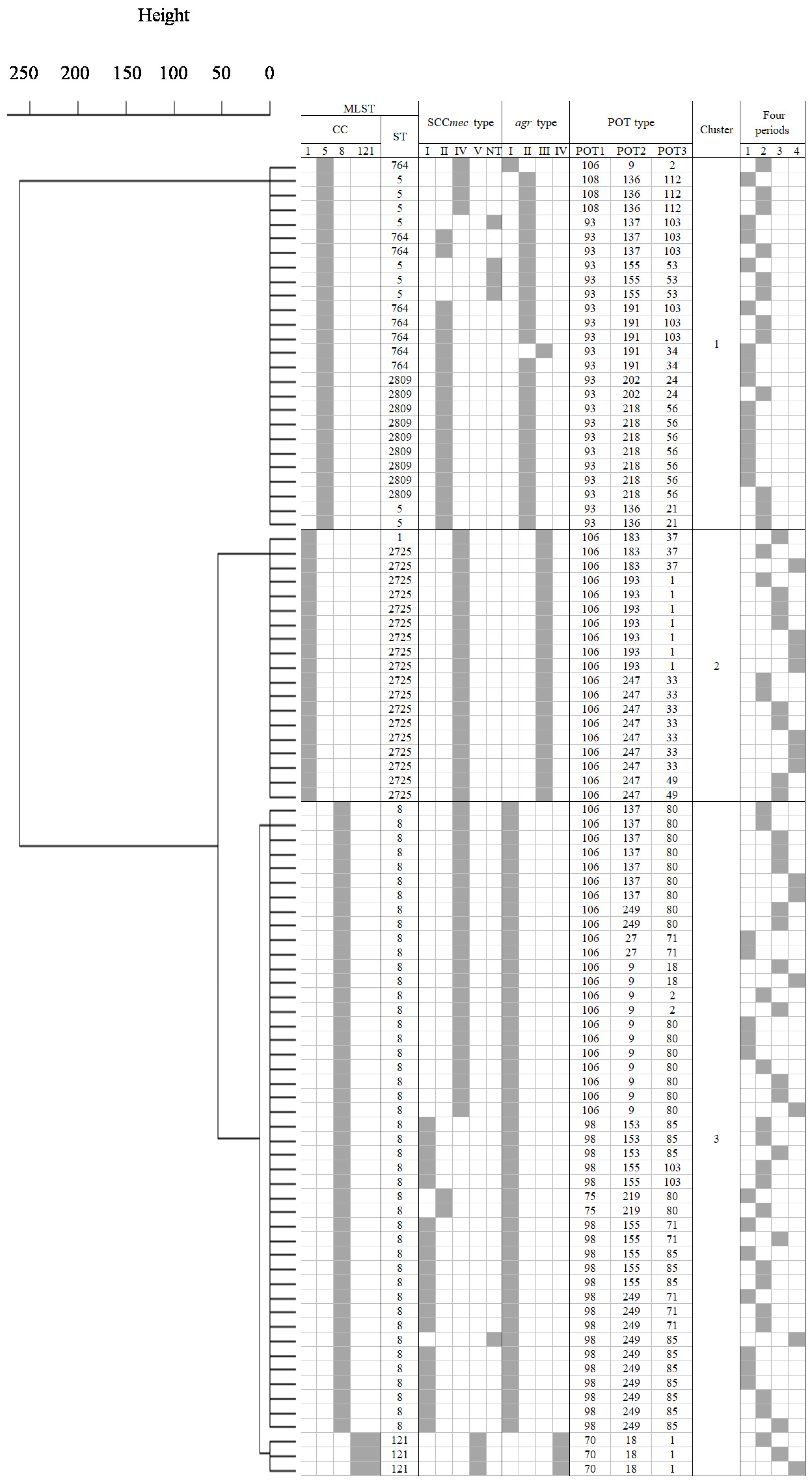
2.5. Cluster Analysis

Among the epidemic clones identified by POT analysis, we randomly selected samples with detection rates ranging from 32% (7/22) to 100% (2/2), totaling 92 strains, for MLST analysis. Figure 5 illustrates the results of cluster analysis based on MLST and POT analyses. The genotypes of epidemic strains showed three clusters with dissimilarity set at 50 (Cluster 1, 26 strains; Cluster 2, 19 strains; Cluster 3, 47 strains). Cluster 1 consisted of strains belonging to CC5, identified as ST2809, ST5, and ST764 and detected only in the first and second periods. The predominant SCCmec type was II (20/26), and the agr type was mainly classified as type II (24/26) within this cluster. Cluster 2 comprised all strains belonging to CC1, with ST identified as ST2725 for all except one isolate. This cluster was primarily detected in the third and fourth periods, and all strains within this cluster were characterized by SCCmec type IV and agr type III. Strains in Cluster 3 were identified as CC8, ST8 (n = 44) and CC121, ST121 (n = 3). ST8 was primarily classified into SCCmec type I (n = 19) and SCCmec type IV (n = 22), with all strains having agr type I. In the first and second periods, ST8 strains with SCCmec type I (n = 16) were predominantly detected, while in the third and fourth periods, strains with SCCmec type IV (n = 13) were mainly detected. ST121 strains were all classified as SCCmec type V and agr type IV, with one strain detected in each of the second, third, and fourth periods. The major five POT types had the following STs: 106-247-33 was ST2725; 98-249-85, 106-137-80, and 106-9-80 were ST8; and 93-218-56 was ST2809 (Table S1).

2.6. Identification of Virulence Genes and Antimicrobial Resistance Profiles

PVL (lukS-PV-lukF-PV) and ACME (arc, opp3) were detected for only one isolate in 2020. The POT type of this strain was identified as 106-77-113, which was the same as USA300. The SCCmec type was not identified (ccr gene: none, mec gene: classB), while agr was type I and ST was ST8 (Table S1: No. 222).

2.7. Antimicrobial Resistance Profiles

Table 1 presents the distributions of the mode of minimum inhibitory concentration (MIC) and resistance rates of antimicrobials over the study period for 244 MRSA blood isolates. All isolates showed susceptibility to both teicoplanin (TEIC) and linezolid (LZD), while one strain exhibited intermediate resistance to vancomycin (VCM) (4 μg/mL). Seven strains (4%) showed no susceptibility to daptomycin (DAP). The resistance rates for the other antimicrobials examined were as follows: arbekacin (ABK), 1% (n = 2); gentamicin (GM), 49% (n = 119); erythromycin (EM), 72% (n = 175); clindamycin (CLDM), 45% (n = 110); minocycline (MINO), 34% (n = 83); levofloxacin (LVFX), 79% (n = 192); and fosfomycin (FOM), 19% (n = 46). Over the 12-year study period, decreasing trends were observed in the detection rates of strains resistant to GM, EM, CLDM, MINO, and FOM (all p < 0.05).

2.8. Relations between Each Genotype (SCCmec and Agr Types) and VCM Susceptibility

A total of 14 strains with VCM MICs of 2 and 4 μg/mL were detected (Table S1, No.19, 55, 63, 66, 74, 81, 91, 118, 152, 160, 180, 207, 231). In the first and second periods, nine strains with VCM MICs of 2 or 4 μg/mL were detected, of which five strains were identified as SCCmec type II/agr type II. On the other hand, in the third and later periods, five strains with VCM MICs of 2 μg/mL were detected, of which three were identified as SCCmec type IV/agr type I and two as SCCmec type II/agr type II.
Strains with agr type II included a significantly higher proportion of strains with VCM MICs of 2 or 4 μg/mL compared to strains with other agr types [13% (9/70) vs. 3% (5/174), respectively; p < 0.01].

3. Discussion

This study was performed to investigate the longitudinal trends in the genotypes of MRSA bloodstream isolates over a period of 12 years. Taken together with previous reports, the duration of monitoring spans over 30 years. Studies investigating the epidemiological trends of bloodstream isolates over such an extended period are exceedingly rare.
In our hospital, the SCCmec types of MRSA bloodstream isolates have undergone a significant transition from types I and II to type IV since 2016 with corresponding changes in drug susceptibility. Similar trends were observed in changes in SCCmec types for non-blood and blood isolates in other healthcare facilities in Japan [14,16,21]. Interestingly, SCCmec type IV, known to be associated with CA-MRSA, was sporadically detected in healthcare facilities in Japan, including our hospital, from the early 1970s to the late 1980s [2,29].
Similar to SCCmec types, the agr type also underwent a significant transition from type II to type III after 2016. While it has been reported that agr type II is predominant in hospital-acquired (HA)-MRSA and that agr type III is more common in CA-MRSA [30], there have been few previous reports regarding the annual trends of agr types in bloodstream isolates. Therefore, the results of agr types in the present study also suggest a transition in the genotype of MRSA from HA-MRSA to CA-MRSA.
Strains identified as agr type II in this study showed a significantly higher proportion of VCM MIC of 2 or 4 μg/mL compared to strains of other agr types. It has been reported that strains of agr type II exhibit lower susceptibility to VCM compared to strains of other agr types [31,32,33]. In this study, among the five MRSA strains with VCM MIC of 2 μg/mL detected in the third period (2016), two strains were identified as SCCmec type II/agr type II, while three strains were identified as SCCmec type IV/agr type I. There have been previous reports of VCM-intermediate S. aureus or heterogeneous VCM-intermediate S. aureus with the SCCmec type IV/agr type I genotype [34,35,36], indicating the need for vigilance regarding the VCM susceptibility of MRSA strains harboring this genotype.
POT analysis has been reported to exhibit high discriminatory power comparable to PFGE, MLST analysis, and repetitive sequence-based PCR [37,38,39,40]. Furthermore, POT analysis has been applied in epidemiological studies of MRSA and for infection control within hospital wards [20,41,42,43].
In this study, the analysis of POT types revealed 128 epidemic clones among MRSA bloodstream isolates, defined as those with the same POT type detected in two or more strains. Among them, the top five POT types accounted for 46% of the isolates. Among the top five POT types, those with characteristics of CA-MRSA, represented by SCCmec type IV strains (POT types 106-247-33, 106-137-80, and 106-9-80), were prevalent not only in our institution but also in other medical facilities in Japan [20,41,42,44,45]. On the other hand, the SCCmec type II associated with the genotype of HA-MRSA, POT type 93-218-56, has not been reported in other facilities, suggesting that it may be unique to our institution. Therefore, the POT type associated with the genotype of HA-MRSA appears to be institution-specific, while the POT type associated with the genotype of CA-MRSA reflects the possibility of regional or national trends.
The detection rate of strains that did not exhibit epidemic expansion in POT analysis (i.e., sporadic clones consisting of a single strain) has increased significantly from 2016 to 2021 compared to the period from 2010 to 2015. This suggested that there has been a diversification of POT types among bloodstream isolates since 2016.
The strains in Cluster 1 were predominantly identified as CC5-SCCmec type II/agr type II, and they dominated as the major causative strains among MRSA bloodstream isolates in our hospital until 2015. This CC5-SCCmec type II/agr type II genetic type was previously a dominant strain in Japan [2,4]. The strains in Cluster 2 were identified as CC1-SCCmec type IV/agr type III and have been detected frequently since 2016. In addition, this cluster, with the exception of one strain, was identified as ST2725. CC1 MRSA bloodstream isolates have recently shown an increasing trend in Japan [17,46], with a particularly high prevalence of ST2725 reported in the Kyushu region [18]. Our analysis also suggested an increase in CC1 MRSA in recent years among bloodstream isolates. Cluster 3 strains were predominantly identified as CC8-ST8 and were detected throughout the study period. Interestingly, CC8-ST8 strains were mainly SCCmec type I/agr type I until 2015, but their prevalence decreased thereafter, with an increase in SCCmec type IV/agr type I, although the reason for this change remains unclear. While it has been reported that diverse SCCmec types exist within CC8-ST8 [47], to our knowledge, there have been no reports of a transition in SCCmec from type I to type IV within CC8-ST8 strains.
In this study, only one strain with the POT type 106-77-113, testing positive for PVL (lukS-PV-lukF-PV) and ACME (arc, opp3), was detected in 2020. It has been reported that many PVL-producing strains, including the possibility of USA300, exhibit the POT type 106-77-113 [48,49,50]. In recent years, the detection rate of PVL-positive strains, including USA300, has been increasing primarily in skin isolates within healthcare facilities [51]. However, the detection rate in blood isolates was reported to be very low [18,36,52], and our results were consistent with this observation.
This study had several limitations. First, this was a retrospective investigation of MRSA bloodstream isolates in a single facility. Second, the study focused exclusively on bloodstream isolates, which limited the ability to track nosocomial transmission. Third, the number of isolates subjected to MLST analysis in this study represented only a subset of epidemic strains. Fourth, we did not perform whole-genome sequencing for detailed analysis, such as the examination of single nucleotide polymorphisms, and therefore missed the opportunity to confirm hidden nosocomial transmissions [36]. However, within the scope of this study, only two instances were observed where strains with the same genotype were detected in the same ward within 1 month. Fifth, the investigation was conducted solely in a single facility, and due to insufficient information on patient backgrounds before transfer, accurate evaluation of epidemiological data was not possible. Nevertheless, this study revealed the long-term genetic trends of MRSA bloodstream isolates within the same healthcare facility in Japan. From a clinical perspective, the fact that there was a shift in MRSA bacteremia isolates in this study from hospital-specific strains that were endemic in hospitals to those that are endemic in the community suggests the importance of a broader public health approach in the prevention of MRSA bacteremia, looking not only at nosocomial infection control but also at the prevention of community-acquired infections in the community.

4. Materials and Methods

4.1. Clinical Isolates

This study was conducted at Fukuoka University Hospital, a university-affiliated, tertiary hospital in southwestern Japan. Over the 12-year period from 2010 to 2021, molecular epidemiological analysis was performed using 245 strains of MRSA isolated from the blood of hospitalized patients, with the exclusion of duplicate strains from the same individuals from a total of 281 strains. Isolates and the associated data were accessed between 1 October 2018 and 29 February 2024. To assess the longitudinal trends in MRSA, we divided this 12-year period into the first period (2010–2012), second period (2013–2015), third period (2016–2018), and fourth period (2019–2021). MRSA isolates were identified by standard phenotypic procedures and stored at −80 °C [53]. Prior to DNA extraction, they were cultivated at 37 °C with shaking in Difco™ M-Staphylococcus Broth (Becton, Dickinson and Company, Franklin Lakes, NJ, USA). Genomic DNA extraction was carried out using a Cica Geneus DNA Extraction Reagent Kit (Kanto Chemical, Tokyo, Japan) in accordance with the manufacturer’s recommended protocol. The obtained genomic DNA was utilized as a template for all analyses.

4.2. SCCmec Typing, Agr Typing, and Identification of Virulence Genes

SCCmec typing [54], agr typing [55], and the detection of PVL (lukS-PV-lukF-PV) [56] and ACME (arc, opp3) [57] were conducted using previously established methods. The ATCC BAA-1556 strain (USA300 clone) was used as a positive control for PVL and ACME detection.

4.3. POT Type Detection

POT analysis was conducted using a Cica Geneus® Staph POT KIT (Kanto Chemical, Tokyo, Japan) [37,58]. POT analysis, a molecular epidemiological method with discriminatory power equivalent to pulsed-field gel electrophoresis (PFGE) [37,38,39], involves the detection of 22 phage-derived open reading frames (ORFs) effective for strain identification. Following the detection of these ORFs, the possession patterns were converted into numerical values, and the POT types (POT1-POT2-POT3) were determined. POT1 targets SCCmec, while POT2 and POT3 target phage-derived ORFs. In this study, strains in which the same POT type was detected in two or more isolates were classified as epidemic clones, whereas strains in which no common POT type was detected were considered to be sporadic clones.

4.4. MLST Analysis

MLST analysis was performed on a randomly selected subset of the epidemic clones identified based on POT types comprising a total of 92 strains [59]. Genotypes were classified into ST and clonal complexes (CC) following the MLST database (https://pubmlst.org).

4.5. Antimicrobial Resistance Profile

We investigated the drug susceptibility of a total of 244 strains, excluding one isolate for which information was not recorded in the medical records, for all drugs except DAP. As DAP was not used at our hospital from 2010 to 2012, there were no data on its MIC and drug sensitivity. Therefore, we investigated the drug susceptibility of a total of 179 isolates from 2013 for DAP.
Antimicrobial susceptibility testing was conducted using VITEK®2 (Sysmex bioMérieux, Tokyo, Japan). The antimicrobial agents investigated included GM, EM, CLDM, MINO, LVFX, FOM, ABK, TEIC, VCM, LZD, and DAP. Sensitivity to antimicrobial agents, excluding FOM, was determined according to the guidelines of the Clinical and Laboratory Standards Institute (CLSI) [60]. Sensitivity to FOM was determined following the guidelines of the European Committee on Antimicrobial Susceptibility Testing (EUCAST Version 10, 2020). The resistance breakpoints of the following antimicrobial agents were determined according to the CLSI: GM, ≥16 μg/mL; EM, ≥8 μg/mL; CLDM, ≥4 μg/mL; MINO, ≥16 μg/mL; LVFX, ≥4 μg/mL; ABK, ≥16 μg/mL; TEIC, ≥32 μg/mL; VCM, ≥16 μg/mL; LZD, ≥8 μg/mL; and DAP, not defined (N/D). As there was no defined breakpoint for ABK, the breakpoint for GM was used as a substitute. The resistance breakpoint for FOM was determined according to the guidelines provided by the EUCAST: FOM, >32 μg/mL.

4.6. Statistical Analysis

Differences in genotype detection rates were assessed using the χ2 test. For genotype clustering of MRSA bloodstream isolates, hierarchical clustering was performed using seven housekeeping gene numbers of MLST and POT1, 2, and 3 numbers. The analysis utilized the Ward method with Euclidean squared distance. All statistical analyses were conducted using EZR software version 1.62 (Jichi Medical University, Saitama Medical Center, Japan). In all analyses, p < 0.05 was taken to indicate statistical significance.

5. Conclusions

In conclusion, this study showed that the genotype of MRSA bloodstream isolates in our institution shifted from SCCmec type II/agr type II to SCCmec type IV/agr type III around 2016, with a notable increase in CC1 MRSA. The significant increase in sporadic isolates suggested the diversification of genotypes in recent years.

Supplementary Materials

The following supporting information can be downloaded at: https://www.mdpi.com/article/10.3390/antibiotics13070670/s1, Table S1: Molecular epidemiological analysis and antimicrobial susceptibility testing results of MRSA blood isolates detected at Fukuoka University Hospital (245 isolates). The four periods were as follows: Period 1, 2010–2012; Period 2, 2013–2015; Period 3, 2016–2018; and Period 4, 2019–2021). N/T, non-typeable. N/A, not available. * Non-susceptibility to DAP.

Author Contributions

Conceptualization, M.M., Y.N. and T.T.; methodology, M.M., T.T. and N.K.; formal analysis, Y.M. and M.M.; investigation, Y.M., M.K. and S.M.; resources, M.M. and T.T.; data curation, Y.M., M.K. and T.T.; writing—original draft preparation, Y.M. and M.M.; writing—review and editing, T.T. and N.K.; visualization, Y.M.; supervision, T.S. and N.K.; project administration, T.T.; funding acquisition, T.T. and N.K. All authors have read and agreed to the published version of the manuscript.

Funding

This study was partially supported by a Grant-in-Aid for Scientific Research (C) (Grant Number 21K08519).

Institutional Review Board Statement

This study was performed according to the principles of the Declaration of Helsinki and approved by the Ethics Committee of Fukuoka University Hospital (Approval number: 18-9-07).

Informed Consent Statement

All data were fully anonymized before their access. As this was a retrospective clinical study, the patients underwent no medical interventions, and only medical information was utilized. Therefore, patient consent was implemented on an opt-out basis based on the consent of the Ethics Committee.

Data Availability Statement

All data are provided in the manuscript and Supplementary Information.

Acknowledgments

We would like to thank Chiemi Tokushige (Department of Clinical Laboratory, Fukuoka University Hospital) for collecting the MRSA isolates, and we thank Hiromi Toh and Makiko Yuki for their technical assistance. The authors gratefully acknowledge the excellent student technical support by Ryo Noguchi and Namie Tanamachi.

Conflicts of Interest

The authors declare no conflicts of interest. The funders had no role in the design of the study; in the collection, analyses, or interpretation of data; in the writing of the manuscript; or in the decision to publish the results.

References

  1. van Hal, S.J.; Jensen, S.O.; Vaska, V.L.; Espedido, B.A.; Paterson, D.L.; Gosbell, I.B. Predictors of mortality in Staphylococcus aureus Bacteremia. Clin. Microbiol. Rev. 2012, 25, 362–386. [Google Scholar] [CrossRef] [PubMed]
  2. Ma, X.X.; Ito, T.; Chongtrakool, P.; Hiramatsu, K. Predominance of clones carrying Panton-Valentine leukocidin genes among methicillin-resistant Staphylococcus aureus strains isolated in Japanese hospitals from 1979 to 1985. J. Clin. Microbiol. 2006, 44, 4515–4527. [Google Scholar] [CrossRef] [PubMed]
  3. Taneike, I.; Otsuka, T.; Dohmae, S.; Saito, K.; Ozaki, K.; Takano, M.; Yamamoto, T. Molecular nature of methicillin-resistant Staphylococcus aureus derived from explosive nosocomial outbreaks of the 1980s in Japan. FEBS Lett. 2006, 580, 2323–2334. [Google Scholar] [CrossRef] [PubMed]
  4. Yanagihara, K.; Araki, N.; Watanabe, S.; Kinebuchi, T.; Kaku, M.; Maesaki, S.; Yamaguchi, K.; Matsumoto, T.; Mikamo, H.; Takesue, Y.; et al. Antimicrobial susceptibility and molecular characteristics of 857 methicillin-resistant Staphylococcus aureus isolates from 16 medical centers in Japan (2008–2009): Nationwide survey of community-acquired and nosocomial MRSA. Diagn. Microbiol. Infect. Dis. 2012, 72, 253–257. [Google Scholar] [CrossRef] [PubMed]
  5. Yamamoto, T.; Nishiyama, A.; Takano, T.; Yabe, S.; Higuchi, W.; Razvina, O.; Shi, D. Community-acquired methicillin-resistant Staphylococcus aureus: Community transmission, pathogenesis, and drug resistance. J. Infect. Chemother. 2010, 16, 225–254. [Google Scholar] [CrossRef] [PubMed]
  6. Turner, N.A.; Sharma-Kuinkel, B.K.; Maskarinec, S.A.; Eichenberger, E.M.; Shah, P.P.; Carugati, M.; Holland, T.L.; Fowler, V.G., Jr. Methicillin-resistant Staphylococcus aureus: An overview of basic and clinical research. Nat. Rev. Microbiol. 2019, 17, 203–218. [Google Scholar] [CrossRef] [PubMed]
  7. Centers for Disease Control and Prevention (CDC). Four pediatric deaths from community-acquired methicillin-resistant Staphylococcus aureus—Minnesota and North Dakota, 1997–1999. MMWR Morb. Mortal. Wkly. Rep. 1999, 48, 707–710. [Google Scholar]
  8. Vandenesch, F.; Naimi, T.; Enright, M.C.; Lina, G.; Nimmo, G.R.; Heffernan, H.; Liassine, N.; Bes, M.; Greenland, T.; Reverdy, M.E.; et al. Community-acquired methicillin-resistant Staphylococcus aureus carrying Panton-Valentine leukocidin genes: Worldwide emergence. Emerg. Infect. Dis. 2003, 9, 978–984. [Google Scholar] [CrossRef] [PubMed]
  9. Baba, T.; Takeuchi, F.; Kuroda, M.; Yuzawa, H.; Aoki, K.; Oguchi, A.; Nagai, Y.; Iwama, N.; Asano, K.; Naimi, T.; et al. Genome and virulence determinants of high virulence community-acquired MRSA. Lancet 2002, 359, 1819–1827. [Google Scholar] [CrossRef]
  10. Diep, B.A.; Gill, S.R.; Chang, R.F.; Phan, T.H.; Chen, J.H.; Davidson, M.G.; Lin, F.; Lin, J.; Carleton, H.A.; Mongodin, E.F.; et al. Complete genome sequence of USA300, an epidemic clone of community-acquired meticillin-resistant Staphylococcus aureus. Lancet 2006, 367, 731–739. [Google Scholar] [CrossRef]
  11. David, M.Z.; Daum, R.S. Community-associated methicillin-resistant Staphylococcus aureus: Epidemiology and clinical consequences of an emerging epidemic. Clin. Microbiol. Rev. 2010, 23, 616–687. [Google Scholar] [CrossRef] [PubMed]
  12. Miller, L.G.; Perdreau-Remington, F.; Rieg, G.; Mehdi, S.; Perlroth, J.; Bayer, A.S.; Tang, A.W.; Phung, T.O.; Spellberg, B. Necrotizing fasciitis caused by community-associated methicillin-resistant Staphylococcus aureus in Los Angeles. N. Engl. J. Med. 2005, 352, 1445–1453. [Google Scholar] [CrossRef]
  13. Yamaguchi, T.; Okamura, S.; Miura, Y.; Koyama, S.; Yanagisawa, H.; Matsumoto, T. Molecular Characterization of Community-Associated Methicillin-Resistant Staphylococcus aureus Isolated from Skin and Pus Samples of Outpatients in Japan. Microb. Drug Resist. 2015, 21, 441–447. [Google Scholar] [CrossRef]
  14. Nakaminami, H.; Takadama, S.; Ito, A.; Hasegawa, M.; Jono, C.; Noguchi, M.; Shoshi, M.; Wajima, T.; Fujii, T.; Maruyama, H.; et al. Characterization of SCCmec type IV methicillin-resistant Staphylococcus aureus clones increased in Japanese hospitals. J. Med. Microbiol. 2018, 67, 769–774. [Google Scholar] [CrossRef] [PubMed]
  15. Miura, Y.; Yamaguchi, T.; Nakamura, I.; Koyama, S.; Tamai, K.; Okanda, T.; Matsumoto, T. Epidemiological Trends Observed from Molecular Characterization of Methicillin-Resistant Staphylococcus aureus Isolates from Blood Cultures at a Japanese University Hospital, 2012–2015. Microb. Drug Resist. 2018, 24, 70–75. [Google Scholar] [CrossRef]
  16. Kaku, N.; Yanagihara, K.; Morinaga, Y.; Yamada, K.; Harada, Y.; Migiyama, Y.; Nagaoka, K.; Matsuda, J.; Uno, N.; Hasegawa, H.; et al. Influence of antimicrobial regimen on decreased in-hospital mortality of patients with MRSA bacteremia. J. Infect. Chemother. 2014, 20, 350–355. [Google Scholar] [CrossRef]
  17. Aung, M.S.; Urushibara, N.; Kawaguchiya, M.; Ohashi, N.; Hirose, M.; Kimura, Y.; Kudo, K.; Ito, M.; Kobayashi, N. Molecular Epidemiological Characterization of Methicillin-Resistant Staphylococcus aureus from Bloodstream Infections in Northern Japan: Increasing Trend of CC1 and Identification of ST8-SCCmec IVa USA300-Like Isolate Lacking Arginine Catabolic Mobile Element. Microb. Drug Resist. 2023, 30, 63–72. [Google Scholar] [CrossRef]
  18. Kaku, N.; Sasaki, D.; Ota, K.; Miyazaki, T.; Yanagihara, K. Changing molecular epidemiology and characteristics of MRSA isolated from bloodstream infections: Nationwide surveillance in Japan in 2019. J. Antimicrob. Chemother. 2022, 77, 2130–2141. [Google Scholar] [CrossRef]
  19. Kawasuji, H.; Ikezawa, Y.; Morita, M.; Sugie, K.; Somekawa, M.; Ezaki, M.; Koshiyama, Y.; Takegoshi, Y.; Murai, Y.; Kaneda, M.; et al. High Incidence of Metastatic Infections in Panton-Valentine Leucocidin-Negative, Community-Acquired Methicillin-Resistant Staphylococcus aureus Bacteremia: An 11-Year Retrospective Study in Japan. Antibiotics 2023, 12, 1516. [Google Scholar] [CrossRef] [PubMed]
  20. Shimizu, M.; Mihara, T.; Ohara, J.; Inoue, K.; Kinoshita, M.; Sawa, T. Relationship between mortality and molecular epidemiology of methicillin-resistant Staphylococcus aureus bacteremia. PLoS ONE 2022, 17, e0271115. [Google Scholar] [CrossRef] [PubMed]
  21. Hamada, M.; Yamaguchi, T.; Sato, A.; Ono, D.; Aoki, K.; Kajiwara, C.; Kimura, S.; Maeda, T.; Sasaki, M.; Murakami, H.; et al. Increased Incidence and Plasma-Biofilm Formation Ability of SCCmec Type IV Methicillin-Resistant Staphylococcus aureus (MRSA) Isolated from Patients With Bacteremia. Front. Cell. Infect. Microbiol. 2021, 11, 602833. [Google Scholar] [CrossRef] [PubMed]
  22. Nikolaras, G.P.; Papaparaskevas, J.; Samarkos, M.; Tzouvelekis, L.S.; Psychogiou, M.; Pavlopoulou, I.; Goukos, D.; Polonyfi, K.; Pantazatou, A.; Deliolanis, I.; et al. Changes in the rates and population structure of methicillin-resistant Staphylococcus aureus (MRSA) from bloodstream infections: A single-centre experience (2000–2015). J. Glob. Antimicrob. Resist. 2019, 17, 117–122. [Google Scholar] [CrossRef] [PubMed]
  23. Hwang, Y.J. Comparing the Phylogenetic Distribution of Multilocus Sequence Typing, Staphylococcal Protein A, and Staphylococcal Cassette Chromosome Mec Types in Methicillin-Resistant Staphylococcus aureus (MRSA) in Korea from 1994 to 2020. Antibiotics 2023, 12, 1397. [Google Scholar] [CrossRef]
  24. Niek, W.K.; Teh, C.S.J.; Idris, N.; Sit, P.S.; Lee, Y.Q.; Thong, K.L.; Sri La Sri Ponnampalavanar, S. Methicillin-resistant Staphylococcus aureus bacteraemia, 2003–2015: Comparative evaluation of changing trends in molecular epidemiology and clinical outcomes of infections. Infect. Genet. Evol. 2020, 85, 104567. [Google Scholar] [CrossRef]
  25. Ellington, M.J.; Hope, R.; Livermore, D.M.; Kearns, A.M.; Henderson, K.; Cookson, B.D.; Pearson, A.; Johnson, A.P. Decline of EMRSA-16 amongst methicillin-resistant Staphylococcus aureus causing bacteraemias in the UK between 2001 and 2007. J. Antimicrob. Chemother. 2010, 65, 446–448. [Google Scholar] [CrossRef]
  26. Hsieh, Y.C.; Lin, Y.C.; Huang, Y.C. Vancomycin, teicoplanin, daptomycin, and linezolid MIC creep in methicillin-resistant Staphylococcus aureus is associated with clonality. Medicine 2016, 95, e5060. [Google Scholar] [CrossRef] [PubMed]
  27. Popovich, K.J.; Weinstein, R.A.; Hota, B. Are community-associated methicillin-resistant Staphylococcus aureus (MRSA) strains replacing traditional nosocomial MRSA strains? Clin. Infect. Dis. 2008, 46, 787–794. [Google Scholar] [CrossRef]
  28. Choi, S.H.; Lee, J.; Jung, J.; Kim, E.S.; Kim, M.J.; Chong, Y.P.; Kim, S.H.; Choi, S.H.; Lee, S.O.; Kim, Y.S. A Longitudinal Study of Adult Patients with Staphylococcus aureus Bacteremia over 11 Years in Korea. J. Korean Med. Sci. 2021, 36, e104. [Google Scholar] [CrossRef] [PubMed]
  29. Takata, T.; Miyazaki, M.; Li, H.; Healy, M.; Frye, S.; Tanaka, K.; Hara, S.; Kamimura, H.; Yoshimura, H.; Matsunaga, A.; et al. Methicillin-resistant Staphylococcus aureus bloodstream infections in a Japanese University Hospital between 1987 and 2004. J. Infect. Chemother. 2012, 18, 199–206. [Google Scholar] [CrossRef]
  30. Naimi, T.S.; LeDell, K.H.; Como-Sabetti, K.; Borchardt, S.M.; Boxrud, D.J.; Etienne, J.; Johnson, S.K.; Vandenesch, F.; Fridkin, S.; O’Boyle, C.; et al. Comparison of community- and health care-associated methicillin-resistant Staphylococcus aureus infection. JAMA 2003, 290, 2976–2984. [Google Scholar] [CrossRef]
  31. Park, M.J.; Kim, H.S.; Kim, H.S.; Kim, J.S.; Song, W.; Kim, M.Y.; Lee, Y.K.; Kang, H.J. Accessory Gene Regulator Polymorphism and Vancomycin Minimum Inhibitory Concentration in Methicillin-Resistant Staphylococcus aureus. Ann. Lab. Med. 2015, 35, 399–403. [Google Scholar] [CrossRef]
  32. Moise, P.A.; Smyth, D.S.; Robinson, D.A.; El-Fawal, N.; McCalla, C.; Sakoulas, G. Genotypic and phenotypic relationships among methicillin-resistant Staphylococcus aureus from three multicentre bacteraemia studies. J. Antimicrob. Chemother. 2009, 63, 873–876. [Google Scholar] [CrossRef] [PubMed]
  33. Cázares-Domínguez, V.; Cruz-Córdova, A.; Ochoa, S.A.; Escalona, G.; Arellano-Galindo, J.; Rodríguez-Leviz, A.; Hernández-Castro, R.; López-Villegas, E.O.; Xicohtencatl-Cortes, J. Vancomycin tolerant, methicillin-resistant Staphylococcus aureus reveals the effects of vancomycin on cell wall thickening. PLoS ONE 2015, 10, e0118791. [Google Scholar] [CrossRef] [PubMed]
  34. Wang, W.Y.; Chiu, C.F.; Lee, Y.T.; Hsueh, P.R.; Tsao, S.M. Molecular epidemiology and phenotypes of invasive methicillin-resistant vancomycin-intermediate Staphylococcus aureus in Taiwan. J. Microbiol. Immunol. Infect. 2022, 55 Pt 2, 1203–1210. [Google Scholar] [CrossRef] [PubMed]
  35. Wang, W.Y.; Lee, S.Y.; Chiueh, T.S.; Lu, J.J. Molecular and phenotypic characteristics of methicillin-resistant and vancomycin-intermediate Staphylococcus aureus isolates from patients with septic arthritis. J. Clin. Microbiol. 2009, 47, 3617–3623. [Google Scholar] [CrossRef] [PubMed]
  36. Shen, P.; Zhou, K.; Wang, Y.; Song, J.; Liu, Y.; Zhou, Y. High prevalence of a globally disseminated hypervirulent clone, Staphylococcus aureus CC121, with reduced vancomycin susceptibility in community settings in China. J. Antimicrob. Chemother. 2019, 74, 2537–2543. [Google Scholar] [CrossRef] [PubMed]
  37. Suzuki, M.; Tawada, Y.; Kato, M.; Hori, H.; Mamiya, N.; Hayashi, Y.; Nakano, M.; Fukushima, R.; Katai, A.; Tanaka, T.; et al. Development of a rapid strain differentiation method for methicillin-resistant Staphylococcus aureus isolated in Japan by detecting phage-derived open-reading frames. J. Appl. Microbiol. 2006, 101, 938–947. [Google Scholar] [CrossRef] [PubMed]
  38. Nada, T.; Yagi, T.; Ohkura, T.; Morishita, Y.; Baba, H.; Ohta, M.; Suzuki, M. Usefulness of phage open-reading frame typing method in an epidemiological study of an outbreak of methicillin-resistant Staphylococcus aureus infections. Jpn. J. Infect. Dis. 2009, 62, 386–389. [Google Scholar] [CrossRef] [PubMed]
  39. Ogihara, S.; Saito, R.; Sawabe, E.; Kozakai, T.; Shima, M.; Aiso, Y.; Fujie, T.; Nukui, Y.; Koike, R.; Hagihara, M.; et al. Molecular typing of methicillin-resistant Staphylococcus aureus: Comparison of PCR-based open reading frame typing, multilocus sequence typing, and Staphylococcus protein A gene typing. J. Infect. Chemother. 2018, 24, 312–314. [Google Scholar] [CrossRef]
  40. Osawa, K.; Shigemura, K.; Jikimoto, T.; Shirakawa, T.; Fujisawa, M.; Arakawa, S. Comparison between phage-open-reading frame typing and automated repetitive-sequence-based PCR for typing MRSA isolates. J. Antibiot. 2014, 67, 565–569. [Google Scholar] [CrossRef]
  41. Nakaie, K.; Yamada, K.; Park, K.; Nakamura, Y.; Okada, Y.; Fujita, A.; Fujimoto, H.; Kaneko, Y.; Kakeya, H. Effectiveness of weekly polymerase chain reaction-based open reading frame typing analysis of all newly isolated methicillin-resistant Staphylococcus aureus strains for controlling nosocomial infections. J. Infect. Chemother. 2016, 22, 733–737. [Google Scholar] [CrossRef] [PubMed]
  42. Kishita, M.; Matsumura, Y.; Yamamoto, M.; Nagao, M.; Takemura, M.; Sumi, M.; Osawa, M.; Nakano, Y.; Tanikawa, S.; Tsukaguchi, F.; et al. Increase in the frequency of community-acquired methicillin-resistant Staphylococcus aureus clones among inpatients of acute care hospitals in the Kyoto and Shiga regions, Japan. J. Infect. Chemother. 2023, 29, 458–463. [Google Scholar] [CrossRef] [PubMed]
  43. Kawamura, K.; Kitaoka, K.; Kimura, K.; Wachino, J.I.; Kondo, T.; Iinuma, Y.; Murakami, N.; Fujimoto, S.; Arakawa, Y. Spread of seb-Positive Methicillin-Resistant Staphylococcus aureus SCCmec Type II-ST764 Among Elderly Japanese in Nonacute Care Settings. Microb. Drug Resist. 2019, 25, 915–924. [Google Scholar] [CrossRef] [PubMed]
  44. Kato, H.; Ide, K.; Fukase, F.; Shimura, Y.; Yasuda, S.; Goto, H.; Fukuyama, A.; Nakajima, H. Polymerase chain reaction-based open reading frame typing (POT) method analysis for a methicillin-resistant Staphylococcus aureus (MRSA) outbreak through breast-feeding in the neonatal intensive care unit. IDCases 2018, 12, 1–3. [Google Scholar] [CrossRef] [PubMed]
  45. Miyamoto, H.; Murakami, S.; Saito, M.; Tamaki, M.; Ochi, C.; Iyoda, M. Molecular Epidemiology of Methicillin-Resistant Staphylococcus aureus (MRSA) by POT Method, Antimicrobial Susceptibility Patterns, and Toxin-Producing Types. Rinsho Biseibutshu Jinsoku Shindan Kenkyukai Shi 2020, 29, 45–52. (In Japanese) [Google Scholar]
  46. Sato, T.; Yamaguchi, T.; Aoki, K.; Kajiwara, C.; Kimura, S.; Maeda, T.; Yoshizawa, S.; Sasaki, M.; Murakami, H.; Hisatsune, J.; et al. Whole-genome sequencing analysis of molecular epidemiology and silent transmissions causing meticillin-resistant Staphylococcus aureus bloodstream infections in a university hospital. J. Hosp. Infect. 2023, 139, 141–149. [Google Scholar] [CrossRef] [PubMed]
  47. Enright, M.C.; Robinson, D.A.; Randle, G.; Feil, E.J.; Grundmann, H.; Spratt, B.G. The evolutionary history of methicillin-resistant Staphylococcus aureus (MRSA). Proc. Natl. Acad. Sci. USA 2002, 99, 7687–7692. [Google Scholar] [CrossRef] [PubMed]
  48. Konuma, T.; Takahashi, S.; Suzuki, M.; Tojo, A. Molecular characterization of healthcare and community-associated methicillin-resistant Staphylococcus aureus using phage open-reading frame typing. Iran. J. Microbiol. 2021, 13, 560–564. [Google Scholar] [CrossRef] [PubMed]
  49. Ogihara, S.; Inoue, O.; Yamagami, T.; Yanagimoto, K.; Uematsu, K.; Hisada, Y.; Uchida, T.; Ohta, M.; Suzuki-Inoue, K. Clinical characteristics and molecular analysis of USA300 and ST 764 methicillin-resistant Staphylococcus aureus isolates from outpatients in Japan by PCR-Based open reading frame typing. J. Infect. Chemother. 2021, 27, 466–472. [Google Scholar] [CrossRef]
  50. Osaka, S.; Okuzumi, K.; Koide, S.; Tamai, K.; Sato, T.; Tanimoto, K.; Tomita, H.; Suzuki, M.; Nagano, Y.; Shibayama, K.; et al. Genetic shifts in methicillin-resistant Staphylococcus aureus epidemic clones and toxin gene profiles in Japan: Comparative analysis among pre-epidemic, epidemic and post-epidemic phases. J. Med. Microbiol. 2018, 67, 392–399. [Google Scholar] [CrossRef]
  51. Takadama, S.; Nakaminami, H.; Sato, A.; Shoshi, M.; Fujii, T.; Noguchi, N. Dissemination of Panton-Valentine leukocidin-positive methicillin-resistant Staphylococcus aureus USA300 clone in multiple hospitals in Tokyo, Japan. Clin. Microbiol. Infect. 2018, 24, 1211.e1–1211.e7. [Google Scholar] [CrossRef] [PubMed]
  52. Kimura, Y.; Morinaga, Y.; Akamatsu, N.; Matsuda, J.; Yamaryo, T.; Kawakami, K.; Matsuo, H.; Kosai, K.; Uno, N.; Hasegawa, H.; et al. Antimicrobial susceptibility and molecular characteristics of methicillin-resistant Staphylococcus aureus in a Japanese secondary care facility. J. Infect. Chemother. 2016, 22, 14–18. [Google Scholar] [CrossRef] [PubMed]
  53. Tang, Y.W.; Waddington, M.G.; Smith, D.H.; Manahan, J.M.; Kohner, P.C.; Highsmith, L.M.; Li, H.; Cockerill, F.R., 3rd; Thompson, R.L.; Montgomery, S.O.; et al. Comparison of protein A gene sequencing with pulsed-field gel electrophoresis and epidemiologic data for molecular typing of methicillin-resistant Staphylococcus aureus. J. Clin. Microbiol. 2000, 38, 1347–1351. [Google Scholar] [CrossRef] [PubMed]
  54. Kondo, Y.; Ito, T.; Ma, X.X.; Watanabe, S.; Kreiswirth, B.N.; Etienne, J.; Hiramatsu, K. Combination of multiplex PCRs for staphylococcal cassette chromosome mec type assignment: Rapid identification system for mec, ccr, and major differences in junkyard regions. Antimicrob. Agents Chemother. 2007, 51, 264–274. [Google Scholar] [CrossRef] [PubMed]
  55. Lina, G.; Boutite, F.; Tristan, A.; Bes, M.; Etienne, J.; Vandenesch, F. Bacterial competition for human nasal cavity colonization: Role of Staphylococcal agr alleles. Appl. Environ. Microbiol. 2003, 69, 18–23. [Google Scholar] [CrossRef] [PubMed]
  56. Lina, G.; Piémont, Y.; Godail-Gamot, F.; Bes, M.; Peter, M.O.; Gauduchon, V.; Vandenesch, F.; Etienne, J. Involvement of Panton-Valentine leukocidin-producing Staphylococcus aureus in primary skin infections and pneumonia. Clin. Infect. Dis. 1999, 29, 1128–1132. [Google Scholar] [CrossRef] [PubMed]
  57. Du, X.; Zhu, Y.; Song, Y.; Li, T.; Luo, T.; Sun, G.; Yang, C.; Cao, C.; Lu, Y.; Li, M. Molecular analysis of Staphylococcus epidermidis strains isolated from community and hospital environments in China. PLoS ONE 2013, 8, e62742. [Google Scholar] [CrossRef]
  58. Suzuki, M.; Matsumoto, M.; Takahashi, M.; Hayakawa, Y.; Minagawa, H. Identification of the clonal complexes of Staphylococcus aureus strains by determination of the conservation patterns of small genomic islets. J. Appl. Microbiol. 2009, 107, 1367–1374. [Google Scholar] [CrossRef]
  59. Enright, M.C.; Day, N.P.; Davies, C.E.; Peacock, S.J.; Spratt, B.G. Multilocus sequence typing for characterization of methicillin-resistant and methicillin-susceptible clones of Staphylococcus aureus. J. Clin. Microbiol. 2000, 38, 1008–1015. [Google Scholar] [CrossRef]
  60. CLSI. Performance Standards for Antimicrobial Susceptibility Testing, 30th ed.; CLSI supplement M100; Clinical and Laboratory Standards Institute: Wayne, NY, USA, 2020. [Google Scholar]
Figure 1. Trends of changes in SCCmec types. The data are presented as percentages relative to the total isolates for each period. The figure illustrates the distribution of SCCmec types across the four periods.
Antibiotics 13 00670 g001
Figure 2. Trends in agr types. The data are presented as percentages relative to the total isolates for each period. The figure illustrates the distribution of agr types across the four periods.
Antibiotics 13 00670 g002
Figure 3. Distribution of POT types among epidemic clones. The distribution of the 128 isolates identified as epidemic clones is based on POT types. The total percentage is 99% as each component ratio is rounded to the nearest whole number.
Antibiotics 13 00670 g003
Figure 4. Trends in major epidemic clones according to POT analysis. The data are presented as percentages of total isolates for each period. The figure illustrates the longitudinal trends of the top 5 major epidemic clones classified by POT type, along with other epidemic clones and sporadic clones, for each of the four periods.
Antibiotics 13 00670 g004
Figure 5. Dendrogram and molecular epidemiological analysis of 92 randomly selected epidemic clones. First column: CC analyzed by MLST. Second column: ST analyzed by MLST. Third column: SCCmec typing classification. Fourth column: agr typing classification. Fifth column: POT number. Sixth column: Cluster number. Seventh column: Periods of detection (first period: 2010–2012; second period: 2013–2015; third period: 2016–2018; and fourth period: 2019–2021, respectively).
Antibiotics 13 00670 g005
Table 1. Annual distribution of the mode of MIC values and resistance rates to antimicrobial agents for 244 MRSA blood isolates.
Antimicrobial Drug a2010–2012
(n = 65)
2013–2015
(n = 59)
2016–2018
(n = 68)
2019–2021
(n = 52)
Total
Mode MIC
[μg/mL] b
R (%) cMode MIC
[μg/mL]
R (%)Mode MIC
[μg/mL]
R (%)Mode MIC
[μg/mL]
R (%)Mode MIC
[μg/mL]
R (%)
GM≥1657%≥1653%≥1651%≤0.529%≥1648%
EM≥883%≥881%≥865%≥855%≥872%
CLDM≥862%≥864%≤0.2532%≤0.2519%≤0.2545%
MINO≥1646%≥1653%≤0.521%≤0.515%≤0.534%
LVFX≥877%≥885%≥876%≥875%≥879%
FOM≥823%≥834%≥812%≥86%≥819%
ABK≤12%≤10%≤11%≤10%≤11%
TEIC≤0.50%≤0.50%≤0.50%≤0.50%≤0.50%
VCM10%10%10%10%10%
LZD20%20%20%20%20%
DAPN/D dN/D1N/D0.25N/D0.25N/D0.25N/D
a ABK, arbekacin; CLDM, clindamycin; DAP, daptomycin; EM, erythromycin; FOM, fosfomycin; GM, gentamicin; LVFX, levofloxacin; LZD, linezolid; MINO, minocycline; TEIC, teicoplanin; VCM, vancomycin. b Mode MIC, the mode of MIC (μg/mL). c R, resistant. d N/D, not defined.
Disclaimer/Publisher’s Note: The statements, opinions and data contained in all publications are solely those of the individual author(s) and contributor(s) and not of MDPI and/or the editor(s). MDPI and/or the editor(s) disclaim responsibility for any injury to people or property resulting from any ideas, methods, instructions or products referred to in the content.

