Article

Bacterial and Genetic Features of Raw Retail Pork Meat: Integrative Analysis of Antibiotic Susceptibility, Whole-Genome Sequencing, and Metagenomics

1
Centre for Healthcare Association Infections, Antimicrobial Resistance and Mycoses, National Institute for Communicable Diseases, a Division of the National Health Laboratory Service, Johannesburg 2192, South Africa
2
Department of Clinical Microbiology and Infectious Diseases, School of Pathology, University of Witwatersrand, Johannesburg 2193, South Africa
3
Sequencing Core Facility, National Institute for Communicable Diseases, a Division of the National Health Laboratory Service, Johannesburg 2192, South Africa
4
Department of Biochemistry, Genetics and Microbiology, Forestry and Agricultural Biotechnology Institute, University of Pretoria, Pretoria 0002, South Africa
*
Author to whom correspondence should be addressed.
Antibiotics 2024, 13(8), 700; https://doi.org/10.3390/antibiotics13080700
Submission received: 28 June 2024 / Revised: 23 July 2024 / Accepted: 23 July 2024 / Published: 26 July 2024

Abstract

:
The global antibiotic resistance crisis, driven by overuse and misuse of antibiotics, is multifaceted. This study aimed to assess the microbiological and genetic characteristics of raw retail pork meat through various methods, including the isolation, antibiotic susceptibility testing (AST), whole-genome sequencing (WGS) of selected indicator bacteria, antibiotic residue testing, and metagenomic sequencing. Samples were purchased from 10 pre-selected retail stores in Gauteng, South Africa. The samples were aseptically separated, with portions sent to an external laboratory for isolating indicator bacteria and testing for antibiotic residues. Identification of the isolated bacteria was reconfirmed using matrix-assisted laser desorption/ionization time-of-flight mass spectrometry (MALDI-TOF MS). AST was performed using the Microscan Walkaway system (Beckman Coulter, Brea, CA, USA). WGS and metagenomic sequencing were performed using the Illumina NextSeq 550 instrument (San Diego, CA, USA). The isolated E. coli and E. faecalis exhibited minimal phenotypic resistance, with WGS revealing the presence of tetracycline resistance genes. Both the isolated bacteria and meat samples harboured tetracycline resistance genes and the antibiotic residue concentrations were within acceptable limits for human consumption. In the metagenomic context, most identified bacteria were of food/meat spoilage and environmental origin. The resistome analysis primarily indicated beta-lactam, tetracycline and multidrug resistance genes. Further research is needed to understand the broader implications of these findings on environmental health and antibiotic resistance.

1. Introduction

Since 2005, pork consumption in South Africa has seen a significant increase [1,2]. The country has a well-established pork production industry, with the largest contributions coming from the Limpopo and North West provinces, accounting for 24% and 20% of the total production, respectively [1]. In South Africa, meat is sold through formal and informal channels, including butcheries, supermarkets, farms, and open markets [2]. The formal sector adheres to safety checks before selling meat to consumers; informal outlets often skip these checks, posing risks of microbial contamination, antibiotic resistance, and poor meat quality [2].
Antibiotic resistance is multifaceted and has been linked to overuse and misuse of antibiotics in humans and animals, improper prescribing practices, and the widespread use of antibiotics in livestock production [3,4]. Antibiotic resistance is a critical concern in pork production and other food production systems, as it can negatively impact human health [5]. Antibiotic-resistant bacteria and antibiotic resistance genes (ARGs) can contaminate food at any stage, from field to retail [2,5]. Previous studies have found that food products not only serve as a reservoir for antibiotic-resistant bacteria and ARGs but also act as a mediator, facilitating the transfer of antibiotic-resistant bacteria and ARGs between the environment and humans through the consumption of contaminated foods [4,6,7,8]. ARGs can also be shared among different bacterial species through horizontal gene transfer [9]. Among these species are commensal flora and pathogenic foodborne pathogens, including Campylobacter spp., Enterococcus spp., Escherichia coli, and non-typhoidal Salmonella spp. (NTS), which can cause diseases in both humans and animals [2,9].
In animals, antibiotics such as sulphonamides, tetracyclines, and fluoroquinolones are widely used for therapeutic, metaphylactic, and prophylactic purposes or as growth promotors in animal feed [8,10,11]. Understanding the diversity and abundance of ARGs, virulence factor (VF) genes, and antibiotic residues in food, especially retail pork meat, is important for controlling antibiotic resistance [4,10]. This involves implementing effective antibiotic stewardship programs, regulating the use of antibiotics in food production, improving hygiene and sanitation practices, and promoting responsible antibiotic usage in both human and veterinary medicine [8]. To address this issue, the Food and Agriculture Organization and the European Union have established tolerance levels, known as maximum residue limits (MRLs), for antibiotic residues in animal-derived food products [12]. In South Africa, these MRLs are regulated by the Foodstuffs, Cosmetics and Disinfectants Act (Act No. 54 of 1972) and the Meat Safety Act (Act No. 40 of 2000) [13,14].
The aim of this study was (1) to isolate and characterise four common indicator bacteria, Campylobacter spp., Enterococcus spp., E. coli, and NTS, that overlap between humans and animals; (2) to test for antibiotic residues; and (3) to assess the microbial community as well as the resistome present in purchased raw retail pork meat.

2. Results

2.1. Isolation of Indicator Bacteria

All 10 raw meat samples were tested for the selected four indicator bacteria (Table 1). E. coli was detected and isolated from one butchery raw meat sample (PC9-B4). E. faecalis were detected and isolated from two supermarket raw meat samples (PC3-S3 and PC4-S4) and one butchery raw meat sample (PC10-B5). Campylobacter spp. and Salmonella spp. were not detected in any of the collected raw meat samples (Supplementary Table S1).

2.2. Antibiotic Susceptibility Testing (AST) and Whole-Genome Sequencing (WGS) of Isolated Indicator Bacteria

A single E. coli isolate was obtained from sample PC9-B4, while one E. faecalis isolate was obtained from each of the samples PC3-S3, PC4-S4, and PC10-B5. The IDs of all the isolates were reconfirmed and AST was performed. All three E. faecalis isolates had the same AST profile (Table 2). There are no AST guidelines for isolates obtained from raw meat samples; thus, the AST results were interpreted using the 2023 EUCAST guidelines for human isolates [15].
The isolated E. coli (PC9-B4) and E. faecalis (PC3-S3, PC4-S4, and PC10-B5) isolates were subjected to WGS (Table 3). The E. coli isolate exhibited ARGs conferring resistance to aminoglycoside (aadA1), fluoroquinolone (gyrA), tetracycline (tetB), trimethoprim (dfrA1), and sulphonamide (sul2). All three E. faecalis isolates exhibited ARGs conferring resistance to tetracycline (tetM) and lincosamides (isaA).
A total of eight VF genes were identified in E. coli, and nine VF genes were identified in E. faecalis. VF genes associated with E. coli included colicins (cba, cea, cia, and cma), as well as the toxin gene (astA). VF genes associated with E. faecalis isolates included adhesins (ace and efaAfs), toxins (cyl-A, -L, and -M), and genes associated with biofilm formation (ebp-A and -B) and pheromone production (cad, camE, cCF10, and cOB1).
The E. coli isolate harboured the IncB/O/K/Z and IncFII (pCoo) plasmids, while the E. faecalis isolates harboured the repUS43, repUS11, and rep9a plasmids.

2.3. Antibiotic Residue Testing

The average antibiotic residue concentration detected in all the raw meat samples was less than 50 µg/kg for the majority of the tested antibiotics (Table 4). Sample PC5-S5 had an antibiotic residue concentration of 71.5 µg/kg for chlortetracycline. All tested residue levels were below the acceptable limits set by Codex/SA MRL [12].

2.4. Metagenomics

2.4.1. Read Statistics

Approximately 7 million host reads (ranging from 5,797,550–8,900,728 sequencing reads) were removed from each sample prior to the metagenomic analysis (Table 5). The remaining reads were subject to further bacterial community profiling and ARG prediction analyses.

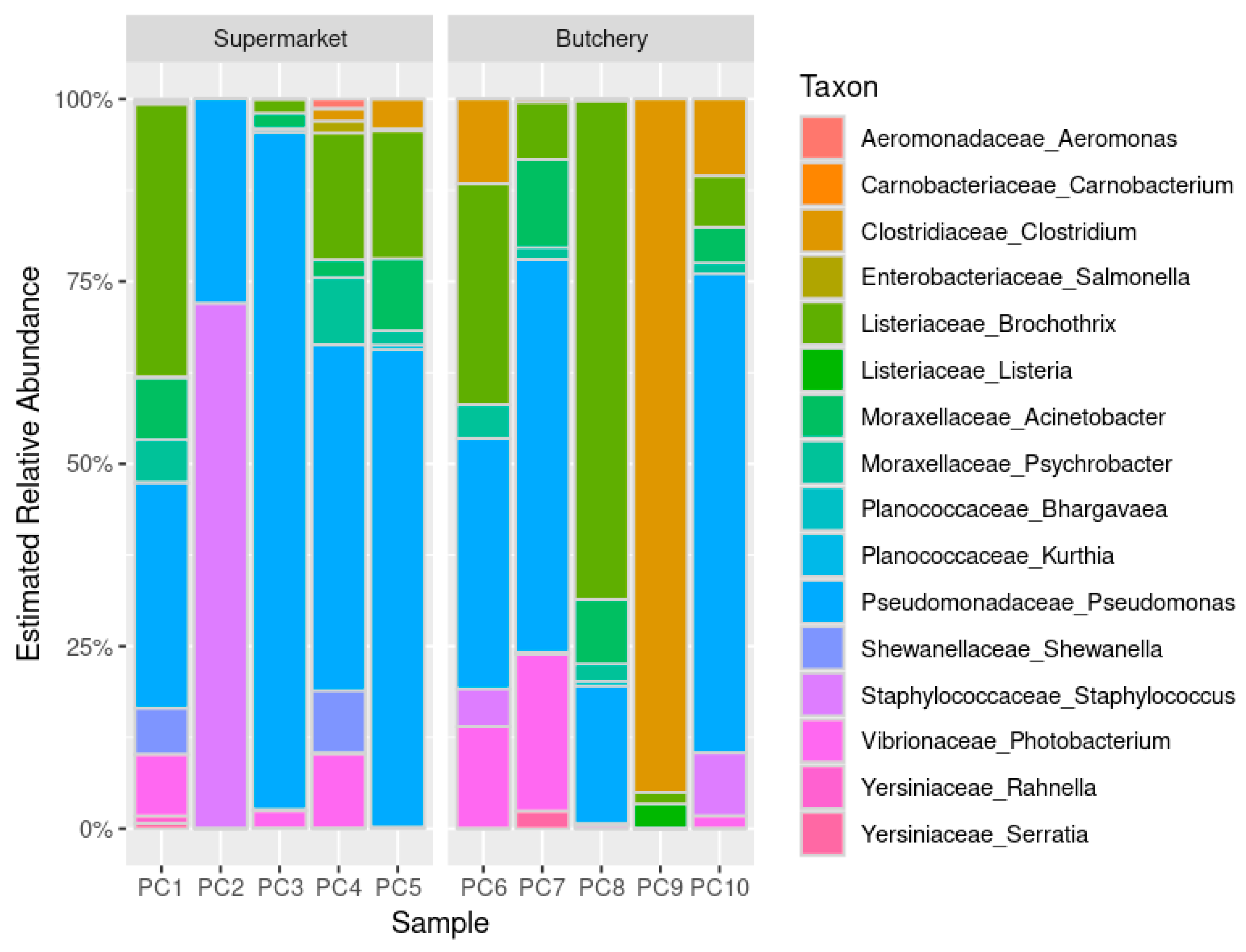
2.4.2. Estimated Relative Abundance

The distribution of the bacterial community structures in the raw meat samples reveals functional diversity among bacteria (Figure 1). The relative abundance was similar between raw meat samples from supermarkets and butcheries, but PC9 was different.
Food and meat spoilage organisms such as Pseudomonas (90%; 9/10), Acinetobacter (80%; 8/10), Brochothrix (80%; 8/10), Psychrobacter (70%; 7/10), Photobacterium (60; 6/10), and Clostridium (50%; 5/10) were detected in the majority of raw meat samples [16,17,18,19,20].

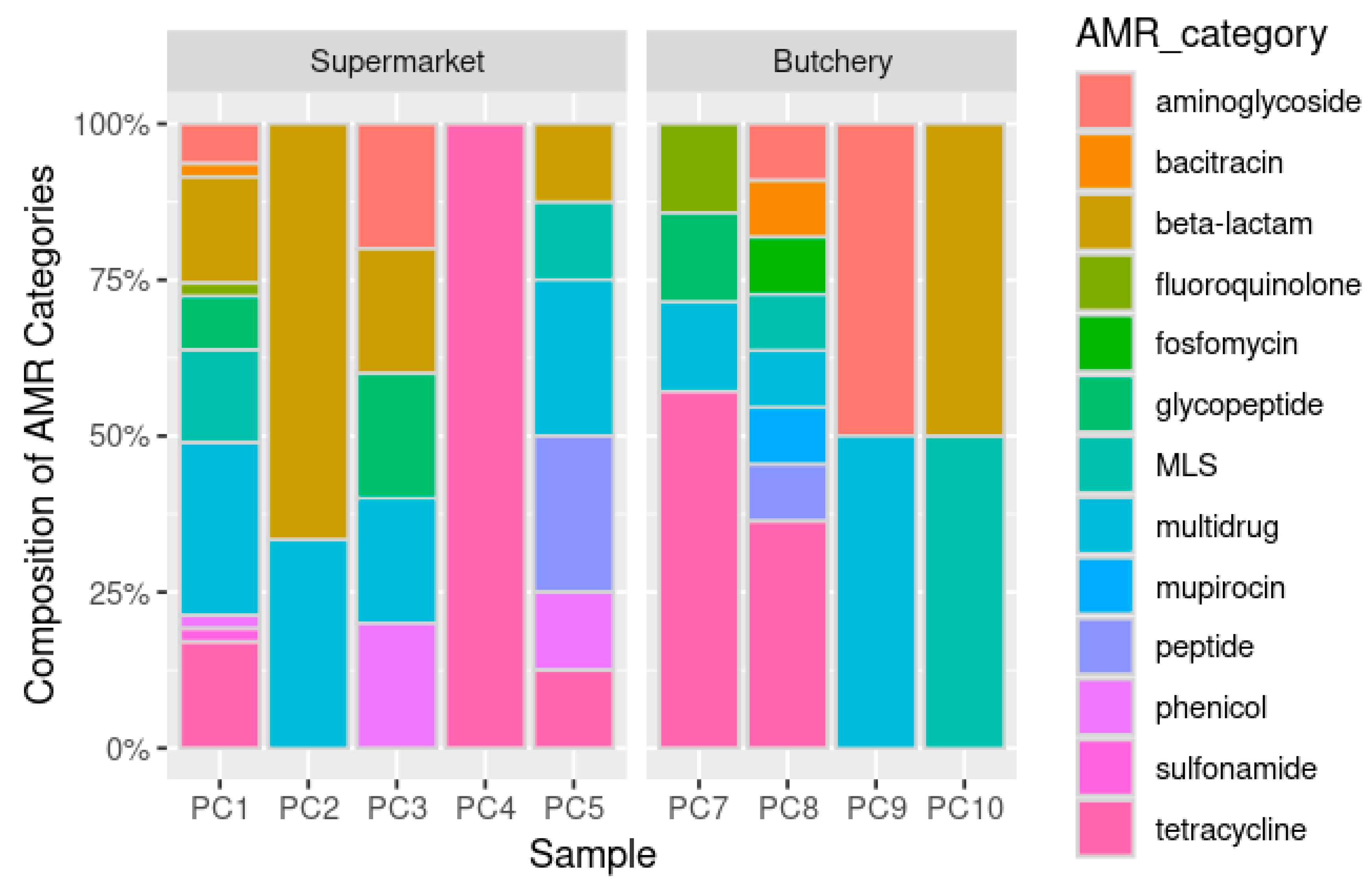
2.4.3. Resistome Prediction

Using the assembled contigs generated by MEGAHIT from the 10 raw meat samples, the PathoFact pipeline predicted a total of 61,665 ORFs. Among these, 89 ORFs were associated with ARGs, 138 with VF secretion, and 40 with toxin secretion (Supplementary Figure S1).

Antibiotic Resistance Gene Prediction

A total of 89 ORFs were predicted to be ARGs (Table 5; Supplementary Table S2). The raw meat samples from supermarkets were dominated by multidrug (26.8%; 18/67), beta-lactam (20.9%; 14/67), and tetracycline (14.9%; 10/67) resistance genes. The raw meat samples from butcheries were dominated by tetracycline (36.4%; 8/22) and multidrug (13.6%; 3/22) (Figure 2 and Figure 3A,C) resistance genes.
The majority of the ARGs were involved with resistance mechanisms such as antibiotic efflux (48.3%; 43/89), antibiotic inactivation (23.6%; 21/89), and antibiotic target alteration (12.36%; 11/89) (Supplementary Table S3). No ARGs were predicted in sample PC6 (butchery—B1) (Figure 3B,D; Supplementary Figure S2).

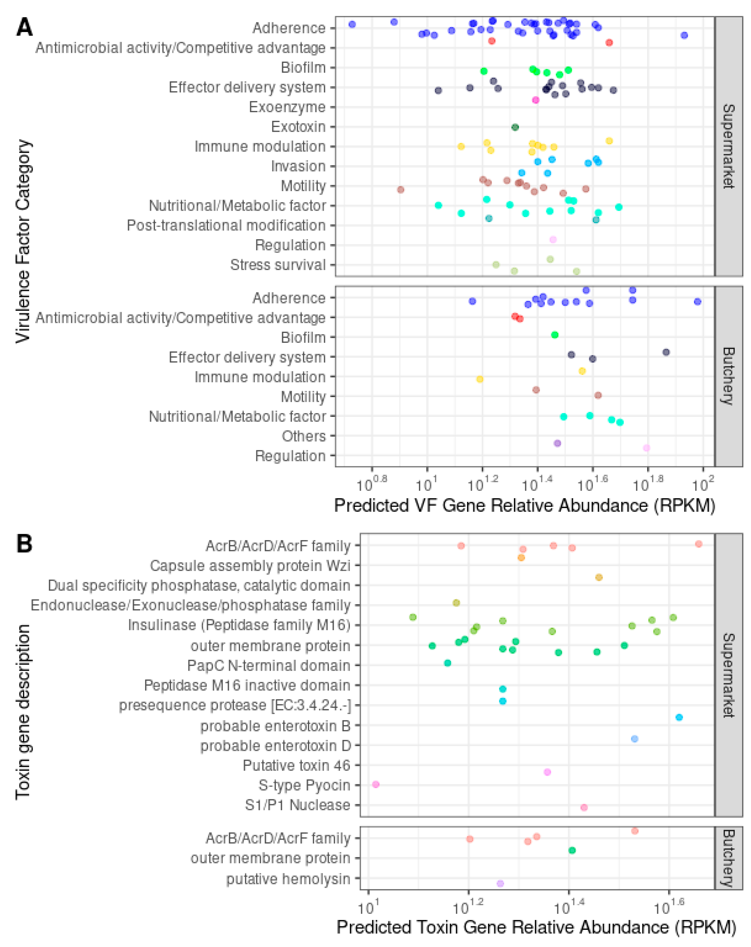
Virulence Factor and Toxin Gene Prediction

A total of 138 ORFs were predicted to be secreted VF genes. These were categorised as follows: adherence (36.9%; 51/138), antimicrobial activity/competitive advantage (2.9%; 4/138), biofilm (5.1%; 7/138), effector delivery system (13.8%; 19/138), exoenzyme (0.7%; 1/138), exotoxin (0.7%; 1/138), immune modulation (8%; 11/138), invasion (5.1%; 7/138), motility (9.4%; 13/138), nutritional/metabolic factor (10.9%; 15/138), post-translational modification (1.4%; 2/138), regulation (1.4%; 2/138), and stress survival (3%; 4/138) (Table 5; Figure 4A; Supplementary Table S4; Supplementary Figure S3).
VF genes from all categories were present in the raw meat samples collected from supermarkets (Supplementary Table S5). No exotoxin, invasion, post-translational modification, or stress survival VF genes were detected in the raw meat samples collected from butcheries. Fifteen types of toxin genes were detected in the raw meat samples collected from supermarkets and butcheries (Figure 4B).

3. Materials and Methods

3.1. Ethical Clearance and Study Definitions

The study was conducted in accordance with the Declaration of Helsinki and approved by the Human Research Ethics Committee of the University of the Witwatersrand (M190244; 10 May 2019).
Supermarkets were defined as stores that sold raw meat and various other grocery items, while butcheries were defined as stores selling primarily raw meat commodities. Raw meat samples were defined as pork chops (alternative names included loin, rib, sirloin, top loin, and blade chops). Indicator bacteria in this study were defined as Campylobacter spp., Enterococcus spp., E. coli, and NTS spp.

3.2. Study Setting and Sampling

Raw meat samples (i.e., pork chops) were purchased on the 4 January 2022, from 10 pre-selected supermarkets and butcheries in Johannesburg and Pretoria, Gauteng. Convenience sampling was used based on the location of the laboratory, Centre for Healthcare-Associated Infections, Antimicrobial Resistance and Mycoses (CHARM), National Institute for Communicable Diseases (NICD). The stores selected for sample collection included both supermarkets (n = 5) and butcheries (n = 5). Pork chops were chosen as it is the most popular cut among consumers [21]. Raw meat samples (containing at least two pork chops in the same container/packet) were randomly selected from the store shelves. All the raw meat samples were collected within the recommended dates for human consumption. Store demographics were captured by the study investigators on the day of sample collection (Supplementary Table S6). A unique number was assigned to each raw meat sample as well as the sampled stores to maintain anonymity and to ensure that the results cannot be linked back to a specific store.
The purchased raw meat samples were transported on ice to CHARM, NICD. The outsides of all the meat containers/packets were wiped with 70% ethanol before segregation and processing to avoid cross-contamination. The raw meat samples were separated aseptically. One raw meat sample from each store was sent on ice within 24 h after collection to a subcontracted laboratory in Gauteng, South Africa, for (1) the isolation of four selected indicator bacteria, and (2) antibiotic residue testing using liquid chromatography–tandem mass spectrometry (LC-MS/MS). The remaining raw meat samples underwent metagenomics sequencing at the Sequencing Core Facility (SCF), NICD. All the raw meat samples were processed within 24 h to 48 h after sample collection.

3.3. AST and WGS of Isolated Indicator Bacteria from Raw Meat Samples

The bacteria isolated from the raw meat samples were transported at room temperature (20 °C to 25 °C) from the subcontracted laboratory to CHARM, NICD within 24 h after isolation. Organism identification was reconfirmed at CHARM, NICD, with matrix-assisted laser desorption/ionization time-of-flight mass spectrometry (MALDI-TOF MS) (Microflex, Bruker Daltonics, Bremen, Germany). AST was performed using the Microscan Walkaway System with the Gram-negative NM44 (Beckman Coulter, Brea, CA, USA) and Gram-positive PM33 (Beckman Coulter, USA) cards. The AST results were interpreted using the 2023 European Committee on antimicrobial susceptibility testing (EUCAST) guidelines [15].
The genomic DNA (gDNA) of four isolated organisms from the raw meat samples was extracted with the QIAamp mini kit (Qiagen, Hilden, Germany) with the inclusion of lysozyme (10 mg/mL; Sigma-Aldrich, St. Louis, MO, USA) to ensure sufficient lysis. The quantity and quality of the extracted gDNA were determined on Qubit 4.0 (Invitrogen, Pittsburgh, PA, USA) with the high sensitivity assay kit (Invitrogen, USA). The tagging and fragmentation of the gDNA were performed using the Nextera DNA Flex Library preparation kit. Multiplexed paired-end libraries were prepared using the Nextera DNA preparation kit, followed by sequencing (2 × 150 bp) on a NextSeq 550 instrument (Illumina Inc., San Diego, CA, USA) with 100× coverage at the SCF, NICD. Raw paired-end reads were analysed using the Jekesa pipeline (v1.0; https://github.com/stanikae/jekesa (accessed on 3 April 2023) [22]. Briefly, Trim Galore! (v0.6.7) was used to filter the paired-end reads (Q > 30 and length > 50 bp) [23]. De novo assembly was performed using SKESA (v2.3.0; https://github.com/ncbi/SKESA (accessed on 3 April 2023)) and the assembled contigs were polished using Shovill (v1.1.0; https://github.com/tseemann/shovill (accessed on 3 April 2023)) [24]. Assembly metrics were calculated using QUAST (v5.0.2) [25]. The multilocus sequence typing (MLST) profiles were determined using the MLST tool (version 2.16.4; https://pubmlst.org/; https://github.com/tseemann/mlst (accessed on 3 April 2023)) [26]. The VF gene and ARG search was performed using ABRicate (version 1.0.1; https://github.com/tseemann/abricate (accessed on 3 April 2023)), against the Comprehensive Antibiotic Resistance Database (CARD), CARD-prevalence, Virulence Factor Database (VFDB), and ResFinder—Center for Genomic Epidemiology (CGE) database; with the gene alignment coverage cut-off of ≥95% and blastn sequence similarity of ≥95% [27,28,29,30,31,32]. A plasmid search was performed using PlasmidFinder (version 2.0; https://cge.food.dtu.dk/services/PlasmidFinder/ (accessed on 3 April 2023)). The assembled genome files were deposited in the National Center for Biotechnology Information GenBank under BioProject number PRJNA1006163.

3.4. Metagenomics

Total gDNA was extracted using the QIAamp Fast DNA stool mini kit (Qiagen, Hilden, Germany) and host depletion was performed using the NEBNext Microbiome DNA enrichment kit (New England Biolabs, Ipswich, MA, USA) with the inclusion of controls (the ZymoBIOMICS Gut Microbiome Standard was used as a positive control). Sequencing was performed on the NextSeq 550 (2 × 150 bp and 10 M reads) (Illumina, USA).
Initial sequence analysis was performed by the SCF, NICD. This included de-multiplexing (assigning reads to the respective sample using the barcodes that were assigned during the library preparation stage), quality checking using FastQC (https://www.bioinformatics.babraham.ac.uk/projects/fastqc/ (accessed on 15 April 2023)), and trimming and discarding of reads with a Q-score of less than 20 (TrimGalore, v0.6.2; https://github.com/FelixKrueger/TrimGalore (accessed on 15 April 2023)). Subsequent sequence analysis included the removal of host sequences (Suis scrofa v11.1 and Homo sapiens GRCh38.p13; https://www.ensembl.org/index.html (accessed on 15 April 2023)) using Bowtie2 v2.5.0 (https://github.com/BenLangmead/bowtie2 (accessed on 15 April 2023)). Taxonomic assignment was performed using the host-depleted metagenomic sequence reads, where the microbial diversity profile analyses were carried out with Kraken (v2.1; https://github.com/DerrickWood/kraken (accessed on 15 April 2023)) using the Standard-16 database [33]. This was followed by calculating the estimated related abundance of genera using Bracken (v2.5; https://ccb.jhu.edu/software/bracken/ (accessed on 15 April 2023)), and the proportion of related abundance was visualised using R (v4.2.1; https://www.r-project.org/ (accessed on 29 April 2023)). The assembled genome files were deposited in the National Center for Biotechnology Information GenBank under BioProject number PRJNA1137389.

3.5. Resistome Gene Abundance Estimates

With the host-depleted metagenomic sequence reads, standard de novo assembly of the metagenomic data was performed using MEGAHIT (v1.2.9; https://github.com/voutcn/megahit (accessed on 16 June 2023)) [34]. Using the assembled contigs, the ARGs, toxin genes, and VF genes were predicted using the PathoFact pipeline (v1.0; https://github.com/samnooij/PathoFact (accessed on 18 June 2023)) [35]. The predicted ARGs, as well as the toxin genes and VF genes with high confident prediction (1: secreted toxin or 1: secreted VF), were selected for relative abundance analysis, using ShortBRED (v0.9.4; https://github.com/biobakery/shortbred (accessed on 25 June 2023)) [36] against the CARD database (v2023-06) and VFDB (database update: 6 August 2023). The predicted resistome relative abundance was quantified by ShortBRED-Quantify calls USEARCH, where reads with 95% identity to the resistome were counted and normalized by reads per kilobase of reference sequence per million sample reads (RPKMs). The antimicrobial resistance (AMR) category “multidrug” was defined as bacterial strains that have become resistant to multiple classes of antibacterial drugs or other agents (https://card.mcmaster.ca/ontology/41472 (accessed on 25 June 2023)).

4. Discussion

The overuse and misuse of antibiotics in livestock production systems have led to residual contamination in food, resulting in antibiotic-resistant bacteria and ARGs [3,4]. In this study, we investigated and categorised four indicator bacteria (Campylobacter spp., Enterococcus spp., E. coli, and NTS spp.) that overlap between human and animals. We examined the diversity and abundance of bacterial communities, as well as ARGs and antibiotic residues in raw pork meat samples.
Out of the 10 raw meat samples collected, six did not contain any of the tested indicator organisms. A single E. coli isolate was obtained from sample PC9-B4, while one E. faecalis isolate was obtained from each of the samples PC3-S3, PC4-S4, and PC10-B5. The isolated E. coli and E. faecalis isolates showed minimal phenotypic resistance. The E. coli isolate showed resistance to tigecycline and trimethoprim/sulfamethoxazole, while all three E. faecalis isolates showed resistance to moxifloxacin only. WGS data for the E. coli (PC9-B4) and E. faecalis isolates (PC3-S3, PC4-S4, and PC10-B5) showed mainly the presence of tetracycline resistance genes.
International studies have shown that E. faecalis is the most dominant Enterococcus spp. isolated from pork samples (>80%) [37,38,39,40]. High tetracycline resistance and associated resistance genes found in this study are consistent with other reports [37,38,39,40,41]. In contrast, Aslam et al. (2012) found additional ARGs in enterococci isolated from retail meats in Canada, including genes for aminoglycosides (aac, aphA3, aad-A and -E, and sat4), macrolides (erm-B and -A), streptogramin (vatE), bacitracin (bcrR), and lincosamide (linB) [37]. Similarly, Hart et al. (2004) reported widespread tetracycline resistance in E. coli isolated from pigs in Australia [42]. The broad-spectrum antibiotic tetracycline, used to treat various infections and promote animal growth, has contributed to the high tetracycline resistance rates observed in this study [8,10,43,44,45].
The WGS data further revealed eight and nine different VF gene categories associated with the sequenced E. coli (PC9-B4) and E. faecalis (PC3-S3, PC4-S4, and PC10-B5) isolates, respectively. VF genes enable pathogenic bacteria to colonise host niches and establish infections, contributing both directly and indirectly to disease processes [35]. Aslam et al. (2012) reported that cytolysin (cyl-A, -B, -L and -M) and aggregation substances (agg) are associated with ARGs in retail meats, although the latter were not detected in our study [37,46]. Other VF genes, such as ace, ebp, and efaA have been reported in E. faecalis isolates from livestock and raw meat in Ghana and hospitalised patients in Iran [40,47].
The E. coli ST10 detected in this study is a known reservoir for ARG and mobile genetic elements, such as class 1 integrons and plasmids [45,48,49]. The IncB/O/K/Z and IncFII (pCoo) plasmids detected in the E. coli isolates have also been reported in E. coli from hospitalised patients in South Africa and in carbapenem-resistant Enterobacterales from hospitalised patients in Thailand [50,51]. To our knowledge, E. faecalis ST30 has only been identified in hospitalised patients [52,53,54]. The repUS43, repUS11, and rep9a plasmids detected in the E. faecalis isolates have been reported in other E. faecalis isolates from livestock and raw meat in Ghana as well as hospitalised patients in the USA [40,55]. The repUS43 plasmid is integrated chromosomally near the tetracycline resistance gene (tetM), while the rep9a plasmid encodes the cytolysin VF gene [55]. Another study also reported that the tet-M and -L and ermB resistance genes were harboured on the repUS43 and rep9a plasmids [40]. Therefore, sequencing and plasmid monitoring are essential for future surveillance studies to track the spread and evolution of these genetic elements in humans, animals, and food products.
The antibiotic residue concentrations in the 10 tested raw meat samples were within acceptable limits for human consumption according to the published guidelines [12]. Ramatla et al. (2017) reported higher antibiotic residue concentrations in raw meat samples collected in North West, South Africa [10]. The discrepancy between their findings and ours could be due to differences in sampling frequency and sample types.
The metagenomic data revealed that most of the identified bacteria were associated with food/meat spoilage and environmental sources [16,17,18,19,20]. Their pathogenicity or ability to cause disease was not confirmed in this study. Resistome analysis revealed the presence of beta-lactam, tetracycline, and multidrug resistance genes in the 10 raw meat samples, consistent with previous local and internal studies [44,56,57].
Further research and analysis are needed to fully understand the potential implications of these findings in terms of environmental health and antibiotic resistance. This study has several limitations. Firstly, a high number of host reads (i.e., pig DNA) in the metagenomic data limited our ability to recover bacterial metagenome-assembled genomes (MAGs) from the raw meat samples. Consequently, this limited our ability to validate ARG, VF, and toxin gene predictions. Using the MAGs approach could provide more comprehensive information about the bacterial species in the meat samples. Secondly, increasing the sequencing depth or using alternative microbiome enrichment methods is recommended. Thirdly, the study’s limited sample size may affect the generalisability of the findings to other supermarkets and butcheries across South Africa.

5. Conclusions

In conclusion, this study highlights the importance of evidence-based investigation and laboratory testing for indicator bacteria and antibiotic susceptibility in food production systems. While none of the indicator bacteria were detected in significant colony-forming units in the raw meat samples, isolated E. coli and E. faecalis strains exhibited minimal phenotypic resistance. Notably, they exhibited resistance to tigecycline, trimethoprim/sulfamethoxazole, and moxifloxacin. WGS data highlighted prevalent tetracycline resistance genes, consistent with other studies on tetracycline use in pork production. The presence of various ARGs and VF genes in these supermarket and butchery meat samples highlight the need for effective antimicrobial practices in pork production. To prevent contamination by antibiotic-resistant bacteria, supermarkets and butcheries selling pork products should rigorously monitor microbial levels and adhere to sanitation standards. Although antibiotic residue concentrations remain within acceptable limits, further research is needed to understand the broader implications of these findings for environmental health and antibiotic resistance. Overall, this study emphasises the need for vigilance and comprehensive strategies to combat antibiotic resistance in food production systems.

Supplementary Materials

The following supporting information can be downloaded at: https://www.mdpi.com/article/10.3390/antibiotics13080700/s1, Table S1: Captured demographic data collected from the sampled stores (five supermarkets and five butcheries); Table S2: Summary of antimicrobial resistance categories by number of open reading frame genes from 10 raw retail meat samples; Table S3: Summary of antimicrobial resistance mechanisms by number of ORF genes from 10 raw retail meat samples; Table S4: Summary of virulence factor category by number of ORF genes from 10 raw retail meat samples; Table S5: Summary of toxin gene description by number of ORF genes from 10 raw retail meat samples; Table S6: Demographic data collected from the sampled stores (five supermarkets and five butcheries); Figure S1: Relative abundance of ARGs, VFs, and toxin genes of 10 raw meat samples (normalized as reads per kilobase of reference sequence per million mapped sample reads (RPKMs), grouped by source); Figure S2: Mobile genetic element prediction of ARGs of all raw meat samples from both supermarket and butchery groups; Figure S3. Mobile genetic element prediction of VF and toxin genes of all raw meat samples from both supermarket and butchery groups.

Author Contributions

Conceptualisation, O.P. and W.S.; methodology, M.L. and W.S.; software, M.L. and W.Y.C.; validation, M.L. and W.S.; formal analysis, M.L. and W.Y.C.; investigation, M.L. and W.S.; resources, O.P.; data curation, M.L., W.S. and W.Y.C.; writing—original draft preparation, M.L.; writing—review and editing, M.L., W.S., W.Y.C. and O.P.; visualisation, M.L. and W.Y.C.; supervision, O.P.; project administration, M.L., W.S. and O.P.; funding acquisition, O.P. All authors have read and agreed to the published version of the manuscript.

Funding

The research reported in this publication was supported by the South African Medical Research Council (SAMRC) as a sub-grant received from the Bill and Melinda Gates Foundation (Grant number: 96086). The content and findings reported/illustrated are the sole deduction, view, and responsibility of the authors and do not reflect the official position and sentiments of the SAMRC or the Bill and Melinda Gates Foundation. WS was also supported by a Fogarty International Center Global Infectious Disease research-training grant, National Institutes of Health, to the University of Pittsburgh and the National Institute for Communicable Diseases (D43TW011255).

Institutional Review Board Statement

The study was conducted in accordance with the Declaration of Helsinki and approved by the Human Research Ethics Committee of the University of the Witwatersrand (Protocol No: M190244).

Informed Consent Statement

Not applicable.

Data Availability Statement

The assembled genome files were deposited in the National Center for Biotechnology Information GenBank and are available under the following BioProject numbers: PRJNA1006163 and PRJNA1137389. The Supplementary Materials contain additional data.

Acknowledgments

We would like to thank Arshad Ismail and the rest of the staff members of the SCF, NICD for guidance and for the sequencing of our isolate and retail pork meat selection.

Conflicts of Interest

The authors declare no conflicts of interest.

References

  1. Department of Agriculture Land Reform and Rural Development. A Profile of the South African Pork Market Value Chain. Available online: http://webapps1.daff.gov.za/AmisAdmin/upload/Pork%20Market%20Value%20Chain%20Profile%202021.pdf (accessed on 26 October 2023).
  2. Manyi-Loh, C.E.; Lues, R. A South African perspective on the microbiological and chemical quality of meat: Plausible public health implications. Microorganisms 2023, 11, 2484. [Google Scholar] [CrossRef] [PubMed]
  3. Eckstrom, K.; Barlow, J.W. Resistome metagenomics from plate to farm: The resistome and microbial composition during food waste feeding and composting on a Vermont poultry farm. PLoS ONE 2019, 14, e0219807. [Google Scholar] [CrossRef] [PubMed]
  4. Li, Y.; Cao, W.; Liang, S.; Yamasaki, S.; Chen, X.; Shi, L.; Ye, L. Metagenomic characterization of bacterial community and antibiotic resistance genes in representative ready-to-eat food in southern China. Sci. Rep. 2020, 10, 15175. [Google Scholar] [CrossRef]
  5. Samtiya, M.; Matthews, K.R.; Dhewa, T.; Puniya, A.K. Antimicrobial resistance in the food chain: Trends, mechanisms, pathways, and possible regulation strategies. Foods 2022, 11, 2966. [Google Scholar] [CrossRef] [PubMed]
  6. Bezanson, G.S.; MacInnis, R.; Potter, G.; Hughes, T. Presence and potential for horizontal transfer of antibiotic resistance in oxidase-positive bacteria populating raw salad vegetables. Int. J. Food Microbiol. 2008, 127, 37–42. [Google Scholar] [CrossRef]
  7. Sultana, F.; Kamrunnahar; Afroz, H.; Jahan, A.; Fakruddin, M.; Datta, S. Multi-antibiotic resistant bacteria in frozen food (ready to cook food) of animal origin sold in Dhaka, Bangladesh. Asian Pac. J. Trop. Biomed. 2014, 4, S268–S271. [Google Scholar] [CrossRef]
  8. Patel, S.J.; Wellington, M.; Shah, R.M.; Ferreira, M.J. Antibiotic stewardship in food-producing animals: Challenges, progress, and opportunities. Clin. Ther. 2020, 42, 1649–1658. [Google Scholar] [CrossRef]
  9. Urban-Chmiel, R.; Marek, A.; Stepien-Pysniak, D.; Wieczorek, K.; Dec, M.; Nowaczek, A.; Osek, J. Antibiotic resistance in bacteria-A review. Antibiotics 2022, 11, 1079. [Google Scholar] [CrossRef]
  10. Ramatla, T.; Ngoma, L.; Adetunji, M.; Mwanza, M. Evaluation of antibiotic residues in raw meat using different analytical methods. Antibiotics 2017, 6, 34. [Google Scholar] [CrossRef]
  11. World Health Organization. WHO Guidelines on Use of Medically Important Antimicrobials in Food-Producing Animals. Available online: https://www.who.int/publications/i/item/9789241550130 (accessed on 31 October 2023).
  12. Food and Agriculture Organization of the United Nations; World Health Organization. CX/MRL 2-2021: Maximum Residue Limits (MRLs) and Risk Management Recommendations (RMRs) for Residues of Veterinary Drugs in Foods. Available online: https://www.fao.org/fao-who-codexalimentarius/codex-texts/maximum-residue-limits/en/ (accessed on 20 July 2023).
  13. Foodstuffs Cosmetics and Disinfectants Amendment Act 39 of 2007. Available online: https://www.gov.za/sites/default/files/gcis_document/201409/a39-07.pdf (accessed on 26 October 2023).
  14. Meat Safety Act 40 of 2000. Available online: https://www.gov.za/sites/default/files/gcis_document/201409/a40-000.pdf (accessed on 26 October 2023).
  15. EUCAST. EUCAST Breakpoint Tables for Interpretation of MICs and Zone Diameters (v. 13.1). 2023. Available online: https://www.eucast.org/eucast_news/news_singleview?tx_ttnews%5Btt_news%5D=518&cHash=2509b0db92646dffba041406dcc9f20c (accessed on 1 February 2023).
  16. Stellato, G.; La Storia, A.; De Filippis, F.; Borriello, G.; Villani, F.; Ercolini, D. Overlap of spoilage-associated microbiota between meat and the meat processing environment in small-scale and large-scale retail distributions. Appl. Environ. Microbiol. 2016, 82, 4045–4054. [Google Scholar] [CrossRef]
  17. Stellato, G.; Utter, D.R.; Voorhis, A.; De Angelis, M.; Eren, A.M.; Ercolini, D. A few Pseudomonas oligotypes dominate in the meat and dairy processing environment. Front. Microbiol. 2017, 8, 264. [Google Scholar] [CrossRef] [PubMed]
  18. Fuertes-Perez, S.; Hauschild, P.; Hilgarth, M.; Vogel, R.F. Biodiversity of Photobacterium spp. isolated from meats. Front. Microbiol. 2019, 10, 2399. [Google Scholar] [CrossRef] [PubMed]
  19. Lulietto, M.F.; Sechi, P.; Borgogni, E.; Cenci-Goga, B.T. Meat spoilage: A critical review of a neglected alteration due to ropy slime producing bacteria. Ital. J. Anim. Sci. 2016, 14, 4011. [Google Scholar] [CrossRef]
  20. Cha, M.H.; Kim, S.H.; Kim, S.; Lee, W.; Kwak, H.S.; Chi, Y.M.; Woo, G.J. Antimicrobial resistance profile of Acinetobacter spp. isolates from retail meat samples under Campylobacter-selective conditions. J. Microbiol. Biotechnol. 2021, 31, 733–739. [Google Scholar] [CrossRef]
  21. Schulz, L. Consumers Respond to Meat Price Differences. Available online: https://www.extension.iastate.edu/agdm/articles/schulz/SchApr22.html (accessed on 31 October 2023).
  22. Kwenda, S.; Allam, M.; Khumualo, Z.T.H.; Mtshali, S.; Mnyameni, F.; Ismail, A. Jekesa: An Automated Easy-to-Use Pipeline for Bacterial Whole Genome Typing. 2020. Available online: https://github.com/stanikae/jekesa (accessed on 3 April 2023).
  23. Krueger, F.; James, F.; Ewels, P.; Afyounian, E.; Schuster-Boeckler, B. FelixKrueger/TrimGalore: v0.6.7—DOI via Zenodo (0.6.7). Zenodo. 2021. Available online: https://zenodo.org/records/5127899 (accessed on 1 February 2023).
  24. Souvorov, A.; Agarwala, R.; Lipman, D.J. SKESA: Strategic k-mer extension for scrupulous assemblies. Genome Biol. 2018, 19, 153. [Google Scholar] [CrossRef] [PubMed]
  25. Gurevich, A.; Saveliev, V.; Vyahhi, N.; Tesler, G. QUAST: Quality assessment tool for genome assemblies. Bioinformatics 2013, 29, 1072–1075. [Google Scholar] [CrossRef]
  26. Jolley, K.A.; Maiden, M.C. BIGSdb: Scalable analysis of bacterial genome variation at the population level. BMC Bioinform. 2010, 11, 595. [Google Scholar] [CrossRef] [PubMed]
  27. Jia, B.; Raphenya, A.R.; Alcock, B.; Waglechner, N.; Guo, P.; Tsang, K.K.; Lago, B.A.; Dave, B.M.; Pereira, S.; Sharma, A.N.; et al. CARD 2017: Expansion and model-centric curation of the comprehensive antibiotic resistance database. Nucleic Acids Res. 2017, 45, D566–D573. [Google Scholar] [CrossRef]
  28. Chen, L.; Zheng, D.; Liu, B.; Yang, J.; Jin, Q. VFDB 2016: Hierarchical and refined dataset for big data analysis—10 years on. Nucleic Acids Res. 2016, 44, D694–D697. [Google Scholar] [CrossRef] [PubMed]
  29. Zankari, E.; Hasman, H.; Cosentino, S.; Vestergaard, M.; Rasmussen, S.; Lund, O.; Aarestrup, F.M.; Larsen, M.V. Identification of acquired antimicrobial resistance genes. J. Antimicrob. Chemother. 2012, 67, 2640–2644. [Google Scholar] [CrossRef]
  30. Bortolaia, V.; Kaas, R.S.; Ruppe, E.; Roberts, M.C.; Schwarz, S.; Cattoir, V.; Philippon, A.; Allesoe, R.L.; Rebelo, A.R.; Florensa, A.F.; et al. ResFinder 4.0 for predictions of phenotypes from genotypes. J. Antimicrob. Chemother. 2020, 75, 3491–3500. [Google Scholar] [CrossRef] [PubMed]
  31. Zankari, E.; Allesoe, R.; Joensen, K.G.; Cavaco, L.M.; Lund, O.; Aarestrup, F.M. PointFinder: A novel web tool for WGS-based detection of antimicrobial resistance associated with chromosomal point mutations in bacterial pathogens. J. Antimicrob. Chemother. 2017, 72, 2764–2768. [Google Scholar] [CrossRef] [PubMed]
  32. Camacho, C.; Coulouris, G.; Avagyan, V.; Ma, N.; Papadopoulos, J.; Bealer, K.; Madden, T.L. BLAST+: Architecture and applications. BMC Bioinform. 2009, 10, 421. [Google Scholar] [CrossRef]
  33. Lu, J.; Rincon, N.; Wood, D.E.; Breitwieser, F.P.; Pockrandt, C.; Langmead, B.; Salzberg, S.L.; Steinegger, M. Metagenome analysis using the Kraken software suite. Nat. Protoc. 2022, 17, 2815–2839. [Google Scholar] [CrossRef] [PubMed]
  34. Li, D.; Liu, C.M.; Luo, R.; Sadakane, K.; Lam, T.W. MEGAHIT: An ultra-fast single-node solution for large and complex metagenomics assembly via succinct de Bruijn graph. Bioinformatics 2015, 31, 1674–1676. [Google Scholar] [CrossRef]
  35. de Nies, L.; Lopes, S.; Busi, S.B.; Galata, V.; Heintz-Buschart, A.; Laczny, C.C.; May, P.; Wilmes, P. PathoFact: A pipeline for the prediction of virulence factors and antimicrobial resistance genes in metagenomic data. Microbiome 2021, 9, 49. [Google Scholar] [CrossRef] [PubMed]
  36. Kaminski, J.; Gibson, M.K.; Franzosa, E.A.; Segata, N.; Dantas, G.; Huttenhower, C. High-specificity targeted functional profiling in microbial communities with ShortBRED. PLoS Comput. Biol. 2015, 11, e1004557. [Google Scholar] [CrossRef] [PubMed]
  37. Aslam, M.; Diarra, M.S.; Checkley, S.; Bohaychuk, V.; Masson, L. Characterization of antimicrobial resistance and virulence genes in Enterococcus spp. isolated from retail meats in Alberta, Canada. Int. J. Food Microbiol. 2012, 156, 222–230. [Google Scholar] [CrossRef]
  38. Tyson, G.H.; Nyirabahizi, E.; Crarey, E.; Kabera, C.; Lam, C.; Rice-Trujillo, C.; McDermott, P.F.; Tate, H. Prevalence and antimicrobial resistance of Enterococci isolated from retail meats in the United States, 2002 to 2014. Appl. Environ. Microbiol. 2018, 84, e01902-17. [Google Scholar] [CrossRef]
  39. de Jong, A.; Simjee, S.; Garch, F.E.; Moyaert, H.; Rose, M.; Youala, M.; Dry, M.; Group, E.S. Antimicrobial susceptibility of enterococci recovered from healthy cattle, pigs and chickens in nine EU countries (EASSA Study) to critically important antibiotics. Vet. Microbiol. 2018, 216, 168–175. [Google Scholar] [CrossRef]
  40. Amuasi, G.R.; Dsani, E.; Owusu-Nyantakyi, C.; Owusu, F.A.; Mohktar, Q.; Nilsson, P.; Adu, B.; Hendriksen, R.S.; Egyir, B. Enterococcus species: Insights into antimicrobial resistance and whole-genome features of isolates recovered from livestock and raw meat in Ghana. Front. Microbiol. 2023, 14, 1254896. [Google Scholar] [CrossRef] [PubMed]
  41. Novais, C.; Freitas, A.R.; Silveira, E.; Antunes, P.; Silva, R.; Coque, T.M.; Peixe, L. Spread of multidrug-resistant Enterococcus to animals and humans: An underestimated role for the pig farm environment. J. Antimicrob. Chemother. 2013, 68, 2746–2754. [Google Scholar] [CrossRef] [PubMed]
  42. Hart, W.S.; Heuzenroeder, M.W.; Barton, M.D. Antimicrobial resistance in Campylobacter spp., Escherichia coli and Enterococci associated with pigs in Australia. J. Vet. Med. B Infect. Dis. Vet. Public Health 2004, 51, 216–221. [Google Scholar] [CrossRef] [PubMed]
  43. Strasheim, W.; Etter, E.M.C.; Lowe, M.; Perovic, O. Method to Assess Farm-Level Vaccine and Antibiotic Usage Utilizing Financial Documentation: A Pilot Study in a Commercial Pig Farm in South Africa From 2016 to 2018. Front. Vet. Sci. 2022, 9, 856729. [Google Scholar] [CrossRef] [PubMed]
  44. Liu, Z.; Klumper, U.; Shi, L.; Ye, L.; Li, M. From pig breeding environment to subsequently produced pork: Comparative analysis of antibiotic resistance denes and cacterial community composition. Front. Microbiol. 2019, 10, 43. [Google Scholar] [CrossRef] [PubMed]
  45. Peng, Z.; Hu, Z.; Li, Z.; Zhang, X.; Jia, C.; Li, T.; Dai, M.; Tan, C.; Xu, Z.; Wu, B.; et al. Antimicrobial resistance and population genomics of multidrug-resistant Escherichia coli in pig farms in mainland China. Nat. Commun. 2022, 13, 1116. [Google Scholar] [CrossRef] [PubMed]
  46. Bertels, F.; Silander, O.K.; Pachkov, M.; Rainey, P.B.; van Nimwegen, E. Automated reconstruction of whole-genome phylogenies from short-sequence reads. Mol. Biol. Evol. 2014, 31, 1077–1088. [Google Scholar] [CrossRef] [PubMed]
  47. Kafil, H.S.; Mobarez, A.M.; Moghadam, M.F. Adhesion and virulence factor properties of Enterococci isolated from clinical samples in Iran. Indian J. Pathol. Microbiol. 2013, 56, 238–242. [Google Scholar] [CrossRef] [PubMed]
  48. Reid, C.J.; Wyrsch, E.R.; Roy Chowdhury, P.; Zingali, T.; Liu, M.; Darling, A.E.; Chapman, T.A.; Djordjevic, S.P. Porcine commensal Escherichia coli: A reservoir for class 1 integrons associated with IS26. Microb. Genom. 2017, 3, e000143. [Google Scholar] [CrossRef]
  49. Reid, C.J.; DeMaere, M.Z.; Djordjevic, S.P. Australian porcine clonal complex 10 (CC10) Escherichia coli belong to multiple sublineages of a highly diverse global CC10 phylogeny. Microb. Genom. 2019, 5, e000225. [Google Scholar] [CrossRef] [PubMed]
  50. Perovic, O.; Singh-Moodley, A.; Lowe, M. In vitro activity of ceftolozane-tazobactam against Escherichia coli, Klebsiella pneumoniae and Pseudomonas aeruginosa obtained from blood cultures from sentinel public hospitals in South Africa. Antibiotics 2023, 12, 453. [Google Scholar] [CrossRef] [PubMed]
  51. Paveenkittiporn, W.; Kamjumphol, W.; Ungcharoen, R.; Kerdsin, A. Whole-genome sequencing of clinically isolated carbapenem-resistant Enterobacterales harboring mcr genes in Thailand, 2016–2019. Front Microbiol 2020, 11, 586368. [Google Scholar] [CrossRef]
  52. Rathnayake, I.U.; Hargreaves, M.; Huygens, F. Genotyping of Enterococcus faecalis and Enterococcus faecium isolates by use of a set of eight single nucleotide polymorphisms. J. Clin. Microbiol. 2011, 49, 367–372. [Google Scholar] [CrossRef] [PubMed]
  53. Freitas, A.R.; Novais, C.; Ruiz-Garbajosa, P.; Coque, T.M.; Peixe, L. Clonal expansion within clonal complex 2 and spread of vancomycin-resistant plasmids among different genetic lineages of Enterococcus faecalis from Portugal. J. Antimicrob. Chemother. 2009, 63, 1104–1111. [Google Scholar] [CrossRef]
  54. Kawalec, M.; Pietras, Z.; Danilowicz, E.; Jakubczak, A.; Gniadkowski, M.; Hryniewicz, W.; Willems, R.J. Clonal structure of Enterococcus faecalis isolated from Polish hospitals: Characterization of epidemic clones. J. Clin. Microbiol. 2007, 45, 147–153. [Google Scholar] [CrossRef]
  55. Sharon, B.M.; Arute, A.P.; Nguyen, A.; Tiwari, S.; Reddy Bonthu, S.S.; Hulyalkar, N.V.; Neugent, M.L.; Palacios Araya, D.; Dillon, N.A.; Zimmern, P.E.; et al. Genetic and functional enrichments associated with Enterococcus faecalis isolated from the urinary tract. mBio 2023, 14, e0251523. [Google Scholar] [CrossRef] [PubMed]
  56. Abdalla, S.E.; Abia, A.L.K.; Amoako, D.G.; Perrett, K.; Bester, L.A.; Essack, S.Y. From farm-to-fork: E. coli from an intensive pig production system in South Africa shows high resistance to critically important antibiotics for human and animal use. Antibiotics 2021, 10, 178. [Google Scholar] [CrossRef]
  57. Barroga, T.R.M.; Morales, R.G.; Benigno, C.C.; Castro, S.J.M.; Caniban, M.M.; Cabullo, M.F.B.; Agunos, A.; de Balogh, K.; Dorado-Garcia, A. Antimicrobials used in backyard and commercial poultry and swine farms in the Philippines: A qualitative pilot study. Front. Vet. Sci. 2020, 7, 329. [Google Scholar] [CrossRef]
Figure 1. Estimated relative abundance of the bacterial taxonomic profiling (aggregated by family and genus) in 10 raw meat samples. PC = pork chop; PC1–5 = sampled from supermarkets; PC6–10 = sampled from butcheries.
Antibiotics 13 00700 g001
Figure 2. Composition of predicted AMR categories in 10 raw meat samples as predicated by PathoFact database. MLS = macrolides, lincosamides, and streptogramins; AMR category = default assignment by the PathoFact pipeline; PC6 = no AMR was detected.
Antibiotics 13 00700 g002
Figure 3. Relative abundance of individual ARGs of 10 raw meat samples (normalized as reads per kilobase of reference sequence per million mapped sample reads (RPKMs)), grouped by the AMR category (A,C), and by AMR resistance mechanisms (B,D), stratified by supermarket and butchery (C,D). Each circle indicating the relative abundance of an ARG gene in a single sample and genes within the same AMR category or AMR resistance mechanism are represented with the same colour.
Antibiotics 13 00700 g003
Figure 4. Relative abundance of the individual predicted virulence factors (VFs) and toxin genes in 10 raw meat samples (normalized as reads per kilobase of reference sequence per million mapped sample reads (RPKMs)), grouped by the VF category according to VFDB (A), and by toxin gene description according to the PathoFact database (B). Each circle indicates the relative abundance of a VF/toxin gene in a single sample. Genes within the same VF category or toxin gene description are represented with the same colour.
Antibiotics 13 00700 g004
Table 1. Indicator organisms isolated from raw meat samples.
Sample IDE. coli
(CFUs)
Salmonella spp.
(CFUs)
Enterococci spp.
(CFUs)
Campylobacter spp.
(CFUs)
PC1-S1AbsentAbsentAbsentAbsent
PC2-S2AbsentAbsentAbsentAbsent
PC3-S3AbsentAbsent16 *Absent
PC4-S4AbsentAbsent1 *Absent
PC5-S5AbsentAbsentAbsentAbsent
PC6-B1AbsentAbsentAbsentAbsent
PC7-B2AbsentAbsentAbsentAbsent
PC8-B3AbsentAbsentAbsentAbsent
PC9-B420 *AbsentAbsentAbsent
PC10-B5AbsentAbsent3 *Absent
PC = pork chop; S = supermarket; B = butchery; CFUs = colony-forming units; * = isolated bacteria from raw meat samples were subsequently processed (AST and WGS).
Table 2. Antibiotic susceptibility testing of one Escherichia coli isolate and three Enterococcus faecalis isolates isolated from four raw meat samples.
Antibiotic ClassAntibioticE. coli PC9-B4
(µg/mL)
MIC Interpretation #E. faecalis PC3-S3
(µg/mL)
E. faecalis PC4-S4
(µg/mL)
E. faecalis PC10-B5
(µg/mL)
MIC Interpretation #
AminoglycosideAmikacin≤8S323232NI *
Gentamicin≤2S444NI *
Gentamicin synergyNT-≤500≤500≤500NI
Streptomycin synergyNT-≤1000≤1000≤1000NI
Tobramycin≤2S≤2/38≤2/38≤2/38NI *
Beta-lactam
(penicillins)
Ampicillin≤8S444S
Ampicillin/sulbactam≤8/4SNTNTNT-
Amoxicillin/clavulanic acid≤8/4S≤4/2≤4/2≤4/2S
OxacillinNT->2>2>2NI
PenicillinNT-888NI
Piperacillin≤8SNTNTNT-
Piperacillin/tazobactam≤8SNTNTNT-
Beta-lactam
(cephalosporins)
Cefepime≤1SNTNTNT-
Cefotaxime≤1SNTNTNT-
Cefotaxime/clavulanic acid≤0.5NINTNTNT-
Cefoxitin≤8SNTNTNT-
Cefuroxime≤4SNTNTNT-
Ceftazidime≤1SNTNTNT-
Ceftazidime/clavulanic acid≤0.25NINTNTNT-
Cephalothin≤8NINTNTNT-
Beta-lactam
(carbapenems)
Doripenem≤1SNTNTNT-
Ertapenem≤0.5SNTNTNT-
Imipenem≤1S≤4≤4≤4S
Meropenem≤1SNTNTNT-
Beta-lactam
(monobactams)
Aztreonam≤1SNTNTNT-
AmphenicolChloramphenicol≤8S≤8≤8≤8NI
Cyclic lipopeptideDaptomycinNT-≤1≤1≤1NI
Fluoroquinolone Ciprofloxacin≤0.5S≤1≤1≤1S
Levofloxacin≤1S≤1≤1≤1S
MoxifloxacinNT-≤256≤256≤256R
Norfloxacin≤0.5S≤4≤4≤4NI
FusidaneFusidic acidNT-≤2≤2≤2NI
Lincosamides ClindamycinNT->2>2>2NI
PristinamycinNT-222NI
MacrolideErythromycinNT-111NI
Protein synthesis inhibitor Mupirocin>16NINTNTNT-
NitrofuranNitrofurantoin≤32S≤32≤32≤32S
Phosphonic acidFosfomycin≤32S≤32≤32≤32NI
PolymyxinColistin≤2SNTNTNT-
RifamycinRifampinNT-≤0.5≤0.5≤0.5NI
Tetracycline Minocycline>8NI≤1≤1≤1NI
Tetracycline>8NI888NI
Tigecycline≤1RNTNTNT-
Glycopeptide and lipoglycopeptideTeicoplaninNT-≤1≤1≤1S
VancomycinNT-222S
OxazolidinoneLinezolidNT-222S
SulfonamideTrimethoprim/sulfamethoxazole>4/76RNTNTNT-
PC = pork chop; S = supermarket; B = butchery; NT = not tested (antibiotic not included in the antibiotic panel); NI = no EUCAST MIC interpretation; * = all E. faecalis are intrinsically resistant to aminoglycosides; # = [15].
Table 3. Antibiotic resistance genes, virulence factors, and plasmids detected in one Escherichia coli isolate and three Enterococcus faecalis isolates isolated from four raw meat samples.
Sample IDPC9-B4 *PC3-S3 *PC4-S4 *PC10-B5 *
OrganismE. coliE. faecalisE. faecalisE. faecalis
CH type11–54---
O typeO69---
H typeH32---
MLST10 ^30 #30 #30 #
ARGsAminoglycosideaadA1Y---
FluoroquinolonegyrAY---
LincosamideisaA-YYY
Sulphonamidesul2Y---
TetracyclinetetBY---
tetM-YYY
TrimethoprimdfrA1Y---
VF genesAdhesinace-YYY
efaAfs-YYY
ColicincbaY---
ceaY---
ciaY---
cmaY---
Cytolysin toxincylA-YYY
cylL-YYY
cylM-YYY
Endocarditis and biofilm-associated pili genesebpA-YYY
ebpB-YYY
Enterococcus faecalis leucine-rich protein AelrA-YYY
Glutamate decarboxylasegadY---
gelE-YYY
Heat stable toxinastAY---
Hyaluronidase hylA-YYY
Increased serum survivalissY---
Outer membrane proteaseompTY---
Plasmid-encoded catalase peroxidasekatPY---
Sex pheromonecad-YYY
camE-YYY
cCF10-YYY
cOB1-YYY
Tellurium ion resistanceterCY---
Thiol peroxidasetpx-YYY
Outer membrane protein complement resistancetraTY---
SortaseSrtA-YYY
Plasmids IncB/O/K/ZY---
IncFII(pCoo)Y---
repUS43-YYY
repUS11-YYY
rep9a-YYY
PC = pork chop; S = supermarket; B = butchery; ARG = antibiotic resistance genes; VF = virulence factor; MLST = multilocus sequence typing (https://github.com/tseemann/mlst (accessed on 3 April 2023)); Y = present/detected; - = absent/not detected; ^ = adk 10/fumC 11/gyrB 4/icd 8/mdh 8/purA 8/recA 2; # = aroE 10/ghd 7/gki 1/gyd 1/pstS 11/xpt 2/yqiL 1; * = the corresponding raw meat sample numbers were assigned to each isolate for traceability.
Table 4. Antibiotic residue testing in 10 raw meat samples using liquid chromatography–tandem mass spectrometry (LC-MS/MS).
Antibiotic Class Antibiotic Sample IDAcceptable Maximum Residue Level (µg/kg)
PC1-S1 *
(µg/kg)
PC2-S2 *
(µg/kg)
PC3-S3 *
(µg/kg)
PC4-S4 *
(µg/kg)
PC5-S5 *
(µg/kg)
PC6-B1 *
(µg/kg)
PC7-B2 *
(µg/kg)
PC8-B3 *
(µg/kg)
PC9-B4 *
(µg/kg)
PC10-B5 *
(µg/kg)
Fluoroquinolones Ciprofloxacin<50<50<50<50<50<50<50<50<50<50100 #
Enrofloxacin<50<50<50<50<50<50<50<50<50<50100 #
Norfloxacin<50<50<50<50<50<50<50<50<50<50-
Lincosamides Lincomycin<50<50<50<50<50<50<50<50<50<50200 ^
Macrolides Tylosin<50<50<50<50<50<50<50<50<50<50100 ^
Sulfonamides Sulfadiazine<50<50<50<50<50<50<50<50<50<50-
Sulfadimidine<50<50<50<50<50<50<50<50<50<50100 ^
Sulfamethoxazole<50<50<50<50<50<50<50<50<50<50-
Tetracyclines Chlortetracycline<50<50<50<5071.5<50<50<50<50<50200 ^
Doxycycline<50<50<50<50<50<50<50<50<50<50-
Oxytetracycline<50<50<50<50<50<50<50<50<50<50200 ^
Tetracycline<50<50<50<50<50<50<50<50<50<50200 ^/600 #
Pleuromutilin Tiamulin<50<50<50<50<50<50<50<50<50<50-
Diaminopyrimidines Trimethoprim<50<50<50<50<50<50<50<50<50<50-
Quindoxin Olaquindox metabolite<50<50<50<50<50<50<50<50<50<50-
PC = pork chop; S = supermarket; B = butchery; * = muscle; - = no acceptable maximum residue level listed; ^ = [12]; # = [10].
Table 5. Metagenomics read statistics of 10 raw meat samples.
Sample IDRaw Paired-End Reads (n = NGS Reads)Paired-End Reads after Host Removal
(n = NGS Reads)
Paired-End Reads Mapped to Bacteria
(n = NGS Reads)
Predicted ARG
(n = Annotated ORF)
Predicted
Secreted VF Genes
(n = Annotated ORF)
Predicted Secreted Toxin Genes
(n = Annotated ORF)
PC1-S16,559,325761,775249,495478624
PC2-S27,276,334372,2054242625
PC3-S39,008,555407,82759,1595112
PC4-S48,346,930353,75925,353100
PC5-S57,706,014423,01948,2848103
PC6-B16,948,972260,6382460010
PC7-B26,318,244388,51747,2857123
PC8-B36,811,831400,45671,94111153
PC9-B48,083,308336,6362049200
PC10-B56,727,542295,4662043210
PC = pork chop; S = supermarket; B = butchery; NGS = next-generation sequencing; ARG = antibiotic resistance genes; ORF = open reading frame/s; VF = virulence factor.
Disclaimer/Publisher’s Note: The statements, opinions and data contained in all publications are solely those of the individual author(s) and contributor(s) and not of MDPI and/or the editor(s). MDPI and/or the editor(s) disclaim responsibility for any injury to people or property resulting from any ideas, methods, instructions or products referred to in the content.

