Article

Precision Phage Cocktail Targeting Surface Appendages for Biocontrol of Salmonella in Cold-Stored Foods

1
Department of Food Science & Biotechnology, College of Life Science, Sejong University, Seoul 05006, Republic of Korea
2
Carbohydrate Bioproduct Research Center, College of Life Science, Sejong University, Seoul 05006, Republic of Korea
3
Department of Food Biotechnology, Dong-A University, Busan 49315, Republic of Korea
4
Department of Food and Animal Biotechnology, Research Institute of Agriculture and Life Sciences, Seoul National University, Seoul 08826, Republic of Korea
5
Department of Agricultural Biotechnology, Seoul National University, Seoul 08826, Republic of Korea
6
Center for Food and Bioconvergence, Seoul National University, Seoul 08826, Republic of Korea
*
Authors to whom correspondence should be addressed.
These authors contributed equally to this work.
Antibiotics 2024, 13(9), 799; https://doi.org/10.3390/antibiotics13090799 (registering DOI)
Submission received: 22 July 2024 / Revised: 22 August 2024 / Accepted: 22 August 2024 / Published: 24 August 2024

Abstract

:
Salmonella enterica is a major food-borne pathogen causing food poisoning. The use of bacteriophages as alternative biocontrol agents has gained renewed interest due to the rising issue of antibiotic-resistant bacteria. We isolated and characterized three phages targeting Salmonella: SPN3US, SPN3UB, and SPN10H. Morphological and genomic analyses revealed that they belong to the class Caudoviricetes. SPN3UB, SPN3US, and SPN10H specifically target bacterial surface molecules as receptors, including O-antigens of lipopolysaccharides, flagella, and BtuB, respectively. The phages exhibited a broad host range against Salmonella strains, highlighting their potential for use in a phage cocktail. Bacterial challenge assays demonstrated significant lytic activity of the phage cocktail consisting of the three phages against S. typhimurium UK1, effectively delaying the emergence of phage-resistant bacteria. The phage cocktail effectively reduced Salmonella contamination in foods, including milk and pork and chicken meats, during cold storage. These results indicate that a phage cocktail targeting different host receptors could serve as a promising antimicrobial strategy to control Salmonella.

1. Introduction

Salmonella is a leading cause of foodborne illness, responsible for 1.35 million infections, 26,500 hospitalizations, and 420 deaths in the United States every year [1]. According to the Interagency Food Safety Analytics Collaboration, over 23% of foodborne Salmonella illnesses are associated with the consumption of poultry meat [2]. Salmonella has more than 2,600 different serotypes identified, of which Salmonella typhimurium and Salmonella enteritidis are the two major causes of foodborne illnesses [3,4]. These species are transmitted from humans, leading to severe gastrointestinal diseases, characterized by symptoms such as diarrhea, fever, and abdominal cramps [5]. Unfortunately, controlling Salmonella has become increasingly challenging due to the development of strains resistant to multiple antibiotics [6,7]. The emergence and widespread prevalence of antibiotic-resistant bacteria pose a significant global public health threat, necessitating the exploration of alternative antibacterial strategies.
In recent years, the application of phages, viruses that specifically infect and cause bacterial lysis, has attracted significant attention as an alternative to antibiotics because of their host specificity, effective host–cell lysis, and safety to humans [8,9]. In particular, phages with a lytic life cycle have been researched for various applications in the food industry [10]: the prevention of pathogen colonization in livestock, decontamination of carcasses and other ready-to-eat (RTE) foods, disinfection of pathogen contact surfaces, and preservation of foods [11]. A phage cocktail composed of phages with different infection strategies has also been employed to enhance host growth inhibition as it can broaden the host range and reduces the chance of resistance development [12,13]. Recent studies have focused on using a phage cocktail to control S. typhimurium in foods, providing several promising outcomes. For example, Abhisingha et al. investigated the effectiveness of a phage cocktail containing two Salmonella phages, ENT101 and TYM101, in controlling S. typhimurium on chicken meat [14]. This phage cocktail achieved a 0.4–1 log CFU/cm2 reduction in Salmonella quantity. The research also involved the evaluation of the antibacterial activity of phage cocktails at different temperatures, demonstrating the potential of phages in reducing Salmonella contamination in poultry products under various conditions. Another novel phage cocktail consisting of three phages (BSPM4, BSP101, and BSP22A) that target different host receptors was developed [15]. The phage cocktail not only delayed the emergence of Salmonella resistance but also significantly reduced viable Salmonella cell numbers in fresh produce. These findings encourage the development of new strategies using a phage cocktail to reduce the incidence of foodborne illnesses caused by Salmonella contamination.
In this study, we isolated and characterized three phages targeting Salmonella—SPN3US, SPN3UB, and SPN10H—by analyzing their morphology, host range, bacterial growth inhibition, and genomic characteristics. A phage cocktail composed of the three phages was effective in killing S. typhimurium on various food matrices at a refrigerated temperature and delayed the emergence of resistance.

2. Results and Discussion

2.1. Morphological and Genomic Features of Phages

To characterize the morphology of three isolated phages, Transmission Electron Microscopy (TEM) analysis was performed. The analysis revealed that the phages belong to the class Caudoviricetes but each phage has distinct morphological characteristics [16]. The result revealed that SPN3US had a non-flexible and contractile tail (200 ± 22 nm) with an icosahedral head (110 ± 9 nm), indicative of a myovius-like morphology (Figure 1A). In contrast, both SPN3UB and SPN10H exhibited smaller heads with a diameter of 55 ± 3 nm and 65 ± 5 nm, respectively, and flexible tails with a length of 156 ± 11 nm and 220 ± 12, respectively (Figure 1B, C). These features suggest SPN3UB and SPN10H have a sipovius-like morphology (Figure 1B, C). According to the International Committee on Taxonomy of Viruses (ICTV, https://ictv.global/, accessed on 20 August 2024), SPN3US belongs to the Seoulvirus genus of the Chimalliviridae family and SPN3UB shares genetic homologies with phages in the Lederbergvirus genus. SPN10H belongs to the Demerecviridae family, genus Epseptimavirus.
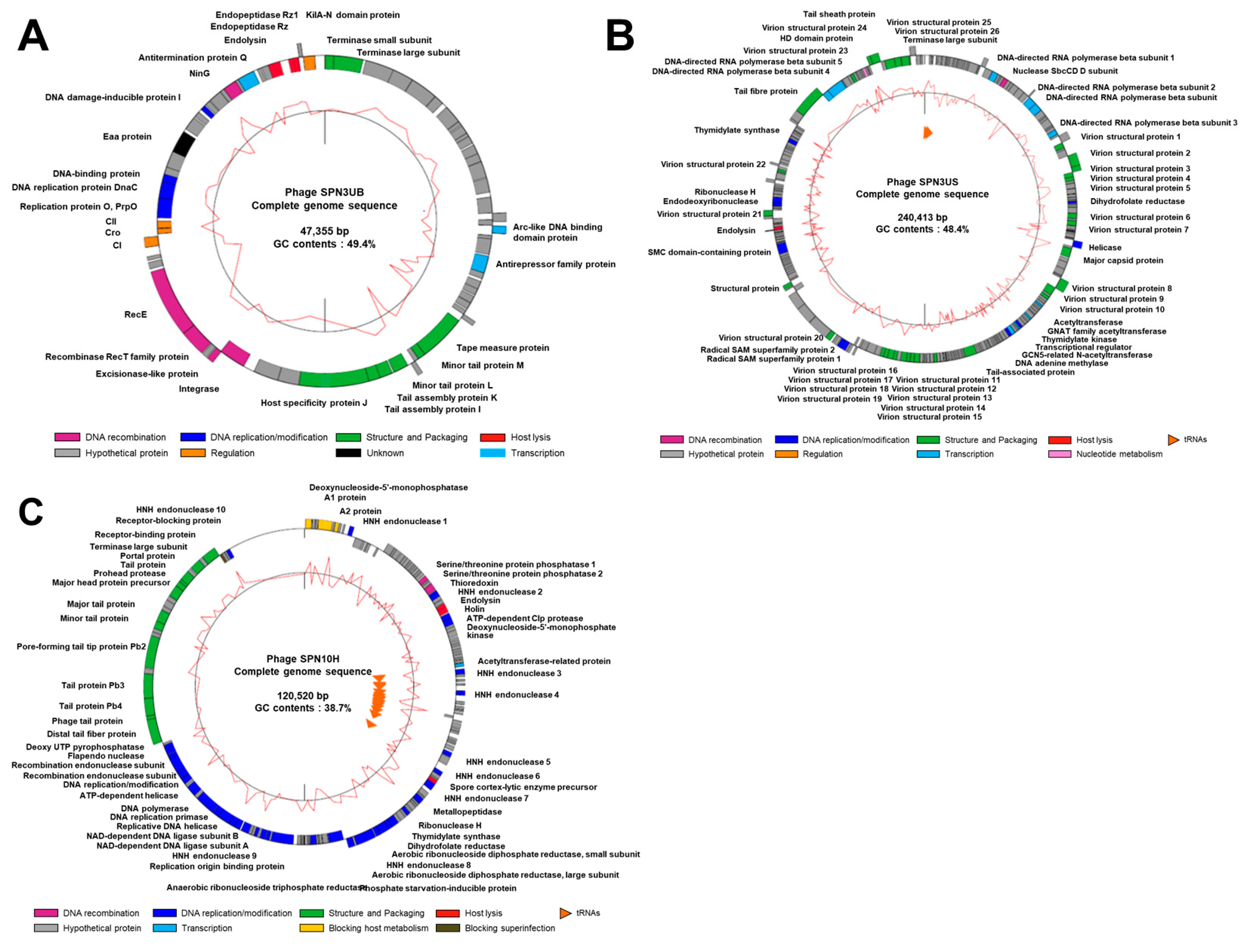
The SPN3UB genome encodes phage structural proteins, phage packaging terminases, lysogeny control proteins, phage replication proteins, host–cell lysis enzymes and peptidases, and proteins with various other functions (Figure 2A; [17]). SPN3US genome comprises functional genes associated with phage structure and packaging, tail structure, replication/transcription, host lysis and additional accessary proteins (Figure 2B; [18]). The SPN10H genome includes functional genes responsible for head/tail structure, phage replication, and host–cell lysis enzymes and peptidases (Figure 2C).
The phylogenetic analysis of the three phages, based on their large subunit of terminase, was conducted to elucidate the evolutionary relationships at the DNA level. SPN3UB aligns closely with enterobacteria phage ES18, a lysogenic phage targeting Salmonella, suggesting a potential lysogenic cycle in SPN3UB, as indicated by the presence of lysogeny control proteins and an integrase (Figure 3B; [19]). SPN3US shows the closest homology to phage SaP7, a polyvalent phage infecting both Salmonella and Esherichia coli (Figure 3A; [20]). SPN10H shares a close relationship with the Salmonella phage Stitch and Seabear, T5-like phages that are capable of infecting both Salmonella and E. coli (Figure 3C; [21,22,23]). Given SPN10H’s significant genomic resemblance to T5-like phages and its host range, including Salmonella and E. coli (Table 1), it is likely to be classified as a T5-like phage.

2.2. The Determination of the Host Range of the Salmonella-Targeting Phages

The host range of the three phages was determined against a total of 42 bacterial strains, including 38 Salmonella strains (comprising S. typhimurium and S. enteritidis), 4 other Gram-negative bacterial strains, and one Gram-positive strain, Bacillus cereus. SPN3UB created clear plaques against 17 out of 27 S. typhimurium strains and one S. enteritidis isolate strain. SPN3US was able to effectively kill 15 S. typhimurium strains and 3 S. enteritidis strains, displaying an expanded host range beyond that of SPN3UB. SPN10H showed a relatively broad host range, infecting 15 S. typhimurium, 9 S. enteritidis and 2 E. coli strains. In conclusion, the host range of each phage covered approximately 90% of the tested Salmonella strains. These results suggest the potential use of SPN3US, SPN3UB, and SPN10H as phage cocktail components to control Salmonella infections (Table 1).

2.3. Bacterial Challenge Assay

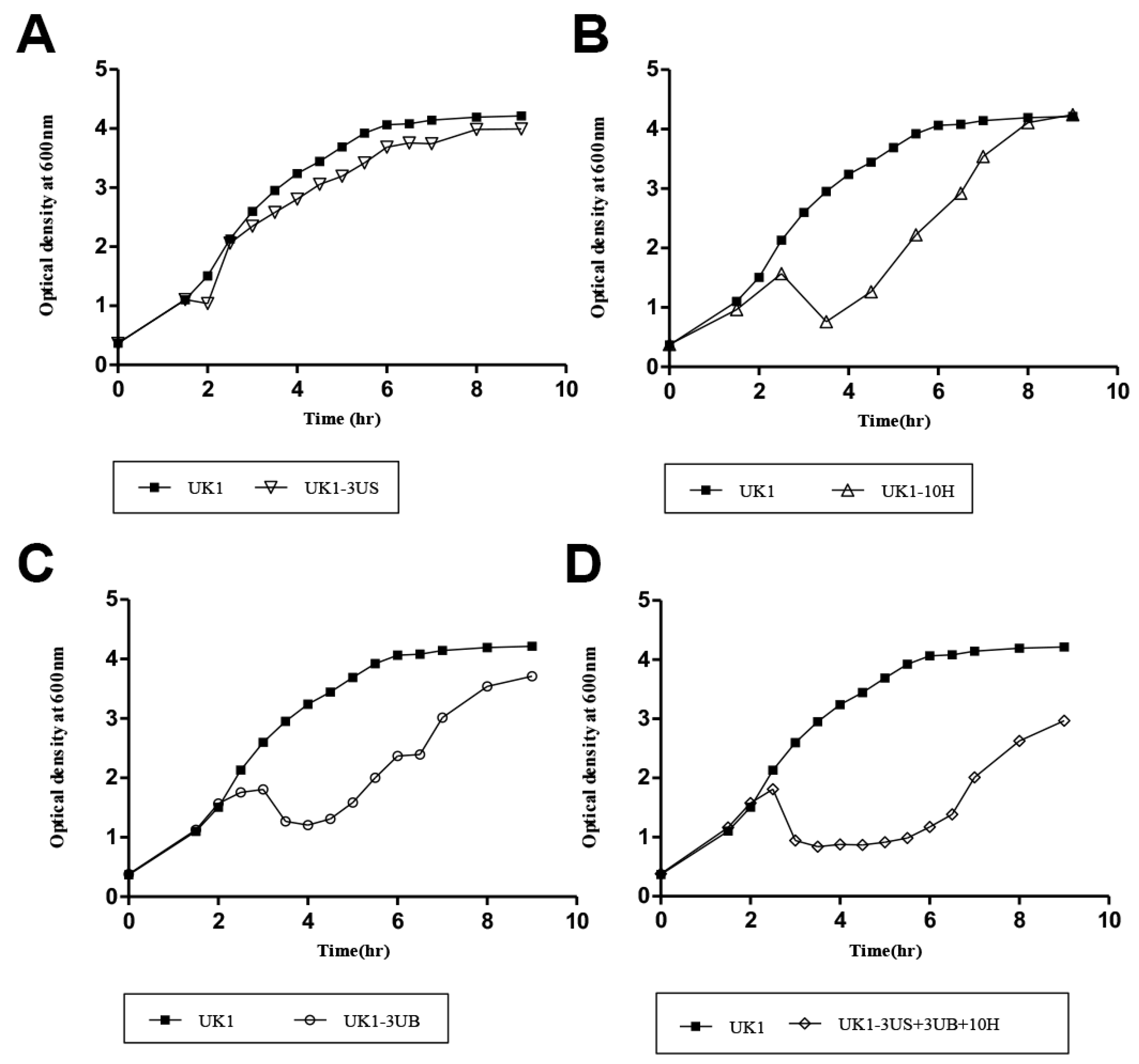
To test the lytic activity of each phage against S. typhimurium UK1, a strain effectively infected by all three phages, we challenged the host strain with SPN3UB, SPN3US, and SPN10H at an MOI of 1. The lysis of the bacterial host was evaluated by measuring the optical density at 600 nm at indicated time points. SPN3US caused only slight growth retardation at about 30 min rather than complete growth inhibition, after which SPN3US-infected S. typhimurium UK1 grew similarly to the uninfected control (Figure 4A). A significant decrease in OD600 was observed at about 4 h post-infection with SPN3UB or SPN10H, indicating their effective bacteria lysis (Figure 4B,C). However, this growth inhibition was maintained for only 1 h or less before the bacteria resumed growth, suggesting the emergence of phage-resistant bacteria. The three phages were formulated into a phage cocktail to control S. typhimurium. Despite SPN3UB being predicted to be a temperate phage, it was included in the phage cocktail due to its strong lytic activity and broad host range. While strictly lytic phages are generally preferred for therapeutic purposes, some studies have explored the potential of temperate phages, with some being engineered to remove unwanted genes for therapeutic use [30]. The treatment with phage cocktail inhibited the growth of S. typhimurium UK1 for 4 h, demonstrating a four times greater efficacy in delaying the emergence of resistance compared to single-phage treatment (Figure 4D). These results suggest that the phage cocktail can inhibit host bacterial growth more effectively and delay the emergence of phage resistance compared to a single phage infection. Previous studies have shown that the simultaneous use of several different phages, targeting different host surface receptors, can effectively suppress the development of anti-phage pathogens [31,32,33]. Notably, SPN3UB, SPN3US, and SPN10H target distinct surface receptors, including O-antigens of lipopolysaccharides [17], flagella [18] and BtuB [34], respectively. We reasoned that this superior efficacy of our phage cocktail in delaying the emergence of phage-resistant bacteria arises from the simultaneous targeting of varied host receptors, unlike single-phage use.

2.4. The Application of the Phage Cocktail to Prevent Salmonella Contamination in Foods

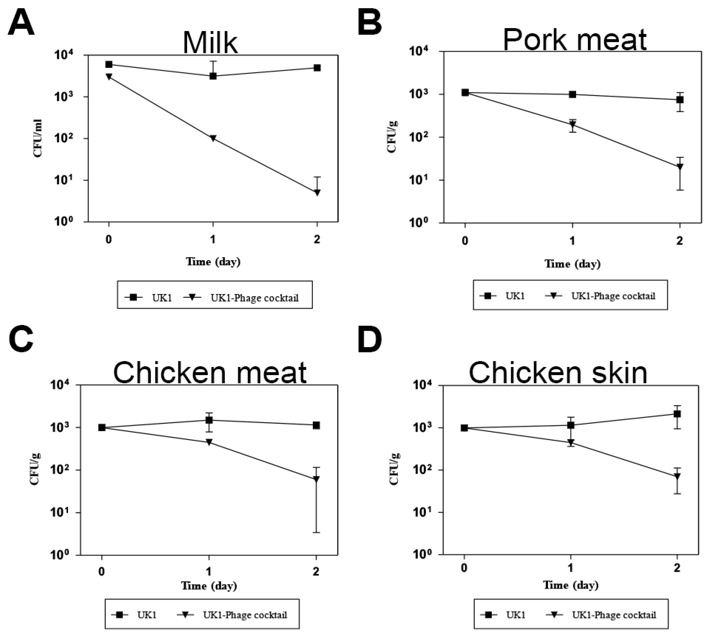
Given that the phage cocktail containing SPN3US, SPN3UB, and SPN10H effectively controlled Salmonella bacterial growth in vitro (Figure 4D), we aimed to evaluate its lytic activity in pasteurized milk, pork meat, chicken meat, and chicken skin, which are commonly contaminated by Salmonella. To this end, these foods were artificially inoculated with S. typhimurium UK1 and treated with the phage cocktail. The growth inhibition was monitored at 4 °C because dairy or meat products are still at risk of Salmonella contamination even when refrigerated. The results demonstrated a significant 3-log reduction in bacterial growth in milk after a 2-day incubation (Figure 5A). However, a lesser reduction was observed on raw pork, chicken tender, and chicken skin, with decreases of 1.5 log CFU/g, 1 log CFU/g, and 1.4 log CFU/g, respectively (Figure 5B–D). These results are consistent with previously reported studies that phages exhibited stronger host bacterial reductions in liquid foods compared to solid or semi-solid food matrices [35,36,37] as phages are allowed to be diffused in liquid, making them more accessible to bacterial populations.
It is generally known that storing foods at cold temperatures, ranging from 5 °C to 8 °C, is beneficial as it can effectively inhibit Salmonella growth by suppressing its metabolic and enzymatic activities [38,39]. These physiological changes can limit the lytic activity of phages against the host by prolonging the proliferation and latent period, thus diminishing their replication rate and progeny production [40,41]. In light of this, a phage cocktail in this study is a promising biocontrol agent as it can effectively control Salmonella contamination at cold temperatures. Taken together, our findings suggest that the phage cocktail may effectively reduce Salmonella during cold storage of foods, and it could potentially serve as an alternative to antibiotics for controlling Salmonella contamination in various foods (Figure 5).

3. Materials and Methods

3.1. Bacterial Strains and Growth Condition

A total of 43 bacterial strains including Salmonella, E. coli, and Cronobacter strains used in this study are listed in Table 1. All bacterial strains were aerobically grown in Luria–Bertani medium (LB) broth (Difco, Detroit, MI, USA) at 37 °C.

3.2. Bacteriophage Isolation and Propagation

Phages were isolated from the sewage, feces, and litter samples collected in traditional markets and poultry farms (Seoul and Chuncheon, Republic of Korea). The samples were mixed with sterile Butterfield’s phosphate-buffered dilution water (0.25 M KH2PO4, pH 7.2) and homogenized using a blender (BacMixer 400; Interscience Laboratory Inc., St. Nom, France). The mixture was centrifuged at 9000× g for 10 min at 4 °C and the supernatant was filtered using a 0.22 µm polyethersulfone (PES) membrane filter (Millipore, Billerica, MA, USA) to remove bacterial cells. Then, 25 mL of the filtrate was mixed with an equal volume of LB broth and incubated for 12 h at 37 °C. Following centrifugation (9000× g, 10min) and filtration, 10 mL of the filtrate was mixed with 40 mL of LB broth and S. typhimurium SL1344, and the mixture was incubated at 37 °C for 12–18 h with shaking (220 rpm). The culture was centrifuged, and the supernatant was filtered as above. Phage plaques were confirmed by spotting the filtrate on 0.4% LB agar (soft agar) containing S. typhimurium SL1344. The agar plates were incubated overnight at 37 °C and monitored for plaque formation. Each single plaque was picked with a sterile tip and eluted in sterilized sodium chloride–magnesium sulfate (SM) buffer (50 mM Tris-HCl, pH 7.5, 100 mM NaCl, 10 mM MgSO4·7H2O). This plaque purification step was repeated at least three times. Finally, we purified 3 phages and named them: SPN3US, SPN3UB, and SPN10H.
For phage propagation, the phage lysate was added to the prophage-cured S. typhimurium strain LT2 [referred to as LT2(c)] that is exponentially grown at a multiplicity of infection (MOI) of 1, followed by incubation at 37 °C with shaking for 3–4 h. Cell debris was removed by subsequent centrifugation and filtration using 0.22 μm pore size filters, and phage particles were precipitated with polyethylene glycol (PEG) 6000 (Sigma, St. Louis, MO, USA). Finally, the propagated phages were concentrated by cesium chloride (CsCl) density gradient ultracentrifugation (78,500× g for 2 h at 4 °C). Viral particles were recovered and dialyzed with SM buffer stirring for 1 h at 4 °C.

3.3. Bacteriophage Host Range

The bacterial strains listed in Table 1 were incubated overnight at 37 °C. A 100 μL of each bacterial culture was mixed with 6 mL of soft agar and overlaid on LB agar plates. Subsequently, 10 μL of serially diluted phage lysates was spotted onto host bacterial lawns and incubated at 37 °C overnight. After incubation, the infectivity was determined based on the appearance of the spots: “C”, clear single plaques; “T”, turbid single plaques; “I”, inhibited growth without single plaques; “—”, no lysis.

3.4. Morphological Analysis by TEM

The three purified phages were subjected to TEM analysis to characterize their morphology. Briefly, 5 μL of high-titer phage stock (1 × 10¹⁰ plaque-forming units (PFU)/mL) was placed on carbon-coated copper grids and negatively stained with 2% aqueous uranyl acetate (pH 4.0). The samples were examined with a TEM (LIBRA 120, Carl Zeiss, Jena, Germany) at an 80 kV accelerating voltage at the National Institute of Agricultural Science and Technology (Suwon, Republic of Korea). The phages were morphologically classified according to the guidelines of the International Committee on Taxonomy of Viruses [42].

3.5. Phage DNA Extraction

Phage DNA was extracted by the phenol–chloroform method as previously described [43]. Briefly, phage lysate (109 PFU/mL) was treated with RNaseA and DNase for 1 h at 37 °C to remove bacterial DNA and RNA contaminants. To degrade the phage capsid, phage lysates were then treated with lysis buffer containing 0.5 mol/L ethylene–diamine–tetraacetic acid (EDTA), 10 mg/mL proteinase K, and 1% sodium dodecyl sulfate (SDS) for 2 h at 56 °C.

3.6. Whole-Genome Sequencing and Genomic Analysis

The purified phage DNA was sequenced using a Genome Sequencer FLX (GS-FLX) Titanium sequencer (Roche, Mannheim, Germany) and assembled with 454 Newbler 2.3 assembler (Roche) at Macrogen Inc., Seoul, Republic of Korea. The open reading frames (ORFs) were identified with the ORF Finder at the National Center of Bioinformatics site (http://www.ncbi.nlm.nih.gov/gorf, accessed on 20 August 2011) and GenMark.hmm prokaryotic version 2.4 (http://opal.biology.gatech.edu/GeneMark/gmhmm2_prok.cgi, accessed on 20 August 2011). Sequence manipulations and genomic analysis were performed using CLC Genomics work-bench version 3.6.1 on a workstation at the Biopolymer Research Center for Advanced Materials, Sejong University. Phylogenetic analysis of amino acid sequences from the large subunit of the phage terminase was performed. The distances among the phages were determined by aligning their sequences using MUSCLE [44] and visualized using MEGA11 with 1000 bootstrap replicates, based on the neighbor-joining method [45,46].

3.7. Bacterial Challenge Assay

An exponentially grown S. typhimurium UK1 culture (optical density at 600 nm = 0.5) was infected with each phage or phage cocktail at an MOI of 1 and the optical density was monitored at 600 nm every 30 min with an uninfected culture as a negative control.

3.8. Biocontrol of Bacteria in Foods

Milk, Pork, chicken tender, and skin were purchased from the same retail store. First, milk was inoculated with S. typhimurium UK1 cells (~103 CFU diluted from an overnight culture), then incubated at 5 °C after the addition of phage cocktail (~107 PFU). The viable cells were counted at each time point by plating each sample on xylose lysine deoxycholate citrate (XLD) agar. Other food samples (pork, chicken tender, and chicken skin) were aseptically cut into 2 cm × 2 cm pieces in petri dishes. Subsequently, 20 μL of S. typhimurium UK1 cells (~103 CFU) was spotted onto the surface of the samples and dried for 10 min at room temperature for bacterial attachment to the samples. Then, 200 μL of prepared phage cocktail was added to cover the entire surface of the food samples and incubated at 5 °C. The intact food samples were treated with SM buffer without the phage cocktail and incubated in parallel as a negative control. At the indicated time points, each sample was harvested and homogenized with SM buffer in a stomacher to detach bacterial cells from the samples. The homogenized samples were then serially diluted and plated on the XLD agar to enumerate viable Salmonella. All experiments were conducted in duplicate.

4. Conclusions

We evaluated the efficacy of a phage cocktail comprising SPN3US, SPN3UB, and SPN10H as a novel biocontrol strategy against Salmonella enterica in various food products. Through morphological and genomic analyses, these phages were identified as members of the Caudoviricetes class. Each phage targets a distinct bacterial surface component as receptors, which broadens their antimicrobial spectrum. The phage cocktail exhibited significant lytic activity against S. typhimurium UK1, effectively delaying the emergence of phage-resistant bacteria. Our findings demonstrate that phage treatment effectively reduces Salmonella levels on chicken meat/skin and in milk at a refrigerated temperature. This suggests that phages can be used to inhibit cross-contamination by Salmonella and serve as an antimicrobial agent during the cold storage of foods, which is a crucial factor in food distribution. Considering that the phage cocktail can effectively target both S. typhimurium and S. Enteriditis, the most prevalent serovars in poultry [47], our phage cocktail could be applied to the poultry industry. Overall, it could potentially serve as an effective and sustainable alternative to antibiotics for controlling Salmonella contamination in various food industries.

Author Contributions

Conceptualization, S.R. and H.S.; methodology, H.K.; data curation, S.K., H.K., B.S. and H.S.; data interpretation, S.K.; writing—original draft preparation, S.K., B.S. and H.S.; writing—review and editing, S.K, B.S., H.S. and S.R.; supervision, S.R. All authors have read and agreed to the published version of the manuscript.

Funding

This research was funded by a grant from the Ministry of Science and ICT (MSIT) through the National Research Foundation (NRF) of Korea (NRF-RS-2023-0021200) (B.S.), and by the Biomaterials Specialized Graduate Program through the Korea Environmental Industry & Technology Institute (KEITI) funded by the Ministry of Environment (MOE) (B.S.). This research was also supported by the Korea Basic Science Institute (National Research Facilities and Equipment Center) grant funded by the Ministry of Education (2023R1A6C101A045), and the NRF funded by the Ministry of Education (2022R1A6A1A03055869).

Institutional Review Board Statement

Not applicable.

Informed Consent Statement

Not applicable.

Data Availability Statement

The original contributions presented in the study are included in the article; further inquiries can be directed to the corresponding authors.

Conflicts of Interest

The authors declare no conflicts of interest.

References

  1. Center for Disease Control and Prevention. Salmonella. Available online: https://www.cdc.gov/Salmonella/index.html (accessed on 15 February 2024).
  2. Interagency Food Safety Analytics Collaboration. Foodborne Illness Source Attribution Estimates for Salmonella, Escherichia coli O157, and Listeria monocytogenes Using Multi-Year Outbreak Surveillance Data, United States. Centers for Disease Control and Prevention, 2021. Available online: https://www.cdc.gov/foodsafety/ifsac/annual-reports.html (accessed on 1 July 2024).
  3. Naushad, S.; Ogunremi, D.; Huang, H. Salmonella: A Brief Review; IntechOpen: London, UK, 2023. [Google Scholar]
  4. Eng, S.K.; Pusparajah, P.; Ab Mutalib, N.S.; Ser, H.L.; Chan, K.G.; Lee, L.H. A review on pathogenesis, epidemiology and antibiotic resistance. Front. Life Sci. 2015, 8, 284–293. [Google Scholar] [CrossRef]
  5. Ferrari, R.G.; Rosario, D.K.; Cunha-Neto, A.; Mano, S.B.; Figueiredo, E.E.; Conte-Junior, C.A. Worldwide epidemiology of Salmonella serovars in animal-based foods: A meta-analysis. Appl. Environ. Microb. 2019, 85, e00591-19. [Google Scholar] [CrossRef] [PubMed]
  6. VT Nair, D.; Venkitanarayanan, K.; Kollanoor Johny, A. Antibiotic-resistant Salmonella in the food supply and the potential role of antibiotic alternatives for control. Foods 2018, 7, 167. [Google Scholar] [CrossRef] [PubMed]
  7. Wang, X.; Biswas, S.; Paudyal, N.; Pan, H.; Li, X.; Fang, W.; Yue, M. Antibiotic resistance in Salmonella Typhimurium isolates recovered from the food chain through national antimicrobial resistance monitoring system between 1996 and 2016. Front. Microbiol. 2019, 10, 985. [Google Scholar] [CrossRef] [PubMed]
  8. Azeredo, J.; Sutherland, I.W. The use of phages for the removal of infectious biofilms. Curr. Pharm. Biotechnol. 2008, 9, 261–266. [Google Scholar] [CrossRef]
  9. Lin, D.M.; Koskella, B.; Lin, H.C. Phage therapy: An alternative to antibiotics in the age of multi-drug resistance. World J. Gastrointest. Pharmacol. Ther. 2017, 8, 162. [Google Scholar] [CrossRef]
  10. Moye, Z.D.; Woolston, J.; Sulakvelidze, A. Bacteriophage applications for food production and processing. Viruses 2018, 10, 205. [Google Scholar] [CrossRef]
  11. Garcia, P.; Martinez, B.; Obeso, J.; Rodriguez, A. Bacteriophages and their application in food safety. Lett. Appl. Microbiol. 2008, 47, 479–485. [Google Scholar] [CrossRef]
  12. Lenneman, B.R.; Fernbach, J.; Loessner, M.J.; Lu, T.K.; Kilcher, S. Enhancing phage therapy through synthetic biology and genome engineering. Curr. Opin. Biotechnol. 2021, 68, 151–159. [Google Scholar] [CrossRef]
  13. Abedon, S.T.; Danis-Wlodarczyk, K.M.; Wozniak, D.J. Phage cocktail development for bacteriophage therapy: Toward improving spectrum of activity breadth and depth. Pharmaceuticals 2021, 14, 1019. [Google Scholar] [CrossRef]
  14. Abhisingha, M.; Dumnil, J.; Pitaksutheepong, C. Efficiency of phage cocktail to reduce Salmonella Typhimurium on chicken meat during low temperature storage. LWT 2020, 129, 109580. [Google Scholar] [CrossRef]
  15. Bai, J.; Jeon, B.; Ryu, S. Effective inhibition of Salmonella Typhimurium in fresh produce by a phage cocktail targeting multiple host receptors. Food Microbiol. 2019, 77, 52–60. [Google Scholar] [CrossRef] [PubMed]
  16. Zhu, Y.; Shang, J.; Peng, C.; Sun, Y. Phage family classification under Caudoviricetes: A review of current tools using the latest ICTV classification framework. Front. Microbiol. 2022, 13, 1032186. [Google Scholar] [CrossRef]
  17. Lee, J.H.; Shin, H.; Ryu, S. Complete genome sequence of Salmonella enterica serovar Typhimurium bacteriophage SPN3UB. J. Virol. 2012, 86, 3404–3405. [Google Scholar] [CrossRef]
  18. Lee, J.H.; Shin, H.; Kim, H.; Ryu, S. Complete genome sequence of Salmonella bacteriophage SPN3US. J. Virol. 2011, 85, 13470–13471. [Google Scholar] [CrossRef]
  19. Kuo, T.T.; Stocker, B.A. ES18, a general transducing phage for smooth and nonsmooth Salmonella typhimurium. Virology 1970, 42, 621–632. [Google Scholar] [CrossRef] [PubMed]
  20. Otto, E.C.; Blanchette, R.A. Antagonistic interactions between native fungi of Minnesota and the root rot pathogen. For. Pathol. 2023, 53, e12836. [Google Scholar] [CrossRef]
  21. Patil, K.; Zeng, C.; O’Leary, C.; Lessor, L.; Kongari, R.; Gill, J.; Liu, M. Complete genome sequence of Salmonella enterica serovar Typhimurium siphophage Seabear. Microbiol. Resour. Announc. 2019, 8, 10-1128. [Google Scholar] [CrossRef]
  22. Grover, J.M.; Luna, A.J.; Wood, T.L.; Chamakura, K.R.; Kuty Everett, G.F. Complete genome of Salmonella enterica serovar Typhimurium T5-Like siphophage Stitch. Genome Announc. 2015, 3, e01435-14. [Google Scholar] [CrossRef]
  23. Hong, J.; Kim, K.P.; Heu, S.; Lee, S.J.; Adhya, S.; Ryu, S. Identification of host receptor and receptor-binding module of a newly sequenced T5-like phage EPS7. FEMS Microbiol. Lett. 2008, 289, 202–209. [Google Scholar] [CrossRef]
  24. McClelland, M.; Sanderson, K.E.; Spieth, J.; Clifton, S.W.; Latreille, P.; Courtney, L.; Porwollik, S.; Ali, J.; Dante, M.; Du, F.; et al. Complete genome sequence of Salmonella enterica serovar Typhimurium LT2. Nature 2001, 413, 852–856. [Google Scholar] [CrossRef] [PubMed]
  25. Zhang, X.; Kelly, S.M.; Bollen, W.S.; Curtiss, R., 3rd. Characterization and immunogenicity of Salmonella typhimurium SL1344 and UK-1 delta crp and delta cdt deletion mutants. Infect. Immun. 1997, 65, 5381–5387. [Google Scholar] [CrossRef] [PubMed]
  26. Poppe, C.; Smart, N.; Khakhria, R.; Johnson, W.; Spika, J.; Prescott, J. Salmonella typhimurium DT104: A virulent and drug-resistant pathogen. Can. Vet. J. 1998, 39, 559–565. [Google Scholar] [PubMed]
  27. Marti, R.; Zurfluh, K.; Hagens, S.; Pianezzi, J.; Klumpp, J.; Loessner, M.J. Long tail fibres of the novel broad-host-range T-even bacteriophage S16 specifically recognize Salmonella OmpC. Mol. Microbiol. 2013, 87, 818–834. [Google Scholar] [CrossRef] [PubMed]
  28. Hayashi, K.; Morooka, N.; Yamamoto, Y.; Fujita, K.; Isono, K.; Choi, S.; Ohtsubo, E.; Baba, T.; Wanner, B.L.; Mori, H.; et al. Highly accurate genome sequences of Escherichia coli K-12 strains MG1655 and W3110. Mol. Syst. Biol. 2006, 2, 2006-0007. [Google Scholar] [CrossRef]
  29. Hanahan, D. Studies on transformation of Escherichia coli with plasmids. J. Mol. Biol. 1983, 166, 557–580. [Google Scholar] [CrossRef]
  30. Monteiro, R.; Pires, D.P.; Costa, A.R.; Azeredo, J. Phage therapy: Going temperate? Trends Microbiol. 2019, 27, 368–378. [Google Scholar] [CrossRef]
  31. Stalin, N.; Srinivasan, P. Efficacy of potential phage cocktails against Vibrio harveyi and closely related Vibrio species isolated from shrimp aquaculture environment in the south east coast of India. Vet. Microbiol. 2017, 207, 83–96. [Google Scholar] [CrossRef]
  32. Islam, M.S.; Zhou, Y.; Liang, L.; Nime, I.; Liu, K.; Yan, T.; Wang, X.; Li, J. Application of a phage cocktail for control of Salmonella in foods and reducing biofilms. Viruses 2019, 11, 841. [Google Scholar] [CrossRef]
  33. Kim, M.; Kim, S.; Park, B.; Ryu, S. Core lipopolysaccharide-specific phage SSU5 as an auxiliary component of a phage cocktail for Salmonella bioncontrol. Appl. Environ. Microb. 2014, 80, 1026–1034. [Google Scholar] [CrossRef]
  34. Shin, H.; Lee, J.H.; Kim, H.; Choi, Y.; Heu, S.; Ryu, S. Receptor diversity and host interaction of bacteriophages infecting Salmonella enterica serovar Typhimurium. PLoS ONE 2012, 7, e43392. [Google Scholar] [CrossRef] [PubMed]
  35. Xu, Y. Phage and phage lysins: New era of bio-preservatives and food safety agents. J. Food Sci. 2021, 86, 3349–3373. [Google Scholar] [CrossRef]
  36. Bao, H.; Zhang, P.; Zhang, H.; Zhou, Y.; Zhang, L.; Wang, R. Bio-control of Salmonella enteritidis in foods using bacteriophages. Viruses 2015, 7, 4836–4853. [Google Scholar] [CrossRef] [PubMed]
  37. Zinno, P.; Devirgiliis, C.; Ercolini, D.; Ongeng, D.; Mauriello, G. Bacteriophage P22 to challenge Salmonella in foods. Int. J. Food Microbiol. 2014, 191, 69–74. [Google Scholar] [CrossRef]
  38. D’Aoust, J.Y. Psychrotrophy and foodborne Salmonella. Int. J. Food Microbiol. 1991, 13, 207–215. [Google Scholar] [CrossRef]
  39. Morey, A.; Singh, M. Low-temperature survival of Salmonella spp. in a model food system with natural microflora. Foodborne Pathog. Dis. 2012, 9, 218–223. [Google Scholar] [CrossRef] [PubMed]
  40. Duc, H.M.; Son, H.M.; Yi, H.P.S.; Sato, J.; Ngan, P.H.; Masuda, Y.; Honjoh, K.I.; Miyamoto, T. Isolation, characterization and application of a polyvalent phage capable of controlling Salmonella and Escherichia coli O157:H7 in different food matrices. Food Res. Int. 2020, 131, 108977. [Google Scholar] [CrossRef]
  41. Hooton, S.P.; Atterbury, R.J.; Connerton, I.F. Application of a bacteriophage cocktail to reduce Salmonella Typhimurium U288 contamination on pig skin. Int. J. Food Microbiol. 2011, 151, 157–163. [Google Scholar] [CrossRef]
  42. Lefkowitz, E.J.; Dempsey, D.M.; Hendrickson, R.C.; Orton, R.J.; Siddell, S.G.; Smith, D.B. Virus taxonomy: The database of the International Committee on Taxonomy of Viruses (ICTV). Nucleic Acids Res. 2018, 46, D708–D717. [Google Scholar] [CrossRef]
  43. Sambrook, J.; Russell, D.W. Molecular Cloning: A Laboratory Manual, 3rd ed.; Cold Spring Harbor Laboratory Press: Cold Spring Harbor, NY, USA, 2001. [Google Scholar]
  44. Edgar, R.C. MUSCLE: Multiple sequence alignment with high accuracy and high throughput. Nucleic Acids Res. 2004, 32, 1792–1797. [Google Scholar] [CrossRef]
  45. Tamura, K.; Stecher, G.; Kumar, S. MEGA11: Molecular Evolutionary Genetics Analysis version 11. Mol. Biol. Evol. 2021, 38, 3022–3027. [Google Scholar] [CrossRef] [PubMed]
  46. Saitou, N.; Nei, M. The neighbor-joining method: A new method for reconstructing phylogenetic trees. Mol. Biol. Evol. 1987, 4, 406–425. [Google Scholar] [CrossRef] [PubMed]
  47. Karabasanavar, N.S.; Madhavaprasad, C.B.; Gopalakrishna, S.A.; Hiremath, J.; Patil, G.S.; Barbuddhe, S.B. Prevalence of serotypes S. Enteritidis and S. Typhimurium in poultry and poultry products. J. Food Saf. 2020, 40, e12852. [Google Scholar] [CrossRef]
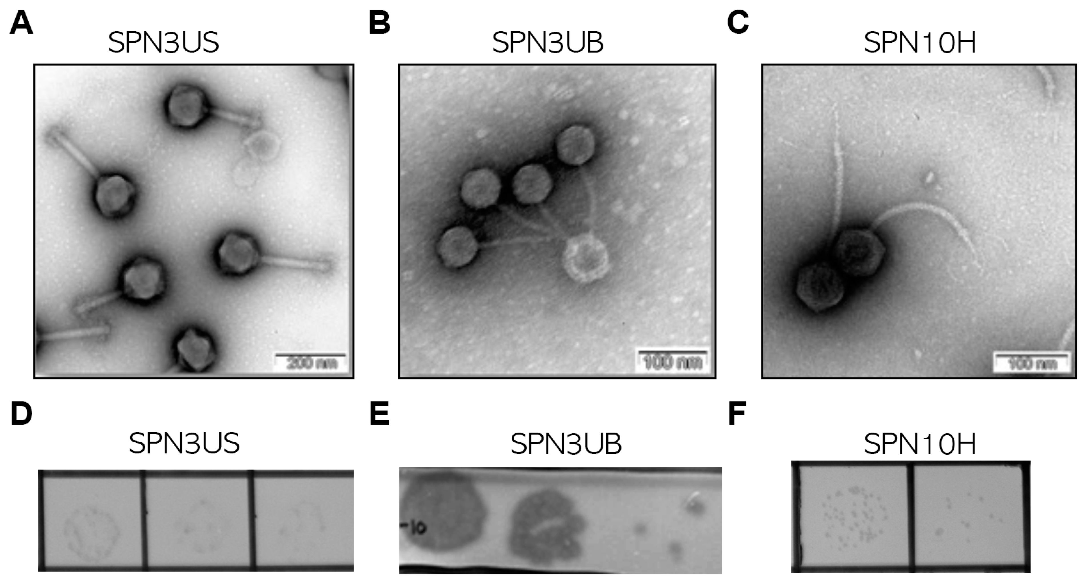
Figure 1. Morphological features of phages. TEM image of phage SPN3US (A), SPN3UB (B), and SPN10H (C) with head/tail structure. Scale bar: 200 nm (A) and 100 nm (B,C). Images of plaques formed by phage SPN3US (D), SPN3UB (E), and SPN10H (F), respectively.
Antibiotics 13 00799 g001
Figure 2. Complete genome maps of (A) SPN3UB, (B) SPN3US, and (C) SPN10H. The outer histogram map indicates gene coding regions by strands. The color of each gene represents the functional group: DNA recombination (magenta), DNA replication/modification (blue), nucleotide metabolism (pink), structure and packaging (green), host lysis (red), regulation (orange), unknown function (black), transcription (sky blue), blocking host metabolism (yellow), blocking superinfection (brown), and hypothetical protein (gray). The inner circles with the red line indicate the GC contents, and the orange arrowheads represent the location of tRNAs. Genome maps were generated using DNASTAR GeneScene v.0.99.8.0 (dnastar.com).
Antibiotics 13 00799 g002
Figure 3. Neighbor-joining phylogenetic trees of phage SPN3UB (A), phage SPN3US (B), and phage SPN10H (C). The amino acid sequences of a terminase large subunit were obtained from the NCBI database and aligned using MUSCLE. The phylogenetic trees were generated with MEGA 11.0. The numbers at the branch nodes indicate the bootstrap value (%) built on 1000 replications.
Antibiotics 13 00799 g003
Figure 4. Bacterial challenge test results for the phages SPN3US (A), SPN10H (B), SPN3UB (C), and a phage cocktail (D) against S. typhimurium UK1. A representative graph was shown from 10 biological replicates, which displays the optical density (OD) at 600 nm, monitored every 30 min. S. typhimurium UK1 was challenged with each phage or the phage cocktail when the OD at 600 nm reached 0.5.
Antibiotics 13 00799 g004
Figure 5. The evaluation of a phage cocktail’s efficacy in reducing Salmonella contamination across various food matrices. The assay was conducted in milk (A), pork meat (B), chicken tenders (C), and chicken skin (D) at 4 °C for 2 days.
Antibiotics 13 00799 g005
Table 1. Host range of the phages SPN3UB, SPN3US, and SPN10H.
Host StrainsLytic Activity of Phage 1
SPN3UBSPN3USSPN10HSource or Reference 2
S. typhimuriumLT2CCC[24]
UK1CCC[25]
SL1344CCCNCTC
14028SICIATCC
DT104CCI[26]
ATCC 19586CCCATCC
ATCC 43174CCIATCC
3068CCILaboratory collection
ATCC 12023CCCATCC
BJ 3505-CCLaboratory collection
CS 634IITLaboratory collection
CS 800ICCLaboratory collection
KCTC 1425-CCKCTC
KCTC 1925-ICKCTC
S.T 4174CCCLaboratory collection
ST DB7155CCC[27]
NCTC 12023ICCNCTC
Isolate 1I--Laboratory collection
Isolate 2C--Laboratory collection
Isolate 3C-ILaboratory collection
Isolate 4I-TLaboratory collection
Isolate 5C-ILaboratory collection
Isolate 6CCCLaboratory collection
Isolate 7C-ILaboratory collection
Isolate 8-IILaboratory collection
Isolate 9CCCLaboratory collection
Isolate 10C-CLaboratory collection
S. enteritidisATCC 13076-ICLaboratory collection
Isolate 1-ICLaboratory collection
Isolate 2-CCLaboratory collection
Isolate 3-ITLaboratory collection
Isolate 4C-TLaboratory collection
Isolate 5-CCLaboratory collection
Isolate 6IICLaboratory collection
Isolate 7IICLaboratory collection
Isolate 8-CCLaboratory collection
Isolate 9-ICLaboratory collection
Isolate 10IICLaboratory collection
Other Gram-negative bacteriaE. coli MG1655--C[28]
E. coli DH5a--C[29]
E. coli O157:H7 ATCC 35150--IATCC
Cronobacter sakazakii ATCC 29544 -I-ATCC
Gram-positive bacteriaB. cereus NRRL B-569---NCTC
1 C, clear single plaques; T, turbid single plaques; I, inhibited growth without single plaques; -, no lysis. 2 ATCC, American Type Culture Collection; NCTC, National Collection of Type Cultures; KCTC, Korean Collection of Type Cultures.
Disclaimer/Publisher’s Note: The statements, opinions and data contained in all publications are solely those of the individual author(s) and contributor(s) and not of MDPI and/or the editor(s). MDPI and/or the editor(s) disclaim responsibility for any injury to people or property resulting from any ideas, methods, instructions or products referred to in the content.