Review

Infective Spondylitis in Adults: A Journey Through Diagnosis, Management, and Future Directions

1
Department of Orthopedic Surgery, Ewha Womans University Seoul Hospital, Ewha Womans University College of Medicine, Seoul 07804, Republic of Korea
2
Department of Orthopedic Surgery, College of Medicine, International St. Mary’s Hospital, Catholic Kwandong University, Incheon 22711, Republic of Korea
3
Department of Orthopedic Surgery, Spine and Spinal Cord Institute, Gangnam Severance Hospital, Yonsei University College of Medicine, Seoul 06273, Republic of Korea
4
Department of Orthopedic Surgery, Yonsei University College of Medicine, Seoul 03722, Republic of Korea
*
Author to whom correspondence should be addressed.
Antibiotics 2025, 14(4), 391; https://doi.org/10.3390/antibiotics14040391
Submission received: 28 February 2025 / Revised: 3 April 2025 / Accepted: 7 April 2025 / Published: 9 April 2025

Abstract

:
Infective spondylitis is a rare but potentially devastating spinal infection that requires timely diagnosis and appropriate treatment to prevent severe complications, including neurological deficits and spinal deformity. Despite advancements in diagnostic imaging, microbiological techniques, and antimicrobial therapies, clinical challenges persist because of the disease’s insidious onset, varied etiologies, and increasing antimicrobial resistance. This review article provides a comprehensive analysis of the current literature on the epidemiology, pathophysiology, diagnostic approaches, and treatment strategies for infective spondylitis.

1. Introduction

Since the 1990s, the incidence of spinal infections has risen because of factors such as an aging population, longer life expectancy among patients with chronic debilitating diseases, a growing number of immunocompromised individuals and intravenous (IV) drug receivers, and the increased application of spinal instrumentation surgeries, acupuncture, and epidural catheters for pain management. Further, the widespread availability of magnetic resonance imaging (MRI) has contributed to the increase in the definitive diagnoses of spinal infections. Achieving favorable outcomes in these cases depends on prompt diagnosis, followed by aggressive medical treatment and, when necessary, surgical intervention. The mortality rate of vertebral osteomyelitis is particularly high in patients with comorbidities, such as hemodialysis or impaired liver function, and the determination of prognostic factors and treatment strategies is crucial in these cases. This review investigates the epidemiology, pathophysiology, clinical features, investigations, and treatment of spondylodiscitis based on current evidence [1,2,3,4,5,6,7]. This review focuses exclusively on infective spondylitis in adults, as the pathophysiology, clinical presentation, and management can differ significantly from pediatric cases [8].

2. Vertebral Osteomyelitis

2.1. Overall

Vertebral osteomyelitis predominantly presents with nonspecific symptoms, which can delay diagnosis for several months. Spinal trauma, postsurgical complications, or hematogenous spread from a nearby site of infection may cause vertebral osteomyelitis. Without appropriate treatment, vertebral osteomyelitis poses a high risk of severe complications, including spinal deformity, paraplegia, and even death. Improving the clinical recognition of this condition is crucial for reducing the associated morbidity and mortality [2,5].

2.2. Epidemiology

The incidence of vertebral osteomyelitis differs depending on the region and population being investigated. The estimated incidence of vertebral osteomyelitis in the United States of America (USA) is 4.8 cases per 100,000 person-year and has been increasing over the last few decades. Vertebral osteomyelitis accounts for approximately 3–5% of all osteomyelitis cases annually in the USA [9,10]. Before the introduction of antibiotics, the mortality rate within 1 year was >25%, but currently, it is approximately 11% [9].

2.3. Etiology

A single pathogen infection typically causes vertebral osteomyelitis, with Staphylococcus aureus being the most prevalent, especially with the presence of hematogenous dissemination [11]. Therefore, if a patient has had S. aureus bacteremia within the last 3 months and demonstrates compatible findings on spine MRI, a vertebral osteomyelitis diagnosis can be presumed without requiring disc space aspiration or other tissue sampling. Conversely, coagulase-negative staphylococci and Propionibacterium acnes are predominantly implicated in exogenous osteomyelitis cases after spinal surgery, particularly with spinal fixation devices [5]. However, vertebral osteomyelitis caused by different pathogens may occur in endemic regions and immunocompromised individuals. Endemic pathogens include Mycobacterium tuberculosis and Brucella spp., whereas the Mycobacterium avium complex is frequently observed in patients with human immunodeficiency virus. Although rare, fungal vertebral osteomyelitis affects patients in endemic regions (e.g., histoplasmosis, blastomycosis, and Coccidioidomycosis) and those who are immunocompromised (Aspergillus), as well as IV drug receivers and individuals with indwelling IV catheters (Candida and Aspergillus) [5,11].

3. Discitis

3.1. Overall

Intervertebral discs are frequently referred to as the largest avascular structures of the human body. These conditions cause discitis to be an uncommon medical diagnosis. Discitis is relatively rare and is difficult to treat because of the minimal blood supply. Therefore, it often requires prolonged antibiotic treatment but may still leave behind an uncomplicated symptom resolution. Figure 1 illustrates vertebral osteomyelitis and discitis with psoas abscess [12,13].

3.2. Epidemiology

The incidence of discitis in the USA is approximately 0.4–2.4 per 100,000 person-year [12]. In general, discitis occurs more prevalently in pediatric patients compared to adults, probably related to the vascular supply of the intervertebral discs, which decreases with age [14]. However, a bimodal distribution of ages was observed, with another surge in incidence observed at approximately 50 years of age. Moreover, it exhibits a higher prevalence in males than in females [12]. In children, discitis is part of a spectrum of conditions including discitis, spinal osteomyelitis, and soft-tissue abscesses [14].

3.3. Etiology

Discitis may spread to the affected intervertebral disc via the bloodstream from a systemic infection such as a urinary tract infection (UTI). Various origins have been implicated, with UTIs, pneumonia, and soft-tissue infections being the most prevalent. No conclusive evidence has associated direct trauma with discitis. IV drug administration with contaminated needles provides direct access for various organisms to enter the bloodstream. In most cases, no other infection site is identified [15,16,17].
S. aureus is the most predominantly identified organism in discitis, although Escherichia coli and Proteus species are more prevalent in patients with UTIs. Pseudomonas aeruginosa and Klebsiella species are other Gram-negative bacteria observed in IV drug receivers, although less prevalent than S. aureus. Medical conditions that predispose patients to infections in other areas of the body, such as diabetes, acquired immunodeficiency syndrome, steroid use, cancer, and chronic renal insufficiency, are associated with discitis [18].
Disc space infection may also occur postoperatively, although it is relatively uncommon. Anterior cervical discectomy and fusion (ACDF) is associated with a low postoperative infection rate, typically ranging from 0.1% to 1.6% [19], while that of lumbar discectomy ranges between 0.2% and 1.2%, depending on patient factors and surgical conditions [12,14,18,20].

4. Epidural Abscess

4.1. Overall

Epidural abscess is a rare but significant purulent infection of the central and peripheral nervous system. Abscesses that are confined within the bony structures of the skull or spinal column can expand and compress the neurological structures, causing severe symptoms, permanent complications, or even death. An epidural abscess, unlike other spinal infections, directly compresses the nerve root and spinal cord, potentially causing neurological involvement such as sensory deficits or motor weakness. Early diagnosis and appropriate treatment prevent complications and cure many cases. Figure 2 illustrates a cervical epidural abscess with spinal cord compression [21,22,23,24,25,26].

4.2. Epidemiology

Overall, spinal epidural abscesses are rare. In particular, a retrospective study conducted from 2004 to 2014 at a large academic hospital in the USA reported an incidence of 5.1 cases per 10,000 admissions. A spinal epidural abscess is particularly rare in children, with less than one-third of affected children having an identifiable risk factor [27]. This incidence indicates an increase compared to previous decades. From 1990 to 2000, the incidence in Minnesota was 0.88 cases per 100,000 person-years [28]. Part of this increase may be related to improved diagnosis sensitivity and accuracy through MRI [27]. In the USA, the increasing incidence of spinal epidural abscesses may also be associated with the aging population and the growing number of patients undergoing invasive spinal procedures for anesthesia or pain control.
The median age at the onset of spinal epidural abscess is approximately 50 years, with the highest prevalence between ages 50 and 70 years, although it occurs at any age. Some studies have shown a higher incidence in male patients [21].

4.3. Etiology

Spinal epidural abscess develops as a secondary complication from any bacteremia-causing conditions [21]. Examples include injection drug administration, dental abscesses, infected catheters, and infective endocarditis, all of which are risk factors for spinal epidural abscesses.
Moreover, spinal procedures can cause spinal epidural abscesses, with epidural catheter placement being a significant risk factor. The incidence of spinal epidural abscess after epidural catheter placement ranges from 0.5% to 3% [29]. This risk is considerably lower when catheters are used for short durations, such as in obstetrical anesthesia [30]. Further, paraspinal glucocorticoid or analgesic injections cause spinal epidural abscess development [31]. Patients with vertebral osteomyelitis frequently develop spinal epidural abscesses. A study revealed that of 167 patients with hematogenous vertebral osteomyelitis, 64 (38%) had spinal epidural abscesses [32].
The primary bacterial pathogen responsible for spinal epidural abscess is S. aureus, accounting for approximately two-thirds of cases caused by pyogenic bacteria [33]. Among microorganisms isolated from the tips of epidural catheters, coagulase-negative staphylococci were the most commonly identified pathogens, accounting for approximately 41% of cases, followed by Staphylococcus aureus (35%) [34]. Other bacteria, such as Gram-negative bacilli and streptococci, can also cause the infection. Although rare, M. tuberculosis can also result in epidural abscesses [33,35].

5. Subdural or Intradural Abscess

Although extremely rare, subdural and/or intradural abscesses exist either as primary or secondary conditions and may require more emergent surgical decompression and antibiotic therapy [36,37,38].
Spinal subdural abscesses are extremely rare, with only a handful of cases reported in the literature. Clinical presentations typically include progressive back pain, neurological deficits, and evidence of subarachnoid block. Surgical findings have described subdural granulation tissue and encapsulated purulent collections, most commonly involving the thoracic or lumbar regions. Pathogens such as Staphylococcus aureus have been isolated. Although some patients recover with surgical drainage, neurological outcomes are often limited [39,40].
Intradural spinal abscesses are considerably less common than epidural abscesses, with most cases believed to result from hematogenous spread. Associations with prior epidural procedures or underlying spondylodiscitis have been reported but remain uncommon, possibly due to the filtering function of the epidural space. These abscesses typically affect older male patients, with a predilection for the lumbar spine. Despite their rarity, intradural abscesses represent a serious clinical concern. A high index of suspicion is warranted in patients presenting with intense back pain, progressive neurological deficits, and elevated inflammatory markers. Timely diagnosis is essential, as early surgical intervention combined with appropriate antibiotic therapy can significantly improve outcomes [36,41].

6. Paraspinal Muscle Abscess

6.1. Overall

To address the spread of infection into the paraspinal tissues typically, treatments of the underlying spinal infection are involved. Any adjacent anterior and lateral abscesses are usually drained concurrently when surgical debridement is performed for conditions such as spondylodiscitis. The iliopsoas abscess, which is the most prevalent paraspinal infection site, is classified into primary and secondary types. Primary iliopsoas abscess is rare and is typically caused by the hematogenous or lymphatic spread of a pathogen from a distant infection. In contrast, most cases are secondary iliopsoas abscesses, which usually originate from inflammatory conditions of the spine or skeleton [42].

6.2. Epidemiology

Paraspinal abscesses are rare medical emergencies, occurring in approximately 0.2–1.2 cases per 10,000 hospital admissions. A meta-analysis determined diabetes (15–53.7% of cases), spinal surgery (22%), and IV drug administration (8.8%) as significant risk factors for developing a paraspinal abscess. Further, some cases are associated with alcoholism and trauma. The source of infection in approximately one-third of cases remains unidentified. If untreated, the clinical outcome can be catastrophic [23].

6.3. Etiology

The most prevalent pathogens responsible are Gram-positive bacteria, particularly S. aureus. Gram-negative infections may also occur and are frequently related to ascending UTIs [43]. Tuberculosis (TB) should also be considered in the presence of epidemiologic risk factors. Less common pathogens include Nocardia, Actinomyces species, and fungi. Risk factors include previous back surgery, spinal surgery or instrumentation, back injury, and bacteremia. Patients receiving injection medications, immunosuppressed, and with diabetes were at a higher risk [42,43].

7. Diagnosis of the Spinal Infection

7.1. Symptoms

Spinal infections often present with nonspecific symptoms, which delays diagnosis. A systematic review [2] revealed that >90% of patients reported experiencing localized neck or back pain, making it the most prevalent symptom. However, only approximately half of the patients presented with a fever during diagnosis [44]. Further, the spinal range of motion is frequently restricted because of localized pain and muscle spasms [45,46]. Focal back pain, often exacerbated by percussion, has been reported in up to 90% of patients with spinal infections. Radicular symptoms and signs of spinal cord involvement have also been observed in up to 59% and 29% of cases, respectively, although the prevalence may vary depending on the underlying pathology and stage at diagnosis [47,48].

7.2. Laboratory Tests

The 2015 Clinical Practice Guidelines for the Diagnosis and Treatment of Native Vertebral Osteomyelitis in Adults, published by the Infectious Disease Society of America, recommend that clinicians consider the possibility of spinal infections in patients presenting with any of the following signs and symptoms [11]:
  • Worsening local pain accompanied by fever;
  • New or worsening local pain along with increased ESR or CRP levels;
  • New or worsening local pain with sepsis or infective endocarditis;
  • Fever combined with new neurological symptoms, with or without local pain;
  • New local pain after a recent episode of sepsis caused by S. aureus.
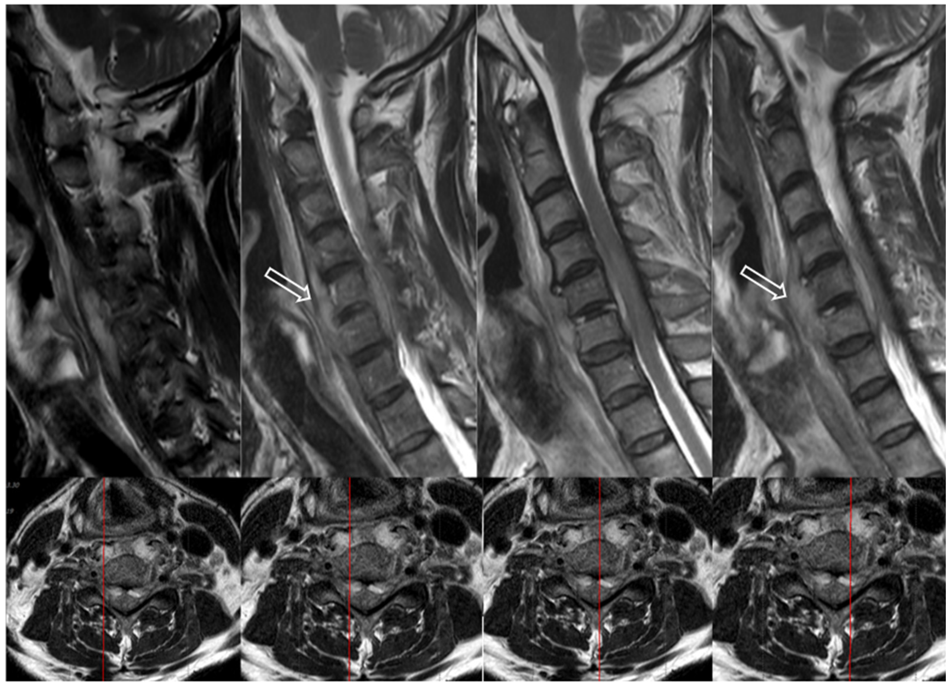
Although nonspecific, elevated ESR and CRP levels demonstrate high sensitivity—ranging from 94% to 100%—for detecting underlying spinal infections. These markers are particularly useful in evaluating patients with persistent back pain, helping to rule out infection or malignancy. Notably, white blood cell counts may remain within normal limits in up to 40% of patients with native vertebral osteomyelitis. Among these markers, CRP tends to normalize more quickly with appropriate treatment and may better reflect the patient’s clinical progress. However, these markers should be interpreted in the clinical context, as they may also be elevated in non-infectious inflammatory conditions [11,49].
Plasma procalcitonin has recently emerged as an important biomarker for distinguishing between bacterial infections and systemic inflammatory diseases. Patients with any bacterial infection typically have a procalcitonin level of >0.5 ng/mL (normal range: <0.05 ng/mL). Procalcitonin demonstrates variable sensitivity, which may be influenced by the presence of concurrent infections [50,51].

7.3. Imaging Tests

In the initial disease stages, plain radiographs can be obtained, although they are frequently normal, as characteristic features may not appear until after two to three weeks. Typical radiographic signs of spondylodiscitis involve the destruction of two adjacent vertebral bodies accompanied by the collapse of the intervening disc space. In rare cases, an infection that involves only a single vertebral body may present as a vertebral collapse comparable to a compression fracture [1].
MRI is considered the gold standard for diagnosing discitis, providing 92% specificity, 96% sensitivity, and 94% accuracy [52]. In acute cases, MRI typically demonstrates high fluid intensity. On T1-weighted images, the infection initially affected the anterolateral portion of the vertebral body, resulting in irregular signal intensity in the end plates, with edema spreading to the rest of the vertebral body and the adjacent disc. A pyogenic infection usually appears as a low signal on T1-weighted images and a high signal on T2-weighted images, with vertebral body contrast enhancements on T1 images after contrast administration.
CT scans are valuable in detecting paraspinal abscesses and bony sequestrate, as well as determining the best location and approach for a biopsy. Noteworthily, radiological results do not always align with the clinical presentation, as clinical symptoms may improve while radiological appearances are worsening. A reduction in contrast enhancement on an MRI can be an early sign of resolving infection; therefore, patients should be closely monitored with follow-up MRIs and careful clinical correlation during result interpretation [53,54].
The appearance of less common pathogens in spinal infections can include features, such as skip lesions, intraosseous abscesses, large paravertebral abscesses, and posterior element, spinal canal, and nerve root involvement, which indicate TB infection [55]. Brucella infection is characterized by an intact vertebral structure with signs of widespread intervertebral infection. The absence of T2-weighted enhancement after contrast administration is a notable feature of fungal infections [53].
A radioisotope bone scan with gallium is highly sensitive in detecting early changes in the disease process, demonstrating a 94% accuracy rate [12]. However, caution is required when interpreting gallium scans in leukopenic or elderly patients, as these individuals may experience relative ischemia of the end plate, which can influence the results [56].
Nuclear medicine imaging modalities, such as positron emission tomography—Computed tomography (CT) using fluorine-18 fluorodeoxyglucose and single-photon emission computed tomography/CT are also employed in diagnosing spondylodiscitis and are known to demonstrate higher sensitivity and specificity than bone scans [57].

7.4. Bacteriological Tests

To select the appropriate antibiotics, the causative organism needs to be identified. In patients with suspected spinal infections, it is recommended to culture specimens from the infection site and obtain two blood samples before starting antibiotic treatment [11]. For suspected cases of Mycobacterium tuberculosis spinal infection or in patients with a UTI history, sputum or urine cultures may also be required.
Specimens from the infection site are typically collected through percutaneous needle biopsy, guided by CT or fluoroscopy. The success rate of pathogen detection using this method varies between 41% and 90% [11,58]. The likelihood of detecting the pathogen is significantly higher in the presence of a psoas abscess, although specimens are not directly taken from the abscess [59]. If cultures are negative but a spinal infection remains suspected, a second biopsy is recommended. Detection rates were notably lower in patients who had received antibiotics before the biopsy. In such cases, a biopsy is advisable 1 or 2 weeks after stopping antibiotics, although detection rates may remain low [59]. An open biopsy is an alternative for patients with multiple negative biopsies, as it yields higher positive results compared to a percutaneous biopsy [11]. Overall, organisms are determined in 67–100% of spinal infection cases [58].
A PCR can be performed on spinal biopsy specimens and provides a rapid diagnostic tool for detecting M. tuberculosis. Although PCR may detect DNA from nonviable organisms, leading to false positives in patients who are undergoing or have completed treatment, during the initial diagnostic workup, a positive PCR is generally considered indicative of active infection. Compared to mycobacterial culture, which can take 6–8 weeks, PCR offers faster results with high sensitivity (around 88%) and specificity (approximately 95%), making it a valuable adjunct in early diagnosis [60,61,62].

8. Postoperative Infection

8.1. Overall

Postoperative spine infection can be a severe complication after spine surgery, affecting both short-term and long-term outcomes. Such infections significantly increase the risk of pseudoarthrosis, chronic pain, repeat surgeries, adverse neurological effects, poor long-term prognosis, and even death [63]. Figure 3 illustrates late postoperative infection [64].

8.2. Epidemiology

The incidence of infection after spine surgery varies widely, ranging from 0% to 18%, depending on the surgical type. Simple lumbar decompression or microdiscectomy exhibits a lower infection rate, approximately 0.6–3%, compared with instrumented fusion, which demonstrates an infection rate of approximately 6–18%. Further, the surgical approach influences the infection rate, with posterior surgery having a higher incidence of infection than anterior spinal surgeries [65].

8.3. Etiology

Several factors related to pathophysiology and microbiology cause postoperative spinal infections. The use of instrumentation in spinal surgery plays a crucial role in the development of these infections. Instrumentation causes local soft-tissue irritation, resulting in inflammation and seroma formation, thereby establishing an ideal environment for microorganisms to thrive. The adherence of bacteria to implant surfaces is facilitated by a polysaccharide biofilm called glycocalyx, which acts as a barrier against the host’s defense mechanisms and antibiotics [66]. Further, metallosis from the micromotion of the instrumentation causes granuloma formation, which provides another medium for bacterial colonization [66,67,68].
Various studies investigated the effect of preoperative epidural steroid injections (ESIs) on postoperative infection [69,70,71,72,73,74]. Kreitz et al. published results indicating an increased infection rate in patients who underwent fusion surgery and received ESI but not in decompression surgery. However, Lee et al. revealed that preoperative ESI is not a risk factor for postoperative infection [75,76]. ESI and acupuncture are frequently used as treatment methods for lumbar degenerative disease. Sung et al. reported that both acupuncture and ESIs performed more than 2 weeks before spinal surgery did not increase the risk of postoperative infection [7,77].
S. aureus remains the most commonly isolated organism in spinal SSIs. The prevalence of methicillin-resistant strains (MRSAs), however, varies depending on the clinical setting and geographic location [78,79,80]. Further, Staphylococcus epidermidis is increasingly predominant in postoperative infections. E. coli and E. faecalis are more prevalent in patients with incontinence or fecal contamination. Low-virulence microorganisms, such as P. acnes, can infect surgical wounds in patients with compromised immune systems. Late hardware infections, although generally rare, can involve Gram-negative rods and are more frequent in patients with trauma, those with severe neurological injuries, and immunocompromised individuals (with an injury severity score of >18). Patients with neuromuscular scoliosis, such as those with cerebral palsy or Duchenne’s muscular dystrophy, are at a higher risk due to poor bowel and bladder control, coupled with a lack of baseline mobility.
Polymicrobial infections are almost exclusively due to direct wound contamination during the postoperative period, frequently involving fecal or urinary contamination in patients with neuromuscular disorders [81,82].

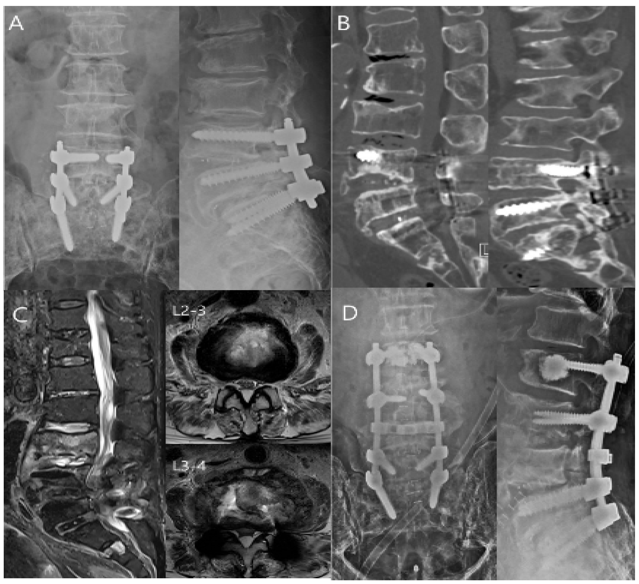
9. Treatment of the Spinal Infection

Spinal infections typically require prolonged IV antibiotics or antifungal therapy, frequently resulting in extended hospital stays. Immobilization may be advised in cases of significant pain or potential spine instability. The initial treatment approach should be conservative, especially in early-stage cases with no or minor neurological deficits or when severe comorbidities limit surgical options [1,65,83,84]. Pyogenic vertebral osteomyelitis generally exhibits a favorable prognosis in the absence of structural instability and neurological deficits, considering that appropriate antibiotic therapy is administered [2,85]. However, in elderly patients with comorbidities, pyogenic spondylitis diagnosis is predominantly delayed, and concurrent infections in other organ systems are frequently observed [86]. These infections significantly affect clinical outcomes, potentially resulting in mortality despite appropriate treatment [85,87,88]. Clinically, negative initial nonoperative culture results are predominant. In such cases, spinal decompression and abscess drainage can be considered for patients with neurological deficits, progressive deterioration, or intolerable pain caused by an abscess, and the procedure can be performed using an endoscope [1,89,90,91]. Additionally, if mechanical instability is present before or after spinal decompression, early spinal instrumentation may be performed, considering the presence of a psoas abscess [92].
A broad range of antibiotics is available for the treatment of spinal infections. In critically ill patients or when cultures remain negative, empirical dual-agent therapy is typically recommended. This usually includes a combination of a third-generation cephalosporin or fluoroquinolone with an anti-Gram-positive agent such as clindamycin or vancomycin. Empirical regimens should be selected to cover both Gram-positive and Gram-negative organisms until the causative pathogen is identified, with options including clindamycin, vancomycin, or flucloxacillin combined with ceftriaxone, cefepime, or ciprofloxacin. Once culture results are available, antibiotics should be tailored accordingly. If methicillin-sensitive Staphylococcus aureus is isolated, anti-staphylococcal penicillins (e.g., nafcillin or flucloxacillin) or a first-generation cephalosporin is preferred. In cases involving methicillin-resistant strains, glycopeptides such as vancomycin or teicoplanin are recommended, with alternatives including linezolid or quinupristin-dalfopristin. For Streptococcus spp., penicillin G remains the first-line agent. When Gram-negative bacilli are implicated—particularly in infections originating from urinary tract sources—second- or third-generation cephalosporins or fluoroquinolones are appropriate. Anaerobic infections should be treated with metronidazole or clindamycin [4,83]. Appropriate antibiotics should be administered via IV for 2–4 weeks or a significant decrease in CRP levels, followed by oral antibiotics for a total duration of 6–12 weeks [3]. The optimal duration and route of antibiotic treatment remain debated due to a presumed association between treatment duration and relapse or failure. Further, conservative treatment should include bed rest and/or the use of an orthosis for at least 6 weeks, based on pain levels during mobilization [93]. Conservative treatments are frequently effective, but they are not always sufficient in every case.
Recurrence and mortality are key indicators of treatment success in patients with infective spondylitis [94,95]. A clinical outcome assessment needs to consider various risk factors [96]. Previous studies have revealed that medical comorbidities, undrained abscesses, and causative pathogens, such as MRSA, are significant predictors of postoperative recurrence and mortality [95,97,98]. Recurrence and mortality in older patients with infective spondylitis are significantly associated with neurological deficits; however, the severity of comorbid conditions also plays a crucial role [6,68,92,95,99,100,101].
Surgical treatment is considered in cases of acute or progressive neurological deficit, when an intraspinal lesion limits the effectiveness of conservative treatment, in instability instances, and when conservative treatment has failed. First, simple decompression or excision can be performed for an epidural abscess without spondylodiscitis, and surgical open drainage is an option. Second, only decompression or excision may be performed for an epidural abscess with spondylodiscitis in the absence of associated instability. However, posterior instrumentation is required in the presence of instability, and according to the extent of discitis, interbody fusion may be necessary. Third, a corpectomy may be performed for spondylodiscitis accompanied by bony destruction, and instrumentation of the anterior column or simultaneous instrumentation of both the anterior and posterior columns may be required [102,103].
Structural reinforcement using autobone or allobone is essential in the presence of bony destruction. The evolution of bone grafting in spinal surgeries has seen the development of various techniques and materials aimed at improving outcomes. Tricortical iliac autograft, which is a traditional bone grafting method, is widely recognized for its safety and consistently excellent results, making it the gold standard for promoting bony fusion [104]. Alternatively, structural bone allograft provides benefits such as reduced operative time and donor site morbidity elimination [105]. Recent studies indicate that combining structural bone grafts with recombinant human bone morphogenetic protein-2 (rhBMP-2) improves fusion rates [106]. However, rhBMP-2 carries potential risks, including seroma formation, radiculitis, and undesirable cell growth. Therefore, patients with a history of cancer and cerebrospinal fluid leaks or those undergoing cervical or thoracic spine surgeries should be specifically informed about these risks.
The use of transforaminal interbody debridement and fusion with antibiotic-impregnated bone graft for the treatment of pyogenic discitis and vertebral osteomyelitis has also been proposed by some clinicians, particularly in studies involving Asian populations [107].
Some researchers have proposed transpedicular curettage and drainage as an effective treatment option for infectious spondylodiscitis, particularly in patients who cannot tolerate conventional combined anterior and posterior surgeries due to multiple comorbidities, extensive infectious lesions, and poor general condition [108,109].
Vacuum-assisted closure is a form of negative pressure wound therapy with or without continuous irrigation that has appeared as a promising alternative for managing complex postoperative spinal infections. Further, it is considered a safe and effective treatment option for deep SSIs even in cases with exposed dura, especially after posterior instrumented spinal surgery [110,111,112,113,114,115,116,117,118,119].
Several strategies have been proposed to prevent postoperative infections after fusion surgery, including the application of vancomycin powder and taurolidine irrigation [120,121]; however, postoperative spine infection is more difficult to treat and exhibits a poorer prognosis than simple spine infection. The principles of open surgical debridement involve investigating the wound to identify whether the infection is deep or superficial, followed by a thorough necrotic and infected tissue removal. For early postoperative infections (within 3 months), the spinal hardware is advised not to be removed to avoid destabilizing the spine. Loose bone grafts should be removed during debridement, but graft material firmly attached to bony structures should be left in place [122]. Hardware removal for late postoperative infections is often required for several reasons. One reason is that the spinal anchorage points and the area directly beneath the rods are hard to access without hardware removal. Removing the hardware enables more thorough wound debridement if a solid fusion has occurred. Late-onset infections are frequently indolent and caused by organisms, such as coagulase-negative Staphylococci or P. acnes, which are prone to biofilm formation. Di Silvestre et al. demonstrated that not removing implants in delayed infections leaves a 50% chance of the infection to persist [123]. The advantage of removing biofilm must be weighed against the risk of destabilizing the spine before bone fusion. The hardware can be removed if fusion has occurred; however, long fusions involve a risk of fracturing the fusion mass, losing alignment, or settling into kyphosis [124]. If fusion has not occurred, an autograft and/or an allograft can be utilized to achieve bone fusion. Using an allograft does not significantly increase the postoperative infection rate [125]. Choi et al. revealed that using an intraoperative toothbrushing technique during the initial surgical debridement for postoperative spine infections can significantly lower the failure rate and decrease the necessity for revision surgery by effectively eliminating the biofilm of pathogenic bacteria [64].
In addition to multiple debridements, continuing antibiotic therapy is equally crucial. As previously mentioned, patients in stable conditions should not be given antibiotics before obtaining the culture results. However, empirical antibiotic administration is necessary for patients with sepsis and unstable conditions to prevent further clinical deterioration. Broad-spectrum antibiotics should be initiated before the final culture results are available. The duration of antibiotic therapy is debated and varies according to the type of infection. A shorter course of antibiotics is usually sufficient for postoperative infections without hardware. Postoperative discitis or osteomyelitis typically requires over 3 months of antibiotics, based on the inflammatory markers. Patients generally need at least 4–6 weeks of IV antibiotics, tailored to culture results and inflammatory markers for deep infections with hardware in place [126]. If ESR/CRP levels increase after discontinuing antibiotics and fusion has occurred, then hardware removal is recommended. Clear consensus on the duration of antibiotic treatment remains unavailable; however, a course of oral suppressive antibiotics is recommended to follow long-term IV antibiotics if hardware is retained. A shorter treatment course may be appropriate if the instrumentation is removed.
However, antibiotic-induced allergic reactions can increase inflammatory markers, and the use of broad-spectrum antibiotics may cause antibiotic-related diarrhea due to Clostridioides difficile; thus, these potential issues should be considered [127,128,129,130,131,132]. Hensgens et al. reported that antibiotic administration increases the risk for C. difficile infection not only while taking them but also for up to three months after stopping, with the highest risk occurring during treatment and in the first month afterward [133]. Also, the use of multiple antibiotics has been associated with an increased risk of Clostridium difficile infection [134].
In patients requiring surgical treatment for infective spondylitis after previous spinal instrumented surgery, a decision must be made between a less invasive noninstrumented approach, which retains the previous instrumentation, or a more invasive additional instrumented surgery which involves thorough infected tissue removal and stabilization. Spinal infections frequently occur in elderly patients with comorbidities, making clinical decision-making challenging because the use of instrumentation can inadvertently interfere with infection management by promoting biofilm formation [67,135]. Outcomes vary according to factors, such as the presence of severe comorbidities, psoas abscess, and MRSA infection; however, studies have revealed comparable recurrence and mortality rates between patients who undergo additional instrumentation and those in the noninstrumented surgery group. Therefore, individual risk factors need to be carefully considered when selecting the most appropriate surgical approach for each patient [68].
Surgical intervention is rarely required in cases of isolated psoas abscesses. Instead of immediate surgery, nonsurgical treatments, such as antibiotics and percutaneous drainage, are prioritized. However, caution is required as infective spondylitis may be the primary source of infection [136,137,138].
Hyperbaric oxygen therapy has been applied as a supplementary treatment for spinal infections. This therapy may help in infection treatment by restoring intramedullary bone oxygen tension, normalizing phagocyte activity, stimulating neovascularization at the edges of healing wounds, inducing vasodilation in healing tissues, and suppressing biofilm formation. Side effects associated with this adjunct therapy were not reported [139]. Further research is warranted to determine if hyperbaric oxygen therapy reduces the number of revision surgeries or debridements and improves long-term outcomes compared with standard treatment alone.

10. Conclusions

Universally applicable guidelines for managing spinal infections remain unavailable because of the diverse and often comorbid nature of the patient population and the broad range of treatment options available, making treatment challenging. Patients should consider both medical and surgical options based on a review of the literature and personal experience. In cases of neurological symptom worsening or medical treatment failure, surgery may be required with or without instrumentation. Patients who have severe comorbidities limit invasive procedures; thus, a less invasive approach, such as transpedicular irrigation, can be a viable alternative. More prospective randomized trials are warranted to confirm and refine these treatment strategies.

Author Contributions

W.-S.J.: writing—original draft, review, and editing; S.-R.C.: visualization and resources; J.-W.K.: resources; K.-S.S.: validation; H.-S.K.: validation; S.-H.M.: validation; S.-Y.P.: validation; J.-W.S.: validation; B.-H.L.: conceptualization, validation, and supervision. All authors have read and agreed to the published version of the manuscript.

Funding

This research received no external funding.

Institutional Review Board Statement

Ethical review and approval were waived for this study because this article is a review article.

Informed Consent Statement

Not applicable.

Data Availability Statement

Not applicable.

Acknowledgments

The authors received professional English language editing assistance.

Conflicts of Interest

The authors declare that there are no conflicts of interest regarding the publication of this paper.

References

  1. Govender, S. Spinal infections. J. Bone Jt. Surg. Br. Vol. 2005, 87, 1454–1458. [Google Scholar] [CrossRef] [PubMed]
  2. Mylona, E.; Samarkos, M.; Kakalou, E.; Fanourgiakis, P.; Skoutelis, A. Pyogenic vertebral osteomyelitis: A systematic review of clinical characteristics. In Seminars in Arthritis and Rheumatism; Elsevier: Amsterdam, The Netherlands, 2009; Volume 39, pp. 10–17. [Google Scholar]
  3. Skaf, G.S.; Domloj, N.T.; Fehlings, M.G.; Bouclaous, C.; Sabbagh, A.S.; Kanafani, Z.A.; Kanj, S. Pyogenic spondylodiscitis: An overview. J. Infect. Public Health 2010, 3, 5–16. [Google Scholar] [CrossRef] [PubMed]
  4. Yunoki, M. A Comprehensive Review of Pyogenic Spondylitis Management for Neurosurgeons. Asian J. Neurosurg. 2023, 18, 724–733. [Google Scholar] [CrossRef] [PubMed]
  5. Zimmerli, W. Vertebral osteomyelitis. N. Engl. J. Med. 2010, 362, 1022–1029. [Google Scholar] [CrossRef]
  6. Kim, J.; Kang, H.S.; Kim, J.W.; Kim, S.W.; Oh, J.-K.; Kim, Y.-W.; Park, M.S.; Kim, T.-H. Treatment outcomes in patients with pyogenic vertebral osteomyelitis who have cirrhosis. Sci. Rep. 2019, 9, 15223. [Google Scholar] [CrossRef]
  7. Sung, S.; Kwon, J.W.; Lee, S.B.; Kim, E.H.; Jung, I.; Moon, S.H.; Suk, K.S.; Kim, H.S.; Park, S.Y.; Lee, B.H. Effect of Preoperative Acupuncture and Epidural Steroid Injection on Early Postoperative Infection After Lumbar Spinal Fusion. J. Bone Jt. Surg. Am. 2024, 107, 229–236. [Google Scholar] [CrossRef]
  8. Principi, N.; Esposito, S. Infectious discitis and spondylodiscitis in children. Int. J. Mol. Sci. 2016, 17, 539. [Google Scholar] [CrossRef]
  9. Issa, K.; Diebo, B.G.; Faloon, M.; Naziri, Q.; Pourtaheri, S.; Paulino, C.B.; Emami, A. The Epidemiology of Vertebral Osteomyelitis in the United States From 1998 to 2013. Clin. Spine Surg. 2018, 31, E102–E108. [Google Scholar] [CrossRef]
  10. Nickerson, E.K.; Sinha, R. Vertebral osteomyelitis in adults: An update. Br. Med. Bull. 2016, 117, 121–138. [Google Scholar] [CrossRef]
  11. Berbari, E.F.; Kanj, S.S.; Kowalski, T.J.; Darouiche, R.O.; Widmer, A.F.; Schmitt, S.K.; Hendershot, E.F.; Holtom, P.D.; Huddleston, P.M., 3rd; Petermann, G.W.; et al. 2015 Infectious Diseases Society of America (IDSA) Clinical Practice Guidelines for the Diagnosis and Treatment of Native Vertebral Osteomyelitis in Adults. Clin. Infect. Dis. 2015, 61, e26–e46. [Google Scholar] [CrossRef]
  12. Cottle, L.; Riordan, T. Infectious spondylodiscitis. J. Infect. 2008, 56, 401–412. [Google Scholar] [CrossRef] [PubMed]
  13. Rezvani, M.; Ahmadvand, A.; Yazdanian, T.; Azimi, P.; Askariardehjani, N. Value of Spinal Infection Treatment Evaluation Score, Pola Classification, and Brighton Spondylodiscitis Score from Decision to Surgery in Patients with Spondylodiscitis: A Receiver-Operating Characteristic Curve Analysis. Asian Spine J. 2024, 18, 218. [Google Scholar] [CrossRef] [PubMed]
  14. Fernandez, M.; Carrol, C.L.; Baker, C.J. Discitis and vertebral osteomyelitis in children: An 18-year review. Pediatrics 2000, 105, 1299–1304. [Google Scholar] [CrossRef] [PubMed]
  15. Subramanian, V.; Hughes, P.; Soni, B.; Oo, T. Discitis following urinary tract infection manifesting as recurrent autonomic dysreflexia related to truncal movements in a person with tetraplegia. BMJ Case Rep. 2020, 13, e238202. [Google Scholar] [CrossRef]
  16. Chuo, C.-Y.; Fu, Y.-C.; Lu, Y.-M.; Chen, J.-C.; Shen, W.-J.; Yang, C.-H.; Chen, C.-Y. Spinal infection in intravenous drug abusers. Clin. Spine Surg. 2007, 20, 324–328. [Google Scholar] [CrossRef]
  17. Samy, D.A.; Gandham, S.; DeMatas, M. The diagnosis and management of discitis and spinal infection. Surgery 2021, 39, 540–546. [Google Scholar]
  18. Conaughty, J.M.; Chen, J.; Martinez, O.V.; Chiappetta, G.; Brookfield, K.F.; Eismont, F.J. Efficacy of linezolid versus vancomycin in the treatment of methicillin-resistant Staphylococcus aureus discitis: A controlled animal model. Spine 2006, 31, E830–E832. [Google Scholar] [CrossRef]
  19. Epstein, N.E. A review of complication rates for anterior cervical diskectomy and fusion (ACDF). Surg. Neurol. Int. 2019, 10, 100. [Google Scholar] [CrossRef]
  20. Zuo, Q.; Zhao, K.; Dong, B.; Zhou, Y.; Wang, X.; Maimaiti, A.; Tian, P. Analysis of risk factors for surgical site infection in spinal surgery patients and study of direct economic losses. BMC Musculoskelet. Disord. 2024, 25, 1096. [Google Scholar] [CrossRef]
  21. Sendi, P.; Bregenzer, T.; Zimmerli, W. Spinal epidural abscess in clinical practice. QJM Int. J. Med. 2008, 101, 1–12. [Google Scholar] [CrossRef]
  22. Darouiche, R.O. Spinal epidural abscess. N. Engl. J. Med. 2006, 355, 2012–2020. [Google Scholar] [CrossRef] [PubMed]
  23. Pradilla, G.; Ardila, G.P.; Hsu, W.; Rigamonti, D. Epidural abscesses of the CNS. Lancet Neurol. 2009, 8, 292–300. [Google Scholar] [CrossRef] [PubMed]
  24. Reihsaus, E.; Waldbaur, H.; Seeling, W. Spinal epidural abscess: A meta-analysis of 915 patients. Neurosurg. Rev. 2000, 23, 175–204. [Google Scholar] [CrossRef] [PubMed]
  25. Tompkins, M.; Panuncialman, I.; Lucas, P.; Palumbo, M. Spinal Epidural Abscess. J. Emerg. Med. 2010, 39, 384–390. [Google Scholar] [CrossRef]
  26. Baker, A.S.; Ojemann, R.G.; Swartz, M.N.; Richardson, E.P., Jr. Spinal Epidural Abscess. N. Engl. J. Med. 1975, 293, 463–468. [Google Scholar] [CrossRef]
  27. Sørensen, P. Spinal epidural abscesses: Conservative treatment for selected subgroups of patients. Br. J. Neurosurg. 2003, 17, 513–518. [Google Scholar] [CrossRef]
  28. Ptaszynski, A.E.; Hooten, W.M.; Huntoon, M.A. The incidence of spontaneous epidural abscess in Olmsted County from 1990 through 2000: A rare cause of spinal pain. Pain Med. 2007, 8, 338–343. [Google Scholar] [CrossRef]
  29. Cook, T.M.; Counsell, D.; Wildsmith, J.A. Major complications of central neuraxial block: Report on the Third National Audit Project of the Royal College of Anaesthetists. Br. J. Anaesth. 2009, 102, 179–190. [Google Scholar] [CrossRef]
  30. D’Angelo, R.; Smiley, R.M.; Riley, E.T.; Segal, S. Serious complications related to obstetric anesthesia: The serious complication repository project of the Society for Obstetric Anesthesia and Perinatology. Anesthesiology 2014, 120, 1505–1512. [Google Scholar] [CrossRef]
  31. Gaul, C.; Neundörfer, B.; Winterholler, M. Iatrogenic (para-) spinal abscesses and meningitis following injection therapy for low back pain. Pain 2005, 116, 407–410. [Google Scholar] [CrossRef]
  32. Park, K.H.; Cho, O.H.; Jung, M.; Suk, K.S.; Lee, J.H.; Park, J.S.; Ryu, K.N.; Kim, S.H.; Lee, S.O.; Choi, S.H.; et al. Clinical characteristics and outcomes of hematogenous vertebral osteomyelitis caused by gram-negative bacteria. J. Infect. 2014, 69, 42–50. [Google Scholar] [CrossRef] [PubMed]
  33. Vakili, M.; Crum-Cianflone, N.F. Spinal Epidural Abscess: A Series of 101 Cases. Am. J. Med. 2017, 130, 1458–1463. [Google Scholar] [CrossRef] [PubMed]
  34. Holt, H.; Andersen, S.; Andersen, O.; Gahrn-Hansen, B.; Siboni, K. Infections following epidural catheterization. J. Hosp. Infect. 1995, 30, 253–260. [Google Scholar] [CrossRef] [PubMed]
  35. Nussbaum, E.S.; Rigamonti, D.; Standiford, H.; Numaguchi, Y.; Wolf, A.L.; Robinson, W.L. Spinal epidural abscess: A report of 40 cases and review. Surg. Neurol. 1992, 38, 225–231. [Google Scholar] [CrossRef]
  36. Kulkarni, A.G.; Chu, G.; Fehlings, M.G. Pyogenic intradural abscess. Spine 2007, 32, E354–E357. [Google Scholar] [CrossRef]
  37. Bartels, R.H.; de Jong, T.R.; Grotenhuis, J.A. Spinal subdural abscess. J. Neurosurg. 1992, 76, 307–311. [Google Scholar] [CrossRef]
  38. Velissaris, D.; Aretha, D.; Fligou, F.; Filos, K.S. Spinal Subdural Staphylococcus Aureus Abscess: Case report and review of the literature. World J. Emerg. Surg. 2009, 4, 31. [Google Scholar] [CrossRef]
  39. Freedman, H.; Alpers, B.J. Spinal subdural abscess. Arch. Neurol. Psychiatry 1948, 60, 49–60. [Google Scholar] [CrossRef]
  40. Sorar, M.; Er, U.; Seçkin, H.; Ozturk, M.H.; Bavbek, M. Spinal subdural abscess: A rare cause of low back pain. J. Clin. Neurosci. 2008, 15, 292–294. [Google Scholar] [CrossRef]
  41. Romano, A.; Blandino, A.; Romano, A.; Palizzi, S.; Moltoni, G.; Acqui, M.; Miscusi, M.; Bozzao, A. Intradural abscess: A challenging diagnosis. Case series and review of the literature. Radiol. Case Rep. 2023, 18, 4140–4144. [Google Scholar] [CrossRef]
  42. Shields, D.; Robinson, P.; Crowley, T.P. Iliopsoas abscess—A review and update on the literature. Int. J. Surg. 2012, 10, 466–469. [Google Scholar] [CrossRef] [PubMed]
  43. López, V.N.; Ramos, J.M.; Meseguer, V.; Pérez Arellano, J.L.; Serrano, R.; Ordóñez, M.A.G.; Peralta, G.; Boix, V.; Pardo, J.; Conde, A.; et al. Microbiology and outcome of iliopsoas abscess in 124 patients. Medicine 2009, 88, 120–130. [Google Scholar] [CrossRef] [PubMed]
  44. Sapico, F.L.; Montgomerie, J.Z. Pyogenic vertebral osteomyelitis: Report of nine cases and review of the literature. Rev. Infect. Dis. 1979, 1, 754–776. [Google Scholar] [CrossRef] [PubMed]
  45. Nagashima, H.; Tanishima, S.; Tanida, A. Diagnosis and management of spinal infections. J. Orthop. Sci. 2018, 23, 8–13. [Google Scholar] [CrossRef]
  46. Rawlings, C.E., III.; Wilkins, R.H.; Gallis, H.A.; Goldner, L.J.; Francis, R. Postoperative intervertebral disc space infection. Neurosurgery 1983, 13, 371–376. [Google Scholar] [CrossRef]
  47. Sato, K.; Yamada, K.; Yokosuka, K.; Yoshida, T.; Goto, M.; Matsubara, T.; Iwahashi, S.; Shimazaki, T.; Nagata, K.; Shiba, N. Pyogenic spondylitis: Clinical features, diagnosis and treatment. Kurume Med. J. 2018, 65, 83–89. [Google Scholar] [CrossRef]
  48. Kapeller, P.; Fazekas, F.; Krametter, D.; Koch, M.; Roob, G.; Schmidt, R.; Offenbacher, H. Pyogenic infectious spondylitis: Clinical, laboratory and MRI features. Eur. Neurol. 1997, 38, 94–98. [Google Scholar] [CrossRef]
  49. Siemionow, K.; Steinmetz, M.; Bell, G.; Ilaslan, H.; McLain, R.F. Identifying serious causes of back pain: Cancer, infection, fracture. Cleve. Clin. J. Med. 2008, 75, 557–566. [Google Scholar] [CrossRef]
  50. Irwin, A.D.; Carrol, E.D. Procalcitonin. Arch. Dis. Child. Educ. Pract. Ed. 2011, 96, 228–233. [Google Scholar] [CrossRef]
  51. Jeong, D.-K.; Lee, H.-W.; Kwon, Y.-M. Clinical value of procalcitonin in patients with spinal infection. J. Korean Neurosurg. Soc. 2015, 58, 271–275. [Google Scholar] [CrossRef]
  52. Modic, M.T.; Feiglin, D.H.; Piraino, D.W.; Boumphrey, F.; Weinstein, M.A.; Duchesneau, P.M.; Rehm, S. Vertebral osteomyelitis: Assessment using MR. Radiology 1985, 157, 157–166. [Google Scholar] [CrossRef] [PubMed]
  53. Mavrogenis, A.F.; Megaloikonomos, P.D.; Igoumenou, V.G.; Panagopoulos, G.N.; Giannitsioti, E.; Papadopoulos, A.; Papagelopoulos, P.J. Spondylodiscitis revisited. EFORT Open Rev. 2017, 2, 447–461. [Google Scholar] [CrossRef]
  54. Sans, N.; Faruch, M.; Lapègue, F.; Ponsot, A.; Chiavassa, H.; Railhac, J.J. Infections of the spinal column—Spondylodiscitis. Diagn. Interv. Imaging 2012, 93, 520–529. [Google Scholar] [CrossRef] [PubMed]
  55. Sharif, H.S. Role of MR imaging in the management of spinal infections. AJR Am. J. Roentgenol. 1992, 158, 1333–1345. [Google Scholar] [CrossRef]
  56. Stäbler, A.; Reiser, M.F. Imaging Of Spinal Infection. Radiol. Clin. 2001, 39, 115–135. [Google Scholar] [CrossRef]
  57. Fuster, D.; Solà, O.; Soriano, A.; Monegal, A.; Setoain, X.; Tomás, X.; Garcia, S.; Mensa, J.; Rubello, D.; Pons, F. A Prospective Study Comparing Whole-Body FDG PET/CT to Combined Planar Bone Scan With 67Ga SPECT/CT in the Diagnosis of Spondylodiskitis. Clin. Nucl. Med. 2012, 37, 827–832. [Google Scholar] [CrossRef]
  58. Nagashima, H.; Nanjo, Y.; Tanida, A.; Dokai, T.; Teshima, R. Clinical features of spinal infection in individuals older than eighty years. Int. Orthop. 2012, 36, 1229–1234. [Google Scholar] [CrossRef]
  59. de Lucas, E.M.; González Mandly, A.; Gutiérrez, A.; Pellón, R.; Martín-Cuesta, L.; Izquierdo, J.; Sánchez, E.; Ruiz, E.; Quintana, F. CT-guided fine-needle aspiration in vertebral osteomyelitis: True usefulness of a common practice. Clin. Rheumatol. 2009, 28, 315–320. [Google Scholar] [CrossRef]
  60. Kim, C.W.; Currier, B.L.; Eismont, F.J. Infections of the Spine. In Rothman-Simeone the Spine, 6th ed.; Herkowitz, H.N., Garfin, S.R., Eismont, F.J., Bell, G.R., Balderston, R.A., Eds.; Elsevier Saunders: Philadelphia, PA, USA, 2011; pp. 1513–1570. [Google Scholar]
  61. Chawla, K.; Gupta, S.; Mukhopadhyay, C.; Rao, P.S.; Bhat, S.S. PCR for M. tuberculosis in tissue samples. J. Infect. Dev. Ctries 2009, 3, 83–87. [Google Scholar] [CrossRef]
  62. Sritharan, V.; Barker, R.H., Jr. A simple method for diagnosing M. tuberculosis infection in clinical samples using PCR. Mol. Cell. Probes 1991, 5, 385–395. [Google Scholar] [CrossRef]
  63. Bible, J.E.; Biswas, D.; Devin, C.J. Postoperative infections of the spine. Am. J. Orthop. 2011, 40, E264–E271. [Google Scholar] [PubMed]
  64. Choi, S.R.; Kwon, J.W.; Suk, K.S.; Kim, H.S.; Moon, S.H.; Park, S.Y.; Moon, S.E.; Lee, B.H. Effectiveness of Toothbrushing Technique for Biofilm Removal and Postoperative Infection Control after Spinal Fusion Surgery: A Retrospective Study. Bioengineering 2023, 10, 1143. [Google Scholar] [CrossRef]
  65. Lener, S.; Hartmann, S.; Barbagallo, G.M.V.; Certo, F.; Thome, C.; Tschugg, A. Management of spinal infection: A review of the literature. Acta Neurochir. 2018, 160, 487–496. [Google Scholar] [CrossRef] [PubMed]
  66. Parchi, P.D.; Evangelisti, G.; Andreani, L.; Girardi, F.; Darren, L.; Sama, A.; Lisanti, M. Postoperative spine infections. Orthop. Rev. 2015, 7, 5900. [Google Scholar]
  67. Høiby, N.; Bjarnsholt, T.; Givskov, M.; Molin, S.; Ciofu, O. Antibiotic resistance of bacterial biofilms. Int. J. Antimicrob. Agents 2010, 35, 322–332. [Google Scholar] [CrossRef]
  68. Kim, J.; Lee, J.H.; Kim, S.W.; Oh, J.-K.; Kim, Y.-W.; Kim, T.-H. Outcomes of additional instrumentation in elderly patients with pyogenic vertebral osteomyelitis and previous spinal instrumentation. Spine J. 2019, 19, 1498–1511. [Google Scholar] [CrossRef]
  69. Hartveldt, S.; Janssen, S.J.; Wood, K.B.; Cha, T.D.; Schwab, J.H.; Bono, C.M.; Jenis, L.G. Is there an association of epidural corticosteroid injection with postoperative surgical site infection after surgery for lumbar degenerative spine disease? Spine 2016, 41, 1542–1547. [Google Scholar] [CrossRef]
  70. Seavey, J.G.; Balazs, G.C.; Steelman, T.; Helgeson, M.; Gwinn, D.E.; Wagner, S.C. The effect of preoperative lumbar epidural corticosteroid injection on postoperative infection rate in patients undergoing single-level lumbar decompression. Spine J. 2017, 17, 1209–1214. [Google Scholar] [CrossRef]
  71. Hooten, W.M.; Eberhart, N.D.; Cao, F.; Gerberi, D.J.; Moman, R.N.; Hirani, S. Preoperative Epidural Steroid Injections and Postoperative Infections After Lumbar or Cervical Spine Surgery: A Systematic Review and Meta-Analysis. Mayo Clin. Proc. Innov. Qual. Outcomes 2023, 7, 349–365. [Google Scholar] [CrossRef]
  72. Patel, H.A.; Cheppalli, N.S.; Bhandarkar, A.W.; Patel, V.; Singla, A. Lumbar spinal steroid injections and infection risk after spinal surgery: A systematic review and meta-analysis. Asian Spine J. 2022, 16, 947. [Google Scholar] [CrossRef]
  73. Donnally, C.J., III.; Rush, A.J., III.; Rivera, S.; Vakharia, R.M.; Vakharia, A.M.; Massel, D.H.; Eismont, F.J. An epidural steroid injection in the 6 months preceding a lumbar decompression without fusion predisposes patients to post-operative infections. J. Spine Surg. 2018, 4, 529. [Google Scholar] [CrossRef] [PubMed]
  74. Singla, A.; Yang, S.; Werner, B.C.; Cancienne, J.M.; Nourbakhsh, A.; Shimer, A.L.; Hassanzadeh, H.; Shen, F.H. The impact of preoperative epidural injections on postoperative infection in lumbar fusion surgery. J. Neurosurg. Spine 2017, 26, 645–649. [Google Scholar] [CrossRef] [PubMed]
  75. Lee, Y.; Issa, T.Z.; Kanhere, A.P.; Lambrechts, M.J.; Ciesielka, K.-A.; Kim, J.; Hilibrand, A.S.; Kepler, C.K.; Schroeder, G.D.; Vaccaro, A.R. Preoperative epidural steroid injections do not increase the risk of postoperative infection in patients undergoing lumbar decompression or fusion: A systematic review and meta-analysis. Eur. Spine J. 2022, 31, 3251–3261. [Google Scholar] [CrossRef]
  76. Kreitz, T.M.; Mangan, J.; Schroeder, G.D.; Kepler, C.K.; Kurd, M.F.; Radcliff, K.E.; Woods, B.I.; Rihn, J.A.; Anderson, D.G.; Vaccaro, A.R. Do preoperative epidural steroid injections increase the risk of infection after lumbar spine surgery? Spine 2021, 46, E197–E202. [Google Scholar] [CrossRef] [PubMed]
  77. Sung, S.; Kim, E.H.; Kwon, J.W.; Lee, J.S.; Lee, S.B.; Moon, S.H.; Lee, H.M.; Jung, I.; Lee, B.H. Invasive dental procedures as risk factors for postoperative spinal infection and the effect of antibiotic prophylaxis. J. Clin. Periodontol. 2021, 48, 1270–1280. [Google Scholar] [CrossRef]
  78. Abdul-Jabbar, A.; Berven, S.H.; Hu, S.S.; Chou, D.; Mummaneni, P.V.; Takemoto, S.; Ames, C.; Deviren, V.; Tay, B.; Weinstein, P.; et al. Surgical site infections in spine surgery: Identification of microbiologic and surgical characteristics in 239 cases. Spine 2013, 38, E1425–E1431. [Google Scholar] [CrossRef]
  79. Patel, H.; Khoury, H.; Girgenti, D.; Welner, S.; Yu, H. Burden of surgical site infections associated with select spine operations and involvement of Staphylococcus aureus. Surg. Infect. 2017, 18, 461–473. [Google Scholar] [CrossRef]
  80. Ter Gunne, A.F.P.; Mohamed, A.S.; Skolasky, R.L.; Van Laarhoven, C.J.; Cohen, D.B. The presentation, incidence, etiology, and treatment of surgical site infections after spinal surgery. Spine 2010, 35, 1323–1328. [Google Scholar] [CrossRef]
  81. Alexiades, N.G.; Shao, B.; Ahn, E.S.; Blount, J.P.; Brockmeyer, D.L.; Hankinson, T.C.; Nesvick, C.L.; Sandberg, D.I.; Heuer, G.G.; Saiman, L. High prevalence of gram-negative and multiorganism surgical site infections after pediatric complex tethered spinal cord surgery: A multicenter study. J. Neurosurg. Pediatr. 2022, 30, 357–363. [Google Scholar] [CrossRef]
  82. Ramo, B.A.; Roberts, D.W.; Tuason, D.; McClung, A.; Paraison, L.E.; Moore, H.G., IV.; Sucato, D.J. Surgical site infections after posterior spinal fusion for neuromuscular scoliosis: A thirty-year experience at a single institution. J. Bone Jt. Surg. Am. 2014, 96, 2038–2048. [Google Scholar] [CrossRef]
  83. Tsantes, A.G.; Papadopoulos, D.V.; Vrioni, G.; Sioutis, S.; Sapkas, G.; Benzakour, A.; Benzakour, T.; Angelini, A.; Ruggieri, P.; Mavrogenis, A.F. Spinal infections: An update. Microorganisms 2020, 8, 476. [Google Scholar] [CrossRef] [PubMed]
  84. Ganesh, D.; Gottlieb, J.; Chan, S.; Martinez, O.; Eismont, F. Fungal infections of the spine. Spine 2015, 40, E719–E728. [Google Scholar] [CrossRef]
  85. Carragee, E.J. Pyogenic vertebral osteomyelitis. J. Bone Jt. Surg. 1997, 79, 874–880. [Google Scholar] [CrossRef] [PubMed]
  86. Courjon, J.; Lemaignen, A.; Ghout, I.; Therby, A.; Belmatoug, N.; Dinh, A.; Gras, G.; Bernard, L.; Group, D.S. Pyogenic vertebral osteomyelitis of the elderly: Characteristics and outcomes. PLoS ONE 2017, 12, e0188470. [Google Scholar] [CrossRef] [PubMed]
  87. Kim, D.; Kim, J.; Kim, T. Clinical characteristics of patients with pyogenic vertebral osteomyelitis and concurrent infections and their clinical outcomes. J. Pers. Med. 2022, 12, 541. [Google Scholar] [CrossRef]
  88. Zou, X.; Li, X.; He, K.; Song, Q.; Yin, R. Current knowledge of vertebral osteomyelitis: A review. Eur. J. Clin. Microbiol. Infect. Dis. 2024, 44, 213–231. [Google Scholar] [CrossRef]
  89. Wang, X.; Zhou, S.; Bian, Z.; Li, M.; Jiang, W.; Hou, C.; Zhu, L. Unilateral percutaneous endoscopic debridement and drainage for lumbar infectious spondylitis. J. Orthop. Surg. Res. 2018, 13, 306. [Google Scholar] [CrossRef]
  90. Yang, Y.; Li, J.; Chang, Z. A comprehensive clinical analysis of the use of percutaneous endoscopic debridement for the treatment of early lumbar epidural abscesses. Front. Surg. 2023, 10, 1215240. [Google Scholar] [CrossRef]
  91. Wu, T.; Liu, D.; Meng, F.-H.; Lu, J.-H.; Fan, Z. Full-Endoscopic Transforaminal Debridement and Decompression for Brucellar Thoracic Spinal Epidural Abscess: A Minimally Invasive Alternative to Open Surgery. Orthop. Surg. 2024, 16, 1480–1486. [Google Scholar] [CrossRef]
  92. Lee, S.H.; Kim, J.; Kim, T.-H. Treatment guideline for patients with native culture-negative pyogenic vertebral osteomyelitis. Clin. Orthop. Relat. Res. 2022, 480, 124–136. [Google Scholar] [CrossRef]
  93. Cheung, W.Y.; Luk, K.D. Pyogenic spondylitis. Int. Orthop. 2012, 36, 397–404. [Google Scholar] [CrossRef] [PubMed]
  94. Kilinc, F.; Setzer, M.; Gessler, F.; Prinz, V.; Jussen, D.; Czabanka, M.; Freiman, T.; Behmanesh, B. Key Predictors of Treatment Failure in Conservatively Managed Spondylodiscitis: A Long-Term Retrospective Cohort Study. J. Clin. Med. 2025, 14, 1973. [Google Scholar] [CrossRef] [PubMed]
  95. Schindler, M.; Walter, N.; Reinhard, J.; Pagano, S.; Szymski, D.; Alt, V.; Rupp, M.; Lang, S. Midterm survival and risk factor analysis in patients with pyogenic vertebral osteomyelitis: A retrospective study of 155 cases. Front. Surg. 2024, 11, 1357318. [Google Scholar] [CrossRef]
  96. Vasconcellos, F.D.N.; Mashiach, E.; Bhanja, D.; Page, J.; Halalmeh, D.R. Outcomes, complications, and prognosis. In Osteomyelitis and Discitis of the Spine; Elsevier: Amsterdam, The Netherlands, 2025; pp. 405–428. [Google Scholar]
  97. Waheed, G.; Soliman, M.A.; Ali, A.M.; Aly, M.H. Spontaneous spondylodiscitis: Review, incidence, management, and clinical outcome in 44 patients. Neurosurg. Focus 2019, 46, E10. [Google Scholar] [CrossRef]
  98. Akiyama, T.; Chikuda, H.; Yasunaga, H.; Horiguchi, H.; Fushimi, K.; Saita, K. Incidence and risk factors for mortality of vertebral osteomyelitis: A retrospective analysis using the Japanese diagnosis procedure combination database. BMJ Open 2013, 3, e002412. [Google Scholar] [CrossRef]
  99. Lee, J.H.; Kim, J.; Kim, T.-H. Clinical outcomes in older patients aged over 75 years who underwent early surgical treatment for pyogenic vertebral osteomyelitis. J. Clin. Med. 2021, 10, 5451. [Google Scholar] [CrossRef]
  100. Kim, J.; Lee, S.; Jung, J.; Kim, S.; Oh, J.; Park, M.; Chang, H.; Kim, T. The outcome following spinal instrumentation in haemodialyzed patients with pyogenic spondylodiscitis. Bone Jt. J. 2019, 101, 75–82. [Google Scholar] [CrossRef]
  101. Herren, C.; von der Hoeh, N.H.; Zwingenberger, S.; Sauer, D.; Jung, N.; Pieroh, P.; Drange, S.; Pumberger, M.; Scheyerer, M.J.; Spine Section of the German Society for Orthopaedics and Trauma; et al. Spondylodiscitis in geriatric patients: What are the issues? Glob. Spine J. 2023, 13, 73S–84S. [Google Scholar] [CrossRef]
  102. Stüer, C.; Stoffel, M.; Hecker, J.; Ringel, F.; Meyer, B. A staged treatment algorithm for spinal infections. J. Neurol. Surg. A Cent. Eur. Neurosurg. 2013, 74, 87–95. [Google Scholar] [CrossRef]
  103. Hadjipavlou, A.G.; Mader, J.T.; Necessary, J.T.; Muffoletto, A.J. Hematogenous pyogenic spinal infections and their surgical management. Spine 2000, 25, 1668–1679. [Google Scholar] [CrossRef]
  104. Dimar, J.R.; Carreon, L.Y.; Glassman, S.D.; Campbell, M.J.; Hartman, M.J.; Johnson, J.R. Treatment of pyogenic vertebral osteomyelitis with anterior debridement and fusion followed by delayed posterior spinal fusion. Spine 2004, 29, 326–332. [Google Scholar] [CrossRef] [PubMed]
  105. Lu, D.C.; Wang, V.; Chou, D. The use of allograft or autograft and expandable titanium cages for the treatment of vertebral osteomyelitis. Neurosurgery 2009, 64, 122–129. [Google Scholar] [CrossRef] [PubMed]
  106. Allen, R.T.; Lee, Y.P.; Stimson, E.; Garfin, S.R. Bone morphogenetic protein-2 (BMP-2) in the treatment of pyogenic vertebral osteomyelitis. Spine 2007, 32, 2996–3006. [Google Scholar] [CrossRef] [PubMed]
  107. Chang, C.-C.; Chang, H.-K.; Lu, M.-L.; Wegner, A.; Wu, R.-W.; Yin, T.-C. Transforaminal interbody debridement and fusion with antibiotic-impregnated bone graft to treat pyogenic discitis and vertebral osteomyelitis: A comparative study in Asian population. Asian Spine J. 2025, 19, 38. [Google Scholar] [CrossRef]
  108. Lee, J.S.; Suk, K.S.; Kwon, J.W.; Moon, S.H.; Ju, H.J.; Lee, B.H.; Abdou, M. Cervical transpedicular irrigation and drainage in a patient with cervical spondylodiscitis after hypopharyngeal cancer treatment: A case report. J. Clin. Med. Images 2023, 4. [Google Scholar]
  109. Lee, B.H.; Lee, H.-M.; Kim, T.-H.; Kim, H.-S.; Moon, E.-S.; Park, J.-O.; Chong, H.-S.; Moon, S.-H. Transpedicular Curettage and Drainage of Infective Lumbar Spondylodiscitis: Technique and Clinical Results. Clin. Orthop. Surg. 2012, 4, 200–208. [Google Scholar] [CrossRef]
  110. Wang, J.; Yang, Y.; Xing, W.; Xing, H.; Bai, Y.; Chang, Z. Safety and efficacy of negative pressure wound therapy in treating deep surgical site infection after lumbar surgery. Int. Orthop. 2022, 46, 2629–2635. [Google Scholar] [CrossRef]
  111. Jones, G.A.; Butler, J.; Lieberman, I.; Schlenk, R. Negative-pressure wound therapy in the treatment of complex postoperative spinal wound infections: Complications and lessons learned using vacuum-assisted closure. J. Neurosurg. Spine SPI 2007, 6, 407–411. [Google Scholar] [CrossRef]
  112. Li, J.; Chang, Z. Case Report: A spinal infection with bilateral psoas abscesses was treated with NPWT to enhance the local infection by increasing the infiltration of neutrophil cells and draining the pus. Front. Cell. Infect. Microbiol. 2023, 13, 1228376. [Google Scholar] [CrossRef]
  113. Sergeenko, M.O.; Korostelev, Y.M.; Shikhaleva, G.N.; Savin, M.D.; Filatov, Y.E.; Ryabykh, O.S. Negative pressure wound therapy in cases of spinal surgery and exposed dura: A case-based review. Child’s Nerv. Syst. 2021, 37, 2975–2981. [Google Scholar] [CrossRef]
  114. Canavese, F.; Marengo, L.; Corradin, M.; Mansour, M.; Samba, A.; Andreacchio, A.; Rousset, M.; Dimeglio, A. Deep postoperative spine infection treated by negative pressure therapy in patients with progressive spinal deformities. Arch. Orthop. Trauma Surg. 2018, 138, 463–469. [Google Scholar] [CrossRef] [PubMed]
  115. Ousey, K.J.; Atkinson, R.A.; Williamson, J.B.; Lui, S. Negative pressure wound therapy (NPWT) for spinal wounds: A systematic review. Spine J. 2013, 13, 1393–1405. [Google Scholar] [CrossRef] [PubMed]
  116. Xing, W.; Yang, Y.; Bai, Y.; Yu, X.; Chang, Z. A Comparison of Negative Pressure and Conventional Therapy in Spine Infections: A Single-Center Retrospective Study. J. Pers. Med. 2023, 13, 162. [Google Scholar] [CrossRef] [PubMed]
  117. Adogwa, O.; Fatemi, P.; Perez, E.; Moreno, J.; Gazcon, G.C.; Gokaslan, Z.L.; Cheng, J.; Gottfried, O.; Bagley, C.A. Negative pressure wound therapy reduces incidence of postoperative wound infection and dehiscence after long-segment thoracolumbar spinal fusion: A single institutional experience. Spine J. 2014, 14, 2911–2917. [Google Scholar] [CrossRef]
  118. Zwolak, P.; König, M.A.; Osterhoff, G.; Wilzeck, V.; Simmen, H.P.; Jukema, G.N. Therapy of acute and delayed spinal infections after spinal surgery treated with negative pressure wound therapy in adult patients. Orthop. Rev. 2013, 5, e30. [Google Scholar] [CrossRef]
  119. Kiyokawa, K.; Takahashi, N.; Rikimaru, H.; Yamauchi, T.; Inoue, Y. New Continuous Negative-Pressure and Irrigation Treatment for Infected Wounds and Intractable Ulcers. Plast. Reconstr. Surg. 2007, 120, 1257–1265. [Google Scholar] [CrossRef]
  120. Bakhsheshian, J.; Dahdaleh, N.S.; Lam, S.K.; Savage, J.W.; Smith, Z.A. The use of vancomycin powder in modern spine surgery: Systematic review and meta-analysis of the clinical evidence. World Neurosurg. 2015, 83, 816–823. [Google Scholar] [CrossRef]
  121. Eun, D.C.; Suk, K.S.; Kim, H.S.; Kwon, J.W.; Moon, S.H.; Lee, Y.H.; Lee, B.H. Is Vancomycin More Effective than Taurolidine? Comparative Analysis of Their Preventive Effect against Spinal Infection in 1000 Patients with Spinal Fusion. Antibiotics 2022, 11, 1388. [Google Scholar] [CrossRef]
  122. Hegde, V.; Meredith, D.S.; Kepler, C.K.; Huang, R.C. Management of postoperative spinal infections. World J. Orthop. 2012, 3, 182–189. [Google Scholar] [CrossRef]
  123. Di Silvestre, M.; Bakaloudis, G.; Lolli, F.; Giacomini, S. Late-developing infection following posterior fusion for adolescent idiopathic scoliosis. Eur. Spine J. 2011, 20 (Suppl. S1), S121–S127. [Google Scholar] [CrossRef]
  124. Sedney, C.L.; Daffner, S.D.; Stefanko, J.J.; Abdelfattah, H.; Emery, S.E.; France, J.C. Fracture of fusion mass after hardware removal in patients with high sagittal imbalance. J. Neurosurg. Spine 2016, 24, 639–643. [Google Scholar] [CrossRef] [PubMed]
  125. Mikhael, M.M.; Huddleston, P.M.; Nassr, A. Postoperative culture positive surgical site infections after the use of irradiated allograft, nonirradiated allograft, or autograft for spinal fusion. Spine 2009, 34, 2466–2468. [Google Scholar] [CrossRef] [PubMed]
  126. Dowdell, J.; Brochin, R.; Kim, J.; Overley, S.; Oren, J.; Freedman, B.; Cho, S. Postoperative Spine Infection: Diagnosis and Management. Glob. Spine J. 2018, 8, 37s–43s. [Google Scholar] [CrossRef] [PubMed]
  127. Bartlett, J.G. Antibiotic-associated pseudomembranous colitis. Rev. Infect. Dis. 1979, 1, 530–539. [Google Scholar] [CrossRef]
  128. McFarland, L.V. Epidemiology, risk factors and treatments for antibiotic-associated diarrhea. Dig. Dis. 1999, 16, 292–307. [Google Scholar] [CrossRef]
  129. Ma, H.; Zhang, L.; Zhang, Y.; Liu, Y.; He, Y.; Guo, L. Combined administration of antibiotics increases the incidence of antibiotic-associated diarrhea in critically ill patients. Infect. Drug Resist. 2019, 12, 1047–1054. [Google Scholar] [CrossRef]
  130. Eom, K.S. Antibiotic-induced increase in inflammatory markers in cured infectious spondylitis: Two case reports. J. Korean Neurosurg. Soc. 2019, 62, 487–491. [Google Scholar] [CrossRef]
  131. Bignardi, G.E. Risk factors for Clostridium difficile infection. J. Hosp. Infect. 1998, 40, 1–15. [Google Scholar] [CrossRef]
  132. Slimings, C.; Riley, T.V. Antibiotics and hospital-acquired Clostridium difficile infection: Update of systematic review and meta-analysis. J. Antimicrob. Chemother. 2014, 69, 881–891. [Google Scholar] [CrossRef]
  133. Hensgens, M.P.; Goorhuis, A.; Dekkers, O.M.; Kuijper, E.J. Time interval of increased risk for Clostridium difficile infection after exposure to antibiotics. J. Antimicrob. Chemother. 2012, 67, 742–748. [Google Scholar] [CrossRef]
  134. Sung, S.; Kwon, J.-W.; Lee, S.-B.; Lee, H.-M.; Moon, S.-H.; Lee, B.H. Risk factors of clostridium difficile infection after spinal surgery: National health insurance database. Sci. Rep. 2020, 10, 4438. [Google Scholar] [CrossRef] [PubMed]
  135. Stewart, P.S.; Costerton, J.W. Antibiotic resistance of bacteria in biofilms. Lancet 2001, 358, 135–138. [Google Scholar] [CrossRef] [PubMed]
  136. Dave, B.R.; Kurupati, R.B.; Shah, D.; Degulamadi, D.; Borgohain, N.; Krishnan, A. Outcome of percutaneous continuous drainage of psoas abscess: A clinically guided technique. Indian J. Orthop. 2014, 48, 67–73. [Google Scholar] [CrossRef] [PubMed]
  137. Mückley, T.; Schütz, T.; Kirschner, M.; Potulski, M.; Hofmann, G.; Bühren, V. Psoas Abscess: The Spine as a Primary Source of Infection. Spine 2003, 28, E106–E113. [Google Scholar] [CrossRef]
  138. Yacoub, W.N.; Sohn, H.J.; Chan, S.; Petrosyan, M.; Vermaire, H.M.; Kelso, R.L.; Towfigh, S.; Mason, R.J. Psoas abscess rarely requires surgical intervention. Am. J. Surg. 2008, 196, 223–227. [Google Scholar] [CrossRef]
  139. Larsson, A.; Uusijärvi, J.; Lind, F.; Gustavsson, B.; Saraste, H. Hyperbaric oxygen in the treatment of postoperative infections in paediatric patients with neuromuscular spine deformity. Eur. Spine J. 2011, 20, 2217–2222. [Google Scholar] [CrossRef]
Figure 1. An 80-year old female patient presented to the emergency department with persistent lower back pain and fever. Magnetic resonance imaging(MRI) demonstrated signs consistent with spondylodiscitis at the L4–5 level, including disc space narrowing and endplate irregularity, as well as a rim-enhancing fluid collection in the right psoas muscle suggestive of an abscess. A white arrows indicate the location of the right psoas abscess on the axial image and the spondylodiscitis on the sagittal image.
Antibiotics 14 00391 g001
Figure 2. A 64-year-old male patient presented to the emergency room with quadriplegia. Magnetic resonance imaging(MRI) revealed an abscess within both longus coli muscles and discitis at the C5–6 level, along with an epidural abscess extending from C3 to C6, causing spinal cord compression. The white arrows indicate the abscess in the longus coli muscles.
Antibiotics 14 00391 g002
Figure 3. A 70-year-old woman with L4–L5–S1 spinal stenosis underwent partial laminectomy, posterior lumbar interbody fusion, and instrumentation. After four months, she returned with sudden severe back pain. The surgical site exhibited swelling and warmth, resulting in a suspected and confirmed late-postoperative infection via X-ray and CT scan (A,B). No evidence of implant loosening was observed. An initial incision and drainage surgery provided temporary relief, but signs of infection persisted, and the MRI revealed spondylodiscitis and worsening kyphotic deformity (C). She ultimately underwent revision surgery, including screw removal and extended fusion to L2 (D). The patient consented to publish her clinical images.
Antibiotics 14 00391 g003
Disclaimer/Publisher’s Note: The statements, opinions and data contained in all publications are solely those of the individual author(s) and contributor(s) and not of MDPI and/or the editor(s). MDPI and/or the editor(s) disclaim responsibility for any injury to people or property resulting from any ideas, methods, instructions or products referred to in the content.

