Article

Frequency, Distribution, and Antimicrobial Resistance of Methicillin-Resistant Staphylococci and Mammaliicoccus sciuri Isolated from Dogs and Their Owners in Rio de Janeiro

by
Fernanda Cruz Bonnard
,
Luciana Guimarães
,
Izabel Mello Teixeira
,
Sandryelle Mercês Freire
,
Alessandra Maia
,
Patrícia Câmara de Castro Abreu Pinto
,
Thais Veiga Blanchart
and
Bruno Penna
*
Laboratório de Cocos Gram Positivos, Instituto Biomédico, Universidade Federal Fluminense (UFF), Niterói 24210-130, Brazil
*
Author to whom correspondence should be addressed.
Antibiotics 2025, 14(4), 409; https://doi.org/10.3390/antibiotics14040409
Submission received: 7 February 2025 / Revised: 21 February 2025 / Accepted: 9 April 2025 / Published: 16 April 2025

Abstract

:
Background: Staphylococcus spp. and Mammaliicoccus sciuri (M. sciuri) are Gram-positive cocci that inhabit mammals’ and birds’ skin and mucous membranes, part of the microbiota. An imbalance in local immunity can increase colonization, resulting in various infections. Inappropriate use of antimicrobials leads to Staphylococci and M. sciuri becoming resistant to conventional treatments. The transmission of methicillin-resistant staphylococci and M. sciuri (MRS and MRMs, respectively) between humans and animals is still underreported in Brazil. Objectives: this study aimed to describe the frequency, distribution, resistance pattern, and evaluation of potential sharing of MRS and MRMs in isolates from asymptomatic dogs and their owners in Rio de Janeiro. Methods: Samples from 50 asymptomatic dogs and 34 from their owners were collected. Isolates were identified by mass spectrometry. The mecA gene was confirmed by conventional PCR. Antimicrobial activity of samples that carried the mecA gene was evaluated by disk diffusion method. Results: In this study, MRS and MRMs were analyzed in 50 dogs and their owners (34) by identifying strains carrying the mecA gene. A total of 185 strains were isolated. The mecA gene was found in 33.5% of the isolates. The most prevalent species carrying the mecA gene was S. epidermidis (33.9%). MRMs showed 14.5%. Fourteen dogs had the same strain carrying the mecA gene as their owners. Of these, 50% exhibited the same antimicrobial resistance pattern, determined by the disk diffusion. The highest percentage of resistance observed in the MRS isolated from dogs and the owners was to Erythromycin (51.3% and 56.5%, respectively). Conclusions: The presence of methicillin-resistant staphylococci is worrisome because there is the potential to transfer these strains between dogs and humans. These strains may act as a reservoir of resistance genes.

1. Introduction

Staphylococci are part of the normal microbiota of the skin and mucous membranes of humans and animals [1]. Up to now, 89 species and 30 subspecies have been described for the genus [2,3]. Thus, species of the genus are divided into Coagulase-Positive Staphylococcus (CPS) and Coagulase-Negative Staphylococcus (CNS) [4,5]. Among the CPS species, the most well-known are S. aureus and S. pseudintermedius [6].
The species most associated with human diseases is Staphylococcus aureus [4]. Although human colonization and infection by S. aureus are at the forefront on a global scale, the organism has a long evolutionary history as a multi-host opportunistic pathogen [7]. This species can cause various infections, from skin lesions to pneumonia, meningitis, endocarditis, toxic shock syndrome, and septicemia [8]. Methicillin-resistant S. aureus (MRSA) has emerged as a significant pathogen in hospitals, communities, and veterinary environments, compromising public health and livestock production. Additionally, the high pathogenicity of MRSA, due to a series of virulence factors produced by S. aureus, combined with antibiotic resistance, helps to breach host immunity and is responsible for causing severe infections in humans and animals [9]. MRSA strains have been considered a global public health problem.
Staphylococcus pseudintermedius is a significant component of the cutaneous microbiome of healthy dogs. It is also the leading cause of canine skin, ear, urinary tract infections, and post-surgical wounds. Methicillin-resistant S. pseudintermedius (MRSP) has also been reported as an emerging problem in veterinary medicine. These microorganisms are highly relevant in the clinical practice of small animals, as they are resident skin bacteria in dogs and are responsible for opportunistic infections [10]. Although not typically isolated from humans, cross-species transmissions have already been documented. Nonetheless, this zoonotic potential of S. pseudintermedius strains cannot be neglected, since these samples may be misidentified as S. aureus. Close contact between pets and their owners can facilitate zoonotic transmission [11].
First classified as an occasional pathogen, M. sciuri, previously known as Staphylococcus sciuri [12,13], is emerging as a significant agent in gene resistance dissemination among Staphylococcaceae [14]. M. sciuri has garnered particular attention after several studies suggested that the mecA1 gene, ubiquitous in all methicillin-susceptible strains of M. sciuri, may be an evolutionary precursor to the homologous methicillin resistance gene mecA found in pathogenic MRSA strains. Of great concern, the resistance to methicillin in M. sciuri has been increasingly reported in recent years, particularly in Brazil [15]. Staphylococci and M. sciuri transmission among humans and animals can occur through direct contact or exposure to contaminated environments. These agents are a concern both for treating animal diseases and for potential public health consequences [16], making it increasingly necessary to expand studies in this area and control multidrug-resistant strains.
Bacterial antimicrobial resistance raises treatment costs, prolongs patient hospital stays, and can increase mortality rates. Additionally, the improper administration and use of antimicrobials compromise the patient’s clinical response, increase hospitalization costs, and can contribute to the emergence of multidrug-resistant bacteria [17]. The effort to discover and synthesize new drugs can take years; therefore, the rational use of antimicrobials is necessary to prevent resistance [18]. According to the World Health Organization (WHO), it is estimated that ten million deaths will occur annually by 2050 due to microbial resistance, and this impact will be significant on the global economy, approximately one hundred trillion dollars [19]. According to the WHO, the international phenomenon of antimicrobial resistance is as dangerous as a pandemic and threatens to destroy a century of medical progress [20].
Considering the potential exchange of different staphylococci species between humans and animals, this study aimed to identify and characterize samples of MRS and MRMs isolated from asymptomatic dogs and their owners in terms of species diversity and antimicrobial susceptibility patterns.

2. Results

2.1. Distribution of Staphylococci and M. sciuri Amongst Owners and Dogs

A total of 185 strains were recovered from dogs and owner samples, 127 isolates from asymptomatic dogs, and 58 from their respective owners. Of the strains isolated from dogs, 56.7% (72/127) were obtained from the nasal cavity, while 43.3% (55/127) were obtained from the perineum.
Regarding the species distribution, it was possible to confirm that of the 185 isolated strains, 22.1% (41/185) were identified as S. epidermidis, 18.9% (35/185) as S. pseudintermedius, 16.2% (30/185) as M. sciuri, 9.1% (17/185) as S. aureus, 8.1% (15/185) as S. saprophyticus, 7% (13/185) as S. urealyticus, 5.9% (11/185) as S. hominis, 3.2% (6/185) as S. warneri and S. schleiferi each, 2.7% (5/185) as S. haemolyticus, 1.6% (3/185) as S. arlettae, 1% (2/185) as S. xylosus, and 0.5% (1/185) as S. intermedius.
Considering the nasal isolates from dogs (72/127), the distribution of percentages by species was as follows: 22.2% (16/72) of S. pseudintermedius, 19.4% (14/72) of M. sciuri, 11.1% (8/72) of S. saprophyticus, 9.7% (7/72) of S. epidermidis, 8.3% (6/72) of S. aureus, 6.9% (5/72) of S. urealyticus and S. hominis each, 5.6% (4/72) of S. warneri, 4.2% (3/72) of S. haemolyticus, 2.8% (2/72) of S. arlettae, and 1.4% (1/72) of S. schleiferi and S. xylosus each. In the perineum (55/127), the distribution by species was as follows: 27.3% (15/55) of S. pseudintermedius, 20% (11/55) of M. sciuri, 10.9% (6/55) of S. urealyticus, 9.1% (5/55) of S. epidermidis and S. schleiferi each, 7.3% (4/55) of S. hominis, 5.4% (3/55) of S. aureus, 3.6% (2/55) of S. saprophyticus, and 1.8% (1/55) of S. haemolyticus, S. warneri, S. arlettae, and S. xylosus each.
A total of 58 strains were isolated from the nostrils of the owners. The distribution of percentages by species was as follows: 50% (29/58) of S. epidermidis, 13.8% (8/58) of S. aureus, 8.6% (5/58) of M. sciuri and S. saprophyticus each, 6.9% (4/58) of S. pseudintermedius, 3.5% (2/58) of S. hominis and S. urealyticus each, and 1.7% (1/58) of S. haemolyticus, S. intermedius, and S. warneri. No S. schleiferi, S. arlettae, or S. xylosus were isolated from the owners. All results of species distributions are depicted in Table 1.

2.2. Identification of MRS and MRMs

The mecA gene was detected in 33.5% (62/185) of the samples, comprising the majority—62.9% (39/62) from dogs and 37.1% (23/62) from their owners. When evaluating the percentage based on the number of dogs and owners, 43% (24/50) and 52.9% (18/34) carried microorganisms resistant to methicillin. Further considering all the mecA gene strains, the prevalence by species was 33.9% (21/62) in S. epidermidis, 16.1% (10/62) in S. saprophyticus, 14.5% in M. sciuri (9/62), 9.7% in S. hominis (6/62), 6.4% in S. urealyticus and S. haemolyticus each (4/62), 4.8% in S. aureus (3/62), 3.2% in S. pseudintermedius and S. warneri each (2/62), and 1.6% in S. schleiferi (1/62), as shown in Table 2.
In the dogs, among the isolates carrying the mecA gene, the prevalence by species was as follows: 20.5% (8/39) in M. sciuri, 17.9% (7/39) in S. saprophyticus, 15.4% (6/39) in S. epidermidis, 12.8% (5/39) in S. hominis, followed by 7.7% (3/39) in S. urealyticus, S. haemolyticus, and S. aureus each, 5.1% (2/39) in S. warneri, and finally, 2.6% (1/39) in S. pseudintermedius and S. xylosus each.
In the owners, the prevalence of the mecA gene by species was 65.2% (15/23) in S. epidermidis, 13% (3/23) in S. saprophyticus, followed by 4.4% (1/23) in S. pseudintermedius, S. haemolyticus, S. hominis, S. urealyticus, and M. sciuri.

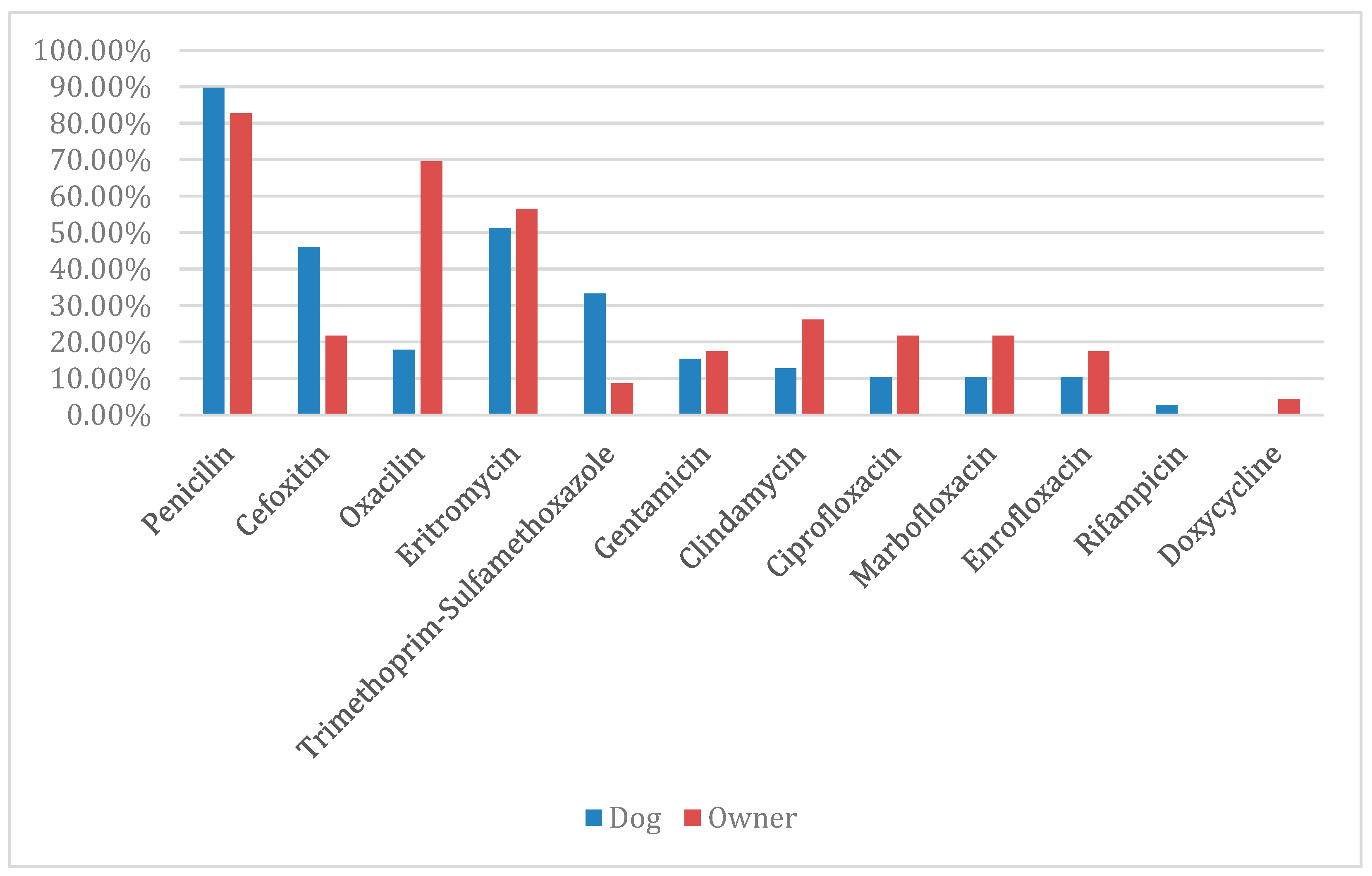
2.3. Antimicrobial Resistance Pattern of MRS and MRMs

In the methicillin-resistant specimens isolated from the dogs, resistance to β -lactams was already expected. The results found were as follows: penicillin with 89.7% (35/39), cefoxitin with 46.1% (18/39) and oxacillin with 17.9% (7/39). Additionally, the highest percentage of resistance observed was to erythromycin (51.3%; 20/39), followed by trimethoprim–sulfamethoxazole (33.3%; 13/39). Considering the other antimicrobials, the following results were obtained: gentamicin (15.4%; 6/39) and clindamycin (12.8%; 5/39), with two isolates resistant to clindamycin due to erythromycin induction, forming a D zone; followed by ciprofloxacin, marbofloxacin, and enrofloxacin (10.3%; 4/39) each, and rifampicin (2.6%; 1/39). There were no resistant specimens to doxycycline.
In the methicillin-resistant specimens isolated from the owners, resistance to β-lactams was also already expected. The results found were as follows: penicillin with 82.6% (19/23), oxacillin with 69.6% (16/23), and cefoxitin with 21.7% (5/23). The other percentages of resistance found were to erythromycin (56.5%; 13/23), followed by clindamycin (26.1%; 6/23), with three isolates being resistant due to erythromycin induction, forming a D zone. Considering the other antimicrobials, the following results were obtained: marbofloxacin and ciprofloxacin (21.7%; 5/23) each, enrofloxacin and gentamicin (17.4%; 4/23) each, sulfamethoxazole–trimethoprim (8.7%; 2/23), and doxycycline (4.4%; 1/23). There were no specimens resistant to rifampicin, as demonstrated in Figure 1.

2.4. Comparative Study Between Dog–Owner Pairs MRS and MRMs

Fourteen pairs (dog–owner) sharing the same bacterial species were identified. Of these 14 pairs, 71% (10) had strains carrying the mecA gene in both hosts. The other four pairs presented the same bacterial species without the presence of the mecA gene, except for only one strain of M. sciuri isolated from a dog that was positive for the mecA gene (Table 3). In the antimicrobial susceptibility test, it can be observed that of the 14 pairs, 50% of them (7/14) presented the same antimicrobial susceptibility pattern. The rest showed different patterns, as shown in Table 3.

3. Discussion

In all 185 strains, the presence of the mecA gene, which confers resistance to β-lactam antimicrobial agents, was investigated. The gene was found in 33.5% of the isolates (62/185). When evaluating the percentage based on the number of dogs and their owners assessed, 43% (24/50) and 52.9% (18/34) carried microorganisms harboring the mecA gene, respectively. This gene can be transferred to other staphylococci occupying the same habitat [21].
Fourteen dog–owner pairs shared the same bacterial species carrying the mecA gene. Furthermore, seven of these pairs also exhibited the same antimicrobial resistance pattern, suggesting that there may have been transmission between the dog and its owner. In a study conducted by Gómez-Sans and collaborators [22], it was observed that nine out of ten dogs investigated were colonized by identical strains (characterized by the MLST technique) to those colonizing their owners, highlighting the sharing of this bacterium. Further analyses, such as the MLST technique, are necessary to confirm the sharing observed in this study.
No MRSA was simultaneously observed among the isolates from the dogs and their owners. This finding is consistent with the data reported by Suepaul and collaborators [23]. This may be because dogs are usually not colonized with S. aureus, although the temporary carriage of human S. aureus strains, especially for those of MRSA, is possible. Previous studies have shown that about 30% of healthy humans are colonized with S. aureus, while a third to half of humans are intermittently colonized, and the remainder are never colonized [23].
A methicillin-resistant S. pseudintermedius (MRSP) sample was found in a dog and its owner. Although S. pseudintermedius is not a common commensal in humans, this bacterium has been isolated from the nostrils of owners of dogs with staphylococcal pyoderma, with zoonotic origin confirmed through molecular testing [24]. This factor highlights the risk of transmitting S. pseudintermedius, including MRSP [25], especially for pet owners, due to the increasingly close contact between the hosts. As an aggravating factor, the antimicrobials used in the treatment of dogs are similar to those used in humans, and in most cases, the use is inappropriate [26]. According to Martins [27], the prevalence of MRSP has significantly increased and was more frequent in animals that had received previous antibiotic therapy.
Of the 12 antimicrobials tested in this study, we found resistance to 11, with strains demonstrating resistance to up to 10 different drugs. We also observed that MRS colonized seven dogs and their respective owners, and they showed identical resistance patterns, similar to the findings by Castro [28]. Only molecular analysis of these samples will allow for the identification of the sharing of the same MRS lineage among these hosts, indicating transmission of the bacterial species.
The canine samples demonstrated resistance to more antimicrobials when compared to human samples, which was also observed by Dotto [29]. This may have occurred because antimicrobial therapies in companion animals are often used without correct identification of the agent or without performing antimicrobial susceptibility tests. Additionally, easy access to veterinary medications increases the population’s inappropriate use of these products.
The increasing bacterial resistance to antimicrobials has become a global public health issue. It is important to emphasize that several studies support the evidence that methicillin resistance can be transmitted between microorganisms of animals and humans [30,31]. The role of companion animals as reservoirs of antimicrobial resistance, exacerbated by the horizontal transmission capability of resistance genes (mecA) among staphylococci species, should be researched in depth. This transmission is facilitated by close physical contact and by the fact that the antimicrobials used in small animal clinics are virtually the same as those used in human medicine. The quantification of this risk is highly problematic, as there are little data available on antimicrobial consumption in small animal clinics and antimicrobial susceptibility, as well as the prevalence of resistance genes among bacterial pathogens of pets [32].
In Brazil, antimicrobial drugs for veterinary use can still be purchased without a prescription. The frequent use of these antimicrobials may contribute to the population’s selective pressures on staphylococci. Methicillin and multidrug resistance are now common in both veterinary and human medicine, leading to findings that may foster future efforts to develop alternative approaches to control the staphylococcal infection burden.
Controlling AMR requires an integrated One Health approach that considers the interconnections between animals, humans, and environmental health. Control strategies, such as the rational use of antibiotics, the development of infection control programs in veterinary hospitals, and the continuing education of veterinarians, are essential to mitigate the spread of antimicrobial resistance [33]. We are aware that the present study still has some limitations, such as the reduced number of samples shared between dogs and their owners, as well as more in-depth analyses of this transmission, including data such as SCCmec type or clonality.

4. Material and Methods

4.1. Study Area, Population, and Samples

Samples were collected in veterinary clinics in Rio de Janeiro from May 2018 to March 2019. Sterile cotton swabs (Copan Diagnostic, Brescia, Italy) were used to collect samples from the nostrils and perineum of 50 dogs and the nostrils of their respective owners (34) during their clinic visits for routine check-ups or vaccinations—veterinarians collected the samples from the humans and animals. The samples were transported to the laboratory, where they were processed.

4.2. Isolation and Identification of Staphylococci and M. sciuri

Each swab was transferred to 3 mL of Tryptone Soy Broth (TSB) (Tryptone Soy Broth, Kasvi®, Pinhais, Italy) and incubated for 24 h at 37 °C. After incubation, the tubes were vortexed, and an entire loop of this culture was then plated on Mannitol Salt Agar (MSA) (Mannitol Salt Agar, Kasvi®, Italy) to isolate presumptive staphylococci and M. sciuri. The MSA plates were incubated for 24 h at 37 °C. After this initial incubation, three sample colonies with bacterial growth compatible with staphylococci and M. sciuri were selected for Gram staining and catalase testing. Gram-positive bacteria and those positive for the catalase test were stocked in a −20 °C freezer. All isolates were identified through Matrix-Assisted Laser Desorption Ionization Time-of-Flight Mass Spectrometry (MALDI-TOF Biotyper, Bruker, Billerica, MA, USA) following the manufacturer’s instructions, described as follows. A thin film of bacterial colony was applied in duplicate onto a 96-well steel plate (Bruker Daltonics, Billerica, MA, USA). Following drying at room temperature, 1 μL of 70% formic acid was applied to each colony and allowed to dry again at room temperature. Subsequently, 1 μL of matrix (a saturated solution of α-cyano-4-hydroxycinnamic acid [HCCA; Bruker Daltonics] in 50% acetonitrile and 2.5% trifluoroacetic acid) was applied to each colony and allowed to dry at room temperature before testing. The spectra were analyzed using a MALDI Biotyper automation control and the Bruker Biotyper 2.0 software and library (version 2.0, 3740 entries; Bruker Daltonics). Only results with scores ≥ 2000 were considered for this study.

4.3. Identification of the mecA Gene

The release of bacterial DNA from staphylococci and M. sciuri was performed using Chelex® (Bio-Rad, Hercules, CA, USA) according to the methodology described by Walsh et al. [34]. Following DNA extraction, the samples were subjected to Polymerase Chain Reaction (PCR) for the detection of the mecA gene using the Promega Kit (Madison, WI, USA, GoTaq® G2 DNA Polymerase) with the following concentrations of reagents: 5× Green Reaction Buffer, 0.2 mM dNTPs, 1.5 mM MgCl2, 10 pmol of forward primer (mecAF (5′TCCAGATTACAACTTCACCAGG3′)), 10 pmol of reverse primer (mecAR (5′CCACTTCATATCTTGTAACG3′)), 2 U of GoTaq, and 100 ng of DNA template in a total reaction volume of 25 μL. Amplification was carried out in a thermocycler (Applied Biosystems, Foster City, CA, USA), as described by Zhang et al. [35]. The reaction products were visualized through electrophoresis in 1% agarose gel in 0.5× TBE buffer (0.05 nM Tris, 1.25 mM EDTA, and 0.05 M boric acid). A 1 Kb molecular size marker (Invitrogen, Waltham, MA, USA) was used. The expected product size was 162 bp. The S. aureus USA100 strain was used as positive control.

4.4. Antimicrobial Susceptibility Testing of MRS and MRM Strains

Samples that carried the mecA gene were analyzed for resistance to other antimicrobial agents using the disk diffusion method (Kirby–Bauer) on Mueller–Hinton agar (Becton, Dickinson and Company BD, Franklin Lakes, NJ, USA), according to the recommendations of the Clinical and Laboratory Standards Institute [36,37]. After bacterial growth on TSA medium at 37 °C for 24 h, a bacterial suspension was prepared in sterile 0.85% saline, achieving a turbidity of 0.5 on the McFarland scale. Subsequently, using a sterile swab, the suspension was inoculated onto a plate containing Mueller–Hinton agar. Twelve antimicrobial discs were used, belonging to eight classes: beta-lactams (cefoxitin 30 µg, oxacillin 1 µg, penicillin G 10 µg), fluoroquinolones (ciprofloxacin 5 µg, enrofloxacin 5 µg, marbofloxacin 5 µg), lincosamide (clindamycin 2 µg), macrolide (erythromycin 15 µg), aminoglycoside (gentamicin 10 µg), rifampicin 5 µg, tetracycline (doxycycline 30 µg), and sulfonamide–trimethoprim (cotrimoxazole 25 µg) (CECON, Vila Sonia, São Paulo, Brazil). The plates were incubated aerobically at 37 °C for 24 h, and the diameters of the zones were measured manually using an Antibiotic Zone Scale-C™ (HiMedia, Thane, India). The S. aureus ATCC 25923 strain was used as positive control.

5. Conclusions

Several species of staphylococci were isolated from dogs and their owners. The most prevalent were S. pseudintermedius in the dogs and S. epidermidis in the owners. MRS and MRMs strains were isolated from both the dogs and their owners. The most prevalent methicillin-resistant strain in the dogs was M. sciuri, while in the owners, it was S. epidermidis. The MRS strains from the dogs exhibited high resistance rates to other antimicrobials such as erythromycin and sulfamethoxazole–trimethoprim. Meanwhile, the MRS strains from the owners showed interesting resistance rates to erythromycin and clindamycin.

Author Contributions

Conceptualization, L.G., I.M.T., and B.P.; methodology, F.C.B., L.G., I.M.T., S.M.F., A.M., P.C.d.C.A.P., T.V.B., and B.P.; validation, S.M.F., A.M., P.C.d.C.A.P., and T.V.B.; formal analysis, F.C.B. and B.P.; investigation, F.C.B., L.G., A.M., P.C.d.C.A.P., and T.V.B.; writing—original draft, F.C.B., I.M.T., S.M.F., and B.P.; writing—review and editing, L.G., I.M.T., and B.P.; project administration, B.P.; funding acquisition, B.P. All authors have read and agreed to the published version of the manuscript.

Funding

This work was supported by Fundação Carlos Chagas Filho de Amparo à Pesquisa do Estado do Rio de Janeiro—FAPERJ (Projeto REDES: E26/211.554/2019; E-26/201.328/2021), Conselho Nacional de Desenvolvimento Científico e Tecnológico—CNPq (406057/2016-8; 443764/2018-2), Bill & Melinda Gates Foundation (Grand Challenges Brazil), and by the Coordenação de Aperfeiçoamento de Pessoal de Nível Superior—Brasil (CAPES) Finance Code 001.

Institutional Review Board Statement

The Animal Use Ethics Committee (CEUA certificate 1008) and Human Ethics Council (CAAE: 14348319.4.0000.5243) approved this study.

Informed Consent Statement

Not applicable.

Data Availability Statement

The data are contained within the article.

Acknowledgments

The authors would like to thank the team of LCGP for all their support.

Conflicts of Interest

The authors declare no conflicts of interest.

References

  1. Prado, R.; Freitas, E.; Júnior, E.V.; Costa, P.; Siqueira, M.; Rossi, D. Staphylococcus spp.: Importantes riscos à saúde pública. Pubvet 2015, 9, 363–368. [Google Scholar] [CrossRef]
  2. List of Prokariotic Names with Standing in Nomenclature—LPSN. Available online: www.ncbi.nlm.nih.gov/Taxonomy/Browser/wwwtax.cgi (accessed on 4 December 2024).
  3. Urbaneja, M.E. Implicações da resistência bacteriana por Staphylococcus spp. na medicina veterinária. Pubvet 2024, 18, e1586. [Google Scholar] [CrossRef]
  4. Murray, P.R.; Rosenthal, K.S.; Pfaller, M.A. Medical Microbiology, 7th ed.; Elsevier: Amsterdam, The Netherlands, 2014. [Google Scholar]
  5. Pereira, E.P.L.; da Cunha, M.d.L.R.d.S. Avaliação da colonização nasal por Staphylococcus spp. resistenteà oxacilina em alunos de enfermagem. J. Bras. Patol. Med. Lab. 2009, 45, 361–369. [Google Scholar] [CrossRef]
  6. Devriese, L.A.; Vancanneyt, M.; Baele, M.; Vaneechoutte, M.; De Graef, E.; Snauwaert, C.; Cleenwerck, I.; Dawyndt, P.; Swings, J.; Decostere, A.; et al. Staphylococcus pseudintermedius sp. nov., a coagulase-positive species from animals. Int. J. Syst. Evol. Microbiol. 2005, 55, 1569–1573. [Google Scholar] [CrossRef]
  7. Howden, B.P.; Giulieri, S.G.; Lung, T.W.F.; Baines, S.L.; Sharkey, L.K.; Lee, J.Y.H.; Hachani, A.; Monk, I.R.; Stinear, T.P. Staphylococcus aureus host interactions and adaptation. Nat. Rev. Microbiol. 2023, 380–395. [Google Scholar] [CrossRef]
  8. dos Santos, A.L.; Santos, D.O.; de Freitas, C.C.; Ferreira, B.L.A.; Afonso, I.F.; Rodrigues, C.R.; Castro, H.C. Staphylococcus aureus: Visitando uma cepa de importância hospitalar. J. Bras. Patol. Med. Lab. 2007, 43, 413–423. [Google Scholar] [CrossRef]
  9. Shoaib, M.; Aqib, A.I.; Muzammil, I.; Majeed, N.; Bhutta, Z.A.; Kulyar, M.F.-E.; Fatima, M.; Zaheer, C.-N.F.; Muneer, A.; Murtaza, M.; et al. MRSA compendium of epidemiology, transmission, pathophysiology, treatment, and prevention within one health framework. Front. Microbiol. 2023, 13, 1067284. [Google Scholar] [CrossRef]
  10. Botoni, L.S.; Filho, N.P.R.; Scherer, C.B.; Braga, L.; Leme, F.O.P.; Bicalho, A.C.P.V. Superficial pyoderma due to meticillin resistant Staphylococcus pseudintermedius (MRSP). Medvep Derm.—J. Contin. Educ. Dermatol. Vet. Allergol. 2014, 3, 270–277. [Google Scholar]
  11. Teixeira, I.M.; Assumpção, Y.D.M.; Paletta, A.C.C.; Aguiar, L.; Guimarães, L.; da Silva, I.T.; Côrtes, M.F.; Botelho, A.M.N.; Jaeger, L.H.; Ferreira, R.F.; et al. Investigation of antimicrobial susceptibility and genetic diversity among Staphylococcus pseudintermedius isolated from dogs in Rio de Janeiro. Sci. Rep. 2023, 13, 20219. [Google Scholar] [CrossRef]
  12. Madhaiyan, M.; Wirth, J.S.; Saravanan, V.S. Phylogenomic analyses of the Staphylococcaceae family suggest the reclassification of five species within the genus Staphylococcus as heterotypic synonyms, the promotion of five subspecies to novel species, the taxonomic reassignment of five Staphylococcus species to Mammaliicoccus gen. Nov., and the formal assignment of Nosocomiicoccus to the family Staphylococcaceae. Int. J. Syst. Evol. Microbiol. 2020, 70, 5926–5936. [Google Scholar] [CrossRef]
  13. Tang, S.; Prem, A.; Tjokrosurjo, J.; Sary, M.; Van Bel, M.A.; Rodrigues-Hoffmann, A.; Kavanagh, M.; Wu, G.; Van Eden, M.E.; Krumbeck, J.A. The canine skin and ear microbiome: A comprehensive survey of pathogens implicated in canine skin and ear infections using a novel next-generation-sequencing-based assay. Veter Microbiol. 2020, 247, 108764. [Google Scholar] [CrossRef] [PubMed]
  14. de Carvalho, A.; Giambiagi-Demarval, M.; Rossi, C.C. Mammaliicoccus sciuri’s Pan-Immune System and the Dynamics of Horizontal Gene Transfer Among Staphylococcaceae: A One-Health CRISPR Tale. J. Microbiol. 2024, 62, 775–784. [Google Scholar] [CrossRef] [PubMed]
  15. Sacramento, A.G.; Fuga, B.; Monte, D.F.; Cardoso, B.; Esposito, F.; Dolabella, S.S.; Barbosa, A.A.; Zanella, R.C.; Cortopassi, S.R.; da Silva, L.C.; et al. Genomic features of mecA-positive methicillin-resistant Mammaliicoccus sciuri causing fatal infections in pets admitted to a veterinary intensive care unit. Microb. Pathog. 2022, 171, 105733. [Google Scholar] [CrossRef] [PubMed]
  16. Lima, D.A. S. pseudintermedius e S. aureus Resistant Isolates from Dogs with Superficial Pyoderma. Master’s Thesis, Federal University of Goiás, Goiás, Brazil, 2021. [Google Scholar]
  17. Dantas, C.C.S.; de Oliveira, A.S.; de Almeida, S.P.; de Oliveira, C.M.S. Importância do Uso Racional de Antimicrobianos em Ambiente Hospitalar. Arquivos 2023, 9, 2366–2374. [Google Scholar] [CrossRef]
  18. Andrade, S.F. Manual de Terapêutica Veterinária, 2nd ed.; Roca: São Paulo, Brazil, 2002. [Google Scholar]
  19. Loureiro, R.J.; Roque, F.; Rodrigues, A.T.; Herdeiro, M.T.; Ramalheira, E. O uso de antibióticos e as resistências bacterianas: Breves notas sobre a sua evolução. Rev. Port. Saúde Pública 2016, 34, 77–84. [Google Scholar] [CrossRef]
  20. Llor, C.; Bjerrum, L. Antimicrobial resistance: Risk associated with antibiotic overuse and initiatives to reduce the problem. Ther. Adv. Drug Saf. 2014, 5, 229–241. [Google Scholar] [CrossRef]
  21. Nemeghaire, S.; Argudín, M.A.; Feßler, A.T.; Hauschild, T.; Schwarz, S.; Butaye, P. The ecological importance of the Staphylococcus sciuri species group as a reservoir for resistance and virulence genes. Veter Microbiol. 2014, 171, 342–356. [Google Scholar] [CrossRef]
  22. Gómez-Sanz, E.; Torres, C.; Lozano, C.; Zarazaga, M. High diversity of Staphylococcus aureus and Staphylococcus pseudintermedius lineages and toxigenic traits in healthy pet-owning household members. Underestimating normal household contact? Comp. Immunol. Microbiol. Infect. Dis. 2012, 36, 83–94. [Google Scholar] [CrossRef]
  23. Suepaul, S.; Georges, K.; Unakal, C.; Boyen, F.; Sookhoo, J.; Ashraph, K.; Yusuf, A.; Butaye, P. Determination of the frequency, species distribution and antimicrobial resistance of staphylococci isolated from dogs and their owners in Trinidad. PLoS ONE 2021, 16, e0254048. [Google Scholar] [CrossRef]
  24. Guimarães, L.; Teixeira, I.M.; da Silva, I.T.; Antunes, M.; Pesset, C.; Fonseca, C.; Santos, A.L.; Côrtes, M.F.; Penna, B. Epidemiologic case investigation on the zoonotic transmission of Methicillin-resistant Staphylococcus pseudintermedius among dogs and their owners. J. Infect. Public Health 2023, 16, 183–189. [Google Scholar] [CrossRef]
  25. Carroll, K.C.; Burnham, C.D.; Westblade, L.F. From canines to humans: Clinical importance of Staphylococcus pseudintermedius. PLoS Pathog. 2021, 17, e1009961. [Google Scholar] [CrossRef] [PubMed]
  26. Frank, L.A.; Kania, S.A.; Kirzeder, E.M.; Eberlein, L.C.; Bemis, D.A. Risk of colonization or gene transfer to owners of dogs with meticillin-resistant Staphylococcus pseudintermedius. Vet. Dermatol. 2009, 20, 496–501. [Google Scholar] [CrossRef] [PubMed]
  27. Matins, C. Staphylococcal Infections in Dogs: Prevalence, Antibacterial Resistance, Risk and Virulence Factors. Ph.D. Thesis, Federal University of Uberlândia, Uberlândia, Brazil, 2020. [Google Scholar]
  28. Castro, E.M. Occurrence and Characterization of Staphylococcus spp. Methicillin-Resistant Isolates from Dogs and Their Human Contacts in Municipalities in Rio de Janeiro. Master’s Thesis, Federal Fluminense University, Niterói, Brazil, 2018. [Google Scholar]
  29. Dotto, E.K. Antimicrobial Resistance Profile of Coagulase-Positive Staphylococcus Isolated from Dogs, Cats and Humans: Meta-Analysis. Bachelor’s Thesis (Residency in Preventive Veterinary Medicine), Federal University of Santa Maria, Santa Maria, Brazil, 2018. [Google Scholar]
  30. Weese, J.S.; van Duijkeren, E. Methicillin-resistant Staphylococcus aureus and Staphylococcus pseudintermedius in veterinary medicine. Veter Microbiol. 2010, 140, 418–429. [Google Scholar] [CrossRef] [PubMed]
  31. Jin, M.; Osman, M.; Green, B.A.; Yang, Y.; Ahuja, A.; Lu, Z.; Cazer, C.L. Evidence for the transmission of antimicrobial resistant bacteria between humans and companion animals: A scoping review. One Health 2023, 17, 100593. [Google Scholar] [CrossRef]
  32. Tunon, G.I.L.; Silva, E.P.; Faierstein, C.C. Isolation of multiresistant staphylococci from canine otitis and its importance for public health. Bepa 2008, 5, 4. [Google Scholar]
  33. Gomes, M.F.A.; Chaves, L.K.M.; Pereira, G.E.S.; Albuquerque, D.L.; Miranda, M.L.A.F.; Zorzi, L.R.M.; Waterloo, M.M.L. Emergence of antimicrobial resistance in pets: A narrative review of the literature and its implications for Public Health. Res. Soc. Dev. 2024, 13, e45131047090. [Google Scholar] [CrossRef]
  34. Walsh, P.S.; Metzger, D.A.; Higuvhi, R. Chelex 100 as a Medium for Simple Extraction of DNA for PCR-Based Typing from Forensic Material. BioTechniques 1991, 10, 506–513. [Google Scholar] [CrossRef] [PubMed]
  35. Zhang, K.; McClure, J.-A.; Elsayed, S.; Louie, T.; Conly, J.M. Novel Multiplex PCR Assay for Characterization and Concomitant Subtyping of Staphylococcal Cassette Chromosome mec Types I to V in Methicillin-Resistant Staphylococcus aureus. J. Clin. Microbiol. 2005, 43, 5026–5033. [Google Scholar] [CrossRef]
  36. Clinical & Laboratory Standards Institute (CLSI). Performance Standards for Antimicrobial Susceptibility Testing; Supplement Document, M100-ed 32; Clinical & Laboratory Standards Institute (CLSI): Malvern, PA, USA, 2022. [Google Scholar]
  37. Clinical & Laboratory Standards Institute (CLSI). Performance Standards for Antimicrobial Disk and Dilution Susceptibility Tests for Bacteria Isolated from Animals; Second Informational Supplement. VET01-S02; Clinical & Laboratory Standards Institute (CLSI): Malvern, PA, USA, 2018; Volume 33. [Google Scholar]
Figure 1. Percentage of antimicrobial resistance pattern of MRS and MRMs.
Antibiotics 14 00409 g001
Table 1. Distribution of staphylococci and M. sciuri isolated from dogs and their owners (N: number).
SpeciesIsolated from
Dogs % (N)
Isolated from Owners % (N)TOTAL
% (N)
NostrilsPerineumNostrilsGeneral
S. pseudintermedius22.2 (16)27.3 (15)6.9 (4)18.9 (35)
M. sciuri19.4 (14)20 (11)8.6 (5)16.2 (30)
S. epidermidis9.7 (7)9.1 (5)50 (29)22.1 (41)
S. urealyticus6.9 (5)10.9 (6)3.5 (2)7 (13)
S. saprophyticus11.1 (8)3.6 (2)8.6 (5)8.1 (15)
S. aureus8.3 (6)5.4 (3)13.8 (8)9.1 (17)
S. hominis6.9 (5)7.3 (4)3.5 (2)5.9 (11)
S. schleiferi1.4 (1)9.1 (5)03.2 (6)
S. warneri5.6 (4)1.8 (1)1.7 (1)3.2 (6)
S. haemolyticus4.2 (3)1.8 (1)1.7 (1)2.7 (5)
S. arlettae2.8 (2)1.8 (1)01.6 (3)
S. xylosus1.4 (1)1.8 (1)01 (2)
S. intermedius001.7 (1)0.5 (1)
Total % (N)56.7 (72)43.3 (55)100 (58)100 (185)
Table 2. Prevalence of mecA gene.
SpeciesmecA Gene (%)
S. epidermidis33.9
S, saprophyticus16.1
M. sciuri14.5
S. hominis9.7
S. urealyticus6.4
S. haemolyticus6.4
S. aureus4.8
S. pseudintermedius3.2
S. warneri3.2
S. schleiferi1.6
Table 3. Demonstration of the 14 dog–owner pairs.
PairsSample IdentificationBacterial SpeciesmecA GeneSiteCFOPENOXAGENCIPENOMBFSTERICLIRIFDOXMultidrug-Resistant
17 (D)S. warneri+N3418----2830303222RR42+36*
333 (O)S. warneri+N2416----2428323026RR3814*
233.1 (D)S. epidermidis+N----1812303434302212304224
340 (O)S. epidermidis+N----3412243230302010284030
383.2 (D)S. epidermidis+P----1414303436342828284230
353 (O)S. epidermidis+N----1014243632342630304234
4107.1 (D)S. saprophyticus+P2614----303234303630263834
358.2 (O)S. saprophyticus+N1810----323236303430264038
5316.2 (D)S. epidermidis+N----36143036363624343442+30
410 (O)S. epidermidis+N----3618263436362432324230
699 (D)S. hominis+N28R----8RR12R12364236*
347 (O)S. hominis+N22R----10RR10RR284232*
7144 (D)S. pseudintermedius+N----101612RR8R10184236*
367 (O)S. pseudintermedius+N----1024RRRRRR163634*
823 (D)S. haemolyticus+N10R----RRRRRRR42+28*
338 (O)S. haemolyticus+N1212----R26243024R204022*
9216 (D)S. epidermidis+N----1612163036342428301222
387 (O)S. epidermidis+N----1614263030282010284232
10223.2 (D)S. saprophyticus+P1610----283028263028303834
388 (O)S. saprophyticus+N2012----R32343228R24 (D)4216*
11173 (D)S. aureus-P3420----14263026326263634
365 (O)S. aureus-N3618----12283026306263630*
12319 (D)S. aureus-N3018----22282626306263434
412 (O)S. aureus-N3416----2222282830R26 (D)3230*
13420 (D)S. aureus-N3214----1028242830626 (D)3830*
416 (O)S. aureus-N3618----2426303032R30 (D)3830*
14126 (D)M. sciuri-P2628----262626262626183230
130 (D)M. sciuri+N2628----262626242626203230
356 (O)M. sciuri-N2630----242424242626183028
(D—dog; O—owner; N—nostril; P—perineum; CFO—cefoxitin; PEN—penicillin; OXA—oxacillin; GEN—gentamicin; CIP—ciprofloxacin; ENO—enrofloxacin; MBF—marbofloxacin; ST—sulfamethoxazole–trimethoprim; ERI—erythromycin; CLI—clindamycin; RIF—rifampicin; DOX—doxycycline; “D” in red—isolates that showed D zone; *—multidrug-resistant samples).
Disclaimer/Publisher’s Note: The statements, opinions and data contained in all publications are solely those of the individual author(s) and contributor(s) and not of MDPI and/or the editor(s). MDPI and/or the editor(s) disclaim responsibility for any injury to people or property resulting from any ideas, methods, instructions or products referred to in the content.

