Article

An Efficient Disinfectant, Composite Material {SLS@[Zn3(CitH)2]} as Ingredient for Development of Sterilized and Non Infectious Contact Lens

1
Inorganic Chemistry laboratory, Department of Chemistry, University of Ioannina, 45110 Ioannina, Greece
2
Medical Physics Laboratory, Medical School, University of Ioannina, 45110 Ioannina, Greece
3
Department of Physics, University of Ioannina, 45110 Ioannina, Greece
4
Laboratory of Analytical Chemistry, Department of Chemistry, University of Ioannina, 45110 Ioannina, Greece
5
Institute of Nanoscience and Nanotechnology, NCSR “Demokritos”, 15341 Athens, Greece
6
Institute of Biology, Medicinal Chemistry and Biotechnology, National Hellenic Research Foundation, 11635 Athens, Greece
7
Organic Chemistry Laboratory, Department of Chemistry, National and Kapodistrian University of Athens, 15571 Athens, Greece
8
Cancer Biobank Center, University of Ioannina, 45110 Ioannina, Greece
*
Authors to whom correspondence should be addressed.
Antibiotics 2019, 8(4), 213; https://doi.org/10.3390/antibiotics8040213
Submission received: 28 September 2019 / Revised: 30 October 2019 / Accepted: 5 November 2019 / Published: 7 November 2019

Abstract

:
The [Zn3(CitH)2] (1) (CitH4= citric acid), was dispersed in sodium lauryl sulphate (SLS) to form the micelle of SLS@[Zn3(CitH)2] (2). This material 2 was incorporated in hydrogel made by hydroxyethyl-methacrylate (HEMA), an ingredient of contact lenses, toward the formation of pHEMA@(SLS@[Zn3(CitH)2]) (3). Samples of 1 and 2 were characterized by UV-Vis, 1H-NMR, FT-IR, FT-Raman, single crystal X-ray crystallography, X-ray fluorescence analysis, atomic absorption and TG/DTA/DSC. The antibacterial activity of 13 as well as of SLS against Gram-positive (Staphylococcus epidermidis (St. epidermidis) and Staphylococcus aureus (St. aureus)) and Gram-negative (Pseudomonas aeruginosa (PAO1), and Escherichia coli (E. coli)) bacteria was evaluated by the means of minimum inhibitory concentration (MIC), minimum bactericidal concentration (MBC) and inhibitory zone (IZ). 2 showed 10 to 20-fold higher activity than 1 against the bacteria tested. Moreover the 3 decreases the abundance of Gram-positive microbes up to 30% (St. aureus) and up to 20% (PAO1) the Gram-negative ones. The noteworthy antimicrobial activity of the obtained composite 3 suggests an effective antimicrobial additive for infection-free contact lenses.

1. Introduction

Microbial keratitis often leads to blindness, unless if it is immediately treated. Ocular infection is either caused by misuse contact lens or a wide variety of pathogens [1,2,3]. However, contact lenses have become a major etiology for microbial keratitis, nowadays [4,5,6,7]. Therefore, the development of contact lenses built by long term or permanent, non-infected materials is a research, technological and financial issue challenge. The soft contact lenses are generally made by the hydrogel, poly (2-hydroxyethyl methacrylate) (pHEMA) and they have already been investigated for ocular delivery of several drugs [2,8,9].
Gram-positive bacteria are the major contributor of bacterial ocular infections, with the most commonly reported belong to the genus Staphylococci such as Staphylococcus epidermidis (St. epidermidis) and Staphylococcus aureus (St. aureus) [3]. Among Gram-negatives, on the other hand, frequent isolate from ocular infections include Pseudomonas aeruginosa (PAO1) and Escherichia coli (E. coli) [3].
Zinc is an essential element for the microorganisms since it is involved in vital cellular reactions even at its low endogenous concentrations. The antimicrobial properties of Zn2+ and its complexes have been known since long time now [10,11,12,13,14].
SLS is an anionic surfactant and detergent with microbicidal activity, due to its ability to induce protein denaturation [15]. It is use in the personal care products industry (e.g., toothpastes and shampoos) [15,16]. It has been classified as readily biodegradable, with no any concern in respect to human health by United Nations Environment Programme (UNEP) [17]. SLS forms micelles by encapsulating bioactive materials adjusting their hydrophilicity [18]. This makes possible the loading non water soluble antimicrobial agents to be incorporated in hydrogels, acting, by thus, as a vehicle [18].
In the course of our studies on the development of new antimicrobial materials for medical devices [11,19,20,21], the slightly water soluble [Zn3(CitH)2] (1) which contains CitH4 (C6H8O7) was encapsulated in a micelle of SLS with the aim to enhance its dispersion inside a hydrogel. The compound and its composite SLS@[Zn3(CitH)2] (2) were characterized by their physical properties and spectral data. Either in solid state or in solution the concentration of 1 in the composite material is evaluated. This is used for the determination of MIC, MBC, IZ, biofilm elimination concentration (BEC) and the % cells viability upon treated with 3, which is impossible otherwise to be determined. The antimicrobial activities of 2 and its components SLS and 1 were evaluated against the Gram-positive or negative microbes. The influence of 2 against the formation of biofilm of St. aureus was also evaluated. The composite 2 was incorporated in hydroxyethyl methacrylate (HEMA), an ingredient of contact lenses to form pHEMA@(SLS@[Zn3(CitH)2]) (3) which was evaluated for its antibacterial efficiency. Although zinc citrate in combination with SLS has been already studied for the inhibition of plague [22], here we described the extensive characterization of the composite material derived by the combination of [Zn3(CitH)2] (1) with SLS for the first time. Moreover, this work mainly aims to the development of sterilized and non infectious contact lens, where the composite SLS@[Zn3(CitH)2] (2)is an ingredient by its dispersion into pHEMA.

2. Results

2.1. General Aspects

Compound 1 was synthesized as follows: trisodium citrate reacted with CitH4 in 1:2 molar ratio, in 2 mL dd-water (Scheme 1). Disodium citrate solution (2 mmol) was added to a clear solution of Zn(NO3)2 (3 mmol) in dd water (Scheme 1) under stirring for 10 min.
Crystals of 1 were grown by slow evaporation of the solution. The formula of 1 was initially determined by spectroscopic methods and its molecular structure was confirmed by single crystal X-ray diffraction analysis. The structure of 1 was confirmed as previously reported by Che et al. [23] (1: a = 6.144(4) Å, b = 14.577(4) Å, c = 9.585(4) Å, β = 102.50(2)°, V = 838(1) Å3; the reported: monoclinic, space group P21/c, a = 6.1552(13) Å, b = 14.546(3) Å, c = 9.581(4) Å, β = 102.66(2)°, V = 836.9(4) Å3 [23]). Compound, 1 was prepared previously, under hydrothermal conditions which include heating at 150 °C for 20 h [23]. However, this is synthesized here, at ambient condition upon few minutes stirring of its components. The variation in preparation conditions and the absence of detailed spectroscopic studies of 1 led us to collect these data for its structure evaluation. The crystals of 1 are air stable when they are stored in darkness at room temperature. The 1 is poorly soluble in H2O, and it is insoluble in organic solvents.

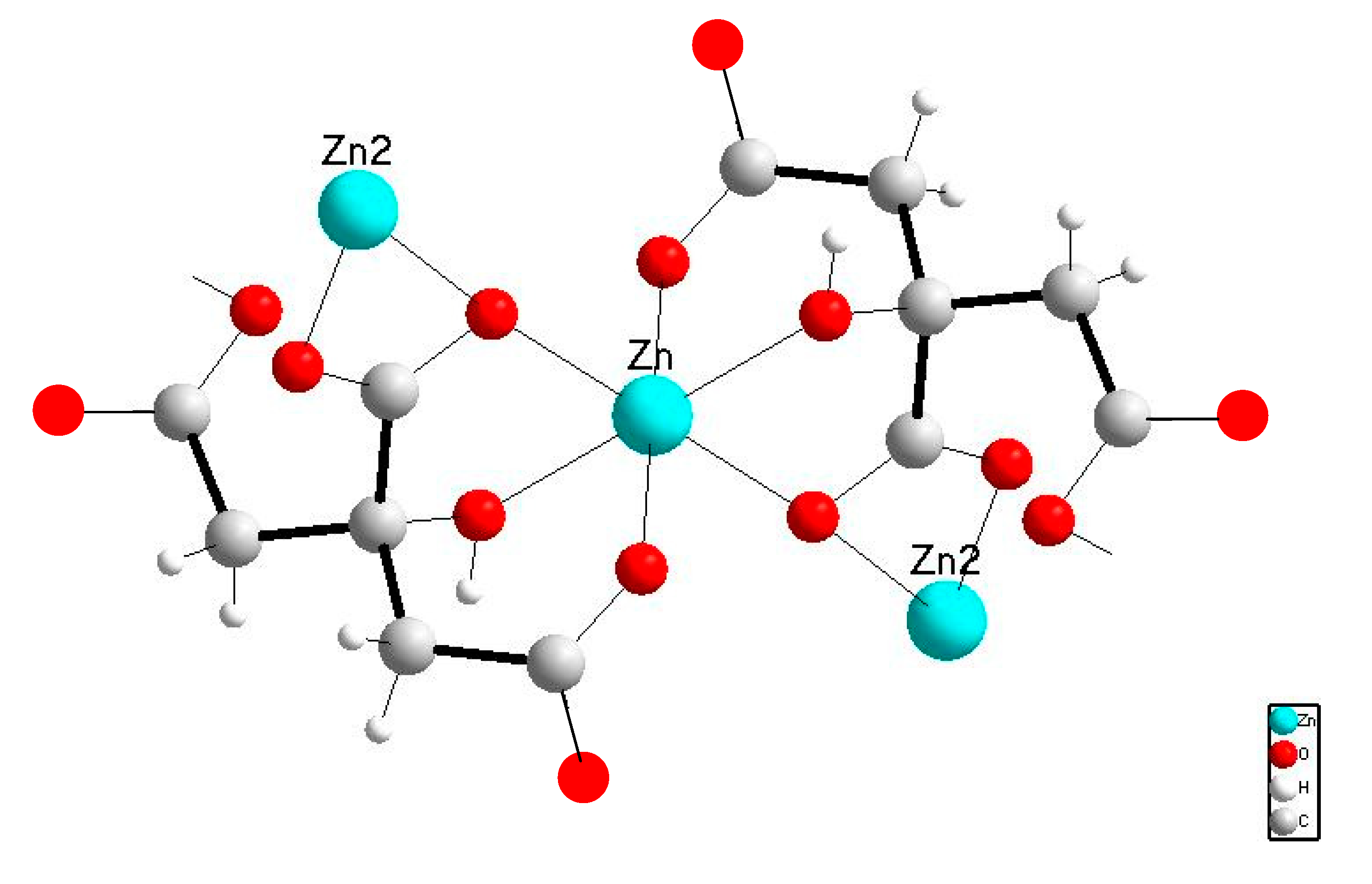
2.2. Characterization of 1

2.2.1. Solid State Studies

Crystal and molecular structure of1: The crystal and molecular structure of 1 is known [23], however the refinement of its X-ray diffraction data was proceeded for the verification of the product. The formula of the building block of the entire compound 1 is shown in Figure 1.
Two different environments are identified around the zinc atoms in this structural unit. One is bonded to the citrate ligand via hydroxyl group, the central carboxyl group and one of the terminal carboxyl groups of the ligand in a distorted octahedral arrangement. In the second one, the zinc ion is coordinated to a water oxygen atom as well as to six carboxyl oxygen atoms of three carboxyl groups forming seven-coordinated metal center.
Vibrational spectroscopy: The FT-IR spectrum of 1 shows vibration bands at 1400 cm−1 and at 1557 cm−1, which are assigned to symmetric (vs) and asymmetric (vas) bands of (COO) respectively (Figure S1). The corresponding vibration bands of the trisodium citrate (CitHNa3) are observed at 1394 cm−1 and at 1560 cm−1, respectively. The difference in Δv[vas(COO-)-vs(COO-)] values of CitHNa3 (166 cm−1) and 1 (157 cm−1) suggests the coordination of the ligand in to the metal [24].

2.2.2. Solution Studies

Stability studies: The stability of 1 in water solution was tested by UV-Vis and by 1H-NMR spectroscopies. UV-Vis spectra were recorded initially and for a period of 24 h. No differences were observed between the initial UV-Vis spectrum and the corresponding ones after 24 h suggesting the stability of 1 in solution for 24 h, at least (Figure S2).Moreover the double signals of methylene protons (Hb and Hb’) of CitH4 are assigned at 2.99–2.95 and 2.81–2.77 ppm (Figure 2) and they are shifted in the case of 1 at 2.83–2.79 and 2.67–2.63 ppm, respectively, indicating the coordination of the metal to the ligand. The assignment of the 1H-NMR spectrum of 1 is based on its crystal structure data suggesting the retention of the structure in solution.
On the other hand, despite of the already examples on the effect of pH on the stability and biological activities of Zn(II) complexes [13,14], no such study was performed in case of 1, since our work aims in the formation of a water soluble micelle of 1 which will be used as an ingredient in the hydrogel 3.

2.3. Preparation and Characterization of Micelle

Determination of critical micelle concentration (CMC): Due to the low solubility of the 1 in aqueous media, the micelle 2 is formed, using the SLS as surfactant. In order to assert the formation of the 2, the determination of the CMC was performed via potentiometric titration at 36 °C [25] and the graph of conductivity values vs [SLS]/[1] is drawn (Figure S3). Above the CMC value, surfactant molecules self-agglomerate to form the micelles [26]. Upon the formation of the micelles, a turning point (change of slope) is observed in the graph [26]. The CMC in case of 2, was determined as follows: a solution of SLS (2 × 10⁻² M) in ddH2O was added to 20 mL ddH2O solution of 1 (8.9 × 10⁻⁴ M) in portions of 1 mL, while the conductivity values were being recorded subsequently. The CMC value is obtained at [SLS]:[1]molar ratio of 8.7 (Figure S3). Thus, by mixing its components in a molar ratio [SLS]:[1] = 8.7 the composite 2, is formed. This ratio corresponds to a concentration of 5.7 mM of SLS. The corresponding concentration for the formation of micelle of free SLS is 8.3 mM. This is expected since upon the formation of micelles of drug with surfactants thereby decrease the CMC of surfactant [27].

2.3.1. Solid State Studies

Vibrational Spectroscopy

FT-IR: The IR spectra of the 1, SLS and 2 were recorded (Figure S4). The vibration band at 1557 cm−1 in the spectrum of 1 is attributed to νasym of the COO− of the CitH4 and it is absent from the corresponding spectrum of SLS. The presence of this band in the spectrum of 2 suggests the encapsulation of 1 in SLS.
FT-Raman: Raman spectra of 1, 2, and SLS are shown in Figure 3. The spectrum of SLS show intense Raman bands related to the ν(C–C) stretching vibration in the region between 1000 and 1150 cm−1, a strong peak at 1297 cm−1 related to ν(SO4) stretching and at 1437 cm−1 due to δ(CH2) deformations. The strong bands at 2848 and 2881 cm−1 are attributed to the α-CH2 scissoring and (CH2)n symmetric stretch.
In the spectrum of compound 1, the antisymmetric stretching vibration of the carboxylate groups vas(COO−) yields the band at 1604 cm−1. The corresponding symmetric stretching vs(COO−) generates the intense Raman line at 1407 cm−1. These peaks are typically used as guidelines to determine the coordination mode of the ligand. Together with the absence of a band at ~1735 cm−1 assigned to the free carboxylic acid group, the asymmetric and symmetric COO− stretching are characteristic for the ligand coordination scheme. The strong Raman band at 971 cm−1 is assigned to ν(C-C) mode. At 671 cm−1, the δ(COO−) deformational mode is shown. Metal–ligand (Zn–O) vibrations appear as an intense Raman band at 472 cm−1 while vibrations related to ν(CH2) and ν(C–H) modes are visible as strong Raman lines at 2930, 2970, and 2983 cm−1. The majority of the remaining bands cannot be assigned unambiguously. However, the presence of the COO−, and the C–H stretching bands in the spectrum of 2 shifted to 688, and 1598 cm−1, and 2930 cm−1 respectively, confirms the encapsulation of 1 in SLS.

Thermo Gravimetric Analysis of the Composite 2

Differential scanning calorimetry (DSC): Encapsulation of 1 in SLS aims in the adjustment of its solubility in polar or non-polar solvents and through this, in the control of its releasing in the cytoplasm or other intracellular organelles.
To confirm whether 1 interacts with SLS in the solid state to give a mixture, DSC studies were carried out. DSC thermodiagrams of SLS and 2 are shown in Figure 4. The broad endothermic peak between 70–120 °C may be attributed to the boiling of the water in the SLC or in compounds 1 and 2. Two sharp endothermic transitions at 187.5 and 206.4 °C are observed in the DSC diagram of SLS, while only one at 182.5 °C for 2. The melting point of SLS is 204–207 °C, while the corresponding one of 1 is above 240 °C. The absence of any transition at 205–207 °C in the DSC diagram of the composite 2 suggests the formation of a composite instead of a mixture.
Thermal Decomposition: TG/DTA analysis was performed under nitrogen. The composite 2 decomposes with four exothermic steps at 36.2, 92.5 °C due to the elimination of the solvents and at 198.5 and 521.0 °C (Figure S5A) with total mass loss of 64.06%. The SLS decomposes in two exothermic steps that occur at 213.4 and 424.2 °C (Figure S5B) with total mass loss of 70.80%.
X-ray fluorescence spectroscopy: The XRF spectrum of 2 confirms the presence of Zn in the 2 which is indicative of the encapsulated 1 into the micelle (Figure 5).
The content of micelle 2 in zinc was determined by XRF spectroscopy and was 6.4± 0.8 % wt, which corresponds to dispersion of 1 in the SLS by 18.9% wt. This leads to a [SLS]:[1] molar ratio equal to 8.6 which is in agreement to the value obtained for CMC (8.7).

2.3.2. Solution Studies

Atomic Absorption: In order to ascertain the dispersion of the 1 in its composite 2, graphite furnace atomic absorption spectroscopy (GFAAS) was employed. The mass content of the zinc in the micelle 2 was determined to be 25.06% w/w which leads to a molar ratio [SLS]:[1]= 6.0. The differences in the molar ratios resulted for the contents of the composite, depending to the method used (CMC: 8.7, X-ray fluorescence spectroscopy: 8.6, atomic absorption: 6.0) should be attributed to the physical state in which the measurements were carried out (solid state for CMC and X-ray fluorescence spectroscopy; solution state for the Atomic absorption) and to the accuracy of them.
1H NMR studies: The 1H NMR spectra of 2 and SLS are shown in Figure 2. The multiple resonance signals at 0.83–0.80 ppm are assigned to the protons of the (-CH3)ω group of the SLS [28], while the multiple signals at 1.28–1.23 ppm and 1.62–1.58 ppm are attributed to its (-CH2-)9 and (CH2)β protons. The multiple signals at 3.98–3.94 ppm are assigned to the protons of the (CH2)α group of the SLS. It is noteworthy to mention that the two double signals at 2.69–2.66 and 2.52–2.49 ppm which are assigned to the methylene protons of the CitH4 [28], underline a strong upfield shift towards the corresponding signals observed in the free ligand (2.81–2.77 ppm) and in complex 1(at 2.83–2.79 and 2.67–2.63 ppm). These shifts are indicative of the coordination of the 1 to SLS confirming the formation of the composite instead of a mixture in case of 2 as evidenced by DSC studies.
It is noteworthy to mansion, here, that species distribution for Zn2+—citrate within various pH values involve the identification of the [Zn(cit)], [Zn(cit)H], [Zn(cit)2]4− and [Zn2(cit),H−2]4− in water, which is suggesting pH dependence [29]. However, DSC studies demonstrated the formation of the composite material 2 only, and therefore any shifts in the NMR spectra, in single pH value, can be assigned to the interaction between 1 and SLS in the composite material 2.
Drug loading in pHEMA: pHEMA cross-linked with ethylene glycol dimethacrylate (EGDMA), is the basis of many types of daily wear soft contact lenses [9]. In order to evaluate the sterilizing efficiency of contact lenses which contain antimicrobial agents, 2 was dispersed in pHEMA to form compound 3. The content of the hydrogel in 1 is 2 mM (given that its content in the micelle was found 25.1%). Discs, of 10 mm diameter were subsequently cut, cleaned from monomers and stored either in sterilized NaCl 0.9% w/w solution or they dried at 50 °C.

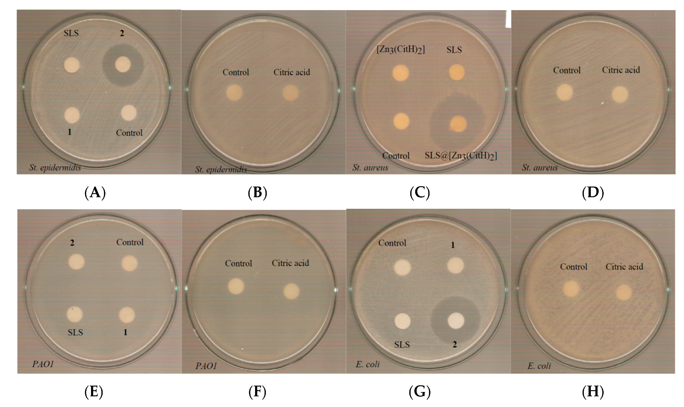
2.4. Biological Studies

Antibacterial Effect of 1-2, SLS andCitH4 on the growth of microbial strains: The antimicrobial potency of 12, SLS, and CitH4, against Gram-positive (St. epidermidis and St. aureus) and Gram-negative (PAO1 and E. coli) is evaluated by the mean of minimum inhibitory concentration (MIC) [30]. MIC is defined as the lowest concentration needed for the inhibition of the bacterial growth [30]. The MICs values of 1, 2, and SLS against microbes studied here are summarized in Table 1 (Figures S6–S9). The CitH4 exhibits no activity against either Gram-positive or negative bacteria (Figures S6–S9). Thus, the composite 2 exhibits up to 21-fold higher antibacterial potency than free 1 and 5-fold stronger than free SLS, in the case of St. aureus. However, in the case of Gram-negative bacteria the composite exhibits either similar (PAO1) of 2-fold higher (E. coli) activity than free SLS, suggesting its sensitivity towards Gram-positive bacteria. Moreover, the encapsulation of the non-active 1, into the SLS, creates a composite material with significant strong activity against Gram-positive bacteria (St. epidermidis and St. aureus) (Table 1).
Minimum bactericidal concentration testing: The lowest concentration of an antibacterial agent that can eliminate the 99.9% of the bacterial inoculum is known as minimum bactericidal concentration (MBC) [31]. The MBC values were: 201.7 μΜ(1), 13.4 μΜ (2) and 100 μΜ (SLS) against St. epidermidis (Figure S10), Table 1), while the corresponding MBC values against St. aureus are: >250 μΜ (1) 12 μΜ (2) and 60 μΜ (SLS) respectively (Figure S11) (Table 1). The MBC values of CitH4 are higher than 250 μΜ against St. epidermidis and St. aureus. The MBC values of 1–2, SLS, and CitH4 are higher than 250 μΜ against PAO1 and E. coli (Figures S12 and S13) (Table 1).
Moreover, the MBC/MIC ratios of 12 and SLS against St. epidermidis and the corresponding ones of 2 and SLS against St aureus are lower to 2 (Table 1). When MBC/MIC value is less than 2, the compound is classified into bactericidal one, indicating that it kills 99.9% of the microorganisms, while if MBC/MIC ≥ 4 then it is bacteriostatic and it inhibits but not kill the organism [32]. Therefore the agents 12 and SLS are classified in bactericidal ones against Gram-positive bacteria.
Determination of the inhibition zone (IZ) through agar disk-diffusion method: The agar disk-diffusion method was used in order to survey the sensitivity of the microorganism to the antibacterial agent studied here [33]. The diameter inhibition zones of bacterial growth caused by of 1-2, SLS, and CitH4 at concentration of 1 mM against St. epidermidis, St. aureus, PAO1, and E. coli were measured after 20 h (Table 1, Figure 6). The corresponding ones caused by 2 are 23.0 mm, 30.5 mm, and 26.5 mm against St. epidermidis, St. aureus, and E. coli respectively. No inhibition zone was observed upon treatment of PAO1 with the composite material. Moreover, no inhibition zone was formed for of 1, SLS, and CitH4 against all tested bacteria. Therefore, the presence of SLS enhances the antibacterial properties of 1 upon the formation of the composite, in agreement with the MIC experiment. The microbe strains may classified in three categories according to the size of IZ, caused by an antimicrobial agent in their agar dilution culture: (i) strains where the agent causes IZ ≥17 mm are susceptible, (ii) those where the agent creates IZ between 13 to 16 mm (13 ≤ IZ ≤ 16 mm) are intermediate, while (iii) those where the agent causes IZ ≤ 12 mm, the microbes are considered as resistant strains [34]. Therefore, the strains of St. epidermidis, St. aureus, and E. coli respond susceptible against 2 [34].
Effects on biofilm formation by 2: The adhesion of bacterial cells to surfaces is the initial stage of the formation of biofilm. Thus, bacterial colonies are attached in a surface and they are protected in a polysaccharides matrix. It is estimated that 80% of all clinical infections are biofilm related. Especially, the ophthalmic biofilm infections are difficult to treat. The necessity of the surgical removal of the infected tissue which is then following in the untreated cases contains high risk for corneal blindness. The biofilm elimination can be achieved by applying metallodrugs or surfactant molecules [35].
The effect of 2 against biofilm formation of St. aureus, was studied by the biofilm elimination concentration (ΒΕC) using crystal violet assay [9]. The ΒΕC is defined as the concentration required to achieve at least a 99.9% reduction in the viability of biofilm bacteria. The composite was able to inhibit the biofilm formation, reducing the 100% of biomass at 700 μΜ, against St. aureus (Figure S14). The BEC value of ciprofloxacin, a known antibiotic for the bacterial keratitis is 897 μΜ for the St. aureus [9]. Thus, the composite is 1.3 times more efficient against biofilm than the commercially available antibiotic ciprofloxaxin.
Antimicrobial activity of3: Since the composite exhibits antimicrobial activity against the tested strains, this prompts us to load it into pHEMA for the development of new non-infectious contact lens (Figure 7). The 3 and pHEMA discs were placed in tests tubes which contain 5 × 10⁵ cfu/mL of St. epidermidis, St. aureus, PAO1, and E. coli microbes.
The calculated bacterial % viability of St. epidermidis, St. aureus, PAO1, and E. coli upon their incubation with 3 discs for 20 h is 72.5±22.2%, 71.0±18.0%, 81.2±6.5%, and 93.5±7.7% respectively (Figure 8). On the contrary, no influence in the bacterial viability was observed against these bacterial strains, upon their treatment by discs of pure pHEMA (Figure 8). Moreover, the 3 is more active against positive than negative bacterial strains. This is in agreement with the MIC values found for the 2.
Discs of pHEMA or 3 with diameter 10 mm, were placed to petri agar dishes and the IZs were determined. The inhibition zones of 3 against St. epidermidis and St. aureus, were 14.0 mm and 17.0 mm respectively, while no inhibition zones were developed against PAO1 and E. coli (Table 1, Figure 9). Moreover, no inhibition zones were developed when pHEMA was used against all tested strains (Figure 9). Given that the IZs, which were developed when paper discs instead of pHEMA ones, were loaded by 2, were significantly greater, a low releasing of 2 from pHEMA, can be concluded.

3. Conclusions

This work aims in the development of sterilized and noninfectious contact lens. The encapsulation of 1 in SLS towards 2 was attempted in order to increase its solubility in polar solvents particularly into water. The composite material 2 is proven to be an efficient disinfectant. Thus, a stronger activity towards Gram-positive than negative bacteria is exhibited by the composite 2, which is up to 21-fold higher than free 1 and 5-fold stronger than free SLS, in the case of St. aureus. The 2, behaves as bactericidal material, while the Gram-positive bacteria as susceptible against it. Also, the 2, is 1.3 times more efficient against biofilm than the commercially available antibiotic ciprofloxacin, which is used in ocular infections. Material 2 is loaded into pHEMA for the development of the new non-infectious contact lens ingredient 3. The calculated % bacterial elimination of St. epidermidis, St. aureus, PAO1 and E. coli bacteria, when they are incubating with 3 discs, is 27.5%, 29.0%, 18.8% and 6.5% respectively. In conclusion 3is an effective candidate towards the development of new non-infectious contact lens.

4. Materials and Methods

Materials and instruments: All solvents used were of reagent grade. Tryptone and soy peptone were purchased from Biolife. Agar was purchased from Sigma-Aldrich. Sodium cloride, D(+)-glucose, di potassium hydrogen phosphate trihydrate were purchased from Merck. Citric acid, trisodium citrate, sodium lauryl sulfate, and zinc nitrate (Sigma-Aldrich, Merck, Darmstadt, Germany) were used without further purification. Dimethylsulfoxide (DMSO) and boric acid were from Riedel-de Haen. Melting points were measured in open tubes with a Stuart Scientific apparatus and are uncorrected. FT-IR spectra in the region of 4000–370 cm−1 were obtained from KBr discs, with a Perkin-Elmer Spectrum GX FT-IR spectrophotometer. Raman spectra were obtained with 785 nm solid state excitation laser coupled to a focusing fiber optic probe with output power of approximately 90 mW, 5 s accumulation time, in the 400–3150 cm−1 spectral range. Stokes photons were detected with a 2048 pixel thermoelectrically cooled CCD. The 1H-NMR spectra were recorded on a Bruker AC 400 MHz FT-NMR instrument in D2O solution. A UV-1600 PC series spectrophotometer of VWR was used to obtain electronic absorption spectra. XRF measurement was carried out using an Am-241 radio isotopic source (exciting radiation 59.5 keV). Conductivity measurements were performed at 36 °C in H2O solutions with a WTF LF-91 conductivity meter.
Synthesis and crystallization of 1: A clear solution of 0.5 mmol CitH4 mono-hydrate (0.105 g) and 1 mmol of trisodium citrate di-hydrate (0.294 g) were stirred in 2 mL of distilled water. Next, 3 mmol Zn(NO3)2 6 H2O (0.892 g) were added in the solution and were stirred. White crystals of 1 suitable for X-ray analysis were grown from slow evaporation of the solution after 2 days.
1: White crystals, MW= 574.258, yield= 0.240 g; melting point: >240 °C. Elemental analysis found:C, 25.35; H, 1.72%; Calc. for C12H10O14Zn3, 25.02; H, 1.75%; IR (cm−1), (KBr) of 1: 1559vs (νasym(-COO-)), 1541s, 1399vs (νsym(-COO-)), 1258s (ν(C-OH)), 1139w, 1083s(ν(C-OH)), 1070s (ν(C-OH)), 934s (νstretching(C-C)), 903s (νstretching(C-C)), 861s (νdeformation(COO)); 1H NMR(ppm) in D2O:2.67–2.63 and 2.83–2.79 (Hb, Hb’)
X-ray Structure Determination: Data collection was performed with a Rigaku R-AXIS SPIDER Image Plate diffractometer, using graphite-monochromated Mo-Ka(λ= 0.71073 Å) radiation at 160 K. Data collection (ω-scans) and processing (cell refinement, data reduction and Empirical absorption correction) were performed using the CrystalClear program package [36].
Micellar Synthesis of 2: 0.051 g (0.09 mmol) of 1 and 0.164 g (0.56 mmol) of SLS were dissolved in 100 mL of distilled H2O and stirred to clearness at 36 °C. The clear solution was then allowed for solvent evaporation within few days. The micelles were collected as white aggregates. IR (cm−1), (KBr) of 2: 1560w, 1457w, 1246w, 1215vs, 1061s, 901w, 828s, 721w, 630s, 590s; IR (cm−1), (KBr) of SLS: 1250w, 1216vs, 1060s, 970w, 827s, 712w, 630s, 590s. 1H NMR (ppm) in D2O 2:0.81, 0.83 (ωCH3), 1.22, 1.24, 1.26 (CH2)9, 1.59, 1.61, 164 (βCH2), 2.53–2.49, 2.70–2.66 (Hb, Hb’), 3.96, 3.97, 3.99 (αCH2).
Synthesis of 3: Hydrogel of pHEMA with incorporated 2 is obtained as follows: 2.7 mL of HEMA were mixed with 2 mL of double distilled water (ddw), which contains 2 (2 mM) and 10 µL of EGDMA. The solution was then degassed by bubbling with nitrogen for 15 min. TPO initiator (6 mg) was added to the solution and mixed for 5 min at 800 rpm. The solution was poured into the mold and was then placing under a UV mercury lamp (λmax=280 nm), 15 watt, where photopolymerization was occurred, for 40 min. Un-reacted monomers were removed, by immersing the gel in boiling water for 15 min. Discs with 10 mm diameter were cut by a puncher, and they were washed by immersion in dd water and NaCl 0.9%, HCl 0.1 M. For the antimicrobial activity tests, the hydrogel discs were stored in sterilized NaCl 0.9% w/w.
Atomic Absorption: A solution of 0.00134 g 2 was dissolved in 500 μL ultra pure nitric acid. The detection of zinc mass in the sample was performed with a graphite furnace atomic absorption spectrophotometer (GFAAS).

Biological Studies

Bacterial Strains: For the antibacterial experiments the strain of Staphylococcus epidermidis (ATCC® 14990™), St. aureus subsp. aureus (ATCC® 25923™), P. aeruginosa PAO1 and Escherichia coli Dh5a (E. coli) were used.
Antibacterial effects of 12and SLS on the growth of microbial strains: This study was performed according standard procedure which is also described elsewhere [9,11,20,21]. Briefly, the bacterial strains were streaked in trypticase soy agar. The plates were incubated for 18–24 h at 37 °C. Three to five isolated colonies are selected of the same morphological appearance from the fresh agar plate using a sterile loop and transfer into a tube containing 2 mL of sterile saline solution. The optical density at 620 nm is adjusted to 0.1 which corresponds to 108 cfu/mL.
For the evaluation of MIC the inoculum size for broth dilution is 5 × 10⁵ cfu/mL. The total volume of the culture solution treated by 12, SLS, and CitH4, as well as the total volume of the positive and negative control was 2 mL. The range of concentrations of 2 is 0.2–250μΜ, for 1 is 50–250μΜ, and SLS or CitH4 is 0.5–100μΜ. The growth is assessed after incubation for 20 h.
For the evaluation of MBC the bacteria were initially cultivated in the presence of 1-2, SLS, and CitH4, in broth culture for 20 h. The MBC values were determined in duplicate, by subculturing 4 μL of the broth an agar plate [9,11,20,21].
The study of IZ agar plates were inoculated with a standardized inoculum (108cfu/mL) of the tested microorganism. Filter paper disks (9 mm in diameter), which have been previously soaked by 12, SLS and CitH4, (1 mΜ), were placed on the agar surface. The Petri dishes were incubated for 20 h. The antimicrobial activity of 3 was performed as previously reported [9].

Supplementary Materials

The following are available online at https://www.mdpi.com/2079-6382/8/4/213/s1, Figure S1. FT-IR spectra of 1 and CitHNa3; Figure S2. UV-Vis spectra of 1 4.5 ×10⁻⁴ M in water at 0 and 24 h; Figure S3. CMC determination for the surfactant SLS via conductivity in presence of 1; Figure S4. FT-IR spectra of 1, 2 and SLS; Figure S5. TG-DTA curve of 2 (A) and SLS (B); Figure S6. Minimum Inhibitory Concentration of 1 (A), 2 (B), SLS (C) and CitH4(D) against St. epidermidis; Figure S7. Minimum Inhibitory Concentration of 1 (A), 2 (B),SLS (C), CitH4 (D) against St. aureus; Figure S8. Minimum Inhibitory Concentration of of 1 (A), 2 (B),SLS (C), CitH4 (D) against PAO1; Figure S9. Minimum Inhibitory Concentration of of 1 (A), 2 (B), SLS (C), CitH4 (D) against E. coli; Figure S10. Minimum bactericidal concentration of 1 (A), 2 (B), SLS (C), CitH4 (D) against St. epidermidis.; Figure S11. Results from MBC assay with of 1 (A), 2 (B), SLS (C), CitH4 (D) against St. aureus; Figure S12. Results from MBC assay with of 1 (A), 2 (B), SLS (C), CitH4 (D) against PAO1; Figure S13. Results from MBC assay with of 1 (A), 2 (B), SLS (C), CitH4 (D) against E. coli; Figure S14. Biofilm inhibition of St. aureus versus increasing concentrations of 2. The trend line function that fits better to the points (higher R2 parameter) from which the BEC value is determined, is also shown in this figure.

Author Contributions

Conceptualization, S.K.H.; Investigation, V.A.K., C.N.B., N.K., C.P., C.D.S., C.P.R., V.P., P.Z., T.M. and I.S.; Methodology, C.N.B. and S.K.H.; Supervision, S.K.H.; Validation, S.K.H.; Writing—original draft, C.N.B. and S.K.H.; Writing—review and editing, C.N.B. and S.K.H.

Funding

This research was co-financial supported by the European Union and Greek national funds through the Operational Program Competitiveness, Entrepreneurship and Innovation, under the call RESEARCH-CREATE-INNOVATE (project code: T1EDK-02990).

Acknowledgments

(a) The International Graduate Program in “Biological Inorganic Chemistry”, which operates at the University of Ioannina within the collaboration of the Departments of Chemistry of the Universities of Ioannina, Athens, Thessaloniki, Patras, Crete and the Department of Chemistry of the University of Cyprus (http://bic.chem.uoi.gr/BIC-En/index-en.html) (b) This research has been co-financed by the European Union and Greek national funds through the Operational Program Competitiveness, Entrepreneurship and Innovation, under the call RESEARCH-CREATE-INNOVATE (project code: T1EDK-02990).

Conflicts of Interest

The authors declare no conflict of interest.

References

  1. Taube, M.-A.; del Mar Cendra, M.; Elsahn, A.; Christodoulides, M.; Hossain, P. Pattern recognition receptors in microbial keratitis. Eye 2015, 29, 1399–1415. [Google Scholar] [CrossRef] [Green Version]
  2. Stapleton, F.; Carnt, N. Contact lens-related microbial keratitis: How have epidemiology and genetics helped us with pathogenesis and prophylaxis. Eye 2012, 26, 185–193. [Google Scholar] [CrossRef]
  3. Teweldemedhin, M.; Gebreyesus, H.; HailuAtsbaha, A.; Weldegebreal Asgedom, S.; Saravanan, M. Bacterial profile of ocular infections: A systematic review. BMC Ophthalmol. 2017, 17, 212. [Google Scholar] [CrossRef] [PubMed]
  4. Zimmerman, A.B.; Nixon, A.D.; Rueff, E.M. Contact lens associated microbial keratitis: Practical considerations for the optometrist. Clin. Optom. 2016, 8, 1–12. [Google Scholar] [CrossRef] [PubMed]
  5. Yepes, N.; Lee, S.B.; Hill, V.; Ashenhurst, M.; Saunders, P.P.; Slomovic, A.R. Infectious keratitis after overnight orthokeratology in Canada. Cornea 2005, 24, 857–860. [Google Scholar] [CrossRef] [PubMed]
  6. Tseng, C.H.; Fong, C.F.; Chen, W.L.; Hou, Y.C.; Wang, I.J.; Hu, F.R. Overnight orthokeratology-associated microbial keratitis. Cornea 2005, 24, 778–782. [Google Scholar] [CrossRef]
  7. Keay, L.; Stapleton, F.; Schein, O. Epidemiology of contact lens-related inflammation and microbial keratitis: A 20-year perspective. Eye Contact Lens. 2007, 33, 346–353. [Google Scholar] [CrossRef]
  8. Lee, D.; Cho, S.; Sung-Park, H.; Kwon, I. Ocular drug delivery through pHEMA-Hydrogel contact lenses co-loaded with lipophilic vitamins. Sci. Rep. 2016, 6, 34194. [Google Scholar] [CrossRef]
  9. Milionis, I.; Banti, C.N.; Sainis, I.; Raptopoulou, C.P.; Psycharis, V.; Kourkoumelis, N.; Hadjikakou, S.K. Silver ciprofloxacin (CIPAG): A successful combination of antibiotics in inorganic-organic hybrid for the development of novel formulations based on chemically modified commercially available antibiotics. J. Biol. Inorg. Chem. 2018, 23, 705–723. [Google Scholar] [CrossRef]
  10. Pasquet, J.; Chevalier, Y.; Pelletier, J.; Couval, E.; Bouvier, D.; Bolzinger, M.-A. The contribution of zinc ions to the antimicrobial activity of zinc oxide. Colloids Surf. A Physicochem. Eng. Asp. 2014, 457, 263–274. [Google Scholar] [CrossRef]
  11. Papadimitriou, A.; Ketikidis, I.; Stathopoulou, M.K.; Banti, C.N.; Papachristodoulou, C.; Zoumpoulakis, L.; Agathopoulos, S.; Vagenas, G.V.; Hadjikakou, S.K. Innovative material containing the natural product curcumin, with enhanced antimicrobial properties for active packaging. Mater. Sci. Eng. C Mater. Biol. Appl. 2018, 84, 118–122. [Google Scholar] [CrossRef] [PubMed]
  12. Supuran, C.T. Inhibition of bacterial carbonic anhydrases and zinc proteases: From orphan targets to innovative new antibiotic drugs. Curr. Med. Chem. 2012, 19, 831–844. [Google Scholar] [CrossRef] [PubMed]
  13. Homzová, K.; Győryová, K.; Hudecová, D.; Koman, M.; Melník, M.; Kovářová, J. Synthesis, thermal, spectral, and biological properties of zinc(II) 4-aminobenzoate complexes. J. Therm. Anal. Calorim. 2017, 129, 1065–1082. [Google Scholar] [CrossRef]
  14. Zelenák, V.; Györyová, K.; Mlynarcík, D. Antibacterial and antifungal activity of Zinc(II) carboxylates with/without n-donor organic ligands. Met. Based Drugs 2002, 8, 269–274. [Google Scholar] [CrossRef] [PubMed]
  15. Urdaneta, S.; Wigdahl, B.; Neely, E.B.; Berlin, C.M.; Schengrund, C.L.; Lin, H.M.; Howett, M.K. Inactivation of HIV-1 in breast milk by treatment with the alkyl sulfate microbicide sodium dodecyl sulfate (SDS). Retrovirology 2005, 2, 28. [Google Scholar] [CrossRef]
  16. Guan, A.; Li, Z.; Phillips, K.S. The effects of non-ionic polymeric surfactants on the cleaning of biofouled hydrogel materials. Biofouling 2015, 31, 689–697. [Google Scholar] [CrossRef]
  17. United Nations Environment Programme (UNEP). SIDS initial assessment report. Sodium dodecyl sulfate (CAS No. 151-21-3). In Screening Information Data Sheet (SIDS) for High Volume Chemicals; UNEP, OECD, UN, IRPTC, Eds.; United Nations: Geneva, Switzerland, 1997; Volume 4, Part 2; pp. 1–39. [Google Scholar]
  18. Zilberman, M.; Kraitzer, A.; Grinberg, O.; Elsner, J.J. Drug-eluting medical implants. In Handbook of Experimental Pharmacology; Springer: Berlin/Heidelberg, Germany, 2010; Volume 197, pp. 299–341. [Google Scholar]
  19. Sainis, I.; Banti, C.N.; Owczarzak, A.M.; Kyros, L.; Kourkoumelis, N.; Kubicki, M.; Hadjikakou, S.K. New antibacterial, non-genotoxic materials, derived from the functionalization of the anti-thyroid drug methimazole with silver ions. J. Inorg. Biochem. 2016, 160, 114–124. [Google Scholar] [CrossRef]
  20. Kyros, L.; Banti, C.N.; Kourkoumelis, N.; Kubicki, M.; Sainis, I.; Hadjikakou, S.K. Synthesis, characterization, and binding properties towards CT-DNA and lipoxygenase of mixed-ligand silver(I) complexes with 2-mercaptothiazole and its derivatives and triphenylphosphine. J. Biol. Inorg. Chem. 2014, 19, 449–464. [Google Scholar] [CrossRef]
  21. Stathopoulou, M.-E.K.; Banti, C.N.; Kourkoumelis, N.; Hatzidimitriou, A.G.; Kalampounias, A.G.; Hadjikakou, S.K. Silver complex of salicylic acid and its hydrogel-cream in wound healing chemotherapy. J. Inorg. Biochem. 2018, 181, 41–55. [Google Scholar] [CrossRef]
  22. Giertsen, E.; Scheie, A.A.; Rölla, G. Plaque inhibition by a combination of zinc citrate and sodium lauryl sulfate. Caries Res. 1989, 23, 278–283. [Google Scholar] [CrossRef]
  23. Che, P.; Fang, D.; Zhang, D.; Feng, J.; Wang, J.; Hu, N.I.; Meng, J. Hydrothermal synthesis and crystal structure of a new two dimensional zinc citrate complex. J. Coord. Chem. 2005, 58, 1581–1588. [Google Scholar] [CrossRef]
  24. Abdellah, M.A.; Hadjikakou, S.K.; Hadjiliadis, N.; Kubicki, M.; Bakas, T.; Kourkoumelis, N.; Simos, Y.V.; Karkabounas, S.; Barsan, M.M.; Butler, I.S. Synthesis, characterization, and biological studies of organotin(IV) derivatives with o- or p-hydroxybenzoic acids. Bioinorg. Chem. Appl. 2009, 2009, 542979. [Google Scholar] [CrossRef]
  25. Wang, L.; Chen, H.; He, Y.-E.; Li, Y.; Li, M.; Li, X. Long chain olefin hydroformylation in biphasic catalytic system—How the reaction is accelerated. Appl. Catal. A 2003, 242, 85–88. [Google Scholar] [CrossRef]
  26. Gkaniatsou, E.I.; Banti, C.N.; Kourkoumelis, N.; Skoulika, S.; Manoli, M.; Tasiopoulos, A.J.; Hadjikakou, S.K. Novel mixed metal Ag(I)-Sb(III)-metallotherapeutics of the NSAIDs, aspirin and salicylic acid: Enhancement of their solubility and bioactivity by using the surfactant CTAB. J. Inorg. Chem. 2015, 150, 108–119. [Google Scholar] [CrossRef]
  27. Rub, M.A.; Naqvi, A.Z. Micellization of mixtures of amphiphilic drugs and cationic surfactants: A detailed study. Colloids Surf. B Biointerfaces 2012, 92, 16–24. [Google Scholar]
  28. Goldshleger, N.F.; Chernyak, A.V.; Kalashnikov, I.P.; Baulin, V.E.; Tsivadze, A.Y. Magnesium Octa (benzo-15-crown-5) phthalocyaninate in the Sodium Dodecyl Sulfate Solutions: A Study Using Electron and 1 H NMR Spectroscopy. Russ. J. Gen. Chem. 2012, 82, 856–865. [Google Scholar] [CrossRef]
  29. Capone, S.; de Robertis, A.; de Stefano, C.; Sammartano, S. Formation and stability of zinc(II) and cadmium(II) citrate complexes in aqueous solution at various temperatures. Talanta 1986, 33, 763–767. [Google Scholar] [CrossRef]
  30. Wiegand, I.; Hilpert, K.; Hancock, R.E.W. Agar and broth dilution methods to determine the minimal inhibitory concentration (MIC) of antimicrobial substances. Nat. Protoc. 2008, 3, 163–175. [Google Scholar] [CrossRef]
  31. Kostenko, V.; Ceri, H.; Martinuzzi, R.J. Increased tolerance of Staphylococcus aureus to vancomycin in Viscous media. FEMS Immunol. Med. Microbiol. 2007, 51, 277–288. [Google Scholar] [CrossRef]
  32. Motyl, M.; Dorso, K.; Barrett, J.; Giacobbe, R. Basic microbiological techniques used in antibacterial drug discovery. Curr. Protoc. Pharmacol. 2006, 31, 13A-3. [Google Scholar]
  33. Matuschek, E.; Brown, D.F.J.; Kahlmeter, G. Development of the EUCAST disk diffusion antimicrobial susceptibility testing method and its implementation in routine microbiology laboratories. Clin. Microbiol. Infect. 2014, 20, o255–o266. [Google Scholar] [CrossRef] [PubMed]
  34. Wayne, P.A.; Clinical and Laboratory Standards Institute (CLSI). Performance Standards for Antimicrobial Susceptibility Testing; approved standard, 25th Informational Supplement, CLSI document M100-S25; Clinical and Laboratory Standards Institute: Wayne, PA, USA, 2015. [Google Scholar]
  35. Doroshenko, N.; Rimmer, S.; Hoskins, R.; Garg, P.; Swift, T.; Spencer, H.L.M.; Lord, R.M.; Katsikogianni, M.; Pownall, D.; MacNeil, S.; et al. Antibiotic functionalised polymers reduce bacterial biofilm and bioburden in a simulated infection of the cornea. Biomater. Sci. 2018, 6, 2101–2109. [Google Scholar] [CrossRef] [Green Version]
  36. Rigaku, M.S. CrystalClear; Rigaku/MSC Inc.: The Woodlands, TX, USA, 2005. [Google Scholar]
Scheme 1. Reaction scheme for the preparations of compounds 13.
Antibiotics 08 00213 sch001
Figure 1. Molecular diagram of 1.
Antibiotics 08 00213 g001
Figure 2. Spectra of 12 and SLS and CitH4 in D2O.
Antibiotics 08 00213 g002
Figure 3. Raman spectrum of the micelle of 2 compared to corresponding ones for sodium lauryl sulphate (SLS) and 1. The spectra are artificially offset for clarity. The data for 2 was baseline corrected due to fluorescence.
Antibiotics 08 00213 g003
Figure 4. DSC thermograms of SLS and 2.
Antibiotics 08 00213 g004
Figure 5. A typical XRF spectrum acquired from sample 2, showing the characteristic Zn Kα and Zn Kβ X-ray peaks.
Antibiotics 08 00213 g005
Figure 6. Inhibition zones which were grown in St. epidermidis (A,B), St. aureus (C,D), PAO1 (E,F) and E. coli (G,H) for 1, 2, SLS, and CitH4.
Antibiotics 08 00213 g006
Figure 7. Dried discs of 3.
Antibiotics 08 00213 g007
Figure 8. Bacteria viability St. epidermidis (A), St. aureus (B), PAO1 (C) and E. coli (D) upon their incubations above discs of pHEMA or 3.
Antibiotics 08 00213 g008
Figure 9. Inhibition zones of pHEMA and 3 against St epidermidis (A), St. aureus (B), PAO1 (C), and E. coli (D).
Antibiotics 08 00213 g009
Table 1. Minimum Inhibitory Concentrations, Bactericidal Concentrations and Inhibition zones of 1, 2, SLS, and CitH4 against St. epidermidis, St. aureus, PAO1, and E. coli.
CompoundGram-PositiveGram-Negative
St. epidermidisSt. aureusPAO1E. coli
MIC (μΜ)
1119.2 ± 18.1183.3 ± 31.4>250>250
211.7 ± 2.18.5 ± 0.3228.5 ± 4.614.9 ± 0.4
SLS49.7 ± 0.442.9 ± 6.2>25039.8 ± 0.6
CitH4>250>250>250>250
MBC (μΜ)
1201.7>250>250>250
213.412>250>250
SLS10060>250>250
CitH4>250>250>250>250
MBC/MIC
11.69NDNDND
21.151.41NDND
SLS2.011.40NDND
CitH4NDNDNDND
IZ (mm)
1NDNDNDND
223.0 ± 2.030.5 ± 5.4ND26.5 ± 1.3
SLSNDNDNDND
CitH4NDNDNDND
314.0 ± 2.817.0 ± 1.4NDND
ND= No inhibitory zone (IZ) was developed. MIC—minimum inhibitory concentration, MBC—minimum bactericidal concentration.

