Article

Improving the CO and CH4 Gas Sensor Response at Room Temperature of α-Fe2O3(0001) Epitaxial Thin Films Grown on SrTiO3(111) Incorporating Au(111) Islands

by
Aída Serrano
1,2,3,*,
Jesús López-Sánchez
2,3,4,
Iciar Arnay
2,3,
Rosalía Cid
2,3,5,
María Vila
2,3,6,
Eduardo Salas-Cólera
2,3,7,
Germán R. Castro
2,3 and
Juan Rubio-Zuazo
2,3
1
Instituto de Cerámica y Vidrio (ICV), Consejo Superior de Investigaciones Científicas (CSIC), 28049 Madrid, Spain
2
Spanish CRG BM25 Beamline-SpLine at the European Synchrotron Radiation Facility (ESRF), 38000 Grenoble, France
3
Instituto de Ciencia de Materiales de Madrid (ICMM), Consejo Superior de Investigaciones Científicas (CSIC), 28049 Madrid, Spain
4
Departamento de Física de Materiales, Universidad Complutense de Madrid, 28040 Madrid, Spain
5
Centre for Cooperative Research on Alternative Energies (CIC energiGUNE), Basque Research and Technology Alliance (BRTA), Alava Technology Park, 01510 Vitoria-Gasteiz, Spain
6
Escuela Técnica Superior de Ingeniería de Telecomunicación (ETSIT), Universidad Rey Juan Carlos, 28933 Madrid, Spain
7
Departamento de Física, Universidad Carlos III de Madrid (UC3M), Leganés, 28911 Madrid, Spain
*
Author to whom correspondence should be addressed.
Coatings 2021, 11(7), 848; https://doi.org/10.3390/coatings11070848
Submission received: 18 June 2021 / Revised: 5 July 2021 / Accepted: 8 July 2021 / Published: 14 July 2021
(This article belongs to the Special Issue Advanced Ceramic Films and Coatings)

Abstract

:
In this work, the functional character of complex α-Fe2O3(0001)/SrTiO3(111) and Au(111) islands/α-Fe2O3(0001)/SrTiO3(111) heterostructures has been proven as gas sensors at room temperature. Epitaxial Au islands and α-Fe2O3 thin film are grown by pulsed laser deposition on SrTiO3(111) substrates. Intrinsic parameters such as the composition, particle size and epitaxial character are investigated for their influence on the gas sensing response. Both Au and α-Fe2O3 layer show an island-type growth with an average particle size of 40 and 62 nm, respectively. The epitaxial and incommensurate growth is evidenced, confirming a rotation of 30° between the in-plane crystallographic axes of α-Fe2O3(0001) structure and those of SrTiO3(111) substrate and between the in-plane crystallographic axes of Au(111) and those of α-Fe2O3(0001) structure. α-Fe2O3 is the only phase of iron oxide identified before and after its functionalization with Au nanoparticles. In addition, its structural characteristics are also preserved after Au deposition, with minor changes at short-range order. Conductance measurements of Au(111)/α-Fe2O3(0001)/SrTiO3(111) system show that the incorporation of epitaxial Au islands on top of the α-Fe2O3(0001) layer induces an enhancement of the gas-sensing activity of around 25% under CO and 35% under CH4 gas exposure, in comparison to a bare α-Fe2O3(0001) layer grown on SrTiO3(111) substrates. In addition, the response of the heterostructures to CO gas exposure is around 5–10% higher than to CH4 gas in each case.

1. Introduction

CH4 is the main component of natural gas and is widely employed in industrial production [1]. This compound, along with CO, is usually generated through coal spontaneous combustion, and both represent a real danger for human life and climate change [1,2]. Hence, the development of gas sensors capable of monitoring CH4 and CO leakage is essential in the current society to ensure a safer and healthier environment. In this line, metal oxide semiconductors are the most attractive materials for gas sensors since they are chemically stable and can change their resistance when a specific gas interacts with their surface [3,4]. These properties render the metal oxide semiconductor-based gas sensors as very viable alternatives compared to the conventional analytical techniques in order to achieve high sensing performance [2]. However, the exploitation of these gas sensors is limited to factors such as the high costs of certain semiconductors and the high temperatures for a good operation [3].
Specifically, α-Fe2O3 (hematite) has been used as gas sensor in many applications due to its interesting properties such as its low toxicity, stability under ambient conditions and low costs [5,6,7]. α-Fe2O3 is one of the most abundant minerals and probably one of the most used iron oxides. Its morphological forms can be assorted controlling the physical properties, which allows employing this iron oxide polymorph in different fields as magnetic data storage technologies, biomedicine, catalysis or gas sensors [5,8,9,10,11,12], demonstrating its high-potential. For example, for water splitting applications, α-Fe2O3 has been considered as an active oxide for CO oxidation at low temperature [13,14]. In addition, a larger photochemical reactivity has been identified in α-Fe2O3 surfaces grown on SrTiO3(111) substrates [15]. As a gas sensor, α-Fe2O3 has been also continuously investigated for monitoring combustible or toxic gases. Mesoporous nanoparticles, nanoporous nanoparticles, hollow balls, thin films and single-crystalline oblique nanoparallelepipeds, among others, are some of the configurations employed with reported gas sensing properties [4,16,17,18,19,20,21].
The incorporation of certain nanoparticles on active surfaces allows enhancing their physical properties for a wide range of attractive applications. In this respect, the functionalization of α-Fe2O3 surfaces with noble metallic nanoparticles has been proven to improve, for example, the catalytic activities [3,4,22,23,24]. However, the transfer of this type of nanoparticles, without modifying their characteristics and properties, is not an easy way and considerable efforts have been expended on it. A common and interesting approach that allows covering large surfaces with functional nanoparticles is the solid state dewetting [25,26,27,28]. Based on this processing method, a one-step strategy has been reported in which noble metal nanostructures are prepared on an oxide support by the deposition of metallic material with the substrate at a specific temperature [29,30,31]. This approach avoids a subsequent annealing process after the deposition of a metallic film [25,26,27,32] and, to the best of our knowledge, it has been used to obtain Au and Pt nanoparticles on iron oxide surfaces [29,31] and Al nanostructures on Si and glass substrates [30]. Using this methodology, we have recently investigated the effect of the substrate temperature and the type of the substrate in the preparation of Au nanostructures on α-Fe2O3 surfaces, reporting a precise control of the morphological characteristics of the noble metal nanostructures tuning accurately the growth parameters [31].
In this work, considering these premises, we have studied the incorporation of Au nanoparticles on α-Fe2O3 thin films grown on SrTiO3(111) substrates and the gas sensing properties of the complex systems for CO and CH4 gases. Several morphological and structural parameters of nanomaterials, such as the composition, particle size, area exposed or the epitaxial character, among others, influence the gas sensor response. Therefore, an exhaustive morphological, structural and compositional characterization of α-Fe2O3/SrTiO3(111) and Au/α-Fe2O3/SrTiO3(111) heterostructures has been carried out, examining specifically the α-Fe2O3 features before and after its surface functionalization with Au islands. In addition, conductance experiments are presented, showing the gas sensing performance of complex systems under CO and CH4 atmospheres and the enhancement of response on the functionalized epitaxial α-Fe2O3 layers when epitaxial Au nanostructures are incorporated on top.

2. Materials and Methods

α-Fe2O3 thin films and Au nanoparticles/α-Fe2O3 heterostructures were grown on SrTiO3 (STO)(111) substrates by pulsed laser deposition according to a previous work [31]. The growth was performed by using an ultraviolet pulsed laser source (355 nm) of high power (1 W) to obtain the plasma, with a base pressure of 10−9 mbar. α-Fe2O3 thin films were deposited on the substrates at 400 °C using an α-Fe2O3 target under an O2 atmosphere (oxygen pressure, PO2, of 10−4 mbar), while Au nanoparticles were grown from a Au target on α-Fe2O3 films keeping the PO2 about 10−4 mbar, in order to prevent the reduction to Fe3O4, and with the sample kept at 250 °C.
During the deposition process, the crystalline character of both layers was analyzed by in situ reflection high-energy electron diffraction (RHEED) using a primary electron beam of 29 keV. Morphology of the samples was studied by scanning electron microscopy (SEM) with an S-4700 Hitachi instrument (Hitachi, Marunouchi, Chiyoda-ku, Tokyo, Japan) at 20 kV and morphological features were examined by using the software ImageJ (Version 1.51).
Grazing incidence X-ray diffraction (GIXRD), X-ray reflectivity (XRR) and X-ray absorption spectroscopy (XAS) experiments were carried out at the Spanish CRG synchrotron beamline BM25-SpLine at the European Synchrotron Radiation Facility (ESRF), in Grenoble (France). GIXRD and XRR measurements were carried out in a high precision six-circle diffractometer (HUBER Diffraktionstechnik GmbH & Co. KG, Rimsting, Germany) in vertical geometry [33] at room temperature using a photon wavelength of 0.826 Å (hν = 15 keV) to ensure the access to a wide reciprocal space region. XAS experiments were performed at the Fe K-edge (7112 eV) at room temperature in fluorescence mode using a 13-elements Si(Li) detector from e2V Scientific Instruments (Sirius House, Watery Lane, Wooburn Green, High Wycombe, Buckinghamshire, UK) located 90° with respect to the incoming X-ray beam. Spectra of reference standards (Fe foil, FeO, Fe3O4 and α-Fe2O3) were measured for comparison in transmission mode with two ionization chambers as detectors. XAS data were analyzed using Demeter package (Version 0.9.26) [34,35].
X-ray photoelectron spectroscopy (XPS) measurements were carried out on the Fe 2p and Au 4f core levels using a standard monochromatic X-ray tube with a Mg Kα radiation anode (hν = 1253.6 eV). The binding energy of XPS spectra was calibrated considering the C 1s binding energy of a small residual amount at 284.8 eV.
The gas sensing characteristics of the samples were measured in a home-made chamber for CO and CH4 gas at room temperature in a continuous gas flux mode. Gases were injected into the chamber separately through a precision leak valve and the gas concentration was regulated by mass flux controllers adjusting the flow rates of gases. The flow inside the chamber was set to 1 bar with a gas flux of 50 mL/min and a gas concentration of 3000 mg/m3. During the experiments, gas sensor tests were performed at a relative humidity (RH) of 40(10)%. Sensor conductance measurements were normalized by the response recorded without gas under a base pressure around 10−2 mbar into the chamber. A multimeter from Keithley instruments (Model 6514, Cleveland, OH, USA) was employed to collect the resistance measurement applying a bias voltage of 10 V. A scheme representing the experimental setup used for gas sensor measurements in real time is shown in Figure 1.

3. Results

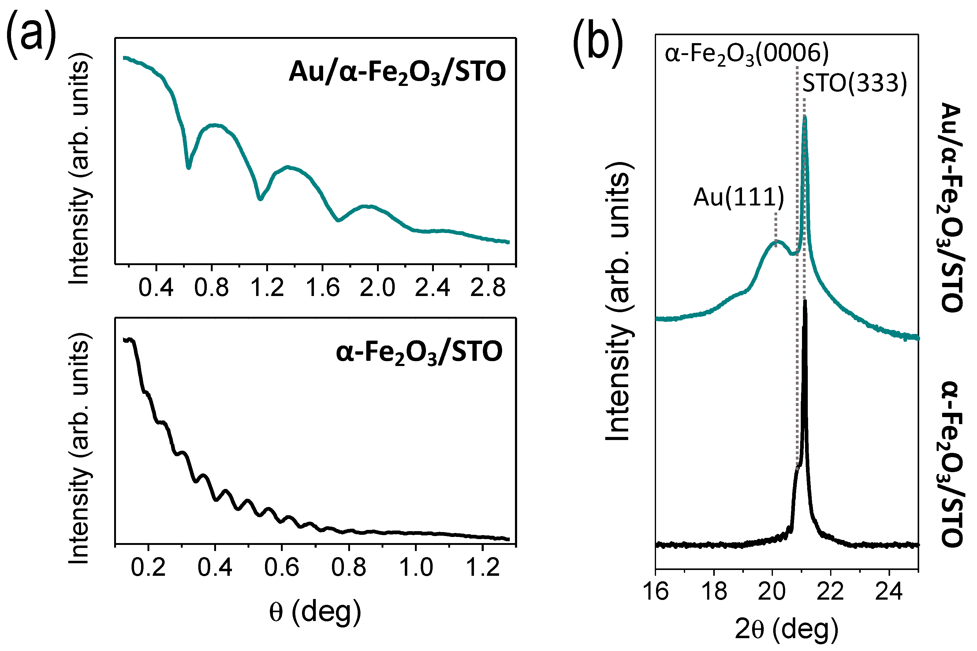
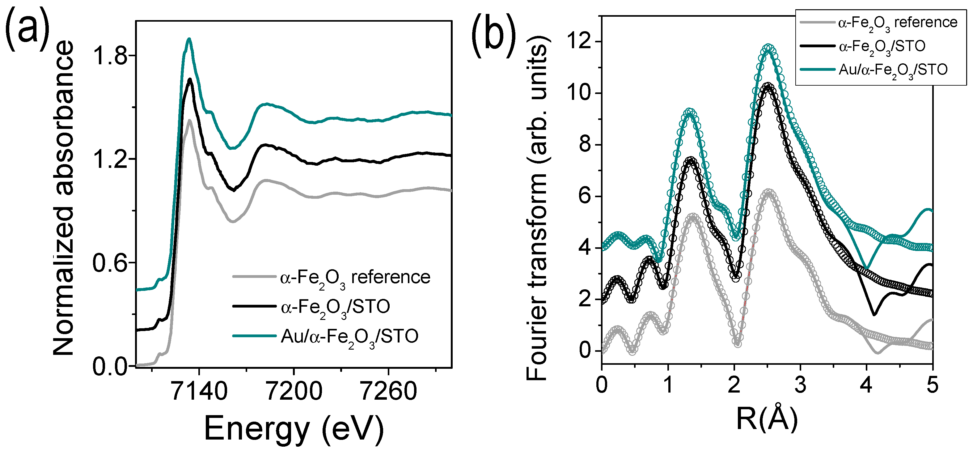
3.1. Morphological and Structural Characterization

During the growth process of both α-Fe2O3 and Au layers by pulsed laser deposition, in situ RHEED technique was employed to monitor their crystalline character. From the initial stages of deposition, intense patterns were observed. Figure 2 shows representative RHEED patterns of the clean STO(111) substrate and those of α-Fe2O3 thin film grown on STO(111) and Au islands grown on α-Fe2O3/STO(111) system at the final growth stages. In both Au and α-Fe2O3 layers, the RHEED patterns show smooth diffraction stripes, related to the growth of homogeneous and crystalline films.
Figure 3 shows the SEM images and the distribution analysis of particle size for α-Fe2O3 thin film and Au/α-Fe2O3 bilayer prepared on oxide STO(111) substrate. α-Fe2O3 thin film grown on STO(111) (Figure 3a) shows a homogeneous coverage of particles with an average size about 62(2) nm (Figure 3c), identifying an island-type growth. The greater interaction of α-Fe2O3 atoms with those of the STO substrate generates the formation of a α-Fe2O3 layer with a nanoparticulate character in order to minimize the system free energy. For the case of the Au/α-Fe2O3 bilayer, a nanostructured top layer related to the formation of Au islands is obtained on α-Fe2O3/STO(111) system. Au islands also show a homogeneous coverage on α-Fe2O3 surface, with an average particle size of 40(2) nm (see Figure 3b,d). This type of growth is induced during the Au growth when the sample substrate is kept at a temperature of 250 °C, as was identified previously in [31], inducing an island-type growth of Au promoted by the surface diffusion. The phenomenon is attributed to a dewetting process by a one-step procedure, instead of the usual methodology based on the post-annealing of the metallic film [25,26,27,28,32,36]. Specifically here, the α-Fe2O3 surface presents a particulate character with a discontinuous profile, which generates a more heterogeneous stress distribution than in a flat surface. These features of the bottom layer favor the nucleation of Au holes and the formation of larger and more separated Au islands in order to minimize the interface energy of the system [37], with qualitative similar final results. The average height for both α-Fe2O3 and Au layers was also determined on equivalent samples in our previous works to be 2.3(4) nm and 6(1) nm, respectively, by atomic force microscopy (AFM) and GIXRD [31,38].
To analyze the in-plane and out-plane crystalline growth of the Au and α-Fe2O3 layer, high resolution GIXRD, XRR, and reciprocal space map (RSM) measurements were performed. From low angle XRR measurements of the α-Fe2O3 layer and Au/α-Fe2O3 bilayer deposited on oxide STO(111), shown in Figure 4a, the thicknesses of the layers were estimated using the period of the oscillations. A thickness of 28(2) nm for the α-Fe2O3 layers and 6(1) nm for the Au layer was obtained. Representative high angle XRR measurements for the α-Fe2O3 layer and Au/α-Fe2O3 bilayer grown on STO(111) substrates are shown in Figure 4b. In the α-Fe2O3/STO(111) system, in addition to the diffraction peak corresponding to the STO(111) substrate, the presence of α-Fe2O3 with (0001) orientation as a single phase is identified, which is not clearly recognized in the diffraction pattern of the Au/α-Fe2O3 bilayer. In the case of the Au/α-Fe2O3 bilayer, the Au nanostructured layer on top of the α-Fe2O3 layer is recognized as a wide peak at lower 2θ values than the diffraction peak related to the α-Fe2O3 layer, which corresponds to epitaxial Au with a (111) orientation and is responsible for masking the diffraction peak signal related to the epitaxial α-Fe2O3 layer. It should be pointed out that some Kiessig fringes around the layer Bragg peaks can be noted, indicating the occurrence of high quality and smooth surfaces with abrupt α-Fe2O3/STO substrate and Au/α-Fe2O3 interfaces. These results are in agreement with results reported in [31].
Representative RSM measurements for the single α-Fe2O3 layer and Au/α-Fe2O3 bilayer grown on STO(111) substrates are displayed in Figure 5. Taking into account the analysis of RSM measurements, an incommensurate growth based on the non-coincidence of the in-plane diffraction peak maxima from both Au and α-Fe2O3 layers and the STO substrate is recognized. Besides, the crystallographic coupling between the layers and the substrate is analyzed. A rotation of 30° is shown between the in-plane crystallographic axes of the α-Fe2O3 layer and the STO(111) substrate and the Au layer and the α-Fe2O3 layer, with the crystallographic axes between Au and STO substrate parallel. Thus, the orientation relationships between the lattices of Au and α-Fe2O3 layers and the STO substrate is: (0001)[100]α-Fe2O3 ||(111)[110]STO for α-Fe2O3/STO(111) and (111)[110]Au || (0001)[100]α-Fe2O3 || (111)[110]STO for Au/α-Fe2O3/STO(111) system, as previously reported [31,38].
Out-of-plane and in-plane lattice parameters were calculated from the positions at several diffraction peaks in reciprocal space respect to the STO(111) substrate obtaining for α-Fe2O3 layers: cα-Fe2O3/STO = 13.72(6) Å, cAu/α-Fe2O3/STO = 13.72(3) Å, aα-Fe2O3 = bα-Fe2O3 = 5.08(4) Å and aAu/α-Fe2O3/STO = bAu/α-Fe2O3/STO = 5.07(1) Å and for Au layer: cAu/α-Fe2O3/STO = 7.04(2) Å and aAu/α-Fe2O3/STO = bAu/α-Fe2O3/STO = 2.89(2) Å. Additionally, the in-plane domain sizes were calculated to be 25(1) nm for the epitaxial α-Fe2O3 layer and Au/α-Fe2O3 system and 17(1) nm for the epitaxial Au, in agreement with previous work [31]. Similar values of both the lattice crystallographic parameters and in-plane domain size are obtained in the α-Fe2O3 layer before and after the deposition of Au islands, indicating no crystallographic alterations of the iron oxide structure and its thermodynamic stability during the Au growth at 250 °C. Besides, the lattice parameters match those of the bulk α-Fe2O3 structure disclosing a stress-free growth of layers.
Possible α-Fe2O3 modifications at short-range order during the growth of Au nanoparticles or amorphous minority phases non identified by GIXRD are analyzed by XAS experiments, as Figure 6 shows. Both X-ray absorption near-edge structure (XANES) and extended X-ray absorption fine structure (EXAFS) measurements were performed. Absorption profiles obtained by XANES measurements at the Fe K-edge of heterostructures follow the absorption features of powder α-Fe2O3 reference (see Figure 6a). Fitting the XANES spectra by a linear combination of different iron oxide reference compounds (Fe, FeO, Fe3O4 and α-Fe2O3 references) [39,40] (not shown here) confirms an oxidation state of Fe3+ and a 100% of α-Fe2O3 phase in both α-Fe2O3 layer and Au/α-Fe2O3 bilayer grown on STO(111) substrates, corroborating the α-Fe2O3 phase as a single iron oxide phase before and after the epitaxial deposition of Au islands.
The short-range ordering of cations around the Fe is studied in both α-Fe2O3 layer and Au/α-Fe2O3 bilayer by EXAFS spectroscopy. Figure 6b shows the modulus of the Fourier transform (FT) of the EXAFS signal at the Fe K-edge. The FT is performed in the k2χ(k) weighted EXAFS signal between 2.5 and 10.5 Å−1. Experimental EXAFS results were fitted in R-space in the range 1.0–3.6 Å using the FEFFIT code [34]. The first intense peak involves two subshells that corresponds to a first neighbor shell with three short plus three long O neighbors around the Fe. The second peak is related to single and multiple scatterings from the nearest Fe–Fe neighbors [41]. The fitting was performed fixing the shift at the edge energy E0, which was previously calculated from α-Fe2O3 powder reference. The coordination number N, the interatomic distance R and the Debye-Waller (DW) factors σ2 were used as free parameters for the fitting. Table 1 shows the parameters obtained from the EXAFS fittings.
In both α-Fe2O3/STO(111) and Au/α-Fe2O3/STO(111) systems, α-Fe2O3 layer presents an increase of the coordination number at the five coordination shells with respect to the α-Fe2O3 reference (see Table 1) that may be related to the greatest local order in the epitaxial thin films. With respect to the neighbor bondlengths, both α-Fe2O3 layers (single and with Au islands) present an Fe-O elongation and Fe–Fe contraction, indicating a local distortion at short-range order of epitaxial α-Fe2O3 structure grown on the STO(111) substrate in comparison with the α-Fe2O3 reference. Specifically for Au/α-Fe2O3/STO(111) system, most of the first neighbors present slightly larger bondlength distances than for α-Fe2O3/STO(111) system, which supposes a short-range order expansion that could be related to a relaxion of the α-Fe2O3 structure promoted by the deposition of the Au islands on the α-Fe2O3(0001)/STO(111) system with the substrate at 250 °C. Finally, the obtained DW factors are of similar order for both α-Fe2O3(0001) layers and we do not observe a structural disorder related to the growth of Au island adlayer.
The incorporation of Au islands on the α-Fe2O3/STO(111) system was also investigated by XPS measurements on the Fe 2p and Au 4f core level, as Figure 7 shows. The binding energy values of the Fe 2p3/2 peak (~710 eV) and the Fe 2p1/2 peak (~723.6 eV), with a distance among both of around 13.6 eV, were determined to be similar in both α-Fe2O3 layer and Au/α-Fe2O3 bilayer and comparable to typical values reported for the Fe 2p core level [42,43,44]. In addition, the satellite peak of Fe 2p3/2 is distinguished, more clearly in the α-Fe2O3/STO(111) sample, and located around 8 eV higher than the main Fe 2p3/2 peak [42,43,44]. These results confirm the oxidation state of the Fe cations to be Fe3+ and the nature of the iron oxide layer as α-Fe2O3, in agreement to GIXRD and XAS results. During the XPS measurements, a lower signal is collected on the Fe 2p core level for the Au/α-Fe2O3 bilayer than that acquired from the α-Fe2O3 layer due to the presence of Au islands. The Fe 2p signal is very low since the α-Fe2O3 is buried 6 nm down (Au layer thickness), which is approximately the depth limit of conventional XPS equipment (see Figure 4). On the Au 4f core level, the photoemission signal related to the Au/α-Fe2O3 bilayer grown on the STO(111) substrate shows a spin–orbit splitting of 3.7 eV between Au 4f5/2 and Au 4f7/2 peaks, with 4f7/2 at 84.0 eV corresponding to that of pure Au [45,46]. Moreover, the Fe 3s signal is imperceptible with respect to that of Au 4f, which means that the nanostructured-Au layer is homogeneous and there is no segregation of Fe through the Au layer. The combination of XPS and XANES techniques demonstrates the absence of mixed Fe-Au phases present on the surface and buried interfaces.

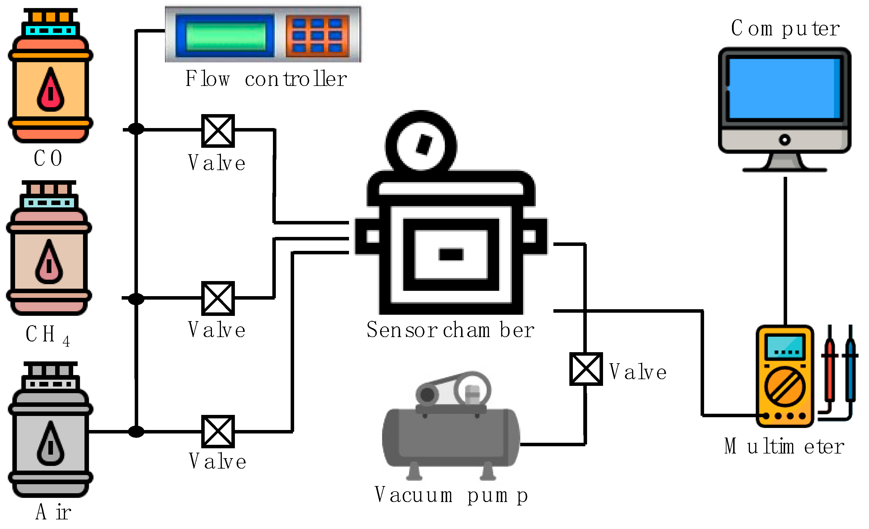
3.2. Conductance Response

The sensibility of the α-Fe2O3 layer and complex Au/α-Fe2O3 bilayer as gas sensing materials under CO and CH4 gas atmospheres at room temperature is shown in Figure 8. For that, the relative conductance response (ΔG/G0) of the complex Au/α-Fe2O3 system is compared in each case with the results obtained for the single α-Fe2O3 layer deposited on the STO(111) substrate under the same conditions. In each case, the gas sensor sensibility is determined by the gas in–gas out difference from the ΔG/G0 curves. α-Fe2O3 is a n-type semiconductor and its gas response involves the formation of an electron depletion layer at the surface and the return of these electrons to the conduction band, increasing the conductance signal after exposure to reducing gases [18,21,47].
On α-Fe2O3/STO(111) system in contact with air, oxygen molecules are adsorbed at the α-Fe2O3 surface. When the gas passes through the α-Fe2O3 surface, it is chemisorbed at the surface with previously adsorbed oxygen and an oxidation reaction occurs. Therefore, the trapped electrons on the surface are released back to the conduction band producing an increase of the electrical conductance of α-Fe2O3, as shown in Figure 8 for the measurements performed at room temperature. Prior to the experiments, the linear behavior of the resistance signal was checked in each sample.
As the α-Fe2O3 is functionalized on top with epitaxial Au(111) islands, forming a complex Au/α-Fe2O3 bilayer, the gas sensor sensibility increases around 25% under CO gas and around 35% under CH4 (see Figure 8), which should be ascribed to the active Au islands that play an important role in catalyzing the surface sensing reactions [4,24]. It should be noted that in both complex Au/α-Fe2O3 bilayer and single α-Fe2O3 layer, the sensor response for the CH4 is higher than for the CO gas (~5–10%). In addition, variations in the curve shape of the gas sensor response and recovery are noted. These changes are related to the adsorption and desorption reaction kinetics of the process on the sensor surface (in both systems) and can explain by different parameters of the reactions, the operating temperature and the gas nature, among other factors [48,49].
Major limitations to increase the technology progress and competitiveness of gas sensors are the high cost of certain semiconductor materials and the usual high temperatures required for their operation. Here, α-Fe2O3 is used, which is one of the most abundant semiconductors with a low cost and its sensor response under CO and CH4 atmospheres is stated at a very low temperature, i.e., at room temperature. In addition, the processing method to prepare the Au(111)/Fe2O3(0001)/SrTiO3(111) heterostructure is an easy way by a one-step method without post-processes and without modifying the structure and composition of the α-Fe2O3 semiconductor sensor. This methodology can be scaled up industrially and extrapolated to other catalytic systems or gas sensor systems with complementary properties. Therefore, these results are very promising for gas sensor applications at room temperature where the incorporation of epitaxial Au(111) islands on epitaxial α-Fe2O3(0001) layers grown on STO(111) substrates improves the gas sensor response for the detection of CO and CH4 gas at room temperature. In addition, the complex system exhibits plasmonic activity as previously reported [31], demonstrating the multifunctional character of these hybrid Au/α-Fe2O3 heterostructures.

4. Conclusions

We have grown epitaxial incommensurate α-Fe2O3(0001) layers and Au(111)/α-Fe2O3(0001) bilayers on STO(111) substrates by pulsed layer deposition with CO and CH4 gas sensor response at room temperature, identifying the influence of the incorporation of epitaxial Au nanostructures on the physical properties of α-Fe2O3 structure. An island-type growth is recognized in both Au and α-Fe2O3. Structurally, both before and after the Au deposition, α-Fe2O3 is the single iron oxide phase identified with a slight variation in the neighbor bondlengths at short-range order. The epitaxial growth is confirmed, with the in-plane crystallographic axes between Au and α-Fe2O3 and those between α-Fe2O3 and STO(111) rotated 30°. Gas sensor response under CO and CH4 atmospheres is measured at room temperature in both α-Fe2O3/STO(111) and Au(111)/α-Fe2O3(0001)/STO(111) systems, detecting an increase of the gas-sensing activity as the α-Fe2O3(0001) surface is functionalized with epitaxial Au nanostructures. The gas sensor sensibility increases around 35% under CH4 and 25% under CO gas atmosphere, being higher for the CH4 gas than for the CO gas (~5–10%).

Author Contributions

Conceptualization, methodology, investigation, formal analysis, data curation, writing—original draft and funding acquisition, A.S.; investigation, formal analysis and review—editing, J.L.-S., I.A., R.C., M.V. and E.S.-C.; methodology, investigation, supervision, review—editing and funding acquisition, G.R.C. and J.R.-Z. All authors have read and agreed to the published version of the manuscript.

Funding

This work has been supported by the Ministerio Español de Ciencia e Innovación (MICINN) and the Consejo Superior de Investigaciones Cientificas (CSIC) through the projects PIE-2010-OE-013-200014, PIE 2021-60-E-030 and RTI2018-095303-A-C52. The ESRF, MICINN and CSIC are acknowledged for the provision of synchrotron radiation facilities. A.S. acknowledges financial support from Comunidad de Madrid for an “Atracción de Talento Investigador” Contract (2017-t2/IND5395).

Institutional Review Board Statement

Not applicable.

Informed Consent Statement

Not applicable.

Data Availability Statement

The data presented in this study are openly available at 10.20944/preprints202106.0539.v1.

Acknowledgments

We especially thank Carlos Beltrán for his technical support during the experiments at the BM25 beamline at The ESRF.

Conflicts of Interest

The authors declare no conflict of interest.

References

  1. Zhang, S.; Li, H.; Zhang, N.; Zhao, X.; Zhang, Z.; Wang, Y. Self-sacrificial templated formation of ZnO with decoration of catalysts for regulating CO and CH4 sensitive detection. Sens. Actuators B Chem. 2021, 330, 129286. [Google Scholar] [CrossRef]
  2. Qiu, X.; Wei, Y.; Li, J.; Zhang, E.; Li, N.; Li, C.; Wei, J. Early detection system for coal spontaneous combustion by laser dual-species sensor of CO and CH4. Opt. Laser Technol. 2020, 121, 105832. [Google Scholar] [CrossRef]
  3. Garcia-Osorio, D.; Hidalgo-Falla, P.; Peres, H.E.M.; Gonçalves, J.M.; Araki, K.; Garcia-Segura, S.; Picasso, G. Silver enhances hematite nanoparticles based ethanol sensor response and selectivity at room temperature. Sensors 2021, 21, 440. [Google Scholar] [CrossRef] [PubMed]
  4. Garcia, D.; Picasso, G.; Hidalgo, P.; Peres, H.E.M.; Sun Kou, R.; Gonçalves, J.M. Sensors based on Ag-loaded hematite (α-Fe2O3) nanoparticles for methyl mercaptan detection at room temperature. Anal. Chem. Res. 2017, 12, 74–81. [Google Scholar] [CrossRef]
  5. Mishra, M.; Chun, D.M. α-Fe2O3 as a photocatalytic material: A review. Appl. Catal. A Gen. 2015, 498, 126–141. [Google Scholar] [CrossRef]
  6. Mirzaei, A.; Hashemi, B.; Janghorban, K. α-Fe2O3 based nanomaterials as gas sensors. J. Mater. Sci. Mater. Electron. 2016, 27, 3109–3144. [Google Scholar] [CrossRef]
  7. Ali, A.; Zafar, H.; Zia, M.; ul Haq, I.; Phull, A.R.; Ali, J.S.; Hussain, A. Synthesis, characterization, applications, and challenges of iron oxide nanoparticles. Nanotechnol. Sci. Appl. 2016, 9, 49–67. [Google Scholar] [CrossRef] [Green Version]
  8. Khatavkar, S.N.; Sartale, S.D. α-Fe2O3 thin films by liquid phase deposition: Low-cost option for supercapacitor. J. Solid State Electrochem. 2017, 21, 2555–2566. [Google Scholar] [CrossRef]
  9. Cornell, R.M.; Schwertmann, U. The Iron Oxides: Structure, Properties, Reactions, Occurrence and Uses, 2nd ed.; WILEY-VCH Verlag GmbH & Co. KGaA: Weinheim, Germany, 2003. [Google Scholar]
  10. Liang, H.; Jiang, X.; Qi, Z.; Chen, W.; Wu, Z.; Xu, B.; Wang, Z.; Mi, J.; Li, Q. Hematite concave nanocubes and their superior catalytic activity for low temperature CO oxidation. Nanoscale 2014, 6, 7199. [Google Scholar] [CrossRef]
  11. Yan, S.; Wu, Q. A novel structure for enhancing the sensitivity of gas sensors-α-Fe2O3 nanoropes containing a large amount of grain boundaries and their excellent ethanol sensing performance. J. Mater. Chem. A 2015, 3, 5982–5990. [Google Scholar] [CrossRef]
  12. Marti, X.; Fina, I.; Jungwirth, T. Prospect for antiferromagnetic spintronics. IEEE Trans. Magn. 2015, 51, 18–21. [Google Scholar] [CrossRef] [Green Version]
  13. Zeng, L.; Li, K.; Wang, H.; Yu, H.; Zhu, X.; Wei, Y.; Ning, P.; Shi, C.; Luo, Y. CO Oxidation on Au/α-Fe2O3-Hollow Catalysts: General Synthesis and Structural Dependence. J. Phys. Chem. C 2017, 121, 12696–12710. [Google Scholar] [CrossRef]
  14. Shen, S.; Lindley, S.A.; Chen, X.; Zhang, J.Z. Hematite heterostructures for photoelectrochemical water splitting: Rational materials design and charge carrier dynamics. Energy Environ. Sci. 2016, 9, 2744–2775. [Google Scholar] [CrossRef]
  15. Schultz, A.M.; Salvador, P.A.; Rohrer, G.S. Enhanced photochemical activity of α-Fe2O3 films supported on SrTiO3 substrates under visible light illumination. Chem. Commun. 2012, 48, 2012–2014. [Google Scholar] [CrossRef] [Green Version]
  16. Aronniemi, M.; Lahtinen, J.; Hautojärvi, P. Characterization of iron oxide thin films. Surf. Interface Anal. 2004, 36, 1004–1006. [Google Scholar] [CrossRef]
  17. Mirzaeian, M.; Ogwu, A.A.; Jirandehi, H.F.; Aidarova, S.; Ospanova, Z.; Tsendzughul, N. Surface characteristics of silver oxide thin film electrodes for supercapacitor applications. Colloids Surf. A Physicochem. Eng. Asp. 2017, 519, 223–230. [Google Scholar] [CrossRef] [Green Version]
  18. Wang, L.; Lou, Z.; Deng, J.; Zhang, R.; Zhang, T. ethanol gas detection using a Yolk-Shell (Core-Shell) α-Fe2O3 nanospheres as sensing material. ACS Appl. Mater. Interfaces 2015, 7, 13098–13104. [Google Scholar] [CrossRef] [PubMed]
  19. Cuong, N.D.; Hoa, T.T.; Khieu, D.Q.; Hoa, N.D.; Van Hieu, N. Gas sensor based on nanoporous hematite nanoparticles: Effect of synthesis pathways on morphology and gas sensing properties. Curr. Appl. Phys. 2012, 12, 1355–1360. [Google Scholar] [CrossRef]
  20. Li, X.; Wei, W.; Wang, S.; Kuai, L.; Geng, B. Single-crystalline α-Fe2O3 oblique nanoparallelepipeds: High-yield synthesis, growth mechanism and structure enhanced gas-sensing properties. Nanoscale 2011, 3, 718–724. [Google Scholar] [CrossRef]
  21. Hung, C.M.; Hoa, N.D.; Van Duy, N.; Van Toan, N.; Le, D.T.T.; Van Hieu, N. Synthesis and gas-sensing characteristics of α-Fe2O3 hollow balls. J. Sci. Adv. Mater. Devices 2016, 1, 45–50. [Google Scholar] [CrossRef] [Green Version]
  22. Wang, L.; Wang, L.; Hu, H.; Truong, N.; Zhang, Y.; Schmuki, P.; Bi, Y. Plasmon-Induced hole-depletion layer on hematite nanoflake photoanodes for highly efficient solar water splitting nano energy plasmon-induced hole-depletion layer on hematite nano flake photoanodes for highly efficient solar water splitting. Nano Energy 2017, 35, 171–178. [Google Scholar] [CrossRef]
  23. Gao, H.; Liu, C.; Jeong, H.E.; Yang, P. Plasmon-enhanced photocatalytic activity of iron oxide on gold nanopillars. ACS Nano 2012, 6, 234–240. [Google Scholar] [CrossRef]
  24. Zhang, J.; Liu, X.; Wang, L.; Yang, T.; Guo, X.; Wu, S.; Wang, S.; Zhang, S. Au-functionalized hematite hybrid nanospindles: General synthesis, gas sensing and catalytic properties. J. Phys. Chem. C 2011, 115, 5352–5357. [Google Scholar] [CrossRef]
  25. Thompson, C. V Solid-state dewetting of thin films. Annu. Rev. Mater. Res. 2012, 42, 399–434. [Google Scholar] [CrossRef]
  26. Leroy, F.; Borowik, Ł.; Cheynis, F.; Almadori, Y.; Curiotto, S.; Trautmann, M.; Barbe, J.C.; Müller, P. How to control solid state dewetting: A short review. Surf. Sci. Rep. 2016, 71, 391–409. [Google Scholar] [CrossRef]
  27. Serrano, A.; De La Fuente, O.R.; García, M.A. Extended and localized surface plasmons in annealed Au films on glass substrates. J. Appl. Phys. 2010, 108, 074303. [Google Scholar] [CrossRef] [Green Version]
  28. Serrano, A.; Fernandez, J.F.; Rodriguez de la Fuente, O.; García, M.A. A novel route to obtain metal and oxide nanoparticles co-existing on a substrate. Mater. Today Chem. 2017, 4, 64–72. [Google Scholar] [CrossRef]
  29. Gatel, C.; Snoeck, E. Epitaxial growth of Au and Pt on Fe3O4 (111) surface. Surf. Sci. 2007, 601, 1031–1039. [Google Scholar] [CrossRef]
  30. Lachebi, I.; Fedala, A.; Djenizian, T.; Hadjersi, T.; Kechouane, M. Morphological and optical properties of aluminum nanoparticles deposited by thermal evaporation on heated substrates. Surf. Coat. Technol. 2018, 343, 160–165. [Google Scholar] [CrossRef]
  31. Serrano, A.; Rubio-Zuazo, J.; López-Sánchez, J.; Enríquez, E.; Salas-Cólera, E.; Castro, G.R. Nanostructured Au(111)/Oxide epitaxial heterostructures with tailoring plasmonic response by a one-step strategy. J. Phys. Chem. C 2019, 123, 25294–25302. [Google Scholar] [CrossRef]
  32. Serrano, A.; Llorca-Hernando, O.; Del Campo, A.; Rubio-Marcos, F.; Rodríguez de La Fuente, O.; Fernández, J.F.; García, M.A. Ag–AgO nanostructures on glass substrates by solid-state dewetting: From extended to localized surface plasmons. J. Appl. Phys. 2018, 124, 133103. [Google Scholar] [CrossRef]
  33. Rubio-zuazo, J.; Ferrer, P.; López, A.; Gutiérrez-león, A.; Silva, I.; Castro, G.R. Nuclear instruments and methods in physics research a the multipurpose X-ray diffraction end-station of the BM25B-SpLine synchrotron beamline at the ESRF. Nucl. Inst. Methods Phys. Res. A 2013, 716, 23–28. [Google Scholar] [CrossRef] [Green Version]
  34. Newville, M.; Ravel, B.; Haskel, D.; Rehra, J.J.; Stern, E.A.; Yacoby, Y. Analysis of multiple-scattering XAFS data using theoretical standards. Phys. B Condens. Matter 1995, 208, 154–156. [Google Scholar] [CrossRef]
  35. Ravel, B.; Newville, M. ATHENA, ARTEMIS, HEPHAESTUS: Data analysis for X-ray absorption spectroscopy using IFEFFIT. J. Synchrotron Radiat. 2005, 12, 537–541. [Google Scholar] [CrossRef] [Green Version]
  36. Sharma, S.K.; Spitz, J. Hillock formation, hole growth and agglomeration in thin silver films. Thin Solid Films 1980, 65, 339–350. [Google Scholar] [CrossRef]
  37. Serrano Rubio, A. Modified Au-Based Nanomaterials Studied by Surface Plasmon Resonance Spectroscopy; Springer: Berlin, Germany, 2015. [Google Scholar]
  38. Serrano, A.; Rubio-Zuazo, J.; López-Sánchez, J.; Arnay, I.; Salas-Colera, E.; Castro, G.R. Stabilization of epitaxial α-Fe2O3 thin films grown by pulsed laser deposition on oxide substrates. J. Phys. Chem. C 2018, 122, 16042–16047. [Google Scholar] [CrossRef]
  39. Rubio-Zuazo, J.; Onandia, L.; Salas-Colera, E.; Muñoz-Noval, A.; Castro, G.R. Incommensurate growth of thin and ultrathin films of single-phase Fe3O4(001) on SrTiO3(001). J. Phys. Chem. C 2015, 119, 1108–1112. [Google Scholar] [CrossRef]
  40. Malferrari, D.; Castellini, E.; Bernini, F.; Rubio, A.S.; Castro, G.R.; Sainz-Díaz, C.I.; Caleffi, M.; Brigatti, M.F.; Borsari, M. Chemical trapping of gaseous H2S at high and low partial pressures by an iron complex immobilized inside the montmorillonite interlayer. Microporous Mesoporous Mater. 2018, 265, 8–17. [Google Scholar] [CrossRef]
  41. Sanson, A.; Mathon, O.; Pascarelli, S. Local vibrational dynamics of hematite (α-Fe₂O₃) studied by extended X-ray absorption fine structure and molecular dynamics. J. Chem. Phys. 2014, 140, 224504. [Google Scholar] [CrossRef] [PubMed]
  42. Oku, M.; Wagatsuma, K.; Matsuta, H. Background subtraction from transition metal 2p XPS by deconvolution using ligand atom XPS: Study on first transition metal cyanide complexes. J. Electron Spectrosc. Relat. Phenom. 1997, 83, 31–39. [Google Scholar] [CrossRef]
  43. Mills, P.; Sullivan, J.L. A study of the core level electrons in iron and its three oxides by means of X-ray photoelectron spectroscopy. J. Phys. D Appl. Phys. 1983, 16, 723–732. [Google Scholar] [CrossRef]
  44. Yamashita, T.; Hayes, P. Analysis of XPS spectra of Fe2+ and Fe3+ ions in oxide materials. Appl. Surf. Sci. 2008, 254, 2441–2449. [Google Scholar] [CrossRef]
  45. Kruse, N.; Chenakin, S. XPS characterization of Au/TiO2 catalysts: Binding energy assessment and irradiation effects. Appl. Catal. A Gen. 2011, 391, 367–376. [Google Scholar] [CrossRef] [Green Version]
  46. Strohmeier, B.R. Copper/silver/gold alloy by XPS. Surf. Sci. Spectra 1994, 3, 175–181. [Google Scholar] [CrossRef]
  47. Široký, K.; Jirešová, J.; Hudec, L.L.; Sirok, K.; Jire, J. Iron oxide thin film gas sensor. Thin Solid Films 1994, 245, 211–214. [Google Scholar] [CrossRef]
  48. Vuong, N.M.; Kim, D.; Kim, H. Surface gas sensing kinetics of a WO3 nanowire sensor: Part 1—Oxidizing gases. Sens. Actuators B Chem. 2015, 220, 932–941. [Google Scholar] [CrossRef]
  49. Vuong, N.M.; Kim, D.; Kim, H. Surface gas sensing kinetics of a WO3 nanowire sensor: Part 2—Reducing gases. Sens. Actuators B Chem. 2016, 224, 425–433. [Google Scholar] [CrossRef]
Figure 1. Experimental setup used to carry out gas sensor experiments in real time.
Coatings 11 00848 g001
Figure 2. Representative RHEED patterns of the clean STO(111) substrate, α-Fe2O3 thin films grown on STO(111) substrate and Au islands grown on α-Fe2O3/STO(111) system. During the deposition process of both α-Fe2O3 and Au layers, intense patterns with smooth diffraction stripes are identified.
Coatings 11 00848 g002
Figure 3. (a,b) SEM images and (c,d) particle size distribution analysis of α-Fe2O3 layer and Au/α-Fe2O3 bilayer grown on STO(111) substrates. An island-type growth for α-Fe2O3 and Au layers is recognized with a homogeneous coverage of particles.
Coatings 11 00848 g003
Figure 4. (a) Low angle XRR and (b) high-angle θ-2θ scans of epitaxial α-Fe2O3 layer and Au/α-Fe2O3 system grown on STO(111) substrates. Thicknesses were calculated to be 28(2) nm for the α-Fe2O3 layer and 6(1) nm for the Au layer. Bragg peaks related to STO(111) substrate, Au(111) and α-Fe2O3(0001) are identified.
Coatings 11 00848 g004
Figure 5. LH reciprocal space maps (RSM) for (a) α-Fe2O3 layer grown on STO(111) substrate and (b) Au/α-Fe2O3 bilayer prepared on STO(111) substrates. An incommensurate epitaxial growth is identified from a non-coincidence of Au(111), α-Fe2O3(0001), and STO(111) substrate in-plane peaks (marked in each figure). The color scale corresponds to the signal intensity.
Coatings 11 00848 g005
Figure 6. (a) XANES spectra and (b) FT modulus of the EXAFS signal (lines with symbols) and best-fitting simulations (continuous lines) at the Fe K-absorption edge of α-Fe2O3 layer and Au/α-Fe2O3 bilayer grown on STO(111) substrates. XANES spectrum and FT of the EXAFS signal of α-Fe2O3 powder reference are presented for comparison.
Coatings 11 00848 g006
Figure 7. XPS measurements performed on the (a) Fe 2p core level of the α-Fe2O3 layer and the Au/α-Fe2O3 bilayer grown on STO(111) substrates and (b) Au 4f core level of the Au/α-Fe2O3 bilayer grown on STO(111) substrate. Lower signal is collected on the Fe 2p core level for the Au/α-Fe2O3 bilayer than for the α-Fe2O3 layer.
Coatings 11 00848 g007
Figure 8. Gas sensor response curves for (a,b) Au/α-Fe2O3 bilayers deposited on STO(111) substrate at a substrate temperature for the Au growth of 250 °C and (c,d) bare α-Fe2O3 thin film under (a,c) CO and (b,d) CH4 atmosphere at room temperature. G0 and G values correspond to the sensor conductance in air and specific gas atmosphere. The gas in to gas out difference in the ΔG/G0 curves is used to determine the gas sensor sensibility. A red bar is represented as an example in each case.
Coatings 11 00848 g008
Table 1. Results of the EXAFS fittings by a five-shell model at the Fe K-edge of α-Fe2O3 layer and Au/α-Fe2O3 bilayer grown on STO(111) substrates. EXAFS parameters are compared with results obtained from powder α-Fe2O3 reference.
SampleShellN R (Å)DW(Å2)
α-Fe2O3 reference Fe–O131.967(4)0.006(1)
Fe–O232.13(1)0.017(2)
Fe–Fe112.977(1)0.006(1)
Fe–Fe233.06(1)0.007(1)
Fe–Fe333.454(1)0.017(1)
α-Fe2O3/STOFe–O13.4(2)1.981(8)0.003(1)
Fe–O23.4(2)2.188(3)0.014(1)
Fe–Fe11.2(1)2.90(4)0.032(9)
Fe–Fe23.6(3)2.983(2)0.003(1)
Fe–Fe33.6(3)3.380(2)0.005(1)
Au/α-Fe2O3/STOFe–O13.7(2)1.993(6)0.003(1)
Fe–O23.7(2)2.230(5)0.013(2)
Fe–Fe11.2(1)2.82(6)0.032(9)
Fe–Fe23.6(3)3.001(3)0.004(2)
Fe–Fe33.6(3)3.394(4)0.005(1)
Publisher’s Note: MDPI stays neutral with regard to jurisdictional claims in published maps and institutional affiliations.

