Article

Polydopamine-Coated Copper-Substituted Mesoporous Silica Nanoparticles for Dual Cancer Therapy

by
Prabhakar Busa
1,
Ravindranadh Koutavarapu
2,* and
Yaswanth Kuthati
1,*
1
Department of Anesthesiology, Cathay General Hospital, Taipei 280, Taiwan
2
Department of Robotics and Intelligent Machine Engineering, College of Mechanical and IT Engineering, Yeungnam University, Gyeongsan 712-749, Korea
*
Authors to whom correspondence should be addressed.
Coatings 2022, 12(1), 60; https://doi.org/10.3390/coatings12010060
Submission received: 5 December 2021 / Revised: 29 December 2021 / Accepted: 31 December 2021 / Published: 5 January 2022

Abstract

:
Combinational therapy using chemodynamictherapy (CDT) and photothermal therapy (PTT) is known to enhance the therapeutic outcome for cancer treatment. In this study, a biocompatible nano formulation was developed by coating polydopamine (PDA) over doxorubicin (DOX)-loaded copper-substituted mesoporous silica (CuMSN) nanoparticles. PDA coating not only allowed selective photothermal properties with an extended DOX release but also enhanced the water solubility and biocompatibility of the nanocomposites. The nanocomposites displayed a monodispersed shape and pH-dependent release characteristics, with an outstanding photothermal conversion and excellent tumor cell inhibition. The cellular-uptake experiments of CuMSN@DOX@PDA in A549 cells indicated that nanoparticles (NPs) aided in the enhanced DOX uptake in tumor cells compared to free DOX with synergistic anti-cancer effects. Moreover, the cell-viability studies displayed remarkable tumor inhibition in combinational therapy over monotherapy. Thus, the synthesized CuMSN@DOX@PDA NPs can serve as a promising platform for dual cancer therapy.

Graphical Abstract

1. Introduction

Cancer remains one of the leading causes of death worldwide [1,2]. Currently, chemotherapy is one of the most commonly used treatments, and often results in drug resistance and adverse effects to the neighboring healthy cells without selectivity [3,4]. To address this, combinational treatment therapies have been developed that can limit resistance and adverse events [5,6,7]. Combination of chemotherapy with phototherapy can greatly enhance the selectivity towards cancer cells. In addition, combination with a photothermal agent can further enhance the anti-cancer effects [8,9,10,11].
Doxorubicin (DOX) is one of the most promising anti-cancer drugs used for the treatment of various types of cancers such as lung, breast, throat, skin, and blood [12,13]. DOX can interfere with DNA synthesis, produce reactive oxygen species (ROS), and induce DNA and mitochondrial damage [14,15]. In recent years, with the advent of nanotechnology, various formulations have been developed using DOX for the selective delivery of drugs into cancer cells [16,17]. Various nanocarriers have been reported for the delivery of DOX, such as polymeric NPs, layered double hydroxide NPs, gold NPs, silver NPs, and mesoporous silica (MSN) NPs [18,19,20,21]. Among all, MSNs have garnered significant attention due to their peculiar characteristics like ease of synthesis, large surface area and pore volume, ability to control size and shape, biocompatibility, and surface functionality [22,23,24]. The large surface area and pore volume aids in a high drug loading of therapeutics like small-molecule drugs, proteins, and genes [25,26,27]. In general, therapeutics are loaded into a mesoporous framework though adsorption [28,29,30]. The open pore structure facilitates the easier release of drugs when necessary, and to promote prolonged drug release, the surface can be modified with polymers [31,32,33].
In general, cancerous cells have lower extra cellular pH (pH = 6.5) than healthy cells. To exploit this selective difference, pH-responsive polymers have gained a great deal of attention in the development of nanocarriers for selective drug release in cancerous cells [34,35,36]. Abnormal pH is a common hallmark in most cancerous cells, a fact that is utilized to design pH-responsive polymers that can alter their physiochemical properties specific to pH. Moreover, nanopolymer size in combination with pH-responsive drug release can strengthen targeting ability [37,38].
In the current work, copper-substituted MSN was prepared by co-condensation. Metal substitution can impart benefits including size reduction, enhanced biocompatibility, reduction of negative charge, improved drug loading, and enhanced stability [39,40,41,42]. PDA is used as a surface polymer for the pH-dependent release of anticancer cargo [43,44]. The PDA coating further served as a photothermal agent, enhancing the synergistic effects of phototherapy and chemotherapy [45,46]. PDA is coated by the oxidative self-polymerization of dopamine monomers in the presence of weak alkaline conditions (pH 8.0–8.5) [47,48]. PDA is coated over NPs’ surface for various biological applications [49]. Under neutral pH, PDA coating can ensure the drug molecules remain intact in the mesoporous framework [50]. However, upon encountering acidic pH, PDA dissolves and releases the drug molecules into the cytosol of cancer cells [51]. Yanhong et al. reported MSN@ DOX NPs, coated with PDA and poly (ethylene gycol) (PEG), for the management of breast cancer. In this report, PDA acted as a pH-sensitive gatekeeper with photothermal properties [52]. In another report, MSN@DOX NPs were coated with PDA and further conjugated with CSNRDARRC peptide for site-specific DOX delivery with enhanced anti-cancer effects [53].
In this study, DOX-loaded PDA-coated copper-substituted (CuMSN) were developed, and their anticancer efficacy is evaluated against A549 (lung cancer) cells. The particle size, zeta potential, drug-loading capacity (LC), and in vitro drug-release studies (pH- and light-responsive) were verified. 3-(4,5-dimethyl-2-thiazolyl)-2,5-diphenyl tetrazolium bromide (MTT) assay is used to evaluate the anti-cancer effects of monotherapy and combinational therapy. The cellular uptake of DOX-loaded and fluorescein isothiocyanate (FITC)-labeled NPs was examined by fluorescence microscopy. The intracellular ROS levels were detected by 2′,7′-dichlorodihydroflurescein diacetate (DACFDA) assay.

2. Materials and Methods

2.1. Materials

DOX, DCFDA, dopamine, tetraethyl orthosilicate (TEOS), cetyltrimethylammonium bromide (CTAB), ammonium nitrate (NH4NO3), copper nitrate, FITC, potassium bromide (FT-IR grade), copper nitrate trihydrate (Cu(NO3)2·3H2O), 4’,6-diamidino-2-phenylindole dihydrochloride (DAPI), and sodium hydroxide were obtained from Sigma Co. Ltd. (St. Louis, MO, USA).

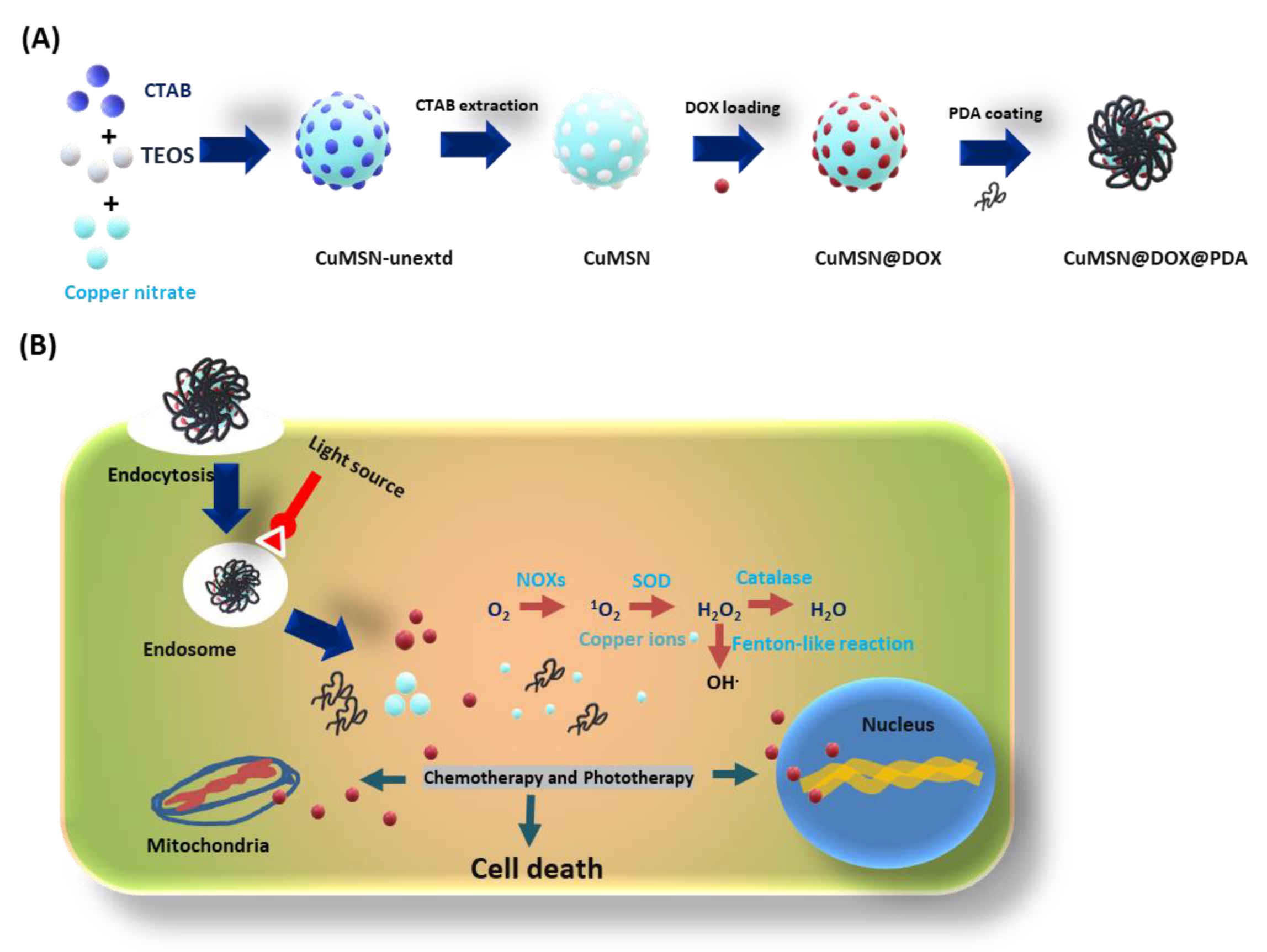
2.2. Synthesis of CuMSN

NPs were prepared by following the previous publications [25]. TEOS is used as silica precursor; (Cu (NO3)2·3H2O) is used as a copper source, and CTAB is employed as a surfactant in the basic solvent to perform the co-condensation reaction to synthesize the uniformly sized CuMSN. In detail, firstly, 0.58 g of CTAB was dissolved in 300 mL of NH4OH (0.51 M) and stirred with constant speed at 40 °C. A total of 5 mL of dilute TEOS (0.2 M TEOS in 5 mL of ethanol) is added and was continuously stirred for 3 h. After 3 h, (Cu (NO3)2·3H2O) and 5 mL of concentrated TEOS (1.0 M in 5 mL of ethanol) was added and stirred for another 2 h. Then, the stirring is stopped, and the reaction mixture is aged for 20 h at 40 °C. The contents are centrifuged and collected, and chemical composition was explained in the supplementary (Table S1). Surfactant CTAB is extracted by suspending 100 mg of CuMSN in a mixture of 0.3 g of NH4NO3 in isopropanol (40 mL) for 16 h. Finally, CuMSN was collected by centrifugation and washed two times with ethanol and isopropanol. The solid was dried under vacuum at room temperature, and the product was named CuMSN-extd.

2.3. DOX Loading into CuMSN

Initially, 6 mg of DOX was dissolved in 6 mL of dry methanol, and the pH was adjusted to 5.8 to 6.0. The DOX solution was transferred into a glass container with 100 mg dried CuMSN NPs, and the contents were stirred at room temperature overnight in a dark environment [54]. The DOX-loaded solid contents were collected by centrifugation and washed twice with ethanol and methanol to remove the unbound DOX molecules. The loading percentage was calculated by using the formula mentioned below.
Drug   Entrapment   Efficacy   %   ( DEE   % ) = weight   of   drug   in   nanoparticle weight   of   drug   used   in   formulation × 100 Drug   Loading   Capacity   %   ( DLC   % ) = weight   of   drug   in   nanoparticle weight   of   nanoparticle × 100
Approximately 8% (w/w) DOX was loaded into CuMSN. The DOX load was measured at 480 nm. The product was named CuMSN@DOX.

2.4. PDA Coating onto CuMSN@DOX

The PDA was coated onto CuMSN@DOX NPs by following the previous publications [48,55]. Initially, 50 mg of CuMSN@DOX NPs were added into 1 mg/mL solution of dopamine hydrochloride in Tris buffer (10 mM, pH 8.5) and continuously stirred under dark conditions at room temperature for 10 h. PDA-coated black-colored solid was collected by centrifugation and washed thrice with distilled water to remove un-polymerized dopamine molecules from the surface. Then, finally, the solids were dried by freeze dryer and stored at 4 °C. The product was denoted CuMSN@DOX@PDA.

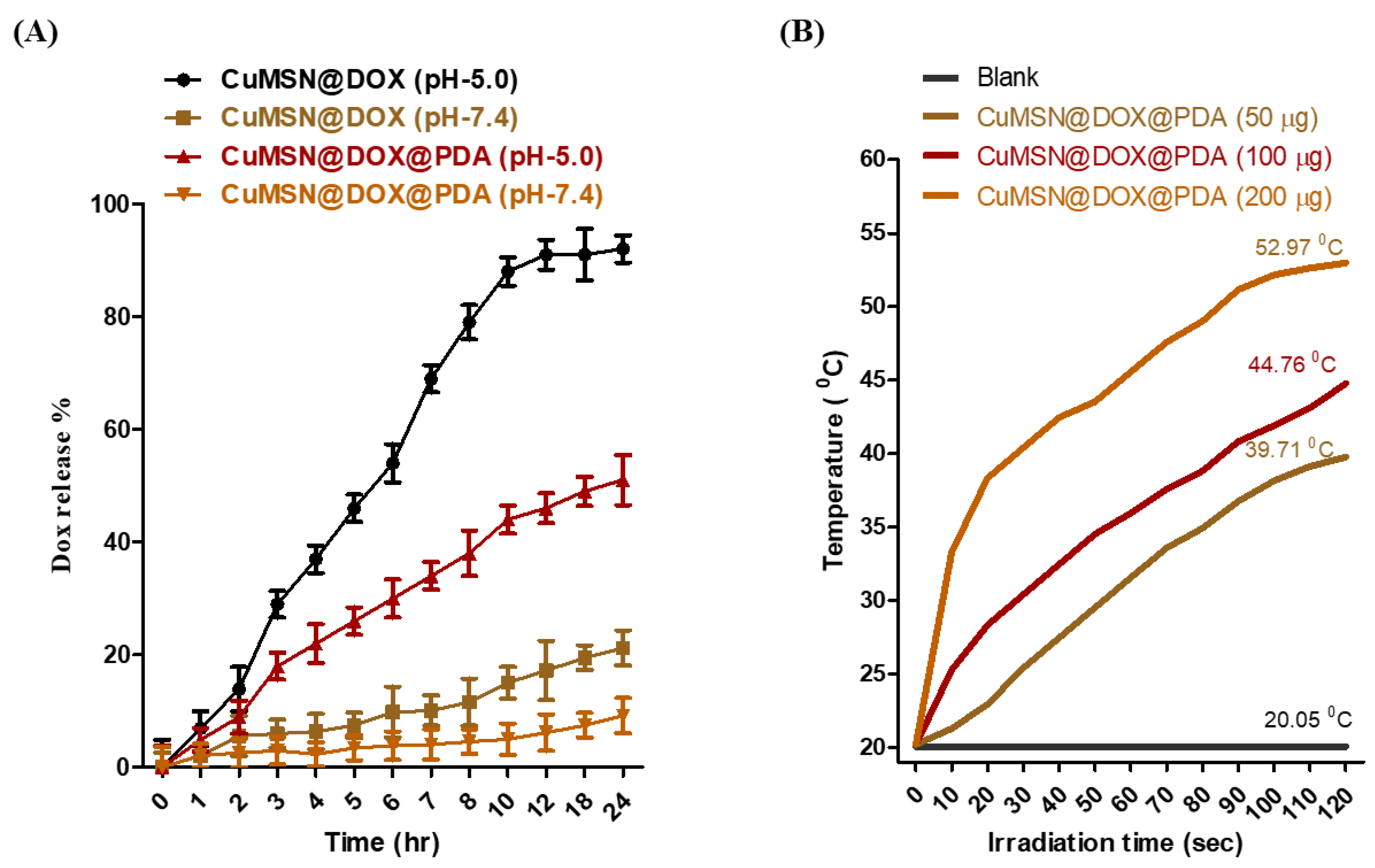
2.5. In Vitro DOX Releasing Study

The DOX release profile from the NPs (CuMSN@DOX and CuMSN@DOX@PDA) was verified in various buffers by following previous protocols [32,56]. A total of 5 mg of NPs were suspended in phosphate buffer saline solution (PBS) (pH 5.0 and 7.4) and placed on a shaker (150 rpm) at 37 °C on a rotary shake machine. The supernatant was collected from the respective buffers at regular time intervals, and the DOX concentration was calculated by Uv-vis spectrometry (Uv-1700 Pharma Spec, Shimadzu, MA, USA) at 480 nm. The collected sample solution was replaced with same amount of fresh PBS. The cumulative DOX-release percentage was calculated periodically; all experiments were performed in triplicate.

2.6. In Vitro PTT Studies

PPT experiments were performed by following the previous reports. A total of 1 mL of quartz cuvette with samples (CuMSN@DOX@PDA) was irradiated with a near-infrared (NIR) light source (808 nm, 1 W/cm2) [57]. The temperature changes were noted by thermal camera with time (FLIR ONE PRO, Wilsonville, OR, USA).

2.7. Cell Culture and Cytotoxicity Assay

A549 and HT-29 cells were obtained from the American Type culture collection (Rockville, MD, USA), cultured in Dulbecco’s Modified Eagle’s Medium (DMEM) and Roswell Park Memorial-1640 (RPMI-1640) respectively, with 10% fetal bovine serum (FBS) and 1% penicillin or streptomycin (antibiotic), passage number 6. The cells were grown in an incubator and maintained at 37 °C with 5% CO2 supply. MTT assay was used to determine the CDT and PTT groups’ cell viability. For CDT and PTT therapy, cells were seeded in the 96-well plate at 1 × 10⁵ cells per well and incubated for 24 h. After 24 h, for CDT therapy, the wells were treated with respective NPs, and PTT experiments are performed by utilizing a light source (810 nm, 1.0 W/cm) for 5 min and incubating for 24 h. Finally, all wells are washed carefully with PBS solution. A MTT reagent was added and incubated for 20 min. Finally, Formazan crystals were dissolved in the dimethyl sulfoxide (DMSO), and the absorbance was recorded by using a micro plate reader [48]. All experiments were performed in triplicate for the individual experiments.

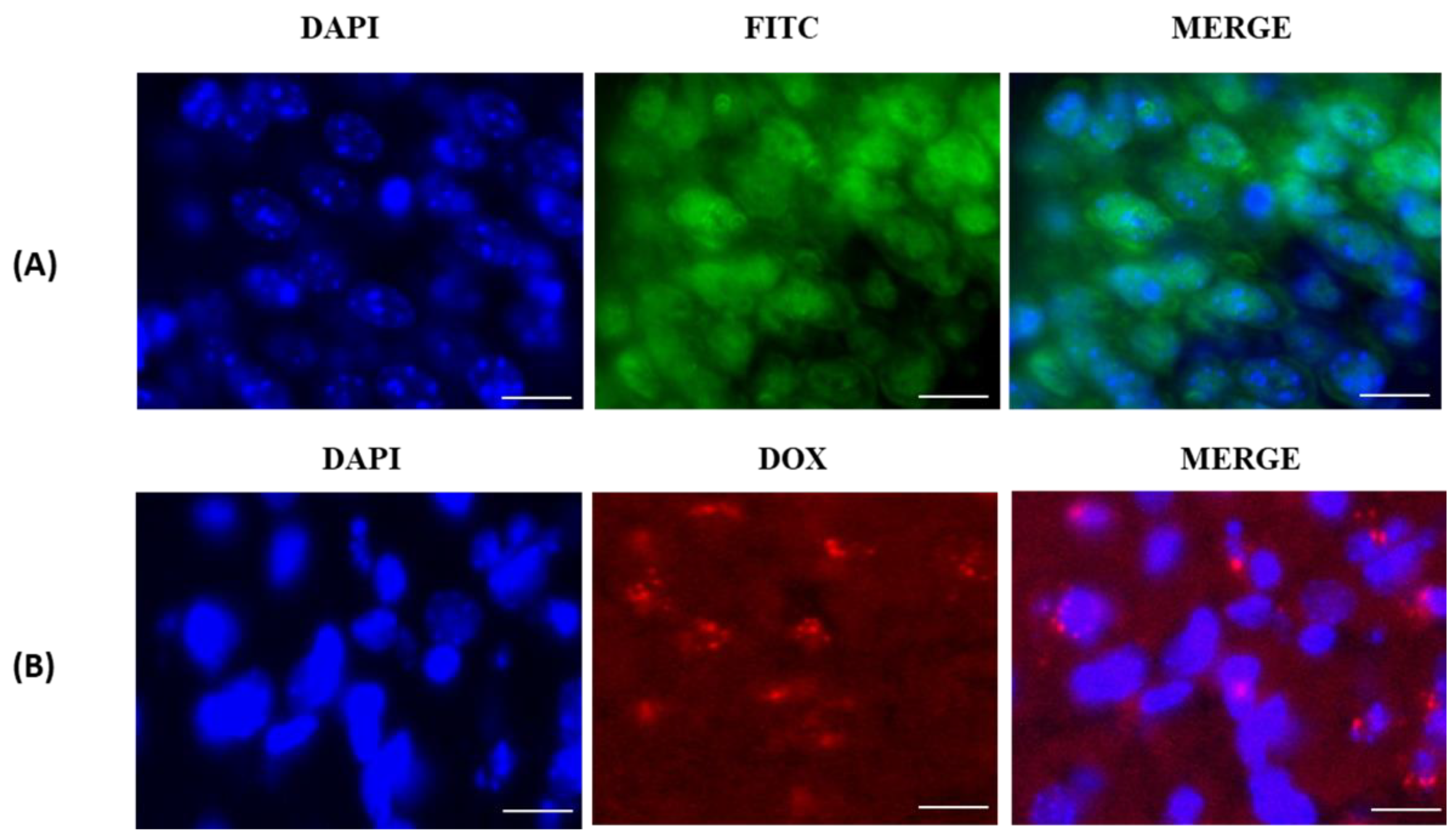
2.8. Cell Uptake Studies

The drug-delivery capacity of CuMSN and its modified counterparts was evaluated in vitro through cell-uptake studies in A549 cells through fluorescence microscopy (Olympus BX51, NY, USA). The DOX self-fluorescence gave the ability to check the internalization capacity. A549 cells were seeded onto 6-well plates at a density of 1 × 10⁵ cells per well and incubated for 24 h. CuMSN and their modified samples were suspended in the FBS-free culture medium with 50 μg/mL and added to the respective wells. After 4 h of incubation, the cells were fixed with 4% paraformaldehyde and incubated for another 10 min. Finally, the nuclei were stained with DAPI, and all wells were carefully washed with PBS solution twice. Ultimately, the images were captured by fluorescence microscopy [58,59].

2.9. ROS Detection Assay

The cellular level ROS generation was assessed by DCFDA assay by following previous protocols [60,61]. A549 cells were seeded in the 96-well plates at a density of 1 × 10⁵ cells per well and incubated for 24 h for the bottom attachment of cells to carry out experiments smoothly. Then, the respective wells are treated with CuMSN and their successive modified nanoparticles with 100 μg/mL; for the PPT group, the experimental procedure was performed with the same experimental condition by using a light source. Herein, after 24 h of nanoparticle treatment, all wells were exposed to a DCFDA solution (5 μg/mL) and incubated for 30 min. The fluorescence intensity was detected by a micro plate reader with Coring (Glendale, AZ, USA) plates having a transparent bottom and black side walls. Hydrogen peroxide was used as negative control, and all experiments were performed in triplets with individual experiments.

2.10. Statistical Analysis

All data are represented as mean ± standard deviation (S.D). In experiments, p ≤ 0.001 (***), p ≤ 0.01 (**), and p ≤ 0.5 (*) were statistically significant. The data were analyzed by a Student t-test to compare the mean values. Graph pad prism-5 software was used to calculate all statistical significance.

3. Results and Discussion

The material design of our multifunctional nanomaterial is depicted in Figure 1. The aim of the proposed design is to deliver DOX into A549 cells and eradicate them by synergistic CDT/PTT. Initially, we synthesized CuMSN by a co-condensation method in the basic medium composed of TEOS and copper silts using CTAB as a template. The presence of copper (Cu (II)) within the MSN framework enables the formation of a coordination link with DOX, which triggers pH-sensitive drug release at acidic pH conditions (endosomal level). The metal substitution can also enhance the DOX loading. Additionally, the PDA coating aims to improve the biocompatibility and cellular internalization of NPs, along with photothermal properties. The PDA layer is coated onto the nanoparticle surface in the presence of alkaline conditions (Tris buffer (pH 8.0–8.5)). Dopamine undergoes oxidative polymerization in the oxygenated environment (oxidant). Firstly, the synthesized NPs were physically characterized by different techniques such as transmission electron microscopy (TEM, Hitachi HT7700, Tokyo, Japan) for morphological analysis, Fourier-transform infrared spectroscopy (FT-IR, Bruker’s Tensors 27 series, TX, USA) for functional group detection, and thermogravimetric analysis (TGA, TG Q500, New castle, DE, USA) studies for the weight-loss determination of respective compounds. Uv-vis (Uv-1700 Pharma Spec, Shimadzu, MA, USA) is performed to determine the loading percentage and drug-release profile of DOX, and dynamic light scattering (DLS, Zetasizer Nano ZS-90, Malvern, UK) is used to evaluate the particle-size distribution and zeta potential. Furthermore, the synergistic CDT/PTT effects of nanocarriers are studied in A549 cancer cells by MTT assay, and ROS levels were determined using DCFDA assay [62,63].

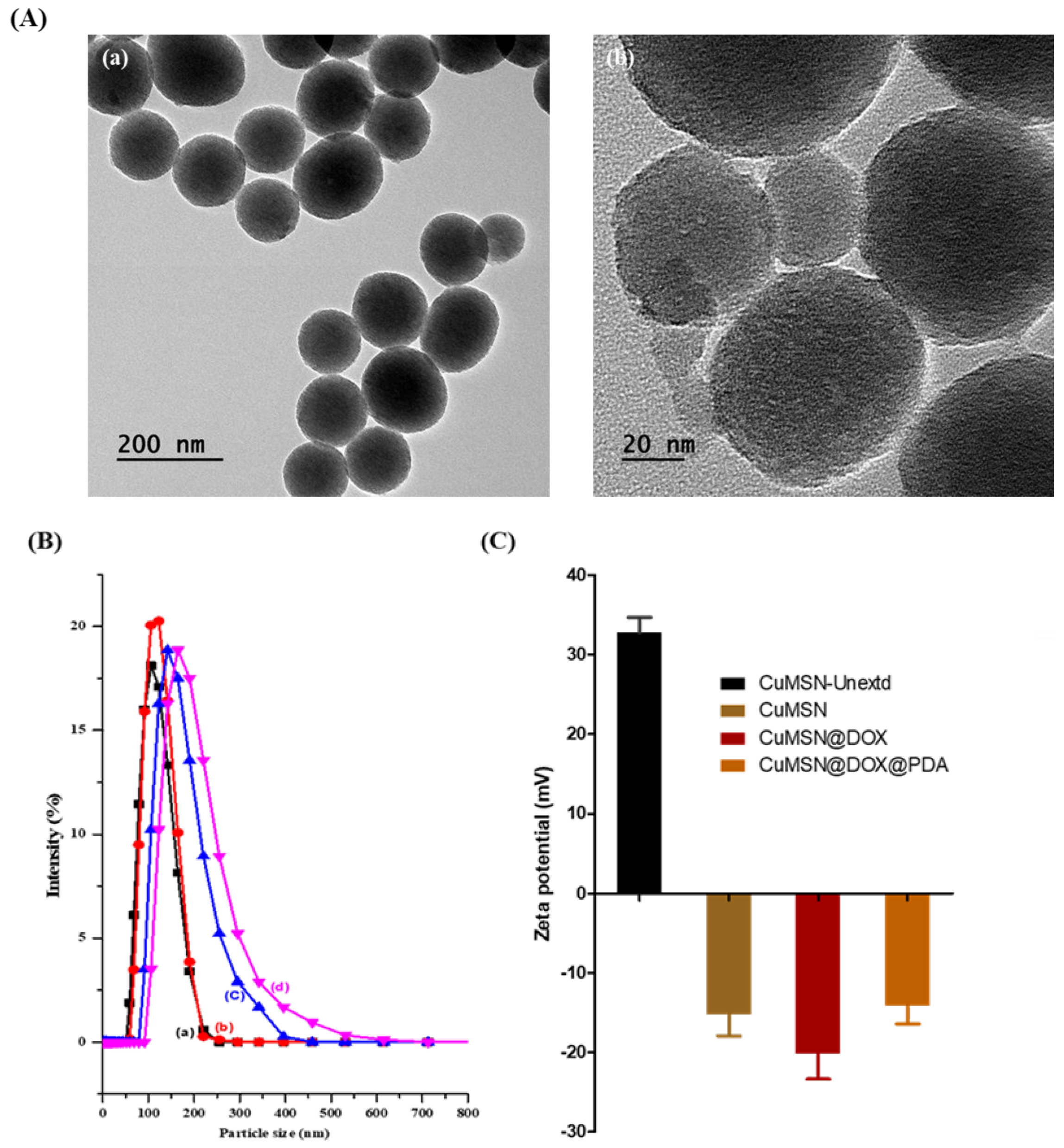
3.1. Physical Characterization

TEM images (Figure 2A) show that CuMSN@DOX-loaded NPs retained their spherical shape after drug loading, without disturbing the mesoporous structure. After PDA coating, a rough surface was detected with a dense core, indicating the successful binding of DOX within the mesopores of CuMSN. PDA coating did not affect the loading of DOX and served as a gatekeeper. The size distribution of NPs in the TEM is consistent with the DLS size-distribution studies shown in Figure 2B. Furthermore, we studied the changes in hydrodynamic size and zeta potential values of CuMSN before and after drug loading and PDA coating. The average hydrodynamic diameter of unextracted CuMSN (CuMSN-Unextd), CTAB extracted (CuMSN-extd), and DOX-loaded (CuMSN@DOX) NPs are around 100–160 nm. Furthermore, PDA coating (CuMSN@DOX@PDA) resulted in a gradual increase of the NPs size to 210 nm, suggesting the successful coating of PDA on the surface of NPs. Liu et al. studied the distribution of the radiolabeled NPs of various sizes ranging from 30–400 nm in tumor tissue. Their studies revealed that NPs within 100–200 nm have four times higher retention than the NPs larger than 300 nm and less than 50 nm [64].
Furthermore, the pristine MSN have a negative surface charge after CTAB extraction, but the extraction of a template resulted in a slight decrease of negative zeta potential (−17 ± 0.2 mV) due to the presence of copper ions in the MSN framework. After DOX loading, the zeta potential shifted to the negative side. However, PDA coating resulted in a positive zeta potential, implying PDA coating on to the surface of NPs.
Furthermore, FT-IR spectral studies gave evidence of various functional groups in the silica framework (Figure 3A). The broad band at 3450 cm−1 can be attributed to O-H stretching vibration and surface Si-OH functional groups from the water molecules (Figure 2B (a–d)). The nanoparticle surface silanol functional groups Si-O-Si are verified at 790 and 1085 cm−1, and Si-O-H functional groups are seen at 970 cm−1, which represent the stable formation of a silica framework in all of the samples. The C-H stretching peaks appeared at around 2800 to 2930 cm−1 in the unextracted CuMSN (Figure 3A (a)). After CTAB extraction by chemical etching, these peaks vanished, denoting the successful removal of surfactants. After CTAB extraction, the characteristic peaks of the silica framework remained the same, demonstrating the stability of the nanocarrier. DOX-loaded CuMSN (Figure 3A (c)) show peaks at around 1690 cm−1, corresponding to the aliphatic carbonyl group. Other peaks at 1549 cm−1 can be attributed to amine groups. The broad peak at 2940 cm−1 can be attributed to C–H vibrations of DOX and is similar to previous publications [65,66,67,68]. The N–H vibrational peaks appearing at 1520 to 1550 cm−1 from DOX gives evidence for coordination interactions and the successful loading of DOX into the mesopores of NPs. Furthermore, DOX loading was confirmed by TGA and Uv-vis spectral analysis. The broad peaks at 3130 to 3600 cm−1 are attributed to N–H and O–H stretching vibration of PDA (Figure 3A (d)), and the peak at 1650 cm−1 is assigned for the C–C resonance stretching vibrations in the aromatic rings. These results suggest the successful coating of PDA onto the surface of NPs without disturbing the DOX and NPs’ framework.
Next, the drug loading and thermal properties of CuMSN and their succeeding modifications were evaluated by TGA studies. Figure 3 B shows the TGA curves of various nanocomposites. The early stage of weight loss before 100 °C in all samples corresponded to a loss of residual water. In unextracted CuMSN (Figure 3B (a)), weight loss at around 240 °C results from the degradation of CTAB. DOX-loaded NPs show a significant weight loss at 310 °C, which is equivalent to the amount of DOX loaded (~8%) into the mesopores. This weight-loss percentage supports the estimated drug load reported through UV-vis measurements. The PDA-loaded samples began a gradual loss of weight at 250 °C and continued until 550 °C. This significant loss of DOX and PDA events overlapped at 420 °C. These findings demonstrated that PDA was successfully coated onto the surface of the nanoparticles and enhanced their thermal stability [69].

3.2. In Vitro DOX Release and PDA Photothermal Studies

In vitro DOX-release behavior is studied in various buffer solutions (PBS, pH 5.0 and 7.4) using a Uv-vis spectrometer. The pH-sensitive bond between copper ions and amine groups were disrupted in acidic pH (pH 5.0) and significantly enhanced the DOX release compared to basic conditions (pH 7.4); similar pH-sensitive release profiles have been reported earlier (Figure 4A) [70]. Furthermore, a similar pattern of release is observed in the PDA-coated samples, wherein PDA acts as a gatekeeper and extends the DOX release from the mesopores of the nanoparticles. The PDA coating can help to extend the DOX release in the tumor environment (Figure 4B) [71].
Photothermal properties of PDA were investigated by NIR light source (810 nm) directed towards the cancer cells. The PDA-coated nanoparticles (50, 100 and 200 μg/mL) were dispersed in PBS solution, and the samples were irradiated with an NIR light source with various time intervals up to 120 s. The significant temperature increment was observed in a time- and concentration-dependent manner that implies the efficacy of PDA as a photo-thermal agent to generate the heat to destroy the cancer cells [72,73].

3.3. In Vitro Cellular-Uptake Studies

We have evaluated cellular internalization, DOX pH-dependent release, DOX participating in ROS generation by CDT, and CDT/PPT combinational therapy. To deliver DOX into the tumor cells, initially the PDA-coated NPs attached to the cell membranes, followed by internalization, and the DOX was released at the cytosol level due to the pH-sensitive effect. The released DOX molecules participated in the reductive metabolic pathway, in which molecular oxygen was converted to superoxide in the presence of NADPH oxidase (NOX) enzymes. Then, the superoxide was converted to hydrogen peroxide (H2O2) in the presence of copper ions (Fenton-like reaction), yielding hydroxy free radicals. These free radicals were simultaneously involved in several significant events, such as the stimulation of cytochrome C, etc. The copper ions were involved in the redox chemistry to generate ROS along with DOX to enhance the CDT action [74,75].
The cellular-uptake studies were conducted in A549 cells by using CuMSN and their modified counterparts. These results demonstrated the time-dependent cellular internalization of NPs after 4 h of incubation. The FITC-labelled CuMSN (Figure 5A) show green fluorescence at the cytosol level, demonstrating the successful internalization of CuMSN into cells. The self-fluorescent property of DOX makes it possible to verify the cell uptake. As shown in Figure 5B, PDA-coated CuMSN@DOX-sample-treated cells showed significant red fluorescence, implying DOX release through the successful internalization of NPs through endocytosis, which is in agreement with previous reports [76].

3.4. In Vitro Cell-Viability Studies
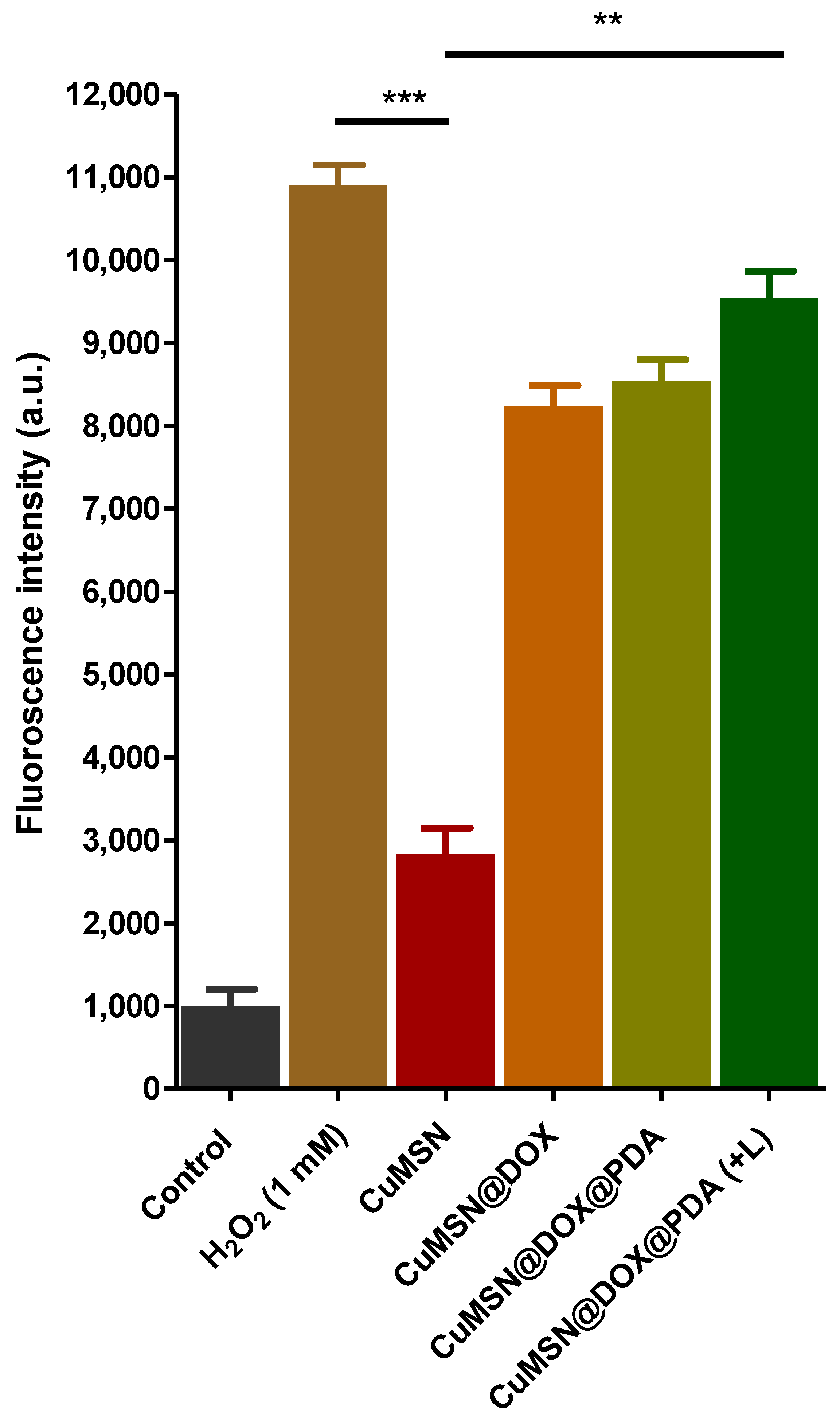
The anticancer efficacy of CuMSN and other nanocomposites were verified by MTT assay in A549 cells. The proposed formulation successfully eradicated the cancer cells by ROS production through combinatorial therapy. As Figure 6A shows the PDA-coated CuMSN@DOX samples with an IC 50 value of 1.6 μg in the presence of light and 2.90 μg in the absence of light. Furthermore, DOX-loaded NPs show better anticancer efficacy at a very low concentration (2.95 μg) than that of pure DOX (IC-50—7.5 μg). Interestingly, the copper-substituted MSN, together with DOX, enhanced the generation of ROS, and PDA serves as a photothermal agent to achieve CDT/PTT synergetic therapy at a low dose [77,78,79].

3.5. ROS Detection

The synergistic anticancer efficacy of designed NPs against A549 cells can be attributed to the production of ROS at the cellular level. Recently, some groups have reported that DOX, in combination with PDA, can significantly enhance anticancer efficacy by increasing the ROS and thermal energy production, thereby killing cancer cells synergistically. The intracellular ROS levels were detected by DCFDA assay, as shown in Figure 7. The ROS levels of PDA-coated CuMSN@DOX samples was evaluated in the presence and absence of light and compared to the remaining sample groups except the H2O2 (negative control) group. Our results demonstrated that light significantly increased the level of ROS production through synergistic CDT/PTT therapy, thereby successfully killing the cancer cells [80,81].

4. Conclusions

In summary, we designed a biocompatible copper-substituted mesoporous silica nanoparticle for the eradication of cancer cells. The anti-tumor drug DOX was loaded into mesopores by establishing a pH-sensitive bond to overcome premature release. Furthermore, PDA coating served as a PTT agent and gatekeeper along with the extension of DOX release. The physical and in vitro studies revealed that the synthesized CuMSN and modifications are advisable for CDT/PTT therapy. DOX participated in the ROS with copper ions, and PDA generates thermal energy (PTT). This promising approach can synergistically eradicate cancer cells effectively.

Supplementary Materials

The following supporting information can be downloaded at: https://www.mdpi.com/article/10.3390/coatings12010060/s1, Table S1: Tabular representation of CuMSN NP chemical composition in detail.

Author Contributions

Conceptualization and methodology, software, formal analysis, writing— original draft preparation, validation, supervision, editing, resources, and funding, P.B.; validation, formal analysis, investigation, and funding acquisition R.K.; conceptualization, and methodology, software, formal analysis, writing—original draft preparation, validation, resources, and funding, Y.K. All authors have read and agreed to the published version of the manuscript.

Funding

This research was supported by the National Research Foundation of Korea (NRF) funded by the Korea Government (2020R1A2C1012439), Republic of Korea.

Institutional Review Board Statement

We have not conducted any experiments on animals or humans.

Informed Consent Statement

Not applicable.

Data Availability Statement

No supporting data is presented in this study.

Conflicts of Interest

The authors declare that they have no known competing financial interests or personal relationships that could have appeared to influence the work reported in this paper.

References

  1. Sung, H.; Ferlay, J.; Siegel, R.L.; Laversanne, M.; Soerjomataram, I.; Jemal, A.; Bray, F. Global Cancer Statistics 2020: GLOBOCAN Estimates of Incidence and Mortality Worldwide for 36 Cancers in 185 Countries. CA A Cancer J. Clin. 2021, 71, 209–249. [Google Scholar] [CrossRef]
  2. Siegel, R.L.; Miller, K.D.; Fuchs, H.E.; Jemal, A. Cancer Statistics, 2021. CA A Cancer J. Clin. 2021, 71, 7–33. [Google Scholar] [CrossRef]
  3. Zahreddine, H.K. Borden Mechanisms and Insights into Drug Resistance in Cancer. Front. Pharmacol. 2013, 4, 28. [Google Scholar] [CrossRef] [Green Version]
  4. Luqmani, Y.A. Mechanisms of Drug Resistance in Cancer Chemotherapy. Med. Princ. Pract. 2005, 14 (Suppl. 1), 35–48. [Google Scholar] [CrossRef]
  5. Pritchard, J.R.; Lauffenburger, D.A.; Hemann, M.T. Hemann Understanding Resistance to Combination Chemotherapy. Drug Resist. Updates Rev. Comment. Antimicrob. Anticancer. Chemother. 2012, 15, 249–257. [Google Scholar]
  6. Wang, H.; Huang, Y. Combination Therapy Based on Nano Codelivery for Overcoming Cancer Drug Resistance. Med. Drug Discov. 2020, 6, 100024. [Google Scholar] [CrossRef]
  7. Poulose, A.C.; Veeranarayanan, S.; Mohamed, M.S.; Aburto, R.R.; Mitcham, T.; Bouchard, R.; Ajayan, P.M.; Sakamoto, Y.; Maekawa, T.; Kumar, D.S. Multifunctional Cu2−xTe Nanocubes Mediated Combination Therapy for Multi-Drug Resistant MDA MB 453. Sci. Rep. 2016, 6, 35961. [Google Scholar] [CrossRef] [Green Version]
  8. Li, Z.; Chen, Y.; Yang, Y.; Yu, Y.; Zhang, Y.; Zhu, D.; Yu, X.; Ouyang, X.; Xie, Z.; Zhao, Y.; et al. Recent Advances in Nanomaterials-Based Chemo-Photothermal Combination Therapy for Improving Cancer Treatment. Front. Bioeng. Biotechnol. 2019, 7, 293. [Google Scholar] [CrossRef]
  9. Fang, T.; Zhang, J.; Zuo, T.; Wu, G.; Xu, Y.; Yang, Y.; Yang, J.; Shen, Q. Chemo-Photothermal Combination Cancer Therapy with ROS Scavenging, Extracellular Matrix Depletion, and Tumor Immune Activation by Telmisartan and Diselenide-Paclitaxel Prodrug Loaded Nanoparticles. ACS Appl. Mater. Interfaces 2020, 12, 31292–31308. [Google Scholar] [CrossRef]
  10. Du, X.-F.; Li, Y.; Long, J.; Zhang, W.; Wang, D.; Li, C.-R.; Zhao, M.-X.; Lai, Y. Fabrication of Cisplatin-Loaded Polydopamine Nanoparticles via Supramolecular Self-Assembly for Photoacoustic Imaging Guided Chemo-Photothermal Cancer Therapy. Appl. Mater. Today 2021, 23, 101019. [Google Scholar] [CrossRef]
  11. Abbasian, M.; Mahmoodzadeh, F.; Salehi, R.; Amirshaghaghi, A. Chemo-Photothermal Therapy of Cancer Cells Using Gold Nanorod-Cored Stimuli-Responsive Triblock Copolymer. New J. Chem. 2017, 41, 12777–12788. [Google Scholar] [CrossRef]
  12. Zhu, H.; Sarkar, S.; Scott, L.; Danelisen, I.; Trush, M.A.; Jia, Z.; Li, Y.R. Doxorubicin Redox Biology: Redox Cycling, Topoisomerase Inhibition, and Oxidative Stress. React. Oxyg. Species 2016, 1, 189–198. [Google Scholar] [CrossRef]
  13. Fan, C.; Zheng, W.; Fu, X.; Li, X.; Wong, Y.-S.; Chen, T. Strategy to Enhance the Therapeutic Effect of Doxorubicin in Human Hepatocellular Carcinoma by Selenocystine, a Synergistic Agent that Regulates the ROS-Mediated Signaling. Oncotarget 2014, 5, 2853–2863. [Google Scholar] [CrossRef] [Green Version]
  14. Ijäs, H.; Shen, B.; Heuer-Jungemann, A.; Keller, A.; Kostiainen, M.A.; Liedl, T.; Ihalainen, J.; Linko, V. Unraveling the Interaction between Doxorubicin and DNA Origami Nanostructures for Customizable Chemotherapeutic Drug Release. Nucleic Acids Res. 2021, 49, 3048–3062. [Google Scholar] [CrossRef]
  15. Qiao, X.; van der Zanden, S.Y.; Wander, D.P.A.; Borràs, D.M.; Song, J.-Y.; Li, X.; van Duikeren, S.; van Gils, N.; Rutten, A.; van Herwaarden, T.; et al. Uncoupling DNA Damage from Chromatin Damage to Detoxify Doxorubicin. Proc. Natl. Acad. Sci. USA 2020, 117, 15182. [Google Scholar] [CrossRef]
  16. Wang, N.; Liu, C.; Yao, W.; Zhou, H.; Yu, S.; Chen, H.; Qiao, W. Endogenous Reactive Oxygen Species Burst Induced and Spatiotemporally Controlled Multiple Drug Release by Traceable Nanoparticles for Enhancing Antitumor Efficacy. Biomater. Sci. 2021, 9, 4968–4983. [Google Scholar] [CrossRef]
  17. Kim, D.; Park, S.; Yoo, H.; Park, S.; Kim, J.; Yum, K.; Kim, K.; Kim, H. Overcoming Anticancer Resistance by Photodynamic Therapy-Related Efflux Pump Deactivation and Ultrasound-Mediated Improved Drug Delivery Efficiency. Nano Converg. 2020, 7, 30. [Google Scholar] [CrossRef]
  18. Ghosn, Y.; Kamareddine, M.H.; Tawk, A.; Elia, C.; El Mahmoud, A.; Terro, K.; El Harake, N.; El-Baba, B.; Makdessi, J.; Farhat, S. Inorganic Nanoparticles as Drug Delivery Systems and Their Potential Role in the Treatment of Chronic Myelogenous Leukaemia. Technol. Cancer Res. Treat. 2019, 18, 1533033819853241. [Google Scholar] [CrossRef]
  19. Shi, Z.; Zhou, Y.; Fan, T.; Lin, Y.; Zhang, H.; Mei, L. Inorganic Nano-Carriers Based Smart Drug Delivery Systems for Tumor Therapy. Smart Mater. Med. 2020, 1, 32–47. [Google Scholar] [CrossRef]
  20. Wang, F.; Li, C.; Cheng, J.; Yuan, Z. Recent Advances on Inorganic Nanoparticle-Based Cancer Therapeutic Agents. Int. J. Environ. Res. Public Health 2016, 13, 1182. [Google Scholar] [CrossRef]
  21. Kuthati, Y.; Busa, P.; Tummala, S.; Rao, V.; Davuluri, V.; Ho, Y.-P.; Wong, C.-S. Mesoporous Polydopamine Nanoparticles Attenuate Morphine Tolerance in Neuropathic Pain Rats by Inhibition of Oxidative Stress and Restoration of the Endogenous Antioxidant System. Antioxidants 2021, 10, 195. [Google Scholar] [CrossRef]
  22. Wang, Y.; Zhao, Q.; Han, N.; Bai, L.; Li, J.; Che, E.; Hu, L.; Zhang, Q.; Jiang, T.; Wang, S. Mesoporous Silica Nanoparticles in Drug Delivery and Biomedical Applications. Nanomed. Nanotechnol. Biol. Med. 2015, 11, 313–327. [Google Scholar] [CrossRef]
  23. Niculescu, V.-C. Mesoporous Silica Nanoparticles for Bio-Applications. Front. Mater. 2020, 7, 36. [Google Scholar] [CrossRef] [Green Version]
  24. Xu, C.; Lei, C.; Yu, C. Mesoporous Silica Nanoparticles for Protein Protection and Delivery. Front. Chem. 2019, 7, 290. [Google Scholar] [CrossRef] [Green Version]
  25. Kuthati, Y.; Kankala, R.K.; Busa, P.; Lin, S.-X.; Deng, J.-P.; Mou, C.-Y.; Lee, C.-H. Phototherapeutic Spectrum Expansion through Synergistic Effect of Mesoporous Silica Trio-Nanohybrids against Antibiotic-Resistant Gram-Negative Bacterium. J. Photochem. Photobiol. B Biol. 2017, 169, 124–133. [Google Scholar] [CrossRef]
  26. Castillo, R.R.; Lozano, D.; Vallet-Regí, M. Mesoporous Silica Nanoparticles as Carriers for Therapeutic Biomolecules. Pharmaceutics 2020, 12, 432. [Google Scholar] [CrossRef]
  27. Zhou, Y.; Quan, G.; Wu, Q.; Zhang, X.; Niu, B.; Wu, B.; Huang, Y.; Pan, X.; Wu, C. Mesoporous Silica Nanoparticles for Drug and Gene Delivery. Acta Pharm. Sinica. B 2018, 8, 165–177. [Google Scholar] [CrossRef]
  28. Lin, C.-H.; Kankala, R.K.; Busa, P.; Lee, C.-H. Hydrophobicity-Tuned Periodic Mesoporous Organo-Silica Nanoparticles for Photodynamic Therapy. Int. J. Mol. Sci. 2020, 21, 2586. [Google Scholar] [CrossRef] [Green Version]
  29. Li, Z.; Zhang, Y.; Feng, N. Mesoporous Silica Nanoparticles: Synthesis, Classification, Drug Loading, Pharmacokinetics, Biocompatibility, and Application in Drug Delivery. Expert Opin. Drug Deliv. 2019, 16, 219–237. [Google Scholar] [CrossRef]
  30. Trzeciak, K.; Chotera-Ouda, A.; Bak-Sypien, I.; Potrzebowski, M. Mesoporous Mesoporous Silica Particles as Drug Delivery Systems—The State of the Art in Loading Methods and the Recent Progress in Analytical Techniques for Monitoring These Processes. Pharmaceutics 2021, 13, 950. [Google Scholar] [CrossRef]
  31. Bhattacharyya, S.; Wang, H.; Ducheyne, P. Polymer-Coated Mesoporous Silica Nanoparticles for the Controlled Release of Macromolecules. Acta Biomater. 2012, 8, 3429–3435. [Google Scholar] [CrossRef]
  32. Bansal, K.K.; Mishra, D.K.; Rosling, A.; Rosenholm, J.M. Therapeutic Potential of Polymer-Coated Mesoporous Silica Nanoparticles. Appl. Sci. 2020, 10, 289. [Google Scholar] [CrossRef] [Green Version]
  33. Xing, L.; Zheng, H.; Cao, Y.; Che, S. Coordination Polymer Coated Mesoporous Silica Nanoparticles for pH-Responsive Drug Release. Adv. Mater. 2012, 24, 6433–6437. [Google Scholar] [CrossRef]
  34. Song, Z.; Shi, J.; Zhang, Z.; Qi, Z.; Han, S.; Cao, S. Mesoporous Silica-Coated Gold Nanorods with a Thermally Responsive Polymeric Cap for Near-Infrared-Activated Drug Delivery. J. Mater. Sci. 2018, 53, 7165–7179. [Google Scholar] [CrossRef]
  35. Choi, S.R.; Jang, D.J.; Kim, S.; An, S.; Lee, J.; Oh, E.; Kim, J. Polymer-Coated Spherical Mesoporous Silica for pH-Controlled Delivery of Insulin. J. Mater. Chem. B 2014, 2, 616–619. [Google Scholar] [CrossRef] [Green Version]
  36. Yang, Y.; Yan, X.; Cui, Y.; He, Q.; Li, D.; Wang, A.; Fei, J.; Li, J. Preparation of Polymer-Coated Mesoporous Silica Nanoparticles Used for Cellular Imaging by a “Graft-from” Method. J. Mater. Chem. 2008, 18, 5731–5737. [Google Scholar] [CrossRef]
  37. Tang, H.; Zhao, W.; Yu, J.; Li, Y.; Zhao, C. Recent Development of pH-Responsive Polymers for Cancer Nanomedicine. Molecules 2019, 24, 4. [Google Scholar] [CrossRef] [Green Version]
  38. Alsehli, M. Polymeric Nanocarriers as Stimuli-Responsive Systems for Targeted Tumor (Cancer) Therapy: Recent Advances in Drug Delivery. Saudi Pharm. J. 2020, 28, 255–265. [Google Scholar] [CrossRef]
  39. Wang, X.; Li, X.; Ito, A.; Sogo, Y.; Watanabe, Y.; Tsuji, N.M.; Ohno, T. Biodegradable Metal Ion-Doped Mesoporous Silica Nanospheres Stimulate Anticancer Th1 Immune Response in Vivo. ACS Appl. Mater. Interfaces 2017, 9, 43538–43544. [Google Scholar] [CrossRef]
  40. Tahmasbi, L.; Sedaghat, T.; Motamedi, H.; Kooti, M. Mesoporous Silica Nanoparticles Supported Copper(II) and Nickel(II) Schiff Base Complexes: Synthesis, Characterization, Antibacterial Activity and Enzyme Immobilization. J. Solid State Chem. 2018, 258, 517–525. [Google Scholar] [CrossRef]
  41. Pan, Y.; Zhou, S.; Li, Y.; Parshad, B.; Li, W.; Haag, R. Novel Dendritic Polyglycerol-Conjugated, Mesoporous Silica-Based Targeting Nanocarriers for Co-Delivery of Doxorubicin and Tariquidar to Overcome Multidrug Resistance in Breast Cancer stem Cells. J. Control. Release 2021, 330, 1106–1117. [Google Scholar] [CrossRef]
  42. Abdullah, C.S.; Ray, P.; Alam, S.; Kale, N.; Aishwarya, R.; Morshed, M.; Dutta, D.; Hudziak, C.; Banerjee, S.K.; Mallik, S.; et al. Chemical Architecture of Block Copolymers Differentially Abrogate Cardiotoxicity and Maintain the Anticancer Efficacy of Doxorubicin. Mol. Pharm. 2020, 17, 4676–4690. [Google Scholar] [CrossRef]
  43. Shi, M.; Zhang, J.; Li, J.; Fan, Y.; Wang, J.; Sun, W.; Yang, H.; Peng, C.; Shen, M.; Shi, X. Polydopamine-Coated Magnetic Mesoporous Silica Nanoparticles for Multimodal Cancer Theranostics. J. Mater. Chem. B 2019, 7, 368–372. [Google Scholar] [CrossRef]
  44. Zhang, X.; Achazi, K.; Steinhilber, D.; Kratz, F.; Dernedde, J.; Haag, R. A Facile Approach for Dual-Responsive Prodrug Nanogels Based on Dendritic Polyglycerols with Minimal Leaching. J. Control. Release 2014, 174, 209–216. [Google Scholar] [CrossRef]
  45. Pada, A.-K.; Desai, D.; Sun, K.; Govardhanam, N.P.; Törnquist, K.; Zhang, J.; Rosenholm, J.M. Comparison of Polydopamine-Coated Mesoporous Silica Nanorods and Spheres for the Delivery of Hydrophilic and Hydrophobic Anticancer Drugs. Int. J. Mol. Sci. 2019, 20, 3408. [Google Scholar] [CrossRef] [Green Version]
  46. Zhu, M.; Shi, Y.; Shan, Y.; Guo, J.; Song, X.; Wu, Y.; Wu, M.; Lu, Y.; Chen, W.; Xu, X.; et al. Recent Developments in Mesoporous Polydopamine-Derived Nanoplatforms for Cancer Theranostics. J. Nanobiotechnology 2021, 19, 387. [Google Scholar] [CrossRef]
  47. Li, X.; Xie, C.; Xia, H.; Wang, Z. pH and Ultrasound Dual-Responsive Polydopamine-Coated Mesoporous Silica Nanoparticles for Controlled Drug Delivery. Langmuir 2018, 34, 9974–9981. [Google Scholar] [CrossRef]
  48. Lei, W.; Sun, C.; Jiang, T.; Gao, Y.; Yang, Y.; Zhao, Q.; Wang, S. Polydopamine-Coated Mesoporous Silica Nanoparticles for Multi-Responsive Drug Delivery and Combined Chemo-Photothermal Therapy. Mater. Sci. Eng. C. 2019, 105, 110103. [Google Scholar] [CrossRef]
  49. Huang, C.; Zhang, L.; Guo, Q.; Zuo, Y.; Wang, N.; Wang, H.; Kong, D.; Zhu, D.; Zhang, L. Robust Nanovaccine Based on Polydopamine-Coated Mesoporous Silica Nanoparticles for Effective Photothermal-Immunotherapy Against Melanoma. Adv. Funct. Mater. 2021, 31, 2010637. [Google Scholar] [CrossRef]
  50. Liao, J.; Zhang, H.; Wang, X. Polydopamine-Doped Virus-like Mesoporous Silica Coated Reduced Graphene Oxide Nanosheets for Chemo-Photothermal Synergetic Therapy. J. Biomater. Appl. 2020, 35, 28–38. [Google Scholar] [CrossRef]
  51. Tran, H.Q.; Bhave, M.; Xu, G.; Sun, C.; Yu, A. Synthesis of Polydopamine Hollow Capsules via a Polydopamine Mediated Silica Water Dissolution Process and Its Application for Enzyme Encapsulation. Front. Chem. 2019, 7, 468. [Google Scholar] [CrossRef] [Green Version]
  52. Duo, Y.; Li, Y.; Chen, C.; Liu, B.; Wang, X.; Zeng, X.; Chen, H. DOX-Loaded pH-Sensitive Mesoporous Silica Nanoparticles Coated with PDA and PEG Induce Pro-Death Autophagy in Breast Cancer. RSC Adv. 2017, 7, 39641–39650. [Google Scholar] [CrossRef] [Green Version]
  53. Wei, Y.; Gao, L.; Wang, L.; Shi, L.; Wei, E.; Zhou, B.; Zhou, L.; Ge, B. Polydopamine and Peptide Decorated Doxorubicin-Loaded Mesoporous Silica Nanoparticles as a Targeted Drug Delivery System for Bladder Cancer Therapy. Drug Deliv. 2017, 24, 681–691. [Google Scholar] [CrossRef] [Green Version]
  54. Day, C.; Sweetman, M.; Song, Y.; Plush, S.; Garg, S. Functionalized Mesoporous Silica Nanoparticles as Delivery Systems for Doxorubicin: Drug Loading and Release. Appl. Sci. 2021, 11, 6121. [Google Scholar] [CrossRef]
  55. Tao, C.; Chen, T.; Liu, H.; Su, S. Preparation and Adsorption Performance Research of Large-Volume Hollow Mesoporous Polydopamine Microcapsules. MRS Commun. 2019, 9, 744–749. [Google Scholar] [CrossRef]
  56. Cheng, W.; Liang, C.; Xu, L.; Liu, G.; Gao, N.; Tao, W.; Luo, L.; Zuo, Y.; Wang, X.; Zhang, X.; et al. TPGS-Functionalized Polydopamine-Modified Mesoporous Silica as Drug Nanocarriers for Enhanced Lung Cancer Chemotherapy against Multidrug Resistance. Small 2017, 13, 1700623. [Google Scholar] [CrossRef]
  57. Busa, P.; Koutavarapu, R.; Lee, D.-Y.; Shim, J.; Kuthati, Y. Hierarchical Two-Dimensional Layered Double Hydroxide Coated Polydopamine Nanocarriers for Combined Chemodynamic and Photothermal Tumor Therapy. Coatings 2021, 11, 1008. [Google Scholar] [CrossRef]
  58. Lin, C.-W.; Lin, S.-X.; Kankala, R.K.; Busa, P.; Deng, J.-P.; Lue, S.-I.; Liu, C.-L.; Weng, C.-F.; Lee, C.-H. Surface-Functionalized Layered Double Hydroxide Nanocontainers as Bile Acid Sequestrants for Lowering Hyperlipidemia. Int. J. Pharm. 2020, 590, 119921. [Google Scholar] [CrossRef]
  59. Jin, R.; Liu, Z.; Bai, Y.; Zhou, Y.; Chen, X. Multiple-Responsive Mesoporous Silica Nanoparticles for Highly Accurate Drugs Delivery to Tumor Cells. ACS Omega 2018, 3, 4306–4315. [Google Scholar] [CrossRef] [Green Version]
  60. Kuthati, Y.; Busa, P.; Davuluri, V.N.G.; Wong, C.S. Manganese Oxide Nanozymes Ameliorate Mechanical Allodynia in a Rat Model of Partial Sciatic Nerve-Transection Induced Neuropathic Pain. Int. J. Nanomed. 2019, 14, 10105–10117. [Google Scholar] [CrossRef] [Green Version]
  61. Dong, K.; Zhao, Z.-Z.; Kang, J.; Lin, L.-R.; Chen, W.-T.; Liu, J.-X.; Wu, X.-L.; Lu, T.-L. Cinnamaldehyde and Doxorubicin Co-Loaded Graphene Oxide Wrapped Mesoporous Silica Nanoparticles for Enhanced MCF-7 Cell Apoptosis. Int. J. Nanomed. 2020, 15, 10285–10304. [Google Scholar] [CrossRef]
  62. Xing, Y.; Zhang, J.; Chen, F.; Liu, J.; Cai, K. Mesoporous Polydopamine Nanoparticles with Co-Delivery Function for Overcoming Multidrug Resistance via Synergistic Chemo-Photothermal Therapy. Nanoscale 2017, 9, 8781–8790. [Google Scholar] [CrossRef]
  63. Li, X.-J.; Li, W.-T.; Li, Z.-H.-R.; Zhang, L.-P.; Gai, C.-C.; Zhang, W.-F.; Ding, D.-J. Iron-Chelated Polydopamine Decorated Doxorubicin-Loaded Nanodevices for Reactive Oxygen Species Enhanced Cancer Combination Therapy. Front. Pharmacol. 2019, 10, 75. [Google Scholar] [CrossRef] [Green Version]
  64. Liu, D.; Mori, A.; Huang, L. Role of Liposome Size and RES Blockade in Controlling Biodistribution and Tumor Uptake of GM1-Containing Liposomes. Biochim. Biophys Acta 1992, 1104, 95–101. [Google Scholar] [CrossRef]
  65. Shen, J.; He, Q.; Gao, Y.; Shi, J.; Li, Y. Mesoporous Silica Nanoparticles Loading Doxorubicin Reverse Multidrug Resistance: Performance and Mechanism. Nanoscale 2011, 3, 4314–4322. [Google Scholar] [CrossRef]
  66. Yuan, Z.; Pan, Y.; Cheng, R.; Sheng, L.; Wu, W.; Pan, G.; Feng, Q.; Cui, W. Doxorubicin-Loaded Mesoporous Silica Nanoparticle Composite Nanofibers for Long-Term Adjustments of Tumor Apoptosis. Nanotechnology 2016, 27, 245101. [Google Scholar] [CrossRef]
  67. Jain, S.; Khan, T.A.; Patil, Y.P.; Pagariya, D.; Kishore, N.; Tapryal, S.; Naik, A.D.; Naik, S.G. Bio-Affinity of Copper(II) Complexes with Nitrogen and Oxygen Donor Ligands: Synthesis, Structural Studies and in Vitro DNA and HSA Interaction of Copper(II) Complexes. J. Photochem. Photobiol. B Biol. 2017, 174, 35–43. [Google Scholar] [CrossRef]
  68. Zeng, X.; Liu, G.; Tao, W.; Ma, Y.; Zhang, X.; He, F.; Pan, J.; Mei, L.; Pan, G. A Drug-Self-Gated Mesoporous Antitumor Nanoplatform Based on pH-Sensitive Dynamic Covalent Bond. Adv. Funct. Mater. 2017, 27, 1605985. [Google Scholar] [CrossRef]
  69. Rahoui, N.; Jiang, B.; Hegazy, M.; Taloub, N.; Wang, Y.; Yu, M.; Huang, Y.D. Gold Modified Polydopamine Coated Mesoporous Silica Nano-Structures for Synergetic Chemo-Photothermal Effect. Colloids Surf. B Biointerfaces 2018, 171, 176–185. [Google Scholar] [CrossRef]
  70. Chen, X.; Cheng, X.; Soeriyadi, A.H.; Sagnella, S.M.; Lu, X.; Scott, J.A.; Lowe, S.B.; Kavallaris, M.; Gooding, J.J. Stimuli-Responsive Functionalized Mesoporous Silica Nanoparticles for Drug Release in Response to Various Biological Stimuli. Biomater. Sci. 2014, 2, 121–130. [Google Scholar] [CrossRef] [Green Version]
  71. Chang, D.; Gao, Y.; Wang, L.; Liu, G.; Chen, Y.; Wang, T.; Tao, W.; Mei, L.; Huang, L.; Zeng, X. Polydopamine-Based Surface Modification of Mesoporous Silica Nanoparticles as pH-Sensitive Drug Delivery Vehicles for Cancer Therapy. J. Colloid Interface Sci. 2015, 463, 279–287. [Google Scholar] [CrossRef]
  72. Tiwari, A.P.; Bhattarai, D.P.; Maharjan, B.; Ko, S.W.; Kim, H.Y.; Park, C.H.; Kim, C.S. Polydopamine-Based Implantable Multifunctional Nanocarpet for Highly Efficient Photothermal-Chemo Therapy. Sci. Rep. 2019, 9, 2943. [Google Scholar] [CrossRef]
  73. Jia, C.; Wu, H.; Luo, K.; Hao, W.; Wang, S.; Huang, M. Magnetic Silica Nanosystems With NIR-Responsive and Redox Reaction Capacity for Drug Delivery and Tumor Therapy. Front. Chem. 2020, 8, 567652. [Google Scholar] [CrossRef]
  74. Ruan, L.; Wang, M.; Zhou, M.; Lu, H.; Zhang, J.; Gao, J.; Chen, J.; Hu, Y. Doxorubicin-Metal Coordinated Micellar Nanoparticles for Intracellular Codelivery and Chemo/Chemodynamic Therapy in Vitro. ACS Appl. Bio Mater. 2019, 2, 4703–4707. [Google Scholar] [CrossRef]
  75. Yao, L.; Tang, Y.; Cao, W.; Cui, Y.; Qian, G. Highly Efficient Encapsulation of Doxorubicin Hydrochloride in Metal-Organic Frameworks for Synergistic Chemotherapy and Chemodynamic Therapy. ACS Biomater. Sci. Eng. 2021, 7, 4999–5006. [Google Scholar] [CrossRef]
  76. Xue, T.; Xu, C.; Wang, Y.; Wang, Y.; Tian, H.; Zhang, Y. Doxorubicin-Loaded Nanoscale Metal-Organic Framework for Tumor-Targeting Combined Chemotherapy and Chemodynamic Therapy. Biomater. Sci. 2019, 7, 4615–4623. [Google Scholar] [CrossRef]
  77. Chen, C.; Tang, W.; Jiang, D.; Yang, G.; Wang, X.; Zhou, L.; Zhang, W.; Wang, P. Hyaluronic Acid Conjugated Polydopamine Functionalized Mesoporous Silica Nanoparticles for Synergistic Targeted Chemo-Photothermal Therapy. Nanoscale 2019, 11, 11012–11024. [Google Scholar] [CrossRef]
  78. Shao, L.; Zhang, R.; Lu, J.; Zhao, C.; Deng, X.; Wu, Y. Mesoporous Silica Coated Polydopamine Functionalized Reduced Graphene Oxide for Synergistic Targeted Chemo-Photothermal Therapy. ACS Appl. Mater. Interfaces 2017, 9, 1226–1236. [Google Scholar] [CrossRef]
  79. Zhang, Y.; Eltayeb, O.; Meng, Y.; Zhang, G.; Shuang, S.; Dong, C. Tumor Microenvironment Responsive Mesoporous Silica Nanoparticles for Dual Delivery of Doxorubicin and Chemodynamic Therapy (CDT) agent. New J. Chem. 2020, 44, 2578–2586. [Google Scholar] [CrossRef]
  80. Xin, J.; Deng, C.; Aras, O.; Zhou, M.; Wu, C.; An, F. Chemodynamic Nanomaterials for Cancer Theranostics. J. Nanobiotechnology 2021, 19, 192. [Google Scholar] [CrossRef]
  81. Poinard, B.; Neo, S.Z.Y.; Yeo, E.L.L.; Heng, H.P.S.; Neoh, K.G.; Kah, J.C.Y. Polydopamine Nanoparticles Enhance Drug Release for Combined Photodynamic and Photothermal Therapy. ACS Appl. Mater. Interfaces 2018, 10, 21125–21136. [Google Scholar] [CrossRef]
Figure 1. (A) Schematic explanation of CuMSN, DOX loading, and PDA coating onto the surface and (B) a graphical description of chemotherapy and photothermal therapy killing the A549 cancer cells.
Coatings 12 00060 g001
Figure 2. (A) TEM images of (a) DOX-loaded and (b) PDA-coated CuMSN and (B) hydrodynamic sizes of (a) CuMSN-Unextd, (b) CuMSN, (c) CuMSN@DOX, and (d) CuMSN@DOX@PDA. (C) Zeta potential charges of CuMSN and their modifications, respectively. Data represent mean ± s.d., n = 3.
Coatings 12 00060 g002
Figure 3. (A) FT-IR analysis of (a) CuMSN-Unextd, (b) CuMSN-extd, (c) CuMSN@DOX, and (d) CuMSN@DOX@PDA and (B) TGA curves of (a) CuMSN-Unextd, (b) CuMSN@DOX, and (c) CuMSN@DOX@PDA.
Coatings 12 00060 g003
Figure 4. (A) DOX releasing from CuMSN@DOX and CuMSN@DOX@PDA over time at various PBS solutions of pH 5.0 and 7.4 (at 37 °C). (B) Temperature increasing profiles of CuMSN@DOX@PDA under 808 nm laser irradiation over time (50, 100, and 200 µg/mL).
Coatings 12 00060 g004
Figure 5. (A) Fluorescence microscopic images of CuMSN (100 µg/mL) cellular uptake with the treatment of FITC-labeled CuMSN (100 µg/mL), and (B) intracellular DOX uptake in A549 cells treated with CuMSN@DOX@PDA (20× magnification) (scale bar 100 µm).
Coatings 12 00060 g005
Figure 6. (A) In vitro cell-viability studies on the dose-dependent cell viability of A549 and HT-29 cells incubated with CuMSN, DOX, CuMSN@DOX, CuMSN@DOX@PDA, and CuAl-LDH@DOX@PDA (+L) for 24 h, and CuAl-LDH@DOX@PDA (+L)-treated groups were irradiated with a light source for 5 min after 24 h treatment (DOX-equivalent dose). (B) Toxicity profile investigations of the A549 and HT-29 cell with CuMSN. ***, **, and * denotes the significant difference between DOX vs CuMSN@DOX and DOX vs CuMSN@DOX@PDA (+L).
Coatings 12 00060 g006
Figure 7. Intracellular ROS production in A549 cells treated with nanoparticles with and without light irradiation by DCFDA assay. The fluorescence signal was detected by a microplate reader (λex = 488 nm, λem = 535 nm). ***, and ** denotes the significant difference between H2O2 vs CuMSN and CuMSN vs CuMSN@DOX@PDA (+L).
Coatings 12 00060 g007
Publisher’s Note: MDPI stays neutral with regard to jurisdictional claims in published maps and institutional affiliations.

