Article

Electrochemical Surface Biofunctionalization of Titanium through Growth of TiO2 Nanotubes and Deposition of Zn Doped Hydroxyapatite

by
Diana Maria Vranceanu
1,
Elena Ungureanu
1,
Ionut Cornel Ionescu
1,
Anca Constantina Parau
2,
Adrian Emil Kiss
2,
Alina Vladescu
2,3 and
Cosmin Mihai Cotrut
1,*
1
Faculty of Materials Science and Engineering, University Politehnica of Bucharest, 60042 Bucharest, Romania
2
Department for Advanced Surface Processing and Analysis by Vacuum Technologies, National Institute of Research and Development for Optoelectronics—INOE 2000, 77125 Magurele, Romania
3
Physical Materials Science and Composite Materials Centre, Research School of Chemistry & Applied Biomedical Sciences, National Research Tomsk Polytechnic University, 634050 Tomsk, Russia
*
Author to whom correspondence should be addressed.
Coatings 2022, 12(1), 69; https://doi.org/10.3390/coatings12010069
Submission received: 7 December 2021 / Revised: 4 January 2022 / Accepted: 5 January 2022 / Published: 7 January 2022
(This article belongs to the Special Issue Surface Modification of Medical Implants)

Abstract

:
The current research aim is to biofunctionalize pure titanium (Ti, grade IV) substrate with titania nanotubes and Zn doped hydroxyapatite-based coatings by applying a duplex electrochemical treatment, and to evaluate the influence of Zn content on the physico-chemical properties of hydroxyapatite (HAp). The obtained nanostructured surfaces were covered with HAp-based coatings doped with Zn in different concentrations by electrochemical deposition in pulsed galvanostatic mode. The obtained surfaces were characterized in terms of morphology, elemental and phasic composition, chemical bonds, roughness, and adhesion. The nanostructured surface consisted of titania nanotubes (NT), aligned, vertically oriented, and hollow, with an inner diameter of ~70 nm. X-ray Diffraction (XRD) analysis showed that the nanostructured surface consists of an anatase phase and some rutile peaks as a secondary phase. The morphology of all coatings consisted of ribbon like-crystals, and by increasing the Zn content the coating became denser due to the decrement of the crystals’ dimensions. The elemental and phase compositions evidenced that HAp was successfully doped with Zn through the pulsed galvanostatic method on the Ti nanostructured surfaces. Fourier Transform Infrared spectroscopy (FTIR) and XRD analysis confirmed the presence of HAp in all coatings, while the adhesion test showed that the addition of a high quantity leads to some delamination. Based on the obtained results, it can be said that the addition of Zn enhances the properties of HAp, and through proper experimental design, the concentration of Zn can be modulated to achieve coatings with tunable features.

1. Introduction

Generally, biomaterials play an important role in our society and have become a key element for human well-being, especially since the rate of elderly population is increasing rapidly [1]. Among these biomaterials, the metallic ones are extensively used in biomedical devices and components, such as hard tissue replacements, cardiac and cardiovascular stents, etc., due to their desirable mechanical properties and biocompatibility [2].
The fundamental requirement of a biomaterial is found to be in its interaction with its surrounding environment, with which it must coexist without either inducing any undesirable effect on the other [3]. Biomaterials are an important and integrated part of modern medicine and their development and improvement are important challenges [4].
Due to their remarkable properties, such as good mechanical properties, a Young modulus that is close to that of hard tissue, a high resistance in aggressive media, and high biocompatibility, titanium and its alloys are the most used biomaterials for manufacturing orthopedic [1,5] and dental implants [6]. Its affinity to form an oxide layer on the surface has led to it being one of the biomaterials most resistant to corrosion [7]. Despite all its favorable properties, however, titanium is also known for its bioinert character. As a result, commercially pure titanium (cp-Ti) implants often become encapsulated by fibrous tissues and the healing time is prolonged [8,9].
To improve the osseoconductivity of metallic implants, researchers around the world are trying to modify its properties through various surface treatments to promote the direct chemical bond between the implant and the bone tissue by obtaining bioactive surfaces that improve the osseointegration of titanium implants [10].
Surface modification of Ti based implants can be achieved by various physical, mechanical, or chemical treatments [11,12,13]. Some of these surface modification techniques include air-born abrasion [14], chemical treatment [15], and anodic oxidation [16]. In addition to these methods, there is another efficient route to modulate the surface characteristic of Ti, namely, by using bioactive coatings [17].
Calcium phosphates (CaP), in particular hydroxyapatite (HAp), are the most investigated bioactive materials. Hydroxyapatite has a Ca/P ratio of 1.67 and is one of the most stable CaPs, possessing properties such as biocompatibility, osseoconductivity, and osseointegration [18]. Due to its compact hexagonal structure, HAp can easily accept the substitution of Ca2+ with other divalent (Sr2+, Mg2+, Zn2+) and trivalent (Ce3+) metallic ions [19].
Recently, zinc has attracted much interest because it can be used as a doping element in the HAp structure [20,21,22] and can improve cell proliferation, viability, and adhesion [23], as well as offering antibacterial effects, which are useful in preventing implant-related infections [24]. Moreover, zinc is one of the essential elements of the human body, with an important role in the regulation of bone formation [25]. Thus, through this strategy, by combining HAp with Zn, coatings with enhanced surface characteristics can be designed.
A study performed by Ito et al. [26] has shown that over a certain amount (higher than 1.2 wt.%), Zn is toxic to osteoblast cells. In terms of antibacterial efficiency, a study by Sergi et al. [22] showed that Zn has a higher efficiency against S. aureus than E. coli, while Tank et al. [27] reported that Zn-doped HAp coatings have a greater antibacterial effect on S. aureus and M. luteus than on B. cereus and S. flexneri. In an in vitro study performed by Candidato et al. [28], it was shown that Zn-doped coatings may inhibit the precipitation of new calcium phosphates. Thus, considering the above, it can be deduced that special attention must be given to the designing stage of the material with multi-functional features.
Zn-doped hydroxyapatite coatings can be deposited by several techniques, such as magnetron sputtering [29], plasma spray [30], pulsed laser deposition [31], sol–gel [32], or electrochemical deposition [33]. Electrochemical deposition is a very interesting deposition technique, which in recent years has gained special attention due to advantages such as low costs, the ability to cover surfaces with complex geometries, and a good control of the coating’s properties [34,35]. However, the disadvantage of electrochemical deposition is the poor adhesion of the coatings [36]. To improve their adhesion, different treatments can be applied before deposition, such as anodic oxidation and pre-treatment with NaOH, followed by a heat treatment [37,38].
Anodic oxidation is a simple and cost-efficient technique, which is extensively used to obtain titanium-based materials with a nanostructured surface that consists of titanium dioxide (TiO2) nanotubes (NT) [39]. TiO2 nanotubes are characterized by pore size and length, features that can be easily modified due to the simplicity of the technique [40].
Thus, the aim of the present research is to functionalize the surface of Ti by applying a duplex electrochemical treatment that consists of an initial anodization of the Ti substrate to obtain TiO2 NT, followed by the deposition of undoped and Zn doped HAp based coatings with different Zn concentrations to develop coatings with a suitable bioactive character and to offer proper antibacterial efficiency. The conceptual design of this duplex electrochemical treatment applied to cp-Ti represent the novelty of the present study. To the best of our knowledge, there are only a few studies on the electrochemical deposition of Zn doped HAp and just Ref. [41] reports on Zn doped HAp deposited on TiO2 NT (using other deposition parameters). Therefore, to achieve the proposed aim, two different quantities of Zn were used to establish the influence of Zn content on the properties of HAp in terms of morphology, elemental and phasic composition, chemical bonds, roughness, and adhesion of the coatings to the substrate.

2. Materials and Methods

2.1. Sample Preparation

A commercially pure titanium bar (cp-Ti), purchased from Bibus Metals AG, Essen, Germany, was cut into discs (ϕ = 20 mm, h = 2 mm), which were used as starting materials. The cp-Ti discs were metallographically prepared on silicon carbide paper of different grit (250–1200 grit) and then polished with 1 μm alumina slurry. The prepared discs were cleaned with ultra-pure water (ASTM I) and iso-propylic alcohol to remove the contaminants accumulated during surface preparation. Subsequently, the discs were ultrasonically degreased in acetone and cleaned with ultrapure water. After these steps, the samples had a mirror aspect, and the duplex electrochemical treatment could be applied.

2.2. Duplex Electrochemical Treatment

2.2.1. Anodization

The electrochemical setup of the anodizing experiment for a nanostructured surface with self-organized TiO2 consisted of a two-electrode configuration with a stainless-steel sheet as a counter electrode and the sample (Ti discs) as working electrodes. The nanostructured surface was obtained in 0.5 wt.% HF electrolyte. The anodization potential was kept constant at 20 V with a DC power supply system (N5771A model, Keysight, Böblingen, Germany) for 30 min. The as-anodized samples were annealed for 2 h at 450 °C in air with a heat treatment furnace (N17/HR model, Nabertherm, Lilienthal, Germany).

2.2.2. Coating Deposition

Electrochemical deposition of hydroxyapatite was conducted in a typical three electrode electrochemical cell configured as follows: the working electrode (WE) was the cp-Ti disc with a nanostructured surface, the reference electrode (RE) was a calomel electrode, and the counter electrode (CE) was a platinum foil. The electrodeposition was controlled using a multichannel Potentiostat/Galvanostat (Parstat MC, Princeton Applied Research–Ametek, Oak Ridge, TN, USA) operating in pulsed galvanostatic mode. One pulse consisted of an activation stage (iON = −0.85 mA/cm2 for 1 s), followed by a relaxation stage (iOFF = 0 mA/cm2 for 1 s) [42]. In total, 1200 pulses were applied.
The electrolyte was prepared by dissolving calcium nitrate tetrahydrate (Ca(NO3)2·4H2O), ammonium dihydrogen phosphate (NH4H2PO4), and zinc nitrate hexahydrate (Zn(NO3)2·6H2O) in ultra-pure water in different concentrations, which are presented in Table 1. All the chemicals were of reagent grade and were purchased from Sigma-Aldrich (Munich, Germany). The electrolyte’s pH was measured and adjusted to 5 by the drop-wise of 1 M NaOH in the presence of a pH/ORP-meter (HI 4221, Hanna Instruments, Smithfield, RI, USA). To minimize the formation of CaCO3 deposits, the electrolyte was de-aerated with nitrogen gas before deposition.
During deposition, the temperature was controlled and kept constant to 75 °C using a basic magnetic hotplate stirrer (KA RCT Basic Safety Control Hotplate/Stirrer and ETS-D6 Temp, IKA, Staufen, Germany). To keep the concentration of the electrolyte uniform, during the deposition, the solution was continuously stirred with a magnetic bar. After the electrodeposition, the samples were rinsed with ultra-pure water and dried at room temperature in a desiccator.

2.3. Characterization

The nanostructured surface of cp-Ti, consisting of titania nanotubes (NT), was analyzed in terms of morphology with a field-emission scanning electron microscope (Scienta Omicron NanoSAM Lab system, Oxford Instruments, High Wycombe, UK), while the coating’s surface morphology was investigated with a Scanning Electron Microscope equipped with an X-ray Energy Dispersive Spectrometer (SEM-EDS, Phenom ProX, Phenom World, Eindhoven, The Netherlands).
The phase composition of the samples (nanostructured surface and coatings) was studied by X-ray diffraction (Rigaku SmartLab, Tokyo, Japan) using an X-ray source of CuKα (λ = 1.5406 Å) in the range θ/2θ of 20–80° at an incident angle of 3°, with a medium resolution parallel beam optical system.
The Debye-Scherrer mathematical relation (Equation (1)) was used to estimate the crystallite size for the (002) diffraction plane, being the most intense diffraction peak in HAp coatings obtained by electrochemical deposition [37].
L ( 002 ) = K   λ   β   c o s θ ( 002 )
where K = 0.9 is a constant shape factor for HAp with hexagonal structure, β is the full width at half maximum (FWHM) measured in radian of (002) diffraction plane, and θ in degrees indicates the Braggs diffraction angle, λ = 1.5406 Å is the wavelength of CuKα radiation.
The crystallinity (χc) was estimated using the following equation (Equation (2)) [43,44,45]:
χ c = ( K A   β ) 3
where KA is a constant found equal to 0.24 for HAp bioceramic, and β is the FWHM of refection (002) in degrees.
The lattice parameters of hydroxyapatite-based coatings were computed using (Equation (3)), the relation between the interplanar spacing distance (dhkl) and lattice constants (a and c) for a hexagonal crystallographic system [46,47], as follows:
1 d h k l 2 = 4 ( h 2 + k 2 + l 2 ) 3 a 2 + 1 c 2
The chemical bonds and identification of the functional groups was achieved by Fourier Transform Infra-Red (FTIR) spectroscopy in attenuated total reflectance (ATR) mode. The FTIR-ATR spectra were collected in the 4000–600 cm−1 wave numbers range using FT-IR, Jasco 6300 (Jasco, Tokyo, Japan) with the universal ATR sampling accessory Pike MIRacle (Pike Technologies, Madison, WI, USA).
Surface roughness was measured over a length of 3000 μm, using a DEKTAK 150 (Veeco Instruments, Plainview, NY, USA) profilometer. The thickness of the coatings were also determined by a DEKTAK 150 profilometer (stylus radius of 12.5 μm), measuring the forming ledge between the deposited film and the uncoated substrate.
The adhesion between the nanostructured Ti surface (NT) substrate and the undoped and Zn doped HAp coating was measured by “tape test” according to the ASTM D3359-17 standard [48] with an Elcometer 107 Cross Hatch Adhesion Tester kit (Ulmer, Aalen, Germany). The coating adhesion was assessed in terms of area removed by analyzing the lattice pattern indentation and was classified in terms of percentages (from highest to weakest): 5B: 0%; 4B: ≤5% 3B: 5%–15%; 2B: 15%–35%; 1B: 35%–65%; 0B: ≥65%. A more detailed presentation of the procedure is available in Ref. [42].

3. Results and Discussions

3.1. Morphology

The HR-SEM images of the anodized samples before and after annealing are presented in Figure 1. It can be observed that the TiO2 nanotubes are highly oriented and uniformly distributed on the surface. The inner diameter before and after annealing was measured and highlighted that the diameter of the NT presents a small decrement from 75 (±5 nm), obtained for the as-anodized one, compared to 70 (±7 nm) for the annealed ones.
Compared to our previous study [49], it can be said that by reducing the anodization time from 1 h to 0.5 h, the diameter has decreased from an average value of 75 nm to 70 nm, and the nanotubes appear to be better organized in terms of density (with smaller spaces between them). The obtained results are in strong correlation with the available literature [50,51]. Thus, it can be said that by reducing the anodization time, nanotubes with a smaller inner diameter can be obtained.
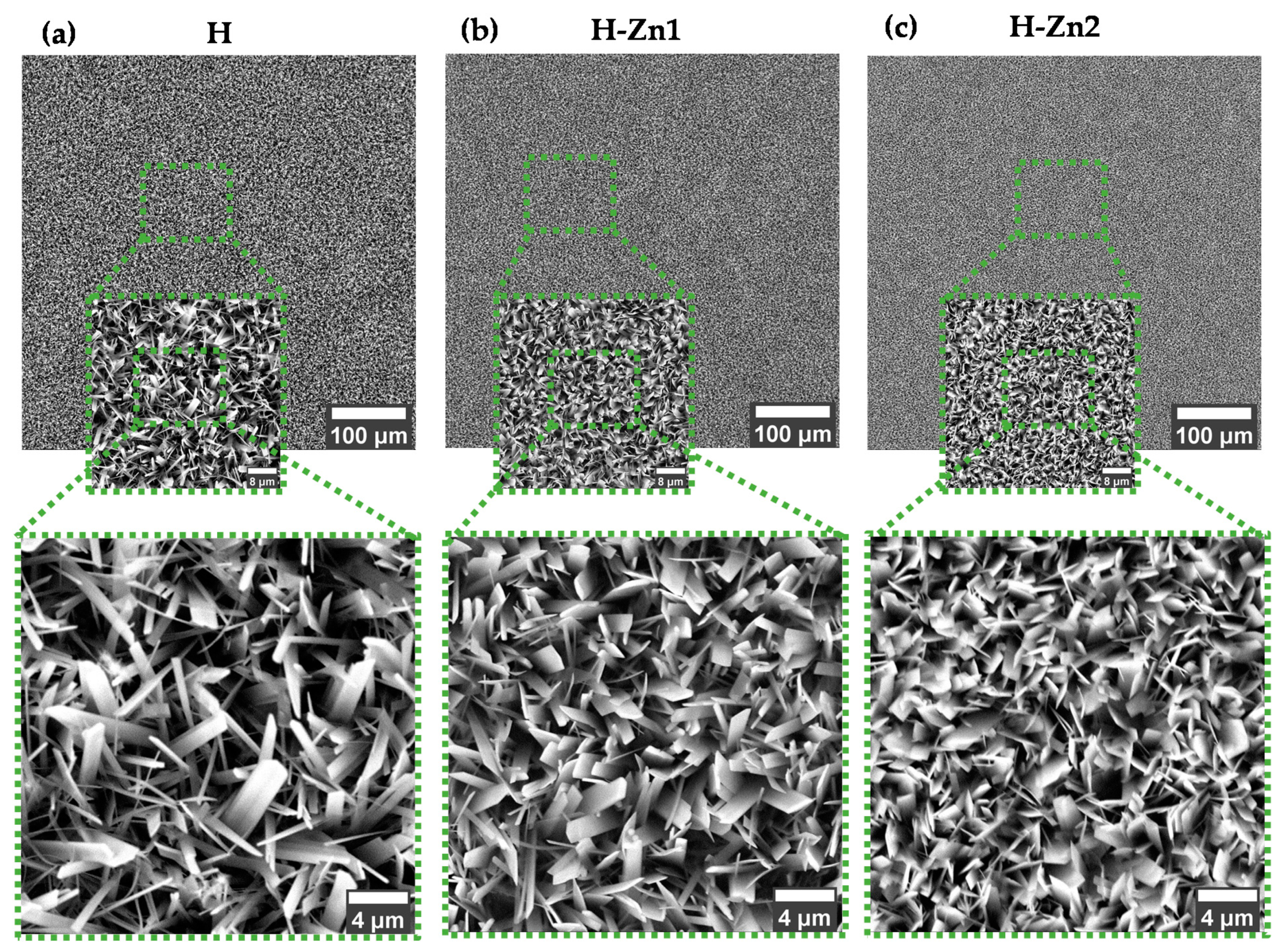
In Figure 2 are presented the SEM images obtained on the undoped and zinc doped HAp based coatings, electrochemically deposited on the TiO2 nanostructured surface (NT). At small magnification, it can be observed that all coatings are covering the surface with a uniform and compact layer, which does not present defects.
At higher magnification, the SEM images highlight that the coatings’ morphology consists of elongated ribbon-like crystals, very thin, within the nanometric scale, forming a complex morphology that resemble a 3D architecture. All coatings have crystallized perpendicular on the substrate, which is characteristic for the electrochemical deposition process [52].
The addition of Zn within the HAp has led to some morphological modifications, which are as follows. Compared to the H coatings, whose morphology is predominant in very thin and narrow crystals, the addition of Zn has led to visible dimensional changes of the ribbon-like crystals, which have become wider, thicker, and smaller, irrespective of Zn content. Moreover, by increasing the Zn concentration, not only are the crystals dimensions reducing, but the layer also begins to appear denser, suggesting that the addition of Zn, even in very small amounts, affects the electro-crystallization of HAp. A possible explanation for this could be attributed to the differences between the ionic radius of Zn2+ (0.074 nm) and Ca2+ (0.099 nm), which can lead to a structural mismatch that prevents HAp crystal growth when Zn is incorporated in the HAp structure [53,54].

3.2. Elemental Composition

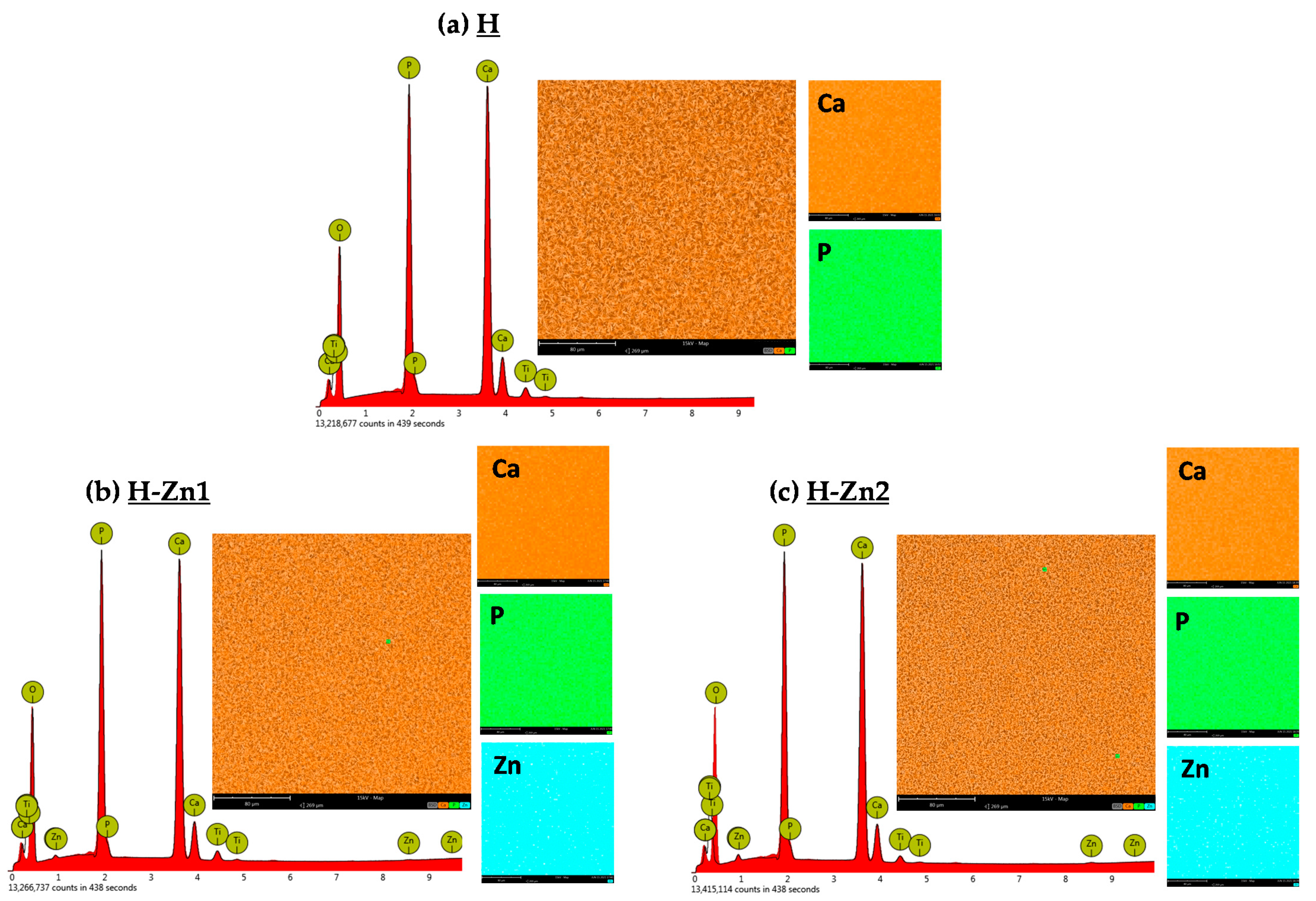
The EDS spectra, elemental composition and distribution of each element are presented in Figure 3 and Table 2. Based on this investigation, it can be stated that all characteristic elements of hydroxyapatite (Ca, O, P), along with the doping elements (Zn) and Ti, as part of the substrate and/or nanostructured surface, are present in all coatings.
Considering that Zn2+ substitutes the Ca2+ found in the HAp structure, the Ca/P ratio was calculated accordingly, and thus the ratio was obtained by summing the Zn to the Ca content, and divided to that of P, as following (Ca + Zn)/P.
All developed coatings presented smaller values in terms of their Ca/P ratio, compared to the natural HAp one, which had a ratio of 1.67. The highest Ca/P ratio was obtained for the H coatings, with a value of 1.59, which further decreased to 1.56 for H-Zn1 and 1.55 for H-Zn2, highlighting that the addition and increment of the Zn content alters the Ca/P ratio. A similar tendency was also reported in Ref [55].
A smaller Ca/P ratio, between 1.33 and 1.67, indicates the presence of a Ca-deficient apatite, which has a composition and structure that resembles the natural bone mineral [56] and is considered to be a suitable candidate for bone regeneration [57]. Moreover, a lower Ca/P ratio can also suggest an enhanced solubility and thus a more bioactive character [58].
Regarding the Zn content, it can be noted that the H-Zn1 coatings present a quantity of 0.78 at.%, which increases by 60% for the H-Zn2, reaching a value of 1.3 at.%.
According to the study performed by Miyaji et al. [59], it has been shown that there is a substituting limit of Zn which was estimated to be at about 15 mol.%, and that a too-high content of zinc favors the formation of amorphous phase, which can lead to rapid degradation. Ito et al. [26] suggested that a low concentration of Zn2+, found to be between 0.6 and 1.2 wt.%, is favorable to avoid cytotoxicity, and enhances cell proliferation. Thus, it can be stated that the H-Zn1 coatings fit within the maximum amount which does not affect the cell behavior.

3.3. Phase Composition

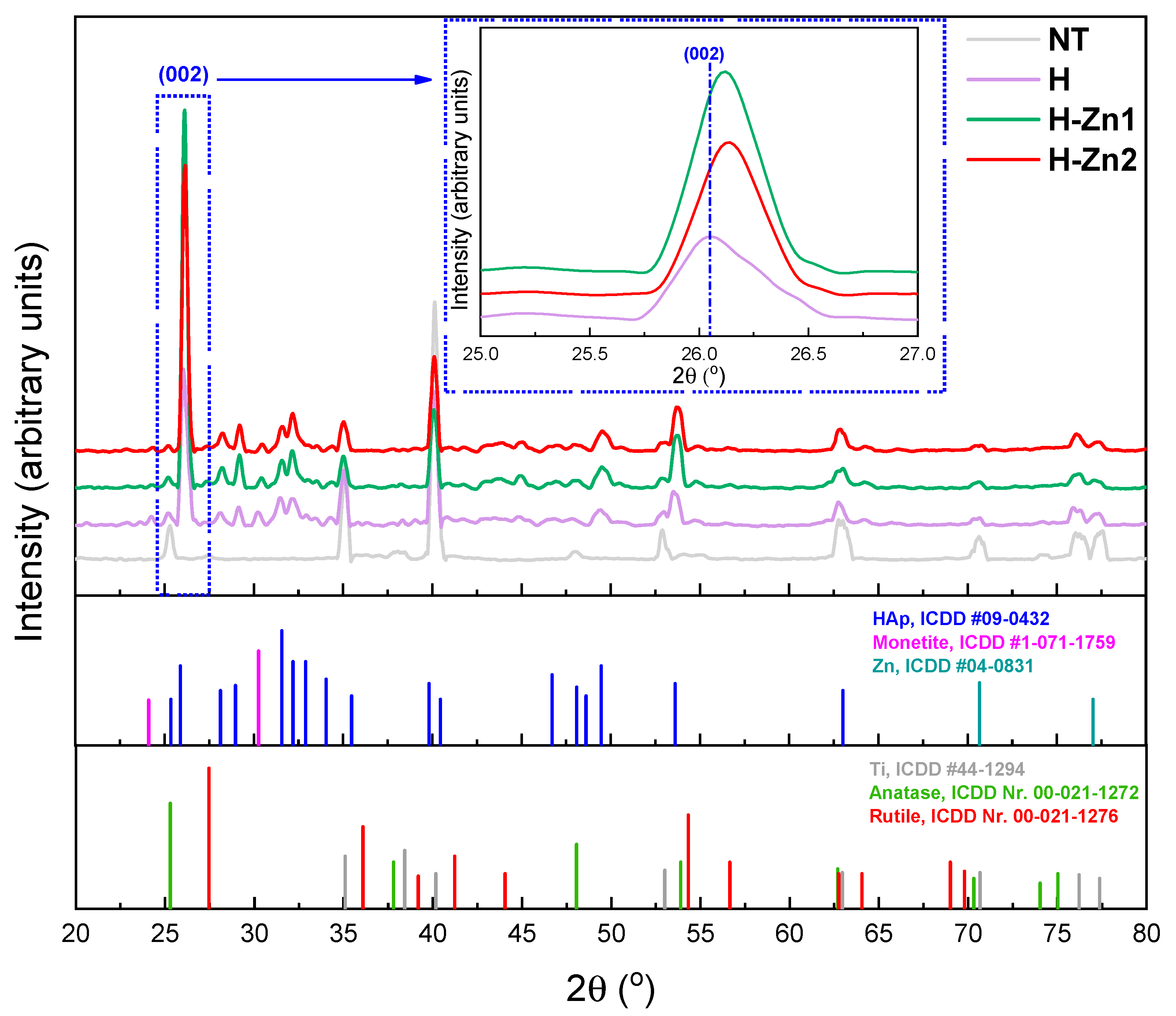
In Figure 4 and Table 3 are presented the obtained XRD diffractograms and the structural characteristic parameters obtained from XRD analysis.
The XRD diffractogram (Figure 4) of the NT substrate revealed the presence of specific diffraction peaks for titanium (ICDD #44-1298), anatase (ICDD Nr. 00-021-1272)—this being also the majority phase—and rutile (Nr. 00-021-1276). Bai et al. [60] consider that an optimal structure would consist of a mixture of phases, namely anatase and rutile, as the mechanical interaction at the bone-implant interface may be favored. Moreover, the rutile phase is expected to improve the chemical resistance of the material during its interaction with the biological environment, thus preventing the release of titanium ions, while the anatase phase, improves the proliferation and osteoblast adhesion [61] and favors the formation of calcium phosphates [62].
The X-ray diffraction of the undoped and doped HAp-based coatings confirmed the presence of HAp according to ICDD #09-0432 as the main crystal phase in all three coatings in accordance with the International Center for Diffraction Data (ICDD).
The most intense diffraction peak, which is characteristic for electrochemical deposited coatings, is located at ~26° and is associated with the (002) plane, indicating that the HAp crystals are oriented perpendicular to the NT surface [42]. According to the literature, the HAp with a c-axis orientation increases cell proliferation, chemical stability, and mechanical properties [63,64]. Moreover, as can be observed in Figure 4 and Table 3, the addition of Zn into the HAp structure has led to shifts towards higher angles, indicating the substitution of higher ionic radius Ca2+ (0.099 nm) ions for those of Zn2+ (0.074 nm), which have a smaller ionic radius, inducing the contraction of the HAp lattice [65].
According to ICDD #1-071-1759, one diffraction peak located at ~31° was assigned to monetite, a precursor of HAp. Its presence can be beneficial in these coatings, as it is known for its potential for bone regeneration and its osteoconductive and osseoinductive character [66,67]. According to the XRD analysis, no peaks could be attributed to pure Zn in its metallic form in the Zn-doped HAp coatings, irrespective of the Zn concentration.
The addition of Zn in the HAp structure leads to a decrease in the lattice parameters a and c (Table 3), which is in accordance with the literature [68,69]. The differences in terms of lattice parameters between the H-Zn1 and H-Zn2 are very small, suggesting that a higher concentration may not induce significant changes.
Both crystallite size and crystallinity contrast with lattice parameters and, based on the obtained results, it was noted that, compared to H, both increase with the addition of Zn. Thus, by comparison with H, the crystallinity of the H-Zn coatings increased by ~50%, while the crystallite dimension increased by ~25%, indicating the beneficial effect of the Zn in the HAp structure. By comparing the Zn doped coatings, it can be observed that by using a higher quantity of Zn, both the crystallinity and crystallite dimensions start to decrease, suggesting that a too-high Zn content inhibits the crystallinity of the HAp coatings. Similar findings have also been reported elsewhere [23,31].

3.4. Roughness

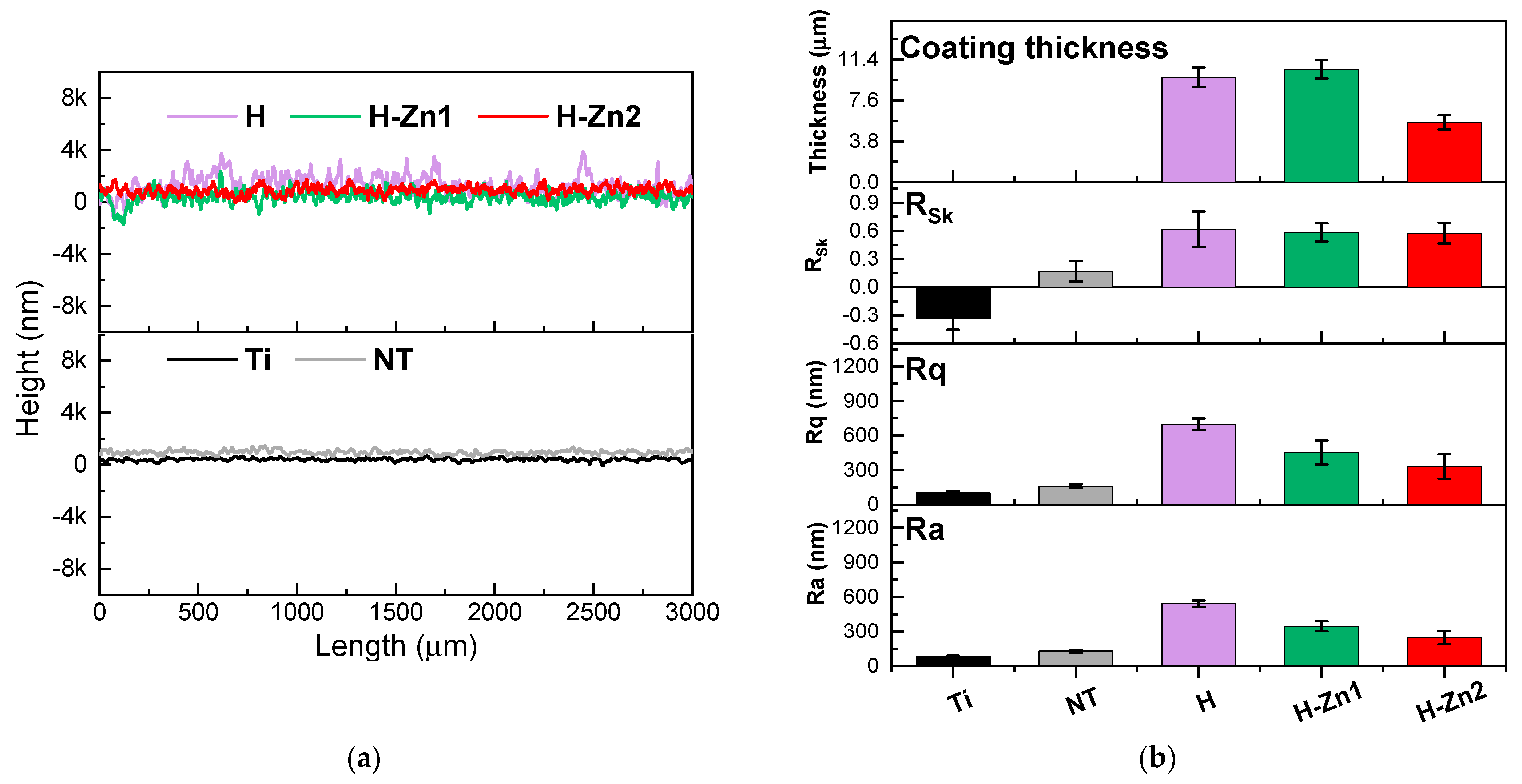
The main roughness parameters and profile line specific to each surface are presented in Figure 5. By comparing the average roughness (Ra) of the Ti with that of the NT, it can be noted that roughness has increased, reaching a value of ~130 nm due to the presence of the nanotubes, which is in good agreement with other studies [49].
By comparison to the NT surface, the H coatings obtained through the pulse galvanostatic technique have led to an increment of the roughness of 4 times more, with a value of the Ra of ~540 nm. The addition of Zn in the HAp structure has led to a decrement of its roughness, from ~540 nm (H coatings) to ~320 nm (H-Zn1) and ~240 nm (H-Zn2), suggesting that Zn smooths the surface. These results are in agreement with the SEM images, in which the size of the ribbon-like crystals decreases after the addition of Zn, leading to a denser coating.
The skewness parameter (Rsk) offers information on the hills (slope < 0) and valleys (slope > 0) distribution. According to the literature, the Rsk parameters can provide valuable information on how the material will act in terms of electrochemical behavior and biodegradation [70,71,72,73]. Moreover, the surfaces with a positive Rsk present favorable coating adhesion compared to surfaces with a negative one [66].
Thus, it can be observed that all coatings registered positive values of the Rsk parameter, which are very close to each other, indicating that the surface is predominantly hills.
In terms of coating thickness, which was determined with the contact profilometer, by measuring the profile of the ledge between the coating and the substrate, it can be observed that the H and H-Zn1 coatings have registered close values of 9.75 µm for the H and of 10.61 µm for the H-Zn1. According to Ref. [74], a thickness of ~10 µm is favorable for bone apposition. If the thickness of the coatings is too high, the coatings can present poor adhesion, while a too-small value, associated with poor crystallinity, can affect the coating stability [75,76]. The smallest thickness, of 5.54 µm, was noted for the H-Zn2 coatings, suggesting that the nucleation and growth of HAp is affected when even a slightly higher amount of Zn is added within the electrolyte, leading to a smaller deposition rate.

3.5. Chemical Bonds

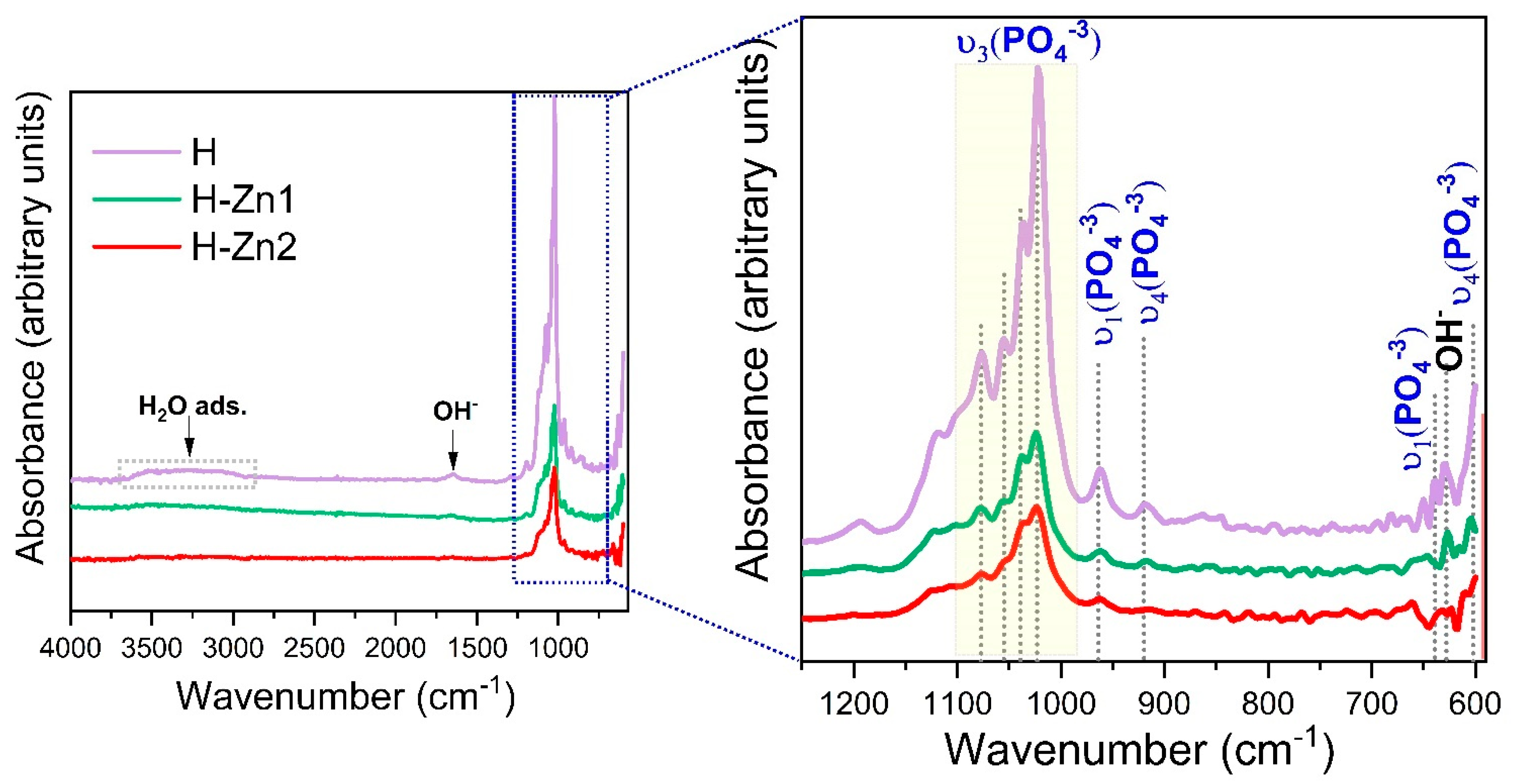
Figure 6 depicts the FTIR spectra obtained for the undoped and Zn doped HAp based coatings. All samples exhibited the main characteristic bands of hydroxyapatite with some variations in the absorption bands, which may indicate the substitution of Zn ions into the apatite structure.
Thus, the spectrum illustrates the main vibration modes of the phosphate group ( PO 4 3 ) , which were identified between 1022 cm−1 and 1100 cm−13), and at 635 cm−1 and 961 cm−14), respectively [77,78]. The υ4 and υ3  ( PO 4 3 ) vibration bands are considered the IR fingerprint of a HAp structure.
A very broad band identified between 3000 cm−1 and 3600 cm−1 in the H coatings was assigned to the adsorbed water [79]. Even though the Zn doped HAp coatings presented the same shape, some differences were noted, as follows. The peaks found between 1022 cm−1 and 1100 cm−1 associated with the υ3  ( PO 4 3 ) vibration and the symmetric stretching mode of ( PO 4 3 ) 1) identified at 961 cm−1 decrease in intensity after zinc addition, being also dependent on the Zn concentration.
All coatings present a weak sharp peak at 628 cm−1 which was assigned to the libration of (OH) groups in HAp [80,81,82]. However, the intensity of this absorption peak, associated with the structural (OH) group at 628 cm−1 observed in the H coatings, has significantly diminished after the addition of Zn, highlighting the loss of OH ions from the unit cell and the apparent substitution of Zn ions into the lattice of HAp [83].

3.6. Adhesion

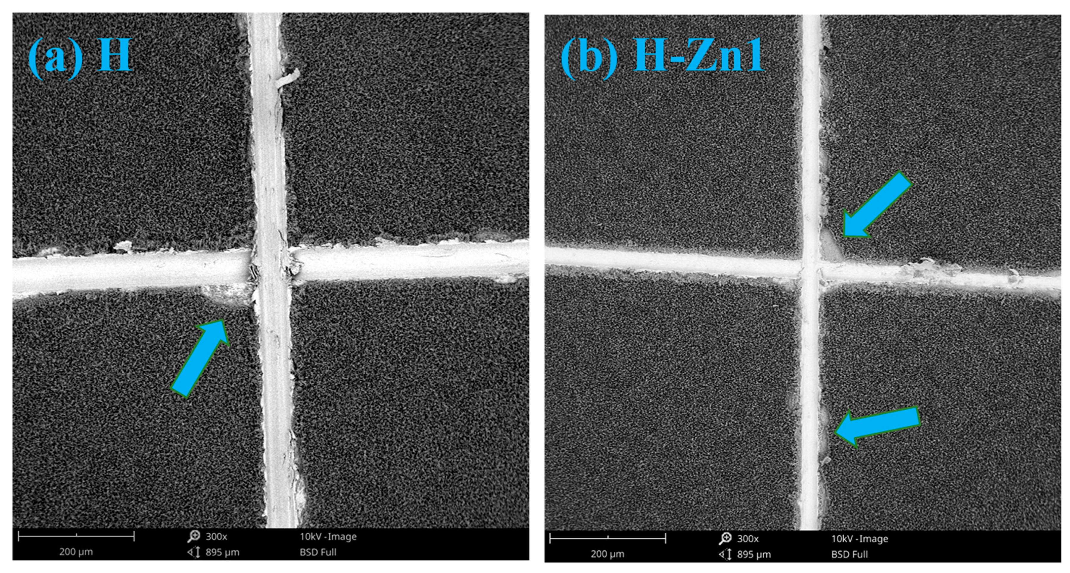
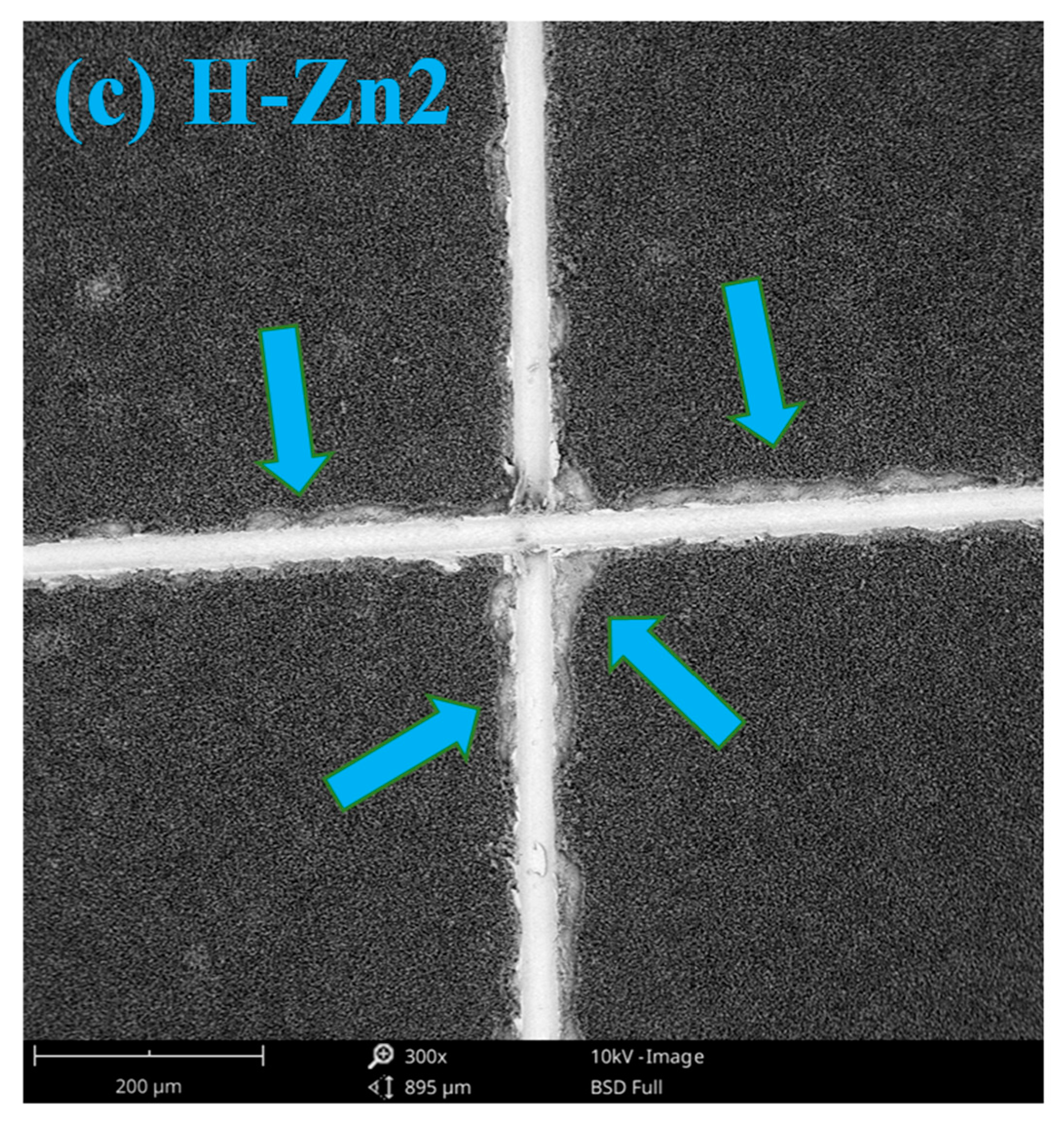
In Figure 7 are presented the SEM images of the coatings after performing the adhesion assays by “tape test” according to the ASTM procedure. Similar tests were also performed by Parcharoen et al. [84], who have investigated the adhesion between HAp based coatings deposited on NT of different lengths.
The results of adhesion tests based on SEM investigation revealed that the H and H-Zn1 have a strong adhesion to the substrate classified according to the standard ASTM D 3359-17 on the top category, namely 4B, highlighting only small detachments of coating along the indentation line or at cuts intersection.
By increasing the amount of Zn, it was noted that the delamination/detached area is found between 5–15%, thus affecting the H-Zn2 coating, which falls into the 3B category of the standard. Based on the results, it can be concluded that by adding a small amount of a doping element, the adhesion of coatings is not affected.
Even though in the present study the nanotubes’ lengths were not evaluated, based on the other literature reports [50,51], it can be argued that the length of the nanotubes obtained in weak hydrofluoric acid electrolyte (0.5 wt.% HF) when a potential of 20 V is applied for 30 min or 60 min is ~400 nm. Thus, it was assumed that the length of the nanotubes is within the range which offers suitable adhesion to the HAp based coatings.
Moreover, in a study performed by Dey et al. [85], it was highlighted that if the NT length is too high, the in vivo behavior could be compromised, due to insufficient mechanical stability, which can cause breakage of the nanotubes from the titanium. Nevertheless, according to Bauer et al. [86], the length of the nanotubes does not influence cell behavior, and thus the length of the nanotubes was not considered in the current study.

4. Conclusions

  • The electrochemical surface functionalization of Ti by the growth of nanotubes with an average diameter of ~70 nm and deposition of undoped HAp doped with Zn in different concentrations was successfully achieved by electrochemical techniques. The present study highlighted that the addition of Zn in even a small amount can enhance the characteristics of HAp. All coatings presented a morphology consisting of ribbon-like crystals, which suffers some alterations by the addition and increment of Zn content. The XRD and FTIR investigations have confirmed that all coatings consist in HAp as a main phase. Irrespective of the Zn content, the crystallinity of the HAp coatings was enhanced after the addition of Zn.
  • Regarding the effect of Zn content on the properties of HAp, it can be said that even though the differences between the two selected concentrations is minor, the coating H-Zn1 (with Zn of 0.78 at.%) has met most of the requirements, being more suitable for medical applications. Thus, the coatings with a smaller Zn amount will be further evaluated in terms of electrochemical behavior in synthetic media, along with their biomineralization ability and in vitro cell behavior, to establish if the proposed coatings can impart a good biocompatibility with a suitable bioactive character and antibacterial efficiency, without inducing a cytotoxic effect.

Author Contributions

Conceptualization, D.M.V. and C.M.C.; methodology, C.M.C.; validation, D.M.V. and A.V.; investigation, D.M.V., E.U., I.C.I., A.C.P. and A.E.K.; resources, D.M.V. and C.M.C.; data curation, D.M.V., E.U. and A.V.; writing—original draft preparation, D.M.V., E.U. and I.C.I.; writing—review and editing, D.M.V., C.M.C. and A.V.; visualization, D.M.V. and E.U.; supervision, C.M.C.; project administration, D.M.V.; funding acquisition, D.M.V. All authors have read and agreed to the published version of the manuscript.

Funding

This research has been supported by a grant of the Romanian Ministry of Education and Research, CNCS-UEFISCDI, project number PN-III-P1-1.1-TE-2019-1331, within PNCDI III (project no. TE 172/2020; 3B-CoatED).

Institutional Review Board Statement

Not applicable.

Informed Consent Statement

Not applicable.

Data Availability Statement

Data sharing is not applicable to this article.

Acknowledgments

A.V. thanks to Tomsk Polytechnic University within the framework of the Tomsk Polytechnic University-Competitiveness Enhancement Program grant.

Conflicts of Interest

The authors declare no conflict of interest.

References

  1. Kurtz, S.; Ong, K.; Lau, E.; Mowat, F.; Halpern, M. Projections of primary and revision hip and knee arthroplasty in the United States from 2005 to 2030. J. Bone Jt. Surg. 2007, 89, 780–785. [Google Scholar] [CrossRef]
  2. Quinn, J.; McFadden, R.; Chan, C.W.; Carson, L. Titanium for orthopedic applications: An overview of surface modification to improve biocompatibility and prevent bacterial biofilm formation. iScience 2020, 23, 101745. [Google Scholar] [CrossRef]
  3. Mariani, E.; Lisignoli, G.; Borzì, R.M.; Pulsatelli, L. Biomaterials: Foreign bodies or tuners for the immune response? Int. J. Mol. Sci. 2019, 20, 636. [Google Scholar] [CrossRef] [PubMed] [Green Version]
  4. Prasad, K.; Bazaka, O.; Chua, M.; Rochford, M.; Fedrick, L.; Spoor, J.; Symes, R.; Tieppo, M.; Collins, C.; Cao, A.; et al. Metallic biomaterials: Current challenges and opportunities. Materials 2017, 10, 884. [Google Scholar] [CrossRef]
  5. Geetha, M.; Singh, A.K.; Asokamani, R.; Gogia, A.K. Ti based biomaterials, the ultimate choice for orthopaedic implants—A review. Prog. Mater. Sci. 2009, 54, 397–425. [Google Scholar] [CrossRef]
  6. Nicholson, J.W. Titanium alloys for dental implants: A review. Prosthesis 2020, 2, 100–116. [Google Scholar] [CrossRef]
  7. Prasad, S.; Ehrensberger, M.; Gibson, M.P.; Kim, H.; Monaco, E.A. Biomaterial properties of titanium in dentistry. J. Oral Biosci. 2015, 57, 192–199. [Google Scholar] [CrossRef] [Green Version]
  8. Szmukler-Moncler, S.; Salama, H.; Reingewirtz, Y.; Dubruille, J.H. Timing of loading and effect of micromotion on bone-dental implant interface: Review of experimental literature. J. Biomed. Mater. Res. 1998, 43, 192–203. [Google Scholar] [CrossRef]
  9. Irandoust, S.; Müftü, S. The interplay between bone healing and remodeling around dental implants. Sci. Rep. 2020, 10, 4335. [Google Scholar] [CrossRef] [Green Version]
  10. He, R.; Lu, Y.; Ren, J.; Wang, Z.; Huang, J.; Zhu, L.; Wang, K. Decreased fibrous encapsulation and enhanced osseointegration in vitro by decorin-modified titanium surface. Colloids Surf. B Biointerfaces 2017, 155, 17–24. [Google Scholar] [CrossRef] [PubMed]
  11. Kurup, A.; Dhatrak, P.; Khasnis, N. Surface modification techniques of titanium and titanium alloys for biomedical dental Applications: A review. Mater. Today Proc. 2020, 39, 84–90. [Google Scholar] [CrossRef]
  12. Xue, T.; Attarilar, S.; Liu, S.; Liu, J.; Song, X.; Li, L.; Zhao, B.; Tang, Y. Surface modification techniques of titanium and its alloys to functionally optimize their biomedical properties: Thematic review. Front. Bioeng. Biotechnol. 2020, 8, 1261. [Google Scholar] [CrossRef] [PubMed]
  13. Li, J.; Zhou, P.; Attarilar, S.; Shi, H. Innovative surface modification procedures to achieve micro/nano-graded Ti-based biomedical alloys and implants. Coatings 2021, 11, 647. [Google Scholar] [CrossRef]
  14. Velasco-Ortega, E.; Ortiz-García, I.; Jiménez-Guerra, A.; Monsalve-Guil, L.; Muñoz-Guzón, F.; Perez, R.A.; Gil, F.J. Comparison between sandblasted acid-etched and oxidized titanium dental implants: In vivo study. Int. J. Mol. Sci. 2019, 20, 3267. [Google Scholar] [CrossRef] [PubMed] [Green Version]
  15. Sartoretto, S.C.; Alves, A.T.N.N.; Resende, R.F.B.; Calasans-Maia, J.; Granjeiro, J.M.; Calasans-Maia, M.D. Early osseointegration driven by the surface chemistry and wettability of dental implants. J. Appl. Oral Sci. 2015, 23, 272–278. [Google Scholar] [CrossRef] [PubMed]
  16. Brammer, K.S.; Frandsen, C.J.; Jin, S. TiO2 nanotubes for bone regeneration. Trends Biotechnol. 2012, 30, 315–322. [Google Scholar] [CrossRef]
  17. Priyadarshini, B.; Rama, M.; Chetan, U.; Vijayalakshmi, U. Bioactive coating as a surface modification technique for biocompatible metallic implants: A review. J. Asian Ceram. Soc. 2019, 7, 397–406. [Google Scholar] [CrossRef] [Green Version]
  18. Eliaz, N.; Metoki, N. Calcium phosphate bioceramics: A review of their history, structure, properties, coating technologies and biomedical applications. Materials 2017, 10, 334. [Google Scholar] [CrossRef] [PubMed] [Green Version]
  19. Šupová, M. Substituted hydroxyapatites for biomedical applications: A review. Ceram. Int. 2015, 41, 9203–9231. [Google Scholar] [CrossRef]
  20. Prosolov, K.A.; Mitrichenko, D.V.; Prosolov, A.B.; Nikolaeva, O.O.; Lastovka, V.V.; Belyavskaya, O.A.; Chebodaeva, V.A.; Glukhov, I.A.; Litvinova, L.S.; Shupletsova, V.V.; et al. Zn-doped CaP-based coatings on Ti–6Al–4V and Ti–6Al–7Nb alloys prepared by magnetron sputtering: Controllable biodegradation, bacteriostatic, and osteogenic activities. Coatings 2021, 11, 809. [Google Scholar] [CrossRef]
  21. Popa, C.L.; Deniaud, A.; Michaud-Soret, I.; Guégan, R.; Motelica-Heino, M.; Predoi, D. Structural and biological assessment of zinc doped hydroxyapatite nanoparticles. J. Nanomater. 2016, 2016, 1062878. [Google Scholar] [CrossRef] [Green Version]
  22. Sergi, R.; Bellucci, D.; Candidato, R.T.; Lusvarghi, L.; Bolelli, G.; Pawlowski, L.; Candiani, G.; Altomare, L.; de Nardo, L.; Cannillo, V. Bioactive Zn-doped hydroxyapatite coatings and their antibacterial efficacy against Escherichia coli and Staphylococcus aureus. Surf. Coat. Technol. 2018, 352, 84–91. [Google Scholar] [CrossRef]
  23. Begam, H.; Kundu, B.; Chanda, A.; Nandi, S.K. MG63 osteoblast cell response on Zn doped hydroxyapatite (HAp) with various surface features. Ceram. Int. 2017, 43, 3752–3760. [Google Scholar] [CrossRef]
  24. Wang, M.; Tang, T. Surface treatment strategies to combat implant-related infection from the beginning. J. Orthop. Transl. 2019, 17, 42–54. [Google Scholar] [CrossRef]
  25. Uysal, I.; Yilmaz, B.; Evis, Z. Zn-doped hydroxyapatite in biomedical applications. J. Aust. Ceram. Soc. 2021, 57, 869–897. [Google Scholar] [CrossRef]
  26. Ito, A.; Ojima, K.; Naito, H.; Ichinose, N.; Tateishi, T. Preparation, solubility, and cytocompatibility of zinc-releasing calcium phosphate ceramics. J. Biomed. Mater. Res. 2000, 50, 178–183. [Google Scholar] [CrossRef]
  27. Tank, K.P.; Chudasama, K.S.; Thaker, V.S.; Joshi, M.J. Pure and zinc doped nano-hydroxyapatite: Synthesis, characterization, antimicrobial and hemolytic studies. J. Cryst. Growth 2014, 401, 474–479. [Google Scholar] [CrossRef]
  28. Candidato, R.T.; Thouzellier, C.; Pawłowski, L. Evaluation of the in-vitro behavior of nanostructured hydroxyapatite and zinc doped hydroxyapatite coatings obtained using solution precursor plasma spraying. J. Biomed. Mater. Res. Part B Appl. Biomater. 2018, 106, 2101–2108. [Google Scholar] [CrossRef]
  29. Codescu, M.M.; Vladescu, A.; Geanta, V.; Voiculescu, I.; Pana, I.; Dinu, M.; Kiss, A.E.; Braic, V.; Patroi, D.; Marinescu, V.E.; et al. Zn based hydroxyapatite based coatings deposited on a novel FeMoTaTiZr high entropy alloy used for bone implants. Surf. Interfaces 2021, 101591. [Google Scholar] [CrossRef]
  30. Ke, D.; Banerjee, D.; Bose, S. In vitro characterizations of Si4+ and Zn2+ doped plasma sprayed hydroxyapatite coatings using osteoblast and osteoclast coculture. ACS Biomater. Sci. Eng. 2019, 5, 1302–1310. [Google Scholar] [CrossRef]
  31. Hidalgo-Robatto, B.M.; López-Álvarez, M.; Azevedo, A.S.; Dorado, J.; Serra, J.; Azevedo, N.F.; González, P. Pulsed laser deposition of copper and zinc doped hydroxyapatite coatings for biomedical applications. Surf. Coat. Technol. 2018, 333, 168–177. [Google Scholar] [CrossRef]
  32. Iconaru, S.L.; Prodan, A.M.; Buton, N.; Predoi, D. Structural characterization and antifungal studies of zinc-doped hydroxyapatite coatings. Molecules 2017, 22, 604. [Google Scholar] [CrossRef] [PubMed] [Green Version]
  33. Thanh, T.T.; Cotrut, C.M.; Vranceanu, M.D.; Ungureanu, E.; Tarcolea, M. Studies of microstructure and composition of modified hydroxyapatite coatings via Sem investigations. UPB Sci. Bull. Ser. B Chem. Mater. Sci. 2020, 82, 145–154. [Google Scholar]
  34. Safavi, M.S.; Walsh, F.C.; Surmeneva, M.A.; Surmenev, R.A.; Khalil-Allafi, J. Electrodeposited hydroxyapatite-based biocoatings: Recent progress and future challenges. Coatings 2021, 11, 110. [Google Scholar] [CrossRef]
  35. Vladescu, A.; Vranceanu, D.M.; Kulesza, S.; Ivanov, A.N.; Bramowicz, M.; Fedonnikov, A.S.; Braic, M.; Norkin, I.A.; Koptyug, A.; Kurtukova, M.O.; et al. Influence of the electrolyte’s PH on the properties of electrochemically deposited hydroxyapatite coating on additively manufactured Ti64 alloy. Sci. Rep. 2017, 7, 4–7. [Google Scholar] [CrossRef] [PubMed]
  36. Li, T.T.; Ling, L.; Lin, M.C.; Peng, H.K.; Ren, H.T.; Lou, C.W.; Lin, J.H. Recent advances in multifunctional hydroxyapatite coating by electrochemical deposition. J. Mater. Sci. 2020, 55, 6352–6374. [Google Scholar] [CrossRef]
  37. Blackwood, D.J.; Seah, K.H.W. Electrochemical cathodic deposition of hydroxyapatite: Improvements in adhesion and crystallinity. Mater. Sci. Eng. C 2009, 29, 1233–1238. [Google Scholar] [CrossRef]
  38. Yan, Y.; Ding, Q.; Huang, Y.; Han, S.; Pang, X. Magnesium substituted hydroxyapatite coating on titanium with nanotublar TiO2 intermediate layer via electrochemical deposition. Appl. Surf. Sci. 2014, 305, 77–85. [Google Scholar] [CrossRef]
  39. Indira, K.; Mudali, U.K.; Nishimura, T.; Rajendran, N. A review on TiO2 nanotubes: Influence of anodization parameters, formation mechanism, properties, corrosion behavior, and biomedical applications. J. Bio-Tribo-Corros. 2015, 1, 28. [Google Scholar] [CrossRef] [Green Version]
  40. Martinez-Marquez, D.; Gulati, K.; Carty, C.P.; Stewart, R.A.; Ivanovski, S. Determining the relative importance of titania nanotubes characteristics on bone implant surface performance: A quality by design study with a fuzzy approach. Mater. Sci. Eng. C 2020, 114, 110995. [Google Scholar] [CrossRef] [PubMed]
  41. Shahmohammadi, P.; Khazaei, B.A. Characterization of Zn/Mg-enriched calcium phosphate coating produced by the two-step pulsed electrodeposition method on titanium substrate. Surf. Interfaces 2021, 22, 110819. [Google Scholar] [CrossRef]
  42. Vranceanu, D.M.; Ionescu, I.C.; Ungureanu, E.; Cojocaru, M.O.; Vladescu, A.; Cotrut, C.M. Magnesium doped hydroxyapatite-based coatings obtained by pulsed galvanostatic electrochemical deposition with adjustable electrochemical behavior. Coatings 2020, 10, 727. [Google Scholar] [CrossRef]
  43. Landi, E.; Tampieri, A.; Celotti, G.; Sprio, S. Densification behaviour and mechanisms of synthetic hydroxyapatites. J. Eur. Ceram. Soc. 2000, 20, 2377–2387. [Google Scholar] [CrossRef]
  44. Gopi, D.; Kanimozhi, K.; Bhuvaneshwari, N.; Indira, J.; Kavitha, L. Novel banana peel pectin mediated green route for the synthesis of hydroxyapatite nanoparticles and their spectral characterization. Spectrochimica Acta Part A Mol. Biomol. Spectrosc. 2014, 118, 589–597. [Google Scholar] [CrossRef] [PubMed]
  45. Rusu, V.; Chuen-How, N.; Wilke, M.; Tiersch, B.; Fratzl, P.; Peter, M. Size-controlled hydroxyapatite nanoparticles as self-organized organic-inorganic composite materials. Biomaterials 2005, 26, 5414–5426. [Google Scholar] [CrossRef]
  46. Li, Z.Y.; Lam, W.M.; Yang, C.; Xu, B.; Ni, G.X.; Abbah, S.A.; Cheung, K.M.C.; Luk, K.D.K.; Lu, W.W. Chemical composition, crystal size and lattice structural changes after incorporation of strontium into biomimetic apatite. Biomaterials 2007, 28, 1452–1460. [Google Scholar] [CrossRef]
  47. Narayanan, R.; Kwon, T.-Y.; Kim, K.-H. Direct nanocrystalline hydroxyapatite formation on titanium from ultrasonated electrochemical bath at physiological PH. Mater. Sci. Eng. C 2008, 28, 1265–1270. [Google Scholar] [CrossRef]
  48. ASTM International. ASTM D3359-17 Standard Test Methods for Rating Adhesion by Tape Test; ASTM International: Calw, Germany, 2017; pp. 1–9. [Google Scholar] [CrossRef]
  49. Cotrut, C.M.; Ionescu, I.C.; Ungureanu, E.; Berbecaru, A.; Zamfir, R.I.; Vladescu, A.; Vranceanu, D.M. Evaluation of surface modification techniques on the ability of apatite formation and corrosion behavior in synthetic body fluid: An in vitro study. Surf. Interfaces 2021, 22, 100866. [Google Scholar] [CrossRef]
  50. Bai, J.; Zhou, B.; Li, L.; Liu, Y.; Zheng, Q.; Shao, J.; Zhu, X.; Cai, W.; Liao, J.; Zou, L. The formation mechanism of titania nanotube arrays in hydrofluoric acid electrolyte. J. Mater. Sci. 2008, 43, 1880–1884. [Google Scholar] [CrossRef]
  51. Lai, Y.K.; Sun, L.; Chen, C.; Nie, C.G.; Zuo, J.; Lin, C.J. Optical and electrical characterization of TiO2 nanotube arrays on titanium substrate. Appl. Surf. Sci. 2005, 252, 1101–1106. [Google Scholar] [CrossRef]
  52. Zielinski, A.; Bartmanski, M. Electrodeposited biocoatings, their properties and fabrication technologies: A review. Coatings 2020, 10, 782. [Google Scholar] [CrossRef]
  53. Bigi, A.; Foresti, E.; Gandolfi, M.; Gazzano, M.; Roveri, N. Inhibiting effect of zinc on hydroxylapatite crystallization. J. Inorg. Biochem. 1995, 58, 49–58. [Google Scholar] [CrossRef]
  54. Kanzaki, N.; Onuma, K.; Treboux, G.; Tsutsumi, S.; Ito, A. Inhibitory effect of magnesium and zinc on crystallization kinetics of hydroxyapatite (0001) face. J. Phys. Chem. B 2000, 104, 4189–4194. [Google Scholar] [CrossRef]
  55. Kaseem, M.; Choe, H.-C. Triggering the hydroxyapatite deposition on the surface of PEO-coated Ti–6Al–4V alloy via the dual incorporation of Zn and Mg ions. J. Alloy. Compd. 2020, 819, 153038. [Google Scholar] [CrossRef]
  56. Mavropoulos, E.; Rossi, A.M.; da Rocha, N.C.C.; Soares, G.A.; Moreira, J.C.; Moure, G.T. Dissolution of calcium-deficient hydroxyapatite synthesized at different conditions. Mater. Charact. 2003, 50, 203–207. [Google Scholar] [CrossRef]
  57. Dorozhkin, S.V. A review on the dissolution models of calcium apatites. Prog. Cryst. Growth Charact. Mater. 2002, 44, 45–61. [Google Scholar] [CrossRef]
  58. Lin, J.H.; Kuo, K.H.; Ding, S.J.; Ju, C.P. Surface reaction of stoichiometric and calcium-deficient hydroxyapatite in simulated body fluid. J. Mater. Sci. Mater. Med. 2001, 12, 731–741. [Google Scholar] [CrossRef] [PubMed]
  59. Miyaji, F.; Kono, Y.; Suyama, Y. Formation and structure of zinc-substituted calcium hydroxyapatite. Mater. Res. Bull. 2005, 40, 209–220. [Google Scholar] [CrossRef]
  60. Bai, Y.; Park, I.S.; Park, H.H.; Lee, M.H.; Bae, T.S.; Duncan, W.; Swain, M. The effect of annealing temperatures on surface properties, hydroxyapatite growth and cell behaviors of TiO2 nanotubes. Surf. Interface Anal. 2011, 43, 998–1005. [Google Scholar] [CrossRef]
  61. He, J.; Zhou, W.; Zhou, X.; Zhong, X.; Zhang, X.; Wan, P.; Zhu, B.; Chen, W. The anatase phase of nanotopography titania plays an important role on osteoblast cell morphology and proliferation. J. Mater. Sci. Mater. Med. 2008, 19, 3465–3472. [Google Scholar] [CrossRef]
  62. Xiao, X.; Yu, J.; Tang, H.; Mao, D.; Wang, C.; Liu, R. TiO2 nanotube arrays induced deposition of hydroxyapatite coating by hydrothermal treatment. Mater. Chem. Phys. 2013, 138, 695–702. [Google Scholar] [CrossRef]
  63. Liu, X.; He, D.; Zhou, Z.; Guo, X.; Liu, Y.; Hou, W.; Li, H. In vitro bioactivity and antibacterial performances of atmospheric plasma sprayed C-axis preferential oriented hydroxyapatite coatings. Surf. Coat. Technol. 2021, 417, 127209. [Google Scholar] [CrossRef]
  64. Liu, X.; He, D.; Zhou, Z.; Wang, G.; Wang, Z.; Wu, X.; Tan, Z. Characteristics of (002) oriented hydroxyapatite coatings deposited by atmospheric plasma spraying. Coatings 2018, 8, 258. [Google Scholar] [CrossRef] [Green Version]
  65. Bi, Q.; Song, X.; Chen, Y.; Zheng, Y.; Yin, P.; Lei, T. Zn-HA/Bi-HA biphasic coatings on titanium: Fabrication, characterization, antibacterial and biological activity. Colloids Surf. B Biointerfaces 2020, 189, 110803. [Google Scholar] [CrossRef] [PubMed]
  66. Motameni, A.; Alshemary, A.Z.; Evis, Z. A review of synthesis methods, properties and use of monetite cements as filler for bone defects. Ceram. Int. 2021, 47, 13245–13256. [Google Scholar] [CrossRef]
  67. Zhou, H.; Yang, L.; Gbureck, U.; Bhaduri, S.B.; Sikder, P. Monetite, an important calcium phosphate compound—Its synthesis, properties and applications in orthopedics. Acta Biomaterialia 2021, 127, 41–55. [Google Scholar] [CrossRef] [PubMed]
  68. Uysal, I.; Severcan, F.; Tezcaner, A.; Evis, Z. Co-doping of hydroxyapatite with zinc and fluoride improves mechanical and biological properties of hydroxyapatite. Prog. Nat. Sci. Mater. Int. 2014, 24, 340–349. [Google Scholar] [CrossRef] [Green Version]
  69. Bulina, N.V.; Vinokurova, O.B.; Eremina, N.V.; Prosanov, I.Y.; Khusnutdinov, V.R.; Chaikina, M.V. Features of solid-phase mechanochemical synthesis of hydroxyapatite doped by copper and zinc ions. J. Solid State Chem. 2021, 296, 121973. [Google Scholar] [CrossRef]
  70. Grzesik, W. Prediction of the functional performance of machined components based on surface topography: State of the art. J. Mater. Eng. Perform. 2016, 25, 4460–4468. [Google Scholar] [CrossRef] [Green Version]
  71. Vranceanu, D.M.; Parau, A.C.; Cotrut, C.M.; Kiss, A.E.; Constantin, L.R.; Braic, V.; Vladescu, A. In vitro evaluation of Ag doped hydroxyapatite coatings in acellular media. Ceram. Int. 2019, 45, 11050–11061. [Google Scholar] [CrossRef]
  72. Monsees, T.K.; Azem, F.A.; Cotrut, C.M.; Braic, M.; Abdulgader, R.; Pana, I.; Birlik, I.; Kiss, A.; Booysen, R.; Vladescu, A. Biodegradable ceramics consisting of hydroxyapatite for orthopaedic implants. Coatings 2017, 7, 184. [Google Scholar] [CrossRef] [Green Version]
  73. Patel, K.; Doyle, C.S.; Yonekura, D.; James, B.J. Effect of surface roughness parameters on thermally sprayed PEEK coatings. Surf. Coat. Technol. 2010, 204, 3567–3572. [Google Scholar] [CrossRef]
  74. Huang, Y.; Ding, Q.; Han, S.; Yan, Y.; Pang, X. Characterisation, corrosion resistance and in vitro bioactivity of manganese-doped hydroxyapatite films electrodeposited on titanium. J. Mater. Sci. Mater. Med. 2013, 24, 1853–1864. [Google Scholar] [CrossRef] [PubMed]
  75. Narayanan, R.; Seshadri, S.K.; Kwon, T.Y.; Kim, K.H. Calcium phosphate-based coatings on titanium and its alloys. J. Biomed. Mater. Res.-Part B Appl. Biomater. 2008, 85, 279–299. [Google Scholar] [CrossRef]
  76. Zhu, X.; Son, D.W.; Ong, J.L.; Kim, K. Characterization of hydrothermally treated anodic oxides containing Ca and P on titanium. J. Mater. Sci. Mater. Med. 2003, 14, 629–634. [Google Scholar] [CrossRef]
  77. Gopi, D.; Murugan, N.; Ramya, S.; Shinyjoy, E.; Kavitha, L. Ball flower like manganese, strontium substituted hydroxyapatite/cerium oxide dual coatings on the AZ91 Mg alloy with improved bioactive and corrosion resistance properties for implant applications. RSC Adv. 2015, 5, 27402–27411. [Google Scholar] [CrossRef]
  78. Dascălu, C.-A.; Maidaniuc, A.; Pandele, A.M.; Voicu, S.I.; Machedon-Pisu, T.; Stan, G.E.; Cîmpean, A.; Mitran, V.; Antoniac, I.V.; Miculescu, F. Synthesis and characterization of biocompatible polymer-ceramic film structures as favorable interface in guided bone regeneration. Appl. Surf. Sci. 2019, 494, 335–352. [Google Scholar] [CrossRef]
  79. Meejoo, S.; Maneeprakorn, W.; Winotai, P. Phase and thermal stability of nanocrystalline hydroxyapatite prepared via microwave heating. Thermochimica Acta 2006, 447, 115–120. [Google Scholar] [CrossRef]
  80. Gopi, D.; Murugan, N.; Ramya, S.; Kavitha, L. Electrodeposition of a porous strontium-substituted hydroxyapatite/zinc oxide duplex layer on AZ91 magnesium alloy for orthopedic applications. J. Mater. Chem. B 2014, 2, 5531–5540. [Google Scholar] [CrossRef]
  81. Gopi, D.; Karthika, A.; Rajeswari, D.; Kavitha, L.; Pramod, R.; Dwivedi, J. Investigation on corrosion protection and mechanical performance of minerals substituted hydroxyapatite coating on HELCDEB-treated titanium using pulsed electrodeposition method. RSC Adv. 2014, 4, 34751–34759. [Google Scholar] [CrossRef]
  82. Maidaniuc, A.; Miculescu, F.; Ciocoiu, R.C.; Butte, T.M.; Pasuk, I.; Stan, G.E.; Voicu, S.I.; Ciocan, L.T. Effect of the processing parameters on surface, physico-chemical and mechanical features of bioceramics synthesized from abundant carp fish bones. Ceram. Int. 2020, 46, 10159–10171. [Google Scholar] [CrossRef]
  83. Robinson, L.; Salma-Ancane, K.; Stipniece, L.; Meenan, B.J.; Boyd, A.R. The deposition of strontium and zinc co-substituted hydroxyapatite coatings. J. Mater. Sci. Mater. Med. 2017, 28, 51. [Google Scholar] [CrossRef] [PubMed]
  84. Parcharoen, Y.; Kajitvichyanukul, P.; Sirivisoot, S.; Termsuksawad, P. Hydroxyapatite electrodeposition on anodized titanium nanotubes for orthopedic applications. Appl. Surf. Sci. 2014, 311, 54–61. [Google Scholar] [CrossRef]
  85. Dey, T.; Roy, P.; Fabry, B.; Schmuki, P. Anodic mesoporous TiO2 layer on Ti for enhanced formation of biomimetic hydroxyapatite. Acta Biomaterialia 2011, 7, 1873–1879. [Google Scholar] [CrossRef] [PubMed]
  86. Bauer, S.; Park, J.; Faltenbacher, J.; Berger, S.; von der Mark, K.; Schmuki, P. Size selective behavior of mesenchymal stem cells on ZrO2 and TiO2 nanotube arrays. Integr. Biol. 2009, 1, 525–532. [Google Scholar] [CrossRef]
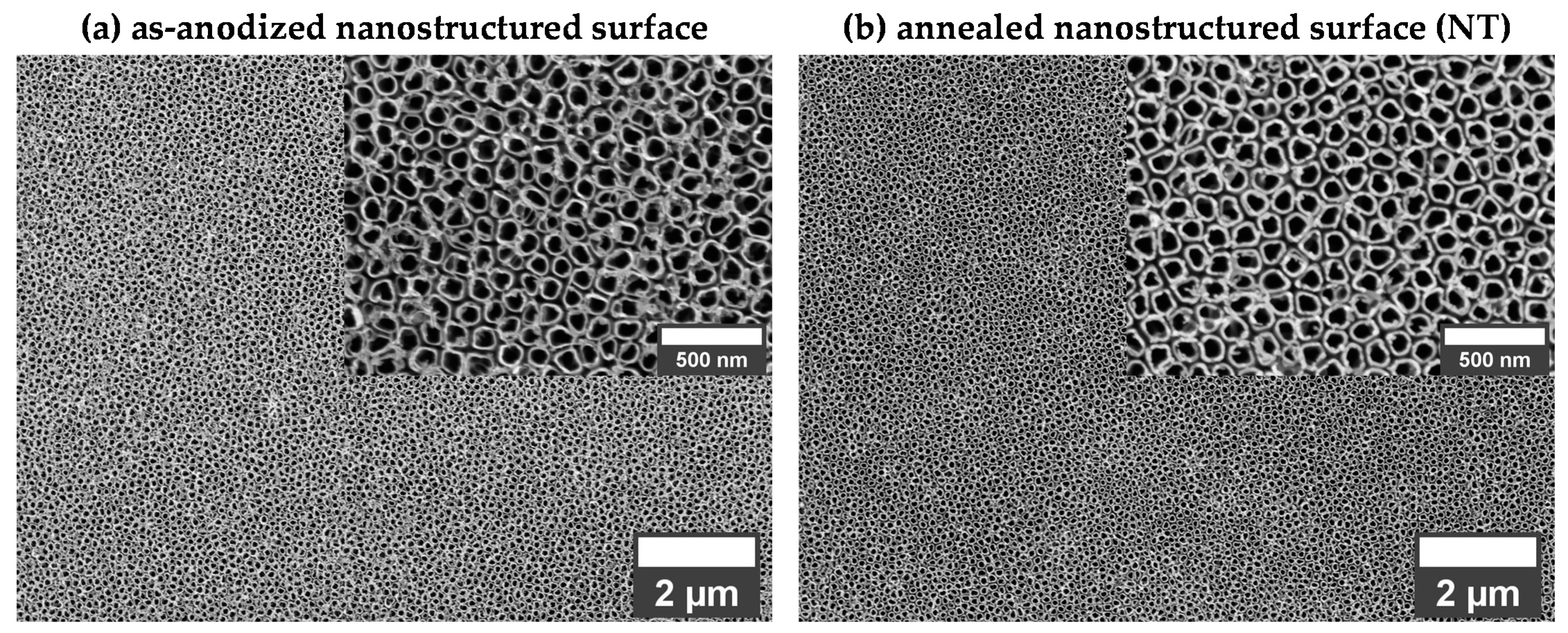
Figure 1. Morphology of the (a) as-anodized and (b) annealed nanostructured surface.
Coatings 12 00069 g001
Figure 2. Morphology of the undoped ((a) H) and zinc doped hydroxyapatite ((b) H-Zn1 and (c) H-Zn2) coatings.
Coatings 12 00069 g002
Figure 3. EDS spectra and elements distribution for the (a) H, (b) H-Zn1, (c) H-Zn2 coatings.
Coatings 12 00069 g003
Figure 4. XRD patterns of the undoped and doped HAp based coatings.
Coatings 12 00069 g004
Figure 5. Specific profile lines (a) and evolution of the main roughness parameters and coating thickness (b) of the investigated material.
Coatings 12 00069 g005
Figure 6. FTIR spectra of the undoped and Zn doped based coatings.
Coatings 12 00069 g006
Figure 7. Representative SEM images of the (a) H, (b) H-Zn1, (c) H-Zn2 coatings after adhesion tests (blue color arow locate the delamination.
Coatings 12 00069 g007aCoatings 12 00069 g007b
Table 1. Sample codification and chemical composition of the electrolyte used for the undoped and Zn doped coatings.
Sample CodificationChemical Composition (mM)Ratio
(Ca + Zn)/P
Ca(NO3)2∙4H2ONH4H2PO4Zn(NO3)2∙6H2O
H106 mM-Ca/P = 1.67
H-Zn19.9850.015(Ca + Zn)/P = 1.67
H-Zn28.9800.020
Table 2. Chemical composition (at.% and wt.%) and Ca/P ratio for the coatings obtained on NT.
MaterialElemental CompositionCa/P
Ratio
(Ca + Zn)/P
Ratio
CaPZn
(at.%)(wt.%)(at.%)(wt.%)(at.%)(wt.%)
H61.4667.3638.5432.64--1.591.59
H-Zn160.2165.7139.0132.900.781.391.541.56
H-Zn259.5464.7739.1632.931.302.301.521.55
Table 3. Lattice parameters, crystallite dimension and crystallinity of the coatings.
Materiala = b (Å)c (Å)(002) (°)Crystallite
Dimension (nm)
Crystallinity (%)
HAp, ICDD #09-04329.4186.88425.88--
H9.4396.87326.0419.9620.90
H-Zn19.4216.86026.1225.6044.30
H-Zn29.4226.85226.1425.4743.60
Publisher’s Note: MDPI stays neutral with regard to jurisdictional claims in published maps and institutional affiliations.