Abstract
Commonwealth war cemeteries commemorate the fallen of both world wars. Every casualty is remembered with a memorial or on a headstone. However, the headstones need to be maintained extensively, as microorganisms easily colonise them, affecting legibility and the stone substrate in the longer term. In the past, pesticides and other chemicals were popular to clean headstones, but due to raised environmental concerns, new treatment strategies are necessary. Within conservation science, enzymes have emerged as a popular tool for restoration. However, studies related to the use of enzymes for stone conservation are limited. Within this preliminary study, we applied commercially available enzyme-based treatments on biofouled natural building stones in the laboratory and in situ. Photography and spectrophotometry were used to monitor the effect of the treatment. The application of enzymes resulted in rapid disintegration of biological pigments, whereas visual improvement occurred more gradually. The successful application of enzymes suggests their potential to replace pesticides as the principal cleaning agent for headstones and natural building stones in a more general fashion.
1. Introduction
To remember the casualties of World War I and II, the Commonwealth War Grave Commission has built war grave cemeteries and memorials. All the cemeteries are characterised by uniform white headstones and monuments, often built with Portland limestone [1]. Just as any natural building stone or construction material, these stones interact with the environment, causing deterioration through physical, chemical and biological action [2].
Although headstones and any rock surface are exposed to extreme conditions of desiccation, oligotrophy, ultraviolet radiation, etc., microorganisms extensively colonise them. The colonisers include lichens, algae, fungi, bacteria and archaea [3]. The environmental conditions, together with the bioreceptivity of the material, determine the colonisation potential [4]. Microorganisms attach to the rock surface and form biofilms or aggregates held together by a matrix of mainly extracellular polymeric substances (EPS) [5,6,7,8]. Colonisation usually starts with autotrophs, such as cyanobacteria and algae [9]. However, heterotrophs could also be the first colonisers and could dominate without the presence of autotrophs [7,10,11].
Microbial growth affects the stone material in several ways. First, biodeterioration could be induced by, e.g., dissolution of minerals by metabolites or physical stress due to contracting or expanding fungal hyphae. Secondly, microbial biofilms induce discolouration and staining due to the presence of pigments [7,8,12,13]. The photosynthetic pigment chlorophyll of algae and cyanobacteria causes greening [14], whereas carotenoids, often from pigmented heterotrophic bacteria, might lead to a reddish discolouration [15,16]. Biological blackening occurs in the absence of a dominating pigment and has also been attributed to melanins [17], scytonemin and cyanobacteria [18]. Further discolouration is possible with EPS, which may trap dust or atmospheric particles, and by microbial oxidation of Fe(II) or Mn(VI) [7,12].
Especially on Commonwealth War Graves headstones, advanced stages of microbiological colonisation, related biofouling and discolouration are mostly undesired. The Commonwealth War Graves are strictly maintained not only for reasons of preventive conservation but also for their intangible heritage. Whereas in other heritage settings, controlled aging, patination and biofouling might be acceptable, their impression conflicts with preserving the memory of the fallen. Therefore, control and prevention of biocolonisation is stringent policy. Cleaning is one of the first steps in the conservation cycle, and it aims to remove organic growth, dirt, crust formations and other harmful substances from the surface of structures without affecting the substrate and inducing environmental harm. With rising awareness of the environmental impacts of such treatments, cleaning alternatives for pesticides have been explored for several years. This also complies with the EU sustainable use directive on pesticide reduction and banning [19]. One of these alternative approaches is the use of commercial enzyme-based products.
Enzyme-based treatments have been abundantly applied in conservation science to clean paper, textiles, paintings, wood and other artefacts [20,21,22,23]. Enzymes could also be applied on natural building stones to remove biocolonisation. However, research is limited to laboratory studies wherein biofilms, patinas and black crusts were removed with glucose oxidase and lipase [24,25]. The results were promising, yet to our knowledge, in situ surveys using enzyme-based treatments are missing. Furthermore, as of today, commercial enzyme-based cleaning products are available. In this preliminary study, we aimed to test the potential of commercial enzyme-based detergents not only in the laboratory but directly on headstones in situ at four sites throughout Belgium and northern France. The effect of the treatment on the biofilms was determined with photography and spectrophotometry.
2. Materials and Methods
2.1. Laboratory Experiments
Laboratory experiments were set up to test (I) the cleaning abilities of a commercially available enzyme-based cleaning product and (II) the hypothesis that cleaning effectiveness is dependent on natural washing by rainfall in the subsequent days and weeks after treatment. The effectiveness of the commercially available enzyme-based cleaning product Biomix ATM (Bionova, Lievegem, Belgium) was tested in the laboratory on two pale limestones, Portland and Savonnières stone, covered with extensive biological growth. The cleaning product is a commercially available liquid biodegradable enzyme-based detergent created to remove biological colonisation on buildings. Portland stone is a Tithonian (Late-Jurassic) oolitic limestone from the Isle of Portland in Dorset, England. This stone has been used to construct major monuments, such as St. Paul’s Cathedral in London, and has also been utilised for monuments and headstones for the casualties of the Commonwealth since WWI [1,26]. It was nominated as a “Global Heritage Stone Resource” [1]. Savonnières stone, on the other hand, is a layered oolitic limestone from France, also deposited during the Tithonian. It is also a popular building stone, commonly used as a replacement stone for restorations [27,28].
In total, we used nine sample blocks of ca. 3 cm × 10 cm × 2 cm retrieved from headstones. They consisted of six Portland and three Savonnières limestone blocks, all severely colonised by microorganisms, including mosses. Six samples (1–6) were treated by spraying ca. 2 mL of 1:1 water-diluted enzyme-based detergent on horizontally orientated samples, whereas the other samples (7–9) were left untreated. The treatment occurred at room temperature, in agreement to the manufacture’s description, above 8 °C and not in the sun. The dilution of 1:1 was chosen based on the intense degree of biocolonisation. The experiment lasted for three weeks at room temperature and included rain simulations through runoff on three treated (1–3) and two untreated (7–8) samples using tap water, based on the setup of De Muynck [14] (Figure 1). An overview of the different samples can be found int Table 1.
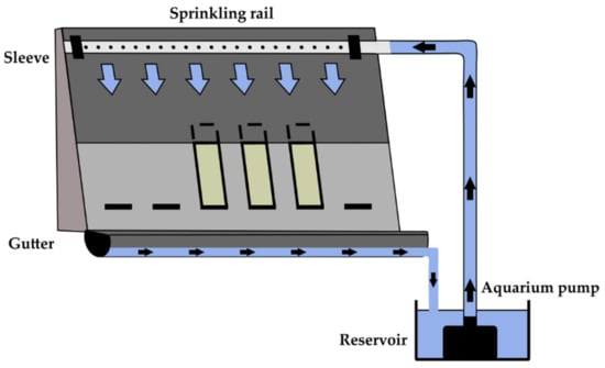
Figure 1.
Schematic drawing of the setup to simulate rain by water runoff.

Table 1.
Overview of the different samples, including stone type, exposed treatment and water exposure during the experiment. Samples 1 to 6 were treated by enzymes. Rain was simulated over the biofouled surface of samples 1–3, 7 and 8 using water runoff, whereas the biofilms of the other samples were periodically wetted by capillary uptake.
A total of 48 h after applying the enzyme-based treatment, water runoff was applied to treated Samples 1 to 3 and untreated Samples 7 and 8 for one hour. The two groups were separated from each other in a different setup to avoid enzyme contamination of the untreated samples. The samples that were not exposed to runoff (treated Samples 4–6 and untreated Sample 9) absorbed water by capillary imbibition for one hour to avoid a complete dry-out to compare the final result to the runoff-exposed samples. After the wetting phase, all samples dried at ambient laboratory conditions for at least one day. This wetting–drying cycle was repeated seven times.
During the experiment, the samples were monitored using photography and spectrophotometry, performed before treatment and at the start of each wetting–drying cycle. The light of all the photographs was white-balanced using a Mini Colour Checker Classic (MacBeth). Colour changes were monitored using a handheld spectrophotometer, which is a non-destructive technique that does not need further sample preparation and allows for fast measurements. Spectrophotometry is a suitable technique to monitor biocolonisation on rocks. It allows for monitoring of biological fouling and to estimate the biofilm biomass on stones. [14,29,30,31]. A CM-600d spectrophotometer (Konica Minolta, Tokyo, Japan) with 8 mm aperture and D65 illuminant determined the visible colour spectrum between 400 and 700 nm (Visible spectroscopy (VIS)) on three fixed points of every sample (measuring points 1, 2 and 3). The exact measuring positions on the samples were captured using a fixed mould. Both the spectral curve and quantitative colour data in the CIE L*a*b* colour space were retrieved from the measurements. The L* value indicates the lightness of the colour and ranges from 0 to 100, representing black and white, respectively. The a* value represents the green (negative) or red (positive) component, and the b* value represents the blue (negative) and yellow (positive) component [32,33]. The spectral data allow for assessment of the colour difference between the start and the end of the experiment by applying the following formula [32,33]:
Furthermore, the spectral data can estimate the specific discolouration of biological pigments. Chlorophyll absorbs visible light between ca. 400 and 500 nm and around 650–670 nm, and it also has a small absorption peak around ca. 610–620 nm. Carotenoids, on the other hand, absorb light between ca. 400 and 500 nm [34]. To describe biological discolouration by chlorophyll during the experiment, we propose the following factor:
Chlorophyll discolouration (CD) = Reflectance700 nm
—Reflectancelocal minimum (between 650–670 nm)
—Reflectancelocal minimum (between 650–670 nm)
2.2. In Situ Survey
Four sites were treated with two different commercial enzyme-based products. For privacy reasons, these sites are undisclosed and hereafter referred to as Sites A, B, C, and D. Sites A and B are located in western Belgium, near the French border. Sites C and D are in northwestern France, close to the Belgian border (Figure 2). The sites are located less than 70 km apart from each other, in rural environments, surrounded by meadows, with three of them bordering a small village. The headstones are, with a few exceptions, made of Portland limestone [1] and are well maintained. In the past, these headstones were cleaned with an ammonium-based cleaning agent. All cleaning treatments at all sites stopped from September 2016 until May 2019. On 2 May 2019, the headstones at all four sites were treated with enzyme-based products for cleaning purposes. On that day, the weather was cloudy with some drizzle or light rain, with temperatures ranging from 12 to 18 °C. The products were applied by low-pressure spraying. Sites A and B were treated with Biomix ATM, the same product as that used in the laboratory experiments. Sites C and D were treated with Enzylan (AlgaVelan, Ermelo, The Netherlands), another commercially available enzyme-based detergent. The approach is performance-based as the exact composition of the products is undisclosed by the manufacturers. Before application, the pH was tested in the laboratory to avoid damage to the material. Both products had a pH of 9.73 and were found suitable for application on limestone. A third enzyme-based cleaning product had a pH of 3.12 and was hence rejected for this study. This shows that attention should be given to the pH before the application of new products. As these products are commercially available enzyme-based detergents, the assumption was made that the enzymes are the active substance to induce the removal of biocolonisation.
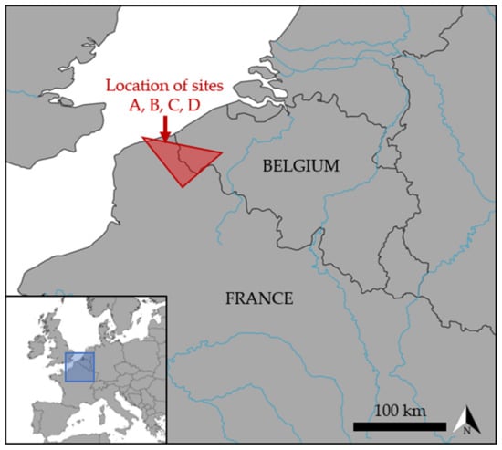
Figure 2.
Location of the four sites treated with enzyme-based products.
On-site monitoring was performed by in situ surveys at all four sites on the same day (Site A in the morning, followed by Sites B, C and D until the afternoon). The procedure was repeated for every measurement day. In total, eight in situ surveys were performed. The first was on 29 April 2019 (29/04/2019), before the application of the treatment. This was repeated three days after the treatment on 6 May 2019 (06/05/2019) and subsequently on 13 May (13/05), 27 May (27/05), 18 June (18/06), 5 August (05/08), 9 September (09/09) and 3 October 2019 (03/10/2019), covering the next five months after application. A minimum of 10 headstones per site were documented by photography, white balanced using a Mini ColourChecker Classic (MacBeth). In addition, five headstones per site were selected to perform spectrophotometry. For these measurements, an SP60 Sphere spectrophotometer (X-rite, Grand Rapids, MA, USA) was used, using the same measurements conditions as the Konica Minolta spectrophotometer. On each headstone, six VIS spectra were recovered: two on the front and four on the back. This resulted in a total of 120 measurements, or 60 measurements per product, divided equally over two sites. The positions of these measurements were captured by using a mould over the headstone. Nevertheless, millimetric variations in the exact position could occur as a result of human handling.
3. Results and Discussion
3.1. Laboratory Experiments
All samples were severely colonised, which affected the aesthetic appearance of the stones. The effect of the enzyme-based treatment on Portland limestone is illustrated and compared with an untreated sample in Figure 3. There was a lower reflectance for small wavelengths, and the biological pigments caused the typical local minima in the spectral reflectance graphs between 400 and 500 nm and around 670 nm, as well as some more subtle minima, such as the one around 610 nm.
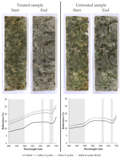
Figure 3.
Appearance of a representative treated (Sample 2) and untreated (Sample 8) biofouled stone at the start and end of the experiment and spectral reflectance graphs showing the evolution of the spectra over time. The areas of chlorophyll absorption are indicated in grey.
The enzyme-based treatment was effective, as in the treated sample, the reflectance increased. Moreover, the local spectral reflectance minima linked to biological pigments became obscure or disappeared, suggesting a decrease in biocolonisation. The local minima between 400 and 500 nm smoothed rapidly after a few cycles, whereas the one around 670 nm smoothed more gradually. The untreated samples also reflected more light, although the spectral reflectance graphs remained similar throughout the whole experiment. Furthermore, the treatment caused the local minimum at 670 nm to shift to 660 or 650 nm, whereas it remained at 670 nm for the untreated samples.
Changes were also visible and captured with photography, but these were subtler and more gradual (Figure 3). Initially, all samples had a bright green appearance, but they turned dull after treatment. The untreated samples also lost some of their green pigmentation, but this was probably caused by the indoor conditions between the wetting phases. Similar observations were made on the other samples and Savonnières limestone. The evolution of discolouration on all samples and accompanying spectral reflectance graphs can be found in the Supplementary Information (Figures S1–S9).
Table 2 provides an overview of the spectrophotometry results from the start and end of the experiment. The data show that after treatment and exposure to runoff, the colour changed (ΔE), and overall, the lightness (L*↑) increased during the experiment, whereas the green and yellow colour component decreased (a*↑, b*↓). Changes in pigmentation were quantified by the chlorophyll discolouration factor (CD), based on the reflectance local minimum induced by chlorophyll [34]. It decreased significantly for the treated samples compared to the untreated samples. In this experiment, the Savonnières limestone experienced relatively larger colour changes and reduced biological pigmentation compared to the Portland stone. This is most likely related to different bioreceptivity. However, due to the lack of replicates, it is not possible to state that the treatment works better on one of the two building stones.

Table 2.
Spectral reflectance data of the three fixed points (X.1–X.3) of every sample (1.X–9.X), with ΔL, Δa and Δb representing changes of the CIE L*a*b* colour space between the start and end; ΔE corresponds to the colour difference, and ΔCD (%) refers to the percentual difference in chlorophyll discolouration.
Water runoff did not seem to improve the results of the enzyme-based treatment. The visual observations and spectrophotometric data were similar for treated samples, regardless of whether they were exposed to runoff. The enzymes caused in both cases reduced biopigmentation on the stones. The colonisers stayed mostly attached to the surfaces, as the water runoff did not succeed in detaching them. This resulted in limited improvement in the aesthetics of the stones. Capillary imbibition had the advantage that it did not wash the enzymes away.
Besides the general trend, there were a few exceptions, especially on Sample 3, where the colour difference and decrease in CD at the end of the experiment was rather limited. It seemed that the enzyme-based treatment did not have a significant effect. Nevertheless, visually, the sample lost biological discolouration (Figure S3). This could relate to the lower initial biofouling, as the CD values were relatively low from the start, combined with high a* values. On the other hand, it could be that the enzyme-based treatment worked less effectively on these biological colonisers. Furthermore, two measuring points, 4.1 and 6.2, indicate a substantial increase in the CD factor. Here, the local minimum became less pronounced; however, there was also an important increase in reflectance at 700 nm. Visual evaluation confirmed that biological colonisation did not increase on those measuring points, which is in agreement with the smoothing of the other spectra between 400 and 630 nm (Figures S4 and S6). Moreover, the green component also decreased (a*↑), suggesting that the enzymes worked, withering the colonisers.
3.2. In Situ Survey
The headstones showed discolouration due to biological colonisation, most likely consisting of algae and lichen (Figure 4). Lichen was identified by its regular form and occurred as crustose epilithic types, found both on the horizontal top surface and on the vertical surface. White stains were present where former lichen growth had etched the surface. Superficial green and grey to black films or spots were interpreted as algae, probably consisting of green algae and cyanobacteria, abundantly present on headstones [35]. The intensity of these biofilms varied significantly. Often, these biofilms showed vertical streaks that were more intense near the horizontal top surface of the headstone because of runoff. A dark (buff) discolouration was visible near the foot of the headstones, probably related to rising damp, associated salts or splashing soil particles.
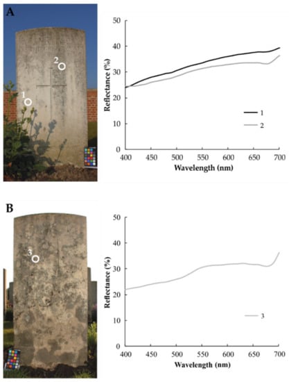
Figure 4.
Spectral reflectance graphs retrieved before treatment from (A) a relatively clean and (B) an intensely biofouled headstone.
Figure 4 gives an example of spectral data measured on three spots of two biofouled headstones. On the first one (Figure 4A), grey to black biofilms were present running down from the horizontal top surface, whereas on the second one (Figure 4B), the headstone was mainly colonised by crustose lichens. Measurement 1 was taken in a relatively clean part of the stone, resulting in a relatively smooth spectral reflectance, with lower reflectance for shorter wavelengths. Measurement 2 was taken on the grey to black biofilm and showed absorption with a local minimum around 650–670 nm and a slight absorption in the 400–500 nm range related to biological pigments. Similar observations were made on the back with measurement 3, displaying even more intense absorption. The resulting chlorophyll discolouration of VIS 1, 2 and 3 was 1.73, 2.99 and 5.05, respectively.
Figure 5 shows an overview of all the CD values over time, with the individual measurements in the background. All the values of all sites can be found in the Supplementary Information (Tables S1–S4). The initial CD values varied strongly, related to the irregular nature of the biofouling. The highest CD was determined at Site B (13.91). Four days after the application of the products, the visual differences were limited, but the CD decreased significantly at all sites. In some cases, this initial reduction in CD was more than 80% (Site C) and often reached reductions of 50% and more at sites B, C and D. In general, the higher the initial CD, the stronger its reduction four days after product application. Therefore, at Site A, where the initial CD was the lowest on average, reductions were mostly lower than 50% but still relevant. Where CD was initially low and biopigmentation thus limited, the effect was neutral.
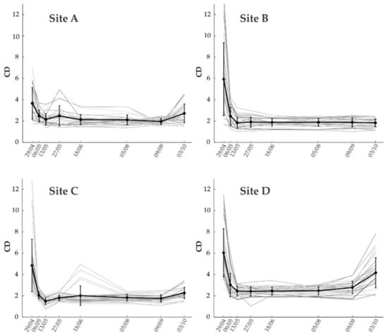
Figure 5.
Evolution of CD values over time in Site A, B, C and D, with the dates of measurements presented on the X-axis (in 2019). Every grey curve shows the evolution of individual measurement locations, whereas the dark black line shows the average values from one site with the error bar.
The decrease in CD stayed put over the next weeks and months, as illustrated by the trend in Figure 5 and in the Supplementary Information (Tables S1–S4). For all sites, CD decreased slightly more in the measurements 11 days after application, shifting to relatively stable CD values of around 2, whereas the initial values were often 4 or more. Over the next five months, until the last measurement, no drastic changes were observed in the reflectance spectra. Most information was retrieved from visual observations and photographs, showing a visual decrease in biofouling persisting over this period. Figure 6, Figure 7 and Figure 8 show time-lapse imaging of three headstones from different sites to illustrate the effect on green and grey to black biofilm, as well as lichen and the stone. The intensity of dark-grey film streaking from the top surface strongly declined, as shown in Figure 6. Much of the dark streaking disappeared after three months. The enzyme-based treatment also decreased grey streaks, together with a general greening of the surface and more intense green streaks (Figure 7). The grey biofilm disappeared after three months, whereas the greening disappeared only four days (or less) after the application. Furthermore, the more intense green streak faded after four days, disappearing completely 47 days after treatment. Finally, the colour of intense green lichens, displayed in Figure 8, faded four days after the treatment, and after three months, the thallus was removed from the surface. It was observed in situ that lichen thalli became dry and easily detached from the surface.
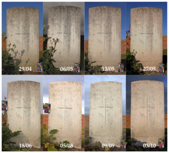
Figure 6.
Evolution of grey-black biofilm in streaks (Site A).
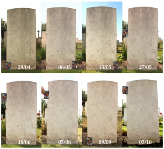
Figure 7.
Evolution of grey-black biofilm in streaks and green biofilm (Site B).
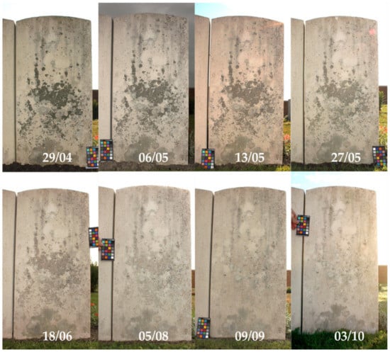
Figure 8.
Evolution of grey-black streaks and lichen (Site A).
Both products seemed effective, and after analysing CD values and visual observations, no clear differences between treatments with different products appeared. Green biofilms disappeared relatively fast after the application of both products, whereas more intense green streaks disappeared in the first weeks after application. Grey to black streaks strongly diminished or completely disappeared in the longer term, which was strongly visible after three months. The lichen degraded in the first weeks after application, and thalli were washed away after a few months. It is assumed that the enzyme-based products negatively affect the microorganisms (resulting in a decrease in chlorophyll, hence CD). Long-term exposure to rain spells acts as natural washing, removing significant amounts of withered organisms and EPS from the surface. This resulted in a visual reduction in biofouling. The stone itself was macroscopically unaffected by the treatment (white, etched zones remained the same), and the treatment did affect other types of discolouration, such as the darkening at the base of the headstones.
During the last survey on 3 October (03/10), five months after applying the products, an increase in CD was observed. Moreover, the full reflectance spectra of individual measurements reveal increased absorption in the 400–500 nm interval and around 670 nm, which occurred at Sites A, C and D, most obviously at the latter sit. It is absent at Site B, which indicates the reactivation of biopigmentation (Figure 5, Figure 6, Figure 7 and Figure 8), and is interpreted as an onset of potential new colonisation.
Exceptions to the general trends in CD in the weeks to months after application were found at Site A and Site C. At Site A, an increase in CD was observed during the measurement of 27 May (27/05), 25 days after application, in 15 of the 30 measurement points (Table S1). A total of 14 of 15 measurements faced a southwest orientation. In the following survey of 18 June (18/06) (47 days after application and 22 days after the previous measurement), this phenomenon disappeared, and CD values fell back to values of around 2. The reason for the short revival is unclear. There were no extreme weather conditions during or before the measurements of 27 May, and this increase was not observed at the other three sites, which are exposed to similar climatic conditions. Therefore, it is assumed that local environmental factors, e.g., airborne nutrients related to nearby farming, might have had a temporary effect on the measurements. At Site C, there was an increase in CD values on four measurement points: one headstone, on both sides of this headstone, in the measurements of 18 June (Table S3). This is also assumed to be related to a local effect. This headstone is neighboured by an equally sized plant leaning against the headstone, blossoming specifically during the measurement of 18 June. This might have resulted in superficial biodeposition on the headstone, affecting CD measurements. Other localised phenomena, such as bird droppings, could be responsible, but in this case, there is no indication of such phenomena.
4. Conclusions and Future Research
Preliminary laboratory and in situ experiments confirmed the effectiveness of enzyme-based treatment to clean natural building stones. The studied treatments decreased biofouling significantly and were especially effective in situ, removing biopigmentation, resulting in a more homogeneous colour. The products reduced chlorophyll discolouration after four days, and the aesthetics of the headstones improved after a few weeks. The treatment was effective during the complete monitoring period of five months, after which very limited potential recolonisation occurred at three out of four studied sites. This suggests that the treatment might have to be repeated on a regular basis, e.g., every (half) year. The cleaning effect was measurable but less pronounced during the laboratory experiments, which is most likely related to the shorter duration of the experiment. Overall, the results suggest enzyme-based detergents as an appropriate cleaning product for outdoor limestones, including headstones, avoiding environmentally harmful pesticides. However, more research is necessary to understand the specific effect of enzymes on biofouling. Biofilms should be characterised in detail, and the effect of enzymes on different types of biofilms, microorganisms and the environment should be further investigated. Moreover, even longer monitoring campaigns could be performed to study recolonisation of treated surfaces. Different enzyme-based products could be applied at the same site to determine the most suitable one, avoiding microclimatic variations. Furthermore, the effect of the products on the stones should be investigated to make sure they do not deteriorate the substrate. It is also important to study how treatment affects microorganisms and reveal any potential negative impact on the environment.
Supplementary Materials
The following supporting information can be downloaded at: https://www.mdpi.com/article/10.3390/coatings12030375/s1, Figure S1: Appearance and spectral reflectance graphs determined at fixed locations 1, 2 and 3 of treated Sample 1 from the start and after different cycles of water runoff and drought.; Figure S2: Appearance and spectral reflectance graphs determined at fixed locations 1, 2 and 3 of treated Sample 2 from the start and after different cycles of water runoff and drought.; Figure S3: Appearance and spectral reflectance graphs determined at fixed locations 1, 2 and 3 of treated Sample 3 from the start and after different cycles of water runoff and drought.; Figure S4: Appearance and spectral reflectance graphs determined at fixed locations 1, 2 and 3 of treated Sample 4 from the start and after different cycles of capillary absorption and drought.; Figure S5: Appearance and spectral reflectance graphs determined at fixed locations 1, 2 and 3 of treated Sample 5 from the start and after different cycles of capillary absorption and drought.; Figure S6: Appearance and spectral reflectance graphs determined at fixed locations 1, 2 and 3 of treated Sample 6 from the start and after different cycles of capillary absorption and drought.; Figure S7: Appearance and spectral reflectance graphs determined at fixed locations 1, 2 and 3 of untreated Sample 7 from the start and after different cycles of water runoff and drought.; Figure S8: Appearance and spectral reflectance graphs determined at fixed locations 1, 2 and 3 of untreated Sample 8 from the start and after different cycles of water runoff and drought.; Figure S9: Appearance and spectral reflectance graphs determined at fixed locations 1, 2 and 3 of untreated Sample 9 from the start and after different cycles of capillary absorption and drought.; Table S1: Initial CD (%) of 30 measurements at Site A before enzyme-based treatment and ∆CD (%) during subsequent field surveys (per date).; Table S2: Initial CD (%) of 30 measurements at Site B before enzyme-based treatment and ∆CD (%) during subsequent field surveys (per date).; Table S3: Initial CD (%) of 30 measurements at Site C before enzyme-based treatment and ∆CD (%) during subsequent field surveys (per date).; Table S4: Initial CD (%) of 30 measurements at Site D before enzyme-based treatment and ∆CD (%) during subsequent field surveys (per date).
Author Contributions
Conceptualization: T.D.K.; methodology: T.D.K., L.S. (Laurenz Schröer) and G.F.; validation: T.D.K. and L.S. (Laurenz Schröer); formal analysis: L.S. (Laurenz Schröer), T.D.K., M.D. and G.F.; investigation: T.D.K., L.S. (Laurenz Schröer), M.D. and G.F.; resources: T.D.K., V.C. and L.S. (Lander Soens); data curation: L.S. (Laurenz Schröer), T.D.K., M.D. and G.F.; writing—original draft preparation: L.S. (Laurenz Schröer) and T.D.K.; writing—review and editing: T.D.K., L.S. (Laurenz Schröer), M.D., G.F., V.C., N.B. and L.S. (Lander Soens); visualization: L.S. (Laurenz Schröer) and T.D.K.; supervision: T.D.K., V.C. and N.B.; project administration: T.D.K.; funding acquisition: T.D.K., V.C. and N.B. All authors have read and agreed to the published version of the manuscript.
Funding
Laurenz Schröer was supported by a PhD fellowship of the Research Foundation—Flanders (FWO, Research Grant No.: 11D4518N and 11D4520N). Tim De Kock was a postdoctoral fellow of the FWO during part of the research and acknowledges their support. Maxim Deprez was supported by the FWO (Project G.0041.15N). Géraldine Fiers was funded by the special research fund of Ghent University (BOF-UGent, BOF16/IOP/001).
Institutional Review Board Statement
Not applicable.
Informed Consent Statement
Not applicable.
Data Availability Statement
The data that support the findings of this study are available from the corresponding author.
Acknowledgments
We want to thank the Commonwealth War Graves Commission (CWGC) for the intense cooperation at the sites and for providing the laboratory material. Furthermore, we acknowledge the support of Sarah Williamson and Daphne Guilbert during the in situ survey and data analysis. We would also like to thank the Laboratory of Wood Technology and Jan Van den Bulcke from Ghent University for lending their spectrophotometer for the in situ survey.
Conflicts of Interest
The authors declare no conflict of interest. The funders had no role in the design of the study; in the collection, analyses, or interpretation of data; in the writing of the manuscript, or in the decision to publish the results.
References
- Hughes, T.; Lott, G.K.; Poultney, M.J.; Cooper, B.J. Portland Stone: A nomination for “Global Heritage Stone Resource” from the United Kingdom. Episodes 2013, 36, 221–226. [Google Scholar] [CrossRef] [PubMed] [Green Version]
- Siegesmund, S.; Weiss, T.; Vollbrecht, A. Natural stone, weathering phenomena, conservation strategies and case studies: Introduction. Geol. Soc. Lond. Spec. Publ. 2002, 205, 1–7. [Google Scholar] [CrossRef]
- Gorbushina, A.A. Life on the rocks. Environ. Microbiol. 2007, 9, 1613–1631. [Google Scholar] [CrossRef] [PubMed]
- Miller, A.Z.; Sanmartín, P.; Pereira-Pardo, L.; Dionísio, A.; Saiz-Jimenez, C.; Macedo, M.F.; Prieto, B. Bioreceptivity of building stones: A review. Sci. Total Environ. 2012, 426, 1–12. [Google Scholar] [CrossRef] [PubMed]
- Flemming, H.C.; Wingender, J.; Szewzyk, U.; Steinberg, P.; Rice, S.A.; Kjelleberg, S. Biofilms: An emergent form of bacterial life. Nat. Rev. Microbiol. 2016, 14, 563–575. [Google Scholar] [CrossRef] [PubMed]
- McNamara, C.J.; Mitchell, R. Microbial deterioration of historic stone. Front. Ecol. Environ. 2005, 3, 445–451. [Google Scholar] [CrossRef]
- Warscheid, T.; Braams, J. Biodeterioration of stone: A review. Int. Biodeterior. Biodegrad. 2000, 46, 343–368. [Google Scholar] [CrossRef]
- Scheerer, S.; Ortega-Morales, O.; Gaylarde, C. Microbial Deterioration of Stone Monuments—An Updated Overview. In Advances in Applied Microbiology; Elsevier Inc.: Amsterdam, The Netherlands, 2009; Volume 66, pp. 97–139. ISBN 9291271195. [Google Scholar]
- Crispim, C.A.; Gaylarde, C.C. Cyanobacteria and Biodeterioration of Cultural Heritage: A Review. Microb. Ecol. 2005, 49, 1–9. [Google Scholar] [CrossRef]
- Schröer, L.; De Kock, T.; Cnudde, V.; Boon, N. Differential colonization of microbial communities inhabiting Lede stone in the urban and rural environment. Sci. Total Environ. 2020, 733, 139339. [Google Scholar] [CrossRef]
- Roeselers, G.; van Loosdrecht, M.C.M.; Muyzer, G. Heterotrophic Pioneers Facilitate Phototrophic Biofilm Development. Microb. Ecol. 2007, 54, 578–585. [Google Scholar] [CrossRef] [Green Version]
- Gadd, G.M. Geomicrobiology of the built environment. Nat. Microbiol. 2017, 2, 16275. [Google Scholar] [CrossRef] [PubMed] [Green Version]
- Schröer, L.; Boon, N.; De Kock, T.; Cnudde, V. The capabilities of bacteria and archaea to alter natural building stones—A review. Int. Biodeterior. Biodegrad. 2021, 165, 105329. [Google Scholar] [CrossRef]
- De Muynck, W.; Ramirez, A.M.; De Belie, N.; Verstraete, W. Evaluation of strategies to prevent algal fouling on white architectural and cellular concrete. Int. Biodeterior. Biodegrad. 2009, 63, 679–689. [Google Scholar] [CrossRef]
- Piñar, G.; Ettenauer, J.; Sterflinger, K. “La vie en rose”: A review of the rosy discoloration of subsurface monuments. In The Conservation of Subterranean Cultural Heritage; Saiz-Jimenez, C., Ed.; CRC Press: Leiden, The Netherlands, 2014; pp. 113–124. ISBN 9781138026940. [Google Scholar]
- Ortega-Morales, B.O.; Gaylarde, C.; Anaya-Hernandez, A.; Chan-Bacab, M.J.; De la Rosa-García, S.C.; Arano-Recio, D.; Montero-Muñoz, J.L. Orientation affects Trentepohlia-dominated biofilms on Mayan monuments of the Rio Bec style. Int. Biodeterior. Biodegrad. 2013, 84, 351–356. [Google Scholar] [CrossRef]
- Saiz-Jimenez, C. Microbial melanins in stone monuments. Sci. Total Environ. 1995, 167, 273–286. [Google Scholar] [CrossRef]
- Gaylarde, C.C.; Ortega-Morales, B.O.; Bartolo-Pérez, P. Biogenic Black Crusts on Buildings in Unpolluted Environments. Curr. Microbiol. 2007, 54, 162–166. [Google Scholar] [CrossRef]
- European Parliament. Council of the European Union Directive 2009/128/EC of the European Parliament and of the Council of 21 October 2009 Establishing a Framework for Community Action to Achieve the Sustainable Use of Pesticides; European Parliament: Brussels, Belgium, 2009. [Google Scholar]
- Alexopoulou, I.; Zervos, S. Paper conservation methods: An international survey. J. Cult. Herit. 2016, 21, 922–930. [Google Scholar] [CrossRef]
- Ahmed, H.E.; Kolisis, F.N. A study on using of protease for removal of animal glue adhesive in textile conservation. J. Appl. Polym. Sci. 2012, 124, 3565–3576. [Google Scholar] [CrossRef]
- Pereira, C.; Busani, T.; Branco, L.C.; Joosten, I.; Sandu, I.C.A. Nondestructive Characterization and Enzyme Cleaning of Painted Surfaces: Assessment from the Macro to Nano Level. Microsc. Microanal. 2013, 19, 1632–1644. [Google Scholar] [CrossRef]
- Hamed, S.A.E.-K.M. A preliminary study on using enzymes in cleaning archaeological wood. J. Archaeol. Sci. 2012, 39, 2515–2520. [Google Scholar] [CrossRef]
- Valentini, F.; Diamanti, A.; Palleschi, G. New bio-cleaning strategies on porous building materials affected by biodeterioration event. Appl. Surf. Sci. 2010, 256, 6550–6563. [Google Scholar] [CrossRef]
- Valentini, F.; Diamanti, A.; Carbone, M.; Bauer, E.M.; Palleschi, G. New cleaning strategies based on carbon nanomaterials applied to the deteriorated marble surfaces: A comparative study with enzyme based treatments. Appl. Surf. Sci. 2012, 258, 5965–5980. [Google Scholar] [CrossRef]
- Palmer, T.J. Limestone Petrography and Durability in English Jurassic Freestones. In Proceedings of the England’s Heritage in Stone, York, UK, 15–27 March 2005; Doyle, P., Ed.; The English Stone Forum: Folkstone, UK, 2005; pp. 64–75. [Google Scholar]
- Fronteau, G. Comportements Telogénétiques des Principaux Calcaires de Champagne-Ardenne, en Relation avec Leur Facies de Dépôt et Leur Séquençage Diagénétique. Ph.D. Thesis, Université de Reims Champagne-Ardenne, Reims, France, 2000. [Google Scholar]
- Dreesen, R.; Dusar, M. Historical building stones in the province of Limburg (NE Belgium): Role of petrography in provenance and durability assessment. Mater. Charact. 2004, 53, 273–287. [Google Scholar] [CrossRef]
- Sanmartín, P.; Vázquez-Nion, D.; Silva, B.; Prieto, B. Spectrophotometric color measurement for early detection and monitoring of greening on granite buildings. Biofouling 2012, 28, 329–338. [Google Scholar] [CrossRef] [PubMed]
- Becerra, J.; Ortiz, P.; Zaderenko, A.P.; Karapanagiotis, I. Assessment of nanoparticles/nanocomposites to inhibit micro-algal fouling on limestone façades. Build. Res. Inf. 2020, 48, 180–190. [Google Scholar] [CrossRef]
- Prieto, B.; Silva, B.; Lantes, O. Biofilm quantification on stone surfaces: Comparison of various methods. Sci. Total Environ. 2004, 333, 1–7. [Google Scholar] [CrossRef]
- Berger-Schunn, A. Practical Color Measurement: A Primer for the Beginner, a Reminder for the Expert; Wiley: Hoboken, NJ, USA, 1994; ISBN 9780471004172. [Google Scholar]
- Brainard, D.H. Color Appearance and Color Difference Specification. In The Science of Color; Elsevier: Amsterdam, The Netherlands, 2003; pp. 191–216. ISBN 9780444512512. [Google Scholar]
- Lichtenthaler, H.K. Chlorophylls and carotenoids: Pigments of photosynthetic biomembranes. In Methods in Enzymology; Academic Press: Cambridge, MA, USA, 1987; Volume 148, pp. 350–382. [Google Scholar]
- Brewer, T.E.; Fierer, N. Tales from the tomb: The microbial ecology of exposed rock surfaces. Environ. Microbiol. 2018, 20, 958–970. [Google Scholar] [CrossRef]
Publisher’s Note: MDPI stays neutral with regard to jurisdictional claims in published maps and institutional affiliations. |
© 2022 by the authors. Licensee MDPI, Basel, Switzerland. This article is an open access article distributed under the terms and conditions of the Creative Commons Attribution (CC BY) license (https://creativecommons.org/licenses/by/4.0/).