Article

Electrochemical and Optical Behavior of ZrN-Ag Coatings Deposited by Means of DC Reactive Magnetron Sputtering Technique

1
Núcleo de Investigación de la Escuela de Ingenierías y Tecnologías (NIEIT), Escuela de Ingenierías y Tecnologías, Fundación Universitaria Monserrate, Bogotá 110221, Colombia
2
Grupo de Investigación Fundamental y Aplicada en Materiales (GIFAM), Departamento de Física, Universidad Antonio Nariño, Bogotá 111511, Colombia
3
Departamento de Mecánica y Mecatrónica, Universidad Nacional de Colombia, Bogotá 111321, Colombia
*
Author to whom correspondence should be addressed.
Coatings 2022, 12(6), 754; https://doi.org/10.3390/coatings12060754
Submission received: 29 April 2022 / Revised: 25 May 2022 / Accepted: 26 May 2022 / Published: 31 May 2022

Abstract

:
The formation of nanostructured transition metal nitride coatings by introducing a small amount of silver (Ag) content has been proven to be a good strategy for enhancing the physical properties of these materials. In this investigation, ZrN coatings with different Ag contents were deposited on an AISI 316L substrate using the DC reactive magnetron sputtering technique. The influence of the silver on the chemical composition, morphology, and microstructure was investigated using energy-dispersive X-ray spectroscopy (EDS), X-ray photoelectron spectroscopy (XPS), scanning electron microscopy (SEM), and X-ray diffraction (XRD). The functional properties, specifically the corrosion resistance and the optical reflectance of the deposited coatings, were investigated using electrochemical impedance spectroscopy (EIS) and UV-Visible-NIR, respectively. The results showed the formation of two nanocrystalline phases, fcc-ZrN and metallic fcc-Ag. On the surface of the deposited coatings, homogeneously distributed silver nanoparticles were observed, and they increased with the Ag atomic content. The chemical composition on the surface showed evidence of the formation of oxides, such as Zr-O and Zr-O-N, before and after the corrosion tests. The corrosion resistance of the AISI 316L substrate and the coatings was improved with the incorporation of Ag, and the optical reflectance increased with increasing the Ag content. Finally, this work investigated the effect of the incorporation of silver into a ZrN matrix for potential use as optical protective coatings.

1. Introduction

AISI 316L stainless steels have been widely used in many fields, such as the food and transportation industries and as a medical implant material, due to their mechanical properties, low cost, ease of fabrication, and corrosion-resistant properties [1,2,3]. However, AISI 316L has a tendency to lose electrons in a solution (experience electrochemical dissolution) and exhibits a low surface hardness and low wear resistance [3,4]. One of the methods for improving the durability and lifetime of these materials is to deposit ceramic coatings on their surfaces [5,6]. Among the large family of ceramic coatings, there are transition metal nitrides, such as titanium nitride (TiN), chromium nitride (CrN), niobium nitride (NbN), and zirconium nitride (ZrN), which have been studied extensively for different applications due to their hardness, wear resistance, corrosion resistance, and attractive colors [7,8,9,10]. For instance, the ZrN coatings have been shown to improve the surface properties of stainless steel, such as corrosion and wear resistance [11,12,13,14,15,16]. Some researchers have found that the formation of two-phase nanocomposite coatings improves the physical and chemical properties of the binary transition metal nitrides by combining hard and soft phases in the proper proportions [8,10,17,18,19,20]. The incorporation of metallic nanoparticles can increase the toughness and hardness of the binary transition metal nitride coatings [17,21] and allow them to have certain functionalities, such as a low friction coefficient [17], hydrophobicity [9], antibacterial properties [18], and the surface plasmon resonance effect [20]; however, their physical properties depend on the microstructure and morphology of the deposited coatings. In binary systems with Ag, various authors have reported that these materials may contain immiscible chemical elements in the crystal structures of TiN or ZrN [4,17,19]. When these materials are deposited together in a sputtering system, the coatings form a nanocomposite structure with metallic silver embedded in the ceramic matrix (transition metal nitride). In addition, ZrN and TiN form more stable nitrides than those formed with Ag, according to their enthalpies of formation. Kelly et al. [17] deposited coatings of CrN-Ag, ZrN-Ag, TiN-Ag, and TiN-Cu and reported that the addition of Ag or Cu tends to decrease the size of the crystallite with increasing silver or copper content. The coefficient of the friction and hardness of these coatings decreased with an increase in the Ag or Cu content. Finally, they showed that the antimicrobial activity in the coatings is higher as the content of Ag or Cu increases. Finally, Ren et al. [21] deposited coatings of Nb-Ag-N using the sputtering technique, and they found that the coating with a silver atomic content of 1.5% increased the hardness of the ternary system to a 28.0 GPa and improved the toughness of the material. With this Ag content, the material’s coefficient of friction decreased to 0.22 compared to the coefficient of the friction of an NbN coating of 0.7. These nanocomposite coatings are often multifunctional since they combine the good resistance to wear and hardness of a transition metal nitride with the antimicrobial nature of Ag or Cu. Therefore, these new coatings are attractive for innovative applications, such as food processing or in the biomedical industries, where the surface is required to be non-toxic, wear-resistant, easy to clean, and resistant to microbial contamination. In addition, metal nanoparticles such as Ag, Cu, or Au embedded in nanostructures based on different materials have shown a plasmonic response in the visible region of the electromagnetic spectrum [20,22,23,24,25,26,27,28]. These plasmonic structures have seen significant applications in the field of photovoltaics for high-efficiency solar cells [22,28]. The aim of the present research is to investigate the nanostructured coatings of ZrN-Ag deposited via the DC reactive magnetron sputtering technique. In particular, the present study investigates the effect of the incorporation of silver into a ZrN matrix on the chemical composition, morphology, microstructure, optical behavior, and corrosion resistance of these coatings immersed in ringer’s lactate for potential use as optical protective coatings.

2. Experimental Details

2.1. Materials and Deposition Method

The ZrN-Ag coatings were deposited by means of the DC reactive magnetron sputtering technique on AISI 316L mirror-like polished stainless steel, (111) silicon, and common glass substrates. The chemical composition of the AISI 316L substrate used in this investigation is shown in Table 1.
The coatings deposited on the AISI 316L were used to evaluate the corrosion resistance and chemical composition. The microstructure and the optical behavior of the coatings were evaluated with the coatings on glass. Finally, the coatings on silicon were used to investigate the morphology. Before deposition, the substrates were ultrasonically cleaned in a bath of acetone and isopropanol for 10 min each, and then they were blow-dried with dry nitrogen. After the cleaning process, the substrates were immediately placed in a deposition chamber. The magnetron was connected to a DC power supply with a metallic target of zirconium 50 mm in diameter at 99.7% purity. To incorporate Ag into the ZrN coatings, a variable number of pellets was placed on the erosion zone of the target. The chamber was evacuated to a base pressure of 4 × 10 4 ± 1 × 10 4   Pa . To eliminate any contaminants from the target and the Ag pellets, pre-sputtering for 5 min at a power of 140 W in argon (Ar) was performed. The deposition was done with a mixture of Ar and N2 at a working pressure of 0.8 Pa, substrate temperature of 200 °C, and power of 140 W. The deposition parameters and the Ar/N2 flow ratio were optimized to obtain zirconium nitride. The deposition time was 60 min for all coatings. Finally, the deposition parameters are listed in Table 2. The coatings were abbreviated as ZrN (without silver), ZrN+1Ag (with a silver pellet on the Zr target), and ZrN+2Ag (with two silver pellets on the Zr target).

2.2. Coating Characterization

The chemical composition of the coatings was investigated using energy-dispersive X-ray spectroscopy in an EDAX Pegasus X4M (EDS/EBSD), while the chemical composition of the surface of the coatings was determined using X-ray photoelectron spectroscopy (XPS) with a monochromatized Al Kα X-ray source (FOCUS 500) operated at 100 W. The acquired spectra were analyzed with the CasaXPS program using the SPECS Prodigy-ACenteno library provided with R.S.F. (response sensitivity factor) determined by the manufacturer. A Shirley baseline was used. The binding energy (BE) scale of the spectra was corrected by reference to the C-(C, H) component of the C 1s peak at 284.8 eV. The morphology and thickness were determined using a NanoSEM–FEI Nova 200 FEG/SEM with a secondary electron detector. The microstructure of the coatings was studied using X-ray diffraction (XRD) with Cu-kα radiation of 0.154 nm with the θ–2θ geometry. The optical reflectance spectra were collected using a UV-vis-NIR spectrophotometer from 300 to 2500 nm. The corrosion behavior was evaluated by means of electrochemical impedance spectroscopy (EIS) using a Gamry REF600+. An acrylic cell with three electrodes was used: test sample (working electrode), platinum (counter electrode), and calomel (reference electrode). The tested sample area was 0.196 cm2. The sample had a 10 mm diameter and 3 mm thickness. A commercial Ringer’s lactate solution with composition reported in Table 3 was used as the electrolyte [29]. Warming of the system at 37.5 °C was done using a thermal water bath with a recirculating system. The tests were performed on the bare and coated AISI 316L stainless steel. The coatings were immersed into a Ringer’s solution with an AC amplitude of 10 mV and a frequency range from 0.01 to 100 kHz, also at 37.5 °C.

3. Results and Discussion

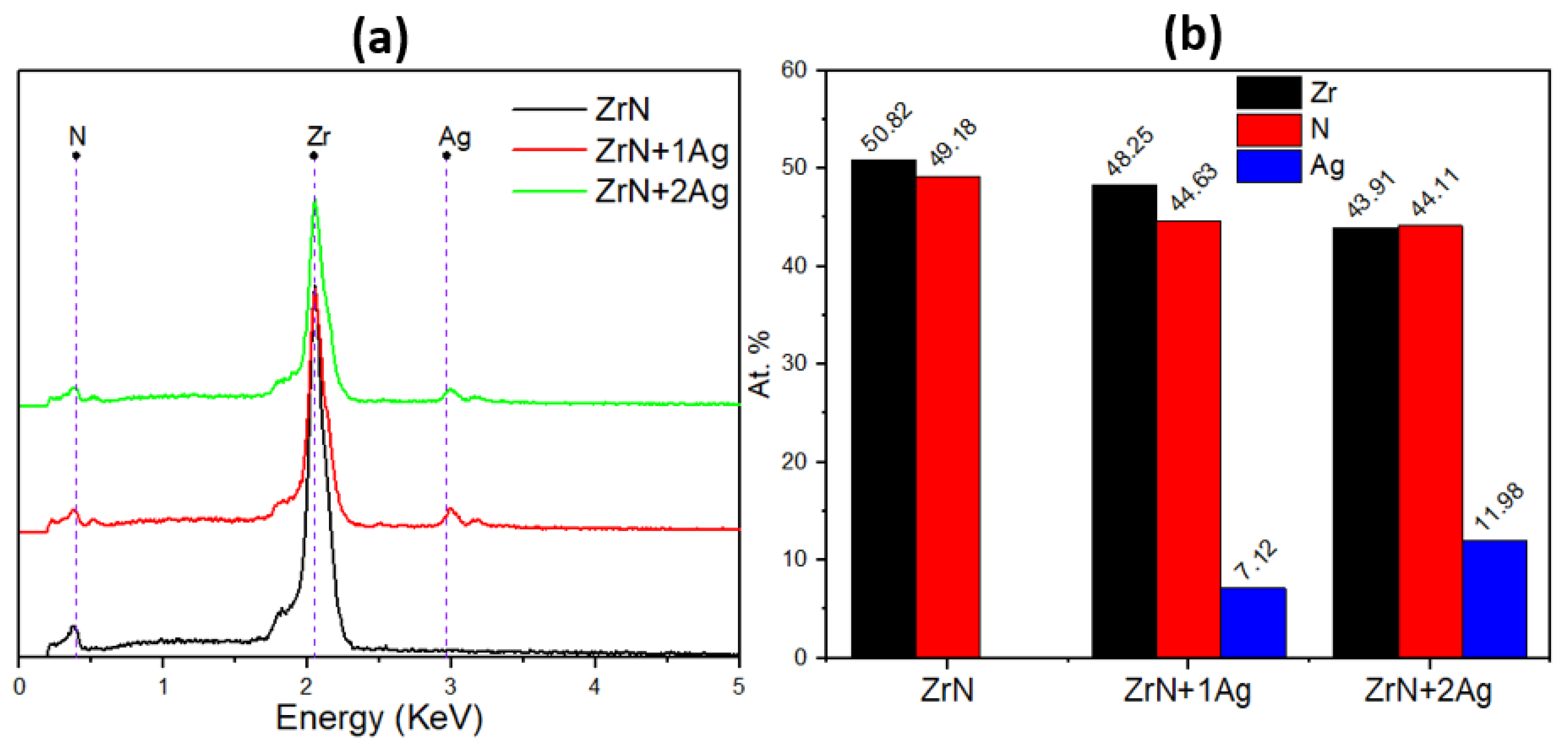
3.1. Chemical Composition of Coatings

The chemical elemental composition of the ZrN-Ag coatings as a function of the Ag pellets is shown in Figure 1. The results confirmed the presence of zirconium (Zr), nitrogen (N), and silver (Ag) in the ZrN-Ag coatings. It can be seen in Figure 1b that, as the number of the Ag pellets increases on the target’s surface, the Zr and N content decreases, while the Ag content increases in the coatings. This decrease of Zr and N is because silver pellets decrease the sputtering area in the Zr target and due to the low reactivity of silver with nitrogen to form a silver nitride phase, according to the enthalpy formation of −291 kJ/mol for the ZrN and +314 kJ/mol for Ag3N [30]. The Ag content was found to be 0.0, 7.12, and 11.98 at.% for the coatings of 0, 1, and 2 Ag pellets, respectively. In the literature, it has been reported that the incorporation of silver into ceramic coatings generates the formation of two nanocrystalline phases, ZrN and metallic Ag, due to the fact that silver is immiscible in these ceramic coatings [8,17,30,31,32].

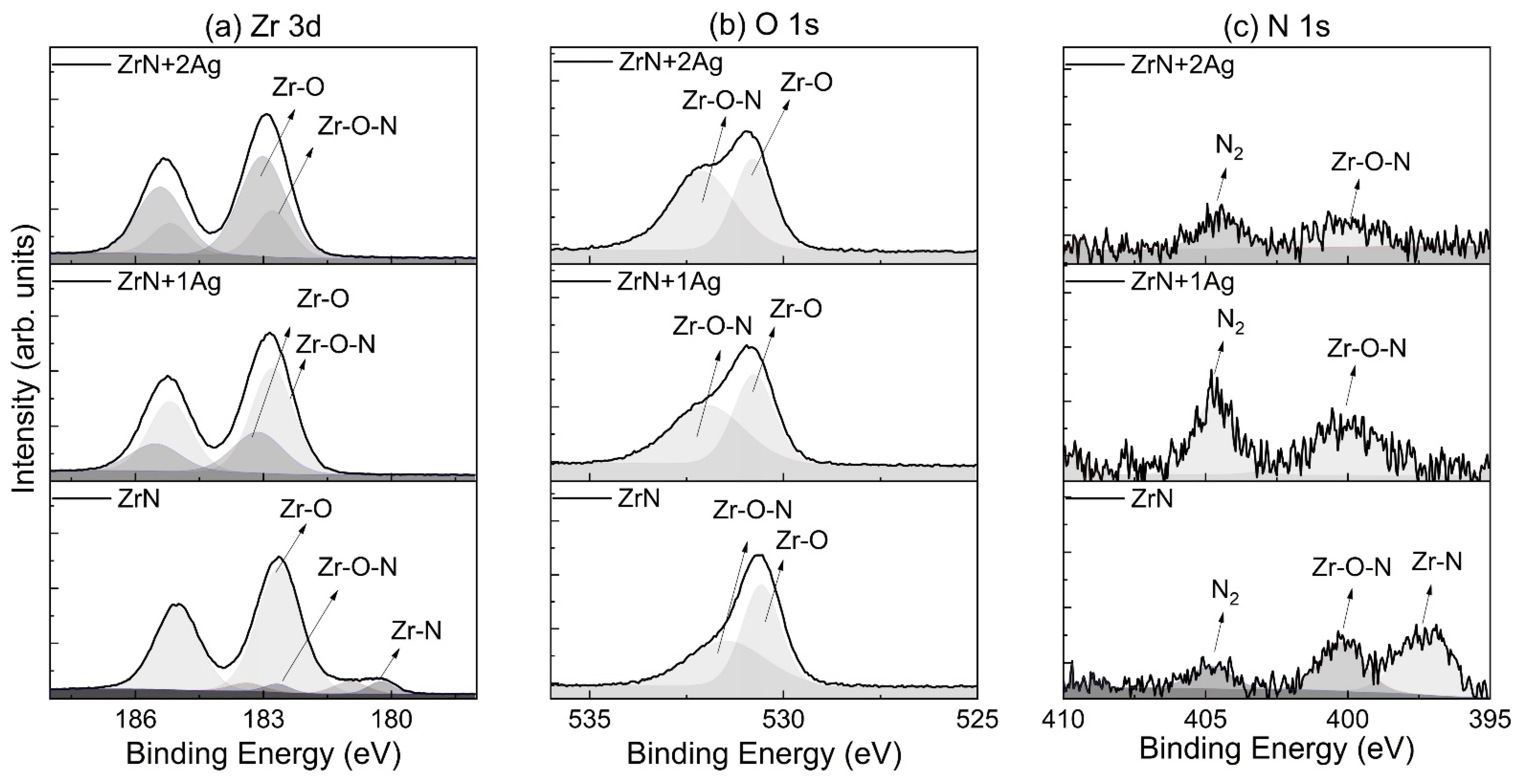
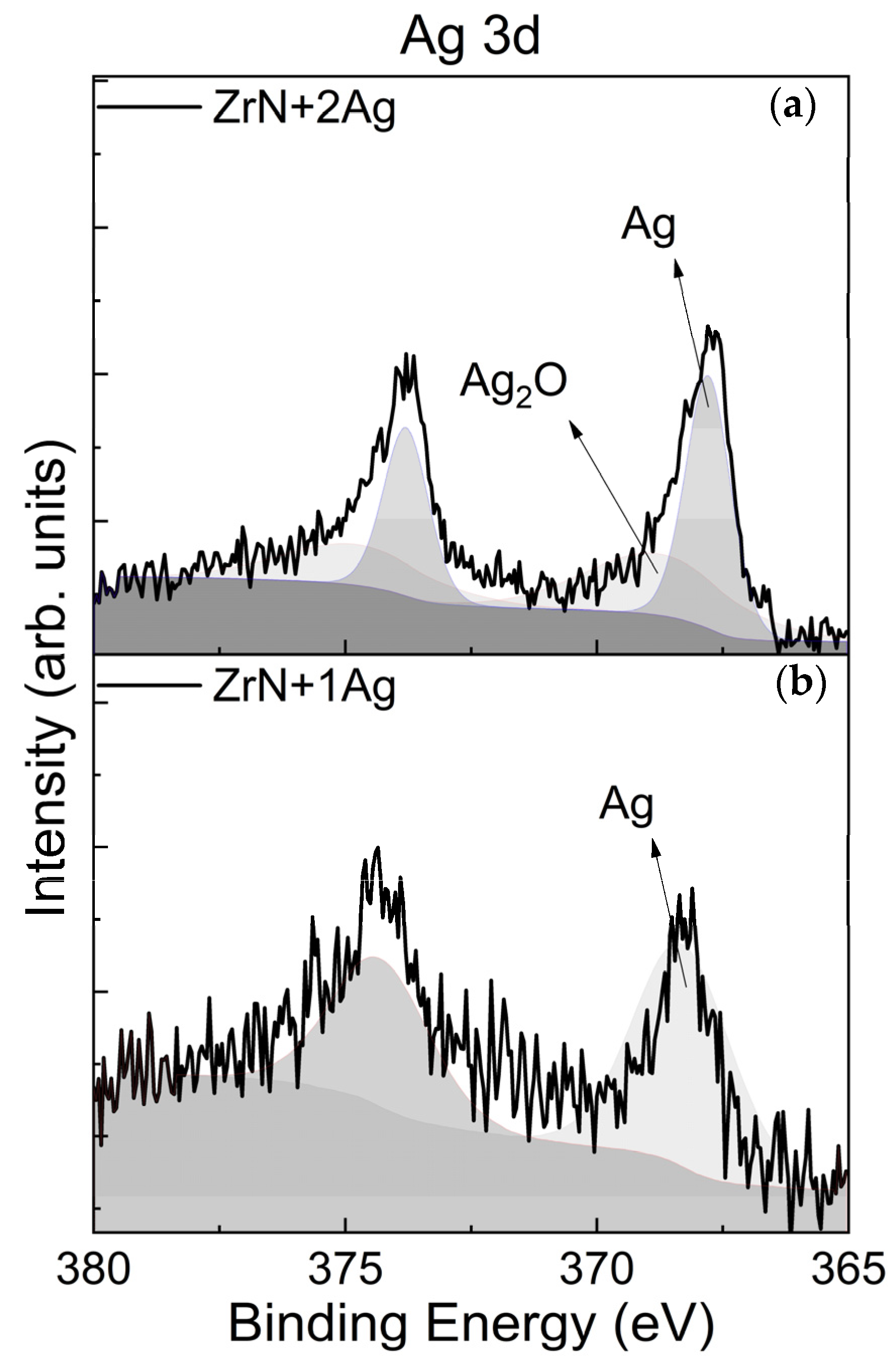
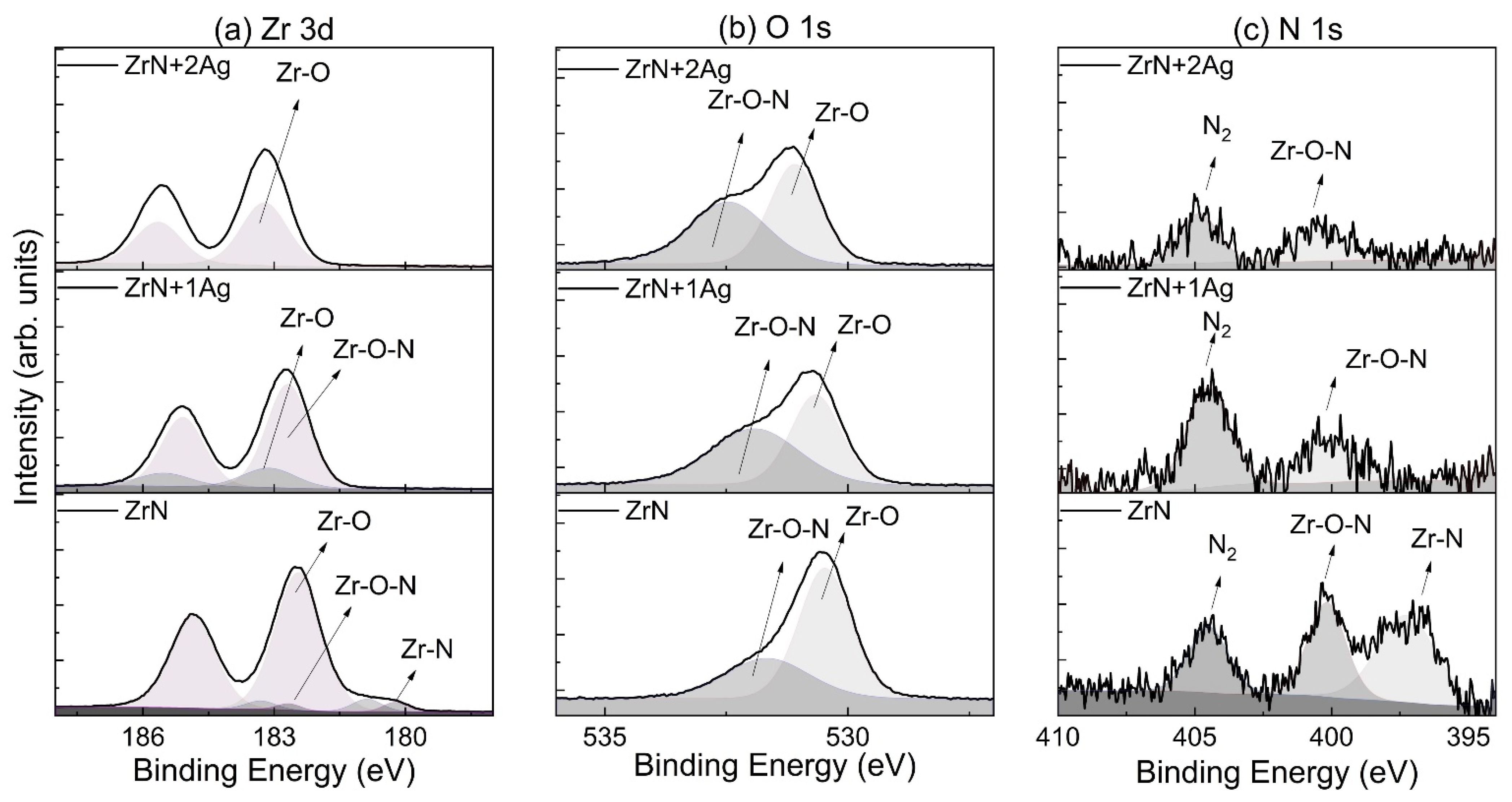
3.2. Chemical Bonding

To obtain information about possible phases present on the surface of the coatings, an XPS analysis was performed. The XPS analysis results were calibrated using the C 1s line with a binding energy of 284.8 eV. The Zr 3d spectra of the ZrN, ZrN+1Ag, and ZrN+2Ag coatings are shown in Figure 2a.
The ZrN coating exhibits the presence of Zr-N, Zr-O-N, and Zr-O bonds at 180.24 eV [33], 180.96 eV [34], and 182.62 eV, respectively. However, with Ag incorporation into the ZrN matrix, no peaks were shown related to the Zr-N bond due to the presence of the Ag nanoparticles and the formation of oxides on the surface, which do not allow them to obtain enough nitrogen signal. No peaks corresponding to Ag-N and Zr-Ag were observed in the spectrum. The presence of oxides was corroborated by the O 1s and N 1s spectra, as seen in Figure 2b. Similar results were obtained by Yu et al. [35], who reported that the presence of oxygen in the coatings could be attributed to contamination by residual oxygen in the chamber during the sputtering process. Figure 3 shows the Ag 3d spectra for the coating with an Ag content of 7.12 at.% (ZrN+1Ag) and 11.98 at.% (ZrN+2Ag). The XPS results show the presence of an Ag-Ag bond at 368.3 eV for the ZrN+1Ag coating, and as the silver content increased (ZrN+2Ag), the Ag-Ag and Ag-O bonds were detected at 368.79 eV and 367.8 eV, respectively.

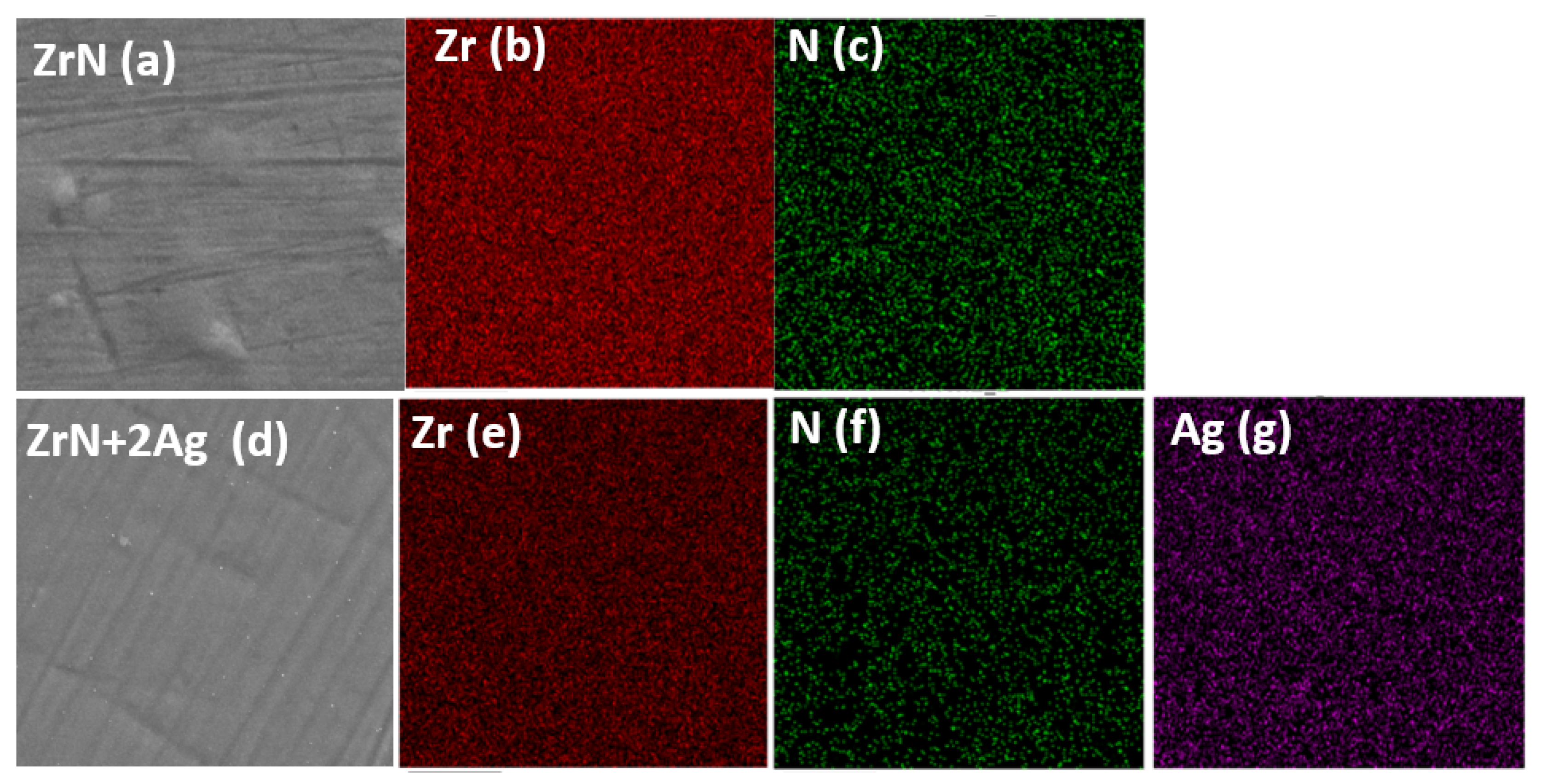
3.3. Surface Morphology of the Coatings

The Ag effect on the morphology of the coatings was investigated by means of SEM, and the results are shown in Figure 4. The morphology of the ZrN coating has been discussed in previous papers [36]. The SEM images show the cross-section and surface morphology of the coatings, with an Ag content of 7.12 at.% (Figure 4a) and 11.98 at.% (Figure 4b), respectively. As shown in Figure 4a, the ZrN+1Ag coating has a smooth surface, while the cross-section morphology reveals that the coating is dense and featureless. However, when the Ag content increases, as seen in Figure 4b, the ZrN+2Ag coating exhibits uniformly distributed nanoparticles in bright contrast, which have been related to metallic silver [37]. The presence of Ag nanoparticles on the surface can be attributed to the Ag diffusion, which is promoted by the temperature during the deposition time of the coating. During the deposition, silver is segregated in the intercolumnar space of the ZrN matrix due to the low solubility of Ag in the ceramic coatings [8,32,37]. Similar results were obtained by other authors, who reported that the segregation of silver to the surface is proportional to the Ag content in the coatings [7,32,37,38,39,40,41].
In order to determine the homogeneity of the coatings, elemental mapping was carried out using EDS on the surface of the ZrN and ZrN+2Ag coatings. Figure 5 shows the concentration of the elements deposited (Zr, N, and Ag) for the samples. The coatings show a homogeneous distribution of zirconium and nitrogen throughout the surface, as expected. In addition, with the Ag incorporation, the EDS mapping shows that metallic silver exhibits homogeneous distribution on the surface, which is in accordance with the SEM results.

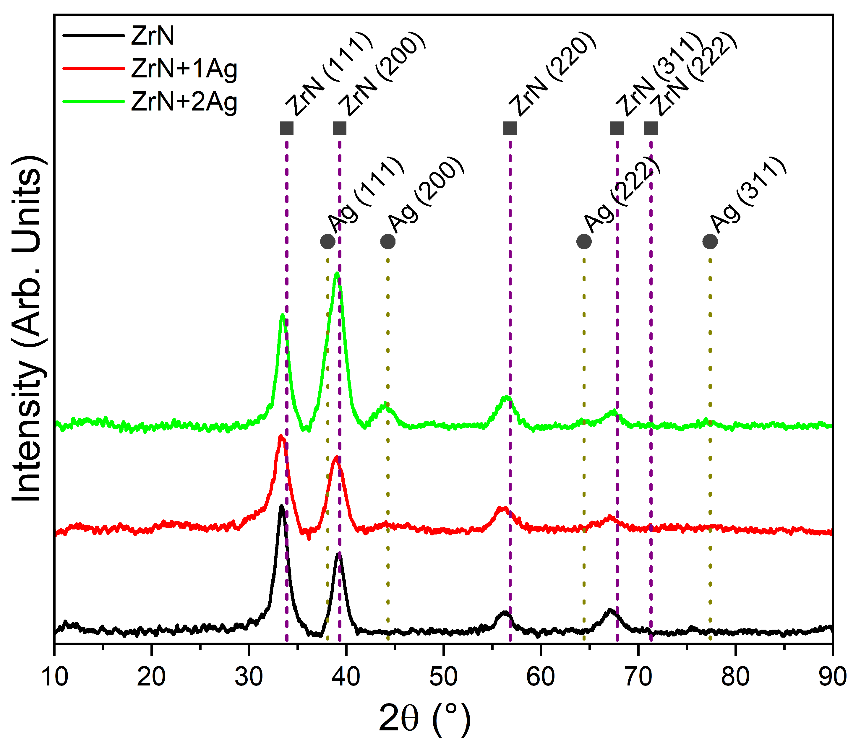
3.4. Microstructure of the Coatings

The structure evolution of ZrN coatings with different silver contents on a common glass substrate was evaluated by means of XRD, and the patterns are shown in Figure 6. All the coatings exhibited four diffraction peaks corresponding to the fcc-ZrN phase, according to pdf 01-078-1420. The broad diffraction peaks suggest a lower degree of structural order and the formation of a nanocrystalline structure, as has been reported by Cavaleiro et al. [37]. No shift of the XRD diffraction peaks was observed with the Ag incorporation. For the ZrN+1Ag coating, no Ag-related diffraction peaks were observed due to the limited precision of the XRD. However, when the Ag content increased, the appearance of a new diffraction peak with a low intensity of around 43.66° was detected and attributed to the fcc-Ag(200) plane, according to pdf. 01-087-0597. In addition, the intensity of the diffraction peak related to the ZrN(200) plane becomes greater and broader with an increasing Ag content due to the overlapping of this peak with that of the fcc-Ag(111) plane. Furthermore, other diffraction peaks corresponding to Ag2O, AgNO3, or AgTi, were not detected in the XRD pattern.
The diffraction peak of around 33.26° was fit with two contributions: one assigned to the ZrN cubic phase and the other to the ZrO2 tetragonal phase due to the high tendency of zirconium to form oxides [42], and the influence of silver in the lattice parameter and grain size was studied. The results showed that all the ZrN-Ag coatings had a grain size <10 nm due to high FWHM values, and the lattice parameter was not affected by the Ag incorporation, as can be seen in Table 4.

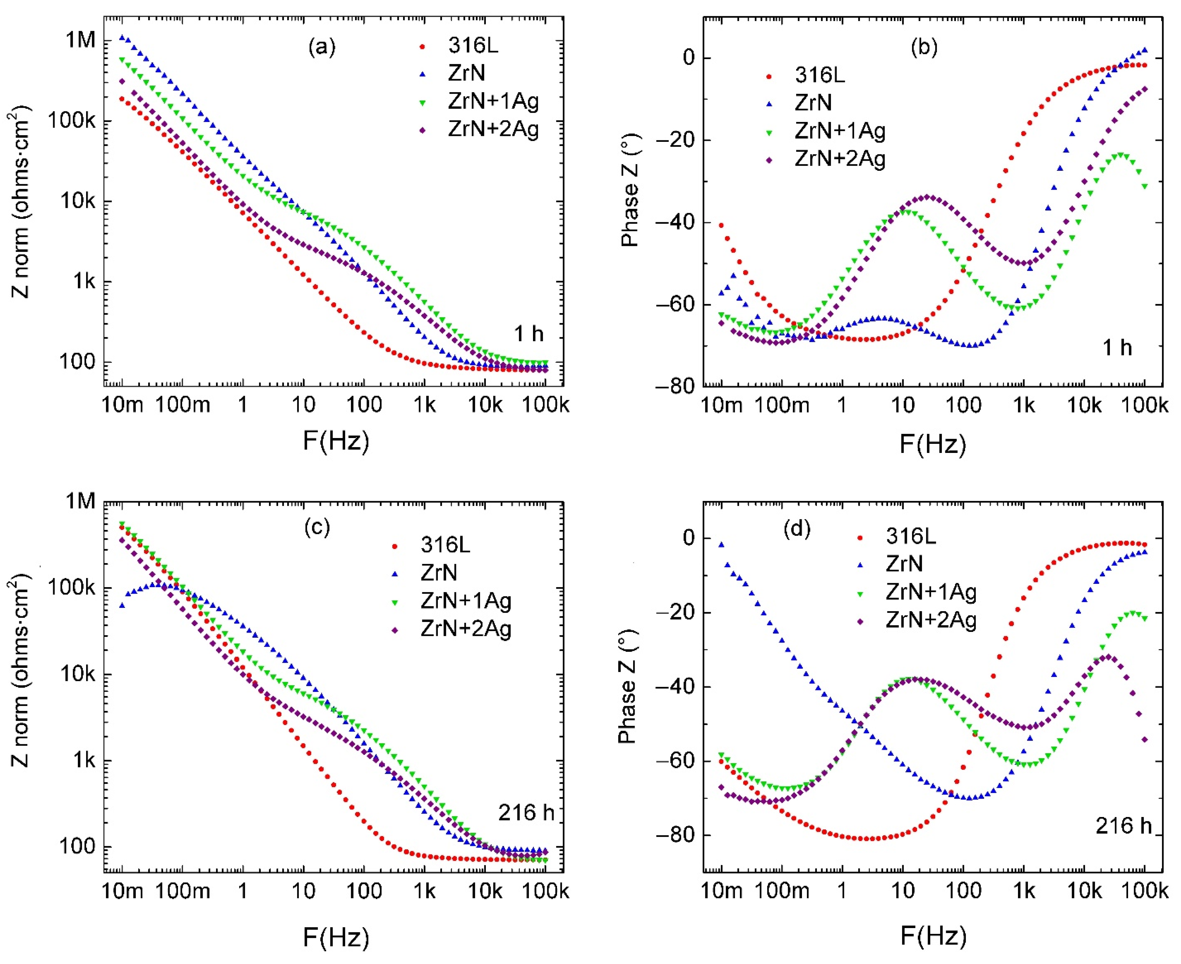
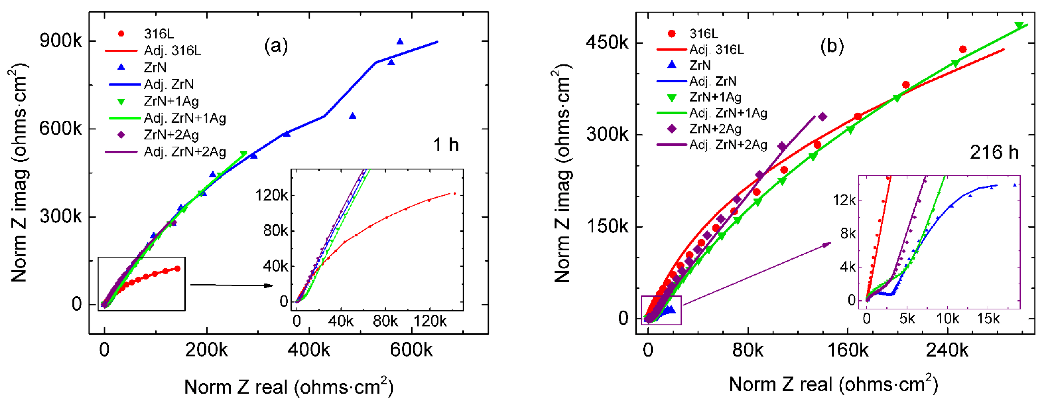
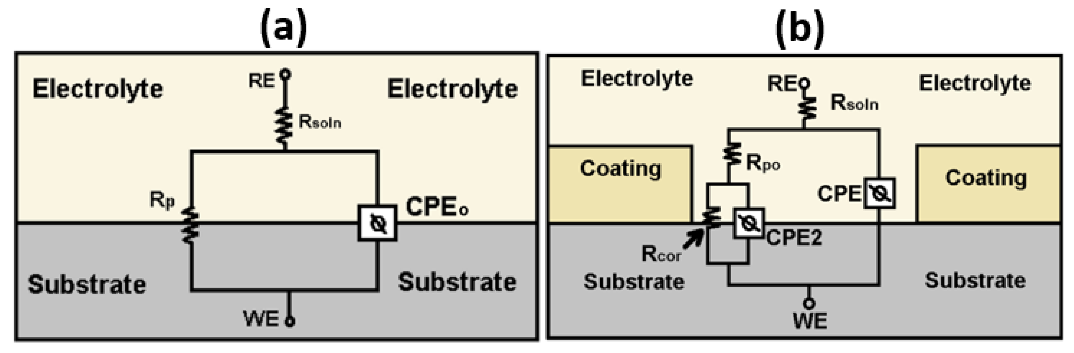
3.5. Corrosion Resistance of the Coatings

The corrosion resistance of the bare substrate and the ZrN, ZrN+1Ag, and ZrN+2Ag coatings was measured using electrochemical impedance spectroscopy (EIS). The experimental data are presented as Bode diagrams for the bare substrate and the deposited coating at a 1 h immersion, impedance module (Figure 7a), and phase angle (Figure 7b). The results show an electrochemical behavior typical for 316L stainless steel with one time constant. The Bode curves for the ZrN, ZrN+1Ag, and ZrN+2Ag coatings show the formation of two-time constants related to the defects in the coating, providing a direct diffusion path for the corrosive media [3] and to the double-layer capacitance [3,43]. Some authors have reported that the presence of two-time constants is typical for porous coatings [3,43,44,45].
Finally, at 1 h, the impedance module diagram (Figure 7a) showed better performance of all the samples compared to the 316L stainless steel. Regarding impedance, the best performance from highest to lowest was: ZrN > ZrN-1Ag > ZrN-2Ag > 316L (bare substrate). In the phase diagram, Figure 7b, the results showed that the more capacitive behavior at low frequencies is similar for coatings with silver; therefore, a better performance related to corrosion resistance is expected in these samples. When the time of immersion increased to 216 h, as seen in Figure 7c,d, the results showed that the bare substrate and the ZrN-1Ag coating had the highest resistance to corrosion. The ZrN coating suffered a dramatic decrease in the impedance module at a low frequency, which can be seen in Figure 7d.
Figure 8 shows the Nyquist spectra for the bare stainless steel, ZrN, ZrN-1Ag, and ZrN-2Ag coatings at (a) 1 h and (b) 216 h immersion. At 1 h of immersion, the results confirm that the coatings have a higher corrosion resistance than the bare substrate. The curve of the bare substrate has the smallest semicircular diameter, indicating a low corrosion resistance, while the deposited coatings increased the diameter of the semicircle. However, at a 216 h immersion, 316L and ZrN-1Ag exhibited the highest corrosion resistance, as can be seen in Figure 8b. To quantify the results, EIS data were fit using an equivalent circuit, shown in Figure 9, and the results obtained are summarized in Table 5, Table 6 and Table 7.
The equivalent circuit used was chosen in accordance with the results of the Bode and Nyquist plots. The electrochemical behavior of the bare substrate can be described using a single-phase constant model, which is summarized in Figure 9a, and the nanostructured coatings can be described using a two-phase constant model, which is summarized in Figure 9b. In the models of Figure 9, RE is a reference electrode, the WE is the working electrode, and Rsoln is the resistance of the electrolyte. For the circuit in Figure 9a, the Rp is the charge transfer resistance between the electrolyte and the substrate, and CPE0 is a constant phase element that relates the electrolyte to the AISI 316L substrate. In Figure 9b, Rpo is the polarization resistance (the charge transfer resistance of the material between the electrolyte and the coating). When the coatings are particularly porous, they provide a response to the impedance of the system, creating a second time constant that corresponds to the capacitance and resistance of the coating and its pores, respectively, until reaching the substrate. Thus, the Rcor indicates the resistance to corrosion due to the pores that allow the coating to interact with the substrate. CPE is the constant phase element that relates the electrolyte to the coating. CPE2 is the constant phase element due to the interaction between the coating and the substrate. The parameters n, m, and ñ are numbers from 0 to 1 and indicate how capacitive CPE0, CPE, and CPE2 are, respectively.
According to the results of the adjustments report in Table 5 and Table 6, at one hour of immersion of Rsoln does not change significantly for all the samples, which is related to a low amount of surface corrosion products of the coatings that are deposited inside the electrolyte. Regarding the ZrN coatings with different amounts of silver, the results of the fit of Table 6 show that at one hour, the parameter ñ is greater in the ZrN, making this sample exhibit high resistance to corrosion at short immersion times compared to the others. After 216 h, in Table 7, the ñ and Rpo parameters are higher in the ZrN+1Ag sample. Therefore, quantitatively, this is the coating that presents the best resistance to long-term corrosion. The polarization resistance value, Rpo, and the ñ parameter decrease for ZrN+2Ag, are due to the fact that silver increases the number of diffusion paths, which allows the electrolyte to penetrate the substrate. These results agree with those obtained via SEM in Figure 4, where it can be seen that as the silver content increases in the coatings, more silver nanoparticles are observed on the surface. This Ag segregation generates the formation of paths that allow the diffusion of the electrolyte to the substrate.
In order to identify the chemical bonds present in the coatings after 216 h of immersion, XPS studies were performed, and the results are shown in Figure 10.
The XPS results did not reveal the formation of new phases on the surface of the coatings after 216 h of immersion in a Ringer’s solution, as can be seen in the Zr 3d, O 1s, and N 1s spectra. The results are similar to those obtained before the corrosion tests. However, the XPS results after 216 h of immersion show a slight shift towards higher binding energy values, which may be due to the presence of more Zr-O or Zr-O-N oxides than Zr-N or Ag on the surface of the coatings.

3.6. Optical Reflectance of the Coatings

The Ag effect on the optical behavior of the ZrN-Ag coatings was investigated by means of UV-Vis spectrophotometry. The reflectance spectra of the ZrN-Ag coatings are shown in Figure 11.
As can be seen, the ZrN coating exhibits high reflectance values in the infrared region and low values in the visible region. Various authors have shown that the optical behavior of the nitride transition metals can be explained using the Drude–Lorentz model [30,46,47,48,49]. The Drude term is based on the free electron gas approximation (metals), while the Lorentz term is related to bound electrons (insulators and semiconductors). However, the reflectance values obtained for the ZrN coating are lower than those reported by other authors [46,49]. Hu et al. [49] showed that the reflectance values depend on the stoichiometry of the coating. They found that the highest values were obtained for the stoichiometric ZrN coating. Therefore, these results are in agreement with those obtained via XPS and XRD, where the results showed the presence of different phases in the coating (ZrO and ZrN), which can affect the optical properties of the coatings.
With the Ag incorporation, the optical reflectance values increased. Plasmonic effects due to localized surface plasmon resonance are not observed in the reflectance spectra due to the wide range of sizes and shapes of Ag nanoparticles on the surface of the coatings. According to Dominguez et al. [20], these metal nanoparticles, dispersed over the surface of the film, can absorb visible and infrared wavelengths over a wide range. In addition, the ZrN exhibits electronic conductivity due to the partially filled valence d orbitals that are not completely hybridized with the electrons, according to [24]. Therefore, the ZrN is not a dielectric matrix that can contribute to the localized surface plasmon resonances [20,23,50]. It is well known that noble metals, such as silver, have the characteristics of a free-electron metal, which increases their optical reflectance values in infrared wavelengths, while, at low values in the visible region, the reflectance is determined by the absorption of light due to the electronic transition between the occupied and unoccupied d state [26]. These results are in agreement with other authors, where the optical transparency is reduced with increased silver content in the coatings [26,51,52].

4. Conclusions

ZrN-Ag nanocomposite coatings were deposited using the DC reactive magnetron sputtering technique onto AISI 316L, (111) silicon, and common glass substrates. The EDS results allowed verifying the presence of the pulverized elements in the coatings, and the XPS results revealed the presence of oxide (ZrO2), oxynitride (ZrON), and nitride (ZrN) in all of the coatings. The presence of oxygen in the coatings can be attributed to contamination by residual oxygen in the chamber during the sputtering process. In addition, the chemical composition showed the presence of silver metallic on the surface of the ZrN+1Ag and ZrN+2Ag coatings. This metallic silver was observed in the SEM images in the form of nanoparticles. The presence of Ag nanoparticles on the surface can be attributed to the Ag diffusion, which is promoted by the temperature during the deposition time of the coating.
The XRD results showed the formation of fcc-ZrN and tetragonal-ZrO2 phases due to the high tendency of zirconium to form oxides, and with the Ag incorporation, the formation of fcc-Ag was observed at 11.98 at.%. EIS tests showed that the polarization resistance increased for the coating, with a 7.12 at.% of silver. However, when the Ag content increased to 11.98 at.%, the polarization resistance value decreased due to an increase in the number of diffusion paths, which allowed the electrolyte to penetrate to the substrate. Finally, the Ag incorporation increased the optical reflectance of the ZrN coatings. In addition, plasmonic effects due to Ag incorporation are not observed in the reflectance spectra due to the wide range of sizes and shapes of Ag and the electronic conductivity of the ZrN matrix.

Author Contributions

Conceptualization, C.P.M. and H.S.V.; coating deposition, H.S.V.; characterization, C.P.M. and H.S.V. data analysis, H.S.V. and C.P.M.; original draft H.S.V. and C.P.M.; writing—review and editing, H.S.V., C.P.M. and J.J.O. Funding acquisition C.P.M., H.S.V. and J.J.O. All authors have read and agreed to the published version of the manuscript.

Funding

This research was supported by the Fundación Universitaria Monserrate (Unimonserrate; Acta 7 del 02/11/2018), Universidad Antonio Nariño (UAN; 2020015-1) and the Universidad Nacional de Colombia (UNAL) through the project: Resistencia a la corrosión del ZrN-Ag mediante impedancia electroquímica No. INV-ING-48224.

Institutional Review Board Statement

Not applicable.

Informed Consent Statement

Not applicable.

Data Availability Statement

Not applicable.

Conflicts of Interest

The authors declare no conflict of interest.

References

  1. Tang, Y.-C.; Katsuma, S.; Fujimoto, S.; Hiromoto, S. Electrochemical study of Type 304 and 316L stainless steels in simulated body fluids and cell cultures. Acta Biomater. 2006, 2, 709–715. [Google Scholar] [CrossRef] [PubMed]
  2. Xi, T.; Shahzad, M.B.; Xu, D.; Sun, Z.; Zhao, J.; Yang, C.; Qi, M.; Yang, K. Effect of copper addition on mechanical properties, corrosion resistance and antibacterial property of 316L stainless steel. Mater. Sci. Eng. C 2017, 71, 1079–1085. [Google Scholar] [CrossRef] [PubMed]
  3. Balasubramanian, S.; Ramadoss, A.; Kobayashi, A.; Muthirulandi, J. Nanocomposite Ti-Si-N Coatings Deposited by Reactive dc Magnetron Sputtering for Biomedical Applications. J. Am. Ceram. Soc. 2012, 95, 2746–2752. [Google Scholar] [CrossRef]
  4. Calderon, S.V.; Oliveira, J.; Evaristo, M.; Cavaleiro, A.; Carvalho, S. Prediction of optimized composition for enhanced mechanical and electrochemical response of Zr-C-N-Ag coatings for medical devices. Appl. Surf. Sci. 2014, 320, 570–580. [Google Scholar] [CrossRef]
  5. Zhou, R.; Ju, H.; Liu, S.; Zhao, Z.; Xu, J.; Yu, L.; Qian, H.; Jia, S.; Song, R.; Shen, J. The influences of Ag content on the friction and wear properties of TiCN–Ag films. Vacuum 2022, 196, 110719. [Google Scholar] [CrossRef]
  6. Shirvani, F.; Shokri, A.; Ravan, B.A. An ab-initio study of structure and mechanical properties of rocksalt ZrN and its bilayers. Solid State Commun. 2021, 328, 114218. [Google Scholar] [CrossRef]
  7. Ju, H.; Yu, L.; Yu, D.; Asempah, I.; Xu, J. Microstructure, mechanical and tribological properties of TiN-Ag films deposited by reactive magnetron sputtering. Vacuum 2017, 141, 82–88. [Google Scholar] [CrossRef]
  8. Hong, C.; Huan, Y.; Zhang, P.; Zhang, K.; Dai, P. Effect of silver content on the microstructure, thermal stability and mechanical properties of CrNx/Ag nanocomposite films. Ceram. Int. 2021, 47, 25324–25336. [Google Scholar] [CrossRef]
  9. Ren, P.; Zhang, K.; Du, S.; Meng, Q.; He, X.; Wang, S.; Wen, M.; Zheng, W. Tailoring the surface chemical bond states of the NbN films by doping Ag: Achieving hard hydrophobic surface. Appl. Surf. Sci. 2017, 407, 434–439. [Google Scholar] [CrossRef]
  10. Prysiazhnyi, V.; Kratochvil, J.; Kaftan, D.; Ctvrtlik, R.; Stranak, V. Growth of hard nanostructured ZrN surface induced by copper nanoparticles. Appl. Surf. Sci. 2021, 562, 150230. [Google Scholar] [CrossRef]
  11. Chen, T.; Yu, L.H.; Ju, H.B.; Geng, Y.X.; Xu, J.H.; Koyama, S.J. Influence of Ag Content on the Microstructure, Mechanical, and Tribological Properties of ZrN-Ag Films. J. Nano Res. 2018, 54, 88–97. [Google Scholar] [CrossRef]
  12. Huang, J.-H.; Ho, C.-H.; Yu, G.-P. Effect of nitrogen flow rate on the structure and mechanical properties of ZrN thin films on Si(100) and stainless steel substrates. Mater. Chem. Phys. 2007, 102, 31–38. [Google Scholar] [CrossRef]
  13. Singh, A.; Kumar, N.; Kuppusami, P.; Prasanthi, T.; Chandramohan, P.; Dash, S.; Srinivasan, M.; Mohandas, E.; Tyagi, A. Tribological properties of sputter deposited ZrN coatings on titanium modified austenitic stainless steel. Wear 2012, 280–281, 22–27. [Google Scholar] [CrossRef]
  14. Hernández-Navarro, C.; Rivera, L.; Flores-Martínez, M.; Camps, E.; Muhl, S.; García, E. Tribological study of a mono and multilayer coating of TaZrN/TaZr produced by magnetron sputtering on AISI-316L stainless steel. Tribol. Int. 2019, 131, 288–298. [Google Scholar] [CrossRef]
  15. Dinu, M.; Parau, A.C.; Vladescu, A.; Kiss, A.E.; Pana, I.; Mouele, E.S.M.; Braic, V. Corrosion improvement of 304l stainless steel by zrsin and zrsi(N,o) mono-and double-layers prepared by reactive cathodic arc evaporation. Coatings 2021, 11, 179–189. [Google Scholar] [CrossRef]
  16. Ghafoor, N.; Petrov, I.; Klenov, D.O.; Freitag, B.; Jensen, J.; Greene, J.E.; Hultman, L.; Odén, M. Self-organized anisotropic (Zr1-xSix)Ny nanocomposites grown by reactive sputter deposition. Acta Mater. 2015, 82, 179–189. [Google Scholar] [CrossRef]
  17. Kelly, P.J.; Li, H.; Benson, P.S.; Whitehead, K.A.; Verran, J.; Arnell, R.D.; Iordanova, I. Comparison of the tribological and antimicrobial properties of CrN/Ag, ZrN/Ag, TiN/Ag, and TiN/Cu nanocomposite coatings. Surf. Coat. Technol. 2010, 205, 1606–1610. [Google Scholar] [CrossRef]
  18. Ju, H.; Yu, D.; Yu, L.; Ding, N.; Xu, J.; Zhang, X.; Zheng, Y.; Yang, L.; He, X. The influence of Ag contents on the microstructure, mechanical and tribological properties of ZrN-Ag films. Vacuum 2018, 148, 54–61. [Google Scholar] [CrossRef]
  19. Baghriche, O.; Kiwi, J.; Pulgarin, C.; Sanjinés, R. Antibacterial Ag–ZrN surfaces promoted by subnanometric ZrN-clusters deposited by reactive pulsed magnetron sputtering. J. Photochem. Photobiol. A Chem. 2012, 229, 39–45. [Google Scholar] [CrossRef]
  20. Domingues, R.P.; Rodrigues, M.S.; Lopes, C.; Pedrosa, P.; Alves, E.; Barradas, N.P.; Borges, J.; Vaz, F. Thin films composed of metal nanoparticles (Au, Ag, Cu) dispersed in AlN: The influence of composition and thermal annealing on the structure and plasmonic response. Thin Solid Film. 2019, 676, 12–25. [Google Scholar] [CrossRef]
  21. Ren, P.; Zhang, K.; He, X.; Du, S.; Yang, X.; An, T.; Wen, M.; Zheng, W. Toughness enhancement and tribochemistry of the Nb-Ag-N films actuated by solute Ag. Acta Mater. 2017, 137, 1–11. [Google Scholar] [CrossRef]
  22. Enrichi, F.; Quandt, A.; Righini, G. Plasmonic enhanced solar cells: Summary of possible strategies and recent results. Renew. Sustain. Energy Rev. 2018, 82, 2433–2439. [Google Scholar] [CrossRef]
  23. Borges, J.; Rodrigues, M.S.; Lopes, C.; Costa, D.; Couto, F.; Kubart, T.; Martins, B.; Duarte, N.; Dias, J.; Cavaleiro, A.; et al. Thin films composed of Ag nanoclusters dispersed in TiO2: Influence of composition and thermal annealing on the microstructure and physical responses. Appl. Surf. Sci. 2015, 358, 595–604. [Google Scholar] [CrossRef] [Green Version]
  24. Patsalas, P.; Kalfagiannis, N.; Kassavetis, S.; Abadias, G.; Bellas, D.V.; Lekka, C.; Lidorikis, E. Conductive nitrides: Growth principles, optical and electronic properties, and their perspectives in photonics and plasmonics. Mater. Sci. Eng. R Rep. 2018, 123, 1–55. [Google Scholar] [CrossRef]
  25. Guler, U.; Shalaev, V.M.; Boltasseva, A. Nanoparticle plasmonics: Going practical with transition metal nitrides. Mater. Today 2015, 18, 227–237. [Google Scholar] [CrossRef]
  26. Das, S.; Alford, T.L. Structural and optical properties of Ag-doped copper oxide thin films on polyethylene napthalate substrate prepared by low temperature microwave annealing. J. Appl. Phys. 2013, 113, 1–7. [Google Scholar] [CrossRef]
  27. Borges, J.; Costa, D.; Antunes, E.; Lopes, C.; Rodrigues, M.S.; Apreutesei, M.; Alves, E.; Barradas, N.P.; Pedrosa, P.; Moura, C.; et al. Biological behaviour of thin films consisting of Au nanoparticles dispersed in a TiO2 dielectric matrix. Vacuum 2015, 122, 360–368. [Google Scholar] [CrossRef]
  28. Placzek-Popko, E.; Gwozdz, K.; Gumienny, Z.; Zielony, E.; Pietruszka, R.; Witkowski, B.S.; Wachnicki, L.; Gieraltowska, S.; Godlewski, M.; Jacak, W.; et al. Si/ZnO nanorods/Ag/AZO structures as promising photovoltaic plasmonic cells. J. Appl. Phys. 2015, 117, 193101. [Google Scholar] [CrossRef]
  29. Mejía, V.C.P.; Chellali, M.R.; Garzón, C.M.; Olaya, J.J.; Hahn, H.; Velasco, L. Effect of discharge current on the corrosion resistance and microstructure of ZrTiSiN coatings deposited by magnetron co-sputtering. Mater Today Commun. 2021, 26, 102151. [Google Scholar] [CrossRef]
  30. Aouadi, S.M.; Debessai, M.; Filip, P. Zirconium nitride/silver nanocomposite structures for biomedical applications. J. Vac. Sci. Technol. B Microelectron Nanom Struct. 2004, 22, 1134. [Google Scholar] [CrossRef]
  31. Dang, C.; Yao, Y.; Olugbade, T.; Li, J.; Wang, L. Effect of multi-interfacial structure on fracture resistance of composite TiSiN/Ag/TiSiN multilayer coating. Thin Solid Film. 2018, 653, 107–112. [Google Scholar] [CrossRef]
  32. Mejía, V.H.D.; Perea, D.; Bejarano, G.G.B. Development and characterization of TiAlN (Ag, Cu) nanocomposite coatings deposited by DC magnetron sputtering for tribological applications. Surf Coat. Technol. 2020, 381, 125095. [Google Scholar] [CrossRef]
  33. Castro, J.D.; Lima, M.J.; Carvalho, I.; Henriques, M.; Carvalho, S. Cu oxidation mechanism on Cu-Zr(O)N coatings: Role on functional properties. Appl. Surf. Sci. 2021, 555, 149704. [Google Scholar] [CrossRef]
  34. Cubillos, G.; Mendoza, M.; Alfonso, J.; Blanco, G.; Bethencourt, M. Chemical composition and microstructure of zirconium oxynitride thin layers from the surface to the substrate-coating interface. Mater. Charact. 2017, 131, 450–458. [Google Scholar] [CrossRef]
  35. Yu, L.; Zhao, H.; Xu, J. Influence of silver content on structure, mechanical and tribological properties of WCN-Ag films. Mater. Charact. 2016, 114, 136–145. [Google Scholar] [CrossRef]
  36. Vanegas, P.H.S.; Calderon, V.S.; Alfonso, O.J.E.; Olaya, F.J.J.; Ferreira, P.J.; Carvalho, S. Influence of silicon on the microstructure and the chemical properties of nanostructured ZrN-Si coatings deposited by means of pulsed-DC reactive magnetron sputtering. Appl. Surf. Sci. 2019, 481, 1249–1259. [Google Scholar] [CrossRef]
  37. Cavaleiro, D.; Carvalho, S.; Cavaleiro, A.; Fernandes, F. TiSiN(Ag) films deposited by HiPIMS working in DOMS mode: Effect of Ag content on structure, mechanical properties and thermal stability. Appl. Surf. Sci. 2019, 478, 426–434. [Google Scholar] [CrossRef]
  38. Rajput, S.S.; Gangopadhyay, S.; Cavaleiro, A.; AL-Rjoub, A.; Kumar, C.S.; Fernandes, F. Influence of Ag additions on the structure, mechanical properties and oxidation behaviour of CrAlNAg coatings deposited by sputtering. Surf Coat. Technol. 2021, 426, 127767. [Google Scholar] [CrossRef]
  39. Al-Rjoub, A.; Cavaleiro, A.; Fernandes, F. Influence of Ag alloying on the morphology, structure, mechanical properties, thermal stability and oxidation resistance of multilayered TiSiN/Ti(Ag)N films. Mater. Des. 2020, 192, 108703. [Google Scholar] [CrossRef]
  40. Zhu, Y.; Dong, M.; Li, J.; Wang, L. The improved corrosion and tribocorrosion properties of TiSiN/Ag by thermal treatment. Surf. Coatings Technol. 2020, 385, 125437. [Google Scholar] [CrossRef]
  41. Yu, D.; Yu, L.; Asempah, I.; Ju, H.; Xu, J.; Koyama, S.; Gao, Y. Microstructure, mechanical and tribological properties of VCN-Ag composite films by reactive magnetron sputtering. Surf. Coatings Technol. 2020, 399, 126167. [Google Scholar] [CrossRef]
  42. Calderon, V.S.; Cavaleiro, A.; Carvalho, S. Chemical and structural characterization of Zr-C-N-Ag coatings: XPS, XRD and Raman spectroscopy. Appl. Surf. Sci. 2015, 346, 240–247. [Google Scholar] [CrossRef]
  43. Vanegas, H.S.; Alfonso, J.E.; Olaya, J.J. Effect of Si content on functional behavior of nanostructured coatings of Zr–Si–N. Mater. Res. Express 2019, 6, 115076. [Google Scholar] [CrossRef]
  44. Rios, J.F.; Calderón, J.A.; Nogueira, R.P. Electrochemical behavior of metals used in drinking water distribution systems: A rotating cylinder electrode’s study. Corrosion 2013, 69, 875–885. [Google Scholar] [CrossRef]
  45. Alves, C.A.; Calderon, S.; Dias, D.; Carvalho, S. Influence of Oxygen content on the electrochemical behavior of Ta1-xOx coatings. Electrochim. Acta 2016, 211, 385–394. [Google Scholar] [CrossRef] [Green Version]
  46. von Fieandt, K.; Pilloud, D.; Fritze, S.; Osinger, B.; Pierson, J.F.; Lewin, E. Optical and electrical properties of hard (Hf,Nb,Ti,V,Zr)Nx thin films. Vacuum 2021, 193, 110517. [Google Scholar] [CrossRef]
  47. Popović, M.; Novaković, M.; Rakočević, Z.; Bibić, N. Tailoring the structural and optical properties of TiN thin films by Ag ion implantation. Nucl. Instrum. Methods Phys. Res. Sect. B Beam Interact. Mater. Atoms 2016, 389–390, 33–39. [Google Scholar] [CrossRef]
  48. Aouadi, S.M.; Maeruf, T.; Debessai, M.; Filip, P. Zirconium nitride/nickel nanocomposite structures. Vacuum 2005, 79, 186–193. [Google Scholar] [CrossRef]
  49. Hu, C.; Guo, K.; Li, Y.; Gu, Z.; Quan, J.; Zhang, S.; Zheng, W. Optical coatings of durability based on transition metal nitrides. Thin Solid Film. 2019, 688, 137339. [Google Scholar] [CrossRef]
  50. Guler, U.; Boltasseva, A.; Shalaev, V.M. Refractory plasmonics. Science 2014, 344, 263–264. [Google Scholar] [CrossRef]
  51. Tseng, C.C.; Hsieh, J.H.; Liu, S.J.; Wu, W. Effects of Ag contents and deposition temperatures on the electrical and optical behaviors of Ag-doped Cu2O thin films. Thin Solid Film. 2009, 518, 1407–1410. [Google Scholar] [CrossRef]
  52. Jalili, S.; Hajakbari, F.; Hojabri, A. Effect of silver thickness on structural, optical and morphological properties of nanocrystalline Ag/NiO thin films. J. Theor. Appl. Phys. 2018, 12, 15–22. [Google Scholar] [CrossRef] [Green Version]
Figure 1. (a) EDS spectrum of the deposited coatings, and (b) the atomic percentage of the coatings (two measurements were done in several areas of the samples).
Coatings 12 00754 g001
Figure 2. XPS spectra of (a) Zr 3d, (b) O 1s, and (c) N 1s of the ZrN, ZrN+1Ag, and ZrN+2Ag coatings.
Coatings 12 00754 g002
Figure 3. XPS spectra of Ag 3d for the (a) ZrN+2Ag and (b) ZrN+1Ag coatings.
Coatings 12 00754 g003
Figure 4. Surface and cross-sectional SEM images of (a) ZrN+1Ag and (b) ZrN+2Ag coatings.
Coatings 12 00754 g004
Figure 5. Surface morphology of (a) ZrN and (d) ZrN+2Ag coatings. EDS mapping of (b) Zr and (c) N and (e) Zr, (f) N and (g) Ag for the surface of the ZrN and ZrN+2Ag coatings, respectively.
Coatings 12 00754 g005
Figure 6. XRD pattern of the deposited coatings: ZrN, ZrN+1Ag, and ZrN+2Ag on a common glass substrate.
Coatings 12 00754 g006
Figure 7. Electrochemical impedance test as a function of the Ag content in a Ringer’s solution at 37.5 °C. (a) Impedance module, (b) phase angle at 1 h immersion, (c) impedance, and (d) phase at 216 h immersion.
Coatings 12 00754 g007
Figure 8. Nyquist spectra for the 316L substrate and the ZrN, ZrN+1Ag, and ZrN+2Ag coatings at (a) 1 h of immersion and (b) 216 h of immersion.
Coatings 12 00754 g008
Figure 9. Equivalent circuit model used for fitting the EIS experimental data for (a) AISI 316L and (b) ZrN, ZrN+1Ag, and ZrN+2Ag coatings.
Coatings 12 00754 g009
Figure 10. XPS spectra of (a) Zr 3d, (b) O 1s and (c) N 1s of the ZrN, ZrN1Ag, and ZrN2Ag coatings after the corrosion test.
Coatings 12 00754 g010
Figure 11. Reflectance spectra of the deposited coatings: ZrN, ZrN+1Ag, and ZrN+2Ag.
Coatings 12 00754 g011
Table 1. Chemical composition of the AISI 316L substrate obtained by EDS.
Cr (at.%)Fe (at.%)Ni (at.%)Mn (at.%)Mo (at.%)
16.469.910.11.52.1
Table 2. Deposition parameter, thicknesses, and deposition rates.
SamplesSilver PelletsPower (W)Substrate Temperature (°C)Thickness (nm)Deposition Rate (nm/s)
ZrN0140200544.90.15
ZrN+1Ag1140200633.70.18
ZrN+2Ag2140200845.10.23
Table 3. Chemical composition of the Ringer’s lactate solution used in EIS test.
NaCl (g/L)C3H5O3Na (g/L)CaCl (g/L)KCl (g/L)
6.03.10.21.5
Table 4. The 2θ and FWHM values obtained from XRD patterns.
CoatingAg ContentPlane2θ (°)FWHMLattice Parameter (nm)
ZrN0 at.%ZrN (111)33.391.580.464
ZrN+1Ag7 at.%ZrN (111)33.411.540.464
ZrN+2Ag12 at.%ZrN (111)33.481.700.463
Ag (200)43.661.540.414
Table 5. EIS fitting parameter of AISI 316L after 1 h and 216 h of immersion in a Ringer’s solution at 37.5 °C.
ParameterUnit316L 1 h316L 216 h
Rsolnohm·cm279.970.0
Rpohm·cm23.4 × 1051.4 × 106
CPE0S·sa/cm23.3 × 10−51.82 × 10⁻⁵
M 7.81 × 10−18.71 × 10−1
Table 6. EIS fitting parameter of ZrN, ZrN+1Ag and ZrN+2Ag coatings after 1 h of immersion on a Ringer’s solution at 37.5 °C.
ParameterUnitZrNZrN+1AgZrN+2Ag
Rsolnohm·cm286.977.971.8
Rcorohm·cm23.91 × 1064.87 × 1063.94 × 106
Rpoohm·cm21.06 × 1048.60 × 1032.66 × 103
CcorS·sa/cm24.47 × 10−61.13 × 10−52.27 × 10−5
n 7.27 × 10−17.74 × 10−17.96 × 10⁻¹
CcS·sa/cm22.37 × 10−62.30 × 10−64.81 × 10−6
ñ 8.95 × 10−17.65 × 10−17.32 × 10⁻¹
Table 7. EIS fitting parameter of ZrN, ZrN+1Ag, and ZrN+2Ag coatings after 216 h of immersion in a Ringer’s solution at 37.5 °C.
ParameterUnitZrNZrN+1AgZrN+2Ag
Rsolnohm·cm281.458.042.0
Rcorohm·cm25.44 × 1042.44 × 1064.58 × 107
Rpoohm·cm22.83 × 1036.30 × 1034.94 × 103
CcorS·sa/cm23.22 × 10−41.17 × 1051.46 × 10−5
n 7.54 × 10−17.93 × 10−18.89 × 10⁻¹
CcS·sa/cm24.13 × 10−62.36 × 10−61.06 × 10−5
ñ 7.76 × 10−17.77 × 10−16.39 × 10⁻¹
Publisher’s Note: MDPI stays neutral with regard to jurisdictional claims in published maps and institutional affiliations.

