Abstract
Environmental pollution caused by heavy metal ions has become a major health problem across the world. In this study, a selective colorimetric sensor based on starch functionalized silver nanoparticles (St-Ag NPs) for rapid detection of Hg2+ in real samples was developed. The environmentally friendly green approach was utilized to synthesize starch functionalized silver nanoparticles (St-AgNPs). A multi-technique approach involving UV-Vis absorption spectroscopy, Fourier transform infrared (FT-IR), X-ray diffraction (XRD), and scanning electron microscope (SEM) was used for the characterization of St-Ag NPs. These starch functionalized AgNPs were tested for the detection of heavy metals at 25 °C. The screening process revealed clear changes in the AgNPs color and absorption intensity only in the presence of Hg2+ due to the redox reaction between Ag0 and Hg2+. The color and absorption intensity of nanoparticles remain unchanged in the presence of all the other tested metals ion. The proposed method has strong selectivity and sensitivity to Hg2+ ions, with a detection limit of 1 ppm revealed by UV-visible spectrophotometry. The proposed procedure was found to be successful for the detection of Hg2+ in real samples of tap water.
1. Introduction
During the last decade, the emerging field of plasmonic-based nanotechnology brought a revolutionary track in the discipline of applied sciences due to many practical applications of nanoparticles in the field of environmental sciences, such as wastewater treatment [1,2]. Heavy metal ions (M+), such as Cr3+, Zn2+, Ni2+, Cu2+, and Hg2+, are essentially mandatory for the growth process of both animals and plants at appropriate concentrations. However, these metal ions are harmful to living creatures at relatively high concentrations because of their input in both human and animal bodies via biological food webs [3,4].
Mercury ion (Hg2+), which is widely distributed in the atmosphere, soil on the earth’s surface, and even in water, is one of the most lethal and hazardous metal pollutants [5,6]. There are various sources of mercury such as the burning of coal in power plants, natural liberation of gases from earth surface during vulcanization, and metals extraction process [7]. Hg2+ is a persistent pollutant because naturally, it cannot decompose in the environment [8]. In water, fishes consume mercury as it is dissolved in water, and through the food web, this is the major way of mercury accumulation in humans [9]. It can damage the brain, the nervous system, and the immune system [6].
Therefore, detection of poisonous metals in the aquatic environment and biological system has become a crucial need of the present-day world [10]. During the last decade, various methods have been developed for Hg2+ detection, including electrochemical methods [11], optical detections [12], atomic absorption spectroscopy, inductively coupled plasma mass spectroscopy [13], and fluorescent spectroscopy [14]. However, most of these approaches are inconvenient because the use of complicated instruments is painstaking and time-consuming [15,16]. Therefore, the introduction of a logical technique that is not just easy and cheap but also useful and reproducible, and able to sense the toxic metal pollutants in the ecological samples, is greatly needed. For these mentioned problems, approaches to low cost and rapid detection of mercury using silver nanoparticles (AgNPs) or gold nanoparticles (AuNPs) are advantageous [17,18].
Synthesizing metallic nanoparticles (AgNPs and AuNPs) in an environmentally friendly manner is a key step in nanotechnology. In the field of selective and sensitive detection methods, the use of environmentally friendly nanotechnology has recently become increasingly significant [19,20]. During the last decade, colorimetric sensors, in particular, have a distinct advantage because of their versatility, rapidity, high selectivity, and ease of use, which includes the ability to perform real-time qualitative [19] and quantitative analysis [21,22].
Nanotechnology has the potential to boost life sciences, healthcare, and industrial technology significantly. For example, Lax man et al. [23] presented an optical process for careful recognition of Hg2+ depending upon the aggregation of AgNPs. Wang et al. [24] reported a highly sensitive method for sensing Hg2+, ascorbic acid, and Cd2+ by using trithiocy anuric acid gold NPs. Senapati and co-workers, in their work, showed the use of tryptophan coated gold nanoparticles for selective and efficient detection of Hg2+ [25]. Chai et al. [26] presented colorimetric detection of Pb2+ using glutathione functionalized AuNPs. However, these approaches typically use some chemicals as reducing agents that frequently produce toxic side products [27]. Some of them used organic reagents as the functional selective reagents, which are unstable and easily oxidized, while some tagging agents found it costly to use these techniques as sensors for real life [28]. Additionally, the production processes of nanoparticles used in sensing systems are complex [29]. These sensors are generally derivatives of fluorescent dyes, usually harmful to the environment [30,31].
Colorimetric detection of particular analytes of interest by using AuNPs and AgNPs is a common practice because of color changes which are simply viewed with naked eyes instead of complicated instruments [32]. These nanoparticles are used as colorimetric probes for sensing lethal metal ions from ecological samples via cheap and simple procedures [33]. Compared to AuNPs, silver nanoparticles have several benefits such as low cost, easy preparation, etc. Further, AgNPs can also be oxidized by Hg2+, which causes color change and decrease in the UV-visible UV spectrum absorption of the AgNPs.
Besides the other advantages, the role of surfactant during the synthesis of AgNPs is very important. The starch and D-glucose are biomolecules that are non-toxic and biocompatible ligands. Starch acts as a protecting agent as it contains many hydroxyl (–OH) groups that will simply attach to the surface of AgNPs through the Ag-O bond and prevent the accumulation of AgNPs [34]. The use of silver nitrate was reduced by D-glucose in the presence of starch synthesizes nanoparticles (NPs). D-Glucose is an ecologically favorable and mild reducing agent, which is activated in the presence of a basic catalyst.
In this paper, a fast and very selective colorimetric method was developed for the detection of Hg2+ by using starch-functionalized AgNPs, with a green synthesis approach. The addition of Hg2+ to AgNPs solution produces instant color change (dark yellow to colorless) which can be seen by the naked eye. The selectivity of this detection system of Hg2+ by using starch stabilized AgNPs is outstanding when compared with other metal ions such as Pb2+, Al3+, Zn2+, Cu2+, and Fe3+. Additionally, the detection process of Hg2+ is outstanding even in the presence of a mixture of the mentioned heavy metal ions. Furthermore, AgNPs were successfully employed for the detection of Hg2+ ion in real water samples.
2. Materials and Methods
2.1. Chemicals
AgNO3, KOH, KCl, NaCl, FeCl3, ZnCl2, HgCl2, NiCl2·6H2O, CuSO4, and AlCl3 were purchased from Sigma Aldrich. D-glucose and soluble starch ((C6H10O5)n) were also purchased from Sigma Aldrich. Distilled water was used during the research. All the required substances were received in pure form so there was no need for additional purification.
2.2. Synthesis of Starch-Stabilized AgNPs
In a typical synthetic procedure, 2 mL of D-glucose (0.1 M), 2 mL of starch (0.2 wt%), and 0.02 mL of KOH (0.1 M) were added into 5 mL of AgNO3 (10 mM). After that, the mixture was heated at 30 °C for 10 min with continuous stirring at (100 rpm). After that formation of AgNPs was indicated by a color change from colorless to deep reddish yellow. The basic steps involved during the synthesis process are mentioned in Scheme 1. This nanoparticle dispersion was centrifuged at 1200 rpm to obtain solid particles. Finally, the prepared nanoparticles were stored at 25 °C for further characterization.
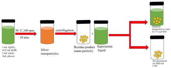
Scheme 1.
Different steps involved in the preparation of AgNPs.
2.3. Characterization
The synthesized AgNPs were analyzed first by UV-vis spectrophotometer. UV-vis absorption studies were done by using UV-1800 double beam spectrophotometer (Shimadzu, Kyoto, Japan), utilizing quartz cuvettes of 1.0 cm path length in the UV range from 200–800 nm. XRD was recorded on a Bruker D-8 powder X-ray diffractometer by Cu-K radiation (λ = 0.15418 nm) over a range of 20–90° with a step size of 0.02°. FT-IR spectra were obtained by using FT-IR 8400S Shimadzu, Japan using KBr disk (4000–400 cm−1).
2.4. General Procedure for the Calorimetric Determination of Hg2+
For detection of Hg2+ using AgNPs dispersion, 1 mL (100 ppm) of aqueous solutions of Pb2+, Cu2+, Al3+, Zn2+, Fe2+, Ni2+, and Hg2+ were added, respectively, into 1 mL of AgNPs dispersion. To find out the detection limit, various concentrations of HgCl2 (1–100 ppm) were prepared from the stock solution by quantitative dilution. Keeping the total volume of mixture constant (2 mL), an equal volume of Ag-NPs and HgCl2 (each concentration) were mixed. To check out the selectivity of the detection system 100 ppm aqueous solutions of Pb2+, Cu2+, Al3+, Zn2+, Fe2+, Ni2+, and Hg2+ were prepared. 1 mL of every solution was added into 1 mL solution of Hg2+ (100 ppm) and 1 mL of AgNPs dispersion. All the solution and dilution processes were carried out at room temperature.
To determine the binding stoichiometry of Hg2+ and AgNPs, Hg2+ (100 ppm) solution ratios from 0.1 to 2 mL were mixed with AgNPs dispersion in opposite ratios of volume. To find out the role of pH on the sensing study, pH of AgNPs was varied from 1 to 12. To study the practical applications of the planned strategy, we used tap water for Hg2+ detection. About 100 ppm Hg2+ solution was prepared in tap water, and then 1 mL of that dispersion was mixed with AgNPs dispersion in tap water. All of these mixtures were kept at room temperature for 10 min to monitor the effect of Hg2+ on AgNPs dispersion in tap water.
3. Results and Discussions
3.1. Visual Detection of AgNPs Synthesis
The color of the reaction mixture changed within 10 min from colorless to reddish yellow (as shown in Figure 1A), after mixing starch, glucose, and KOH solution with AgNO3 solution. Thus, Ag+ reduction was confirmed as the colorless silver nitrate solution altered to yellowish-brown. It is assumed that the production of AgNPs is a redox reaction where Ag+ is reduced to Ag0 with the oxidation of glucose to corresponding gluconic acid which was later on confirmed through FT-IR.
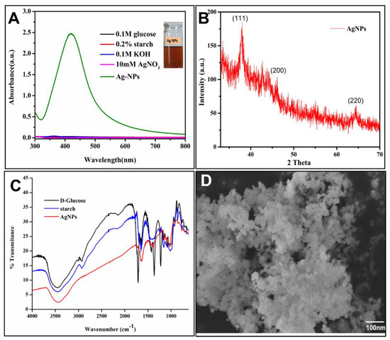
Figure 1.
(A) UV spectra show Glucose, Starch, KOH, and AgNO3 and synthesized AgNPs, (B) XRD image of synthesized silver nanoparticles. (C) FT-IR Spectrum of AgNPs and (D) SEM image of synthesized AgNPs.
3.2. UV–Visible Absorption Spectroscopy
The synthesized AgNPs were analyzed by UV–visible absorption spectroscopy technique because of the surface plasmon resonance (SPR) phenomenon. When light waves interact with free electrons present in the reduced AgNPs surface, plasmon resonance originates. For confirmation of the formation of AgNPs, UV spectra were recorded for a starch solution, D-Glucose solution, and KOH solution, which do not show any characteristic absorption due to the absence of SPR. UV–vis spectrum of the AgNPs suspensions shows an absorption maximum at 430 nm, as shown in Figure 1A. As a function of synthesis/reaction time, the absorbance was found to increase and a maximum was observed after a reaction time of 10 min (as depicted in Figure 1, green curve). Thus, the optimum reaction time for the synthesis of AgNPs was found to be 10 min. Under the influence of high temperature, the rate of reduction of Ag+ increases, and therefore the rapid synthesis of AgNPs could be achieved [35].
3.3. X-ray Diffraction
A characteristic XRD pattern of synthesized AgNPs showed various reflections, at 38.2° (111), 44.3° (200), and 64.5° (220) as shown in Figure 1B. These sharp Bragg peaks maybe produced due to the stabilization of nanoparticles by starch that acts as a capping agent. The peak related to the (111) plane was more prominent than the rest of the planes, signifying that the (111) plane was the major orientation in the face-centered cubic (fcc) structure of AgNPs. The XRD results of AgNPs show a crystalline behavior similar to previously reported literature [36].
3.4. FT-IR Analysis
FT-IR spectra were used to recognize the functional groups in different types of compounds. For comparison, IR spectra were recorded for D-Glucose, starch, and AgNPs. In spectra of D-glucose and starch, there is a wide peak at 3200–3500 cm−1, which reflects the stretching vibration of OH group (i.e., hydrogen-bonded), and a sharp peak at 1725 cm−1 represents the C=O stretch of aldehyde group. The sharp peaks at 2900 cm−1 and 1100 cm−1 are the stretching vibrations of aldehyde C-H and C-O, respectively. Moreover, OH bending vibration is reflected in the region of 1433 cm−1. In IR spectra of AgNPs, the peaks of OH group stretching and bending are less intense. A sharp peak in the region of 1710 cm−1 appeared, which indicates the presence of the COOH group as shown in Figure 1C. It means that during the formation of AgNPs, the C=O and OH groups of Starch and D-Glucose cause the reduction of Ag+ ions from AgNO3 and itself oxidized into respective acid. KumariJyoti et al. reported similar observations in their work [37].
3.5. Scanning Electron Microscopy
The surface morphology monitored by SEM images reveals various shapes in the form of spherical AgNPs. Figure 1D shows the low and high magnification images of AgNPs. One can observe that the particle size lies in the range of 1–100 nm. This may be due to the availability of the different quantities of capping agents during their synthesis process [38].
3.6. Parametric Study of the Synthesis of AgNPs
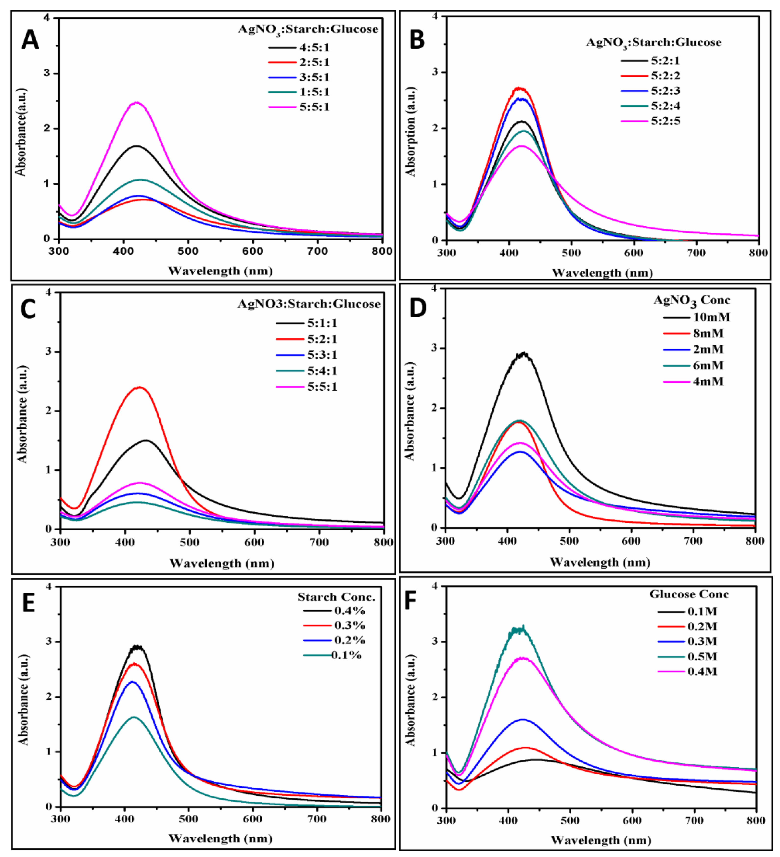
The volume of AgNO3 (10 mM) (NN-A1 to NN-A5), starch (0.2%) (NN-A6 to NN-A10), and glucose (0.1 M) (NNA-11 to NNA-15) varied from 1 to 5 mL as shown in Table 1. UV spectra showed that absorption gradually increases with theAgNO3 volume ratio. This finding can be associated with the rise in the redox reaction rate involving AgNO3 and the OH groups of starch and glucose, whereas absorption gradually decreases with an increase in the volume ratio of starch and glucose (as shown in Figure 2A–C) due to a simple dilution effect.

Table 1.
Experimental details for different volume ratios of AgNO3, starch, and glucose during the synthesis of Ag-NPs.
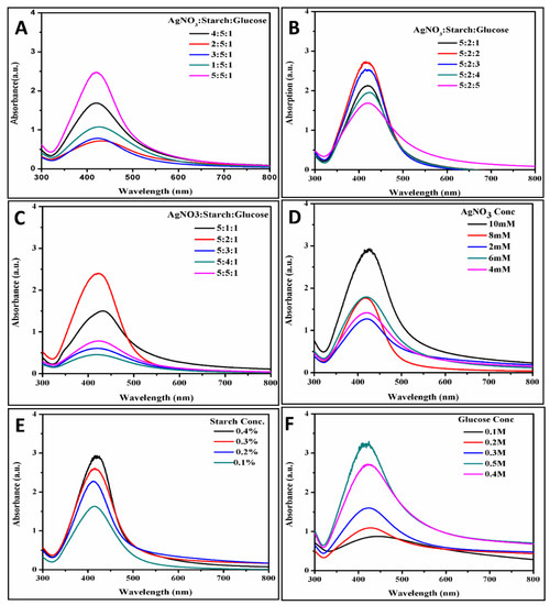
Figure 2.
UV spectra relating to AgNPs’ formation at diverse volume ratios of (A) 10 mM AgNO3 (1 to 5 mL), (B) 0.1 M glucose, (C) 0.2% starch, and (D) at 5 mL of different concentration of AgNO3, (E) at 2 mL of various concentrations of starch, (F) at 2 mL of various concentrations of glucose.
The concentration of AgNO3 (NN-A16 to NNA-20) varied from 2 to 10 mM, while the concentration of glucose (NNA-21 to NNA-25), starch (NN-A26 to NN-A30), and KOH (NNA-31 to NNA-35) varied from 0.1 to 0.5 M, as shown in Table 2. UV spectra show the maximum absorption at 430 nm, which gradually increases with an increase in concern of AgNO3 (as shown in Figure 2D–F). It is expected that a large number of Ag+ would be available during the redox reaction, resulting in a higher yield of AgNPs, which is responsible for the increase in the absorption peak [39]. A similar behavior was observed in the UV spectra of starch, glucose, and KOH, where absorption at 430 nm gradually increases with an increase in concentrations. This result is due to an increase in the rate of nanoparticle synthesis in a highly basic environment and adequate availability of the reducing and stabilizing agent. However, in concentrated solutions the concentration of OH increases, causing aggregation on the surface AgNPs which increases in peak intensity [29,40].
Table 2.
Experimental details for different concentrations. of AgNO3, glucose, starch, and KOH in the synthesis of AgNPs.
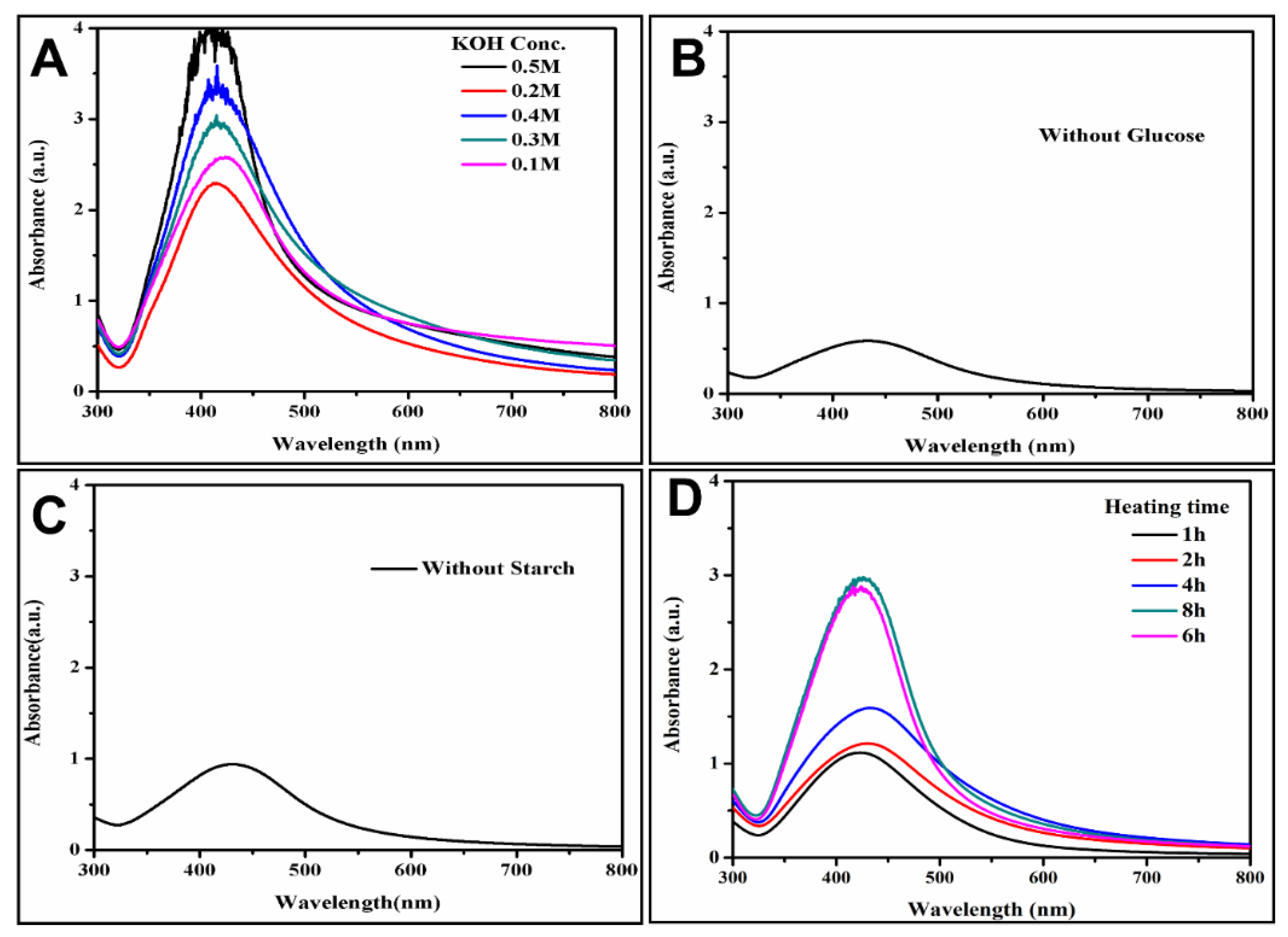
A controlled experiment without glucose and starch was carried out. UV spectrum shows that in the absence of glucose and starch absorption, 430 nm is the minimum (as shown in Figure 3B,C). This might be due to a slight reduction of Ag+ to Ag0 in the absence of glucose that acts as a reducing agent, whereas starch act as a stabilizing agent. In the absence of starch AgNPs are unstable and get aggregated, which results in a decrease in surface plasmon resonance peak [34]. AgNPs can form even in the absence of KOH and UV spectra reveals that absorption of AgNPs gradually increases (as given in Figure 3D), with a raise in heating time in the absence of KOH.

Figure 3.
UV spectrum shows AgNPs formation, (A) at 0.02 mL of different concentrations of KOH, (B) without 2 mL of glucose, (C) without 2 mL of starch, (D) without 0.02 mL of KOH.
It can be concluded that in the absence of base pH the reaction mixture was low, hence the rate of reduction of AgNO3 to AgNPs was time-consuming [41].
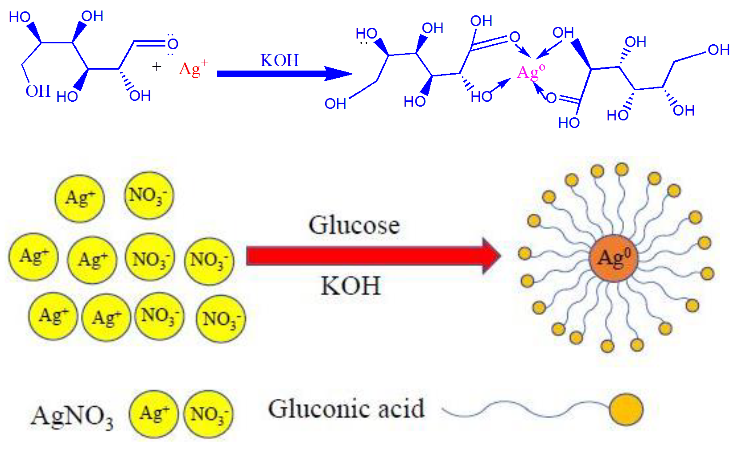
3.7. Possible Mechanism of the Formation of AgNPs
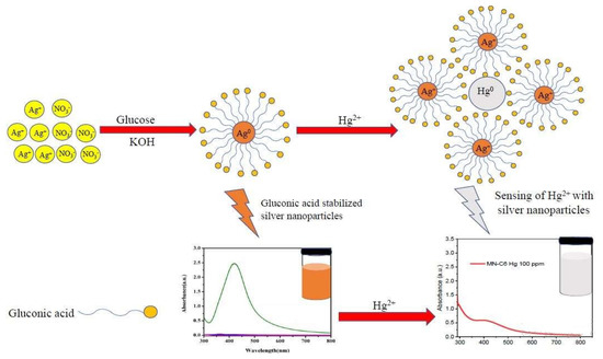
This reaction is an example of hydrolysis of starch that is catalyzed by a base (KOH) giving simpler molecules, like glucose. Glucose is used as a reducing agent for silver nitrate to silver metal. During the reaction, the aldehyde group of glucose reduces Ag+ to Ag0 and is oxidized to gluconic acid (as shown in Scheme 2), while the starch stabilizes silver nanoparticles.

Scheme 2.
Possible route for the synthesis of AgNPs.
3.8. Calorimetric Determination of Hg2+
AgNPs have shown their potential in visual as well as SPR detection-based sensing of heavy metals. Herein, AgNPs are used for Hg2+ sensing in water. The decolorization of AgNPs and decrease in their UV absorption peak intensity is due to the redox reaction that causes the aggregation of the nanoparticles. During the reaction, Ag0 from AgNPs is oxidized to Ag+ while Hg2+ is reduced to Hg0, as confirmed by the standard electrode potential values of Hg2+/Hg (E0 = 0.85 V) and Ag+/Ag (E0 = 0.79 V). Furthermore, because Hg2+ has a greater reduction potential than Ag+, the redox reaction 2Ag+ 2Hg2+ = 2Ag+ + Hg2+ occurs spontaneously. As illustrated for the original colorless solution AgNO3, oxidizing Ag0 to Ag+ changes the color of AgNPs from yellowish brown to colorless (Scheme 3). The colorimetric detection of mercury by AgNPs is based on this redox process. Ag (0) AgNPs to Ag+ cannot oxidize the bulk of transition metals, alkaline, and alkaline earth metals due to their lower potential than Ag+, allowing for extremely selective Hg2+ analyses. Thus, the oxidation of AgNPs leads to the loss of its characteristic color and a decrease in its UV absorption peak intensity.
Scheme 3.
The proposed mechanism of the interaction between Starch coated AgNPs and Hg2+ solution.
This revised mechanism promotes the mechanisms previously proposed [42,43,44,45]. However, after the addition of Hg ions, some colorimetric mercury detection methods with various surfactant or functional AgNPs might yield a colored mixture that would suggest a distinct reaction mechanism. It is found that the role of surfactant is very important during the sensing process [46]. Instead of a direct reaction between Hg2+ ions and Ag(0) of AgNPs, the interaction between Hg ions and capping agents (gluconic acid) to form larger nanoparticles that lead to aggregation plays a crucial role in these reactions. The interaction of Hg2+ ions and Ag(0) of AgNPs in the presence of gluconic acid as a surfactant is shown in Scheme 3.
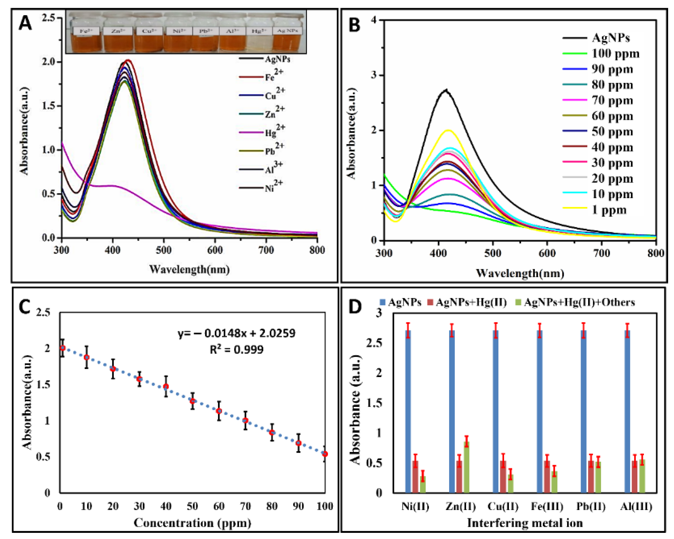
3.8.1. Screening of Heavy Metals
Nanoparticles’ (NPS) behavior towards heavy metals was monitored by UV-Vis spectroscopy. To estimate the detection tendency of AgNPs towards heavy metals, 1 mL of AgNPs was mixed with 1 mL of aqueous solutions of heavy metals (100 ppm) under the experimental conditions reported in Table 3.
Table 3.
Experimental details for the screening of different heavy metals by AgNPs.
The addition of Hg2+ resulted in the destruction of AgNPs that was observable, as after the addition of Hg2+ in AgNPs, the solution suddenly changes its color from reddish yellow to colorless, as shown in vials Figure 4A. UV-Vis spectra reveals broadness and hypochromic shift in the plasmon resonance band. AgNPs dispersion shows utmost absorption intensity at 430 nm, which is dismissed by the addition of Hg2+ as shown in Figure 4A. All other metals, including Cu2+, Na+1, Cr+3,Al3+, Zn2+, Fe2+, Pb+2, and Ni2+, did not make any change in the color of AgNPs as well as in the UV spectrum. Hg2+ was the only metal that showed clear changes in color and absorption intensity of AgNPs, which may be due to the redox reaction occurring between Ag0 and Hg2+. These results reveal outstanding selectivity over a variety of heavy metals, thus AgNPs have binding sites for Hg2+ [42].
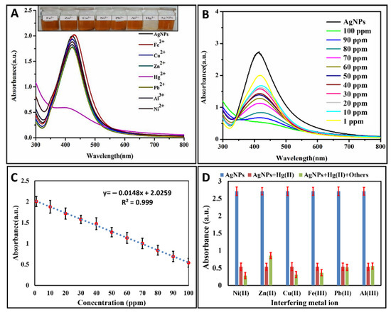
Figure 4.
(A) Photographs and UV absorption spectra of AgNPs dispersions containing 100 ppm of Hg2+, Zn2+,Fe2+, Pb2+, Ni2+, Al3+, Ni2+, or Cu2+ demonstrating the change in the color of nanoparticles upon addition of Hg2+; (B) UV absorption spectra of AgNPs dispersions upon adding various concentrations of Hg2+; (C) linear or direct relationship between the absorbance and Hg2+ concentrations; (D) selectivity of Ag-NPs at 430 nm: the blue bar shows the absorption intensity of AgNPs, the brown bar represents the absorption intensity of AgNPs + Hg2+ and the green bar represents the absorption intensity of AgNPs + Hg2+ in the presence of other metal ions.
3.8.2. Effect of Hg2+ Concentration
The quantitative estimation of the detection limit of Hg2+ ions sensing was studied by altering the concentrations of these Hg2+ (1−100 ppm) while keeping the same concentration of AgNPs at the same laboratory circumstances, as shown in Table 4.
Table 4.
Experimental details for sensing of different concentrations of Hg2+ by Ag NPs.
The surface plasmon resonance band of the AgNPs revealed that mixing of Hg2+ ions in AgNPs solutions produces a steady hypochromic shift in the surface plasmon resonance band at 430 nm. The extent of the shift in the direction of the lower-intensity depends upon the concentrations of Hg2+ ions, as shown in Figure 4B. The decrease in absorbance intensity was observed by an increase in the concentration of Hg2+ ions (1–100 ppm). It can be seen from figure that even 1 ppm concentration of Hg2+ produces significant reduction in the absorption intensity of AgNPS. The value of the linear regression coefficient (R2) for the system under observation was 0.998, with the theoretical detection limit up to 0.2 ppm as shown in Figure 4C. Table S1 shows the comparison of our proposed method with some recent works published in the literature (Supporting Information). These phenomena corroborate with previous results [47].
3.8.3. Interference Study with Other Metal Ions/Selectivity of the Test
To check the selectivity of the above method, 100 ppm aqueous solutions of Na+1, Pb2+, Cu2+, Al3+, Cr+3 Zn2+, Fe2+, Ni2+, and Hg2+ were prepared. Keeping the total volume of mixture constant (3 mL), an equal volume of AgNPs, Hg2+ (100 ppm), and interfering metals solutions were mixed and kept at room temperature to monitor the effect. After a few minutes, the solutions of AgNPs and Hg2+ became colorless even in the presence of all metal ions. The hypochromic shift was observed by UV spectrum as shown in Figure 4D, which indicates that AgNPs can detect Hg2+ ions with high sensitivity even in the presence of an equimolar amount of other interfering cations of any other metal [48].
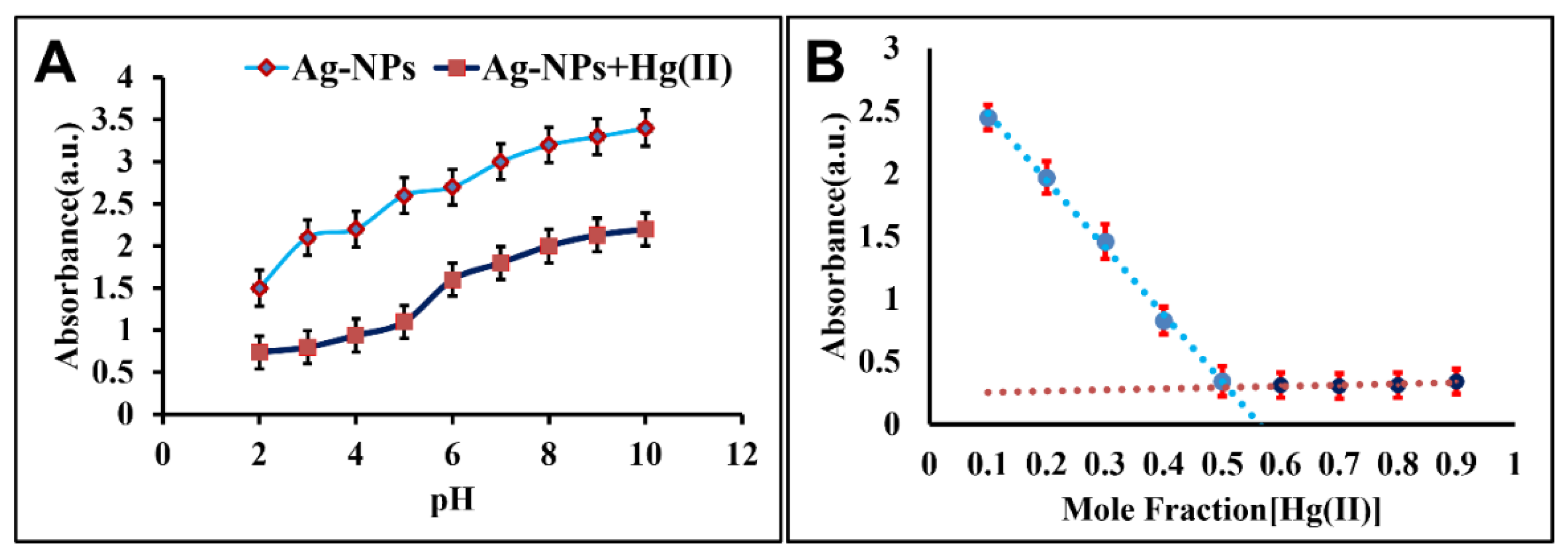
3.8.4. Effect of pH on Detection of Hg2+
If the circumstances of the detection scheme are altered, then there is a noticeable effect on Hg2+ sensing. The variation in pH of the system results in aggregation and destabilization of AgNPs. The pH of the system was altered from 2 to10 by using 0.1 M KOH, and 0.1 M HCl and UV spectra were recorded for the adjusted pH values. The results indicated that AgNPs are stable in basic medium while in acidic medium (pH less than 5) the solution become colorless, and at the same time UV spectra indicate the minimum absorption intensity, as shown in Figure 5A. The stability of AgNPs in basic medium is due to an increase in the rate reduction of Ag+ ions by OH ions from the base, which results in an augmented formation of AgNPs that is degraded by Hg2+ ions. It means that in basic medium Hg2+ are not able to degrade the maximum amount of highly stable AgNPs, while they succeed in acidic conditions.
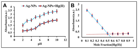
Figure 5.
(A) Effect of pH on the adsorption of AgNPs in the absence and presence of 100 ppm Hg2+ ions, (B) Job’s plot curve showing the binding ratio of AgNPs:Hg2+.
3.8.5. Determination of Required Stoichiometry of AgNPs and Hg2+
The binding stoichiometry of the AgNPs and Hg2+ was detected by Job’s plot method [49]. Different mole fraction ratios of Hg2+ and AgNPs were tested. The absorption intensity at 430 nm obtained via UV was plotted against the molar fraction of Hg2+ (100 ppm) to monitor the results. The mole fraction of the highest absorption intensity revealed the binding stoichiometry of the compound. As shown in Figure 5B, 1 mL of Hg2+ solution and 1 mL of AgNPs suspension were present in the sample, revealing minimum absorption intensity. The results suggest that AgNPs forms a 1:1 complex (AgNPs:Hg2+), which means that the best detection of heavy metals occurs when equal moles of AgNPs and Hg2+ are present [50]. The increased stability of AgNPs in alkaline pH might be due to stronger protection of AgNPs by deprotonated OH groups in the starch [51].
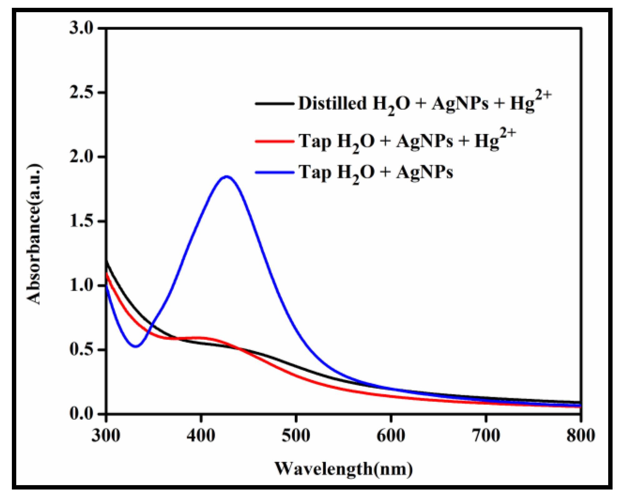
Finally, as shown in Table 2, the proposed method was applied to real tap and lake water samples. In order to verify the recovery and accuracy of the procedure, these samples were also spiked with known levels of Hg2+. A volume of 1 mL of Hg2+ solution was added into 1 mL of tap water sample of AgNPs and after some time the solution became colorless, as was done for Hg2+ sensing in distilled water. Moreover, UV-visible spectra revealed that the peak became broadened at 430 nm (as shown in Figure 6) and the same results observed in distilled water are obtained. Hence, AgNPs are found to be effective for the detection of Hg2+ in tap water samples [52].
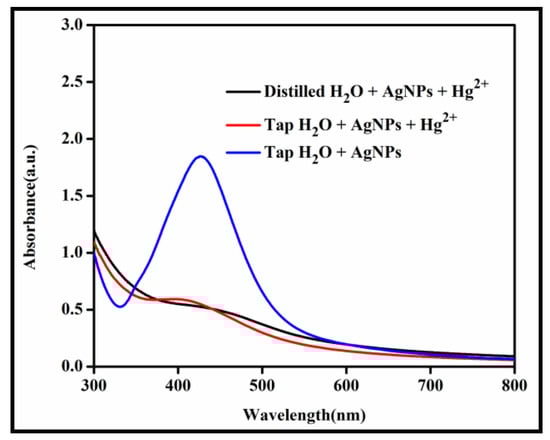
Figure 6.
UV Spectrum shows detection of Hg2+ in laboratory tap water.
4. Conclusions
In this study, we have developed a facile and green approach for the synthesis of starch-coated silver nanoparticles-based colorimetric sensor for the selective recognition of Hg2+ in real samples. Various parameters that regulate the synthesis and stabilization of AgNPs were studied and optimized. The newly synthesized nanoparticles were characterized using various spectroscopic techniques. Moreover, we established the colorimetric as well as SPR detection-based sensing for Hg2+ by starch stabilized AgNPs. The strategy is focused on a redox reaction between Starch-AgNP and Hg2+, which results in a shift in color from yellow to colorless nanoparticles dispersions and a decrease in AgNPs SPR uptake. Additionally, this approach allows a wide range of linear detection, while avoiding interference from other metal ions. The synthesized AgNPs showed admirable selectivity for Hg2+, even in the occurrence of many other heavy metals. The proposed calorimetric chemosensor can be useful for laboratory tap water with a detection limit of 0.1 ppm.
Supplementary Materials
The following supporting information can be downloaded at: https://www.mdpi.com/article/10.3390/coatings12060763/s1, Table S1: Comparison of different methods using nanoparticles as a sensing probe for Hg+2 determination. References [53,54,55] are cited in the supplementary materials.
Author Contributions
Conceptualization, N.M.A., M.U.H. and F.A. (Farid Ahmed); methodology, N.M.A.; validation, N.M.A. and M.U.H.; formal analysis, N.M.A. and M.U.H.; investigation, N.M.A.; data curation, N.M.A.; writing—original draft preparation, N.M.A., M.U.H., N.N., F.A. (Farid Ahmed), F.A. (Faizah Altaf), S.S., S.F. and P.B.; writing—review and editing, N.M.A., S.M.S., P.B. and M.U.H.; visualization, P.B.; supervision, M.U.H. All authors have read and agreed to the published version of the manuscript.
Funding
This research was funded by Higher education commission of Pakistan for Funding, grant 0.3 million PKR number 21-1775/SRGP/R&D/HEC/2017.
Institutional Review Board Statement
Not applicable.
Informed Consent Statement
Not applicable.
Data Availability Statement
The data presented in this study are available on request from author Farid Ahmed which is responsible of the performed experiments.
Conflicts of Interest
The authors declare no conflict of interest.
References
- Ravindran, A.; Elavarasi, M.; Prathna, T.C.; Raichur, A.M.; Chandrasekaran, N.; Mukherjee, A. Selective colorimetric detection of nanomolar Cr (VI) in aqueous solutions using unmodified silver nanoparticles. Sens. Actuators B Chem. 2012, 166, 365–371. [Google Scholar] [CrossRef]
- Singh, R.; Thakur, P.; Thakur, A.; Kumar, H.; Chawla, P.; Rohit, J.V.; Kaushik, R.; Kumar, N. Colorimetric sensing approaches of surface-modified gold and silver nanoparticles for detection of residual pesticides: A review. Int. J. Environ. Anal. Chem. 2021, 101, 3006–3022. [Google Scholar] [CrossRef]
- Domaille, D.W.; Que, E.L.; Chang, C.J. Synthetic fluorescent sensors for studying the cell biology of metals. Nat. Chem. Biol. 2008, 4, 168. [Google Scholar] [CrossRef] [PubMed]
- Georgopoulos, P.G.; Roy, A.; Yonone-Lioy, M.J.; Opiekun, R.E.; Lioy, P.J. Environmental copper: Its dynamics and human exposure issues. J. Toxicol. Environ. Health Part B Crit. Rev. 2001, 4, 341–394. [Google Scholar] [CrossRef]
- Holmes, P.; James, K.; Levy, L. Is low-level environmental mercury exposure of concern to human health? Sci. Total Environ. 2009, 408, 171–182. [Google Scholar] [CrossRef]
- Lavoie, R.A.; Jardine, T.D.; Chumchal, M.M.; Kidd, K.A.; Campbell, L.M. Biomagnification of mercury in aquatic food webs: A worldwide meta-analysis. Environ. Sci. Technol. 2013, 47, 13385–13394. [Google Scholar] [CrossRef]
- Manahan, S. Fundamentals of Environmental and Toxicological Chemistry: Sustainable Science; CRC Press: Boca Raton, FL, USA, 2013. [Google Scholar]
- Firdaus, M.L.; Fitriani, I.; Wyantuti, S.; Hartati, Y.W.; Khaydarov, R.; McAlister, J.A.; Obata, H.; Gamo, T. Colorimetric Detection of Mercury (II) Ion in Aqueous Solution Using Silver Nanoparticles. Anal. Sci. 2017, 33, 831–837. [Google Scholar] [CrossRef] [Green Version]
- Harada, M. Minamata disease: Methylmercury poisoning in Japan caused by environmental pollution. Crit. Rev. Toxicol. 1995, 25, 1–24. [Google Scholar] [CrossRef]
- Aragay, G.; Pons, J.; Merkoçi, A. Recent trends in macro-, micro-, and nanomaterial-based tools and strategies for heavy-metal detection. Chem. Rev. 2011, 111, 3433–3458. [Google Scholar] [CrossRef]
- Zhang, Z.; Tang, A.; Liao, S.; Chen, P.; Wu, Z.; Shen, G.; Yu, R. Oligonucleotide probes applied for sensitive enzyme-amplified electrochemical assay of mercury (II) ions. Biosens. Bioelectron. 2011, 26, 3320–3324. [Google Scholar] [CrossRef]
- Guha, S.; Roy, S.; Banerjee, A. Fluorescent Au@ Ag core–shell nanoparticles with controlled shell thickness and HgII sensing. Langmuir 2011, 27, 13198–13205. [Google Scholar] [CrossRef] [PubMed]
- Han, F.X.; Patterson, W.D.; Xia, Y.; Sridhar, B.B.; Su, Y. Rapid determination of mercury in plant and soil samples using inductively coupled plasma atomic emission spectroscopy, a comprehensive study. Water Air Soil Pollut. 2006, 170, 161–171. [Google Scholar] [CrossRef]
- Fabbrizzi, L.; Licchelli, M.; Parodi, L.; Poggi, A.; Taglietti, A. The molecular design of fluorescent sensors for ionic analytes. J. Fluoresc. 1998, 8, 263–271. [Google Scholar] [CrossRef]
- Vilder, D.S.; Jenkins, R.O.; Hall, J.F.; Harrington, C.F. The determination of methylmercury in biological samples by HPLC coupled to ICP-MS detection. Appl. Organomet. Chem. 2007, 21, 303–310. [Google Scholar]
- Abdelhamid, H.N.; Wu, H.-F. A new binary matrix for specific detection mercury (II) using matrix-assisted laser desorption ionization mass spectrometry. J. Am. Soc. Mass Spectrom. 2019, 30, 2617–2622. [Google Scholar] [CrossRef]
- Duan, J.; Yin, H.; Wei, R.; Wang, W. Facile colorimetric detection of Hg2+ based on anti-aggregation of silver nanoparticles. Biosens. Bioelectron. 2014, 57, 139–142. [Google Scholar] [CrossRef]
- Lou, T.; Chen, L.; Zhang, C.; Kang, Q.; You, H.; Shen, D.; Chen, L. A simple and sensitive colorimetric method for detection of mercury ions based on anti-aggregation of gold nanoparticles. Anal. Methods 2012, 4, 488–491. [Google Scholar] [CrossRef]
- Huang, J.T.; Yang, X.X.; Zeng, Q.L.; Wang, J. A simple green route to prepare stable silver nanoparticles with pear juice and a new selective colorimetric method for detection of cysteine. Analyst 2013, 138, 5296–5302. [Google Scholar] [CrossRef]
- Yoosaf, K.; Ipe, B.I.; Suresh, C.H.; Thomas, K.G. In situ synthesis of metal nanoparticles and selective naked-eye detection of lead ions from aqueous media. J. Phys. Chem. C 2007, 111, 12839–12847. [Google Scholar] [CrossRef]
- Li, H.; Li, F.; Han, C.; Cui, Z.; Xie, G.; Zhang, A. Highly sensitive and selective tryptophan colorimetric sensor based on 4, 4-bipyridine-functionalized silver nanoparticles. Sens. Actuators B Chem. 2010, 145, 194–199. [Google Scholar] [CrossRef]
- Karthiga, D.; Anthony, S.P. Selective colorimetric sensing of toxic metal cations by green synthesized silver nanoparticles over a wide pH range. RSC Adv. 2013, 3, 16765–16774. [Google Scholar] [CrossRef]
- Walekar, L.S.; Gore, A.H.; Anbhule, P.V.; Sudarsan, V.; Patil, S.R.; Kolekar, G.B. A novel colorimetric probe for highly selective recognition of Hg2+ ions in aqueous media based on inducing the aggregation of CPB-capped AgNPs: Accelerating direct detection for environmental analysis. Anal. Methods 2013, 5, 5501–5507. [Google Scholar] [CrossRef]
- Wang, J.; Fang, X.; Cui, X.; Zhang, Y.; Zhao, H.; Li, X.; He, Y. A highly sensitive colorimetric probe for Cd2+, Hg2+ and ascorbic acid determination based on trithiocyanuric acid-AuNPs. Talanta 2018, 188, 266–272. [Google Scholar] [CrossRef] [PubMed]
- Senapati, T.; Senapati, D.; Singh, A.K.; Fan, Z.; Kanchanapally, R.; Ray, P.C. Highly selective SERS probe for Hg (II) detection using tryptophan-protected popcorn shaped gold nanoparticles. Chem. Commun. 2011, 47, 10326–10328. [Google Scholar] [CrossRef] [PubMed]
- Chai, F.; Wang, C.; Wang, T.; Li, L.; Su, Z. Colorimetric detection of Pb2+ using glutathione functionalized gold nanoparticles. ACS Appl. Mater. Interfaces 2010, 2, 1466–1470. [Google Scholar] [CrossRef]
- Ma, Y.; Jiang, L.; Mei, Y.; Song, R.; Tian, D.; Huang, H. Colorimetric sensing strategy for mercury (II) and melamine utilizing cysteamine-modified gold nanoparticles. Analyst 2013, 138, 5338–5343. [Google Scholar] [CrossRef]
- Darbha, G.K.; Singh, A.K.; Rai, U.S.; Yu, E.; Yu, H.; Ray, P.C. Selective detection of mercury (II) ion using nonlinear optical properties of gold nanoparticles. J. Am. Chem. Soc. 2008, 130, 8038–8043. [Google Scholar] [CrossRef] [Green Version]
- Miao, L.-J.; Xin, J.-W.; Shen, Z.-Y.; Zhang, Y.-J.; Wang, H.-Y.; Wu, A.-G. Exploring a new rapid colorimetric detection method of Cu2+ with high sensitivity and selectivity. Sens. Actuators B Chem. 2013, 176, 906–912. [Google Scholar] [CrossRef]
- Su, W.; Yuan, S.; Wang, E. A Rhodamine-Based Fluorescent Chemosensor for the Detection of Pb2+, Hg2+ and Cd2+. J. Fluoresc. 2017, 27, 1871–1875. [Google Scholar] [CrossRef]
- Bumagina, N.A.; Antina, E.V.; Sozonov, D.I. Off-on fluorescent sensor based on the bis (2, 4, 7, 8, 9-pentamethyldipyrrolylmethene-3-yl) methane for detection of Cd2+ and Hg2+ cations. J. Lumin. 2017, 183, 315–321. [Google Scholar] [CrossRef]
- Ding, N.; Zhao, H.; Peng, W.; He, Y.; Zhou, Y.; Yuan, L.; Zhang, Y. A simple colorimetric sensor based on anti-aggregation of gold nanoparticles for Hg2+ detection. Colloids Surf. A Physicochem. Eng. Asp. 2012, 395, 161–167. [Google Scholar] [CrossRef]
- Pienpinijtham, P.; Han, X.X.; Suzuki, T.; Thammacharoen, C.; Ekgasit, S.; Ozaki, Y. Micrometer-sized gold nanoplates: Starch-mediated photochemical reduction synthesis and possibility of application to tip-enhanced Raman scattering (TERS). Phys. Chem. Chem. Phys. 2012, 14, 9636–9641. [Google Scholar] [CrossRef] [PubMed]
- Borbach, M.; Stenzel, W.; Conrad, H.; Bradshaw, A. Hydroxyl formation on Ag (110) studied by HREELS. Surf. Sci. 1997, 377, 796–801. [Google Scholar] [CrossRef]
- Tagad, C.K.; Rajdeo, K.S.; Kulkarni, A.; More, P.; Aiyer, R.; Sabharwal, S. Green synthesis of polysaccharide stabilized gold nanoparticles: Chemo catalytic and room temperature operable vapor sensing application. RSC Adv. 2014, 4, 24014–24019. [Google Scholar] [CrossRef]
- Peng, H.; Yang, A.; Xiong, J. Green microwave-assisted synthesis of silver nanoparticles using bamboo hemicelluloses and glucose in an aqueous medium. Carbohydr. Polym. 2013, 91, 348–355. [Google Scholar] [CrossRef]
- Jyoti, K.; Baunthiyal, M.; Singh, A. Characterization of silver nanoparticles synthesized using Urtica dioica Linn. leaves and their synergistic effects with antibiotics. J. Radiat. Res. Appl. Sci. 2016, 9, 217–227. [Google Scholar] [CrossRef] [Green Version]
- Srirangam, G.; Rao, K.P. Synthesis and charcterization of silver nanoparticles from the leaf extract of Malachracapitata (L). J. Chem. 2017, 10, 46–53. [Google Scholar]
- Ismail, M.; Khan, M.; Akhtar, K.; Khan, M.A.; Asiri, A.M.; Khan, S.B. Biosynthesis of silver nanoparticles: A colorimetric optical sensor for detection of hexavalent chromium and ammonia in aqueous solution. Phys. E Low-Dimens. Syst. Nanostruct. 2018, 103, 367–376. [Google Scholar] [CrossRef]
- Tagad, C.K.; Dugasani, S.R.; Aiyer, R.; Park, S.; Kulkarni, A.; Sabharwal, S. Green synthesis of silver nanoparticles and their application for the development of optical fiber based hydrogen peroxide sensor. Sens. Actuators B Chem. 2013, 183, 144–149. [Google Scholar] [CrossRef]
- Meshram, S.M.; Bonde, S.R.; Gupta, I.R.; Gade, A.K.; Rai, M.K. Green synthesis of silver nanoparticles using white sugar. IET Nanobiotechnol. 2013, 7, 28–32. [Google Scholar] [CrossRef]
- Bothra, S.; Solanki, J.N.; Sahoo, S.K. Functionalized silver nanoparticles as chemosensor for pH, Hg2+ and Fe3+ in aqueous medium. Sens. Actuators B Chem. 2013, 188, 937–943. [Google Scholar] [CrossRef]
- Annadhasan, M.; Rajendiran, N. Highly selective and sensitive colorimetric detection of Hg (II) ions using green synthesized silver nanoparticles. RSC Adv. 2015, 5, 94513–94518. [Google Scholar] [CrossRef]
- Wang, Y.; Yang, F.; Yang, X. Colorimetric detection of mercury (II) ion using unmodified silver nanoparticles and mercury-specific oligonucleotides. ACS Appl. Mater. Interfaces 2010, 2, 339–342. [Google Scholar] [CrossRef] [PubMed]
- Nidya, M.; Umadevi, M.; Rajkumar, B.J. Structural, morphological and optical studies of L-cysteine modified silver nanoparticles and its application as a probe for the selective colorimetric detection of Hg2+. Spectrochim. Acta Part A Mol. Biomol. Spectrosc. 2014, 133, 265–271. [Google Scholar] [CrossRef] [PubMed]
- Chansuvarn, W.; Imyim, A. Visual and colorimetric detection of mercury (II) ion using gold nanoparticles stabilized with a dithia-diaza ligand. Microchim. Acta 2012, 176, 57–64. [Google Scholar] [CrossRef]
- Li, L.; Feng, D.; Fang, X.; Han, X.; Zhang, Y. Visual sensing of Hg2+ using unmodified Au@ Ag core–shell nanoparticles. J. Nanostruct. Chem. 2014, 4, 117. [Google Scholar] [CrossRef] [Green Version]
- Khan, U.; Niaz, A.; Shah, A.; Zaman, M.I.; Zia, M.A.; Iftikhar, F.J.; Nisar, J.; Ahmed, M.N.; Akhter, M.S.; Shah, A.H. Thiamine-functionalized silver nanoparticles for the highly selective and sensitive colorimetric detection of Hg2+ ions. New J. Chem. 2018, 42, 528–534. [Google Scholar] [CrossRef]
- Shah, K.; ul Ain, N.; Ahmed, F.; Anis, I.; Shah, M.R. A new highly selective chemosensor for the detection of iron ion in aqueous medium based on click generated triazole. Sens. Actuators B Chem. 2017, 249, 515–522. [Google Scholar] [CrossRef]
- Kim, H.N.; Ren, W.X.; Kim, J.S.; Yoon, J. Fluorescent and colorimetric sensors for detection of lead, cadmium, and mercury ions. Chem. Soc. Rev. 2012, 41, 3210–3244. [Google Scholar] [CrossRef]
- Kumar, V.V.; Anbarasan, S.; Christena, L.R.; SaiSubramanian, N.; Anthony, S.P. Bio-functionalized silver nanoparticles for selective colorimetric sensing of toxic metal ions and antimicrobial studies. Spectrochim. Acta Part A Mol. Biomol. Spectrosc. 2014, 129, 35–42. [Google Scholar] [CrossRef]
- Luo, Y.; Shen, S.; Luo, J.; Wang, X.; Sun, R. Green synthesis of silver nanoparticles in xylan solution via Tollens reaction and their detection for Hg2+. Nanoscale 2015, 7, 690–700. [Google Scholar] [CrossRef] [PubMed]
- Wang, X.; Su, Y.; Yang, H.; Dong, Z.; Ma, J. Highly sensitive fluorescence probe based on chitosan nanoparticle for selective detection of Hg2+ in water. Colloids Surf. A Physicochem. Eng. Asp. 2012, 402, 88–93. [Google Scholar] [CrossRef]
- Davadhasan, J.P.; Kim, J. A chemically functionalized paper-based microfluidic platform for multiplex heavy metal detection. Sens. Actuator B Chem. 2018, 10, 18–24. [Google Scholar] [CrossRef]
- Chen, M.-M.; Chen, L.; Li, H.-X.; Brammer, L.; Lang, J.-P. Highly selective detection of Hg2+ and MeHgI by di-pyridin-2-yl-[4-(2-pyridin-4-yl-vinyl)-phenyl]-amine and its zinc coordination polymer. Inorg. Chem. Front. 2016, 3, 1297–1305. [Google Scholar] [CrossRef] [Green Version]


Publisher’s Note: MDPI stays neutral with regard to jurisdictional claims in published maps and institutional affiliations. |
© 2022 by the authors. Licensee MDPI, Basel, Switzerland. This article is an open access article distributed under the terms and conditions of the Creative Commons Attribution (CC BY) license (https://creativecommons.org/licenses/by/4.0/).