Article

The Fabrication of Full Chromatography SiO2@PDA Photonic Crystal Structural Colored Fabric with High Thermal Stability

School of Physics and Astronomy, China West Normal University, Nanchong 637002, China
*
Author to whom correspondence should be addressed.
Coatings 2022, 12(8), 1085; https://doi.org/10.3390/coatings12081085
Submission received: 28 June 2022 / Revised: 21 July 2022 / Accepted: 28 July 2022 / Published: 31 July 2022
(This article belongs to the Section Environmental Aspects in Colloid and Interface Science)

Abstract

:
Traditional textile dyeing and finishing industries are two of the most important sources of high pollution, high energy consumption, and high emissions. Structural color, as a clean ecological staining method that does not require any dye or pigment, has received extreme attention from researchers. In this study, core-shell structures of SiO2@PDA microspheres were prepared by coating polydopamine (PDA) formed by rapid polymerization of dopamine (DA) on the surface of SiO2 microspheres. Moreover, the structural colors of full chromatography were successfully prepared by vertical self-assembly on silk. The morphology and chemical structure of the prepared SiO2@PDA microspheres were studied by SEM and FT-IR, and the morphology and optical properties of the structured colored fabrics were characterized by SEM and material microscope. The different structural colors of the entire visible region were obtained by controlling the particle size of SiO2@PDA microspheres and the viewing angle of the SiO2@PDA photonic crystal, which are consistent with Bragg’s diffraction law. Since the SiO2@PDA photonic crystal has thermal stability, the prepared structural color fabric could remain highly saturated in color at temperatures up to 200 °C. This has a previously unreported high thermal stability on structural colors of silk. Therefore, the research work will demonstrate a structural color fabric that can prepare full chromatography with high thermal stability.

1. Introduction

The coloration of traditional textile materials is mainly achieved by using chemical colorants such as dyes or pigments. These chemical colorants belong to the pigmentary color family, which produces colors by selectively absorbing and reflecting certain wavelengths in the range of visible light [1]. Though these chemical colorants can be colored on textiles, they can usually bring about serious environmental pollution [2]. Efficient treatment of printing and dyeing wastewater using dyes, pigments, and heavy metals is still a great challenge in the textile dyeing and finishing industry [3,4]. As such, it is urgent to develop an ecological and clean dyeing method.
Photonic crystals are highly periodically arranged dielectric materials with photonic band gap (PBG) properties. Owing to the existence of the photonic band gap, it can confine and control the propagation ability of light [5]. Therefore, light cannot propagate within a specific frequency range located in the photon band gap, whereas selective reflection produces bright structural color, which is widely used in various fields, such as the new flat-panel display technology [6,7], sensors [8,9], printing [10], anti-counterfeiting [11,12], and textile [13,14,15]. Fortunately, photonic crystals, a structural color material, can be regarded as a colorant to take the place of organic dyes and pigments on account of their no photo-bleaching structural colors and their unique visibility in indoor and outdoor environments [16,17,18]. It is reported that photonic crystals have been well studied on smooth, seamless and rigid hard substrates [19,20,21,22,23], while further exploration is needed on soft, porous and undulating fabrics.
In recent years, photonic crystal microspheres mainly include silica and polystyrene [24,25]. Compared with polystyrene as organic polymer microspheres in aqueous suspension, silica microspheres have higher light/thermal stability and dispersion, which is similar to hard spheres [26], which is considered to be conducive to the formation of photonic crystals on textile substrates under different self-assembly conditions. However, due to the strong scattering effect of the amorphous structure on light, its color saturation decreases significantly, resulting in white or unstructured color [27]. In order to overcome this problem, researchers introduced black materials such as carbon black [28,29], cuttlefish ink [30], and graphene oxide (GO) [31,32,33] into photonic crystals to absorb incoherently scattered light to enhance color saturation. However, carbon black [29] is easy to agglomerate in the self-assembly process and difficult to disperse evenly in the system, so the prepared structural color film may show uneven color. The preparation process for cuttlefish ink [30] and GO [32,33] is relatively complex, with a high cost and uneven structural color membrane, such as carbon black. Polydopamine is a synthetic eumelanin, that is formed by the oxidative self-polymerization of dopamine (DA) in an alkaline environment [34]. It has naturally super-strong adhesion, and can directly adhere to the surface of almost all substances through simple physical and chemical actions, even without grafting modification on the surface of substances [35]. Based on this property, SiO2@PDA microspheres have been used as basic units of structural color materials in photonic crystals. For example, Panmiao Liu et al. [19] initially tried to construct SiO2@PDA photonic crystal structural colors on hard substrates, such as plastic and metal, but did not prepare them on flexible fabrics. Subsequently, Shuai Li et al. [15] initially tried to coat SiO2 nano microspheres with polydopamine, and then, successfully prepared SiO2@PDA photonic crystals by vertical deposition on the surface of polyester fabric. However, only four colors were prepared, and the panchromatic structural color in the visible range was not realized. Moreover, SiO2 has high thermal stability, acting as a precursor of photonic crystals, laying the foundation for the application range of structural color in high thermostability.
In this study, full chromatographic structural colors with bright colors and high thermostability on silk were produced with the vertical deposition method. Specifically, SiO2@PDA microspheres with a core-shell structure were prepared by the oxidative self-polymerization reaction of dopamine, then the crystal array of the face-centered cubic (fcc) structure was obtained by vertical deposition on the silk, producing a stable, bright, and rainbow-like chromatographic structural color.

2. Materials and Methods

2.1. Materials

Tetraethyl orthosilicate (TEOS, >99%, G.C. grade,), ammonium hydroxide (NH3·H2O, 25%–28%, A.R. grade), dopamine hydrochloride (C8H11NO2·HCl, 98%, A.R. grade), and tris - (hydroxymethyl aminomethane) (C4H11NO3, ≥99.9%, standard buffer material) were purchased from Aladdin reagent Co., Ltd. (Shanghai, China). Ethanol (EtOH, A.R. grade) was bought from Chengdu Kelong Chemical, Ltd (Chengdu, China). Black silks were supplied by a local fabric market (Nanchong Yinhai Silk Co. Ltd, Nangchong, China). Deionized water was used throughout the experiment. All chemicals were used without further purification.

2.2. Synthesis of Monodisperse SiO2@PDA Colloidal Microspheres

The SiO2 microspheres with different particle sizes were prepared with an improved Stöber method. First, a certain amount of TEOS and absolute ethanol were added into the beaker and vibrated for 20 min as the monomer solution; next, 5 mL of ammonia water, 15 mL of deionized water, and 20 mL of absolute ethanol were poured into a three-port flask, and the monomer solution was poured into it after magnetic stirring for 10 min. Dispersions of the SiO2 microspheres were obtained after magnetic stirring for 5 h at 30 ℃. Dispersions of the SiO2 microspheres were purified by centrifuging and redispersing, followed by drying and grinding to obtain SiO2 microspheres. Then, 1.21 g of tris - (hydroxymethyl aminomethane) was dispersed in deionized water to prepare 10 mM, PH = 8.5 Tris buffer. Then, 1 g of SiO2 microspheres and 0.05 g of DA were added into a beaker containing 100 mL of Tris buffer. Then, the mixture was put into the ultrasonic cleaning machine, with an adjusted power of 120 W and ultrasonically dispersed for 30 min. The resulting mixed solution was magnetically stirred at room temperature for 20 h. After the reaction, the SiO2@PDA suspension was centrifuged and washed with deionized water. Finally, it was placed in a vacuum drying oven at 60 °C at constant weight to obtain different colors of SiO2@PDA microspheres.

2.3. Preparation of Structured Colored Fabrics

Photonic crystals were successfully prepared on silk by vertical deposition self-assembly. Firstly, 0.5 g of SiO2@PDA nano microspheres were dispersed in 49.5 g of anhydrous ethanol to prepare a suspension with a mass fraction of 1%, which was placed in the ultrasonic cleaning machine, with an adjusted power of 120 W and ultrasonically dispersed for 30 min. Secondly, the black silk (90 mm × 25 mm, length × width) was fixed to the slide, and inserted vertically into the beaker containing the suspension and fixed with fixtures. Lastly, the device was placed in a vacuum drying oven at 60 °C for 12 h. When the suspension completely evaporated, a colorful SiO2@PDA photonic crystal fabric was obtained.

2.4. Characterization

The particle size of SiO2@PDA microspheres was measured with a nano particle size analyzer (Nicomp 380 Z3000, PSS, Santa Barbara, CA, USA). SiO2 microspheres and SiO2@PDA photonic crystals on black silks were observed and photographed with a field emission scanning electron microscopy (Phenom XL, Phenom, Eindhoven, The Netherlands). The crystalline states of SiO2 and SiO2@PDA microspheres were analyzed with an X-ray diffractometer (TD-3500, TongDa, Dandong, China). The infrared spectra of SiO2 and SiO2@PDA microspheres were tested using Fourier transform infrared spectroscopy (Nicolet 6700, Nicolet, Waltham, MA, USA). The structural color of photonic crystals on black silk was observed with a material microscope (axioscope 7, Zeiss, Oberkochen, Germany). The reflection spectrum of the structural color was tested with an ultraviolet spectrophotometer (uv-3600plus, Shimadzu, Kyoto, Japan). The thermal stability of SiO2@PDA microspheres was tested using a simultaneous thermal analyzer (STA2500, Netzsch, selb, Germany).

3. Results

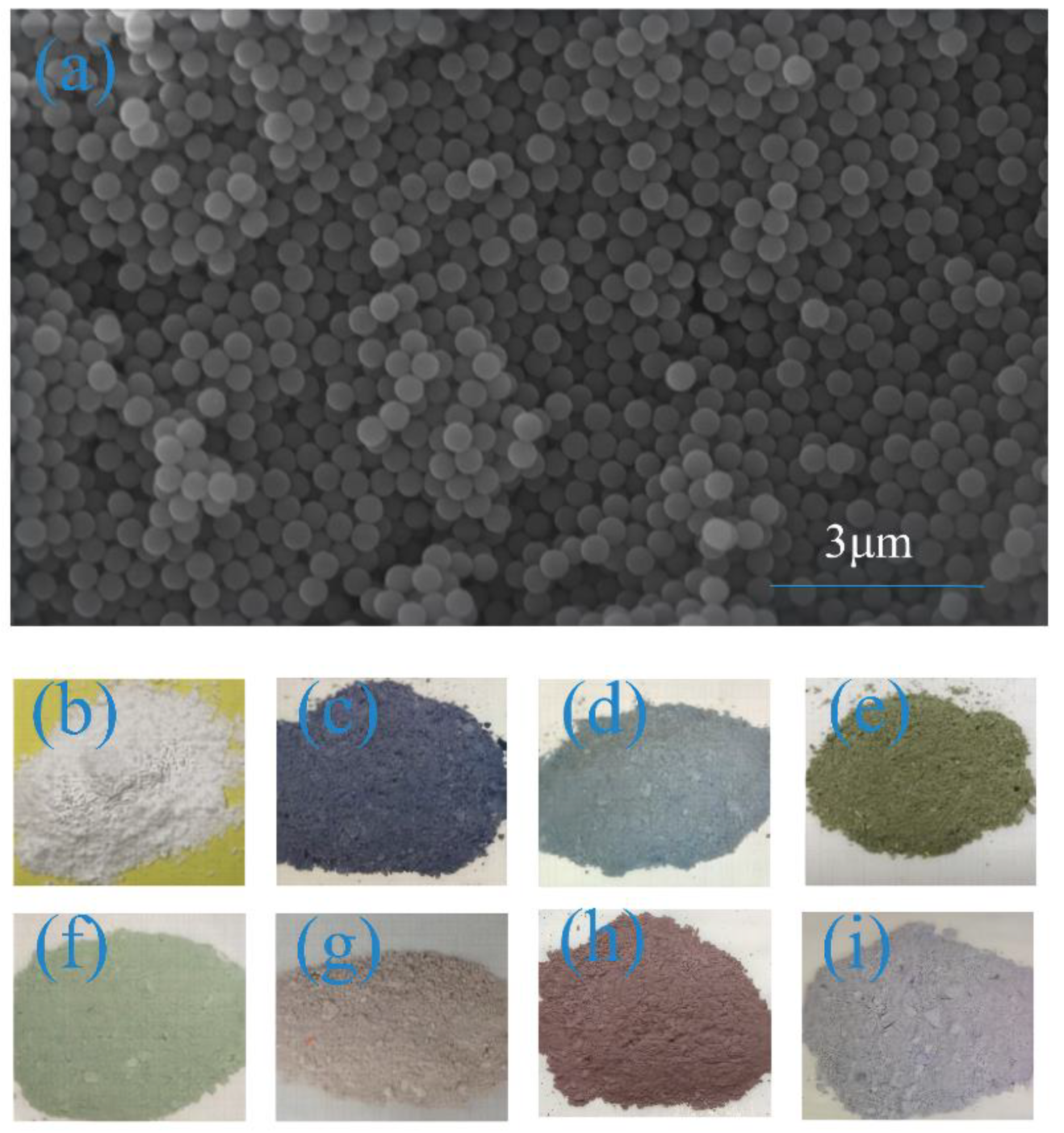
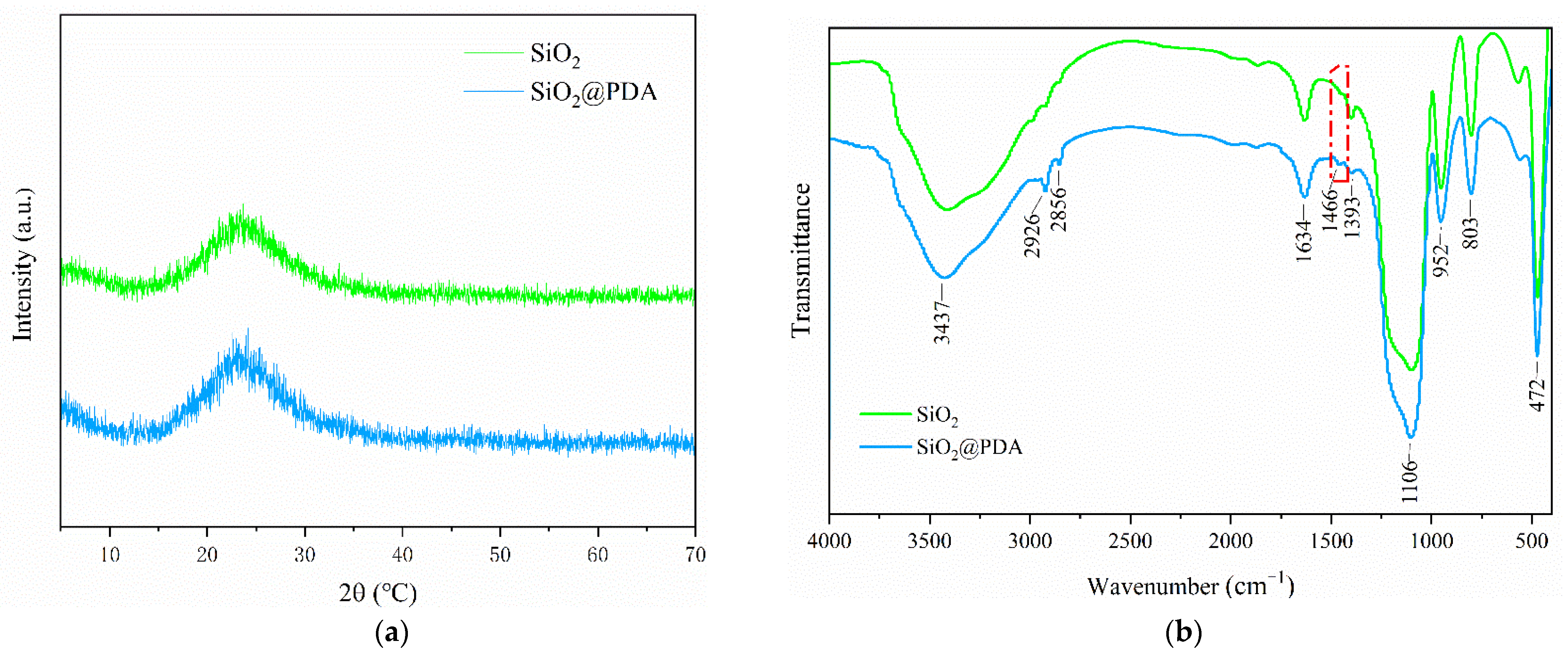
3.1. Characterization and Analysis of SiO2@PDA Core-shell Colloidal Microspheres

Table 1 shows SiO2@PDA microspheres with different particle sizes prepared with different amounts of TEOS (3–10 mL). It could be seen from the table that the particle size of SiO2@PDA microspheres increased with an increase in the TEOS amount. Phenom Image Viewer software (1.1.0_release, Thermo Fisher Scientific, Waltham, MA, USA) was used to read the particle size of 50 SiO2@PDA nanospheres randomly selected by SEM and calculate their polymer dispersity index (PDI), which was compared with the test results of a nano-particle analyzer. It could be seen that the hydration particle size Dh measured with the dynamic light scattering method was greater than or equal to the actual particle size Da directly measured by SEM images, because the nanospheres were dispersed in the solvent, and a solvated layer would be formed on the surface, so the value was larger than the actual value. Moreover, the PDI values were all less than 0.8, indicating that SiO2@PDA microspheres had excellent mono-dispersity, which was conducive to the construction of highly ordered photonic crystal structures. Figure 1a shows the SEM of SiO2@PDA microspheres. It could be seen that SiO2@PDA microspheres had uniform particle size and regular morphology. The digital photographs of the SiO2 and SiO2@PDA microspheres powder under natural light are shown in Figure 1b–i. As can be seen from Figure 1b–i, SiO2 microsphere powder presented a white state, while SiO2@PDA microsphere powder exhibited different colors with the change in the particle size. In general, the drying process of SiO2 microspheres and SiO2@PDA microspheres was similar to the self-assembly process of gravity deposition, which spontaneously assembled into SiO2 and SiO2@PDA photonic crystals. After grinding, many tiny blocky photonic crystals were obtained, which could produce a low-intensity rainbow effect under the reflection of natural light. Since the amorphous structure would produce strong incoherently scattered light under illumination. The SiO2 microsphere powder appeared white [29]. However, black PDA was introduced into the microsphere powder to absorb incoherently scattered light, resulting in structural colors being able to be viewed by the naked eye.
The XRD images of SiO2 microspheres and SiO2@PDA microspheres are shown in Figure 2a, which verifies the amorphous structures of SiO2 and SiO2@PDA microspheres. In addition, it can be found that the introduction of polydopamine did not change the structure of SiO2. The chemical structures of SiO2 microspheres and SiO2@PDA microspheres were characterized by FT-IR and are shown in Figure 2b. The absorption peak of the FT-IR spectrum of SiO2 at 472 cm−1 was caused by the bending vibration of Si-O-Si. The absorption peak of 1106 cm−1 belonged to the antisymmetric stretching vibration peak of Si-O-Si and 3437 cm−1 and was the stretching vibration peak of -OH. In the FT-IR spectra of SiO2@PDA, 3437 cm−1 was attributed to the stretching vibration of -OH/-NH, while the weak peak at 1466 cm−1 was due to the stretching vibration of the benzene ring, indicating that polydopamine was successfully coated on the surface of SiO2 microspheres without changing the original structure of the nanoparticles.

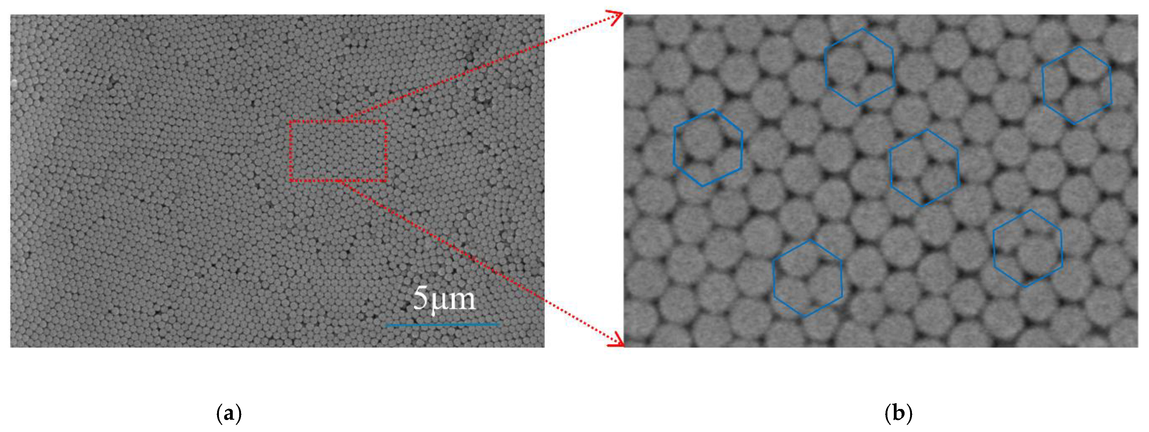
3.2. Ordered Structure of SiO2@PDA Photonic Crystals on Silk

The SEM image of the SiO2@PDA (271 nm) photonic crystal on silk shown in Figure 3a,b is the local enlargement of the red box in Figure 3a. As can be seen from Figure 3a,b, SiO2@PDA microspheres were closely arranged on the surface of the silk, and each in the same layer. The SiO2@PDA microspheres were surrounded by six adjacent photonic crystals, presenting a periodic hexagonal close-packed structure. However, there were some defects in the prepared photonic crystal array, which may be due to the rapid evaporation rate of the solvent or the interference of external airflow during the deposition process, resulting in the formation of vacancies in the microspheres.

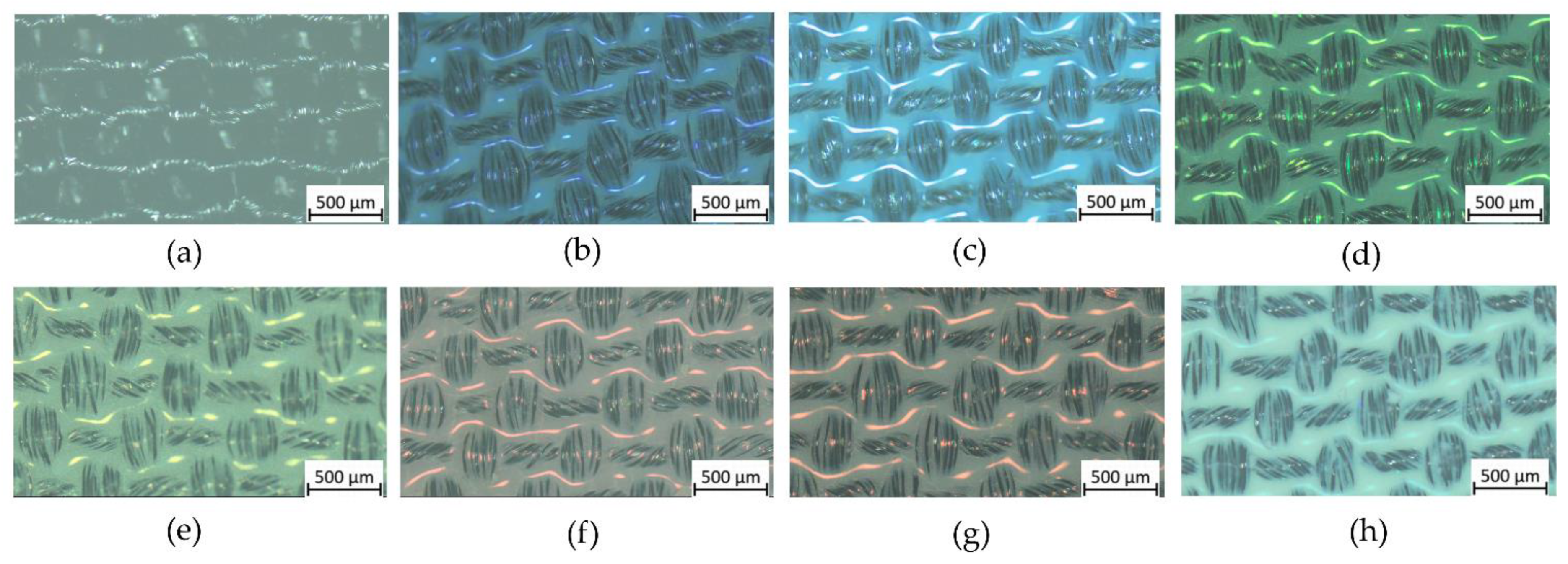
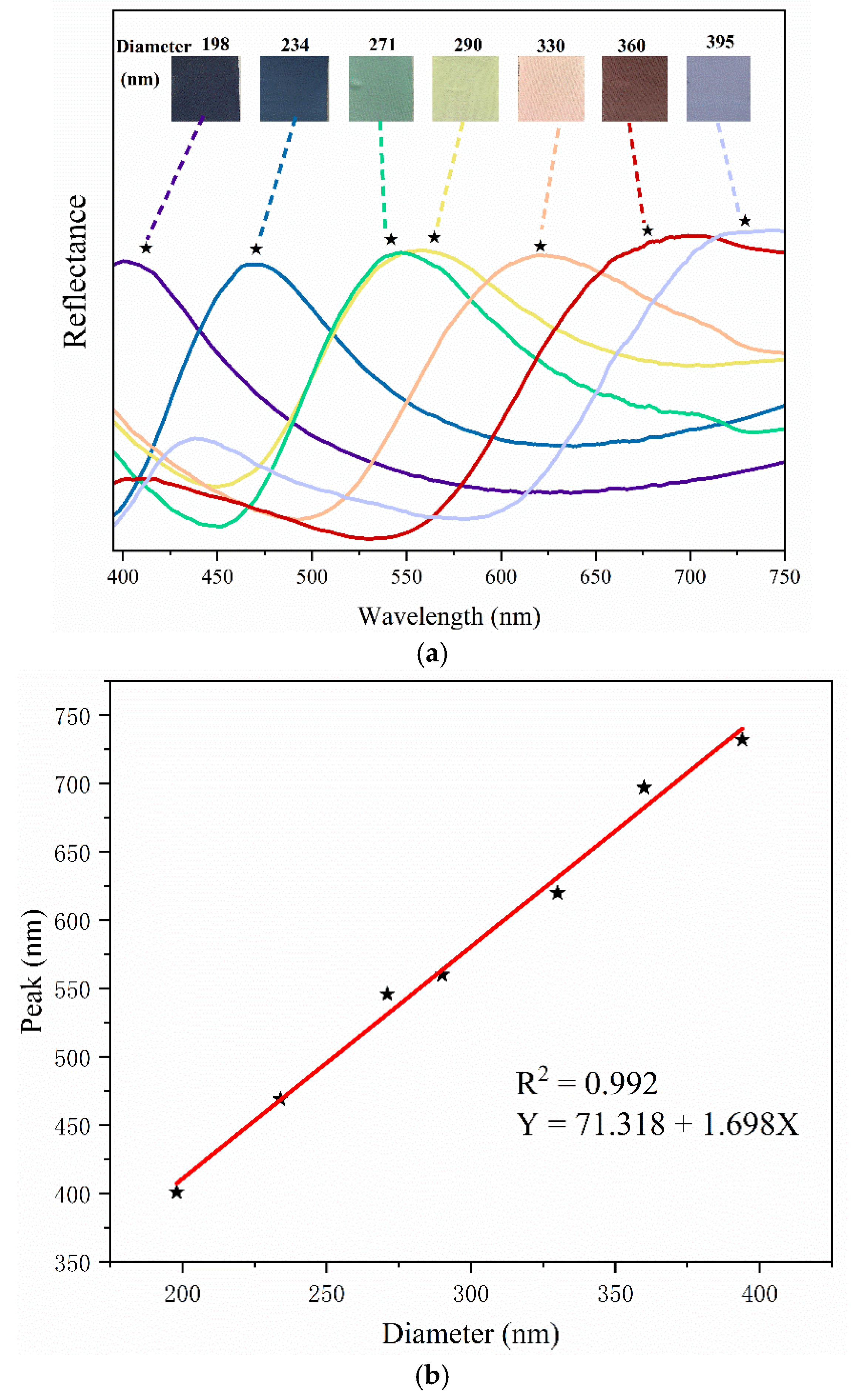
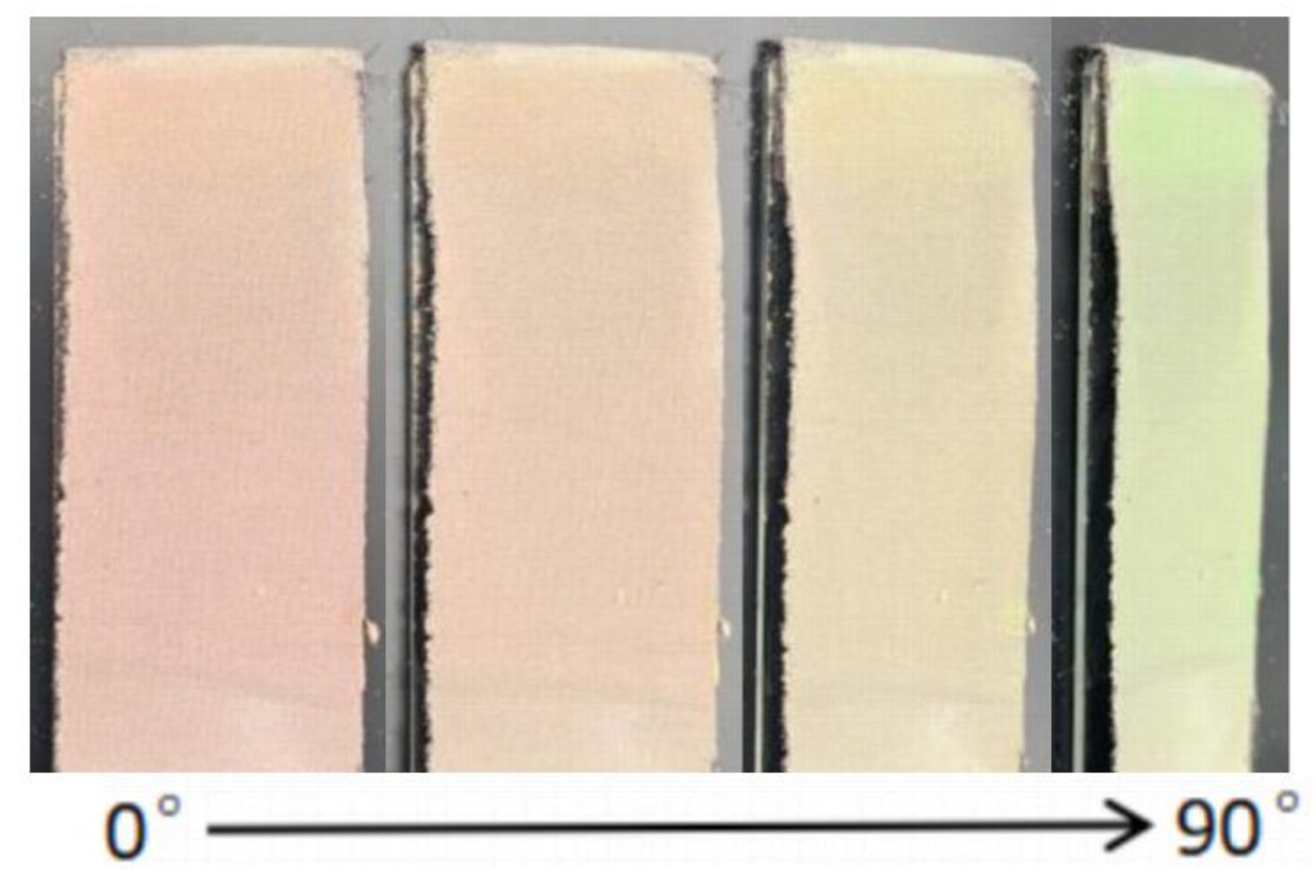
3.3. Optical Properties of Full-Color SiO2@PDA Photonic Crystals on Silk Fabrics

The fabric and the structured colored fabric were photographed by the material microscope, as shown in Figure 4. The SiO2@PDA photonic crystal covered the surface of the black silk, and could interfere/diffract with visible light to produce bright structural colors, which proved that the structural colors of SiO2@PDA photonic crystals were successfully prepared.
The photographs of structural colored fabrics are presented in Figure 5a. As can be seen from Figure 5a, the black silk fabric surface was covered with the SiO2@PDA photonic crystal and presented a bright structural color, which was caused by coherent diffraction on the surface of the periodically arranged SiO2@PDA photonic crystals, and caused by constructive interference, indicating the long-range order of the colloidal crystals directly visualized by SEM, as shown in Figure 3b. Figure 5a shows the various structural colors of purple-red, red, orange, yellow-green, green, blue, and purple as well, constructed by SiO2@PDA microspheres with different diameters of 394 nm, 360 nm, 330 nm, 290 nm, 271 nm, 234 nm, and 198 nm. Figure 5a also shows its corresponding reflection spectrum. It is worth noting that the particle size of SiO2@PDA microspheres increased from 198 nm to 360 nm, and the maximum reflection wavelength of the structural color was red-shifted from 401 nm to 732 nm, which can be explained by Bragg’s Law, and Snell’s refraction should be considered [36,37]:
λ max = 2 d hkl m ( n 2 avg - sin 2 θ ) 1 / 2
where λmax is the wavelength of the maximum reflection peak (i.e., the position of the photonic band gap), m is the order of Bragg diffraction, dhkl is the plane spacing between the diffraction planes, navg is the effective refractive index of the system (medium and colloidal particles), θ is the angle between the incident light and the sample surface normal (the same angle).
As can be seen from Equation (1), the wavelength max of the maximum reflection peak is significantly affected by dhkl, navg, and θ. In this experiment, if other parameters in Equation (1) remain unchanged and the particle size of the SiO2@PDA microsphere is changed, it can be found that the position of the reflection peak moves to a longer wavelength. It means that the position of the photon band gap shifted redshift with the increase in the particle size of the microsphere. On the contrary, blueshift occurred. The linear simulation relationship between the particle size of the SiO2@PDA microsphere and the wavelength of the reflection peak is shown in Figure 5b. The correlation coefficient was as high as 0.992, indicating that the particle size of the SiO2@PDA microsphere was positively correlated with the wavelength of the reflection peak.
In addition to changing particle size, the change in the viewing angle can also change the structural color of photonic crystals, which is called the “rainbow effect”. As shown in Figure 6, the 330 nm SiO2@PDA photonic crystal on silk exhibited different structural colors at different viewing angles from 0° to 90°. With the increase in the observation angle, the structure color showed a blueshift trend. According to Bragg’s law, the reflection peak wavelength λmax decreases with the increase of θ, which is consistent with the experiment.

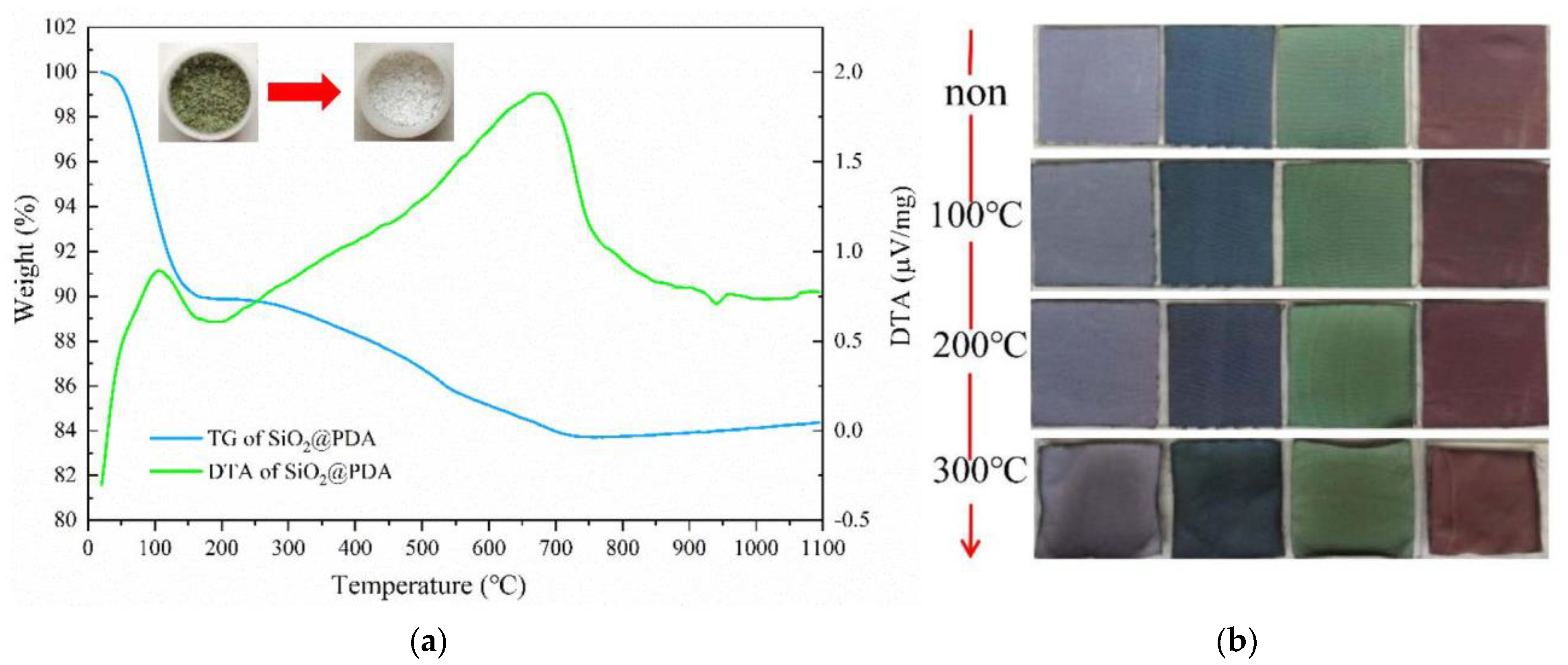
3.4. Thermal Resistance of the Structural Color of Photonic Crystals

Thermogravimetric analysis (TGA) of SiO2@PDA microspheres was performed in an N2 atmosphere. The thermogram (Figure 7a) indicated that the weight loss at 100 °C may be due to the loss of water. Additionally, the weight loss at 700 °C may be attributed to the decomposition of PDA. As illustrated in Figure 7a, the SiO2@PDA microsphere powder initially appeared green and turned white after heat treatment. However, the content of SiO2@PDA remained nearly 84% at 1100 °C, indicating that SiO2@PDA microspheres have high stability.
The thermal stability of the structural color of SiO2@PDA photonic crystal was studied by heat treatment in an air atmosphere. As shown in Figure 7b, the fabric color did not change significantly after being treated at 100 °C for 2 h. Then, the data were recorded every time the heat treatment temperature increased by 100 °C, and the structural color of the SiO2@PDA photonic crystal remained stable at 200 °C. After the heat treatment at 300 °C, the middle part of the structural color fabric was damaged to a certain extent, which may be due to the shrinkage of the silk, but the fabric still maintained most of its own color. Kosuke Nakamae et al. [20] reported that the iridescent structural color on hard substrate glasses was stable at 350 °C, and it can be found that photonic crystal structural colors also had good thermal stability on flexible fabrics, which laid the foundation for the preparation of non-fade materials in a high thermal stability environment.

4. Conclusions

In this study, vertical deposition self-assembly was applied to fabricate the photonic crystals of the fcc arrangement on silk without any chemical dyes or pigments. The 198 nm, 234 nm, 271 nm, 290 nm, 330 nm, 360 nm, and 394 nm SiO2@PDA microspheres were successfully prepared with good sphericity and mono-dispersity, and the full chromatographic structure of the colors was successfully prepared on silk. With the increase in SiO2@PDA microsphere size, the structural color was red-shifted, and the particle size of the microsphere had a highly positive correlation with the maximum wavelength of the reflection peak of the structural color. In addition, under different viewing angles, the blue shift of the structural color will occur with an increase in the viewing angle, which conforms to Bragg’s diffraction law. Last but not least, since the SiO2@PDA microsphere has thermal stability, the prepared structural color fabric remains highly saturated at temperatures up to 200 °C. This has a previously unreported high thermal stability on the structural colors of silk. This method is suitable for physical coloring on flexible fabrics and provides a new idea for clean dyeing in the textile industry.

Author Contributions

Conceptualization, H.L.; methodology, H.L.; validation, H.Y., formal analysis, H.L. and H.Y.; investigation, H.L.; resources, H.Y.; data curation, H.L.; writing—original draft preparation, H.L.; writing—review and editing, H.L., H.Y., M.Z. and T.C.; funding acquisition, H.Y. All authors have read and agreed to the published version of the manuscript.

Funding

This work was supported by the Chunhui Project of the Ministry of Education (No. 2018-19 and Z2017094) and the National General Cultivation Program (No. 17C050).

Institutional Review Board Statement

Not applicable.

Informed Consent Statement

Not applicable.

Data Availability Statement

Not applicable.

Conflicts of Interest

The authors declare no conflict of interest.

References

  1. Chen, G.; Yi, B.; Huang, Y.; Liang, Q.; Shen, H. Development of bright and low angle dependence structural colors from order-disorder hierarchical photonic structure. Dyes Pigment. 2019, 161, 464–469. [Google Scholar] [CrossRef]
  2. Li, Q.; Zhang, Y.; Shi, L.; Qiu, H.; Zhang, S.; Qi, N.; Hu, J.; Yuan, W.; Zhang, X.; Zhang, K.-Q. Additive mixing and conformal coating of noniridescent structural colors with robust mechanical properties fabricated by atomization deposition. ACS Nano 2018, 12, 3095–3102. [Google Scholar] [CrossRef]
  3. Bes-Pia, A.; Mendoza-Roca, J.; Alcaina-Miranda, M.; Iborra-Clar, A.; Iborra-Clar, M. Combination of physico-chemical treatment and nanofiltration to reuse wastewater of a printing, dyeing and finishing textile industry. Desalination 2003, 157, 73–80. [Google Scholar] [CrossRef]
  4. Wang, C.; Yediler, A.; Lienert, D.; Wang, Z.; Kettrup, A. Toxicity evaluation of reactive dyestuffs, auxiliaries and selected effluents in textile finishing industry to luminescent bacteria Vibrio fischeri. Chemosphere 2002, 46, 339–344. [Google Scholar] [CrossRef]
  5. Miyazaki, H.; Hase, M.; Miyazaki, H.T.; Kurokawa, Y.; Shinya, N. Photonic material for designing arbitrarily shaped waveguides in two dimensions. Phys. Rev. B 2003, 67, 235109. [Google Scholar] [CrossRef] [Green Version]
  6. Diao, Y.Y.; Liu, X.Y. Controlled colloidal assembly: Experimental modeling of general crystallization and biomimicking of structural color. Adv. Funct. Mater. 2012, 22, 1354–1375. [Google Scholar] [CrossRef]
  7. Ge, D.; Lee, E.; Yang, L.; Cho, Y.; Li, M.; Gianola, D.S.; Yang, S. A robust smart window: Reversibly switching from high transparency to angle-independent structural color display. Adv. Mater. 2015, 27, 2489–2495. [Google Scholar] [CrossRef]
  8. Chiappini, A.; Armellini, C.; Piccolo, V.; Zur, L.; Ristic, D.; Jovanovic, D.; Vaccari, A.; Zonta, D.; Righini, G.C.; Ferrari, M. Colloidal crystals based portable chromatic sensor for butanol isomers and water mixtures detection. Opt. Mater. 2019, 90, 152–158. [Google Scholar] [CrossRef]
  9. Couturier, J.P.; Sütterlin, M.; Laschewsky, A.; Hettrich, C.; Wischerhoff, E. Responsive inverse opal hydrogels for the sensing of macromolecules. Angew. Chem. Int. Ed. 2015, 54, 6641–6644. [Google Scholar] [CrossRef] [PubMed]
  10. Zhang, J.; Zhu, Z.; Yu, Z.; Ling, L.; Wang, C.-F.; Chen, S. Large-scale colloidal films with robust structural colors. Mater. Horiz. 2019, 6, 90–96. [Google Scholar] [CrossRef]
  11. Liu, P.; Chang, W.; Ju, L.; Chu, L.; Xie, Z.; Chen, J.; Yang, J. Bioinspired noniridescent structural color with hidden patterns for anticounterfeiting. ACS Appl. Nano Mater. 2019, 2, 5752–5760. [Google Scholar] [CrossRef]
  12. Meng, Y.; Qiu, J.; Wu, S.; Ju, B.; Zhang, S.; Tang, B. Biomimetic structural color films with a bilayer inverse heterostructure for anticounterfeiting applications. ACS Appl. Mater. Interfaces 2018, 10, 38459–38465. [Google Scholar] [CrossRef] [PubMed]
  13. Meng, Y.; Tang, B.; Ju, B.; Wu, S.; Zhang, S. Multiple colors output on voile through 3D colloidal crystals with robust mechanical properties. ACS Appl. Mater. Interfaces 2017, 9, 3024–3029. [Google Scholar] [CrossRef] [PubMed]
  14. Liu, G.; Guo, Y.; Zhou, L.; Wu, Y.; Chen, M.; He, Z. Tough, structurally colored fabrics produced by photopolymerization. J. Mater. Sci. 2019, 54, 10929–10940. [Google Scholar] [CrossRef]
  15. Li, S.; Jia, L.; Dong, P.; Shan, G.; Liu, R. Construction of photonic crystal structural colors on white polyester fabrics. Opt. Mater. 2021, 116, 111115. [Google Scholar] [CrossRef]
  16. Liu, P.; Xie, Z.; Zheng, F.; Zhao, Y.; Gu, Z. Surfactant-free HEMA crystal colloidal paint for structural color contact lens. J. Mater. Chem. B 2016, 4, 5222–5227. [Google Scholar] [CrossRef] [PubMed]
  17. Gu, H.; Ye, B.; Ding, H.; Liu, C.; Zhao, Y.; Gu, Z. Non-iridescent structural color pigments from liquid marbles. J. Mater. Chem. C 2015, 3, 6607–6612. [Google Scholar] [CrossRef]
  18. Wang, F.; Zhang, X.; Lin, Y.; Wang, L.; Zhu, J. Structural coloration pigments based on carbon modified ZnS@ SiO2 nanospheres with low-angle dependence, high color saturation, and enhanced stability. ACS Appl. Mater. Interfaces 2016, 8, 5009–5016. [Google Scholar] [CrossRef] [PubMed]
  19. Liu, P.; Chen, J.; Zhang, Z.; Xie, Z.; Du, X.; Gu, Z. Bio-inspired robust non-iridescent structural color with self-adhesive amorphous colloidal particle arrays. Nanoscale 2018, 10, 3673–3679. [Google Scholar] [CrossRef]
  20. Nakamae, K.; Hano, N.; Ihara, H.; Takafuji, M. Thermally stable high-contrast iridescent structural colours from silica colloidal crystals doped with monodisperse spherical black carbon particles. Mater. Adv. 2021, 2, 5935–5941. [Google Scholar] [CrossRef]
  21. Liu, Y. Self-assembly of poly (styrene-methyl methacrylate-acrylic acid)(P (St-MMA-AA)) colloidal microspheres on wood surface by thermal-assisted gravity deposition. Wood Sci. Technol. 2021, 55, 403–417. [Google Scholar] [CrossRef]
  22. Hsieh, C.-H.; Lu, Y.-C.; Yang, H. Self-assembled mechanochromic shape memory photonic crystals by doctor blade coating. ACS Appl. Mater. Interfaces 2020, 12, 36478–36484. [Google Scholar] [CrossRef] [PubMed]
  23. Liu, Y.; Hu, J. Investigation of polystyrene-based microspheres from different copolymers and their structural color coatings on wood surface. Coatings 2020, 11, 14. [Google Scholar] [CrossRef]
  24. Panfilova, E.; Shramko, D.; Ibragimov, A.; Mozer, K. Features of the deposition of photonic crystal films of polystyrene and silica. J. Phys. Conf. Ser. 2021, 1866, 012002. [Google Scholar] [CrossRef]
  25. Shen, H.; Liang, Q.; Song, L.; Chen, G.; Pei, Y.; Wu, L.; Zhang, X. Facile fabrication of mechanically stable non-iridescent structural color coatings. J. Mater. Sci. 2020, 55, 2353–2364. [Google Scholar] [CrossRef]
  26. Miguez, H.; Meseguer, F.; Lopez, C.; Mifsud, A.; Moya, J.; Vazquez, L. Evidence of FCC crystallization of SiO2 nanospheres. Langmuir 1997, 13, 6009–6011. [Google Scholar] [CrossRef]
  27. Iwata, M.; Teshima, M.; Seki, T.; Yoshioka, S.; Takeoka, Y. Bio-Inspired Bright Structurally Colored Colloidal Amorphous Array Enhanced by Controlling Thickness and Black Background. Adv. Mater. 2017, 29, 1605050. [Google Scholar] [CrossRef]
  28. Hu, Y.; Yang, D.; Huang, S. Amorphous photonic structures with brilliant and noniridescent colors via polymer-assisted colloidal assembly. ACS Omega 2019, 4, 18771–18779. [Google Scholar] [CrossRef] [Green Version]
  29. Takeoka, Y.; Yoshioka, S.; Takano, A.; Arai, S.; Nueangnoraj, K.; Nishihara, H.; Teshima, M.; Ohtsuka, Y.; Seki, T. Production of colored pigments with amorphous arrays of black and white colloidal particles. Angew. Chem. 2013, 125, 7402–7406. [Google Scholar] [CrossRef]
  30. Zhang, Y.; Dong, B.; Chen, A.; Liu, X.; Shi, L.; Zi, J. Using cuttlefish ink as an additive to produce non-iridescent structural colors of high color visibility. Adv. Mater. 2015, 27, 4719–4724. [Google Scholar] [CrossRef]
  31. Lee, C.H.; Yu, J.; Wang, Y.; Tang, A.Y.L.; Kan, C.W.; Xin, J.H. Effect of graphene oxide inclusion on the optical reflection of a silica photonic crystal film. RSC Adv. 2018, 8, 16593–16602. [Google Scholar] [CrossRef] [PubMed] [Green Version]
  32. Zhang, Y.; Han, P.; Zhou, H.; Wu, N.; Wei, Y.; Yao, X.; Zhou, J.; Song, Y. Highly brilliant noniridescent structural colors enabled by graphene nanosheets containing graphene quantum dots. Adv. Funct. Mater. 2018, 28, 1802585. [Google Scholar] [CrossRef]
  33. Song, L.; Chen, X.; Xie, Y.; Zhong, L.; Zhang, X.; Cheng, Z. Non-iridescent, crack-free, conductive structural colors enhanced by flexible nanosheets of reduced graphene oxide. Dyes Pigment. 2019, 164, 222–226. [Google Scholar] [CrossRef]
  34. Iwasaki, T.; Tamai, Y.; Yamamoto, M.; Taniguchi, T.; Kishikawa, K.; Kohri, M. Melanin precursor influence on structural colors from artificial melanin particles: PolyDOPA, polydopamine, and polynorepinephrine. Langmuir 2018, 34, 11814–11821. [Google Scholar] [CrossRef] [PubMed]
  35. Zhang, C.; Wu, B.-H.; Du, Y.; Ma, M.-Q.; Xu, Z.-K. Mussel-inspired polydopamine coatings for large-scale and angle-independent structural colors. J. Mater. Chem. C 2017, 5, 3898–3902. [Google Scholar] [CrossRef]
  36. Romanov, S.; Maka, T.; Torres, C.S.; Müller, M.; Zentel, R.; Cassagne, D.; Manzanares-Martinez, J.; Jouanin, C. Diffraction of light from thin-film polymethylmethacrylate opaline photonic crystals. Phys. Rev. E 2001, 63, 056603. [Google Scholar] [CrossRef] [PubMed]
  37. Shao, J.; Zhang, Y.; Fu, G.; Zhou, L.; Fan, Q. Preparation of monodispersed polystyrene microspheres and self-assembly of photonic crystals for structural colors on polyester fabrics. J. Textile Inst. 2014, 105, 938–943. [Google Scholar] [CrossRef]
Figure 1. (a) SEM images of SiO2@PDA microspheres with diameters of 330 nm. (b) Picture of SiO2 microsphere. (ci) SiO2@PDA microsphere powders with different diameters are as follows: 198nm, 234 nm, 271 nm, 290 nm, 330 nm, 360 nm, and 394 nm.
Coatings 12 01085 g001
Figure 2. (a) XRD images of SiO2 and SiO2@PDA microspheres. (b) FT-IR spectra of SiO2 and SiO2@PDA microspheres.
Coatings 12 01085 g002
Figure 3. (a) SEM image of SiO2@PDA (271 nm) photonic crystal on silk (b) is a partial enlargement of the red box in (a).
Coatings 12 01085 g003
Figure 4. (ah) Different structural colors on black silk taken by metallographic microscope. The diameters of SiO2@PDA microspheres are 198 nm, 234 nm, 271 nm, 290 nm, 330 nm, 360 nm, and 394 nm, respectively.
Coatings 12 01085 g004
Figure 5. (a) Structural color and reflection spectrum of SiO2@PDA photonic crystal on silk. (b) Fitting of particle size and peak wavelength.
Coatings 12 01085 g005
Figure 6. Photographs of various structural colors on silk fabricated by the colloidal microspheres with the same diameter of 330 nm at different viewing angles from 0° to 90°.
Coatings 12 01085 g006
Figure 7. (a) TG-DTA measurements of the SiO2@PDA powder in an N2 atmosphere. (b)Thermal stability of SiO2@PDA photonic crystals with diameters of 198, 234, 271, and 360 nm.
Coatings 12 01085 g007
Table 1. Sample diameter size and PDI values.
No.SiO2@PDA(Dh 1)/nmSiO2@PDA(Da 1)/nmPDI
11981960.036
22342340.031
32712670.037
42902840.038
53303280.024
63603560.023
73943860.029
1 Note: Dh is the hydration size of SiO2@PDA microspheres; Da is the actual particle size measured by SEM image.
Publisher’s Note: MDPI stays neutral with regard to jurisdictional claims in published maps and institutional affiliations.

