Review

Diamond-like Carbon Coatings in the Biomedical Field: Properties, Applications and Future Development

1
School of Medicine, South China University of Technology, Guangzhou 510006, China
2
School of Material Science and Engineering, South China University of Technology, Guangzhou 510640, China
3
The First Clinical School, Guangzhou Medical University, Guangzhou 510120, China
*
Authors to whom correspondence should be addressed.
Coatings 2022, 12(8), 1088; https://doi.org/10.3390/coatings12081088
Submission received: 15 June 2022 / Revised: 8 July 2022 / Accepted: 29 July 2022 / Published: 1 August 2022
(This article belongs to the Section Surface Coatings for Biomedicine and Bioengineering)

Abstract

:
Repairment and replacement of organs and tissues are part of the history of struggle against human diseases, in addition to the research and development (R&D) of drugs. Acquisition and processing of specific substances and physiological signals are very important to understand the effects of pathology and treatment. These depend on the available biomedical materials. The family of diamond-like carbon coatings (DLCs) has been extensively applied in many industrial fields. DLCs have also been demonstrated to be biocompatible, both in vivo and in vitro. In many cases, the performance of biomedical devices can be effectively enhanced by coating them with DLCs, such as vascular stents, prosthetic heart valves and surgical instruments. However, the feasibility of the application of DLC in biomedicine remains under discussion. This review introduces the current state of research and application of DLCs in biomedical devices, their potential application in biosensors and urgent problems to be solved. It will be useful to build a bridge between DLC R&D workers and biomedical workers in order to develop high-performance DLC films/coatings, promote their practical use and develop their potential applications in the biomedical field.

1. Introduction

Biomedical materials include metals, alloys, polymers and ceramics, which each have their own merits, but also certain shortcomings. Metallic materials are suitable as substitutes for bone and joint tissues due to high hardness and stiffness, but their compatibility and corrosion resistance have to be considered. Only a few pure metals can be used as biomedical materials alone, including magnesium, titanium [1], and platinum; alloys are more commonly used, which are made by melting, mixing, cooling, and solidifying, because they are harder and have better corrosion resistance [2,3]. For example, titanium alloy is a commonly used alloy material in orthopedics [4]. However, untreated aluminum and aluminum alloys are not suitable for medical use due to their potential to induce irreversible neurotoxicity in humans [5]. Stainless steel is widely used in the biomedicine field, but it is necessary to improve its biocompatibility [6]. An ideal biomedical material should combine corrosion resistance, compressive strength, and biocompatibility. Unfortunately, there is no ideal choice [1]. Surface modification, especially coating, can effectively enhance the biocompatibility, hardness, and corrosion resistance of materials, and maintain the high strength of the substrates. Thus, coating technology has great potential in the field of biomedicine. However, other issues related to coatings have to be considered; for instance, whether they can adhere firmly to the substrate and maintain their function in the harsh biomedical environment [7,8].
Diamond-like carbon coatings (DLCs) are a family of amorphous carbons, which can be made cheaply even at room temperature [9]. The DLC-coating fee is affordable for usual implanted devices, but it is probably expensive for the disposable surgical tools. DLCs possess some merits similar to diamond, such as high hardness, high wear resistance, low coefficient of friction, high insulation, high chemical stability and adjustable optoelectronic performance [9,10]. DLCs have attracted increasing interest in various industries, such as photoelectric devices and advanced manufacturing tools [11]. DLCs have also received a lot of attention in the biomedical field [12,13], due to their excellent biocompatibility, blood compatibility, nontoxicity and noncarcinogenicity, which are important for the application of DLCs in the biomedical fields [14]. DLC coatings have been used in vascular stents, prosthetic heart valves and joint prostheses [12,13]. When coated with DLCs, aluminum and its alloys are suitable for biomedical usage. The other properties of DLCs, such as the high piezoresistivity, have also led them to be considered for biomedical pressure sensors [15]. However, there are issues regarding the effectiveness of DLCs in biomedical applications, particularly related to the adhesion strength between DLCs and substrates in the harsh environment of the human body [16,17].
This review focuses on introducing the pathology of some diseases related to im- planted devices and some help from DLC coating technology. It first briefly describes DLCs and the general requirements of biomedical usage. Then, the status of applications and the potential value of DLCs in the biomedical field are summarized, and proposals to address the current problems are suggested. This review aims to let clinicians better understand DLCs, and enhance their confidence in the use of DLC-coated biomedical devices. We also aim to provide material R&D workers information on the problems of DLCs in biomedical field, and indicate directions for the development of suitable and reliable DLCs for biomedical applications.

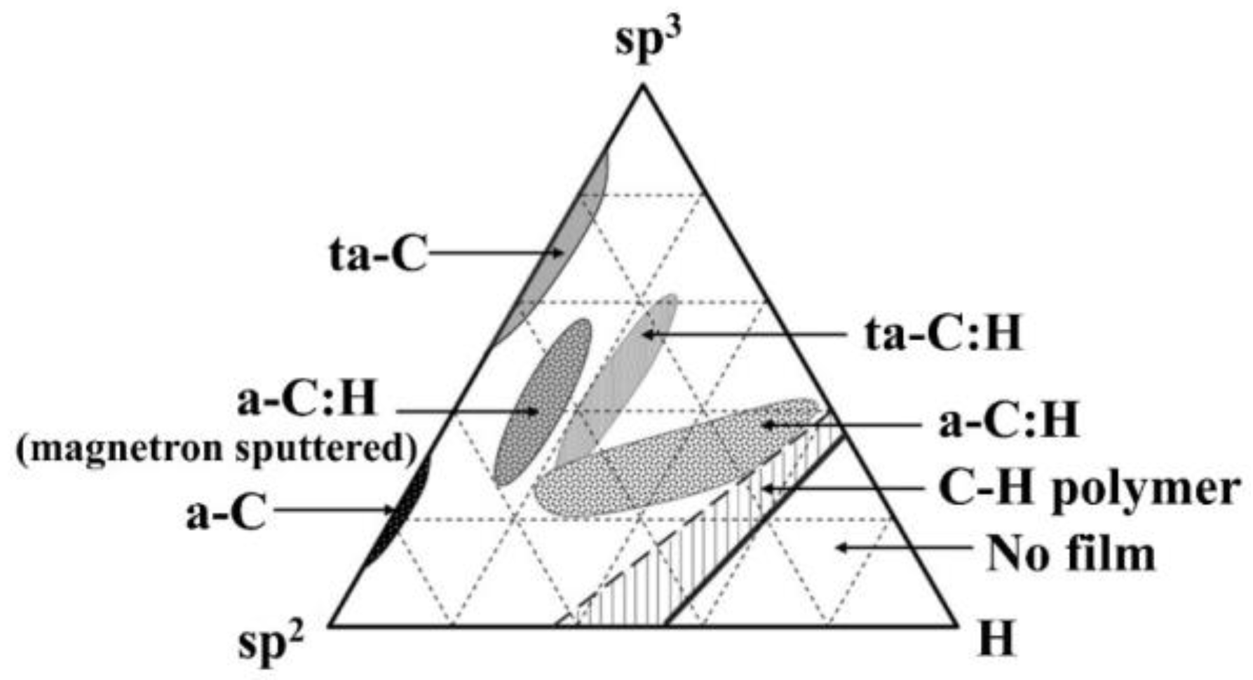
2. DLC Coatings and Their Characteristics

Hybridizations of carbon atoms include sp1, sp2 and sp3 [18], which create the versatility of various carbon materials and new carbon allotropes, including fullerenes, carbon nanotubes, graphene, diamond, DLC films, etc. Diamond has 100% sp3 carbon bond hybridization, in which a carbon atom’s four valence electrons are assigned tetrahedrally to make a strong σ bond to the adjacent atom. The perfect symmetry of the structure sp3 bond hybridization gives diamond extremely high hardness, excellent thermal conductivity, high electrical resistivity, chemical inertness, optical transparency, a wide bandgap and low wear rate in various tribological systems. Graphite has strong anisotropic properties, because in the three-fold coordinated sp2 hybridization, three valence electrons form σ bonds with three atoms in a plane, while the fourth electron in the normal orbit forms a π bond with the neighboring atom. In the perpendicular direction, there is only a weak van der Waals force acting. The sp2 hybridization results in low hardness, low electrical resistivity, high wear rate and low friction. Amorphous carbon films are thermodynamically metastable, in which carbon atoms mainly exist in the modes of sp2 and sp3 to form carbon–carbon bonds by σ bonds and π bonds. Generally, an amorphous carbon film can be classified according to its hydrogen content and sp3C fraction, i.e., graphite-like carbon (GLC) without H and sp3C less than 30%, polymer-like carbon (PLC) with H content over 40% and sp3C-C fraction less than 30%, and further DLCs beyond these ranges [6]. DLCs can also be divided into four categories according to the tertiary diagram (Figure 1) [19]: a-C, a-C:H, ta-C and ta-C:H.
The current mainstream technologies to prepare DLCs are plasma-enhanced physical vapor deposition (PVD) and chemical vapor deposition (CVD). Carbon targets are directly used to produce carbon vapor in PVD, while various hydrocarbon precursors are decomposed through chemical reactions to obtain carbon-rich sources in CVD [19,20]. Many deposition systems have been developed, for example, ion beam deposition, sputtering deposition, cathodic arc deposition, pulsed laser deposition, radio-frequency plasma-enhanced CVD and microwave plasma-enhanced CVD. During DLC deposition, the ion energy impinging the growing surface plays an important role, and currently the model of local thermal spikes induced by ion subplantation to form stabilized sp3C-bonds is well-accepted [18,19]. As presented in Figure 2 [20], during plasma-enhanced vacuum deposition, the mixture of the working gas, the incident energy of the particles, the energy flux, the substrate temperature, and the effective free path of the particles are the main processing parameters for regulating the composition, hybrid structure and performance of the DLCs. Generally, the mechanical properties of DLCs are determined by the sp3C fraction, and its optoelectronic properties mainly arise from the sp2C fraction [9]. DLCs can meet the needs of various industries through the tuning of their composition and hybrid structure.
The main methods to characterize the sp2C and sp3C hybrid structures of DLCs are Raman spectroscopy, X-ray photoelectron spectroscopy (XPS), and infrared spectrometry (IR) [9,10]. Raman spectroscopy is an inelastic light-scattering process that allows for the identification and characterization of the structure of molecules from gas to solid phase, from amorphous to crystals. The typical Raman spectra of micrograin graphite is presented in Figure 3A. The in-plane vibrational modes of various sp2C form a G peak at approximately 1560 cm−1, and the bending vibrations and disorder of aromatic rings and sp2C form a D peak near 1360 cm−1 in the Raman spectra. The position and the full wavelength of G and D peaks are related to the sp2C fraction, carbon clustering and disordering. The lower the intensity ratio of the D peak and the G peak (ID/IG), the greater the sp3 fraction in a DLC film. XPS is one kind of electron spectroscopy to measure the shifts of core levels. Because each element has its own core level (binding energy), the shifts reflect the chemical bonding of each site. The largest shifts are caused usually by the Colombic potential from ionic charges on the adjacent atoms. A typical XPS of the C 1s core of a-C:H coating is shown in Figure 3B; the raw data can be deconvoluted to many peaks to calculate the sp2C/sp3C ratio from the area. Infrared spectroscopy (IR) is a widely used method to characterize the bonding in a-C:H coatings. The IR absorption includes C-H stretching vibration at 2800–3300 cm−1. Similarly, by deconvoluting the raw data, various -CHn bonds and their fraction can be evaluated. In addition, electron energy loss spectroscopy (EELS), nuclear magnetic resonance (NMR), and near-edge X-ray absorption fine structure (NEXAFS) are also usual methods to analyze bonding in DLCs [21,22]. The hydrogen content in the DLC film is mainly measured by Rutherford backscattering spectroscopy (RBS) and elastic recoil detection analysis (EDRA) [23].
Some studies have shown that the density, hardness and elastic modulus of DLCs are inversely dependent on the hydrogen content [25,26,27]. DLC coatings have good chemical inertness, impermeability, biocompatibility, scratch resistance, abrasion resistance and blocking resistance [28]. The friction and wear resistance of DLCs is closely related to the sp2C/sp3C ratio, as well as the environment. DLC coatings have a wide tunable bandgap and are transparent to infrared light, which make them the ideal protective film for optical devices and field-emission electrodes at low voltages. DLCs can be adjusted within 6–8 orders of magnitude of resistivity, which means they can be used not only as insulating high-resistance material, but also as good conductor material, especially electrochemical electrode material in a special corrosive environment [29]. DLCs are also a piezoelectric/piezoresistive material with a huge gage factor [9]. However, the shortcomings of DLCs have to be considered in practical usage: (1) Intrinsic brittleness and large internal stress not only reduce the adhesion strength with the substrates, but also limit the thickness of films [30]; and (2) spontaneous graphitization at high temperatures [31] seriously degrade their performance during service.

3. Application Requirements of Biomedical Materials

Biomedical materials are often classified based on the type of raw material. Sometimes they are classified in terms of the usage of the material (Table 1), or the tissue translation of the material (Table 2). Biomedical materials often need to be in frequent contact with biological tissues with various constituent components. When the material is implanted into the human body, an inflammatory reaction occurs locally. Macrophages recruited due to the inflammatory response may create an acidic environment that corrodes the implant material. Therefore, biomedical materials need to have high corrosion resistance. An appropriate wear resistance of the biomedical material has to be considered because of the potential movement between implanted devices and their neighbor tissues/organs, and even other devices, as in prosthetic joints. In addition to the biocompatibility requirement, the most important property for all biomedical materials is that they must not be toxic or carcinogenic to the body. In general, the most important characteristics for biomedical materials are biocompatibility, blood compatibility, nontoxicity, noncarcinogenicity, abrasion resistance and corrosion resistance. Various artificial tissues made of ceramics, alloys, polymers, composite materials and nanomaterials have achieved clinical application [3,32]. Unfortunately, there is no ideal material to completely fulfil the above requirements. Therefore, the application of surface modification technologies, especially coatings, have significant potential [33,34]. Additionally, DLCs can be prepared without metallic species to avoid potential side effects from metals.
DLC coatings can potentially be used in implantable medical devices to relieve pain. Implantable medical devices can be divided into active and passive types, according to whether energy is needed or not. Active devices include artificial bone, joint prostheses, vascular stents, etc. Passive devices include pacemakers and cochlear implants. The materials used for vascular stents and prosthetic heart valves need to avoid thrombosis as much as possible, which is mainly responsible for the local inflammatory response in clinical practice. Because vascular stents and prosthetic heart valves are inserted into the blood system, the lower the adherence of platelets and macrophages to the scaffold material, the lower the risk of thrombosis. Therefore, these biomedical materials must be nonadherent to platelets and macrophages.
The main purpose of artificial joints is to keep patients at a normal movement; friction and wear are unavoidable. The DLCs used require low coefficient of friction and high resistance, together with high corrosion resistance in the harsh body system during service. To avoid intraoperative infection from surgery, the DLC-coated materials used for surgical equipment should not only be lightweight and durable, but also require antibacterial capabilities. Biosensors and pressure sensors, which are used to characterize the physiological information of blood pressure (BP) and intracranial pressure (ICP), need to be as sensitive and accurate as possible, in order to help clinicians better grasp patients’ condition and formulate a suitable treatment plan. Thus, these DLCs require a high piezoresistive gage factor.

4. Application of DLCs in Biomedical Devices

4.1. Vascular Stent

Blood vessels become diseased due to narrowing of their diameter in some pathological conditions, such as atherosclerosis, Takayasu arteritis, vascular malformation, vascular wall fibromuscular dysplasia, and Budd–Chiari syndrome, resulting in an insufficient blood supply to the organs. Atherosclerosis is a chronic inflammatory disease [45] with a great risk of obesity, which leads to an increase in low-density lipoprotein (LDL). LDL is phagocytosed by macrophages, which causes bone marrow cell responses, including the release of proinflammatory factors and macrophage proliferation. These reactions together lead to inflammation in the blood vessel wall and narrowing of the blood vessel lumen [46,47]. Vascular stenosis can cause a series of problems, such as coronary heart disease and stroke [48], which can be solved by vascular stents.
An inserted and opened vascular stent can open the narrowed artery to allow blood to flow normally. In-stent restenosis (ISR) for inserting vascular stents is the biggest problem, and can be classified as acute (within 24 h), subacute (from 24 h to 30 days), late (30 days to 1 year) and very late (>1 year) in terms of occurrence [49]. There are two main causes of ISR. First, vascular endothelium damage at the stent expansion site results in the proliferation of vascular intimal fibers. Second, the implanted stent stimulates the local tissue, leading to local tissue hyperplasia [50,51]. There are currently two approaches for the prevention and treatment of ISR: systemic and local. Systemic regimens include systemic application of antiplatelet drugs, anticoagulants, antioxidants, calcium channel antagonists and angiotensin-converting enzyme inhibitors for life. Stent-localized radiation therapy, gene therapy and the use of covered stents and drug-coated stents are the main local approaches. At present, drug-coated stents are widely used. One of the main drugs is paclitaxel, which mainly takes effect by stabilizing microtubules and inhibiting mitosis. However, the side effect of paclitaxel is harm to the cardiovascular system [52,53], which increases the risk of a cardiac accident in patients.
DLC-coated stents may be a good solution for ISR. DLC-coated stents have good safety and biocompatibility in the human body [54]. Studies in animal models have shown that DLC-coated stents are better than bare stents to avoid the induction of intimal hyperplasia [55]. Controlled studies in humans have reached similar conclusions [56]. The reduction of ISR using DLC-coated stents may be related to many factors. K. Gutensohn found that DLC-coated metal stents depress the release of ions, reducing platelet activation and thus reducing the risk of thrombosis [57]. A smooth surface can promote platelet adhesion and thrombosis [58]. During DLC coating, the surface roughness of the stents can be effectively controlled, such as through etching by atomic hydrogen and high substrate temperatures [59]. Profiting from surface roughening and surface energy tuning, F-doped fluorinated DLC (F-DLC) can significantly reduce the adhesion of platelets and proteins [60], and improve the antithrombotic ability [61,62]. The inhibition of macrophage activity by DLCs can reduce the risk of ISR [63].
In addition to reducing the risk of ISR as much as possible, research and development of vascular stents must consider the following: (1) low cost; (2) high expandability ratio; (3) sufficient radial hoop strength and negligible recoil; (4) high flexibility; (5) adequate radiopacity/magnetic resonance imaging (MRI) compatibility; and (6) high drug-delivery capacity [64]. In order to improve the antithrombosis resistance of DLC-coated vascular stents, considerable work remains to be completed, including further enhancing the adhesion resistance of platelets, proteins and macrophages, improving the adhesion between the DLCs and the substrates, and increasing the performance stability of DLCs in the blood environment.

4.2. Prosthetic Heart Valve

Heart valves are located between the atrium and the ventricle, or between the ventricle and the artery, act to keep the blood flowing in the same direction and prevent backflow. Heart valve disease (HVD) refers to valve stenosis or insufficiency. Congenital dysplasia or various other diseases can result in an abnormal anatomical structure or function of heart valve and its accessory structures. Excessive production of collagen fibers, infiltration and oxidation of lipids, inflammation, and calcification can induce HVD [65]. HVD can be treated with prosthetic heart valve (PHV) replacement, which was first used in clinical treatment in 1960 [66]. Both biological tissue and mechanical PHVs have been developed. PHVs are different from vascular stents, which simply open blood vessels.
DLCs can only be used in mechanical PHVs. The drawback of mechanical PHVs is a high risk of thrombosis, requiring patients to take anticoagulant drugs for life. DLC-coated mechanical PHVs can physically reduce the risk of thrombosis, possibly reduce the risk of coagulation in patients, reduce the number of anticoagulant drugs used and improve the quality of life of patients.
DLC-coated PHVs are speculated to be suitable for humans to lower the risk of thrombosis [67] because the aggregation tendency of fibrin and platelets was reduced in vitro tests. DLCs can improve the blood compatibility and surface hardness of PHVs [68,69], and also control the surface roughness to reduce the risk of thrombosis. Similarly, F-DLC has received extensive attention due to its low coefficient of friction, controllable surface, and good chemical inertness. Chou et al. reported that F-DLC can reduce platelet adhesion and reduce the risk of thrombosis [70]. Figure 4 shows that Mo-DLC can keep erythrocyte stable with no adhesion and aggregation.
In summary, an ideal DLC-coated PHV should have the following properties: high thrombosis resistance, high biocompatibility, high damage resistance, high blood-flow reflux, high compatibility with cardiac structures, nontoxicity, noncarcinogenicity, no noise during work, low price, no damage to surrounding tissues, easy operation and easy militainment for a long time. At present, DLC-coated PHVs still have some shortcomings, and their antithrombotic property needs improvement. The adhesion between DLCs and the substrates is poor, which limits their practical application in PHVs.

4.3. Joint Prosthesis

The joint is a form of bone connection. Joint-related diseases include end-stage osteoarthritis, rheumatoid arthritis and severe osteonecrosis, among others. Serious damage to the joints induced by various diseases can cause severe pain and loss of mobility. Osteoarthritis is the most common joint disease globally [72,73]. For severely damaged joints and end-stage joint diseases, joint prosthesis replacement is a potential treatment. The replacement of badly worn cartilage can eliminate pain and restore the force lines and joints of the limbs. In fact, joint prosthesis replacement only needs to replace two articular surfaces of the joint, rather than the entire joint. The articular surface is the contact surface of the bones that make up the joint, which reduces friction during movement.
Joint prosthesis replacement often needs to be repeated because of complications, including artificial joint loosening, mechanical damage to the artificial joint, thrombosis, infection around the artificial joint, surgery posterior nerve injury and postoperative pain [74,75,76]. During service, the joint prosthesis suffers forces from multiple directions in the body, demanding high strength, antiwear capacity, corrosion and fatigue resistance [77]. DLCs can improve performance of the joint prosthesis and reduce complications. DLC-coated joint prostheses have shown high biocompatibility without inflammatory reactions or toxic effects both in vivo and in vitro [78]. DLCs increase the surface hardness, improving the wear resistance and friction corrosion resistance of joint prosthesis [79]. The wear rate and injury risk of DLC-coated hip prostheses is reduced [80]. Doped DLCs are expected to further improve the wear resistance of joint prostheses, for example, DLCs doped with tungsten and titanium [81,82]. Silver-doped DLCs have high lubricity and antibacterial ability, which increase the wear resistance and effectively prevent infection around the joint prosthesis [83]. Figure 5 shows that DLC-coated joint prostheses have better wear resistance than non-DLC coated prostheses.
Performance test results of the joint prosthesis must be considered. Taeger et al. reported that DLC-coated femoral heads had a higher failure rate than alumina-coated femoral heads [84]. TiN-coated joint prostheses showed enhanced wear resistance compared to DLC coatings [85,86]. The main reasons for this include the low adhesion and the high defect density (pinholes) of DLCs. Therefore, the DLCs used for joint prostheses should not only reduce the friction coefficient, increase the hardness, increase the biocompatibility, and prevent postoperative infection, but also improve adhesion and density.

4.4. Surgical Instruments

Surgical instruments include scalpels, surgical scissors, surgical forceps, vascular forceps and endoscopes. Advanced and efficient surgical instruments are beneficial for reducing pain and improving treatment efficiency. The surgical site should be as sterile as possible, preoperative disinfection and sterilization are essential. Some studies have shown that surgical instruments cannot be maintained perfectly sterile [87], therefore, it is necessary to reduce bacteria by some suitable materials [88]. It is conducive if the biomaterials of surgical instruments are antibacterial. It is well-known that there is a negative correlation between bacterial adhesion and surface energy [89,90], so DLCs have a potential application in surgical instruments due to their high biocompatibility, and especially the antimicrobial properties. The hydrogen content, sp2C fraction and dopants are effective means to modulate the surface energy [91,92]. Compared with bare biomaterials, the number of staphylococci on the surface of DLC-coated biomaterial is significantly reduced [91]. The bacterial adhesion of DLCs can be lowered greatly by H and dopant control [89,93]. It has been reported that Ag-doped DLCs are antibacterial, because Ag can breakdown DNA and results in rupture of the cell membranes and cell walls [94]. Compared with a DLC without Ag, the release of silver ions plays a role in reducing the number of bacteria that adhere to the surface of Ag-DLCs [95].
Biofilms are communities of microorganisms on a surface where the bacteria adhere, which play an important role in infections [96]. Extracellular matrix is an important constituent of biofilms, which can protect the resident cell from desiccation and chemical perturbation, such as antibiotic [97]. Thus, seeking a suitable method to inhibit the biofilm formation is helpful. The bacterial adhesion on the DLC surface can be reduced by improving its smoothness [98,99]. There are few studies about the inhibition of biofilm by DLC. The biofilm formation of Candida albicans [100], Escherichia coli and Pseudomonas aeruginosa [101] is very hard on the DLC surface. However, Watari et al. reported that DLC could not inhibit the formation of Staphylococcus aureus biofilms [101], while Myllymaa’s study showed the opposite result [102]. Such divergences are probably responsible for different bacterial culture time. Some metal-doped DLCs also perform well in inhibiting biofilm [103]. In future studies, it would be interesting to develop more DLCs against various biofilms.
Prions are special pathogens that only consist of proteins. It is intriguing that the prions misfold normal proteins, achieving self-replication of the prions [104,105]. The infection of prison is usually from surgery. Although surgical instruments have been sterilized carefully, the prions are difficult to kill during the conventional step due to their unique structure [106]. Avoiding prion adhesion is a useful way. DLC-coated surgical instruments prevent the attachment of microorganisms and proteins, reducing the risk of prion infection. Secker et al. reported DLC-coated surgical instruments had lower levels of prion attachment than the uncoated ones [107].
Minimally invasive endoscopic surgery has developed rapidly, using surgical incisions or natural channels for surgical operations. The endoscope consists of five basic systems: the laparoscopic video surveillance system, CO2 inflation system, electric cutting system, irrigation–suction system and surgical instruments. The high-frequency electric knife (HFEK) is a commonly used instrument, and its local surface temperature can reach 200–1000 °C during operation. Two problems of minimally invasive surgery are fog generation and tissue adhesion. Modulating the thickness (nanoscale) of Si-DLC and its surface hydrophobicity may give the lens an improved antifogging ability during surgery, and make the surgical field clearer [108,109,110,111]. In some local anesthesia procedures, DLCs can reduce the friction coefficient of the endoscope surface and reduce patient discomfort [112].
Although the application potential of DLCs in surgical instruments is encouraging, considering the upper temperature of HFEK operation, the local graphitization of DLCs is unavoidable, which can degrade its tissue-adhesion resistance. Thus, enhancing the thermal stability, electrical conductivity and thermal conductivity of DLCs used for HFEK is important; the former can improve the graphitization resistance at elevated temperatures, and the latter can reduce the actual local temperature during operation. The author’s team has prepared a DLC coating with a resistivity lower than 10−4 Ω·cm by doping N and controlling hydrogen, and its conductivity is much higher than that of stainless steel, and slightly higher than that of graphite [113]. Furthermore, increasing the sp3C fraction and the content of metal-carbide compound nanoparticles with a high melting point in DLCs is also likely to be important [114,115,116].
Above all, DLCs have been widely used in some biomedical fields, such as vascular stents and artificial joints. However, some DLC-coated devices do not present superiority over their similar products without DLCs at some time. The mechanisms behind this need deep study.

5. Application of DLCs in Information Sensing in the Biomedical Field

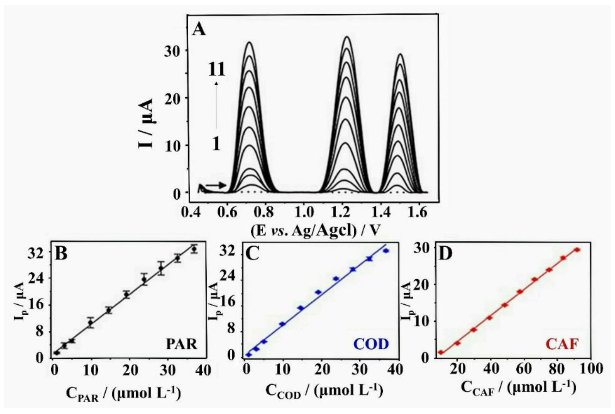
5.1. Biochemical Sensors

Biosensors are sensitive to various chemical and biological substances, which convert the concentration of species into electrical and optical signals for detection. Glucose is of great significance to the normal physiological activities of the human body, but a high level in the blood is a symptom of diabetes. Catechol, methylcatechol and dopamine are neurotransmitters that control the information transmission between neurons, or between neurons and effector cells. Abnormal levels of neurotransmitters can lead to many diseases. A low level of dopamine can induce Parkinson’s disease, while a high level results in mental disorders. Knowing the concentration of these species is useful to understand the progression of disease, to evaluate the efficacy of treatment and to predict the prognosis of disease. Enzymatic biosensors with high resolution are often used; however, their service life and repeatability are poor due to low stability and the bioactivity of the used enzymes. Recently, DLCs have attracted interest in the development of advanced biochemical sensors due to their chemical inertness, wide potential window, low background current, excellent mechanical performance, and long-term stability and repeatability compared to enzymatic biosensors [117,118].
The electronic and catalytic properties of DLC-coated electrodes can be tuned by controlling the fraction of the sp2C hybrid structure [119]. The electrochemical mechanism of DLC-biochemical sensors is to detect the current magnitude of the redox reaction of the certain species on the electrode surface [120]. The electrochemical reactions of catechol and methylcatechol in the same electrolyte are similar to on glassy carbon (GC) electrodes, while dopamine performs very differently because of its coupled chemical reaction [121]. Carbon nanotube (CNT) hybrid electrodes doped with DLCs have shown high sensitivity and stability in the detection of dopamine. Electrodes prepared by depositing DLCs on vertically aligned multiwalled carbon nanotubes (MWCNT) have proven suitable to detect dopamine and epinephrine [122,123]. These experiments showed that electrodes coated with DLCs or their derivatives can be potential biochemical sensors for neurotransmitters. In addition to the species secreted by the human body, DLC-electrodes can also detect exogenous substances such as drugs inside the human body. An overdose of drugs may cause irreversible damage to the patient. DLC-MWCNT electrodes can measure paracetamol, codeine and caffeine in biological fluids with high accuracy and stability [124]. Figure 6 compares the electrochemical performance of different electrolytes on different electrodes. In general, DLCs have great potential in biochemical sensors.

5.2. Pressure Sensor

In the biomedical field, pressure sensors are mainly used to measure blood pressure (BP) and intracranial pressure (ICP). High and low BP often indicate a poor prognosis. Hypertension is generally a symptom of shock, which may cause ischemia damage to organs and can lead a series of critical illnesses such as stroke [125], myocardial infarction, and kidney failure [126,127]. The principle of blood pressure measurement is as follows. The arterial blood flow is blocked, and no blood flow occurs when the external pressure is higher than the systolic blood pressure. Otherwise, the blood flow enters vessels and creates a vortex. When the pressure is further reduced below diastolic pressure, blood flow is fully restored. ICP is made up of three components: brain tissue, blood and cerebrospinal fluid (CSF), and Figure 7A shows their composition ratio and relationship with each other. An elevated ICP may be caused by traumatic brain injury, brain tumor and cerebral hemorrhage. Increased ICP may cause herniation of the brain, leading to respiratory failure and death. It is necessary to monitor ICP in real time and control its stability.
Both noninvasive and invasive BP measurements are currently used. The invasive method is the gold standard of BP measurement, and involves puncturing the blood vessel and placing the pressure sensor directly inside it. The most commonly used noninvasive methods are the oscillometric and auscultatory method [128,129], which have the same accuracy as the gold standard [130]. The accuracy of both BP measurement methods relies on pressure sensors. Similarly, there are two different ways to monitor ICP; invasive and noninvasive approaches. The invasive method is more accurate than the noninvasive method; the noninvasive method is used in patients with mild symptoms. An external ventricular drain (EVD) is one of the invasive methods, and remains the gold standard for measuring CVP. In this method, the pressure sensors are placed inside the ventricle of the brain. Figure 7B is an illustration of an EVD, where a pressure transducer is placed into the ventricle through an incision. Among the invasive methods, implantable microtransducers have lots of advantages, including lower infection rates and risks of hemorrhage [131]. Although it is believed that implantable microtransducers and EVD can be equally accurate [132,133], the experimental data from them still differ to some extent. Improving the accuracy of implantable microtransducers will be a priority in the near future.
Improving the accuracy of pressure measurement is certainly important and urgent for clinical work. The gage factor (GF) characterizes the magnitude of the piezoresistive effect of the materials, which relates to resistance change ΔR and mechanical strain ε.
G F = Δ R / R ε
Many studies have shown that DLCs have an obvious piezoresistive effect [15]. Although the GFs of DLCs differ due to the different production processes [134,135,136,137], they are obviously much larger than that of metal materials [138]. At present, most of the current pressure sensors for BP and ICP are made of metallic materials with a low GF [139,140]. DLCs have great potential for BP and ICP sensors with high accuracy and sensitivity. Although there are no trials of DLCs as a pressure sensor to measure BP/ICP, DLCs have great potential as a pressure sensor for biomedical applications. Today, the developing direction of BP measurement equipment is portable, real-time and continuous [139,141,142]. DLCs with high GF may play an important role in future BP and ICP equipment.
In general, some prospective application can be found when linking the excellent performance of electrochemical and piezoresistive performance of DLCs to the biomedical detection. There is a great expectation for DLCs used for biosensors in the future.

6. Future Outlooks

DLCs have excellent performance in pressure sensors and chemical electrodes, which may be widely used in BP measurement, ICP determination and biochemical sensors. In these reusable devices, the application of DLCs does not increase the cost significantly. However, in some disposable biomedical devices such as HFEK and scalpels, the application of DLCs increases the cost of using these devices, which is unaffordable for some patients. The reliability and feasibility of DLCs for biomedical usage remains under question. In order to promote the application of DLCs in biomedical field, biomedical workers should enhance their confidence in DLCs, as the shortcomings shown by clinical tests can be addressed by optimizing the deposition process, and trying more vitro and vivo tests. R&D workers of DLCs should focus their efforts on the following in the future: (1) improving the adhesion force between DLCs and biomaterial substrates; (2) reducing the defect density of DLCs and make them denser; and (3) developing advanced functions of DLCs based on the service conditions and investigating the mechanisms behind them; (4) decreasing the cost of DLCs. After solving the above problems, the application of DLCs in the biomedical field will be more widespread and feasible.
The application of DLCs in the biomedical field concerns the health, and even life, of the patients. It has to enhance the reliability and feasibility. Some so-called “failure tests” should receive more attention to study the mechanisms behind them. Therefore, R&D workers and clinicians should work together intimately. The clinicians should try more in vitro and in vivo tests of DLC tests, and feed the test results, especially shortcomings, as soon as possible to the R&D workers, who should make further deep investigation and optimization of processing technologies. In the future, R&D workers of DLCs should focus their efforts on the following: (1) to improve the adhesion force between DLCs and the biomaterial substrates; (2) to densify the DLCs and reduce the defect density; (3) to develop advanced functions of DLCs to meet the needs in service and investigate the mechanisms behind them; (4) to do best to decrease the cost of DLCs.

7. Conclusions

This review summarizes and comments on the application DLC-coated biomedical devices, and gives an outlook on the potential DLC-based biomedical sensors. DLCs have been used for experimental research and clinical applications in PHVs, vascular stents, joint prostheses, surgical instruments, etc. DLCs can be tuned to meet certain requirements, such as biocompatibility, wear resistance, corrosion resistance, inhibition of thrombosis and bacterial adsorption, which make it important in implantable biomedical devices. Their excellent electrochemical and piezoresistive properties present a great chance to be applied as biomedical sensors. However, sometimes DLC-coated devices perform unstably in vivo tests. Some deep investigation should be conducted to know the mechanisms and to optimize the performance of DLCs.

Author Contributions

Conceptualization, Y.P. and J.P.; methodology, Y.P.; validation, Y.P., Z.W. and X.Q.; formal analysis, Y.P. and Z.W.; data curation, Y.P., Y.X., Z.W. and X.Q.; writing—original draft preparation, Y.P.; writing—review and editing, J.P. and Y.P.; visualization, Y.P.; supervision, J.P.; project administration, Y.P. and J.P.; funding acquisition, J.P. All authors have read and agreed to the published version of the manuscript.

Funding

This research was funded by Science, Technology & Innovation Commission of Guangzhou Municipality, Grant Nos. 201902010018 and 201807010091.

Institutional Review Board Statement

Not applicable.

Informed Consent Statement

Not applicable.

Data Availability Statement

Not applicable.

Conflicts of Interest

The authors declare no conflict of interest.

List of Abbreviations in This Review

BPBlood pressure
CVDChemical vapor deposition
CSFCerebrospinal fluid
DLCDiamond-like carbon
ERDAElastic recoil detection analysis
EVDExternal ventricular drain
GFGage factor
GLCGraphite-like carbon
HFEKHigh frequency electric knife
HVDHeart valve disease
ICPIntracranial pressure
ISRIn-stent restenosis
LDLLow-density lipoprotein
MRIMagnetic resonance imaging
MWCNTMultiwalled carbon nanotubes
NEXAFSNear-edge X-ray absorption fine structure
NMRNuclear magnetic resonance
PHVProsthetic heart valve
PLCPolymer-like carbon
PVDPlasma-enhanced physical vapor deposition
RBSRutherford backscattering spectroscopy
R&DResearch and development
XPSX-ray photoelectron spectroscopy
XRDX-ray diffraction

References

  1. Anene, F.A.; Jaafar, C.N.A.; Zainol, I.; Hanim, M.A.A.; Suraya, M.T. Biomedical materials: A review of titanium based alloys. Proc. Inst. Mech. Eng. C-J Mech. 2021, 235, 3792–3805. [Google Scholar] [CrossRef]
  2. Wu, T.; Chen, X.; Fan, D.Z.; Pang, X.L. Development and application of metal materials in terms of vascular stents. Bio-Med. Mater. Eng. 2015, 25, 435–441. [Google Scholar] [CrossRef] [PubMed]
  3. Binyamin, G.; Shafi, B.M.; Mery, C.M. Biomaterials: A primer for surgeons. Semin. Pediatr. Surg. 2006, 15, 276–283. [Google Scholar] [CrossRef] [PubMed]
  4. Kaur, M.; Singh, K. Review on titanium and titanium based alloys as biomaterials for orthopaedic applications. Mater. Sci. Eng. C-Mater. 2019, 102, 844–862. [Google Scholar] [CrossRef]
  5. Fernandes, R.M.; Correa, M.G.; Aragao, W.A.B.; Nascimento, P.C.; Cartagenes, S.C.; Rodrigues, C.A.; Sarmiento, L.F.; Monteiro, M.C.; Maia, C.D.F.; Crespo-Lopez, M.E.; et al. Preclinical evidences of aluminum-induced neurotoxicity in hippocampus and pre-frontal cortex of rats exposed to low doses. Ecotoxicol. Environ. Saf. 2020, 206, 111139. [Google Scholar] [CrossRef] [PubMed]
  6. Bordjih, K.J.; Jouzeau, Y.J.; Mainard, D.; Payan, E.; Delagoutte, J.; Netter, P. Evaluation of the effect of three surface treatments on the biocompatibility of 316L stainless steel using human differentiated cells. Biomaterials 1996, 17, 491–500. [Google Scholar] [CrossRef]
  7. Park, S.J.; Lee, K.R.; Ahn, S.H.; Kim, J.G. Instability of diamond-like carbon (DLC) films during sliding in aqueous environment. Diam. Relat. Mater. 2008, 17, 247–251. [Google Scholar] [CrossRef]
  8. Lepicka, M.; Gradzka-Dahlke, M.; Pieniak, D.; Pasierbiewicz, K.; Niewczas, A. Effect of mechanical properties of substrate and coating on wear performance of TiN- or DLC-coated 316LVM stainless steel. Wear 2017, 382, 62–70. [Google Scholar] [CrossRef]
  9. Robertson, J. Diamond-like amorphous carbon. Mater. Sci. Eng. R 2002, 37, 129–281. [Google Scholar] [CrossRef] [Green Version]
  10. Ferrari, A.C.; Robertson, J. Interpretation of Raman spectra of disordered and amorphous carbon. Phys. Rev. B 2000, 61, 14095–14107. [Google Scholar] [CrossRef] [Green Version]
  11. McKenzie, D.R. Tetrahedral bonding in amorphous carbon. Rep. Prog. Phys. 1996, 59, 1611–1664. [Google Scholar] [CrossRef]
  12. Roy, R.K.; Lee, K.R. Biomedical applications of diamond-like carbon coatings: A review. J. Biomed. Mater. Res. B 2007, 83, 72–84. [Google Scholar] [CrossRef] [PubMed]
  13. Dearnaley, G.; Arps, J.H. Biomedical applications of diamond-like carbon (DLC) coatings: A review. Surf. Coat. Technol. 2005, 200, 2518–2524. [Google Scholar] [CrossRef]
  14. Peng, J.; Yang, M.; Bi, J.; Sheng, R.; Li, L. Hydrogen existence state of a hydrogenated amorphous carbon coating and its thermal stability. Diam. Relat. Mater. 2019, 99, 107535. [Google Scholar] [CrossRef]
  15. Petersen, M.; Bandorf, R.; Brauer, G.; Klages, C.P. Diamond-like carbon films as piezoresistors in highly sensitive force sensors. Diam. Relat. Mater. 2012, 26, 50–54. [Google Scholar] [CrossRef]
  16. Horiuchi, T.; Yoshida, K.; Kano, M.; Kumagai, M.; Suzuki, T. Evaluation of Adhesion and Wear Resistance of DLC Films Deposited by Various Methods. Plasma Process. Polym. 2009, 6, 410–416. [Google Scholar] [CrossRef]
  17. Drees, D.C.; Celis, J.P.; Dekempeneer, E.; Meneve, J. The electrochemical and wear behaviour of amorphous diamond-like carbon coatings and multilayered coatings in aqueous environments. Surf. Coat. Technol. 1996, 86–87, 575–580. [Google Scholar] [CrossRef]
  18. Zhang, W.; Zhu, S.Y.; Luque, R.; Han, S.; Hu, L.Z.; Xu, G.B. Recent development of carbon electrode materials and their bioanalytical and environmental applications. Chem. Soc. Rev. 2016, 45, 715–752. [Google Scholar] [CrossRef]
  19. Bewilogua, K.; Hofmann, D. History of diamond-like carbon films—From first experiments to worldwide applications. Surf. Coat. Technol. 2014, 242, 214–225. [Google Scholar] [CrossRef]
  20. Vetter, J. 60 years of DLC coatings: Historical highlights and technical review of cathodic arc processes to synthesize various DLC types, and their evolution for industrial applications. Surf. Coat. Technol. 2014, 257, 213–240. [Google Scholar] [CrossRef]
  21. Konyashin, I.Y. PVD/CVD technology for coating cemented carbides. Surf. Coat. Technol. 1995, 71, 277–283. [Google Scholar] [CrossRef]
  22. Sheng, H.X.; Xiong, W.W.; Zheng, S.S.; Chen, C.; He, S.; Cheng, Q.J. Evaluation of the sp(3)/sp(2) ratio of DLC films by RF-PECVD and its quantitative relationship with optical band gap. Carbon Lett. 2021, 31, 929–939. [Google Scholar] [CrossRef]
  23. Zhou, X.L.; Tunmee, S.; Suzuki, T.; Phothongkam, P.; Kanda, K.; Komatsu, K.; Kawahara, S.; Ito, H.; Saitoh, H. Quantitative NEXAFS and solid-state NMR studies of sp(3)/(sp(2) + sp(3)) ratio in the hydrogenated DLC films. Diam. Relat. Mater. 2017, 73, 232–240. [Google Scholar] [CrossRef]
  24. Merlen, A.; Buijnsters, J.G.; Pardanaud, C. A Guide to and Review of the Use of Multiwavelength Raman Spectroscopy for Characterizing Defective Aromatic Carbon Solids: From Graphene to Amorphous Carbons. Coatings 2017, 7, 153. [Google Scholar] [CrossRef]
  25. Ohtake, N.; Hiratsuka, M.; Kanda, K.; Akasaka, H.; Tsujioka, M.; Hirakuri, K.; Hirata, A.; Ohana, T.; Inaba, H.; Kano, M.; et al. Properties and Classification of Diamond-Like Carbon Films. Materials 2021, 14, 315. [Google Scholar] [CrossRef]
  26. Mabuchi, Y.; Higuchi, T.; Weihnacht, V. Effect of sp(2)/sp(3) bonding ratio and nitrogen content on friction properties of hydrogen-free DLC coatings. Tribol. Int. 2013, 62, 130–140. [Google Scholar] [CrossRef]
  27. Paul, R.; Das, S.N.; Dalui, S.; Gayen, R.N.; Roy, R.K.; Bhar, R.; Pal, A.K. Synthesis of DLC films with different sp(2)/sp(3) ratios and their hydrophobic behaviour. J. Phys. D Appl. Phys. 2008, 41, 5309. [Google Scholar] [CrossRef]
  28. Erdemir, A. The role of hydrogen in tribological properties of diamond-like carbon films. Surf. Coat. Technol. 2001, 146, 292–297. [Google Scholar] [CrossRef]
  29. Tyagi, A.; Walia, R.S.; Murtaza, Q.; Pandey, S.M.; Tyagi, P.K.; Bajaj, B. A critical review of diamond like carbon coating for wear resistance applications. Int. J. Refract. Met. Hard Mater. 2019, 78, 107–122. [Google Scholar] [CrossRef]
  30. Lu, Y.M.; Wang, S.; Huang, G.J.; Xi, L.; Qin, G.H.; Zhu, M.Z.; Chu, H. Fabrication and applications of the optical diamond-like carbon films: A review. J. Mater. Sci. 2022, 57, 3971–3992. [Google Scholar] [CrossRef]
  31. Liu, G.; Wen, Z.; Chen, K.; Dong, L.; Wang, Z.; Zhang, B.; Qiang, L. Optimizing the Microstructure, Mechanical, and Tribological Properties of Si-DLC Coatings on NBR Rubber for Its Potential Applications. Coatings 2020, 10, 671. [Google Scholar] [CrossRef]
  32. Tyan, Y.C.; Yang, M.H.; Chang, C.C.; Chung, T.W. Biocompatibility of Materials for Biomedical Engineering. Adv. Exp. Med. Biol. 2020, 1250, 125–140. [Google Scholar] [CrossRef]
  33. Du, C.; Su, X.W.; Cui, F.Z.; Zhu, X.D. Morphological behaviour of osteoblasts on diamond-like carbon coating and amorphous C-N film in organ culture. Biomaterials 1998, 19, 651–658. [Google Scholar] [CrossRef]
  34. Wickramasinghe, M.L.; Dias, G.J.; Premadasa, K.M.G.P. A novel classification of bone graft materials. J. Biomed. Mater. Res. B 2022, 110, 1724–1749. [Google Scholar] [CrossRef]
  35. Tallant, D.R.P.; Parmeter, J.E.; Siegal, M.P.; Simpson, R.L. The thermal stability of diamond-like carbon. Diam. Relat. Mater. 1995, 4, 191–199. [Google Scholar] [CrossRef]
  36. Serrano-Aroca, A.; Pous-Serrano, S. Prosthetic meshes for hernia repair: State of art, classification, biomaterials, antimicrobial approaches, and fabrication methods. J. Biomed. Mater. Res. A 2021, 109, 2695–2719. [Google Scholar] [CrossRef]
  37. Chethan, K.N.; Satish Shenoy, B.; Shyamasunder Bhat, N. Role of different orthopedic biomaterials on wear of hip joint prosthesis: A review. Mater. Today Proc. 2018, 5, 20827–20836. [Google Scholar] [CrossRef]
  38. Shores, J.T.; Hiersche, M.; Gabriel, A.; Gupta, S. Tendon coverage using an artificial skin substitute. J. Plast. Reconstruct. Aesthet. Surg. 2012, 65, 1544–1550. [Google Scholar] [CrossRef] [PubMed]
  39. Pan, C.; Han, Y.; Lu, J. Structural Design of Vascular Stents: A Review. Micromachines 2021, 12, 770. [Google Scholar] [CrossRef]
  40. Kokubo, K.; Kurihara, Y.; Kobayashi, K.; Tsukao, H.; Kobayashi, H. Evaluation of the Biocompatibility of Dialysis Membranes. Blood Purif. 2015, 40, 293–297. [Google Scholar] [CrossRef]
  41. Greenberg, J.A. The use of barbed sutures in obstetrics and gynecology. Rev. Obstet. Gynecol. 2010, 3, 82–91. [Google Scholar]
  42. Tiwari, G.; Tiwari, R.; Sriwastawa, B.; Bhati, L.; Pandey, S.; Pandey, P.; Bannerjee, S.K. Drug delivery systems: An updated review. Int. J. Pharm. Investig. 2012, 2, 2–11. [Google Scholar] [CrossRef] [Green Version]
  43. Buchanan, S.; Orris, P.; Karliner, J. Alternatives to the mercury sphygmomanometer. J. Public Health Policy 2011, 32, 107–120. [Google Scholar] [CrossRef]
  44. Klinkmann, H.; Vienken, J. Membranes for dialysis. Nephrol. Dial. Transpl. 1995, 10 (Supp. 3), 39–45. [Google Scholar] [CrossRef]
  45. Thomson, L.A.L.; Law, F.C.; Rushton, N.; Franks, J. Biocompatibility of diamond-like carbon coating. Biomaterials 1991, 12, 37–40. [Google Scholar] [CrossRef]
  46. Wolf, D.; Ley, K. Immunity and Inflammation in Atherosclerosis. Circ. Res. 2019, 124, 315–327. [Google Scholar] [CrossRef]
  47. Libby, P. The changing landscape of atherosclerosis. Nature 2021, 592, 524–533. [Google Scholar] [CrossRef]
  48. Zhu, Y.; Xian, X.; Wang, Z.; Bi, Y.; Chen, Q.; Han, X.; Tang, D.; Chen, R. Research Progress on the Relationship between Atherosclerosis and Inflammation. Biomolecules 2018, 8, 80. [Google Scholar] [CrossRef] [Green Version]
  49. Nicolais, C.; Lakhter, V.; Virk, H.U.H.; Sardar, P.; Bavishi, C.; O’Murchu, B.; Chatterjee, S. Therapeutic Options for In-Stent Restenosis. Curr. Cardiol. Rep. 2018, 20, 7. [Google Scholar] [CrossRef]
  50. Holmes, D.R., Jr. In-stent restenosis. Rev. Cardiovasc. Med. 2001, 2, 115–119. [Google Scholar]
  51. Inoue, T.; Croce, K.; Morooka, T.; Sakuma, M.; Node, K.; Simon, D.I. Vascular inflammation and repair: Implications for re-endothelialization, restenosis, and stent thrombosis. JACC Cardiovasc. Interv. 2011, 4, 1057–1066. [Google Scholar] [CrossRef] [Green Version]
  52. Micheletti, P.L.; Carla-da-Silva, J.; Scandolara, T.B.; Kern, R.; Alves, V.D.; Malanowski, J.; Victorino, V.J.; Herrera, A.; Rech, D.; Souza, J.A.O.; et al. Proinflammatory circulating markers: New players for evaluating asymptomatic acute cardiovascular toxicity in breast cancer treatment. J. Chemother. 2021, 33, 106–115. [Google Scholar] [CrossRef]
  53. Senkus, E.; Jassem, J. Cardiovascular effects of systemic cancer treatment. Cancer Treat. Rev. 2011, 37, 300–311. [Google Scholar] [CrossRef]
  54. Salahas, A.; Vrahatis, A.; Karabinos, I.; Antonellis, I.; Ifantis, G.; Gavaliatsis, I.; Anthopoulos, P.; Tavernarakis, A. Success, safety, and efficacy of implantation of diamond-like carbon-coated stents. Angiology 2007, 58, 203–210. [Google Scholar] [CrossRef]
  55. Kim, J.H.; Shin, J.H.; Shin, D.H.; Moon, M.W.; Park, K.; Kim, T.H.; Shin, K.M.; Won, Y.H.; Han, D.K.; Lee, K.R. Comparison of diamond-like carbon-coated nitinol stents with or without polyethylene glycol grafting and uncoated nitinol stents in a canine iliac artery model. Br. J. Radiol. 2011, 84, 210–215. [Google Scholar] [CrossRef] [Green Version]
  56. Ando, K.; Ishii, K.; Tada, E.; Kataoka, K.; Hirohata, A.; Goto, K.; Kobayashi, K.; Tsutsui, H.; Nakahama, M.; Nakashima, H.; et al. Prospective multi-center registry to evaluate efficacy and safety of the newly developed diamond-like carbon-coated cobalt-chromium coronary stent system. Cardiovasc. Interv. Ther. 2017, 32, 225–232. [Google Scholar] [CrossRef] [Green Version]
  57. Gutensohn, K.; Beythien, C.; Bau, J.; Fenner, T.; Grewe, P.; Koester, R.; Padmanaban, K.; Kuehnl, P. In vitro analyses of diamond-like carbon coated stents: Reduction of metal ion release, platelet activation, and thrombogenicity. Thromb. Res. 2000, 99, 577–585. [Google Scholar] [CrossRef]
  58. Karagkiozaki, V.C.; Logothetidis, S.D.; Kassavetis, S.N.; Giannoglou, G.D. Nanomedicine for the reduction of the thrombogenicity of stent coatings. Int. J. Nanomed. 2010, 5, 239–248. [Google Scholar] [CrossRef] [Green Version]
  59. Peng, X.L.; Barber, Z.H.; Clyne, T.W. Surface roughness of diamond-like carbon films prepared using various techniques. Surf. Coat. Technol. 2001, 138, 23–32. [Google Scholar] [CrossRef]
  60. Hasebe, T.; Yohena, S.; Kamijo, A.; Okazaki, Y.; Hotta, A.; Takahashi, K.; Suzuki, T. Fluorine doping into diamond-like carbon coatings inhibits protein adsorption and platelet activation. J. Biomed. Mater. Res. A 2007, 83, 1192–1199. [Google Scholar] [CrossRef]
  61. Maegawa, S.; Hasebe, T.; Yamato, Y.; Bito, K.; Nagashima, S.; Hayashi, T.; Mine, T.; Matsumoto, T.; Hotta, A.; Suzuki, T. Time course analysis of antithrombogenic properties of fluorinated diamond-like carbon coating determined via accelerated aging tests: Quality control for medical device commercialization. Diam. Relat. Mater. 2016, 70, 33–38. [Google Scholar] [CrossRef]
  62. Saito, T.; Hasebe, T.; Yohena, S.; Matsuoka, Y.; Kamijo, A.; Takahashi, K.; Suzuki, T. Antithrombogenicity of fluorinated diamond-like carbon films. Diam. Relat. Mater. 2005, 14, 1116–1119. [Google Scholar] [CrossRef]
  63. Thomas, V.; Halloran, B.A.; Ambalavanan, N.; Catledge, S.A.; Vohra, Y.K. In vitro studies on the effect of particle size on macrophage responses to nanodiamond wear debris. Acta Biomater. 2012, 8, 1939–1947. [Google Scholar] [CrossRef] [PubMed] [Green Version]
  64. Mani, G.; Feldman, M.D.; Patel, D.; Agrawal, C.M. Coronary stents: A materials perspective. Biomaterials 2007, 28, 1689–1710. [Google Scholar] [CrossRef] [PubMed]
  65. Lindman, B.R.; Clavel, M.A.; Mathieu, P.; Iung, B.; Lancellotti, P.; Otto, C.M.; Pibarot, P. Calcific aortic stenosis. Nat. Rev. Dis. Primers 2016, 2, 16006. [Google Scholar] [CrossRef] [PubMed] [Green Version]
  66. Turpie, A.G.; Gent, M.; Laupacis, A.; Latour, Y.; Gunstensen, J.; Basile, F.; Klimek, M.; Hirsh, J. A comparison of aspirin with placebo in patients treated with warfarin after heart-valve replacement. N. Engl. J. Med. 1993, 329, 524–529. [Google Scholar] [CrossRef]
  67. Antonowicz, M.; Kurpanik, R.; Walke, W.; Basiaga, M.; Sondor, J.; Paszenda, Z. Selected Physicochemical Properties of Diamond Like Carbon (DLC) Coating on Ti-13Nb-13Zr Alloy Used for Blood Contacting Implants. Materials 2020, 13, 5077. [Google Scholar] [CrossRef]
  68. Kwok, S.C.H.; Yang, P.; Wang, J.; Liu, X.Y.; Chu, P.K. Hemocompatibility of nitrogen-doped, hydrogen-free diamond-like carbon prepared by nitrogen plasma immersion ion implantation-deposition. J. Biomed. Mater. Res. A 2004, 70, 107–114. [Google Scholar] [CrossRef] [PubMed]
  69. Andara, M.; Agarwal, A.; Scholvin, D.; Gerhardt, R.A.; Doraiswamy, A.; Jin, C.M.; Narayan, R.J.; Shih, C.C.; Shih, C.M.; Lin, S.J.; et al. Hemocompatibility of diamondlike carbon-metal composite thin films. Diam. Relat. Mater. 2006, 15, 1941–1948. [Google Scholar] [CrossRef]
  70. Chou, C.C.; Wu, Y.Y.; Lee, J.W.; Yeh, C.H.; Huang, J.C. Characterization and haemocompatibility of fluorinated DLC and Si interlayer on Ti6Al4V. Surf. Coat. Technol. 2013, 231, 418–422. [Google Scholar] [CrossRef]
  71. Tang, X.S.; Wang, H.J.; Feng, L.; Shao, L.X.; Zou, C.W. Mo doped DLC nanocomposite coatings with improved mechanical and blood compatibility properties. Appl. Surf. Sci. 2014, 311, 758–762. [Google Scholar] [CrossRef]
  72. Mandl, L.A. Osteoarthritis year in review 2018: Clinical. Osteoarthr. Cartil. 2019, 27, 359–364. [Google Scholar] [CrossRef] [PubMed] [Green Version]
  73. Glyn-Jones, S.; Palmer, A.J.; Agricola, R.; Price, A.J.; Vincent, T.L.; Weinans, H.; Carr, A.J. Osteoarthritis. Lancet 2015, 386, 376–387. [Google Scholar] [CrossRef]
  74. Porrino, J.; Wang, A.N.; Moats, A.; Mulcahy, H.; Kani, K. Prosthetic joint infections: Diagnosis, management, and complications of the two-stage replacement arthroplasty. Skelet. Radiol. 2020, 49, 847–859. [Google Scholar] [CrossRef]
  75. Matz, J.; Lanting, B.A.; Howard, J.L. Understanding the patellofemoral joint in total knee arthroplasty. Can. J. Surg. 2019, 62, 57–65. [Google Scholar] [CrossRef] [PubMed] [Green Version]
  76. Ferguson, R.J.; Palmer, A.J.; Taylor, A.; Porter, M.L.; Malchau, H.; Glyn-Jones, S. Hip replacement. Lancet 2018, 392, 1662–1671. [Google Scholar] [CrossRef]
  77. Paul, J.P. Strength requirements for internal and external prostheses. J. Biomech. 1999, 32, 381–393. [Google Scholar] [CrossRef]
  78. Allen, M.; Myer, B.; Rushton, N. In vitro and in vivo investigations into the biocompatibility of diamond-like carbon (DLC) coatings for orthopedic applications. J. Biomed. Mater. Res. 2001, 58, 319–328. [Google Scholar] [CrossRef]
  79. Liu, J.; Wang, X.; Wu, B.J.; Zhang, T.F.; Leng, Y.X.; Huang, N. Tribocorrosion behavior of DLC-coated CoCrMo alloy in simulated biological environment. Vacuum 2013, 92, 39–43. [Google Scholar] [CrossRef]
  80. Wongpanya, P.; Pintitraratibodee, N.; Thumanu, K.; Euaruksakul, C. Improvement of corrosion resistance and biocompatibility of 316L stainless steel for joint replacement application by Ti-doped and Ti-interlayered DLC films. Surf. Coat. Technol. 2021, 425, 127734. [Google Scholar] [CrossRef]
  81. Platon, F.; Fournier, P.; Rouxel, S. Tribological behaviour of DLC coatings compared to different materials used in hip joint prostheses. Wear 2001, 250, 227–236. [Google Scholar] [CrossRef]
  82. Tan, D.; Dai, M.J.; Fu, W.B.; Lin, S.S.; Wei, C.B.; Zhao, M.C. Performance of CoCrMo Alloy with Me-Doped DLC Coatings Prepared by a Magnetron Sputtering Method. Rare Met. Mater. Eng. 2015, 44, 2982–2986. [Google Scholar] [CrossRef]
  83. Jing, P.P.; Su, Y.H.; Li, Y.X.; Liang, W.L.; Leng, Y.X. Mechanism of protein biofilm formation on Ag-DLC films prepared for application in joint implants. Surf. Coat. Technol. 2021, 422, 7553. [Google Scholar] [CrossRef]
  84. Taeger, G.; Podleska, L.E.; Schmidt, B.; Ziegler, M.; Nast-Kolb, D. Comparison of diamond-like-carbon and alumina-oxide articulating with polyethylene in total hip arthroplasty. Mater. Werkst 2003, 34, 1094–1100. [Google Scholar] [CrossRef]
  85. Chen, Y.; Nie, X.Y.; Leyland, A.; Housden, J.; Matthews, A. Substrate and bonding layer effects on performance of DLC and TiN biomedical coatings in Hank’s solution under cyclic impact-sliding loads. Surf. Coat. Technol. 2013, 237, 219–229. [Google Scholar] [CrossRef]
  86. Ortega-Saenz, J.A.; Alvarez-Vera, M.; Hernandez-Rodriguez, M.A.L. Biotribological study of multilayer coated metal-on-metal hip prostheses in a hip joint simulator. Wear 2013, 301, 234–242. [Google Scholar] [CrossRef]
  87. Gaines, S.; Luo, J.N.; Gilbert, J.; Zaborina, O.; Alverdy, J.C. Optimum Operating Room Environment for the Prevention of Surgical Site Infections. Surg. Infect. 2017, 18, 503–507. [Google Scholar] [CrossRef] [PubMed]
  88. Gutiérrez Rodelo, C.; Salinas, R.A.; Armenta Jaime, E.; Armenta, S.; Galdámez-Martínez, A.; Castillo-Blum, S.E.; Astudillo-de la Vega, H.; Nirmala Grace, A.; Aguilar-Salinas, C.A.; Gutiérrez Rodelo, J.; et al. Zinc associated nanomaterials and their intervention in emerging respiratory viruses: Journey to the field of biomedicine and biomaterials. Coord. Chem. Rev. 2022, 457, 214402. [Google Scholar] [CrossRef] [PubMed]
  89. Liu, Y.; Zhao, Q. Influence of surface energy of modified surfaces on bacterial adhesion. Biophys. Chem. 2005, 117, 39–45. [Google Scholar] [CrossRef] [PubMed]
  90. Liu, C.; Zhao, Q. The CQ ratio of surface energy components influences adhesion and removal of fouling bacteria. Biofouling 2011, 27, 275–285. [Google Scholar] [CrossRef]
  91. Del Prado, G.; Terriza, A.; Ortiz-Perez, A.; Molina-Manso, D.; Mahillo, I.; Yubero, F.; Puertolas, J.A.; Manrubia-Cobo, M.; Barrena, E.G.; Esteban, J. DLC coatings for UHMWPE: Relationship between bacterial adherence and surface properties. J. Biomed. Mater. Res. A 2012, 100, 2813–2820. [Google Scholar] [CrossRef]
  92. Wei, C.H.; Peng, K.S.; Hung, M.S. The effect of hydrogen and acetylene mixing ratios on the surface, mechanical and biocompatible properties of diamond-like carbon films. Diam. Relat. Mater. 2016, 63, 108–114. [Google Scholar] [CrossRef]
  93. Ren, D.W.; Zhao, Q.; Bendavid, A. Anti-bacterial property of Si and F doped diamond-like carbon coatings. Surf. Coat. Technol. 2013, 226, 1–6. [Google Scholar] [CrossRef]
  94. Tang, S.H.; Zheng, J. Antibacterial Activity of Silver Nanoparticles: Structural Effects. Adv. Healthc. Mater. 2018, 7, 1503. [Google Scholar] [CrossRef]
  95. Schwarz, F.P.; Hauser-Gerspach, I.; Waltimo, T.; Stritzker, B. Antibacterial properties of silver containing diamond like carbon coatings produced by ion induced polymer densification. Surf. Coat. Technol. 2011, 205, 4850–4854. [Google Scholar] [CrossRef]
  96. Rabin, N.; Zheng, Y.; Opoku-Temeng, C.; Du, Y.; Bonsu, E.; Sintim, H.O. Biofilm formation mechanisms and targets for developing antibiofilm agents. Future Med. Chem. 2015, 7, 493–512. [Google Scholar] [CrossRef]
  97. Yan, J.; Bassler, B.L. Surviving as a Community: Antibiotic Tolerance and Persistence in Bacterial Biofilms. Cell Host Microbe 2019, 26, 15–21. [Google Scholar] [CrossRef]
  98. Hsu Lillian, C.; Fang, J.; Borca-Tasciuc Diana, A.; Worobo Randy, W.; Moraru Carmen, I. Effect of Micro- and Nanoscale Topography on the Adhesion of Bacterial Cells to Solid Surfaces. Appl. Environ. Microbiol. 2013, 79, 2703–2712. [Google Scholar] [CrossRef] [Green Version]
  99. Jones, D.S.; Garvin, C.P.; Dowling, D.; Donnelly, K.; Gorman, S.P. Examination of surface properties and in vitro biological performance of amorphous diamond-like carbon-coated polyurethane. J. Biomed. Mater. Res. Part B Appl. Biomater. 2006, 78, 230–236. [Google Scholar] [CrossRef] [PubMed]
  100. Santos, T.B.; Vieira, A.A.; Paula, L.O.; Santos, E.D.; Radi, P.A.; Khouri, S.; Maciel, H.S.; Pessoa, R.S.; Vieira, L. Flexible camphor diamond-like carbon coating on polyurethane to prevent Candida albicans biofilm growth. J. Mech. Behav. Biomed. Mater. 2017, 68, 239–246. [Google Scholar] [CrossRef] [PubMed]
  101. Watari, S.; Wada, K.; Araki, M.; Sadahira, T.; Ousaka, D.; Oozawa, S.; Nakatani, T.; Imai, Y.; Kato, J.; Kariyama, R.; et al. Intraluminal diamond-like carbon coating with anti-adhesion and anti-biofilm effects for uropathogens: A novel technology applicable to urinary catheters. Int. J. Urol. 2021, 28, 1282–1289. [Google Scholar] [CrossRef] [PubMed]
  102. Myllymaa, K.; Levon, J.; Tiainen, V.-M.; Myllymaa, S.; Soininen, A.; Korhonen, H.; Kaivosoja, E.; Lappalainen, R.; Konttinen, Y.T. Formation and retention of staphylococcal biofilms on DLC and its hybrids compared to metals used as biomaterials. Colloids Surf. B Biointerfaces 2013, 101, 290–297. [Google Scholar] [CrossRef]
  103. Cazalini, E.M.; Miyakawa, W.; Teodoro, G.R.; Sobrinho, A.S.S.; Matieli, J.E.; Massi, M.; Koga-Ito, C.Y. Antimicrobial and anti-biofilm properties of polypropylene meshes coated with metal-containing DLC thin films. J. Mater. Sci. Mater. Med. 2017, 28, 97. [Google Scholar] [CrossRef] [Green Version]
  104. Weissmann, C. The state of the prion. Nat. Rev. Microbiol. 2004, 2, 861–871. [Google Scholar] [CrossRef] [PubMed]
  105. Bartz, J.C. Environmental and host factors that contribute to prion strain evolution. Acta Neuropathol. 2021, 142, 5–16. [Google Scholar] [CrossRef]
  106. Sakudo, A.; Ano, Y.; Onodera, T.; Nitta, K.; Shintani, H.; Ikuta, K.; Tanaka, Y. Fundamentals of prions and their inactivation (review). Int. J. Mol. Med. 2011, 27, 483–489. [Google Scholar] [CrossRef] [PubMed] [Green Version]
  107. Secker, T.J.; Herve, R.; Zhao, Q.; Borisenko, K.B.; Abel, E.W.; Keevil, C.W. Doped diamond-like carbon coatings for surgical instruments reduce protein and prion-amyloid biofouling and improve subsequent cleaning. Biofouling 2012, 28, 563–569. [Google Scholar] [CrossRef] [Green Version]
  108. Ou, K.L.; Weng, C.C.; Sugiatno, E.; Ruslin, M.; Lin, Y.H.; Cheng, H.Y. Effect of nanostructured thin film on minimally invasive surgery devices applications: Characterization, cell cytotoxicity evaluation and an animal study in rat. Surg. Endosc. 2016, 30, 3035–3049. [Google Scholar] [CrossRef]
  109. Su, C.H.; Lin, C.R.; Chang, C.Y.; Hung, H.C.; Lin, T.Y. Mechanical and optical properties of diamond-like carbon thin films deposited by low temperature process. Thin Solid Films 2006, 498, 220–223. [Google Scholar] [CrossRef]
  110. Meskinis, S.; Vasiliauskas, A.; Andrulevicius, M.; Peckus, D.; Tamulevicius, S.; Viskontas, K. Diamond Like Carbon Films Containing Si: Structure and Nonlinear Optical Properties. Materials 2020, 13, 1003. [Google Scholar] [CrossRef] [Green Version]
  111. Huang, L.; Wang, T.; Li, X.; Wang, X.; Zhang, W.; Yang, Y.; Tang, Y. UV-to-IR highly transparent ultrathin diamond nanofilms with intriguing performances: Anti-fogging, self-cleaning and self-lubricating. Appl. Surf. Sci. 2020, 527, 146733. [Google Scholar] [CrossRef]
  112. Sakurai, K.; Hiratsuka, M.; Nakamori, H.; Namiki, K.; Hirakuri, K. Evaluation of sliding properties and durability of DLC coating for medical devices. Diam. Relat. Mater. 2019, 96, 97–103. [Google Scholar] [CrossRef]
  113. Peng, J.; Xiao, Y.; Peng, Y.; Zeng, J. Effect of traceable nitrogen from low-pressure plasma nitriding on diamond growth over WC-co cemented carbides. Diam. Relat. Mater. 2021, 120, 108717. [Google Scholar] [CrossRef]
  114. Al Mahmud, K.A.H.; Kalam, M.A.; Masjuki, H.H.; Mobarak, H.M.; Zulkifli, N.W.M. An updated overview of diamond-like carbon coating in tribology. Crit. Rev. Solid State 2015, 40, 90–118. [Google Scholar] [CrossRef] [Green Version]
  115. Ji, G.W.; Wu, Y.Z.; Wang, X.; Pan, H.X.; Li, P.; Du, W.Y.; Qi, Z.; Huang, A.; Zhang, L.W.; Zhang, L.; et al. Experimental and clinical study of influence of high-frequency electric surgical knives on healing of abdominal incision. World J. Gastroenterol. 2006, 12, 4082–4085. [Google Scholar] [CrossRef] [PubMed]
  116. Zhang, T.F.; Wan, Z.X.; Ding, J.C.; Zhang, S.; Wang, Q.M.; Kim, K.H. Microstructure and high-temperature tribological properties of Si-doped hydrogenated diamond-like carbon films. Appl. Surf. Sci. 2018, 435, 963–973. [Google Scholar] [CrossRef]
  117. Zeng, A.P.; Neto, V.F.; Gracio, J.J.; Fan, Q.H. Diamond-like carbon (DLC) films as electrochemical electrodes. Diam. Relat. Mater. 2014, 43, 12–22. [Google Scholar] [CrossRef]
  118. Compton, R.G.; Foord, J.S.; Marken, F. Electroanalysis at diamond-like and doped-diamond electrodes. Electroanal 2003, 15, 1349–1363. [Google Scholar] [CrossRef]
  119. Grill, A. Electrical and optical properties of diamond-like carbon. Thin Solid Films 1999, 355, 189–193. [Google Scholar] [CrossRef]
  120. Triroj, N.; Saensak, R.; Porntheeraphat, S.; Paosawatyanyong, B.; Amornkitbamrung, V. Diamond-like carbon thin film electrodes for microfluidic bioelectrochemical sensing platforms. Anal. Chem. 2020, 92, 3650–3657. [Google Scholar] [CrossRef] [PubMed]
  121. Palomaki, T.; Chumillas, S.; Sainio, S.; Protopopova, V.; Kauppila, M.; Koskinen, J.; Climent, V.; Feliu, J.M.; Laurila, T. Electrochemical reactions of catechol, methylcatechol and dopamine at tetrahedral amorphous carbon (ta-C) thin film electrodes. Diam. Relat. Mater. 2015, 59, 30–39. [Google Scholar] [CrossRef] [Green Version]
  122. Silva, T.A.; Zanin, H.; May, P.W.; Corat, E.J.; Fatibello, O. Electrochemical Performance of Porous Diamond-like Carbon Electrodes for Sensing Hormones, Neurotransmitters, and Endocrine Disruptors. ACS Appl. Mater. Interface 2014, 6, 21086–21092. [Google Scholar] [CrossRef] [PubMed]
  123. Sainio, S.; Palomaki, T.; Rhode, S.; Kauppila, M.; Pitkanen, O.; Selkala, T.; Toth, G.; Moram, M.; Kordas, K.; Koskinen, J.; et al. Carbon nanotube (CNT) forest grown on diamond-like carbon (DLC) thin films significantly improves electrochemical sensitivity and selectivity towards dopamine. Sens. Actuators B-Chem. 2015, 211, 177–186. [Google Scholar] [CrossRef]
  124. Silva, T.A.; Zanin, H.; Corat, E.J.; Fatibello, O. Simultaneous Voltammetric Determination of Paracetamol, Codeine and Caffeine on Diamond-like Carbon Porous Electrodes. Electroanalysis 2017, 29, 907–916. [Google Scholar] [CrossRef]
  125. O’Brien, E.; White, W.B.; Parati, G.; Dolan, E. Ambulatory blood pressure monitoring in the 21st century. J. Clin. Hypertens. 2018, 20, 1108–1111. [Google Scholar] [CrossRef] [PubMed] [Green Version]
  126. Schwarz, C.E.; Dempsey, E.M. Management of Neonatal Hypotension and Shock. Semin. Fetal Neonatal. Med. 2020, 25, 101121. [Google Scholar] [CrossRef] [PubMed]
  127. Kalkwarf, K.J.; Cotton, B.A. Resuscitation for Hypovolemic Shock. Surg. Clin. N. Am. 2017, 97, 1307–1321. [Google Scholar] [CrossRef]
  128. Raamat, R.; Jagomagi, K.; Talts, J.; Kivastik, J. A Model-Based Retrospective Analysis of the Fixed-Ratio Oscillometric Blood Pressure Measurement. In Proceedings of the 13th IEEE International Conference on BioInformatics and BioEngineering, Chania, Greece, 10–13 November 2013; pp. 632–636. [Google Scholar] [CrossRef]
  129. Benmira, A.; Perez-Martin, A.; Schuster, I.; Aichoun, I.; Coudray, S.; Bereksi-Reguig, F.; Dauzat, M. From Korotkoff and Marey to automatic non-invasive oscillometric blood pressure measurement: Does easiness come with reliability? Expert Rev. Med. Devices 2016, 13, 179–189. [Google Scholar] [CrossRef] [PubMed]
  130. Al-Qatatsheh, A.; Morsi, Y.; Zavabeti, A.; Zolfagharian, A.; Salim, N.A.; Mosadegh, Z.K.B.; Gharaie, S. Blood Pressure Sensors: Materials, Fabrication Methods, Performance Evaluations and Future Perspectives. Sensors 2020, 20, 4484. [Google Scholar] [CrossRef] [PubMed]
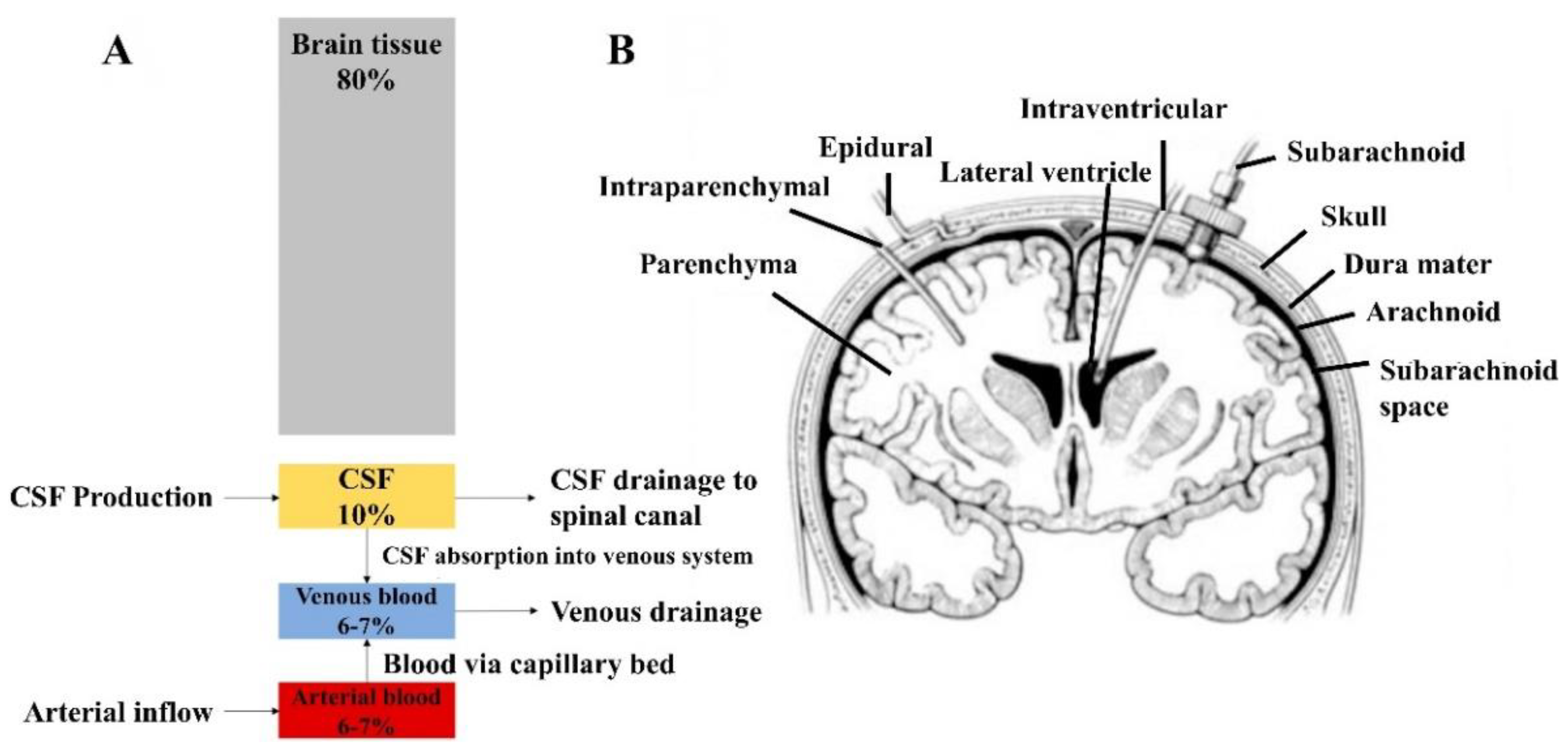
  131. Czosnyka, M.; Pickard, J.D. Monitoring and interpretation of intracranial pressure. J. Neurol. Neurosurg. Psychiatry 2004, 75, 813–821. [Google Scholar] [CrossRef]
  132. Harary, M.; Dolmans, R.G.F.; Gormley, W.B. Intracranial Pressure Monitoring-Review and Avenues for Development. Sensors 2018, 18, 465. [Google Scholar] [CrossRef] [PubMed] [Green Version]
  133. Raboel, P.H.; Bartek, J., Jr.; Andresen, M.; Bellander, B.M.; Romner, B. Intracranial Pressure Monitoring: Invasive versus Non-Invasive Methods-A Review. Crit. Care Res. Pract. 2012, 2012, 950393. [Google Scholar] [CrossRef] [PubMed] [Green Version]
  134. Peinera, E.T.; Tibrewala, A.; Bandorf, R.; Biehl, S.; Lüthje, H.; Doering, L. Micro force sensor with piezoresistive amorphous carbon strain gauge. Sens. Actuators A Phys. 2006, 130–131, 75–82. [Google Scholar] [CrossRef]
  135. Tibrewala, A.P.; Peiner, E.; Bandorf, R.; Biehl, S.; Lüthje, H. Transport and optical properties of amorphous carbon and hydrogenated amorphous carbon films. Appl. Surf. Sci. 2006, 252, 5387–5390. [Google Scholar] [CrossRef]
  136. Fraga, M.A.F.; Furlan, H.; Pessoa, R.S.; Rasia, L.A.; Mateus, C.F.R. Studies on SiC, DLC and TiO2 thin films as piezoresistive sensor materials for high temperature application. Microsyst. Technol. 2012, 18, 1027–1033. [Google Scholar] [CrossRef]
  137. Fraga, M.A.; Furlan, H.; Pessoa, R.S.; Massi, M. Wide bandgap semiconductor thin films for piezoelectric and piezoresistive MEMS sensors applied at high temperatures: An overview. Microsyst. Technol. 2014, 20, 9–21. [Google Scholar] [CrossRef]
  138. Rasia, L.A.; Leal, G.; Koberstein, L.L.; Furlan, H.; Massi, M.; Fraga, M.A. Design and Analytical Studies of a DLC Thin-Film Piezoresistive Pressure Microsensor. Comm. Comput. Inform. Sci. 2017, 742, 433–443. [Google Scholar] [CrossRef]
  139. Meng, K.Y.; Chen, J.; Li, X.S.; Wu, Y.F.; Fan, W.J.; Zhou, Z.H.; He, Q.; Wang, X.; Fan, X.; Zhang, Y.X.; et al. Flexible Weaving Constructed Self-Powered Pressure Sensor Enabling Continuous Diagnosis of Cardiovascular Disease and Measurement of Cuffless Blood Pressure. Adv. Funct. Mater. 2019, 29, 388. [Google Scholar] [CrossRef]
  140. Ion, M.; Dinulescu, S.; Firtat, B.; Savin, M.; Ionescu, O.N.; Moldovan, C. Design and Fabrication of a New Wearable Pressure Sensor for Blood Pressure Monitoring. Sensors 2021, 21, 2075. [Google Scholar] [CrossRef] [PubMed]
  141. Li, S.; Park, Y.; Luan, H.; Wang, H.; Kwon, K.; Rogers, J.A.; Huang, Y. Measurement of Blood Pressure via a Skin-Mounted, Non-Invasive Pressure Sensor. J. Appl. Mech. 2021, 88, 1183. [Google Scholar] [CrossRef]
  142. Ma, X.; Zhang, Q.; Guo, P.; Zhao, Y.; Wang, A. Diamond-like carbon based micro-pressure sensor with ultra-thin sensitive membrane. In Proceedings of the 2021 IEEE Sensors, Sydney, Australia, 31 October–3 November 2021. [Google Scholar] [CrossRef]
Figure 1. Ternary-phase diagram for DLCs. Reprinted with permission from [19]. 2014 Elsevier.
Coatings 12 01088 g001
Figure 2. Methods for tailoring DLC coatings and functional properties. Reprinted with permission from [20]. 2014 Elsevier.
Coatings 12 01088 g002
Figure 3. The typical Raman spectra of a carbon (A) [24] and the XPS of an a-C:H coating (B). Reprinted with permission from [14]. 2019 Elsevier.
Coatings 12 01088 g003
Figure 4. SEM images of whole blood placed on pyrolytic carbon (a) and Mo-DLC nanocomposite coatings with Mo concentrations of 3.8 at. % (b). Reprinted with permission from [71]. 2012 Elsevier.
Coatings 12 01088 g004
Figure 5. DLC-coated joint prosthesis and its wear performance. Surface wear of joint prosthesis with (A) and without (B) DLC; SEM image of the worn DLC-coated joint prosthesis (C). DLC-coated joint prosthesis has a better wear performance than the uncoated one. Reprinted with permission from [84]. 2004 John Wiley and Sons.
Coatings 12 01088 g005
Figure 6. Cyclic voltammogram behavior of different electrolytes on different electrodes. (A): square-wave voltammograms obtained using the DLC: VAMWCNT electrode in 0.1 mol·L−1 H2SO4 solution containing different concentrations of PAR (1:0.00 to 11:3.67 × 10⁻⁵ mol·L−1), COD (1:0.00 to 11:3.67 × 10⁻⁵ mol·L−1) and CAF (1:0.00 to 11:9.17 × 10⁻⁵ mol·L−1); SWV parameters: f = 80 Hz, A = 60 mV, and ΔEs = 9 mV. Analytical curve obtained for (B) PAR (Ip vs. CPAR), (C) COD (Ip vs. CCOD) and (D) CAF (Ip vs. CCAF). Reprinted with permission from [124]. 2016 John Wiley and Sons.
Coatings 12 01088 g006
Figure 7. ICP components and measurement scheme. (A) the Monro–Kellie model of intracranial pressure, and brain tissue including neurons, glia, extracellular fluid and cerebral microvasculature; (B) the sites for invasive ICP monitoring by EVD [132].
Coatings 12 01088 g007
Table 1. The classification of biomedical materials based on use [35,36].
ClassificationUsesCites
Musculoskeletal repair materialsJoint prosthesis and artificial dentures[37]
Soft-tissue repair materialsArtificial skin[38]
Cardiovascular system materialsArtificial valves and vascular stents[39]
Medical membrane materialsMembranes for dialysis and gas selective permeation[40]
Suture materialsSutures[41]
Drug-release carrier materialsDrug-delivery systems[42]
Clinical diagnosis and biosensor materialsSphygmomanometer and ultrasonic probe[43]
Table 2. The classification of biomedical materials based on tissue translation [44].
ClassificationFeatures
Biologically inert material
Bioactive materials
Bioabsorbable material
Placed in the human body with little interaction with surrounding tissue
Placed in the human body with interaction with tissue
Absorbed by the body after being placed in the body
Publisher’s Note: MDPI stays neutral with regard to jurisdictional claims in published maps and institutional affiliations.

