Article

An Antimicrobial Marine Cage Surface Modified with Antibacterial Peptides

Institute of Intelligent Manufacturing and Smart Transportation, Suzhou City University, Suzhou 215104, China
*
Author to whom correspondence should be addressed.
Coatings 2023, 13(10), 1711; https://doi.org/10.3390/coatings13101711
Submission received: 1 September 2023 / Revised: 21 September 2023 / Accepted: 27 September 2023 / Published: 29 September 2023

Abstract

:
Long-term immersion in seawater easily causes surface fouling and affects the marine aquaculture industry. The commonly used method is to apply copper-based coatings on surfaces, however, the release of copper ions will harm marine organisms. Antimicrobial peptides (AMPs) are a substance extracted from organisms that possess environmental friendliness. This study extracted AMPs from traditional Chinese medicine, analyzed their amino acid sequences, and bound them to the surfaces of cage materials based on the strong adhesion of dopamine in weakly alkaline environments. The Fourier transform infrared spectrometer (FTIR) spectrum results showed that the antibacterial peptide was successfully bound to the substrate surface, and the contact angle results demonstrated a significant change in the wettability of the substrate surface. Antibacterial tests were conducted on the surface of the sample using Staphylococcus aureus (S. aureus). The results illustrated that 304 stainless steel (SS) and nylon (PA) surfaces modified by the antibacterial peptide exhibited significant biofilm resistance, with antibacterial adhesion properties reaching 88.68% and 82.61%, respectively, exhibiting the robustness of the antimicrobial efficiency. This study can provide theoretical support for the antifouling performance of the surfaces of marine aquaculture cages.

1. Introduction

Biofouling in aquaculture systems has caused serious impacts on both their products and economic costs [1]. After immersion in seawater, bacteria or other microorganisms can quickly attach to marine cages, causing the surface fouling of the cage materials. The survival ability of fouling organisms in biofilms is high, and different types of attached bacteria are close to each other and cooperate to form a mixed microbial community, leading to the gradual maturation of biofilms. Later, other prokaryote, fungi, algal spores, and the larvae of large fouling organisms develop and grow in membranes, finally forming a complex large fouling biological layer. The composition and quantity of the biofilms formed on these surfaces are not entirely the same due to different substrate materials and environments. After the formation of mature biofilms, large organisms such as seaweed and barnacles will attach, in large quantities, to the surfaces of submerged ships, forming serious biological fouling [2]. Previous studies have shown that the reversible adhesion process of bacteria and other substances on the surfaces of materials is the fundamental determinant of biofilm formation [3]. In addition, the conditions of fluid mechanics, the special surface structures of microorganisms, cell wall composition, and extracellular polymers will also affect the formation of biofilms [2,3].
At present, the main way to inhibit biofilm formation is by preventing bacterial adhesion or killing adherent bacteria. The adhesion of bacteria on the surfaces of materials is closely related to their physical and chemical properties. Biological fouling occurs at the interface between materials and solutions, and the hydrophilicity and hydrophobicity of materials play important roles in the behavior of biological fouling. Superhydrophobic antifouling surfaces have attracted great interest [4]. However, superhydrophobic surfaces do not have stable antifouling properties. The current superhydrophobic surfaces, such as the Lotus effect of hydrophobicity, rely on the typical micro/nano structure of the surface to capture the air to form a stable air layer, which makes it easy for droplets to roll off, thus providing an excellent non-staining performance. However, superhydrophobic surfaces constructed by imitating the Lotus effect have a poor compressive stability, and cannot prevent the infiltration of low-surface-energy liquids and biological liquids [5,6]. Research has shown that the surface energy of materials plays an important role in the formation of biofilms, and is closely related to the adsorption and desorption of marine fouling organisms. Zhao [7] studied the effect of surface energy on bacterial adhesion and found that the minimum value of bacterial adhesion appeared to be between 20 and 30 mJ/m2. Common low-surface-energy coatings are organic silicon coatings and organic fluorine coatings. Although these coatings have an excellent anti fouling and drag reduction performance, they have obvious drawbacks, such as insufficient adhesion, difficulty in research and development, and a high cost. Another method for inhibiting biofilm formation is to cover the sample surface with a bactericidal coating. At present, the commonly used bactericidal coating is still the antifouling coating containing a Copper (I) oxide antifouling agent [8,9]. Although copper-based antifouling agents have a lower toxicity than organic tin, copper ions can also accumulate in the ocean, leading to the death of large numbers of seaweed and causing serious damage to the marine ecological environment. This method will cause large-scale toxic compound pollution, so it is imperative to limit or eliminate the use of such antifouling agents. It is necessary to develop new, alternative green antifouling and drag-reducing materials.
Various non-toxic and harmless green antifouling methods have been proposed by researchers, among which, AMPs have been designed and synthesized as effective antifouling agents [10,11]. AMPs have strong antibacterial/bactericidal effects, which have attracted the extensive attention of surface antifouling and drag reduction scholars at home and abroad. Since Boman’s research group [12] first carried out work on AMPs, in the past few decades, nearly 1000 kinds of AMPs have been isolated and identified. Antimicrobial peptides have great potential in the field of surface antifouling, and they have been widely applied for the surface modification of materials due to their excellent antimicrobial ability [13]. Antimicrobial peptides are able to inhibit bacterial adhesion and biofilm formation, preventing the development of biofouling from its initial stage, and have been validated as effective at targeting a wide range of marine microorganisms, including a variety of bacteria, as well as microalgae [14,15,16]. Some cathelicidin can directly react with metals to obtain new biological metal–organic materials [17,18]. However, the amount of biological peptides bound to metal surfaces through this direct reaction is very small, and the improvement in the antifouling effect is also limited. Surface activation can increase the binding capacity of antimicrobial peptides and enhance their antibacterial and antifouling effects. Piranha solution is a surface activator with a strong activation performance. It endows a large number of active groups on the surfaces of soft materials, such as glass, silicon wafers, and PDMS, etc. [19], and can also activate the surfaces of hard metal materials such as stainless steel [20]. However, due to its strong oxidizing ability, it can damage the surfaces of materials, increase surface roughness, and the residual liquid of piranha solution can cause serious environmental pollution, so it has significant drawbacks. Dopamine is a common coupling agent that can undergo chemical reactions with a large number of solid surfaces through oxidative self-polymerization in weakly alkaline solutions in oxygen-rich environments, thereby improving the surface modification efficiency [21]. Dopamine coatings contain functional groups such as catechol and phenethylamine, which can be bonded to almost all types of materials through non-covalent bonds or covalent bonds. Therefore, dopamine coating has become a very effective method for the surface modification of almost all materials. Lim [22] used dopamine as a coupling agent to graft peptides onto a sample surface and obtained effective antibacterial surfaces. However, the reaction mechanism of dopamine and stainless steel is still unclear, because, during the oxidation process, many intermediates of dopamine are formed through the electron transfer between oxygen and dopamine ions, such as semi-quinone and quinone, etc. [23,24]. Dopamine rarely undergoes oxidative self-polymerization under weakly acidic conditions, and reacts with organic compounds containing carboxyl groups, such as hyaluronic acid, under the action of a catalyst to obtain a new type of organic compound containing dopamine and possessing grafting properties [25]. This method will avoid the reduction in binding sites caused by step bonding and improve the binding amount of the substance, providing a new method for antibacterial peptides to bind to stainless steel surfaces. Majhi [26] proposed a two-step functionalization strategy, in which a stainless steel substrate was immersed in piranha solution for oxidation and hydroxylation, followed by aminosilylation, and finally, the antibacterial peptide was fixed on the stainless steel surface, which could effectively inhibit Escherichia coli and S. aureus. Cao [27] conjugated antimicrobial peptides with dopamine to prepare derived antimicrobial peptide DP to modify stainless steel, and the modified surface had a good biocompatibility and antifouling ability.
Viola philippica is a traditional Chinese medicine that is rich in peptides with antibacterial activity. The peptides were extracted from Viola philippica using high-performance liquid chromatography (HPLC), and the amino acid sequences of the peptides were obtained using a mass spectrometry analysis. There are few studies on reducing the surface antifouling performance of marine fishing facilities by combining antimicrobial peptides with their surfaces. In this study, the extracted peptide was grafted onto the surfaces of marine cage substrate materials such as 304 SS and nylon using dopamine as a coupling agent. The physical and chemical properties of the peptide-modified 304 SS and nylon surfaces were significantly altered. The surfaces possessed relatively stable antibacterial and biofilm properties, and we attempted to explain the antibacterial mechanism of the peptide-modified surfaces. The use of antimicrobial peptide-modified materials in aquaculture net cage materials is a promising approach.

2. Materials and Methods

2.1. Materials and Reagents

The 304 annealed stainless steel samples and nylon (PA6) were purchased from Jiangsu Congbang special steel Co., Ltd. (Suzhou, China) and Dongguan Dalicheng plastic products Co., Ltd. (Dongguan, China), and the elements are displayed in Table S1. Dopamine hydrochloride (98%), Dichloromethane (DCM), ammonium bicarbonate (NH4HCO3), Tris(hydroxymethyl)aminomethane (Tris ≥ 99.9%), Crystal violet (CV, 0.1%), trifluoroacetic acid (TFA), acetone, acetonitrile, and absolute acetic acid were obtained from Aladdin Biochemical Technology Co., Ltd. (Shanghai, China). Methanol, CHCl3, Luria Broth (LB), and Cyclobutane were purchased from Wuhan Feiyoute Biological Technology Co., Ltd. (Wuhan, China). Distilled water was obtained using a Milli-Q water purification system. V. philippica was collected in Anhui Province, China. All the reagents were of analytical grade.

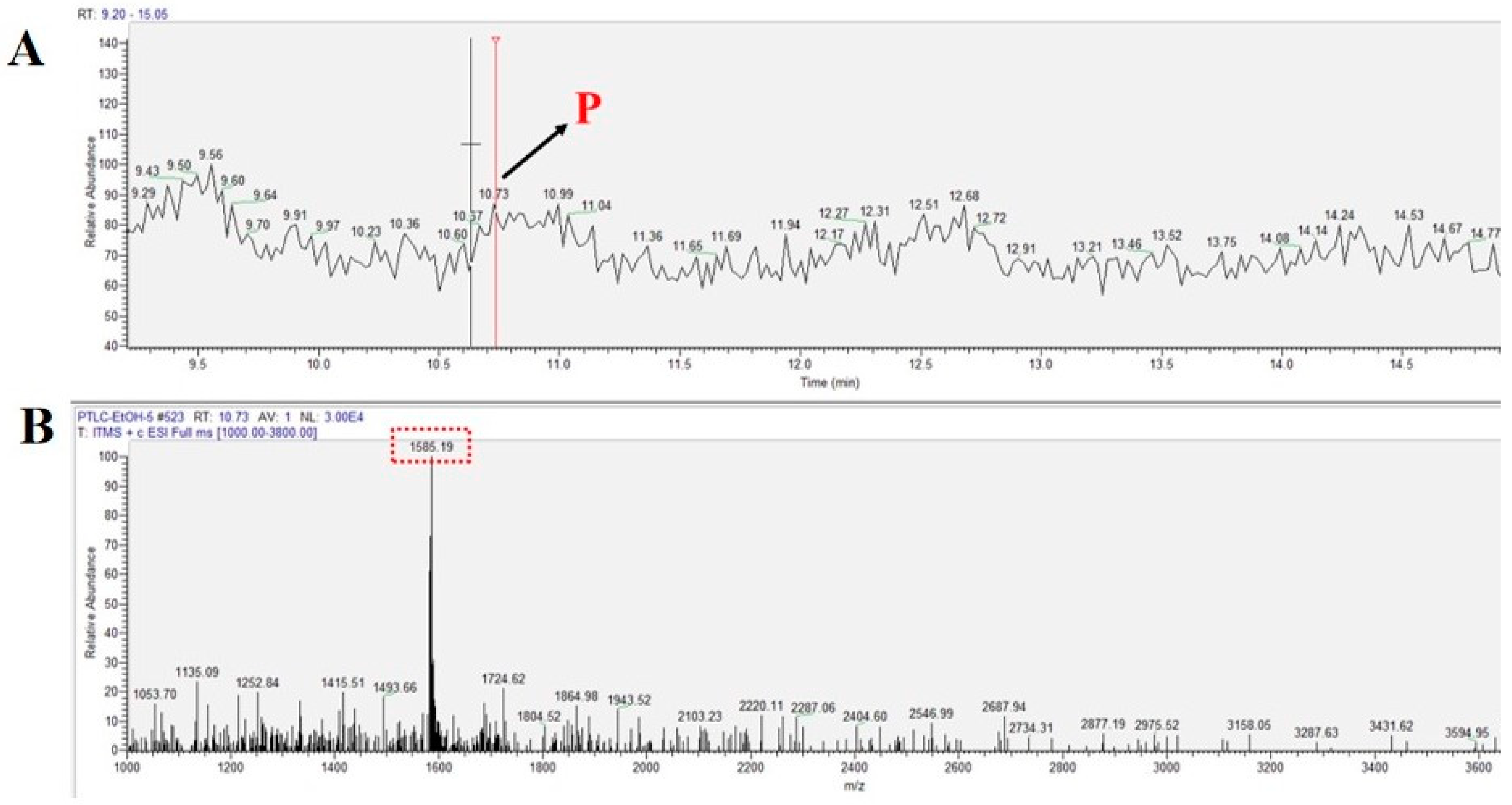
2.2. Extraction and Isolation of Peptide

The extraction procedure of the peptides from V. philippica was carried out according to previous methods [28]. After drying and crushing, preliminary purification was carried out to obtain V. philippica powder. In total, 800 g of coarse powder was pre-extracted with 3.5 L of dichloromethane (DCM), filtered after 24 h, and repeated three times within 96 h; the DCM extract of V. philippica was obtained after evaporating. The residue was dried and immersed in 3.5 L of methanol/water (3:2 by volume) for 72 h, and the MeOH/H2O crude extract was concentrated using rotary evaporation, followed by liquid–liquid distribution using n-butanol and H2O. Cyclobutane was separated from the butanol fraction using gel column chromatography, the sample was washed with the mixture of CHCl3 and MeOH, and the MeOH ratio was increased to 100% at the rate of 70 drops/min. The sample was further separated and purified using high-performance liquid chromatography (HPLC), with liquid A being an aqueous solution of 0.1% trifluoroacetic acid (TFA) and liquid B being an 80% acetonitrile aqueous solution containing 0.1% TFA. The time was set at 15 min, and the percentage of liquid B was increased linearly from 10% to 70%. The flow rate of the eluent in the HPLC was 4 mL/min. The amino acid sequence was analyzed using a mass spectrometer (Agilent 1260 Infinity II, Santa Clara, CA, USA).

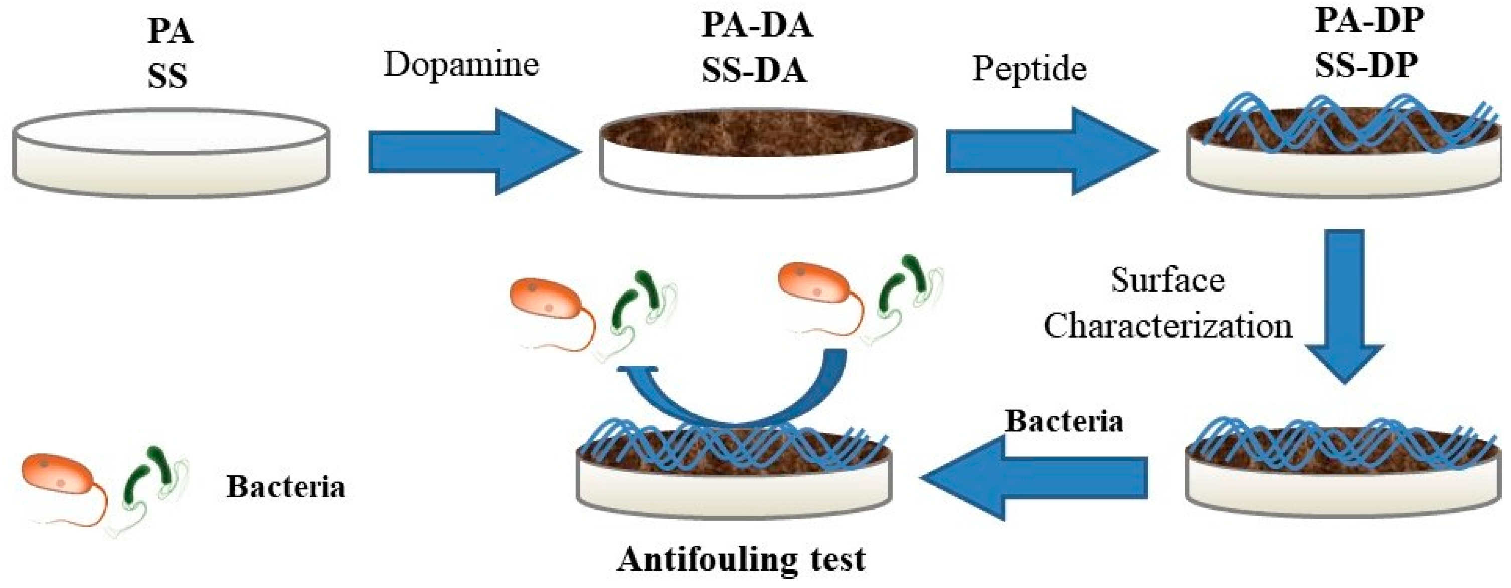
2.3. Fabrication of Peptide Modified Surface

The samples were cut into square shapes with side lengths of 1cm. The 304 stainless steel samples were polished by sandpaper with coast grits from 400, 800, 1200, to 2000# and ultrasonically cleaned for 6 min for further processing. The samples were cleaned with absolute ethanol for 15 min, the steel samples were soaked in acetone for 30 min, and the pretreated samples were obtained after being air dried. The peptide was dissolved in Tris-HCl buffer (pH 8.5) to obtain a peptide solution with concentration of 100 µg/mL. The stainless steel and nylon samples were immersed in 100 µg/mL of fresh dopamine solution in 10 mM of Tris-HCl buffer (pH 8.5) for 24 h. The dopamine-treated samples were then immersed in peptide solution and incubated with 30 rpm shaking at room temperature for 24 h. The nylon sample modified with dopamine was named PA-DA, and PA-DA modified with antimicrobial peptides was named PA-DP. The 304 SS sample modified with dopamine was named SS-DA, and SS-DA modified with antimicrobial peptide was named SS-DP. The peptide-treated sample preparation, characterization, and antimicrobial experimental process are shown in Figure 1. The samples were then cleaned with excessive deionized water and dried in air, and all the samples were stored at room temperature for antimicrobial and characterization tests.

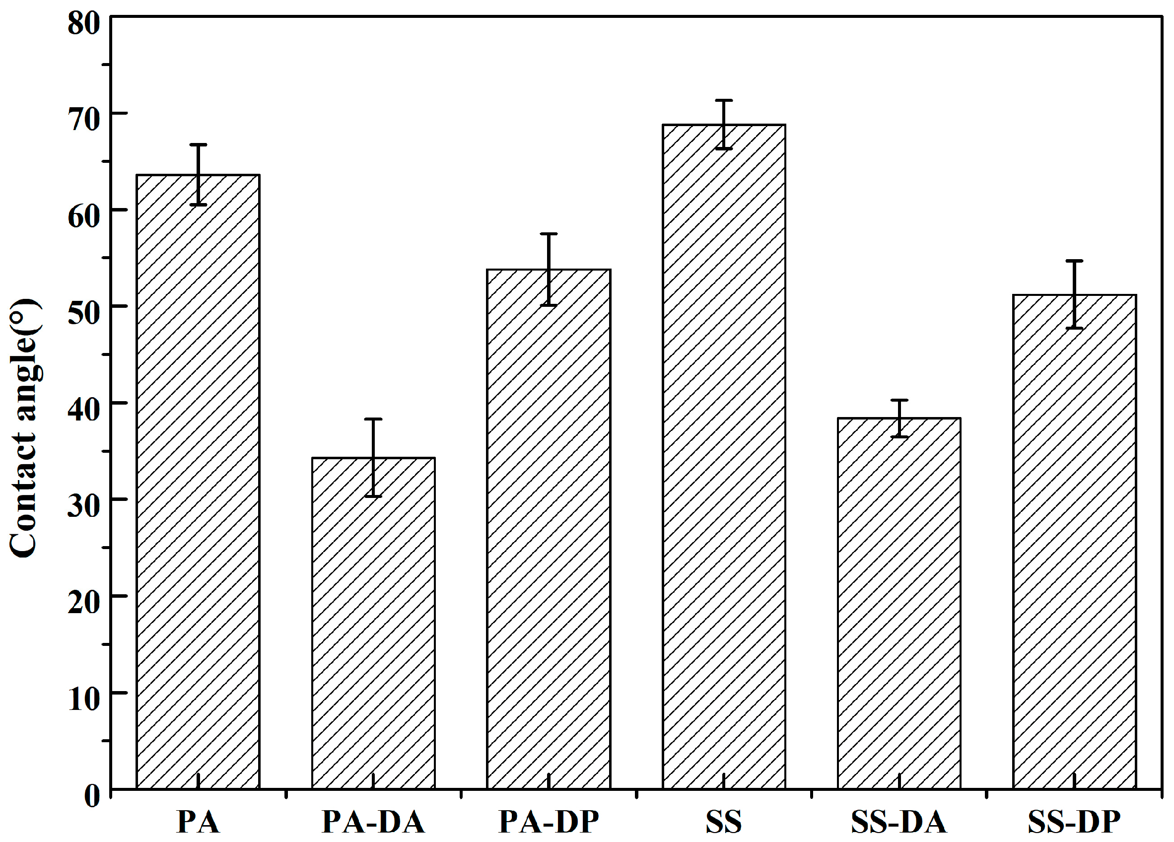
2.4. Surface Characterization

The functional group information on the surfaces of the samples before and after modification was characterized using FTIR (Cary 610/670). During measurement, the infrared spectrometer scanned 128 times with a resolution of 4.0 cm−1. The ATR mode was selected, and 3 points were measured on each sample surface. The adhesion of bacteria on the surfaces of the samples was observed using a field emission scanning electron microscope (FESEM, Gemini SEM300, Oberkochen, Germany). The roughness was characterized using a 3D optical profilometer (Contour GT-K, Bruker, Billerica, MA, USA). The water contact angle of the sample surfaces was evaluated using a contact angle measuring instrument (JC2000D, Shanghai, China). Three points were taken for each sample surface measurement. The optical density (OD) value of the sample surface eluate was analyzed using a UV-Vis spectrophotometer (L5S, Shanghai, China).

2.5. Antimicrobial Tests

The study selected the common Gram-positive bacterium S. aureus to characterize the antibacterial properties of the sample surfaces before and after modification.
Antibiofilm testing: S. aureus was cultured and proliferated in LB liquid culture medium (37 °C, 180 rpm) for 24 h. The bacterial solution was diluted at a ratio of 1:100 and stirred evenly, and the diluted bacterial solution was used to cover the sample before and after modification (24-well bacterial culture plate, 1 mL/well). The plate was sealed and incubated in a shaker at 30 °C for 24 h. The samples were taken out and rinsed with sterile PBS buffer to remove the bacteria that had not adhered tightly, and then the surfaces of the samples were stained with a concentration of 0.1% crystal violet solution for 15 min (500 μL/well). The dyed surfaces were washed with sterile PBS buffer until there was no obvious color in the last cleaning solution, and they were dried in an ultra-clean table. The dyed dry surfaces were immersed in a 30% acetic acid solution for 15 min to collect the washing solution, and the washing solution was added to a colorimetric dish (3 mL/tube) to measure the OD value of the washing solution using a UV-Vis spectrophotometer.
Antibacterial testing: LB agar plates inoculated with S. aureus were cultured in a 37 °C constant-temperature incubator for 24 h, and the monoclonal strains of S. aureus were selected and transferred to LB culture medium, which was shaken in a constant temperature shaker (37 °C, 180 rpm) until the medium became turbid. Finally, the samples were immersed before and after modification in a 1:100 diluted bacterial solution and incubated in a 37 °C constant-temperature incubator for 24 h. During the surface robustness test, the bacterial solution was replaced every 36 h to ensure the concentration of the culture solution. After removing the sample, the treated sample was cleaned with sterile PBS solution to remove non-adhering or loosely adhering bacteria. Then, the sample was immersed in a 2.5% glutaraldehyde solution and left to stand for 12 h in a refrigerator at 4 °C, so that the bacteria could be quickly killed and fixed on the surface of the sample. The samples fixed with bacteria were sequentially treated with ethanol contents of 25% (5 min), 50% (5 min), 75% (5 min), 90% (5 min), and 100% (20 min), and then placed in a vacuum drying oven for 24 h (37 °C) for an FESEM analysis.
Durability testing of the coating: The samples were placed in artificial seawater for 1, 3, 5, and 7 days, respectively. After cleaning, the antibiofilm performances of the artificial-seawater-treated samples were tested using the previous antibiofilm-performance-testing method.

3. Results and Discussion

Peptides were extracted from V. philippica and further purified using semi-preparative reversed-phase high-performance liquid chromatography (HPLC), and their amino acid sequences were identified using a mass spectrometry analysis. Figure 2 showed the HPLC spectrum of the isolated antimicrobial peptides and the MS spectrum of the antimicrobial peptide identification. The separation liquid eluted between 10 min and 11 min was collected and freeze-dried to obtain purified peptide compounds. The molecular weight of the extracted peptide was 3170.43, and the amino acid sequence was CGESCVFIPCISAIIGCSNKVCYKNGSIP according to the mass spectrometry analysis results.
The obtained antibacterial peptide was grafted onto the surfaces of the samples, and the physicochemical properties and antimicrobial performances of the modified sample surfaces were characterized using characterization instruments. The application prospects of the antibacterial peptide isolated from V. philippica on the surfaces of the different substrate materials were analyzed. The durability testing results of the peptide-modified surfaces are displayed in Figure S1, and they demonstrated that the modified surfaces possessed a good stability after 7 days of immersion in artificial seawater. Some antimicrobial peptides were not firmly bound on the surfaces and were prone to detachment from the substrates after immersion in seawater, but the detachment rate gradually decreased and tended to stabilize due to the strong adhesion of polydopamine.
Fourier transform infrared spectrometers are often used to identify known substances, comparing the infrared spectrum of the sample with the standard infrared spectrum. FTIR was also used to determine the similarity and purity of the two compounds based on the consistency of the spectral peak positions, wave numbers, peak shapes, and other characteristics. Figure 3 shows the collected FTIR spectra of the extracted peptide-modified surfaces using PA-DA and SS-DA as backgrounds, respectively; therefore, the peaks that appeared in the spectra did not belong to the PA-DA and SS-DA surfaces. Figure 3A,B are the FTIR spectra of PA-DP and SS-DP. The peaks at 1638 cm1 and 1549 cm1 belonged to the amide I and amide II groups of the peptide, and the peaks near the wavelength 1610 cm1 in the spectrum were formed by the C=O stretching vibration of amide I [29]. The peak with a wavelength of 1450 cm1 was caused by the C-N stretching or N-H bending of amide II [30], while the wide peak near 3300 cm1 in the infrared spectrum was caused by amide A of the peptide [31]. The appearance of the peaks was caused by the binding of the peptide due to PA-DA and SS-DA being used as backgrounds when collecting the spectra. The binding strength of the peptides in the nylon and 304 stainless steel was different, which caused different surface dopamine polymerization thicknesses and hydrogen bonding strengths, resulting in different FTIR peak intensities and deviations when collecting the peptide-modified samples [32].
Figure 4 shows the contact angles of the dopamine and peptide 304 SS and Nylon surfaces. After the surfaces of the 304 stainless steel and nylon were modified with dopamine, the surface contact angle was significantly reduced, which was because dopamine and its polymers are strong hydrophilic substances, and the dopamine attached to the substrate surface would hinder the contact between the measuring liquid and the substrate, only reflecting the contact angle between the titration liquid and the dopamine polymer; similarly, the peptide binding on the surface of the dopamine blocked the contact between dopamine and the titrant, thereby increasing the value of the contact angle. However, this peptide was still a hydrophilic substance, so the increase in the value of the contact angle was not very large. Surface roughness also plays an important role in surface wettability, and the surface roughness (Sa) and surface root mean square deviation (Sq) of the before and after peptide-treated samples were tested (Table S2). The binding of dopamine and antimicrobial peptides to the surfaces of the samples changed their surface roughness, while the value of the surface roughness did not change much due to the relatively uniform and low binding amount of dopamine and antimicrobial peptides on the surfaces. The modification of dopamine and antimicrobial peptides had a greater impact on the surface wettability than the morphology change after binding under the conditions.
After immersing the surfaces in bacteria for 24 h, the samples were stained with crystal violet and the OD values of the acetic acid cleaning solution were measured. The higher the OD value, the more amount of biofilm was removed by the acetic acid, which means that more biofilm was generated on the surfaces of the samples. The OD values of the acetic acid suspensions are presented in Figure 5. The untreated 304 stainless steel and nylon surfaces possessed higher OD values, and the values reached 1.42 and 0.96, respectively, which demonstrated high S. aureus biofilm formation for the bare surfaces. After the dopamine and peptide treatment, the OD values decreased sharply, and it was caused by the mild antibacterial ability of dopamine and the antibacterial/bactericidal ability of the extracted peptide.
SEM was used to observe the bacterial adhesion on the sample surfaces before and after the modification with the antimicrobial peptides to more intuitively observe the antibacterial/antibacterial ability of the antimicrobial peptides bound to the sample surfaces. After co-culturing with a bacterial solution, a large number of bacteria adhered to the surface of the original sample (Figure 6). After being treated with antimicrobial peptides, the adhesion of S. aureus on the surface of the sample was significantly reduced, demonstrating a strong antibacterial performance. This was because the grafting of antimicrobial peptides onto the sample surface endowed the substrate with antibacterial ability. The antibacterial adhesion ability of the sample surfaces was analyzed by counting the number of attached bacteria, and it could be calculated that the S. aureus adhesion resistance ability of the sample surfaces modified with the antimicrobial peptides on the nylon and stainless steel surfaces was 88.68% and 82.61%, respectively. After immersion in the bacterial solution for one week, the samples still exhibited antibacterial properties of 83.95% and 78.69% (Figure 7B), indicating that the surface modified with antimicrobial peptides had a relatively strong antibacterial ability. The nylon surface was relatively soft and could provide more attachment sites for substance attachment, which could improve the binding amount of antimicrobial peptides. Therefore, the antibacterial-peptide-modified surface had a stronger antibacterial adhesion ability. The peptide disrupted the transmembrane potential and osmoregulatory function of cells, inhibiting cellular respiration, and eventually leading to the death of the bacterium (ring pore model) [33], thereby achieving antibacterial/antibacterial effects (Figure 7A).

4. Conclusions

An antimicrobial peptide with a 3170.43 molecular weight was extracted from V. philippica using high-performance liquid chromatography, and the amino acid sequence of the peptide identified using a mass spectrometry analysis. The peptide was grafted onto 304 SS and PA surfaces using dopamine as a coupling agent. The infrared spectroscopy of the modified surfaces verified that the peptide was successfully grafted onto the sample surfaces and slightly increased the contact angle of the sample surfaces; S. aureus immersion tests and the morphology of the bacteria demonstrated that the peptide-modified sample surfaces possessed outstanding antibacterial abilities and a remarkable antibiofilm capacity. The antibacterial stable test results proved that the peptide-modified surface had durability within a certain period of time. This study will provide a new research strategy for the field of bacterial infection and surface fouling prevention.

Supplementary Materials

The following supporting information can be downloaded at: https://www.mdpi.com/article/10.3390/coatings13101711/s1, Table S1. The chemical composition of the samples. Table S2. Surface parameters of the steel samples. Figure S1. The stability of the peptide modified surface in artificial seawater. Figure S2. The photo of Viola philippica.

Author Contributions

Conceptualization, Z.C. and Q.G.; methodology, Z.C.; validation, Z.C. and Q.G.; formal analysis, Z.C.; investigation, Z.C.; data curation, Z.C.; writing—original draft preparation, Z.C.; writing—review and editing, Z.C. and Q.G.; supervision, Q.G.; funding acquisition, Q.G. All authors have read and agreed to the published version of the manuscript.

Funding

This research was funded by the General Program for Basic Science Research (Natural Science) in Higher Education Institutions of Jiangsu Province (No. 23KJB510026).

Institutional Review Board Statement

Not applicable.

Informed Consent Statement

Not applicable.

Data Availability Statement

Not applicable.

Acknowledgments

This work was financially supported by the General Program for Basic Science Research (Natural Science) in Higher Education Institutions of Jiangsu Province (No. 23KJB510026). The authors express their gratitude to Cao Pan for his guidance in the extraction of antimicrobial peptides and antimicrobial experiments.

Conflicts of Interest

The authors declare no conflict of interest.

References

  1. Adams, C.; Shumway, S.; Whitlatch, R.; Getchis, T. Biofouling in marine molluscan shellfish aquaculture: A survey assessing the business and economic implications of mitigation. J. World Aquac. Soc. 2011, 42, 242–252. [Google Scholar] [CrossRef]
  2. Vladkova, T. Surface modification approach to control biofouling. In Marine and Industrial Biofouling; Springer Series on Biofilms; Springer: Berlin/Heidelberg, Germany, 2009; Volume 4, pp. 135–163. [Google Scholar]
  3. Abarzua, S.; Jacubowski, S. Biotechnological investigation for the prevention of biofouling 1. Biological and biochemical principles for the prevention of biofouling. Mar. Ecol. Prog. Ser. 1995, 123, 301–312. [Google Scholar] [CrossRef]
  4. Banerjee, I.; Pangule, R.; Kane, R. Antifouling coatings: Recent developments in the design of surfaces that prevent fouling by proteins, bacteria, and marine organisms. Adv. Mater. 2011, 23, 690–718. [Google Scholar] [CrossRef] [PubMed]
  5. Tesler, A.; Kim, P.; Kolle, S.; Howell, C.; Ahanotu, O.; Aizenberg, J. Extremely durable biofouling-resistant metallic surfaces based on electrodeposited nanoporous tungstite films on steel. Nat. Commun. 2015, 6, 8649. [Google Scholar] [CrossRef] [PubMed]
  6. Sunny, S.; Vogel, N.; Howell, C.; Vu, T.; Aizenberg, J. Lubricant-infused nanoparticulate coatings assembled by layer-by-layer deposition. Adv. Funct. Mater. 2014, 24, 6658–6667. [Google Scholar] [CrossRef]
  7. Anderson, C.; Atlar, M.; Callow, M.; Candries, M.; Milne, A.; Townsin, R. The development of fouling-release coatings for seagoing vessels. J. Mar. Des. Oper. 2003, B4, 11–23. [Google Scholar]
  8. Zhao, Q.; Wang, S.; Müller-Steinhagen, H. Tailored surface free energy of membrane diffusers to minimize microbial adhesion. Appl. Surf. Sci. 2004, 230, 371–378. [Google Scholar] [CrossRef]
  9. Srinivasan, M.; Swain, G. Managing the use of copper-based antifouling paints. Environ. Manag. 2007, 39, 423–441. [Google Scholar] [CrossRef]
  10. Xie, C.; Guo, H.; Zhao, W.; Zhang, L. Environmentally friendly marine antifouling coating based on a synergistic strategy. Langmuir 2020, 36, 2396–2402. [Google Scholar] [CrossRef]
  11. Kazemzadeh-Narbat, M.; Cheng, H.; Chabok, R.; Alvarez, M.; Fuente-Nunez, C.; Phillips, K.; Khademhosseini, A. Strategies for antimicrobial peptide coatings on medical devices: A review and regulatory science perspective. Crit. Rev. Biotechnol. 2021, 41, 94–120. [Google Scholar] [CrossRef]
  12. Boman, H.; Nilsson, I.; Rasmuson, B. Inducible antibacterial defense system in drosophila. Nature 1972, 237, 232–235. [Google Scholar] [CrossRef] [PubMed]
  13. Andrea, A.; Molchanova, N.; Jenssen, H. Antibiofilm peptides and peptidomimetics with focus on surface immobilization. Biomolecules 2018, 8, 27. [Google Scholar] [CrossRef] [PubMed]
  14. Segev-Zarko, L.; Saar-Dover, R.; Brumfeld, V.; Mangoni, M.; Shai, Y. Mechanisms of biofilm inhibition and degradation by antimicrobial peptides. Biochem. J. 2015, 468, 259–270. [Google Scholar] [CrossRef]
  15. Yasir, M.; Willcox, M.; Dutta, D. Action of antimicrobial peptides against bacterial biofilms. Materials 2018, 11, 2468. [Google Scholar] [CrossRef] [PubMed]
  16. Trepos, R.; Cervin, G.; Pile, C.; Pavia, H.; Hellio, C.; Svenson, J. Evaluation of cationic micropeptides derived from the innate immune system as inhibitors of marine biofouling. Biofouling 2015, 31, 393–403. [Google Scholar] [CrossRef] [PubMed]
  17. Davis, E.; Li, D.; Irvin, R. A peptide-stainless steel reaction that yields a new bioorganic—Metal state of matter. Biomaterials 2011, 32, 5311–5319. [Google Scholar] [CrossRef]
  18. Cao, P.; He, X.; Xiao, J.; Yuan, C.; Bai, X. Peptide-modified stainless steel with resistance capacity of Staphylococcus aureus biofilm formation. Surf. Interface Anal. 2018, 50, 1362–1369. [Google Scholar] [CrossRef]
  19. Khan, M.; Wu, Z.; Mao, S.; Lin, J.; Shah, S.; Lin, J. Controlled grafted poly(quaternized-4-vinylpyridine-co-acrylic acid) brushes attract bacteria for effective antimicrobial surfaces. J. Mater. Chem. B 2018, 6, 3782–3791. [Google Scholar] [CrossRef]
  20. Cao, P.; Yuan, C.; Xiao, J.; He, X.; Bai, X. A biofilm resistance surface yielded by grafting of antimicrobial peptides on stainless steel surface. Surf. Interface Anal. 2018, 50, 516–521. [Google Scholar] [CrossRef]
  21. Shang, B.; Wang, Y.; Yang, P.; Peng, B.; Deng, Z. Synthesis of superhydrophobic polydopamine-Ag microbowl/nanoparticle array substrates for highly sensitive, durable and reproducible surface-enhanced Raman scattering detection. Sens. Actuators B Chem. 2018, 255, 995–1005. [Google Scholar] [CrossRef]
  22. Lim, K.; Chua, R.; Bow, H.; Tambyah, P.; Hadinoto, K.; Leong, S. Development of a catheter functionalized by a polydopamine peptide coating with antimicrobial and antibiofilm properties. Acta Biomater. 2015, 15, 127–138. [Google Scholar] [CrossRef] [PubMed]
  23. Kim, H.; McCloskey, B.; Choi, T.; Lee, C.; Kim, M.; Freeman, B.; Park, H. Oxygen concentration control of dopamine-induced high uniformity surface coating chemistry. ACS Appl. Mater. Interfaces 2013, 5, 233–238. [Google Scholar] [CrossRef] [PubMed]
  24. Lee, H.; Dellatore, S.; Miller, W.; Messersmith, P. Mussel-inspired surface chemistry-for multifunctional coatings. Science 2007, 5849, 426–430. [Google Scholar] [CrossRef] [PubMed]
  25. Jeong, Y.; Thuy, L.; Ki, S.; Ko, S.; Kim, S.; Cho, W.; Choi, J.; Kang, S. Multipurpose antifouling coating of solid surfaces with the marine-derived polymer fucoidan. Macromol. Biosci. 2018, 10, 1800137. [Google Scholar] [CrossRef] [PubMed]
  26. Majhi, S.; Peddiraju, V.; Mishra, A. Effect of antimicrobial peptide (AMP) etethered stainless steel surfaces on the bacterial membrane. Mater. Today Chem. 2021, 21, 100541. [Google Scholar] [CrossRef]
  27. Cao, P.; Liu, D.; Zhang, Y.; Xiao, F.; Yuan, C.; Liang, F.; Liu, X.; Zhang, C. Dopamine-assisted sustainable antimicrobial peptide coating with antifouling and anticorrosion properties. Appl. Surf. Sci. 2022, 589, 153019. [Google Scholar] [CrossRef]
  28. Cao, P.; Yang, Y.; Uche, F.; Hart, S.; Li, W.; Yuan, C.Q. Coupling plant-derived cyclotides to metal surfaces: An antibacterial and antibiofilm study. Int. J. Mol. Sci. 2018, 19, 793. [Google Scholar] [CrossRef]
  29. Berisha, A.; Hazimeh, H.; Galtayries, A.; Decorse, P.; Kanoufi, F.; Combellas, C.; Pinson, J.; Podvorica, F. Grafting of an aluminium surface with organic layers. RSC Adv. 2016, 6, 78369–78377. [Google Scholar] [CrossRef]
  30. Lan, Y.; Hiebner, D.; Casey, E. Self-assembly and regeneration strategy for mitigation of membrane biofouling by the exploitation of enzymatic nanoparticles. Chem. Eng. J. 2021, 412, 128666. [Google Scholar] [CrossRef]
  31. Lou, T.; Bai, X.; He, X.; Liu, W.; Yang, Z.; Yang, Y.; Yuan, C. Enhanced antifouling properties of marine antimicrobial peptides by PEGylation. Front. Bioeng. Biotech. 2023, 11, 1124389. [Google Scholar] [CrossRef]
  32. Fevzioglu, M.; Ozturk, O.; Hamaker, B.; Campanella, O. Quantitative approach to study secondary structure of proteins by FT-IR spectroscopy, using a model wheat gluten system. Int. J. Biol. Macromol. 2020, 164, 2753–2760. [Google Scholar] [CrossRef] [PubMed]
  33. Cao, P.; Du, C.; He, X.; Zhang, C.; Yuan, C. Modification of a derived antimicrobial peptide on steel surface for marine bacterial resistance. Appl. Surf. Sci. 2020, 510, 145512. [Google Scholar] [CrossRef]
Figure 1. Schematic illustration of peptide-treated surfaces’ synthesis and antimicrobial tests.
Coatings 13 01711 g001
Figure 2. Target product molecular weight obtained using high-performance liquid chromatography. (A): HPLC spectrum and (B): MS spectrum. P means extracted peptide.
Coatings 13 01711 g002
Figure 3. Infrared spectrum of the surface after grafting antibacterial peptide. (A): Nylon and (B): 304 SS. (-DP represents surface modified with dopamine and peptide).
Coatings 13 01711 g003
Figure 4. Contact angles of 304 SS and Nylon samples before and after surface treatment by dopamine and peptide.
Coatings 13 01711 g004
Figure 5. The OD 590 nm values of different surface eluents.
Coatings 13 01711 g005
Figure 6. SEM images of S. aureus adhesion on different surfaces. (A): 304 SS, (B): Nylon, (A-1): peptide modified 304 SS, (B-1): peptide modified nylon.
Coatings 13 01711 g006
Figure 7. Schematic diagram of antibacterial peptide sterilization mechanism (A) and analysis of surface antibacterial efficacy (B).
Coatings 13 01711 g007
Disclaimer/Publisher’s Note: The statements, opinions and data contained in all publications are solely those of the individual author(s) and contributor(s) and not of MDPI and/or the editor(s). MDPI and/or the editor(s) disclaim responsibility for any injury to people or property resulting from any ideas, methods, instructions or products referred to in the content.

