Article

Study of [Bmim]Cl/LiCl Co-Solvent Dissolution of Waste Wool

1
School of Chemical and Printing-Dyeing Engineering, Henan University of Engineering, Zhengzhou 450007, China
2
Key Laboratory of Science & Technology of Eco-Textile, Ministry of Education, Donghua University, Shanghai 200051, China
3
College of Textile, Zhongyuan University of Technology, Zhengzhou 450007, China
4
Faculty of Humanities, Political Science and Law, Henan University of Engineering, Zhengzhou 451191, China
5
Henan Side Chemical Technology Co., Changyuan County, Xinxiang 450051, China
6
Luthai Textile Co., Ltd., Zibo 255100, China
*
Author to whom correspondence should be addressed.
Coatings 2023, 13(11), 1825; https://doi.org/10.3390/coatings13111825
Submission received: 26 September 2023 / Revised: 20 October 2023 / Accepted: 23 October 2023 / Published: 25 October 2023

Abstract

:
Waste wool was subjected to dissolution using an ionic liquid solvent, 1-butyl-3-methylimidazole chloride salt ([Bmim]Cl), with lithium chloride (LiCl) as an additive. This study’s objective was to examine the impact of LiCl on keratin’s solubility in [Bmim]Cl and characterise the structure and properties of keratin post-dissolution and regeneration. The results indicated that LiCl exhibits efficient solubility in [Bmim]Cl, enhancing keratin dissolution. Investigations employing Infrared, XRD and small-angle X-scattering spectroscopy, along with SDS-PAGE, demonstrated a degradation of the α-helical keratin structure during the dissolution process. This was accompanied by a reduction in molecular weight; however, keratin retained its protein nature. Atomic force microscopy (AFM) results revealed that keratin could proliferate on the slide surface or establish a continuous film. An integrative analysis suggested that wool protein macromolecules could be deconstructed by [Bmim]Cl and LiCl actions, inducing minor structural alterations in wool. This study proposes a groundwork for future research into keratin’s adsorption behaviour on textile materials, without significantly modifying the wool keratin structure or function.

1. Introduction

The textile industry currently discards substantial amounts of coarse wool annually, leading to keratin waste and environmental pollution [1,2,3,4]. However, waste wool is a reprocessable resource rich in keratin; the aromatic amino acids, amino and hydroxyl radicals, and other reactive groups present within allow a degree of covalent cross-linking with fabrics or so-called modified fabrics [5,6,7]. The keratin derived from this waste is often used in fabric finishing. While it can reduce the fabric’s hydrophilicity [8], air permeability, drape, and elongation [9,10,11], it significantly enhances its antistatic capabilities, moisture absorption, quick-drying capacity, strength, and wrinkle resistance, yielding a multifunctional finishing coating [12,13]. Thus, utilizing waste wool is a future-forward approach to protein fibre regeneration and sustainable resource utilisation.
Nonetheless, the -S-S- bonds in keratin molecules, along with hydrogen and ionic bonding [14], contribute to a robust spatial structure showing strong resistance to acid, alkali, and bio-enzymatic degradation [15,16], making dissolution in common solvents challenging [17,18]. Most conventional methods for keratin solubilisation utilise a system of mixed solvents, wherein each component disrupts various forces within keratin molecules to achieve dissolution. These methods, which include acid–base hydrolysis [19,20,21], the enzyme method [22], the oxidation method [23], the reduction method [24], the metal salt method [25,26], and the molten urea dissolution method [27,28], each have distinct dissolution mechanisms. However, a common downside is the resultant reduction in the molecular weight of the dissolved proteins, often leading to challenges in downstream applications. Recent years have seen less success with solubilisation methods like wool oxidation and the mechanical method, partially due to these issues. Traditional solubilisation approaches involve numerous processing steps, which may lead to protein degradation, impacting the physical properties and mechanical strength of the products. Moreover, the volatile molecular reagents used in these processes pose a high environmental pollution risk and are challenging to recover. With the advancement of green chemistry and stricter environmental regulations, ionic liquids are gaining increased recognition as green, designable solvents [29].
Ionic liquids, composed entirely of anions and cations [30,31], stably retain their liquid state between temperatures of −100 °C and 200 °C. They exhibit robust thermal stability and electrical conductivity, and through molecular design [32], they can possess special functionalities. Characterised as odourless, tasteless, non-polluting, and non-flammable, these liquids are easily separated from the product for recovery and repeated use. As an ideal alternative to traditional volatile solvents, the implementation of ionic liquids effectively circumvents problems related to environmental degradation, health hazards, safety issues, and equipment corrosion commonly associated with conventional organic solvents. They effectively serve as undeniably environmentally friendly green solvents. For a majority of inorganic, organic, and polymeric materials, ionic fluids are excellent solvents [33,34,35,36,37,38]. Notably, employing ionic liquids simplifies the process of keratin dissolution.
The straightforward process and limited treatment duration required for keratin dissolution showcases a significant potential for application in keratin dissolution and regeneration [39,40,41,42,43,44]. However, the high cost of the method, coupled with a relatively high dissolution temperature, often causes significant damage to the keratin molecules’ main chain structure. Thus, finding a cost-effective, environmentally friendly method that increases keratin dissolution at relatively low temperatures remains a research priority.
Xu [45] demonstrated that supplementing ionic liquids with LiCl enhances cellulose solubility. Zhang [46] reported increased crystallinity, birefringence, fibre structure density, and strength as the LiCl content augmented. Considering these findings, this study adopted the lithium chloride–ionic liquid co-solvent system for wool keratin dissolution and analysed its effects on the dissolution rate of wool and the extraction rate of regenerated wool keratin. Three variables—dissolution temperature, time, and the mass ratio of wool to the lithium chloride–ionic liquid co-solution system—were optimised. Techniques including infrared spectroscopy, X-ray diffraction, and molecular weight measurement (SDS-PAGE) were used to examine the original and regenerated wool keratin’s structural characteristics. This research aims to understand the co-solubilisation system’s influence on wool keratin structure during the dissolution and regeneration process, in the hope of reducing the dissolution time and temperature without significantly altering wool keratin’s structure and function. It sets a groundwork for future studies of keratin’s adsorption behavior on textile materials.

2. Materials and Methods

Commercial waste wool and [Bmim]Cl were procured from Lanzhou Research Institute of Chemical Physics, the Chinese Academy of Sciences. LiCl was purchased from Shanghai McLean Biochemical Co. Ltd. (Shanghai, China), and anhydrous ethanol was purchased from Tianjin Fuyu Fine Chemicals Co. (Tianjin, China).

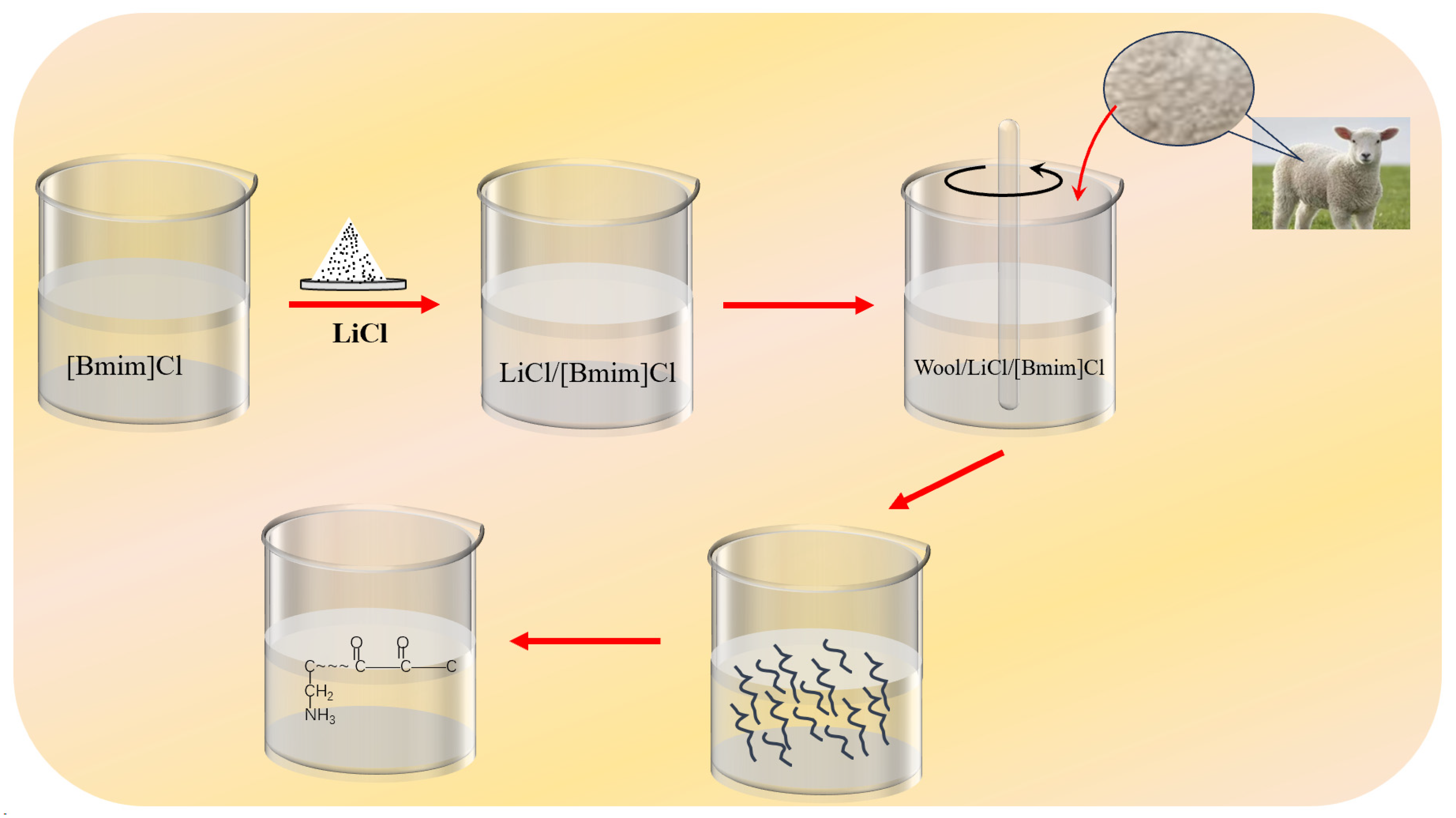
2.1. LiCl/[Bmim]Cl Solution Configuration

In a beaker containing 10 g of [Bmim]Cl ionic liquid, varying amounts of lithium chloride (relative to the ionic liquid mass fraction of 2, 4, 6, 8, and 10%, respectively) were added after the solution reached 80 °C on a magnetic heating stirrer.

2.2. Dissolution and Regeneration of Waste Wool

A specific amount of sheared, dried wool was immersed in the prepared LiCl/[Bmim]Cl mixed solution under controlled dissolution temperature and time. Wool dissolution under a hot stage polarizing microscope was regularly comprehended using a glass rod dipped in the dissolving solution. The wool was deemed completely dissolved if no obvious wool fibres were observed, and the solution appeared clear and transparent; otherwise, stirring was continued until dissolution. This process is depicted in Figure 1.
The solubility was assessed using Equation (1) as follows:
S = 1 m 1 m 0 × 100 %
where, S denotes solubility in %, m0 denotes the initial mass of wool before dissolution (in grams), and m1 denotes the mass of the wool remaining undissolved (in grams).
For the regeneration of keratin, the keratin/LiCl/[Bmim]Cl mixture obtained after the dissolution process was poured into anhydrous ethanol and agitated to reconstitute the keratin. After a period of sedimentation, the solution was filtered using a circulating vacuum pump and rinsed 3–5 times with anhydrous ethanol; subsequently, it was left to dry before experimentation.

2.3. Performance Testing

The solubility measurements of LiCl in [Bmim]Cl were performed at varying temperatures by employing the HX-Y turbidity analyser. A clear glass vial holding an ionic liquid (15 mL) and a magnetic stirrer was positioned in a heated oil bath, and lithium chloride (10 mg) powder was added. The dissolution process was allowed to proceed for at least 15 min after each addition until a steady NTU value was accomplished [47]. This enabled the expression of the solubility as the molar fraction of LiCl in [Bmim]Cl using Equation (2)
C = n/v
where C represents molar concentration, n represents the amount of LiCl substance, and v represents the volume of [Bmim]Cl.
The molecular weights of wool keratin that were attained at various dissolution temperatures and times were determined via the SDS-PAGE method. For these measurements, the keratin concentration dissolved in ionic liquids was maintained at 10% for separating gels and 5% for concentrating gels.
For observing the wool dissolution process, a MICROSCOPEBX51-P hot-stage polarizing microscope acquired from Olympus, Tokyo, Japan, was utilised under a dark field of view. The hot-stage operating temperature was 600 °C (max), with a precision of ±0.5 °C.
Modifications to the keratin complexes before and after the process of regeneration were examined via a D8 ADVANCE X-ray diffractometer (Bruker, Mannheim, Germany). The scanning speed was set at 4°/min, with a 1 mm slit, a step width of 0.02°/s, a tube voltage of 40 kV, and a current of 200 mA.
Similar modifications were also texted using SAXSessMC2, a small-angle X-ray scattering device acquired from Anton Paar GmbH, Graz, Austria, to conduct structural analysis of the keratin.
Infrared spectroscopy was carried out via a Nicolet 6700 infrared spectrometer acquired from Thermo Fisher Company, Waltham, MA, USA, to assess the chemical structure of raw and regenerated wool. For this process, samples were prepared via the potassium bromide press method and heated for 3 min in a quick dryer. Other settings included a scanning speed of 0.2 cm/s with 32 scans and a scanning range of 400 to 4000 cm−1.
The behaviour of keratin assembly that adsorbed onto glass surfaces was investigated using a BX51-P model scanning probe microscope in the tapping mode at ambient temperature. The morphological features of the keratins adsorbed onto glass surfaces were provided using 3D morphology maps.

3. Results

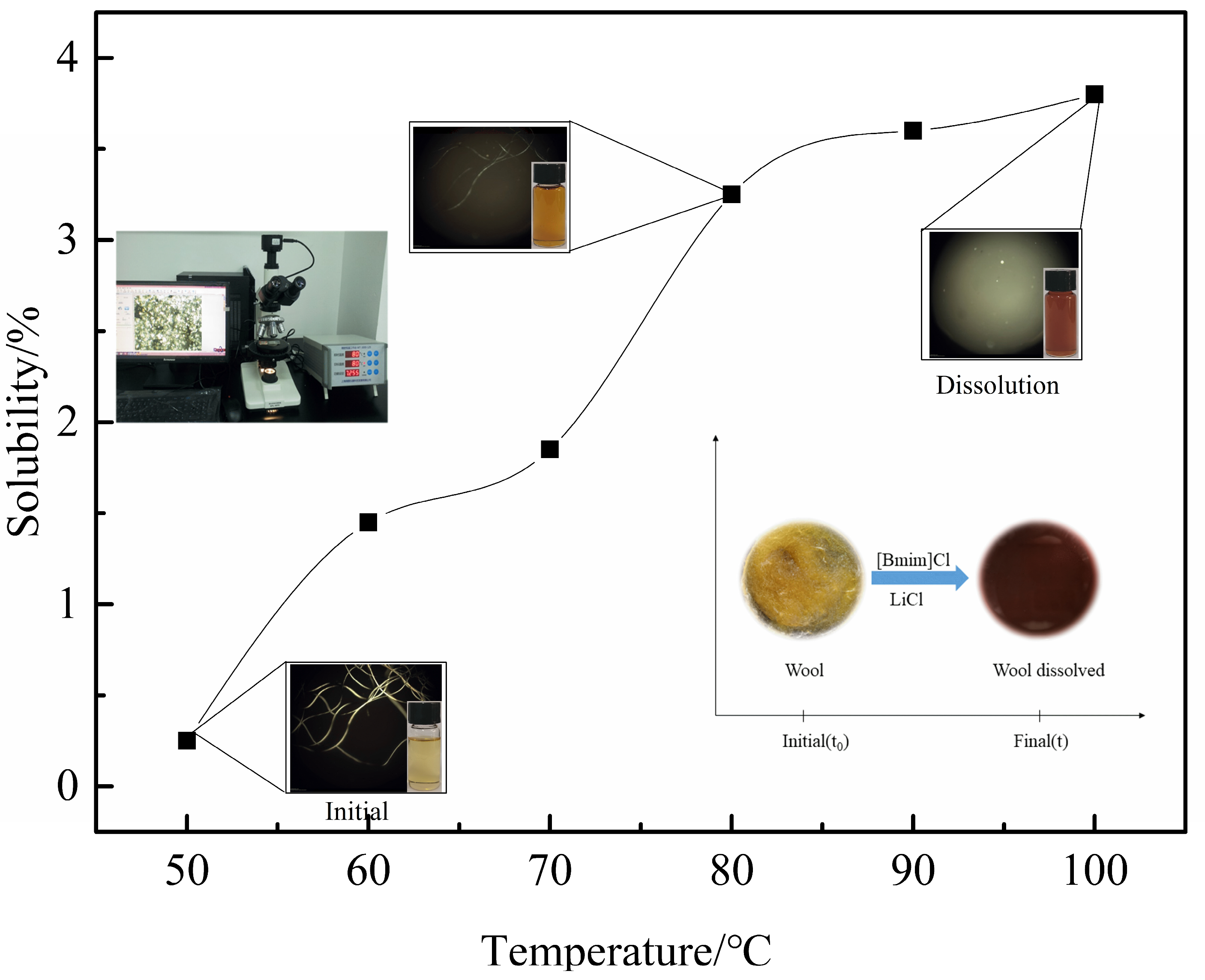
3.1. Solubility of LiCl in [Bmim]Cl

The dissolution process of LiCl in [Bmim]Cl is illustrated in Figure 2.
It is evident from the figure that at A low temperature (50 °C), the solubility rate of LiCl in [Bmim]Cl is slow, and some LiCl remains undissolved. However, as the temperature rises, the dissolution rate of LiCl in [Bmim]Cl accelerates, and when temperatures touch 100 °C, the solubility of anhydrous lithium chloride continues to amplify with increasing temperature, albeit at a lower rate [48,49]. The reduced pace of dissolution is perhaps due to the exothermic characteristic of hydration that occurs when the lithium chloride is dissolved. At 130 °C, the molar fraction reaches 0.60, leading to the formation of a transparent solution. This indicates the excellent solubility of LiCl in [Bmim]Cl.

3.2. Impact of LiCl Content on the Dissolution Process of Waste Wool

The dissolution outcomes of waste wool in a LiCl/[Bmim]Cl mix solution with different LiCl contents (relative to ionic liquid mass fraction 0, 2, 4, 6, 8, and 10%) at a dissolution temperature of 80 °C are tabulated below (refer to Table 1).
From the data tabulated in Table 1, it is clear that with a simultaneous increase in the LiCl fraction in the LiCl/[Bmim]Cl mixture solution, the dissolution time of the same wool mass decreases at a constant temperature. Upon using a 10% LiCl fraction, the dissolution time of the wool was the shortest. This was three times less compared to when an unadulterated ionic liquid was used. Possible reasons for this enhancement in dissolution [44] velocity could be the interaction of the Cl ion in the LiCl/[Bmim]Cl mix solution with H ion in the keratin –OH, fostering keratin dissolution progression, or the increased concentration of Cl ion separated in the LiCl and [Bmim]Cl ionic liquids mix solution, disrupting the multifaceted hydrogen bonding network in the wool fibre, resulting in improved keratin dissolution.

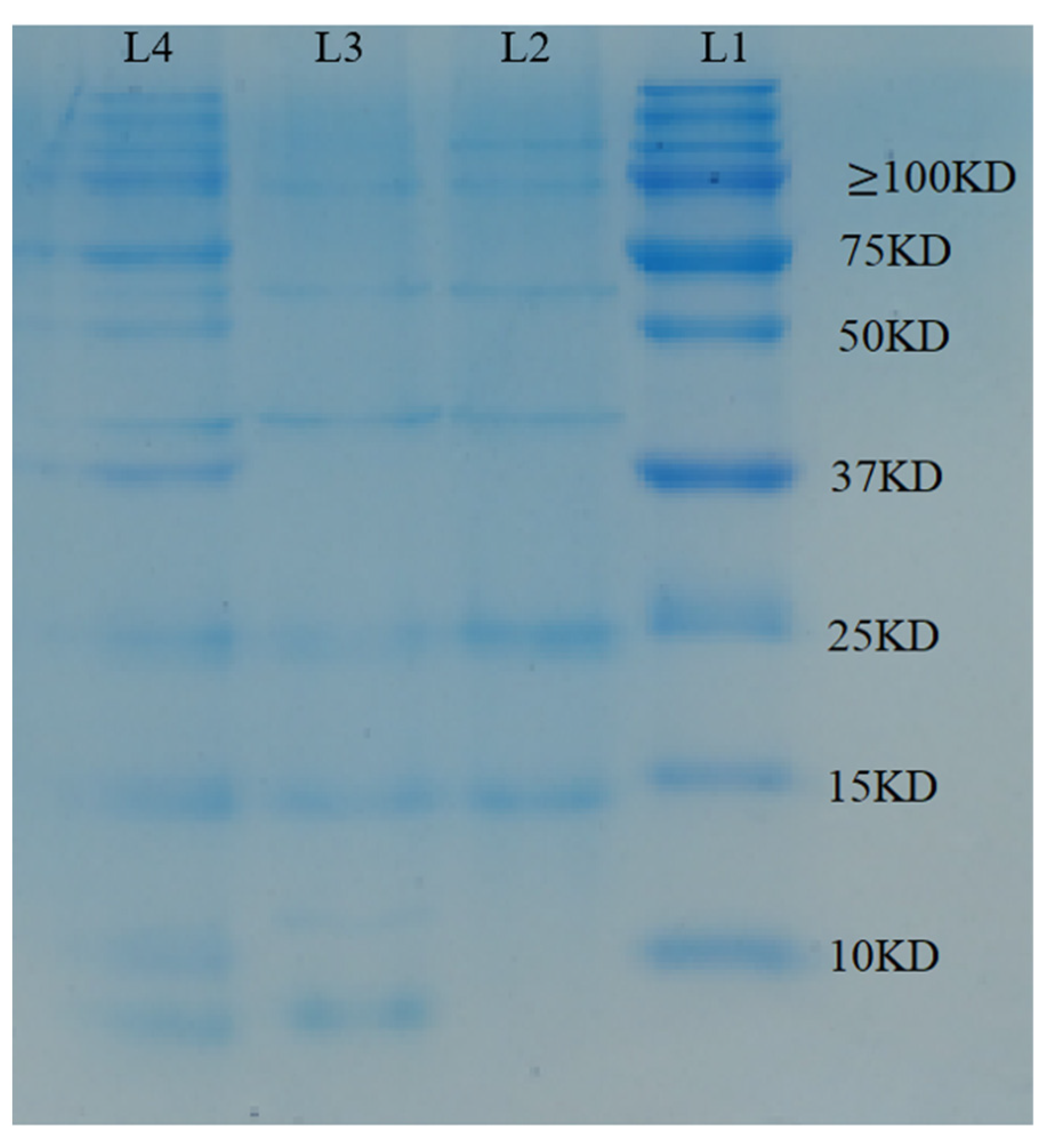
3.3. Impact of Diverse Dissolution Durations on Waste Wool’s Solubility and Molecular Weight

The data in Table 2 indicate that waste wool’s solubility in LiCl/[Bmim]Cl solution progressively increases with the extension of the dissolution duration at an 80 °C dissolution temperature. This increase can be ascribed to the solution’s movement across the scale layer to the cortical layer, which contains less cystine. Over time, the solution penetrates into the scale layer, which is challenging due to the highly cross-linked disulfide bonds and increased dissolution duration, leading to improved solubility.
Molecular weight tests and keratin extraction were performed on the keratin solution formed at the same dissolution temperature of 80 °C with different dissolution time durations of 30, 60, and 90 min. The results are shown in Figure 3.
L1 refers to the standard protein’s molecular weight, and L2 (30 min), L3 (60 min), and L4 (90 min) represent dissolved keratin at different durations.
Figure 3 clearly illustrates that the standard protein’s molecular weight spreads between 10 and 250 kDa. Similarly, the majority of the regenerated keratins also fall within the 10–250 kDa range. An interesting observation is the larger concentration of keratins with molecular weights of less than 10 kDa, an occurrence that correlates with extended dissolution sessions. The molecular weight of proteins is dictated by the length of their backbones and the number of involved amino acids [50,51,52]. The interaction of Cl ions in LiCl/[Bmim]Cl and Bmim+ with the wool’s macromolecular chain results in an interesting dynamic. Over an extended solubilisation period, these ionic liquids diffuse more effectively from the squamous layer to the cortical layer, a region characterised by a lower cystine content, thus inducing significant keratin degradation. This degradation disrupts major chains containing disulphide bonds [53]. Furthermore, the small-molecular-weight free amino acids produced during this process are removed during subsequent dialysis. This combination of effects leads to a reduced molecular weight in the final keratin solution.

3.4. Impact of the Dissolution Temperature on the Waste Wool’s Dissolution Process

(1)
Influence of the Dissolution Temperature on the Waste Wool’s Dissolution Rate
A study was conducted on the LiCL mass fraction of 10% to understand the effect of temperature on the dissolution rate of waste wool in a LiCl/[Bmim]Cl mixed solution. Solubility calculations were carried out according to Equation (2), and the results are displayed in Figure 4.
Figure 4 depicts the impact of the dissolution temperature on wool dissolution. As demonstrated in the figure, between the temperatures of 50 and 60 °C, the dissolution rate escalates noticeably. This phenomenon may be attributed to the rapid dissolution of the wool fibres’ amorphous zone in contact with the ionic liquid. However, with lower dissolution temperatures, the ionic liquid finds it challenging to reach the crystalline zone. At higher temperatures of 60–70 °C, the undissolved wool fibres are enveloped by a concentrated keratin/LiCl/[Bmim]Cl solution, impeding the LiCl/[Bmim]Cl solution’s penetration into the inner layer of the undissolved fibres. This results in non-uniform wool fibre dissolution, causing a decrease in the dissolution rate. Yet, as the temperature increases (70–100 °C), the individual Cl separates from the LiCl and [Bmim]Cl ionic liquids due to the temperature, simultaneously increasing the free Bmim+ ions. During the dissolution process, the free Cl and keratin’s –OH engage the role of hydrogen (H), and the free Bmim+ and –OH undertake the role of oxygen (O), breaking down the complex hydrogen bonding network in the wool fibres and enabling the dissolution of keratin. As the dissolution temperature rises, the ionic liquids’ viscosity diminishes, augmenting the anion and cation mobility, thus facilitating penetration and diffusion into the fibre interior, which, in turn, increases the rate of dissolution, ultimately leading to the dissolution of wool fibres.
(2)
Impact of the Dissolution Temperature on Molecular Weight
Keratin solutions generated at varying dissolution temperatures (L2 = 60 °C, L3 = 80 °C, L4 = 100 °C) underwent keratin extraction for molecular weight tests, and the results are displayed in Figure 5.
As Figure 5 shows, the molecular weight distribution of the standard protein ranges from 10 to 250 kDa. However, at higher temperatures, demonstrated by L4, there’s an increase in molecular weights below 10 kDa. This may be attributed to the higher dissolution temperatures causing an increase in free Cl-, disrupting the complex hydrogen bonding network in wool fibres, thus promoting further keratin protein dissolution. Together with the increasing dissolution temperature, the viscosity of the ionic liquid reduces, surging the mobility of anions and cations. This elevation enables easier penetration and diffusion into the fibre’s interior, breaking the keratin’s molecular chain, resulting in the formation of products with lower molecular weights.

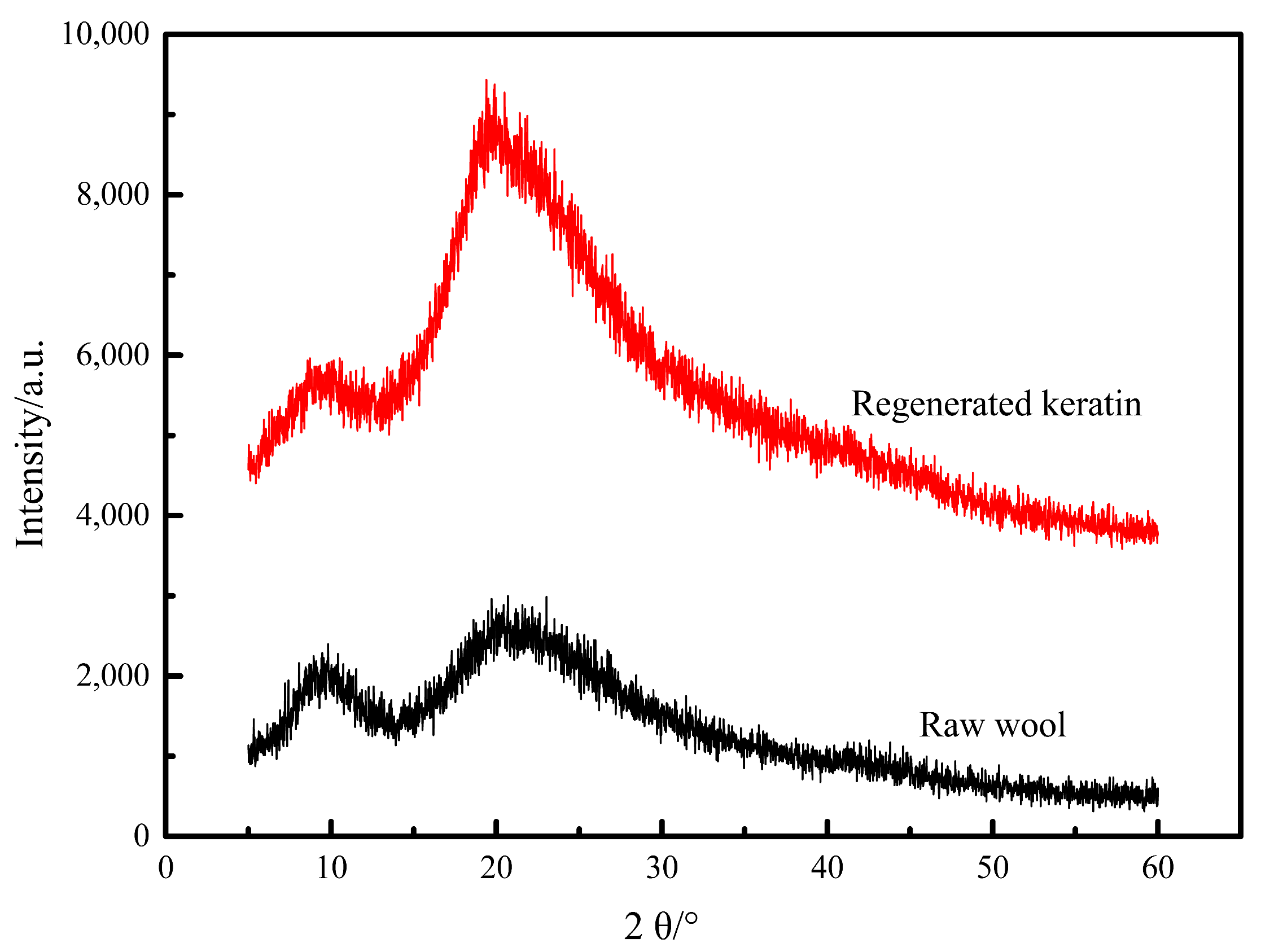
3.5. X-ray Diffraction Test Results Analysis

Both pre- and post-regeneration wool keratin was scrutinised via X-ray diffraction. XRD tests were then conducted to compare any changes within the wool fibres’ crystalline regions before and after regeneration.
It compares the XRD patterns of regenerated keratin to the original wool keratin [53,54]. In both the original and the regenerated wool keratin, two characteristic peaks appear: the diffraction peaks observed at 2θ = 9° and 2θ = 20° are primarily due to α-helix and β-folding structures, respectively. Relative to the original wool, the peak intensity at 2θ = 9° for regenerated wool keratin decreased, while a heightened peak intensity was noted at 2θ = 20°. These observations indicate that while the secondary structure of the original wool is preserved in regenerated wool keratin, damage occurred to the α-helical structure during the solubilisation process, contributing to a reduction in α-helices. Both these diffraction peaks are characteristic of α-crystalline and β-crystalline structures [55]. The area of the diffraction peak for regenerated keratin is larger and narrower compared to that of the original wool keratin. This could suggest that rapid precipitation of LiCl/[Bmim]Cl from the keratin/LiCl/[Bmim]Cl solution during the regeneration process led to increased crystallinity and larger grains in the regenerated keratin. Alternatively, inadequate sample preparation–grinding might have introduced the particle effect, causing an increase in the peak intensity of the crystalline surface.

3.6. Cites an Analysis of the X Small-Angle Scattering Test Results

Figure 6 presents the one-dimensional mapping of small-angle X-ray scattering from the original wool and the regenerated keratin complexes. This mapping signifies the correlation between the scattering intensity vector q and the log of scattering intensity (I). As depicted in Figure 7, very low q values (regenerated keratin = 0.95, raw wool = 0.88) exhibit strong Bragg peaks. As the scattering vector increases, the polymer’s scattering intensity in the solution progressively declines. At relatively higher scattering intensities (q > 0.75), this culmination is characterised by broad, weak scattering intensities with advanced, absent structural peaks. The structural peaks’ absence indicates the lack of advanced structures in the lithium chloride/ionic liquid/keratin mixture.
Interpreting the data from XRD, it can be seen that wool keratin assumes more linear clusters or irregular shapes after the decrease in α-helix and the advance in folding structures during dissolution. Assuming linear and thread clusters can establish a consistent conformation with a regular structure manifesting in the solution [56], it can potentially lead to the formation of further liquid crystals. This finding establishes an indispensable foundation for future industrial applications of keratin complexes as a liquid crystal spinning base solution. Additionally, it paves the way for accurate analysis of the tertiary or higher-order structures of wool keratin by integrating protein structure prediction techniques into the ongoing research.

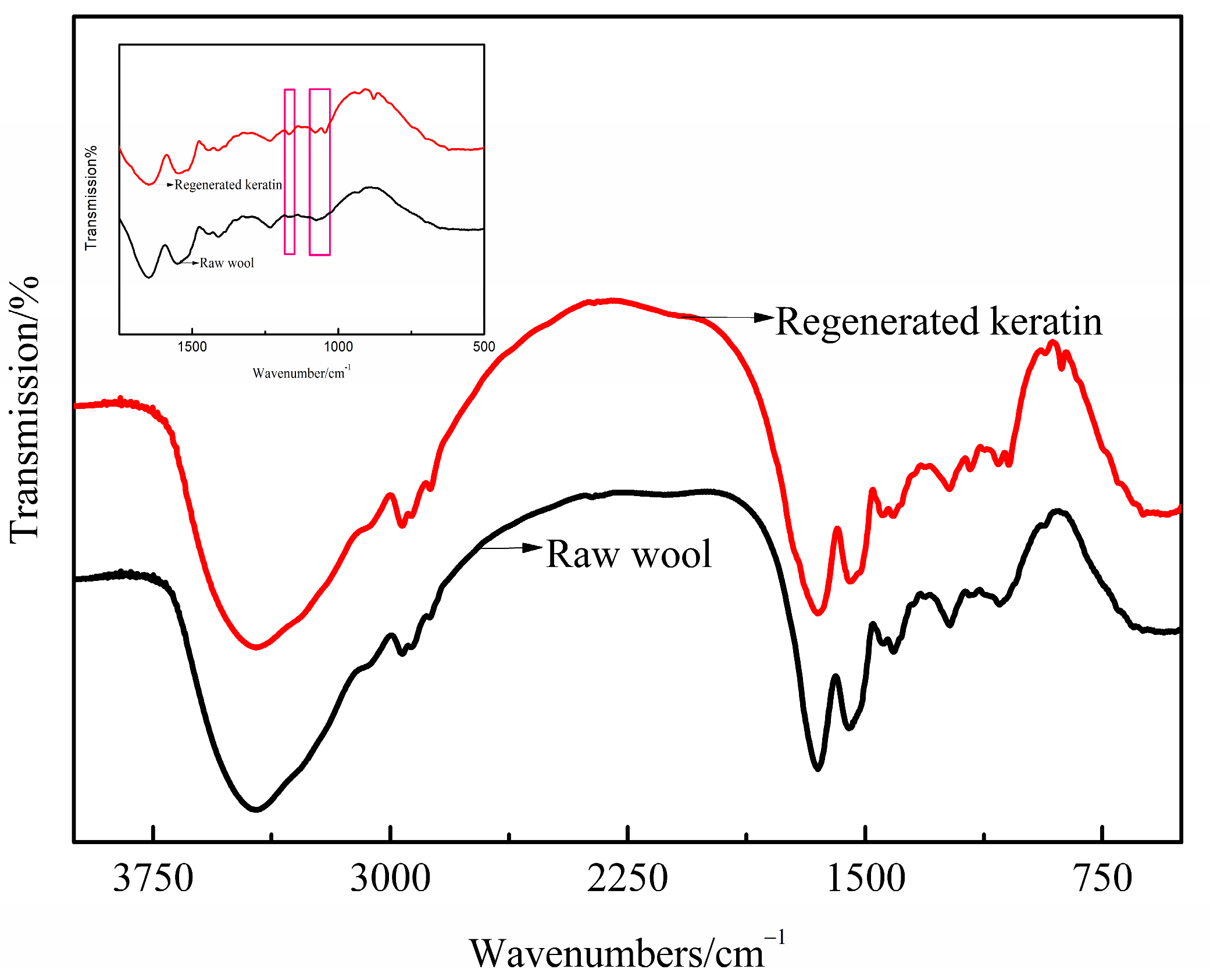
3.7. Consists of an Analysis of Infrared Spectroscopy Results

Before and after regeneration, wool keratin was assessed through infrared spectroscopy to determine any structural changes in the wool fibre.
The broad, strong absorption bands at 3431 cm−1 and 3439 cm−1 represent the stretching bands of N–H and O–H. The keratin’s amide octave peaks are observable at 3001 cm−1. Notably, a characteristic absorption of polypeptide near the 2890 cm−1 wavelength is apparent. The absorption peaks at 2998 cm−1 and 2994 cm−1 are due to the expansion and contraction of the C–H bonds within the molecule’s methyl and ethyl groups. Meanwhile, the absorption peaks at 1649 cm−1 and 1645 cm−1 result from the strong expansion and contraction of the carbonyl group (C=O). This suggests a structural change in the wool from α-helix to β-helix. Absorption peaks at 1532 cm−1 and 1545 cm−1 denote the keratin’s amide, while peaks at 1532 cm−1 and 1535 cm−1 are attributed to the bending vibration of the N-H bond and the stretching vibration of the C–N bond in the amide II band. The peaks at 1452 cm−1, 1448 cm−1, 1384 cm−1 and 1385 cm−1 are caused by the bending vibration of CH3 and CH2. The pronounced peaks at 1319 cm−1 and 1323 cm−1 can mainly be ascribed to the stretching vibration of the C–N bond. Meanwhile, the strong absorption peak of recycled wool at 1143 cm−1 is due to the stretching vibration of the S–O bond in cystine, indicating a conversion from the naturally occurring disulfide bond in cystine into a new cysteine [57,58,59].
In the spectra, wool fibres exhibit strong absorption peaks at wavelengths of 1650 cm−1 and 1537 cm−1, i.e., the amide I and II bands of the α-helical conformation, and at a wavelength of 1232 cm−1, representing the amide III band of the β-folded conformation. This reflects the fact that the wool fibres’ conformation predominantly exists in the α-helical state, with the existence of a β-folded conformation. Strong absorption peaks at the wavelengths of 1653 cm−1 and 1536 cm−1 signify a considerable number of α-helical conformations in the regenerated wool keratin, with amide bonds and polypeptides being present, which belong to the proteins. Consequently, the structure does not significantly change before and after regeneration.
Figure 8 presents two characteristic absorption peaks at 1170 cm−1 and 1080 cm−1, representing sulphonylalanine and cystine monoxide, respectively, along with a weaker shoulder peak at 1040 cm−1 that is allotted to cysteamine sulfonate [60,61]. These absorptions suggest the occurrence of disulfide bond breakage during dissolution, leading to the production of sulphonylalanine. Broadly, three key points arise from a review of similar past research. Firstly, using Li+Cl/[BMIM]+Cl as a solvent triggers the dissolution process from the surface lipids of the wool. The thioester bond of the lipids present on the wool’s fibre surface is targeted by protonophilic Cl. Secondly, under high temperatures, the protein macromolecules within the wool are disassembled and dissolved by Cl, leading to increased molecular mobility and the generation of slippage. Lastly, any ionic liquid and minute quantities of water present in the wool fibre intensify water molecule ionisation to produce H+ and OH, which subsequently react with the cystine disulfide bond [62,63]:
R-S-S-R′ + H+ + OH ⟶ R-SOH + R′-SH
This leads to the fracturing of the disulfide bond. Given the ease with which the resultant sulfenic acid is oxidised in air
2R-SOH + O2 ⟶ 2R-SO2H
5R-SO2H ⟶ R-S-S-R′ + 3R-SO3H + H2O
Therefore, the infrared absorption peak of the sulphoalanine R-SO3H product can hence be observed in the regenerated form.

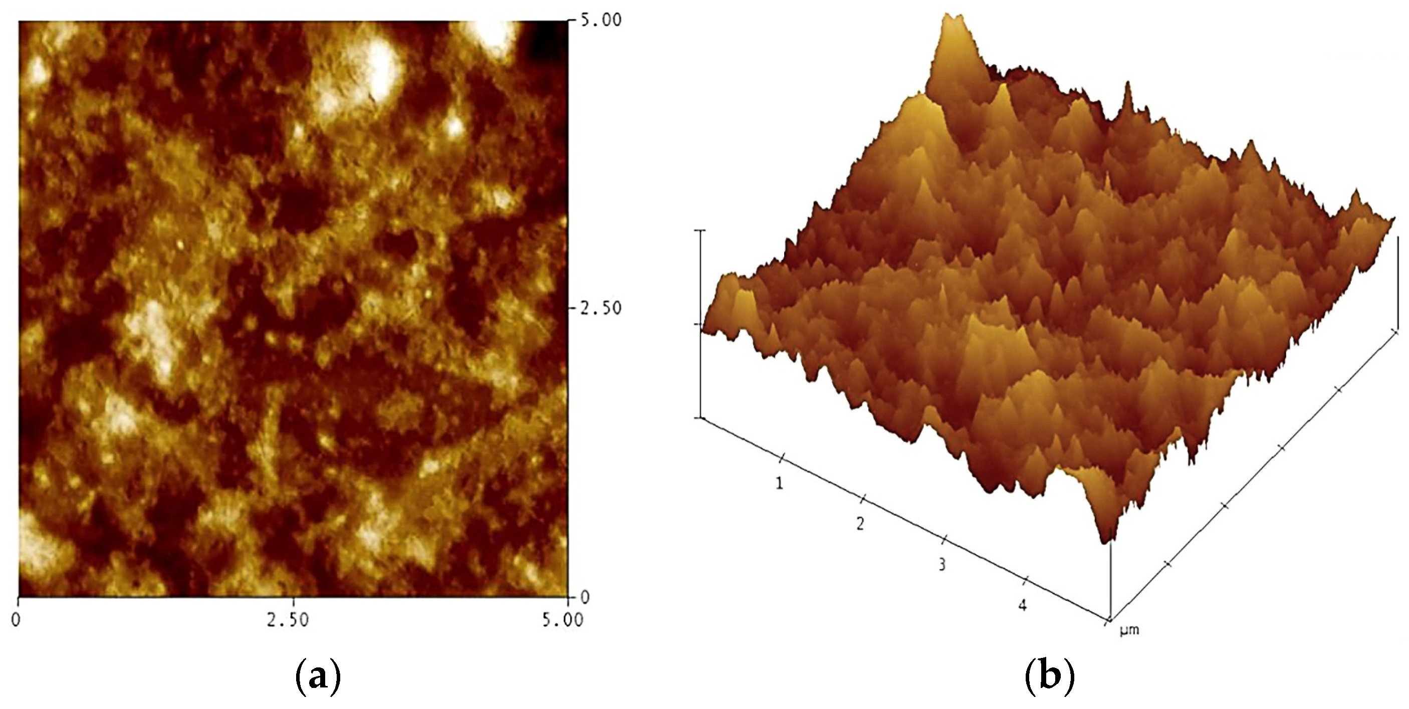
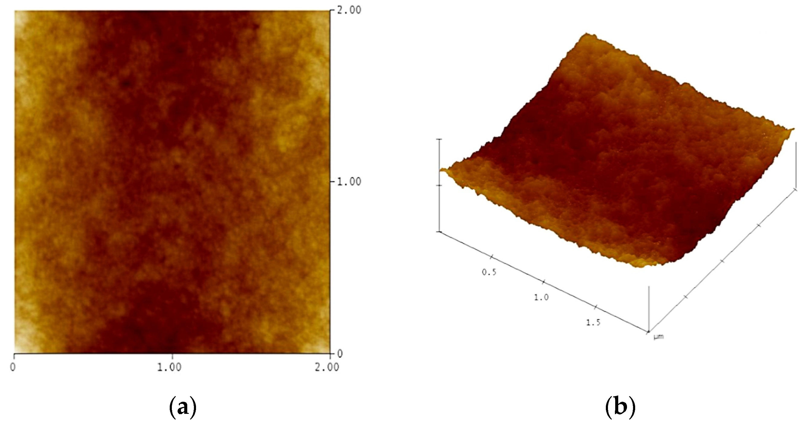
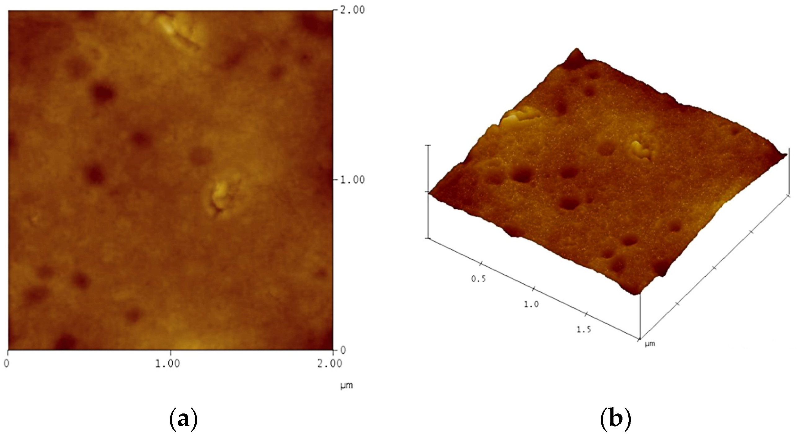
3.8. Presents the Atomic Force Microscopy

The research investigates the keratin’s assembly behaviour adsorbed on glass surfaces in mixed solutions at room temperature, establishing foundational knowledge for subsequent studies of keratin absorption on textile materials. Three-dimensional morphology maps, showcasing morphological features, have been obtained in tap-mode appearance and are displayed in Figure 9, Figure 10 and Figure 11.
The 3D map of the atomic force microscopy (AFM) experimental results demonstrates that, following the removal of the ionic liquid, keratin densely overlays the coverslip substrate’s surface, thereby obscuring the keratin monomolecular layer. With increasing keratin concentration, the surface roughness of the keratin-grown substrate decreases, thus becoming progressively smoother [64].
As keratin concentration increases, the surface roughness of the substrate-grown keratin decreases, resulting in a progressively smoother surface. An increase in substrate keratin leads to a decrease in the absolute value of the image cross-section’s peak amplitude, suggesting a reduction in the height of the image’s keratin surface troughs and crests. This reflects the formation of a more continuous and uniform gel network structure, yielding a smoother and flatter surface [62,65,66]. At lower concentrations, keratin molecules have the opportunity to unfold and grow in the substrate. When the keratin concentration reaches a specific threshold, the aggregation of protein macromolecules begins to occur. Furthermore, as the keratin concentration increases, intra- and intermolecular disulfide bonds and hydrogen bonds within keratin interconnect, forming a network that smooths the surface of macromolecule aggregation.
Aggregates or agglomerates of protein molecules in the keratin/LiCl/[Bmim]Cl mixed solution are typically experienced during crystal growth. These aggregates often exhibit disordered clustering, while orderly stacking throughout the aggregation process is a critical step in crystal growth [67,68,69]. Combining small-angle X-ray scattering (SAXS) results and analysis positions us to explore whether keratin can grow or form a continuous film on fibre (specifically modified fibres) surfaces. The integration of atomic force microscopy (AFM) in these studies holds promise for advancing protein-based biomaterials within the textile finishing domain.

4. Conclusions

LiCl is highly soluble, up to a 0.60 molar fraction, in [Bmim]Cl. Waste wool, and when dissolved in a 10% LiCl/[Bmim]Cl solution, it lowers the dissolution temperature to 80 °C and the dissolution time to 46 min. A combination of XRD spectra and small-angle X-scattering spectra confirmed that the keratin microstructure underwent changes during the dissolution process. The infrared spectra depicted the disassembly of the hydrogen bond in the wool protein macromolecule under the influence of the ionic liquid. This signifies the degradation and disassembly of the disulfide bond, with the wool keratin gradually dissolving in the ionic liquid; given that keratin is abundant in cystine, it consequently has a particularly high concentration of disulfide bonds. These bonds play a critical role in cross-linking within the protein peptide chain, rendering keratin exceptionally stable in terms of its chemical properties and bestowing it with high mechanical strength. SDS-PAGE testing of the dissolved keratin’s molecular weight revealed a broad distribution, signified by a continuous band distribution, primarily within the range of 25–250 kD, indicating a mixture of various products. A keratin extract was procured through filtration following dissolution. Subsequent to the addition of a viscosity-increasing agent to this extract, a high-viscosity spinning solution was formulated. This solution gave rise to robust, pure keratin fibres through wet spinning via dry spraying or utilising microstructures such as the basal protofibrils of keratin. These fibres, upon self-assembly, were found to possess superior physico-mechanical properties. This process, thus, optimised the molecular weight of keratin. As displayed in the AFM 3D diagram, keratin densely covers the coverslip substrate surface after the removal of ionic liquid, making the keratin monomolecular layer hard to detect. This provides the premise to examine whether keratin can grow or form a continuous film on the substrate’s surface.
Employing wool keratin (WK) as a foundational biomaterial for fabricating high-precision protein microstructures could offer numerous benefits. With minimal alterations to its structure and function, wool keratin can be biochemically modified to become photosensitive. This modification enables the coating of relevant fabrics to present photosensitive qualities and allows the creation of high-precision protein prints on fabrics, thereby revolutionising the field. Moreover, as the principal component of wool fibre, keratin aligns 91% structurally with human keratin (K-17), ensuring optimal absorption by the human body. Proteins like functional keratin and cysteine maintain their biological activity under these circumstances. Boasting film-forming properties, keratin has potential in skincare, tissue repair and regeneration, and vast health-related applications.

Author Contributions

Literature research and discussion, M.W., H.C., H.J. and L.D.; writing, M.W.; methodology, J.Z. (Jinli Zhou) and G.Z.; supervision, J.Z. (Jinli Zhou); project administration and funding acquisition, M.W., J.Z. (Junjie Zheng) and G.Z. All authors have read and agreed to the published version of the manuscript.

Funding

This work was supported by the Scientific and Technological Project in Henan Province (No. 212102210550 and No. 212102210036), the Key Laboratory of Science and Technology of Eco-Textiles, Ministry of Education, Donghua University (No. 2232021G-04), ND the Science and Technology Guidance Project of China National Textile and Apparel Council (No. 2022018).

Institutional Review Board Statement

Not applicable.

Informed Consent Statement

Not applicable.

Data Availability Statement

The raw/processed data required to reproduce these findings cannot be shared at this time as the data also form part of an ongoing study.

Conflicts of Interest

The authors declare no conflict of interest.

References

  1. Martin, M.; Herlaar, S. Environmental and social performance of valorizing waste wool for sweater production. Sustain. Prod. Consum. 2021, 25, 425–438. [Google Scholar] [CrossRef]
  2. Sun, Y.; Li, B.; Zhang, Y. The progress and prospect for sustainable development of waste wool resources. Text. Res. J. 2023, 93, 468–485. [Google Scholar] [CrossRef]
  3. Zhang, C.; Xia, L.; Zhang, J. Utilization of waste wool fibers for fabrication of wool powders and keratin: A review. J. Leather Sci. Eng. 2020, 2, 1–15. [Google Scholar] [CrossRef]
  4. Cataldi, P.; Condurache, O.; Spirito, D. Keratin-graphene nanocomposite: Transformation of waste wool in electronic devices. ACS Sustain. Chem. Eng. 2019, 7, 12544–12551. [Google Scholar] [CrossRef]
  5. Giteru, S.G.; Ramsey, D.H.; Hou, Y. Wool keratin as a novel alternative protein: A comprehensive review of extraction, purification, nutrition, safety, and food applications. Compr. Rev. Food Sci. Food Saf. 2023, 22, 643–687. [Google Scholar] [CrossRef]
  6. Ma, H.; Shen, J.; Cao, J. Fabrication of wool keratin/polyethylene oxide nano-membrane from wool fabric waste. J. Clean. Prod. 2017, 161, 357–361. [Google Scholar] [CrossRef]
  7. Patankar, K.C.; Maiti, S.; Singh, G.P. Chemically modified wool waste keratin for flame retardant cotton finishing. Clean. Eng. Technol. 2021, 5, 100319. [Google Scholar] [CrossRef]
  8. Figoli, A.; Ursino, C.; Sanchez Ramirez, D.O. Fabrication of electrospun keratin nanofiber membranes for air and water treatment. Polym. Eng. Sci. 2019, 59, 1472–1478. [Google Scholar] [CrossRef]
  9. El-Sayed, H.; Abou-Taleb, M. Polypropylene fabric of durable hydrophilic character using plasma-mediated surface coating with keratin. J. Adhes. Sci. Technol. 2023, 1–15. [Google Scholar] [CrossRef]
  10. Basuoni, A.S.; Sabaa, M.W.; Mohamed, R.R. An eco-friendly strategy for grafting keratin onto polyacrylonitrile fabrics using bi-functional amino acids as coupling agents. J. Adhes. Sci. Technol. 2023, 1–16. [Google Scholar] [CrossRef]
  11. Kooshamoghadam, N.; Zohoori, S.; Bekrani, M. Enhancing physical properties of viscose by preparing viscose/keratin/nano ZnO composite fabric. J. Nat. Fibers 2022, 19, 4846–4853. [Google Scholar] [CrossRef]
  12. Dickerson, M.B.; Sierra, A.A.; Bedford, N.M. Keratin-based antimicrobial textiles, films, and nanofibers. J. Mater. Chem. B 2013, 1, 5505–5514. [Google Scholar] [CrossRef]
  13. Khalilipourroodi, K.; Dadashian, F.; Solouk, A. Effect of extraction method on properties of feather keratin grafted modified cotton nonwoven fabric for biomedical applications. J. Ind. Text. 2022, 51, 2558S–2575S. [Google Scholar] [CrossRef]
  14. Zhang, Z.; Nie, Y.; Zhang, Q. Quantitative change in disulfide bonds and microstructure variation of regenerated wool keratin from various ionic liquids. ACS Sustain. Chem. Eng. 2017, 5, 2614–2622. [Google Scholar] [CrossRef]
  15. Huson, M.G. Properties of wool. In Handbook of Properties of Textile and Technical Fibres; Woodhead Publishing: Cambridge, UK, 2018; pp. 59–103. [Google Scholar]
  16. Das, S. Physical and Chemical Properties of Wool. Sheep Wool Mutton Prod. Value Addit. 2021, 7, 91–95. [Google Scholar]
  17. Hearle, J.W.S. A critical review of the structural mechanics of wool hair fibers. Int. J. Biol. Macromol. 2000, 27, 123–138. [Google Scholar] [CrossRef] [PubMed]
  18. Rajabinejad, H.; Zoccola, M.; Patrucco, A. Physicochemical properties of keratin extracted from wool by various methods. Text. Res. J. 2018, 88, 2415–2424. [Google Scholar] [CrossRef]
  19. Sehgal, P.K. Studies on solubilized keratins from poultry feathers. Leather Sci. 1986, 33, 333–343. [Google Scholar]
  20. Toran-Diaz, I.; Jain, V.K.; Allais, J.J. Effect of acid or enzymatic hydrolysis on ethanol production by zymomonas mobilis growing on Jerusalem artichoke juice. Biotechnol. Lett. 1985, 7, 527–530. [Google Scholar] [CrossRef]
  21. Zhang, J.; Li, Y.; Li, J. Isolation and characterization of biofunctional keratin particles extracted from wool wastes. Powder Technol. 2013, 246, 356–362. [Google Scholar] [CrossRef]
  22. Eslahi, N.; Dadashian, F.; Nejad, N.H. Optimization of Enzymatic Hydrolysis of Wool Fiber for Nanoparticles Production Using Response Surface Methodology. Adv. Powder Technol. 2013, 24, 416–426. [Google Scholar] [CrossRef]
  23. Mutate, H.; Shigematsu, M.; Abe, K. Molten urea as a novel dissolving solvent for wool and other animal proteins. Sen’i Gakkaishi 2010, 66, 56–159. [Google Scholar]
  24. Cardamone, J.M. Investigating the microstructure of keratin extracted from wool: Peptide sequence (MALDI-TOF /TOF) and protein conformation (FTIR). J. Mol. Struct. 2010, 969, 97–105. [Google Scholar] [CrossRef]
  25. Zeng, C.H.; Lu, Q. Study on the recovery of waste wool by combining reduction and metallic salt methods. Adv. Mater. Res. 2014, 881, 551–555. [Google Scholar] [CrossRef]
  26. Li, B.; Huang, C.; Yang, X. Preparation and characterization of electrospun wool keratin/polyethylene oxide nanofibers for air filtration applications. Dig. J. Nanomater. Biostructures DJNB 2017, 12, 293–301. [Google Scholar]
  27. Sajura; Zhao, X.; Miyag, A.; Tsuyoshi, W.A.; Mitsuhiko, K. Mechanism of wool keratin extraction by molten urea method. South Agric. Sci. 2020, 49, 174–180. [Google Scholar]
  28. Sun, Z.; Li, H.; Sun, M.; Liang, Z. Study on resolubility of recovered wool keratin Textile. Sci. Technol. 2019, 47, 54–59. [Google Scholar]
  29. Pu, Y.; Jiang, N.; Ragauskas, A.J. Ionic liquid as a green solvent for lignin. J. Wood Chem. Technol. 2007, 27, 23–33. [Google Scholar] [CrossRef]
  30. Welton, T. Ionic liquids in green chemistry. Green Chem. 2011, 13, 225. [Google Scholar] [CrossRef]
  31. Mallakpour, S.; Dinari, M. Ionic liquids as green solvents: Progress and prospects. Green Solvents II Prop. Appl. Ion. Liq. 2004, 41, 41–43. [Google Scholar]
  32. Plechkova, N.V.; Seddon, K.R. Ionic liquids: “designer” solvents for green chemistry. In Methods and Reagents for Green Chemistry: An Introduction; John Wiley & Sons, Inc.: Hoboken, NJ, USA, 2007; pp. 103–130. [Google Scholar]
  33. Earle, M.J.; Seddon, K.R. Ionic liquids. Green solvents for the future. Pure Appl. Chem. 2000, 72, 1391–1398. [Google Scholar] [CrossRef]
  34. Hussey, C.L. Room temperature haloaluminate ionic liquids. Novel solvents for transition metal solution chemistry. Pure Appl. Chem. 1988, 60, 1763–1772. [Google Scholar] [CrossRef]
  35. Marsh, K.N.; Deev, A.; Wu, A.C.T. Room temperature ionic liquids as replacements for conventional solvents–A review. Korean J. Chem. Eng. 2002, 19, 357–362. [Google Scholar] [CrossRef]
  36. Tonin, C.; Zoccola, M.; Aluigi, A.; Varesano, A.; Montarsolo, A.; Vineis, C.; Zimbardi, F. Study on the conversion of wool keratin by steam explosion. Biomacromolecules 2006, 7, 3499–3504. [Google Scholar] [CrossRef] [PubMed]
  37. Peng, Z.; Zhang, J.; Du, G. Keratin waste recycling based on microbial degradation: Mechanisms and prospects. ACS Sustain. Chem. Eng. 2019, 7, 9727–9736. [Google Scholar] [CrossRef]
  38. Qiu, J.; Wilkens, C.; Barrett, K. Microbial enzymes catalyzing keratin degradation: Classification, structure, function. Biotechnol. Adv. 2020, 44, 107607. [Google Scholar] [CrossRef]
  39. Holkar, C.R.; Jain, S.S.; Jadhav, A.J. Valorization of keratin based waste. Process Saf. Environ. Prot. 2018, 115, 85–98. [Google Scholar] [CrossRef]
  40. Xie, H.; Li, S.; Zhang, S. Ionic liquids as novel solvents for the dissolution and blending of wool keratin fibers. Green Chem. 2005, 7, 606–608. [Google Scholar] [CrossRef]
  41. Idris, A.; Vijayaraghavan, R.; Rana, U.A. Dissolution of feather keratin in ionic liquids. Green Chem. 2013, 15, 525–534. [Google Scholar] [CrossRef]
  42. Idris, A.; Vijayaraghavan, R.; Rana, U.A. Dissolution and regeneration of wool keratin in ionic liquids. Green Chem. 2014, 16, 2857–2864. [Google Scholar] [CrossRef]
  43. Ghosh, A.; Clerens, S.; Deb-Choudhury, S. Thermal effects of ionic liquid dissolution on the structures and properties of regenerated wool keratin. Polym. Degrad. Degrad. Degrad. Stab. 2014, 108, 108–115. [Google Scholar] [CrossRef]
  44. Salama, A.; Guarino, V. Ionic liquids to process silk fibroin and wool keratin for bio-sustainable and biomedical applications. J. Polym. Environ. 2022, 30, 4961–4977. [Google Scholar] [CrossRef]
  45. Xu, A.; Wang, J.; Wang, H. Effects of anionic structure and lithium salts addition on the dissolution of cellulose in 1-butyl-3-methylimidazolium-based ionic liquid solvent systems. Green Chem. 2010, 12, 268–275. [Google Scholar] [CrossRef]
  46. Zhang, Q.; Song, J.; Liao, W.; Wang, L.; Fu, Z.; Chen, B. Effect of lithium chloride on spinning of cellulose/ionic liquid concentrated solution. J. Henan Inst. Eng. 2014, 26, 6–10. [Google Scholar]
  47. Athieu, M.; Catana, D.A.; Carlos, V.G. Influence of water on the dissolution of cellulose in selected ionic liquids. Cellulose 2009, 16, 207–215. [Google Scholar]
  48. Ejeian, M.; Entezari, A.; Wang, R.Z. Solar powered atmospheric water harvesting with enhanced LiCl/MgSO4/ACF composite. Appl. Therm. Eng. 2020, 176, 115396. [Google Scholar] [CrossRef]
  49. Zhang, C.; Liu, R.; Xiang, J. Dissolution mechanism of cellulose in N, N-dimethylacetamide/lithium chloride: Revisiting through molecular interactions. J. Phys. Chem. B 2014, 118, 9507–9514. [Google Scholar] [CrossRef]
  50. Southan, M.; MacRitchie, F. Molecular weight distribution of wheat proteins. Cereal Chem. 1999, 76, 827–836. [Google Scholar] [CrossRef]
  51. Mittal, G.; Sahana, D.K.; Bhardwaj, V. Estradiol loaded PLGA nanoparticles for oral administration: Effect of polymer molecular weight and copolymer composition on release behavior in vitro and in vivo. J. Control. Release 2007, 119, 77–85. [Google Scholar] [CrossRef]
  52. Goetz, H.; Kuschel, M.; Wulff, T. Comparison of selected analytical techniques for protein sizing, quantitation and molecular weight determination. J. Biochem. Biophys. Methods 2004, 60, 281–293. [Google Scholar] [CrossRef]
  53. Cao, G.; Rong, M.Z.; Zhang, M.Q. Continuous high-content keratin fibers with balanced properties derived from wool waste. ACS Sustain. Chem. Eng. 2020, 8, 18148–18156. [Google Scholar] [CrossRef]
  54. Hameed, N.; Guo, Q. Natural wool/cellulose acetate blends regenerated from the ionic liquid 1-butyl-3-methylimidazolium chloride. Carbohydr. Polym. 2009, 78, 999–1004. [Google Scholar] [CrossRef]
  55. Li, R.; Wang, D. Preparation of regenerated wool keratin films from wool keratin–ionic liquid solutions. J. Appl. Polym. Sci. 2013, 127, 2648–2653. [Google Scholar] [CrossRef]
  56. Svergun, D.I.B.C.; Barberato, C.; Koch, M.H. CRYSOL—A program to evaluate X-ray solution scattering of biological macromolecules from atomic coordinates. J. Appl. Cryst. 1995, 28, 768–773. [Google Scholar] [CrossRef]
  57. Parvulescu, V.I.; Hardacre, C. Catalysis in ionic liquids. Chem. Rev. 2007, 107, 2615–2665. [Google Scholar] [CrossRef] [PubMed]
  58. Greaves, T.; Drummond, C. Protic ionic liquids: Properties and applications. Chem. Rev. 2008, 208, 206–237. [Google Scholar] [CrossRef]
  59. Diz, M.; Jocic, D.; Infante, M. Reaction of a new thiol cationic surfactant with bunte salt in wool fibers. Text. Res. J. 1997, 67, 486–493. [Google Scholar] [CrossRef]
  60. Gomez, N.; Julia, M.R.; Lewis, D.M. The use of FTIR to investigate modifications to wool treated with sodium sulphite and cationic protein hydrolysate. J. Soc. Dye. Colour. 1995, 111, 281–284. [Google Scholar] [CrossRef]
  61. Stahl, W.H.; Mcque, B.; Siu, R.G.H. Decomposition of cystine and wool by treatment in the ball mill and autoclave. J. Biol. Chem. 1949, 177, 69–73. [Google Scholar] [CrossRef]
  62. Alexander, P.; Fox, M.; Hudson, R.F. The reaction of oxidizing agents with wool. Biochem. J. 1951, 49, 129–138. [Google Scholar] [CrossRef]
  63. Zhao, J.; Sun, F.; Li, Y. Modification of gel properties of soy protein isolate by freeze-thaw cycles are associated with changes of molecular force involved in the gelation. Process Biochem. 2016, 52, 200–208. [Google Scholar] [CrossRef]
  64. Lin, M.; Tay, S.H.; Yang, H. Replacement of eggs with soybean protein isolates and polysaccharides to prepare yellow cakes suitable for vegetarians. Food Chem. 2017, 229 (Suppl. C), 663–673. [Google Scholar] [CrossRef] [PubMed]
  65. Guo, F.X.; Xiong, Y.L.; Qin, F.; Jian, H.J.; Huang, X.L.; Chen, J. Examination of the causes of instability of soy protein isolate during storage through probing of the heat-induced aggregation. J. Am. Oil Chem. Soc. 2015, 92, 1075–1084. [Google Scholar] [CrossRef]
  66. Rodriguez-Nogales, J.M.; Garcia, M.C.; Marina, M.L. High-performance liquid chromatography and capillary electrophoresis for the analysis of maize proteins. J. Sep. Sci. 2006, 29, 197–210. [Google Scholar] [CrossRef]
  67. Vu, T.; Xue, Y.; Vuong, T. Comparative study of ultrasonication-induced and naturally self-assembled silk fibroin-wool keratin hydrogel biomaterials. Int. J. Mol. Sci. 2016, 17, 1497. [Google Scholar] [CrossRef]
  68. Lü, X.; Cui, S. Wool keratin-stabilized silver nanoparticles. Bioresour. Technol. 2010, 101, 4703–4707. [Google Scholar] [CrossRef]
  69. Zeng, W.; Yu, D.; Tang, Y. Wool keratin photolithography as an eco-friendly route to fabricate protein microarchitectures. ACS Appl. Bio Mater. 2020, 3, 2891–2896. [Google Scholar] [CrossRef]
Figure 1. A schematic representation of the wool dissolution process.
Coatings 13 01825 g001
Figure 2. Solubility of LiCl in [Bmim]Cl versus temperature.
Coatings 13 01825 g002
Figure 3. Demonstrates the effect of different dissolution durations on molecular weights.
Coatings 13 01825 g003
Figure 4. Influence of the dissolution temperature on the dissolution of wool.
Coatings 13 01825 g004
Figure 5. The effects of different dissolution temperatures on molecular weights, where L1 represents the molecular weight of the standard protein and L2 (60 °C), L3 (80 °C), and L4 (100 °C) signify the molecular weights of regenerated keratin obtained at varying dissolution temperatures.
Coatings 13 01825 g005
Figure 6. The X-ray diffraction pattern.
Coatings 13 01825 g006
Figure 7. The small-angle scattering pattern.
Coatings 13 01825 g007
Figure 8. The infrared spectra of both the original wool and the recycled wool.
Coatings 13 01825 g008
Figure 9. AFM images of 4.5% keratin: (a) 4.5% LiCl/[Bmim]/WK solution removing IL, image size: 200 × 2 μm2; (b) 3D view of keratin.
Coatings 13 01825 g009
Figure 10. AFM images of 11.5% keratin: (a) 11.5% LiCl/[Bmim]/WK solution removing IL, image size: 200 × 2 μm2; (b) 3D view of keratin.
Coatings 13 01825 g010
Figure 11. AFM images of 13.5% keratin: (a) 13.5% LiCl/[Bmim]/WK solution removing IL, image size: 200 × 2 μm2; (b) 3D view of keratin.
Coatings 13 01825 g011
Table 1. Dissolution of wool in a LiCl/[Bmim]Cl mixed solution.
LiCl mass fraction/%0246810
Dissolving time/min15011291735846
Table 2. Illustrates the relationship between solubility and dissolution duration.
SolutionTemperature/°CTime/minSolubility/%
LiCl/[Bmim]Cl80304.5
806011.5
809013.5
Disclaimer/Publisher’s Note: The statements, opinions and data contained in all publications are solely those of the individual author(s) and contributor(s) and not of MDPI and/or the editor(s). MDPI and/or the editor(s) disclaim responsibility for any injury to people or property resulting from any ideas, methods, instructions or products referred to in the content.

