Article

Analysis of Surface and Physicochemical Properties of Novel Hydrogel Materials Supported with Magnetic Nanoparticles

Department of Materials Engineering, Faculty of Materials Engineering and Physics, Cracow University of Technology, 31-864 Krakow, Poland
*
Authors to whom correspondence should be addressed.
Coatings 2023, 13(11), 1907; https://doi.org/10.3390/coatings13111907
Submission received: 25 September 2023 / Revised: 29 October 2023 / Accepted: 6 November 2023 / Published: 7 November 2023

Abstract

:
Nanotechnology is a field of science that has been growing rapidly in recent times. The use of this science in medicine makes it possible to develop new innovative therapies and materials with therapeutic effects. The topic of controlled delivery of therapeutic substances using appropriate carriers is extremely important. Such carriers can be, among others, magnetic nanoparticles. In the present study, magnetic nanoparticles coated with nanosilver were obtained. This carrier was then placed in a hydrogel matrix. The study shows that the properties of the obtained materials indicate their high application potential as transdermal systems. In this work, hydrogel materials modified with magnetic nanoparticles with silver were subjected to a physicochemical analysis. The sorption capacity of these materials was determined, and they were subjected to infrared spectroscopy analysis and incubation tests in simulated body fluids. In addition, the surface of these materials was characterized in detail. The results indicated that all the materials exhibit excellent sorption capacities, and their surfaces are characterized with high roughness.

1. Introduction

Magnetic nanoparticles (MNPs) are nanoscale materials composed of magnetic elements, such as ferromagnets, ferrimagnets, or paramagnets materials. These particles exhibit unique magnetic properties due to their size, shape, and chemical composition. Magnetic nanoparticles often have dimensions below 100 nm, enabling control of their magnetic behaviour [1]. One of their key properties is superparamagnetism, where magnetic nanoparticles switch between magnetic states in the presence of an external magnetic field. This property is essential for various applications (magnetic therapy and molecular diagnostic). Magnetic nanoparticles find extensive use in medicine, particularly as drug carriers for targeted therapy and contrast agents in magnetic resonance imaging (MRI). Their versatility and unique magnetic characteristics make MNPs a subject of intense research and innovation across multiple scientific disciplines [2,3]. Astefanoaei et al. focused on the study of magnetic nanoparticles with different shapes and special magnetic and thermal properties for magnetic hyperthermia therapy [4]. Zhang et al. investigated a novel nanoformulation based on growth factors with doxorubicin that exhibited biocompatibility and was designed to disrupt redox homeostasis in glioblastoma multiforme cells. It increased cellular levels of reactive oxygen species, leading to the induction of ferroptosis, while demonstrating excellent photothermal responsiveness and magnetic resonance imaging (MRI) capability. It demonstrated the ability to effectively aid the penetration of conventional chemotherapeutic drugs across the blood–brain barrier, which is important for the treatment of glioblastoma multiforme [5]. The contrasting ability of magnetic nanoparticles was exploited in their study by Kiru et al. They developed a methodology to label chimeric antigen receptor T cells (CAR T cells) with iron oxide nanoparticles, enabling their non-invasive detection using magnetic resonance imaging (MRI), photoacoustic imaging (PAT), and magnetic particle imaging (MPI). The study showed that CAR T cells labelled with iron oxide nanoparticles were able to aptly migrate to areas with bone tumours and other sites in animals, while unlabelled cells were not visible in these areas [6]. Utilising the ability of magnetic nanoparticles to carry a drug and the induction of local hyperthermia following near-infrared (NIR) irradiation, Wang et al. developed a therapeutic platform used for cancer diagnosis and therapy. Magnetic nanoparticles coated with exosomes could be accumulated in the tumour area using an external magnetic field. They demonstrated that the exosome-based therapeutic platform represents a promising direction for the future development of precision anti-cancer nanoparticles [7]. A similar approach was adopted by Dorjsuren et al. using magnetic nanoparticles and liposomes to develop a new therapeutic strategy against breast cancer, by combining photothermal therapy and targeted chemotherapy [8]. Referring to the exploration of the potential of magnetic hyperthermia, Park et al. conducted a study on the use of magnetic nanoparticles in the treatment of endometriosis. Their study confirmed the ability of magnetic nanoparticles to generate heat when exposed to a magnetic field, opening up new possibilities for their use in the treatment of this disease [9]. Based on the thermal properties of magnetic nanoparticles, Zhang et al. developed a drug delivery system based on these nanoparticles. This system enables photothermal therapy and immunotherapy, resulting in a more effective cancer treatment, i.e., leading to more effective tumour control while minimising side effects [10]. Magnetic nanoparticles can be combined with hydrogels to create advanced biomaterials or drug delivery systems with unique properties. Daya et al. have developed magnetic hydrogels containing magnetic nanoparticles (MNPs) that combine the properties of cytocompatibility, angiogenesis and hyaluronan, and the ability to respond to magnetic fields, and they have potential applications in the fields of tissue engineering and regenerative therapy [11].
Hydrogels are a group of materials with significant properties that have been used in various fields of science. Hydrogels consist of long polymer chains that form networks capable of retaining large amounts of water or body fluids [12]. Using their property of responding to changes in pH, Wang et al. developed a hydrogel containing silver nanoparticles and drugs, enabling controlled drug release in response to inflammation or changing environmental conditions [13]. In order to use hydrogels as carriers for stimuli-responsive drugs, Zeng et al. conducted research on hydrogels containing superparamagnetic iron oxide nanoparticles (Fe3O4) [14]. Ailincai et al. focused on the synthesis and characterisation of hydrogels and drug delivery systems by obtaining hydrogels by reacting PEG-ylated chitosan derivatives with citral at different molar ratios [15]. In contrast, Ji et al. produced a nanocomposite hydrogel with a multilayered porous surface obtained with in situ biosynthesis modification [16]. One of the characteristics of hydrogels is their flexibility, Cai et al. exploited this feature and developed a hydrogel that is not only flexible but also self-healing. The flexibility allows the hydrogel to adapt to the movements of the skin and injured tissue, which is important in the context of wound application [17]. Yang et al. used nanofibres of bacterial cellulose (BC) and cationic polyelectrolyte brushes to reinforce a polydopamine–polyacrylamide hydrogel. In addition to their good elasticity, these hydrogels also showed an adhesion capacity towards wounds, and in vivo studies in rats confirmed that they caused less inflammatory reactions and accelerated the wound-healing process [18].
The aim of the present study was to combine hydrogel materials with silver-modified magnetic nanoparticles. The literature reports described so far mainly concern materials based on synthetic polymers. However, the novelty of this study is the combination of a natural polymer such as chitosan with nanoparticles that can act as a carrier. The resulting system was synthesised using a photopolymerisation method, which is economical and environmentally friendly. The obtained systems were subjected to a detailed physicochemical characterisation with particular emphasis on the effect of the presence of a modifier on selected material properties.

2. Materials and Methods

2.1. Materials

Sodium hydroxide (pure p.a., 98.8%), silver nitrate (pure p.a., 98%), and Arabic gum (powder) were bought from Avantor Performance Materials Poland S.A. (Gliwice, Poland). Iron (II) chloride hexahydrate (97%), iron (III) chloride tetrahydrate (98%), hydrochloric acid (36.5%–38.0%), and hydroxylamine hydrochloride (98%) were purchased from Sigma Aldrich (Saint Louis, MO, USA). Chitosan (deacetylation degree: 75%–85%), diacrylate poly(ethylene) glycol (crosslinker, average molecular weight: 575 g/mol), and 2-hydroxy-2-methylpropiophenone (photoinitiator, d = 1.077 g/mL, 97%) were bought from Merck (Darmstadt, Germany).

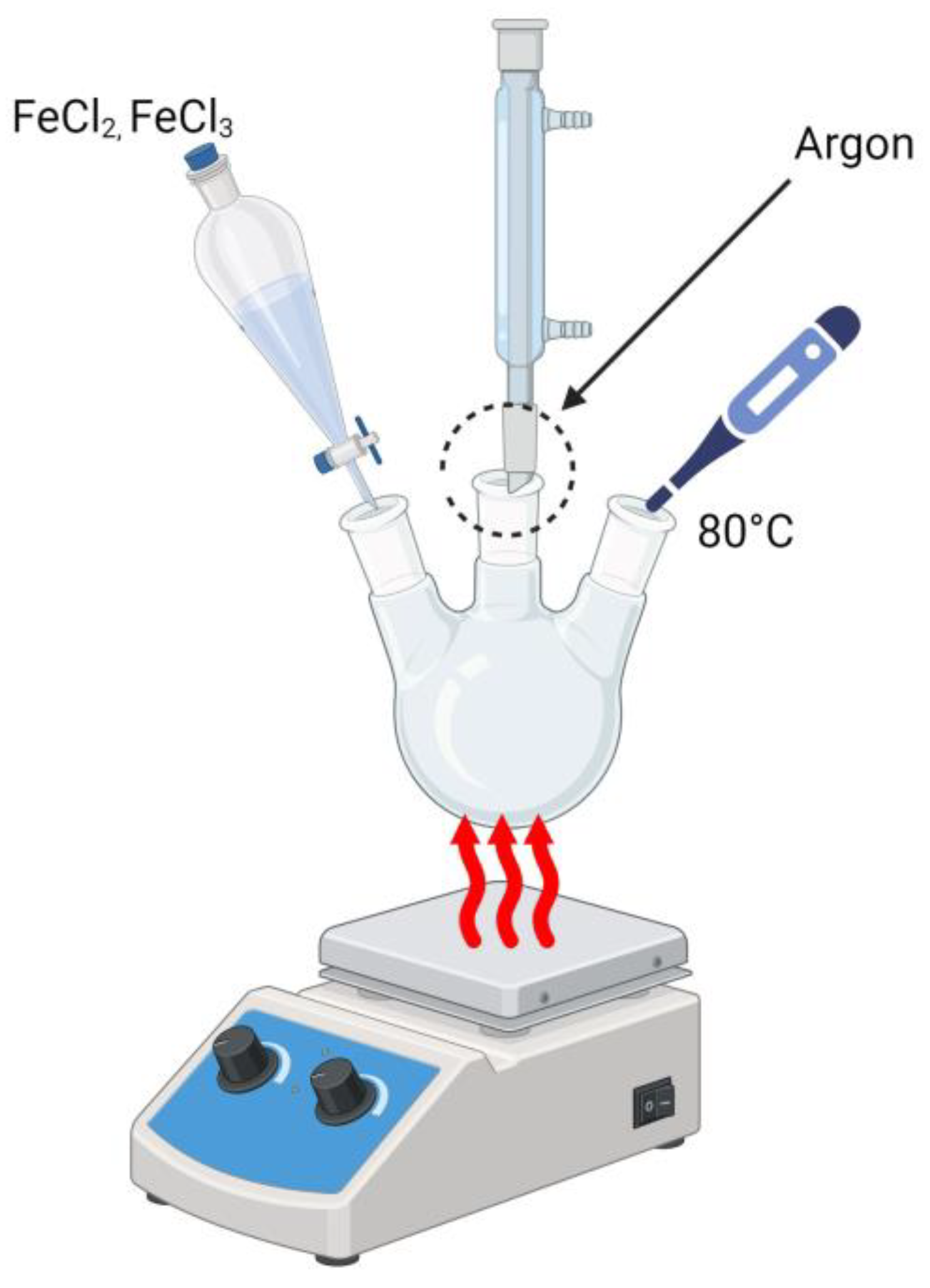
2.2. Synthesis of Magnetic Nanoparticles Modified with Silver

In the first step of the synthesis of hydrogel materials, the reaction to obtain magnetic nanoparticles was carried out using Massart synthesis (Figure 1). In the process, a 1.5 M NaOH solution was obtained. Due to the exothermic nature of the reaction, the process was carried out in a water bath to maintain the appropriate reaction temperature. The resulting solution was placed in a three-neck flask and heated to 80 °C, using a hotplate, in an inert gas atmosphere (argon) and under intense stirring conditions. In the next step, a 10 mM solution of iron precursor in HCl (FeCl2 × 4H2O and FeCl3 × 6H2O) was prepared. Then, 25 mL of this prepared solution was poured into a dropper and slowly dropped into the NaOH solution, heated to 80 °C. The resulting suspension was placed on a magnet to sediment the magnetic nanoparticles. The liquid was then separated from the sediment. The magnetic nanoparticles were washed with distilled water until a neutral pH value was obtained. The resulting Fe3O4 particles were subjected to a physicochemical analysis.
In the context of the synthesis of Fe3O4_Ag nanoparticles, a 3% gum arabic solution was prepared. It was then placed in a three-neck flask, and 7.5 mL of the suspension of magnetic nanoparticles obtained using Massart synthesis was added. The entire mixture was heated to 80 °C in an inert gas (argon) atmosphere under vigorous stirring conditions. When the temperature reached 80 °C, 3.75 mL of a 0.2 M AgNO3 solution and 1.5 mL of a 0.1 M NH2OH × HCl solution were added to the mixture. The reaction was then maintained for 2 h, maintaining the temperature at 80 °C, in an inert gas atmosphere and under vigorous stirring conditions.

2.3. Synthesis of Hydrogel Materials

In making the hydrogel, a 1% chitosan solution was prepared. Using an automatic pipette, the appropriate proportions of the solutions were measured according to the values provided in Table 1.
The prepared solutions were then poured onto Petri dishes and placed under a UV lamp, leaving them for approximately 2 min to allow the hydrogel to crosslink. The crosslinking scheme for hydrogel materials is presented in Figure 2. The resulting samples were obtained using Ag-coated magnetic nanoparticles after the sonification process. The sonification process was carried out in distilled water heated to 37 °C for a period of 20 min. At the end of the process, the hydrogels produced were allowed to dry.

2.4. Characterization of the Obtained Nanoparticles

Due to the optical properties of nanosilver, the resulting magnetic nanoparticles that had a silver envelope were subjected to a UV-Vis analysis. The spectrum was recorded in the wavelength range of 300–600 nm at room temperature. The analysis was performed using a ThermoScientific Evolution 220 UV-Vis spectrometer. Next, FT-IR infrared spectroscopy analysis was performed. For this purpose, a Thermo Scientific Nicolet iS5 FT-IR spectrophotometer (Thermo Fisher Scientific, Waltham, MA, USA) equipped with an iD7 ATR was used. FT-IR spectra were recorded within the 4000–400 cm−1 wavelength range at room temperature. X-ray diffraction (XRD) analysis was performed according to the parameters shown in Table 2 using PANalytical Aeris (Malvern PANalytical, Lelyweg 1, Almelo, The Netherlands).

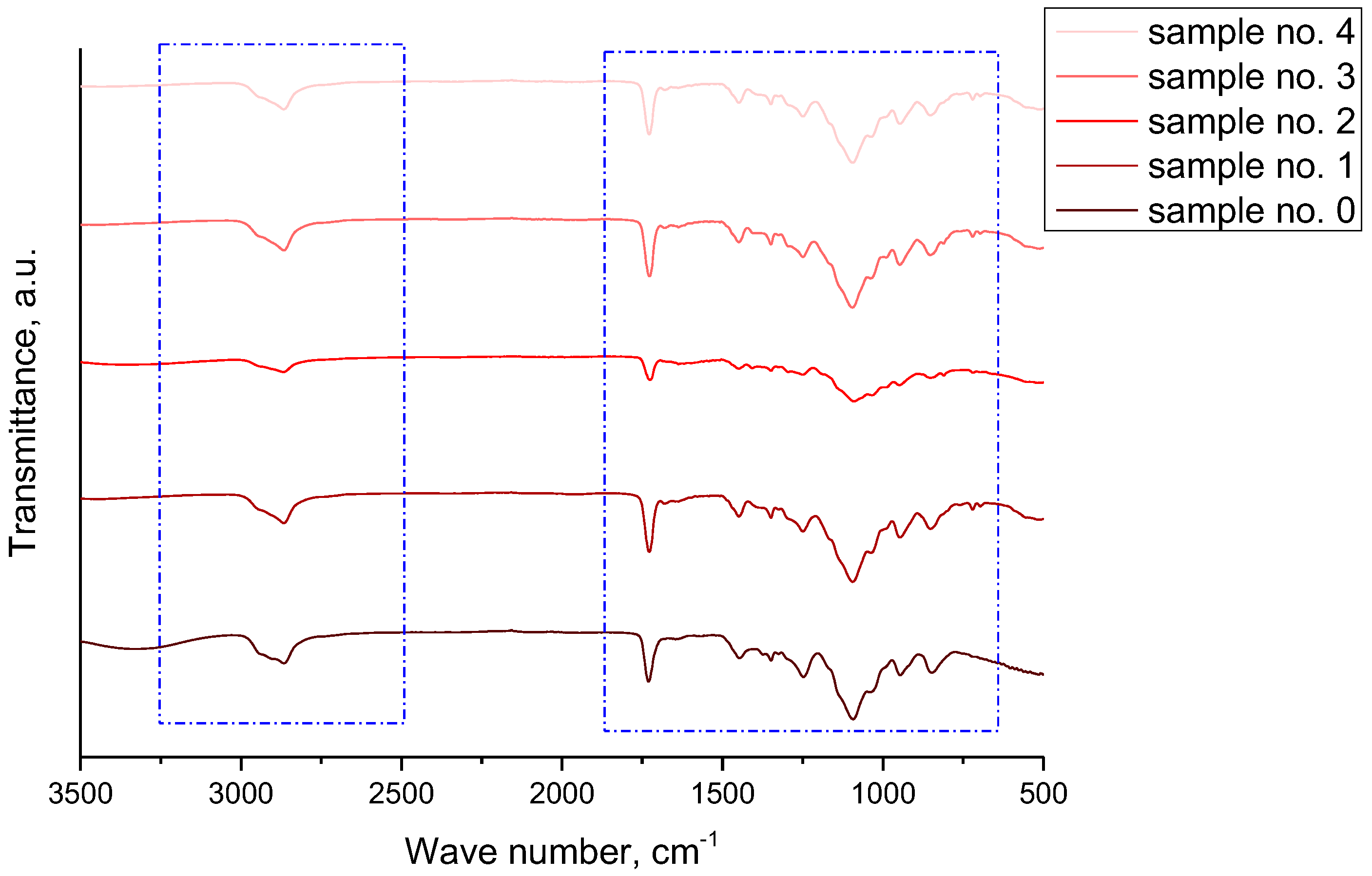
2.5. Infrared Spectroscopy Analysis

Infrared spectroscopy with a Thermo Scientific Nicolet iS5 spectrometer equipped with an ATR attachment was used to evaluate the absorption spectrum. Changes in the composition of the material cause changes in the characteristic distribution of the absorption bands. FT-IR spectra were recorded in the range 4000–500 cm−1 (32 scans, resolution 4.0 cm−1). Samples were analysed after sonification to avoid the agglomeration of hydrogel materials. The analysis was performed at room temperature.

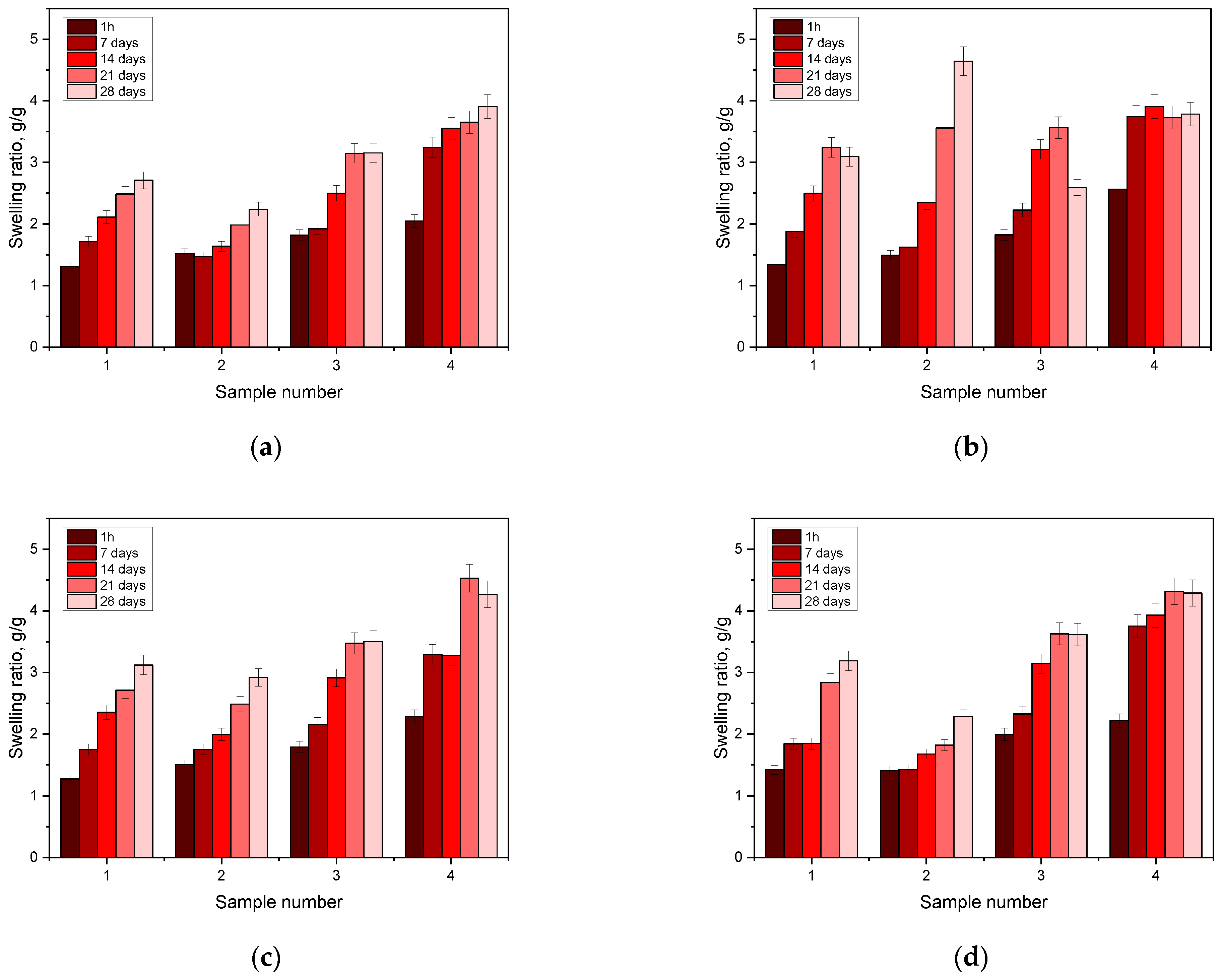
2.6. Testing the Sorption Capacity of Hydrogels

The aim of the analysis was to thoroughly investigate the sorptive capacity of hydrogel matrices in the context of swelling. This study stems from the need to evaluate the potential use of hydrogel dressings for the effective absorption of therapeutic fluids. The analysis was based on the use of previously prepared and dried square hydrogel samples. In the first step, the weight of each sample was accurately measured and then placed in various incubation fluids, distilled water, SBF, Ringer’s solution, and artificial saliva. The next step was to incubate the samples in the aforementioned fluids for 1 h and consecutively at one-week intervals for a period of 1 month. After each time period, excess fluid was drained from the hydrogel surface, and then, the weight of the discs was accurately measured using a precision analytical balance from Radwag (Radom, Poland). In order to minimize errors, each step of the analysis was carried out three times, which made it possible to obtain reproducible results. Finally, the swelling ratio results obtained from the repeated tests were averaged to obtain a representative value of the sorption capacity of the hydrogel matrices. Based on this analysis, the degree of swelling was determined by determining the sorption coefficient using Equation (1).
α = m t m 0 m 0
where
α—swelling ratio, g/g;
mt—mass of swollen sample after time “t”, g;
m0—mass of dry sample (before the study), g.

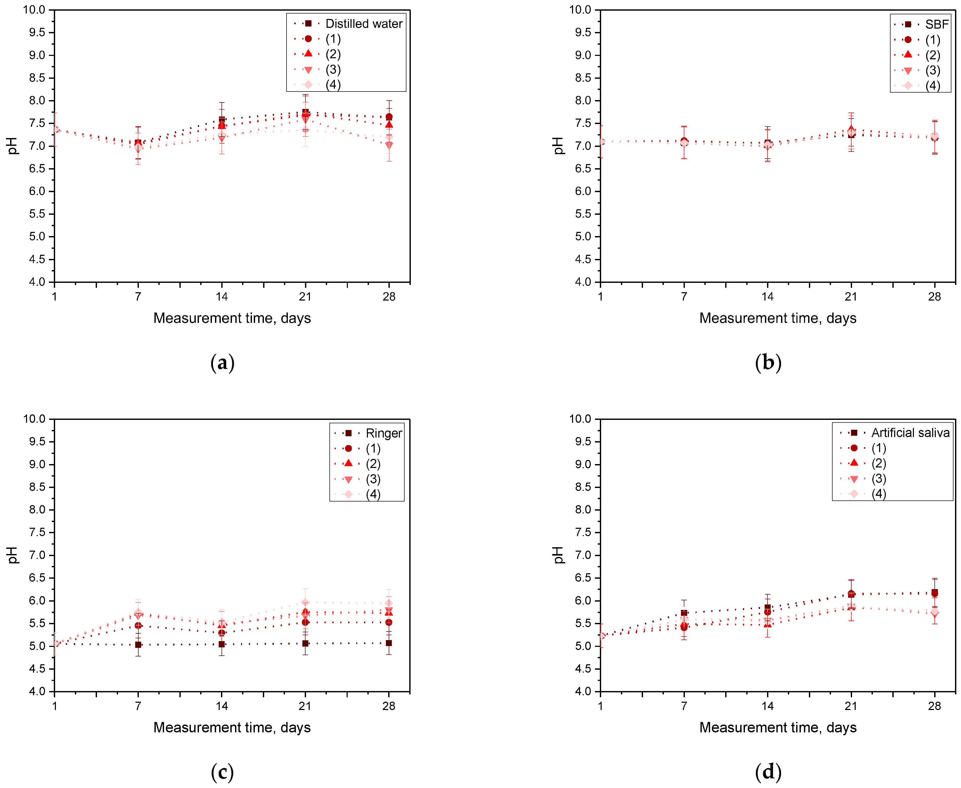
2.7. Incubation Analysis in Simulated Body Fluids

The incubation test was performed to visualize the interactions between hydrogel matrix and solutions simulating human biological fluids. Changes in pH were monitored that indicated the leaching of non-crosslinked substances or the prospective degradation of samples in these fluids. As a part of the experiment, 1 cm square samples were placed in sterile containers and immersed in 50 mL of the above fluids. The samples were incubated at 37 °C in an incubator. The pH values were measured weekly with the ELMETRON CX-701 multifunctional device (Elmetron, Zabrze, Poland) for 28 days.

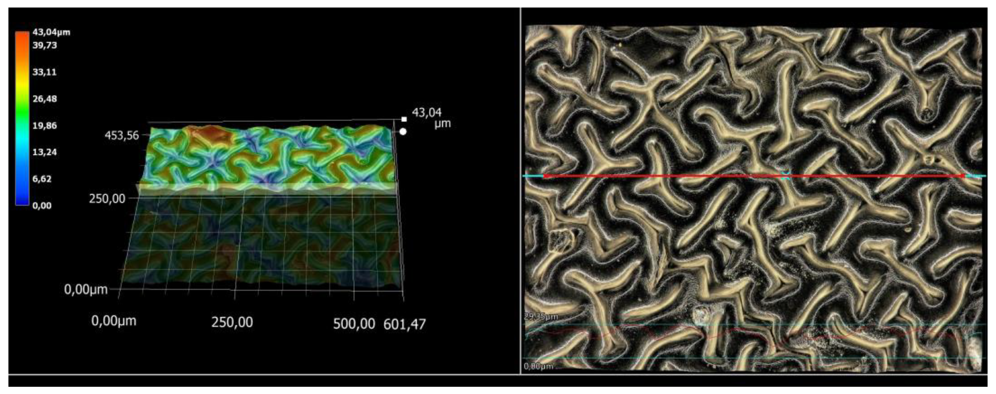
2.8. Microscopic Observations and Surface Roughness Profile Analysis

Next, the advanced VKX-7000 digital microscope from Keyence (Osaka, Japan) was used, which is capable of accurately producing images in 4K resolution. This microscope is equipped with a CEO REMAX optical engine and a 4K CMOS image sensor to guarantee high precision and magnification. The main objective was to analyse the surface morphology of the hydrogels and determine their roughness profiles. In addition, their surface morphology analysis along with their elemental analysis was carried out using scanning electron microscopy with EDS analysis. A scanning electron microscope was used during the observations and was JEOL IT200 (JEOL Ltd., Peabody, MA, USA).

2.9. Statistical Analysis

In order to verify the obtained results and their statistical significance, a two-factor analysis of variance ANOVA with repetition was performed. Statistical analysis was performed for the sorption capacity test and the incubation test, determining the effect of the selected parameters on the properties of the obtained materials, respectively. The composition of the samples and therefore the increasing concentration of silver-encapsulated magnetic nanoparticles and the type of incubation fluid were taken into account.

3. Results

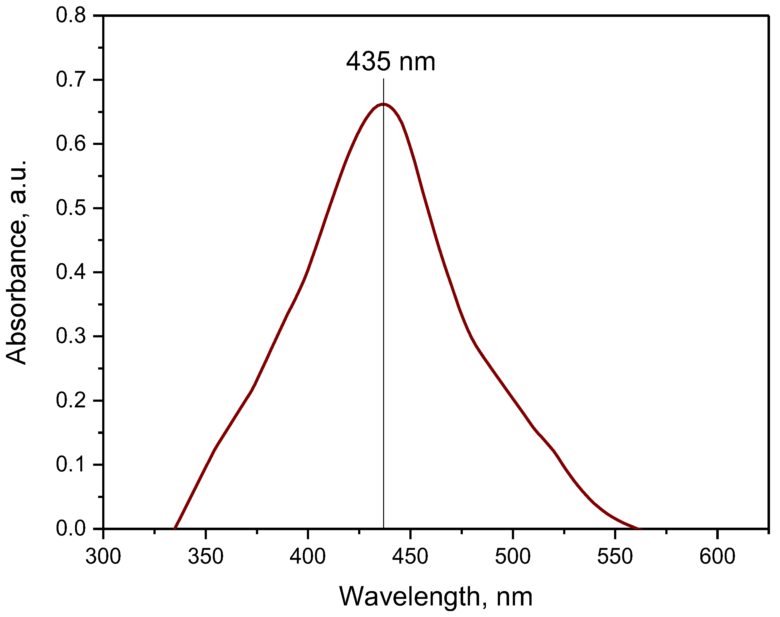
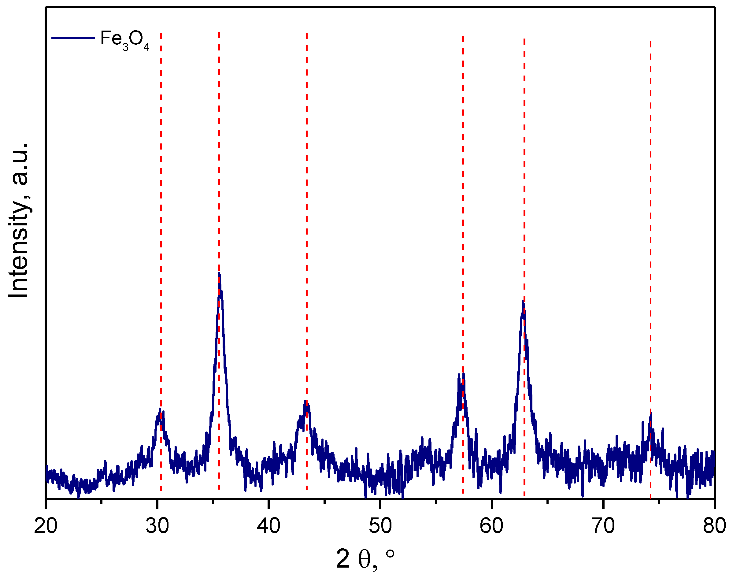
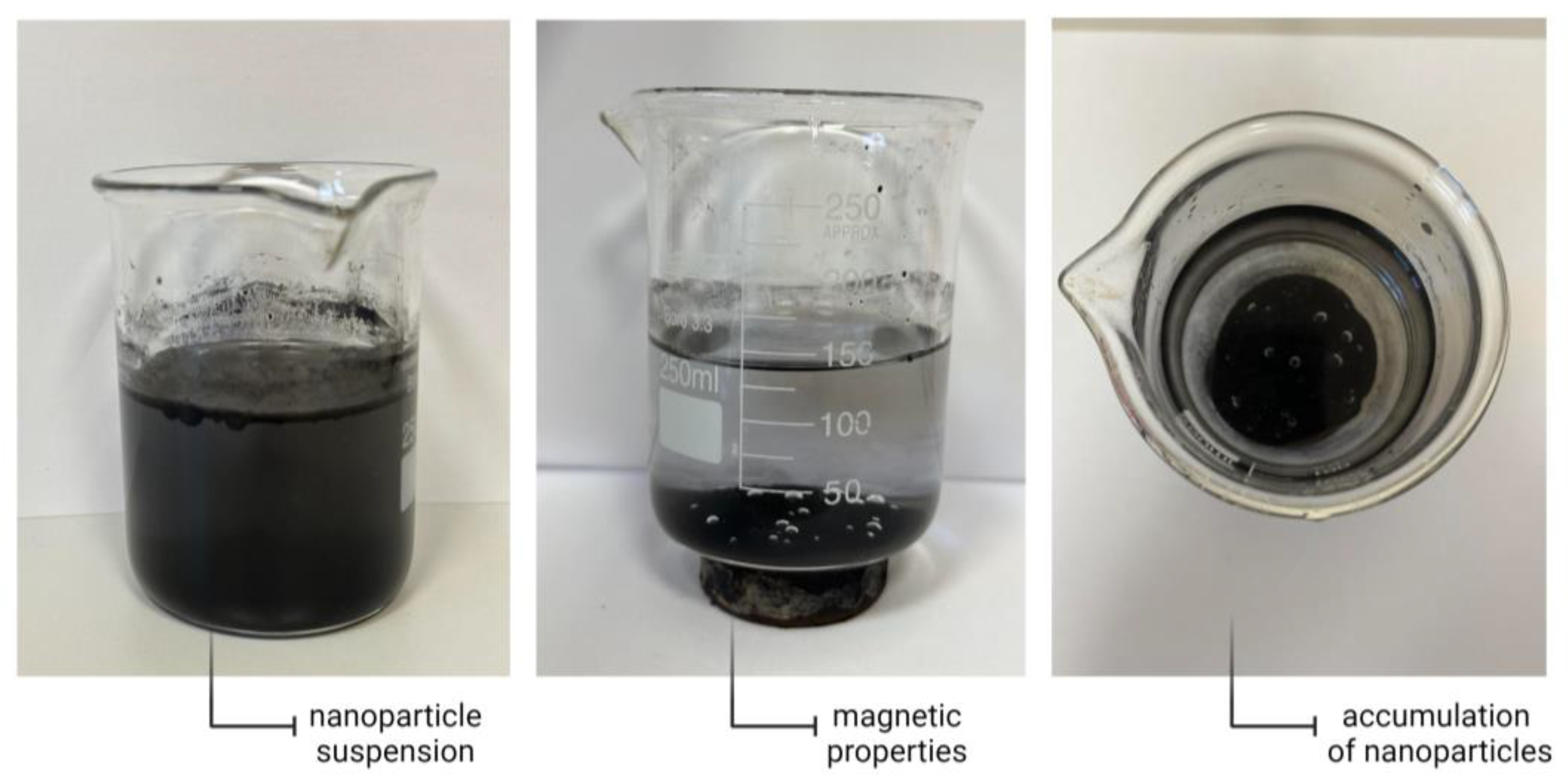
3.1. Characterization of the Obtained Nanoparticles Using FT-IR and UV-Vis Methods

The resulting silver-encapsulated magnetic nanoparticles were subjected to FT-IR and UV-Vis spectroscopic analyses to determine their chemical structures. The results of the FT-IR spectroscopy analysis are presented in Figure 3, while the results of the optical properties obtained from the UV-Vis analysis are shown in Figure 4.
The obtained spectroscopic spectrum presented in Figure 3 indicates the presence of absorption bands characteristic of magnetic nanoparticles. The spectra obtained are analogous to results presented by other researchers. The band with the highest intensity with a maximum at a wave number of about 515 cm−1 can be attributed to the Fe-O bond stretching vibrations, which are characteristic of this type of material and originate from the magnetic core of Fe3O4 nanoparticles. In contrast, the other lower intensity bands occur due to the presence of the stabilizer gum arabic. These interactions may originate from functional groups characteristic of the polysaccharides present in the compound. In addition, the O-H bond is a characteristic of the stabilizer, but its presence may also indicate the presence of interactions between the stabilizer and the magnetic nanoparticles in the form of hydrogen bonding (oxygen derived from Fe3O4 interacts with hydrogen derived from the stabilizer polysaccharides) [19,20,21]. On the other hand, the absorption band with a double peak located in the wave number range of 1600–1400 cm−1 was attributed to the asymmetric and symmetric stretching vibrations of carboxyl groups (COOH) characteristic of the stabilizer Arabic gum [22,23].
Figure 4 refers to the results of UV-Vis spectroscopy analysis of the obtained silver-modified magnetic nanoparticles. Due to the fact that the silver nanoparticles exhibit optical properties and are characterized with an intense colouration related to the phenomenon of excitation of surface plasmons during their exposure to UV-Vis light, it was possible to perform UV-Vis analysis for them. The obtained results show a clear high-intensity band with an absorbance maximum at a wavenumber of about 435 nm that is a characteristic of nanosilver. Thus, it can be concluded that the reduction reaction of silver nitrate performed earlier went well and that the precipitated nanosilver interacts with the magnetic nanoparticles to form the Fe3O4_Ag system. Literature data clearly indicate that this combination reduces the cytotoxicity of the obtained magnetic nanomaterials while providing antimicrobial properties, which is undoubtedly an advantage of the developed system [24,25,26].
The identification of magnetic nanoparticles using the XRD technique involves analysing the diffraction patterns that are produced when X-rays interact with these nanoparticles. Figure 5 shows the diffractogram for the obtained systems, which, according to the ICDD date, shows bands characteristic of the material under study, confirming the presence of both magnetite Fe3O4 (JCPDS 19-629) and maghemite y—Fe2O3 (JCPDS file 39-1346) [27,28,29,30]. Obtaining a magnetic particle system consisting of two or more phases can promote a properly designed drug delivery. The application of an alternating magnetic field can induce the movement of specific magnetic particles, and thus, it is possible to control the delivery profile of the therapeutic substrate, which is of great importance in targeted therapies (Figure 6).

3.2. Infrared Spectroscopy Analysis of Hydrogel Materials

FT-IR analysis was carried out to determine the effect of the incubation process on the chemical structure of the hydrogel matrices. The results obtained are shown in Figure 7.
Infrared spectroscopy analysis was carried out to determine the chemical structures of the obtained hydrogel materials. Analogous spectroscopic spectra were obtained for all the obtained materials. The absence of pronounced deformations may indicate a properly occurring crosslinking process. All spectra are characterized with absorption bands characteristic of chitosan, the main component of the obtained materials [31]. The broad low-intensity band in the wave number range of 3500 cm−1 to 3100 cm−1 is characteristic of the strong hydroxyl bonds of the O-H group and the amine bonds of the N-H group. Then, in the range of around 2950–2800, bands characteristic of the overlapping stretching strains of aliphatic groups such as -CH2 and -CH3 can be distinguished [32,33]. Next, a number of bands characteristic of both the base polymer of the hydrogel matrix and the crosslinking agent PEGDA can be distinguished [34]. A band of relatively high intensity at around 1655 cm−1 indicates the presence of a C-O draconium amide bond. This is followed by a series of bands between 1500 and 1000 cm−1, indicating C=O, C-H, and N-H stretching vibrations [35,36]. No significant differences were observed between the modified material and the material without magnetic nanoparticle addition. The amount of additive may have been too small relative to the sensitivity of the measuring apparatus. In addition, the sonication of the materials ensured a homogeneous distribution of the nanoparticles in the hydrogel matrix, so it was not possible to carry out a measurement for an area with a cumulative amount of nanoparticles that were evenly distributed throughout the material.

3.3. Testing the Sorption Capacity of Hydrogels

A swelling analysis was carried out to investigate and compare the sorption capacity of the hydrogel materials. The study included the effect of the amount of added silver (Ag)-coated magnetic nanoparticles. The results of the analysis for distilled water, SBF liquid, Ringer’s solution, and artificial saliva are shown in the graphs below (Figure 8). The results of the statistical analysis are presented in Table 3.
On the basis of the presented results of the analysis of sorption capacity and the performed statistical analysis, it was found that both the composition of the samples and the type of incubation fluid have a statistically significant effect on the values of the swelling coefficients of the obtained hydrogels. In the case of different types of incubation liquids, different types of monovalent, divalent, or multivalent ions are present in their compositions, and their interactions with the material may change its sorption properties. Divalent ions present, for example, in SBF fluid, can interact with functional groups of hydrogel materials to form additional crosslinks, resulting in an increase in the crosslinking density of such materials. In this case, the change in the internal structure of the polymeric material is associated, for example, with a reduction in the sorption capacity of such a sample. This is different in ion-free distilled water. Next, the composition of the sample to be analysed is extremely important. As the amount of suspended magnetic nanoparticles increases, a clear increase in the swelling coefficients is observed. It is likely that the suspended matter in the pores is released, resulting in an increase in the amount of free space for the penetrating fluid. This is an extremely important phenomenon, as it indicates that during sorption, the hydrogel material gradually swells, loosens the polymer chain network, and releases the residual substances used as modifiers. The resulting material exhibits the ability to release the desired substances in a controlled manner, depending on their amount and the degree of crosslinking of the matrix. This, in turn, allows the design of a hydrogel with the desired properties for the application. It was noted that the greatest increase in mass and therefore in the amount of absorbed incubation fluid occurs in the first hour of analysis. Then, with time, the hydrogel material is gradually filled with the fluid, penetrating the interior of the matrix. After 28 days, a gradual stabilisation of the matrix against the incubation fluid was noted. The analysis confirmed that the compositions of the samples and the incubation liquid affect the sorption properties of the hydrogels. The same type of materials located in other body fluids show different properties. In view of this, it is extremely important to determine the potential application of hydrogels and then adjust the desired properties. The results obtained are consistent with the results of research works of other authors. For example, Vo et al. proved that different volume ratios of polymers included in hydrogels affect their sorption properties. The use of more polyols resulted in an increased ability to absorb liquids than in the case of samples containing only gelatin and chitosan. As in the present study, it was also proven that the presence of Na+/Cl ions in the incubation fluids affected their reduced water absorption capacity due to the shielding effect. The presence of various functional groups is closely related to the sorption properties of the polymer hydrogels due to the occurrence of chemical interactions and interactions between the fluid components and the polymer matrix [37]. This phenomenon was also confirmed by Li et al. by conducting a sorption study in different types of biological fluids. They proved that, as the pH value increases, hydrogels show a greater swelling due to the decreasing amount of cations in a solution [38]. Moreover, Lv et al. proved that the sorption capacity is correlated with the temperature of the incubation fluid. Referring to the literature data, with this correlation between temperature and sorption capacity in mind, the study we conducted was carried out at 37 °C to simulate the temperature of the human body [39].

3.4. Incubation Analysis in Simulated Body Fluids

An incubation study was conducted to determine the hydrogen ion activity (pH) for the fluid in which the hydrogel matrices were incubated for a period of 28 days. The results obtained are shown in the graphs below (Figure 9).
The incubation analysis was designed to determine the compatibility of the tested materials with fluids simulating the human body environment. The developed materials can be used as dressing or coating materials to support the biocompatibility and functionality of current implants and other biomaterials. Based on this analysis, the high stability of these materials was indicated. Simultaneous observations ruled out the degradation of the materials in the form of decomposition, delamination, or dissolution of the samples. The materials retained their shape and form, respectively. In addition, rapid changes in the pH value, which could indicate the release of toxic unreacted substances from within the matrix, were excluded. The obtained results testify to the synthesis of materials that are compatible with the test medium, with a high stability under conditions resembling the organism’s environment. Moreover, there was no significant effect of different types of incubation liquids that differed in their composition. These results indicate the achievement of properly crosslinked systems that remain stable for 28 days in the various tested fluids. This effect is due to the selection of the right amount of crosslinking agent and photoinitiator, allowing the proper photopolymerization of hydrogels with a high stability.

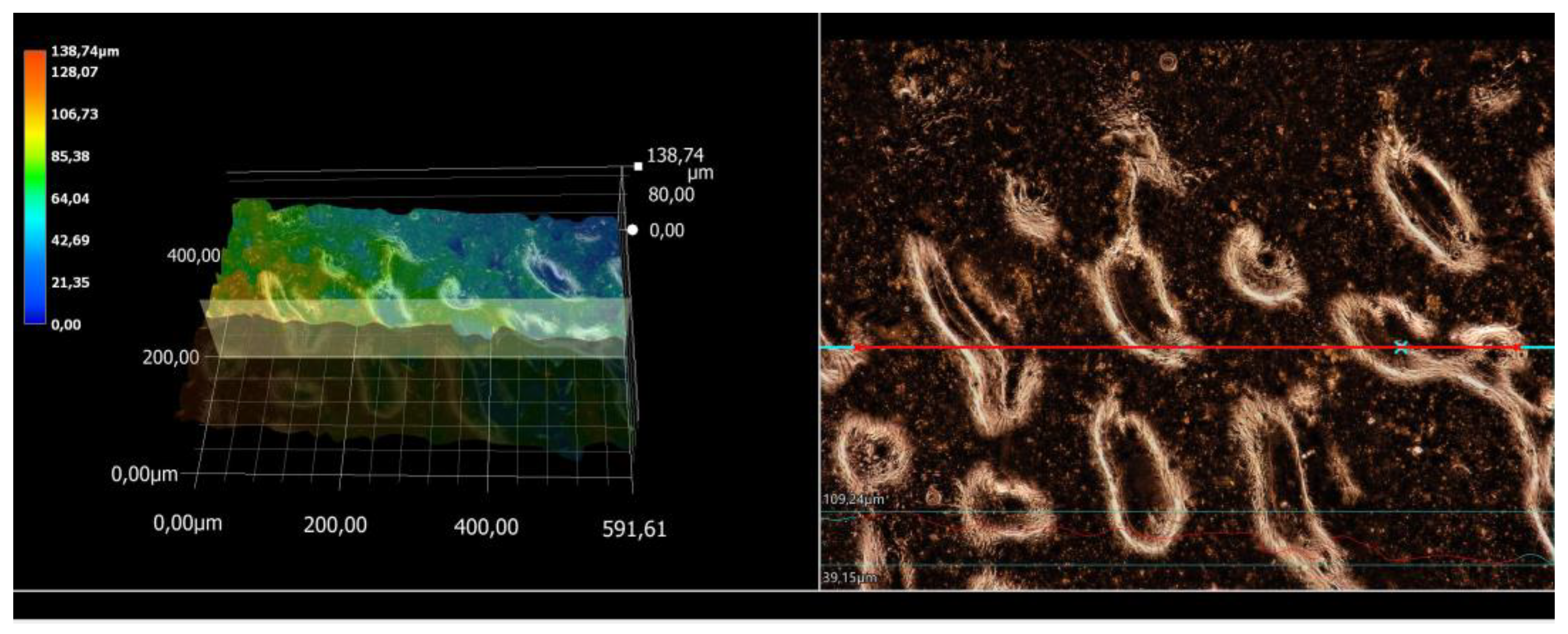
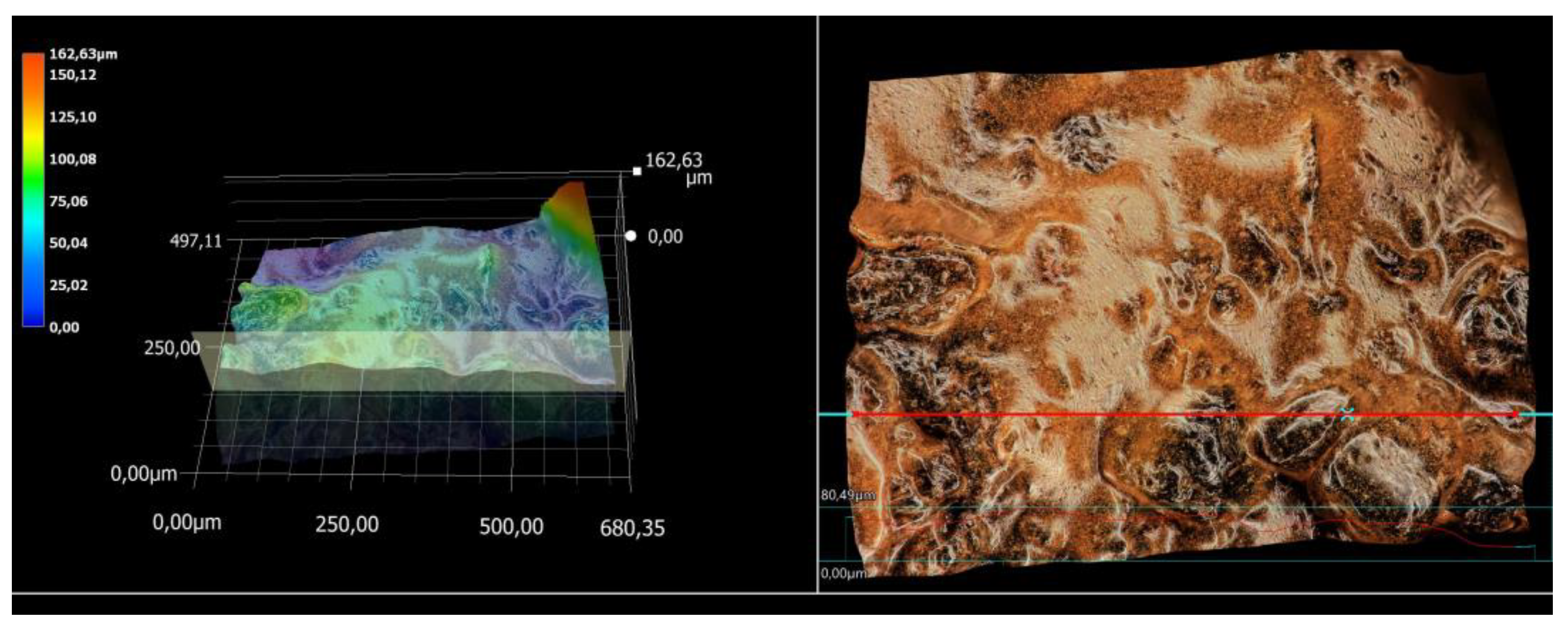
3.5. Microscopic Observations and Surface Roughness Profile Analysis

The roughness parameters along with the surface profile of the tested materials are presented below in Table 4 and Figure 10, Figure 11, Figure 12 and Figure 13.
The surface of a biomaterial plays an extremely important role in cell adhesion and proliferation and thus influences the regenerative properties of damaged/supported tissues. Surfaces with a certain structure and appropriate roughness and corrugation can create an excellent substrate for cells or, on the contrary, hinder their adhesion and growth. In the case of cells with high roughness, the cells may have a difficulty in inserting themselves between high stand-off elevations. Also, surfaces that are very smooth can cause a certain hindrance to their adhesion. On the other hand, an intermediate value of roughness parameters can provide an optimal substrate for cell adhesion and growth [40]. In the case of the tested materials, the unmodified sample showed the lowest roughness value with a Ra parameter of 3.52. With the presence of silver-modified magnetic nanoparticles, an increase in the roughness parameter values, indicating a change in the surface structure of these materials, was noted. These values range from approximately 11 to 16 μm. It was also observed microscopically that the addition of the modifier causes a change in the colour of the hydrogel material. There are no visible clusters, agglomerates, or other defects on the surface of the modified materials. The addition of modified substances can significantly change the structure of the studies hydrogels. In a study conducted by Gradilla-Orozco et al., it was shown that the composition of hydrogels has a significant effect on their surface morphology. Based on their study, they found that increasing the amount of glycerol diacrylate (DAG) leads to a clearer observation of a hydrogel structure. They also observed a critical crosslinking concentration (CCLC) of about 4% by weight, above which the hydrogel surface had a smooth texture, oriented preferentially in one direction. On the other hand, for DAG concentrations below 4% w/w, they observed an irregular alignment of the hydrogel surface. The authors concluded that glycerol diacrylate (DAG) affects the morphology of the hydrogel surface, leading to the formation of structures of different scales and shapes depending on its concentration, which is important for the design of materials with specific surface properties [41]. When designing hydrogel materials, it is important to precisely define their intended use, so that their properties can be controlled, depending on a number of parameters. In addition to the chemical composition, the hydrogel structure is affected by the used drying methods. This was verified by Kaberova et al. who used various drying techniques, such as plunge freezing, freeze-drying, and critical point drying, to prepare hydrogel samples for analysis with scanning electron microscopy (SEM). Rapid freezing at very low temperatures (−196 °C) preserved the original hydrogel morphology. For freeze-drying, temperature and pressure conditions had a significant effect on the final hydrogel morphology. In contrast, critical point drying using specific solvents and CO2 led to visible changes in hydrogel morphology. In our study, we used drying of hydrogel materials in a laboratory dryer, maintaining a constant temperature and ensuring an even drying of biomaterials through ventilation circulation. This controlled drying method allowed us to obtain the desired morphological properties of the hydrogel such as high surface uniformity, which is crucial for their potential applications in the biomedical field [42].

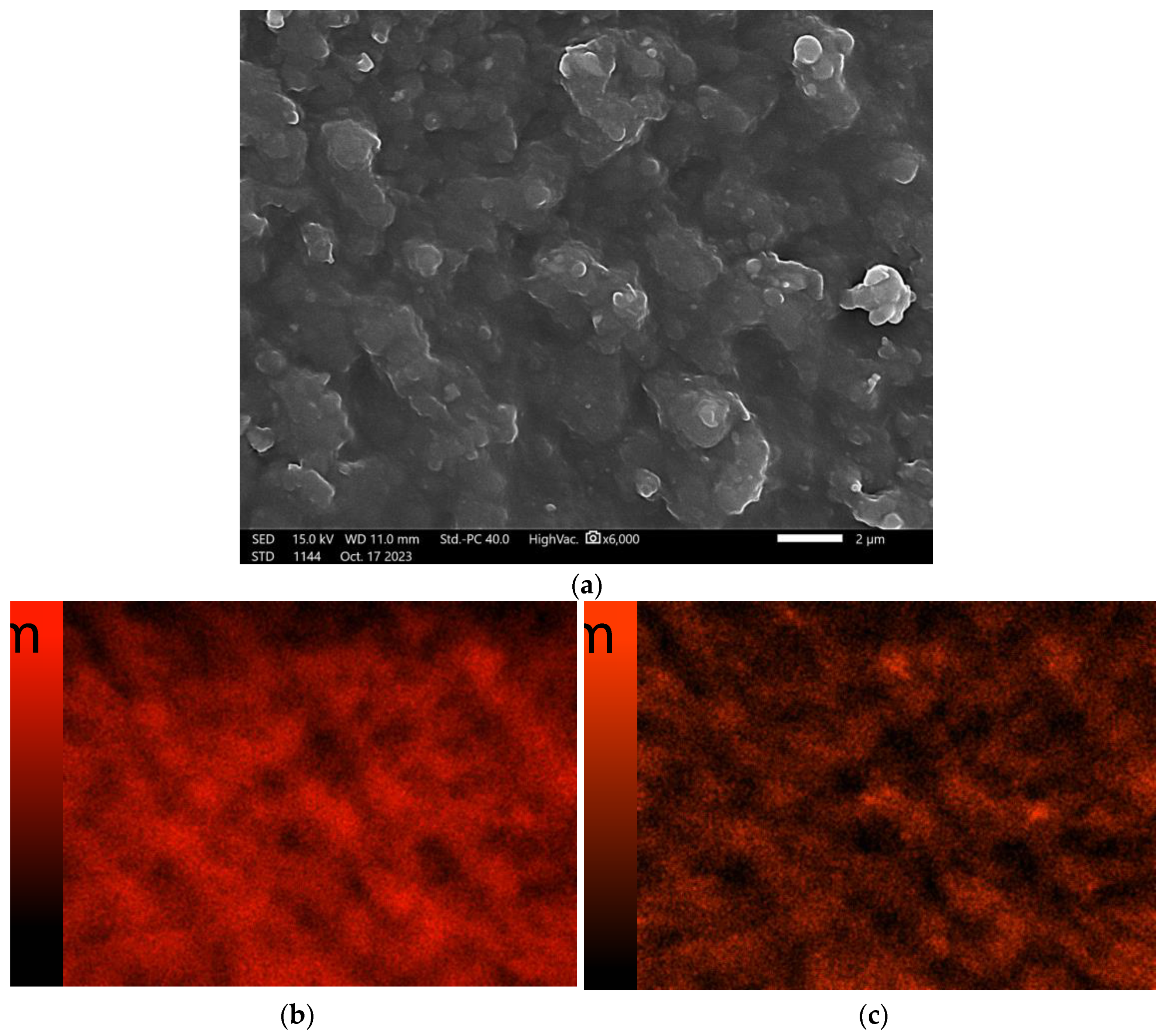
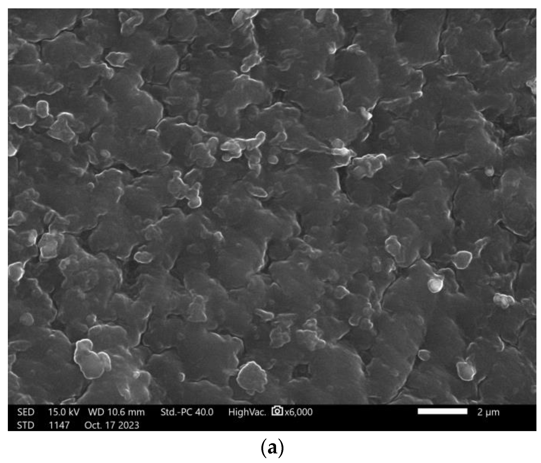
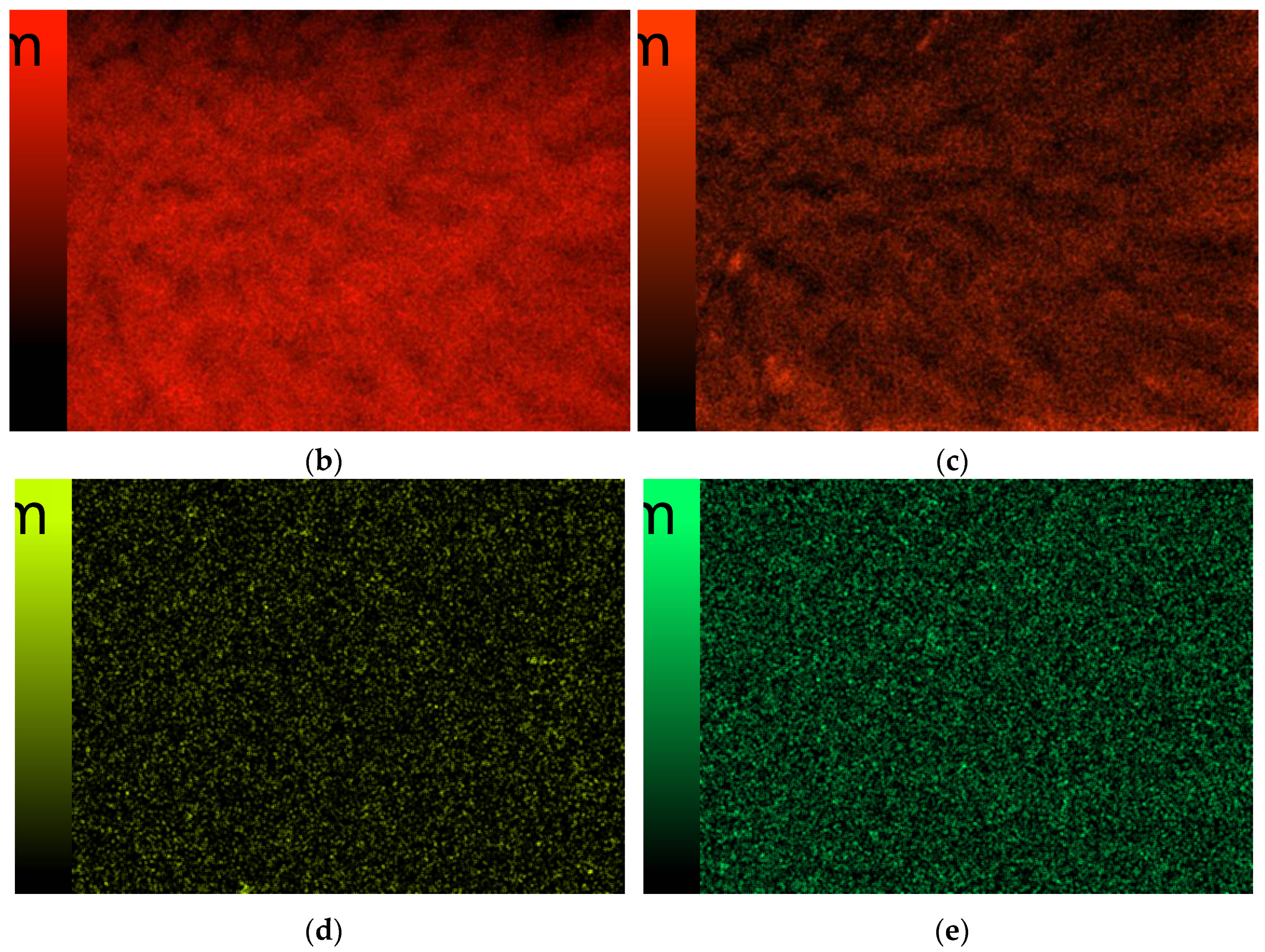
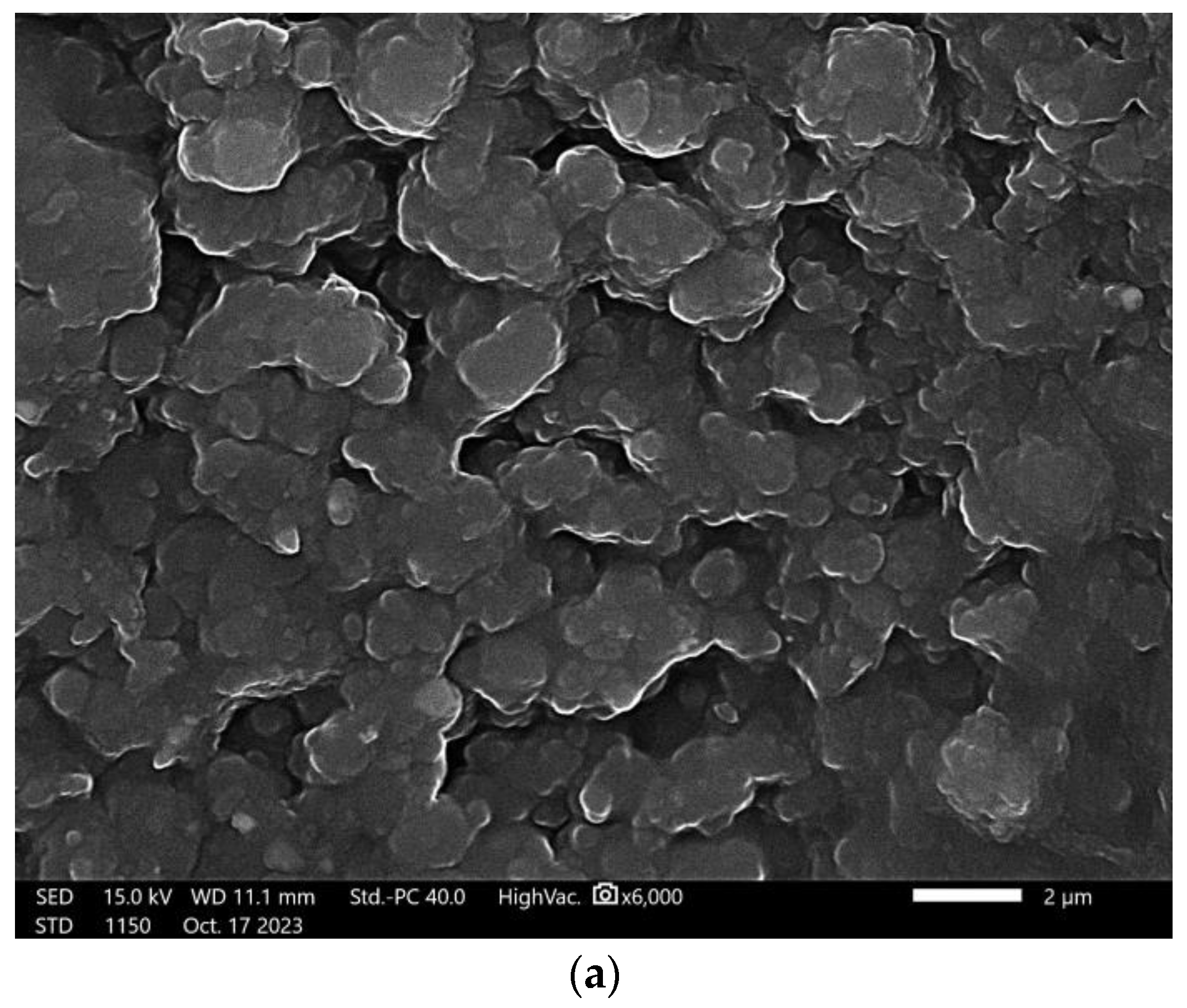
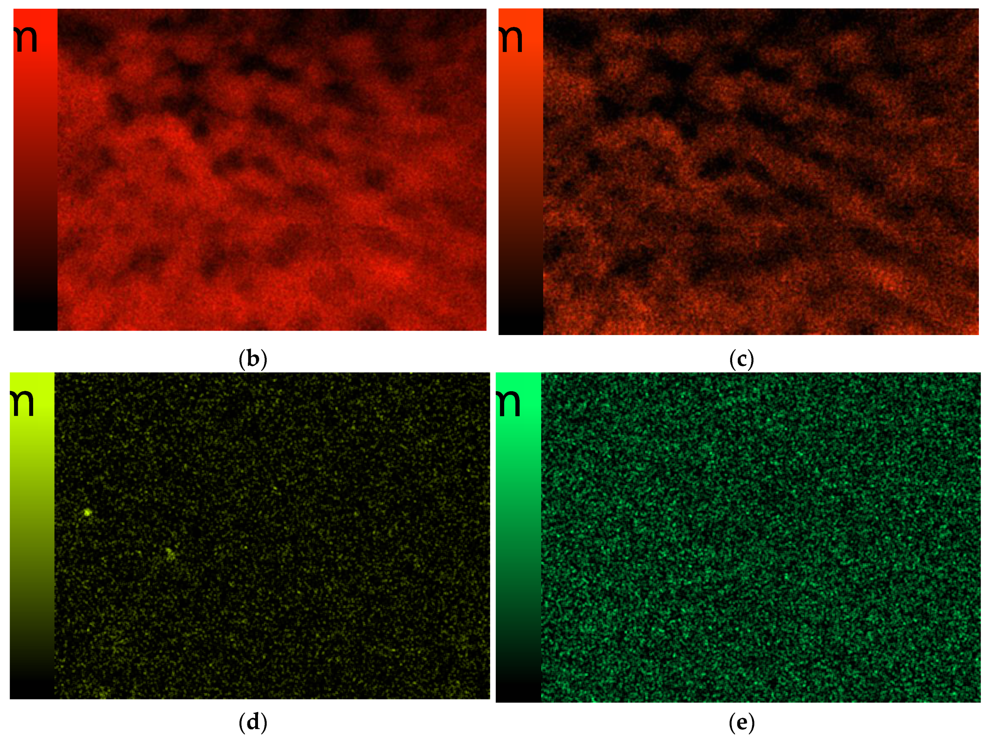
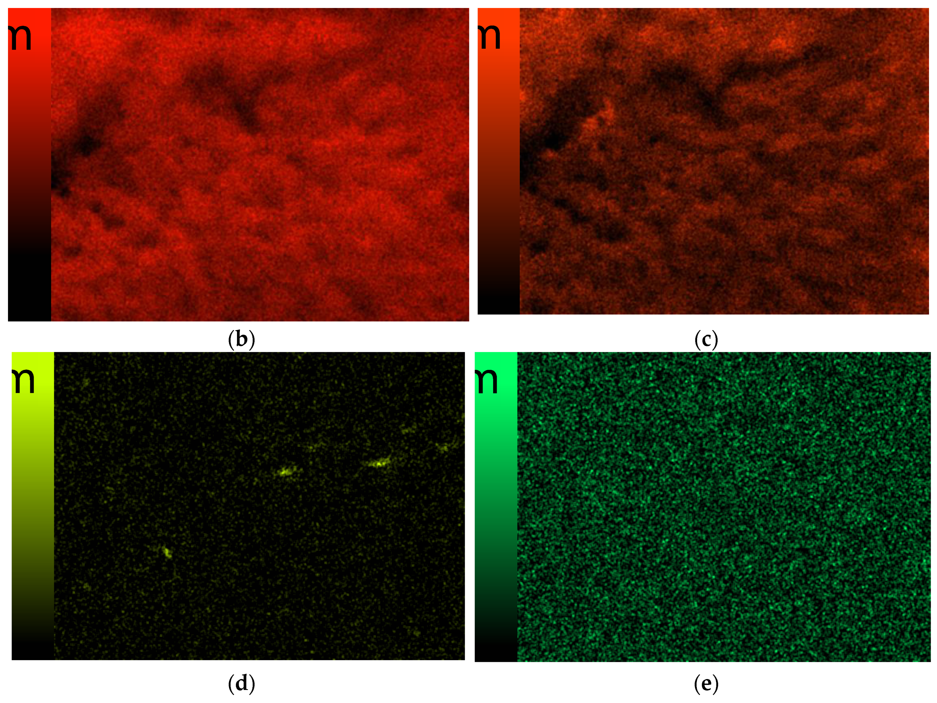
3.6. Scanning Electron Microscopy

Figure 14, Figure 15, Figure 16 and Figure 17 present images obtained from the scanning electron microscope along with the mapping of individual elements.
Based on microscopic observations, it was found that the obtained hydrogel materials have a homogeneous surface structure. Their morphology indicates a large specific surface area, which is well developed. However, no agglomerates or other precipitates were found on the hydrogel surface, which also suggests a proper photopolymerization process. Figure 14 shows the elemental analysis of the unmodified sample with magnetic nanoparticles. In this case, no elements other than the main building blocks of the polymer matrix such as carbon and oxygen were noted. Next, Figure 15, Figure 16 and Figure 17 show the elemental analysis of the surface of hydrogels modified with Fe3O4_Ag nanoparticles. As expected, characteristic signals from iron and silver were identified in the modified materials, indicating that the obtained systems were correctly modified.

4. Conclusions

The aim of the study was to obtain hydrogel materials that can function as dressing or coating materials. The used photopolymerisation method made it possible to obtain hydrogels of variable composition. The compositions obtained differed in the content of nanosilver-coated magnetic nanoparticles. Fe3O4_Ag particles were obtained with the Massart method using silver nitrate reduction for silver modification. The obtained systems were subjected to physicochemical analysis, determining the effect of the used modifier on their selected properties. Spectroscopic analysis indicated that properly crosslinked materials with absorption bands characteristic of the initial components were obtained. It was then demonstrated that an increase in the amount of magnetic nanoparticles results in an increase in the sorption properties of the hydrogels. At the same time, the type of incubation fluid is also important. Incubation analysis and measurement of the pH value and observations carried out together with the study excluded the degradation of the materials and confirmed their compatibility with fluids that simulated the human body environment. Microscopic observations together with the measurement of surface roughness parameters indicated a clear relationship between modification and change in surface structure. All the modified materials showed a surface that was several times rougher than the starting sample without nanoparticles. Microscopic observations did not reveal the presence of any agglomerates that could indicate system heterogeneity. In conclusion, materials capable of absorbing incubation fluids and with a rough surface were obtained, which may find future applications as coating or dressing materials. The novelty of this work was the combination of a polymer matrix with magnetic nanoparticles, which can be successfully used as drug carriers. This work focused on a basic physicochemical analysis, but this work should be extended in the future to incorporate studies on drug-containing systems. The developed materials with magnetic properties, characterized by a high sorption capacity and a developed surface, can serve as coating materials, enriching the currently used therapeutic methods.

Author Contributions

Conceptualization, K.S. and B.T.; methodology, K.S. and M.B.; software, B.T.; validation, K.S. and M.B.; formal analysis, K.S. and M.B.; investigation, K.S., M.B. and K.C.; resources, B.T.; data curation, K.S. and M.B.; writing—original draft preparation, K.S., M.B. and K.C.; writing—review and editing, K.S. and M.B.; visualization, K.S.; supervision, B.T.; project administration, B.T.; funding acquisition, B.T. All authors have read and agreed to the published version of the manuscript.

Funding

This research received no external funding.

Institutional Review Board Statement

Not applicable.

Informed Consent Statement

Not applicable.

Data Availability Statement

No new data were created or analyzed in this study. Data sharing is not applicable to this article.

Acknowledgments

The research work was carried out within the SMART-MAT Functional Materials Science Club at the Faculty of Materials Engineering and Physics of the Cracow University of Technology.

Conflicts of Interest

The authors declare no conflict of interest.

References

  1. Akbarzadeh, A.; Samiei, M.; Davaran, S. Magnetic Nanoparticles: Preparation, Physical Properties, and Applications in Biomedicine. Nanoscale Res. Lett. 2012, 7, 144. [Google Scholar] [CrossRef] [PubMed]
  2. Katz, E. Synthesis, Properties and Applications of Magnetic Nanoparticles and Nanowires—A Brief Introduction. Magnetochemistry 2019, 5, 61. [Google Scholar] [CrossRef]
  3. Issa, B.; Obaidat, I.M.; Albiss, B.A.; Haik, Y. Magnetic Nanoparticles: Surface Effects and Properties Related to Biomedicine Applications. Int. J. Mol. Sci. 2013, 14, 21266–21305. [Google Scholar] [CrossRef] [PubMed]
  4. Astefanoaei, I.; Gimaev, R.; Zverev, V.; Tishin, A.; Stancu, A. Cubic and Sphere Magnetic Nanoparticles for Magnetic Hyperthermia Therapy: Computational Results. Nanomaterials 2023, 13, 2383. [Google Scholar] [CrossRef]
  5. Zhang, Y.; Xi, K.; Fu, X.; Sun, H.; Wang, H.; Yu, D.; Li, Z.; Ma, Y.; Liu, X.; Huang, B.; et al. Versatile Metal-Phenolic Network Nanoparticles for Multitargeted Combination Therapy and Magnetic Resonance Tracing in Glioblastoma. Biomaterials 2021, 278, 121163. [Google Scholar] [CrossRef]
  6. Wang, J.; Chen, P.; Dong, Y.; Xie, H.; Wang, Y.; Soto, F.; Ma, P.; Feng, X.; Du, W.; Liu, B.-F. Designer Exosomes Enabling Tumor Targeted Efficient Chemo/Gene/Photothermal Therapy. Biomaterials 2021, 276, 121056. [Google Scholar] [CrossRef] [PubMed]
  7. Kiru, L.; Zlitni, A.; Tousley, A.M.; Dalton, G.N.; Wu, W.; Lafortune, F.; Liu, A.; Cunanan, K.M.; Nejadnik, H.; Sulchek, T.; et al. In Vivo Imaging of Nanoparticle-Labeled CAR T Cells. Proc. Natl. Acad. Sci. USA 2022, 119, e2102363119. [Google Scholar] [CrossRef]
  8. Dorjsuren, B.; Chaurasiya, B.; Ye, Z.; Liu, Y.; Li, W.; Wang, C.; Shi, D.; Evans, C.E.; Webster, T.J.; Shen, Y. Cetuximab-Coated Thermo-Sensitive Liposomes Loaded with Magnetic Nanoparticles and Doxorubicin for Targeted EGFR-Expressing Breast Cancer Combined Therapy. Int. J. Nanomed. 2020, 15, 8201–8215. [Google Scholar] [CrossRef]
  9. Park, Y.; Demessie, A.A.; Luo, A.; Taratula, O.R.; Moses, A.S.; Do, P.; Campos, L.; Jahangiri, Y.; Wyatt, C.R.; Albarqi, H.A.; et al. Targeted Nanoparticles with High Heating Efficiency for the Treatment of Endometriosis with Systemically Delivered Magnetic Hyperthermia. Small 2022, 18, e2107808. [Google Scholar] [CrossRef]
  10. Zhang, F.; Lu, G.; Wen, X.; Li, F.; Ji, X.; Li, Q.; Wu, M.; Cheng, Q.; Yu, Y.; Tang, J.; et al. Magnetic Nanoparticles Coated with Polyphenols for Spatio-Temporally Controlled Cancer Photothermal/Immunotherapy. J. Control. Release 2020, 326, 131–139. [Google Scholar] [CrossRef]
  11. Daya, R.; Xu, C.; Nguyen, N.-Y.T.; Liu, H.H. Angiogenic Hyaluronic Acid Hydrogels with Curcumin-Coated Magnetic Nanoparticles for Tissue Repair. ACS Appl. Mater. Interfaces 2022, 14, 11051–11067. [Google Scholar] [CrossRef] [PubMed]
  12. Ho, T.-C.; Chang, C.-C.; Chan, H.-P.; Chung, T.-W.; Shu, C.-W.; Chuang, K.-P.; Duh, T.-H.; Yang, M.-H.; Tyan, Y.-C. Hydrogels: Properties and Applications in Biomedicine. Molecules 2022, 27, 2902. [Google Scholar] [CrossRef] [PubMed]
  13. Wang, Y.; Wu, Y.; Long, L.; Yang, L.; Fu, D.; Hu, C.; Kong, Q.; Wang, Y. Inflammation-Responsive Drug-Loaded Hydrogels with Sequential Hemostasis, Antibacterial, and Anti-Inflammatory Behavior for Chronically Infected Diabetic Wound Treatment. ACS Appl. Mater. Interfaces 2021, 13, 33584–33599. [Google Scholar] [CrossRef] [PubMed]
  14. Zeng, N.; He, L.; Jiang, L.; Shan, S.; Su, H. Synthesis of Magnetic/PH Dual Responsive Dextran Hydrogels as Stimuli-Sensitive Drug Carriers. Carbohydr. Res. 2022, 520, 108632. [Google Scholar] [CrossRef] [PubMed]
  15. Ailincai, D.; Mititelu-Tartau, L.; Marin, L. Citryl-Imine-PEG-Ylated Chitosan Hydrogels—Promising Materials for Drug Delivery Applications. Int. J. Biol. Macromol. 2020, 162, 1323–1337. [Google Scholar] [CrossRef] [PubMed]
  16. Ji, L.; Zhang, F.; Zhu, L.; Jiang, J. An In-Situ Fabrication of Bamboo Bacterial Cellulose/Sodium Alginate Nanocomposite Hydrogels as Carrier Materials for Controlled Protein Drug Delivery. Int. J. Biol. Macromol. 2021, 170, 459–468. [Google Scholar] [CrossRef] [PubMed]
  17. Cai, X.; He, Y.; Cai, L.; Zhan, J.; Li, Q.; Zhong, S.; Hou, H.; Wang, W.; Qiu, X. An Injectable Elastic Hydrogel Crosslinked with Curcumin-Gelatin Nanoparticles as a Multifunctional Dressing for the Rapid Repair of Bacterially Infected Wounds. Biomater. Sci. 2023, 11, 3227–3240. [Google Scholar] [CrossRef] [PubMed]
  18. Yang, Z.; Huang, R.; Zheng, B.; Guo, W.; Li, C.; He, W.; Wei, Y.; Du, Y.; Wang, H.; Wu, D.; et al. Highly Stretchable, Adhesive, Biocompatible, and Antibacterial Hydrogel Dressings for Wound Healing. Adv. Sci. 2021, 8, 2003627. [Google Scholar] [CrossRef]
  19. Lobato, N.; Mansur, M.; Ferreira, A. Characterization and Chemical Stability of Hydrophilic and Hydrophobic Magnetic Nanoparticles. Mater. Res. 2017, 20, 736–746. [Google Scholar] [CrossRef]
  20. Silva, C.; Silva, R.E.; De Figueiredo, A.; Alves, V. Magnetic Solid-Phase Microextraction for Lead Detection in Aqueous Samples Using Magnetite Nanoparticles. J. Braz. Chem. Soc. 2020, 31, 109–115. [Google Scholar] [CrossRef]
  21. Nalbandian, L.; Patrikiadou, E.; Zaspalis, V.; Patrikidou, A.; Hatzidaki, E.; Papandreou, C. Magnetic Nanoparticles in Medical Diagnostic Applications: Synthesis, Characterization and Proteins Conjugation. Curr. Nanosci. 2015, 12, 455–468. [Google Scholar] [CrossRef]
  22. Ibekwe, C.; Oyatogun, G.; Esan, T.; Oluwasegun, K. Synthesis and Characterization of Chitosan/Gum Arabic Nanoparticles for Bone Regeneration. Am. J. Mater. Sci. Eng. 2017, 5, 28–36. [Google Scholar] [CrossRef]
  23. Palumbo, G.; Górny, M.; Banaś, J. Corrosion Inhibition of Pipeline Carbon Steel (N80) in CO2-Saturated Chloride (0.5 M of KCl) Solution Using Gum Arabic as a Possible Environmentally Friendly Corrosion Inhibitor for Shale Gas Industry. J. Mater. Eng. Perform. 2019, 28, 6458–6470. [Google Scholar] [CrossRef]
  24. Park, S.B.; White, S.B.; Steadman, C.S.; Pechan, T.; Pechanova, O.; Clemente, H.J.; Thirumalai, R.V.K.G.; Willard, S.T.; Ryan, P.L.; Feugang, J.M. Silver-Coated Magnetic Nanocomposites Induce Growth Inhibition and Protein Changes in Foodborne Bacteria. Sci. Rep. 2019, 9, 17499. [Google Scholar] [CrossRef] [PubMed]
  25. Iglesias-Silva, E.; Rivas, J.; León Isidro, L.M.; López-Quintela, M.A. Synthesis of Silver-Coated Magnetite Nanoparticles. J. Non-Cryst. Solids 2007, 353, 829–831. [Google Scholar] [CrossRef]
  26. Mehrabi, F.; Shamspur, T.; Sheibani, H.; Mostafavi, A.; Mohamadi, M.; Hakimi, H.; Bahramabadi, R.; Salari, E. Silver-Coated Magnetic Nanoparticles as an Efficient Delivery System for the Antibiotics Trimethoprim and Sulfamethoxazole against E. Coli and S. Aureus: Release Kinetics and Antimicrobial Activity. BioMetals 2021, 34, 1237–1246. [Google Scholar] [CrossRef] [PubMed]
  27. Bertolucci, E.; Galletti, A.; Antonetti, C.; Marracci, M.; Tellini, B.; Piccinelli, F.; Visone, C. Chemical and Magnetic Properties Characterization of Magnetic Nanoparticles. In Proceedings of the 2015 IEEE International Instrumentation and Measurement Technology Conference (I2MTC) Proceedings, Pisa, Italy, 11–14 May 2015; Volume 2015, pp. 1492–1496. [Google Scholar] [CrossRef]
  28. Shagholani, H.; Ghoreishi, S.M.; Mousazadeh, M. Improvement of Interaction between PVA and Chitosan via Magnetite Nanoparticles for Drug Delivery Application. Int. J. Biol. Macromol. 2015, 78, 130–136. [Google Scholar] [CrossRef]
  29. Loh, K.-S.; Lee, Y.H.; Musa, A.; Salmah, A.A.; Zamri, I. Use of Fe3O4 Nanoparticles for Enhancement of Biosensor Response to the Herbicide 2,4-Dichlorophenoxyacetic Acid. Sensors 2008, 8, 5775–5791. [Google Scholar] [CrossRef]
  30. Bhosale, M.; Ummineni, D.; Sasaki, T.; Nishio-Hamane, D.; Bhanage, B. Magnetically Separable γ-Fe2O3 Nanoparticles: An Efficient Catalyst for Acylation of Alcohols, Phenols, and Amines Using Sonication Energy under Solvent Free Condition. J. Mol. Catal. A Chem. 2015, 404, 8–17. [Google Scholar] [CrossRef]
  31. Akakuru, O.; Isiuku, B. Chitosan Hydrogels and Their Glutaraldehyde-Crosslinked Counterparts as Potential Drug Release and Tissue Engineering Systems—Synthesis, Characterization, Swelling Kinetics and Mechanism. J. Phys. Chem. Biophys. 2017, 7, 1000256. [Google Scholar] [CrossRef]
  32. Arulmoorthy, M.P.; Anbarasi, G.; Srinivasan, M.; Vishnupriya, B. Biosynthesis and Characterization of Chitosan Based Hydrogel: A Potential in Vitro Wound Healing Agent. Mater. Today Proc. 2022, 48, 263–275. [Google Scholar] [CrossRef]
  33. Jamnongkan, T.; Kaewpirom, S. Potassium Release Kinetics and Water Retention of Controlled-Release Fertilizers Based on Chitosan Hydrogels. J. Polym. Environ. 2010, 18, 413–421. [Google Scholar] [CrossRef]
  34. Imani, M.; Sharifi, S.; Ziaee, F. Monitoring of Polyethylene Glycol-Diacrylate-Based Hydrogel Formation by Real Time NMR Spectroscopy. Iran. Polym. J. 2007, 16, 13–20. [Google Scholar]
  35. ALSamman, M.T.; Sánchez, J. Chitosan- and Alginate-Based Hydrogels for the Adsorption of Anionic and Cationic Dyes from Water. Polymers 2022, 14, 1498. [Google Scholar] [CrossRef] [PubMed]
  36. Indrani, D.J.; Lukitowati, F.; Yulizar, Y. Preparation of Chitosan/Collagen Blend Membranes for Wound Dressing: A Study on FTIR Spectroscopy and Mechanical Properties. IOP Conf. Ser. Mater. Sci. Eng. 2017, 202, 12020. [Google Scholar] [CrossRef]
  37. Vo, T.S.; Vo, T.T.B.C.; Tran, T.T.; Pham, N.D. Enhancement of Water Absorption Capacity and Compressibility of Hydrogel Sponges Prepared from Gelatin/Chitosan Matrix with Different Polyols. Prog. Nat. Sci. Mater. Int. 2022, 32, 54–62. [Google Scholar] [CrossRef]
  38. Li, Y.; Tan, Y.; Xu, K.; Lu, C.; Liang, X.; Wang, P. In Situ Crosslinkable Hydrogels Formed from Modified Starch and O-Carboxymethyl Chitosan. RSC Adv. 2015, 5, 30303–30309. [Google Scholar] [CrossRef]
  39. Lv, Q.; Wu, M.; Shen, Y. Enhanced Swelling Ratio and Water Retention Capacity for Novel Super-Absorbent Hydrogel. Colloids Surfaces A Physicochem. Eng. Asp. 2019, 583, 123972. [Google Scholar] [CrossRef]
  40. Majhy, B.; Priyadarshini, P.; Sen, A.K. Effect of Surface Energy and Roughness on Cell Adhesion and Growth—Facile Surface Modification for Enhanced Cell Culture. RSC Adv. 2021, 11, 15467–15476. [Google Scholar] [CrossRef]
  41. Gradilla-Orozco, J.L.; Hernández-Jiménez, J.Á.; Robles-Vásquez, O.; Cortes-Ortega, J.A.; Renteria-Urquiza, M.; Lomelí-Ramírez, M.G.; Rendón, J.G.T.; Jiménez-Amezcua, R.M.; García-Enriquez, S. Physicomechanical and Morphological Characterization of Multi-Structured Potassium-Acrylate-Based Hydrogels. Gels 2022, 8, 627. [Google Scholar] [CrossRef]
  42. Kaberova, Z.; Karpushkin, E.; Nevoralová, M.; Vetrík, M.; Šlouf, M.; Dušková-Smrčková, M. Microscopic Structure of Swollen Hydrogels by Scanning Electron and Light Microscopies: Artifacts and Reality. Polymers 2020, 12, 578. [Google Scholar] [CrossRef]
Figure 1. Diagram of Massart synthesis.
Coatings 13 01907 g001
Figure 2. Scheme of synthesis of hydrogel materials (1—preparation of the reaction mixture; 2—photopolymerization; 3—hydrogel material; and 4—polymeric hydrogel network).
Coatings 13 01907 g002
Figure 3. Results of FT-IR spectroscopy analysis of magnetic nanoparticles (red box indicates absorption bands that are characteristic of magnetic nanoparticles).
Coatings 13 01907 g003
Figure 4. Results of UV-Vis spectroscopy analysis of Fe3O4_Ag (the black line indicates the absorption maximum).
Coatings 13 01907 g004
Figure 5. Results of XRD analysis of magnetic nanoparticles (the red line indicates signals characteristic of iron oxide).
Coatings 13 01907 g005
Figure 6. Magnetic properties of the obtained suspension based on the example of the used magnet.
Coatings 13 01907 g006
Figure 7. FT-IR analysis results (the characteristic bands are marked on the chart with a blue box).
Coatings 13 01907 g007
Figure 8. Results of sorption analysis in distilled water (a), SBF liquid (b), Ringer’s fluid (c) and artificial saliva (d) (n—number of repetitions; n = 3).
Coatings 13 01907 g008
Figure 9. Results of incubation analysis in distilled water (a), SBF liquid (b), Ringer’s fluid (c) and artificial saliva (d) (n—number of repetitions; n = 3).
Coatings 13 01907 g009
Figure 10. Roughness profile of hydrogel sample 1 (without nanoparticles).
Coatings 13 01907 g010
Figure 11. Roughness profile of hydrogel sample 2 (with 3 mL nanoparticles).
Coatings 13 01907 g011
Figure 12. Roughness profile of hydrogel sample 3 (with 5 mL nanoparticles).
Coatings 13 01907 g012
Figure 13. Roughness profile of hydrogel sample 4 (with 7 mL nanoparticles).
Coatings 13 01907 g013
Figure 14. SEM image of hydrogel sample 1 (without nanoparticles) (a); elemental identification of carbon (b) and oxygen (c).
Coatings 13 01907 g014
Figure 15. SEM image of hydrogel sample 2 (with 3 mL nanoparticles) (a); elemental identification of carbon (b), oxygen (c), iron (d), and silver (e).
Coatings 13 01907 g015aCoatings 13 01907 g015b
Figure 16. SEM image of hydrogel sample 3 (with 5 mL nanoparticles) (a); elemental identification of carbon (b), oxygen (c), iron (d), and silver (e).
Coatings 13 01907 g016aCoatings 13 01907 g016b
Figure 17. SEM image of hydrogel sample 4 (with 7 mL nanoparticles) (a); elemental identification of carbon (b), oxygen (c), iron (d), and silver (e).
Coatings 13 01907 g017aCoatings 13 01907 g017b
Table 1. Composition of hydrogel samples.
Sample Number1% Solution of Chitosan in 0.05% Acetic Acid Solution (mL)Ag-Coated Magnetic
Nanoparticles (mL)
Photoinitiator (mL)Crosslinking Agent Mn = 575 g/mol (mL)
1.1000.252.5
2.3
3.5
4.7
Table 2. XRD analysis parameters.
ParameterValue
Angular range20–80° 2θ
Measurement step0.0027166° 2θ
ComponentsNickel filter on the lamp
Mask 13 mm
Slot 1°
Knife in position LOW
Table 3. Statistical analysis of obtained data based on the two-way analysis of variance (ANOVA) with repetitions.
Independent VariableSum of SquaresMean SquareF-Valuep-Value
Type of incubation fluid0.094700.0315641.593.759 × 10−11
Nanoparticles content6.216792.072262730.601.273 × 10−38
Interaction0.402380.0447058.912.223 × 10−17
At the 0.05 level, the population means of “type of incubation fluid” are significantly different. At the 0.05 level, the population means of “nanoparticles content” are significantly different. At the 0.05 level, the interaction between both of these factors is significant.
Table 4. Roughness parameters of hydrogel materials.
Ag-Coated Magnetic Nanoparticles (mL)Ra (μm)Rz (μm)
03.5219.28
311.3156.76
511.3859.94
715.9567.16
Disclaimer/Publisher’s Note: The statements, opinions and data contained in all publications are solely those of the individual author(s) and contributor(s) and not of MDPI and/or the editor(s). MDPI and/or the editor(s) disclaim responsibility for any injury to people or property resulting from any ideas, methods, instructions or products referred to in the content.

