Article

Electrochemically Obtained Poly(3,4-ethylenedioxythiophene) Layers for Electroanalytical Determination of Lipoic Acid

by
Vasilena Karabozhikova
and
Vessela Tsakova
*
Institute of Physical Chemistry, Bulgarian Academy of Sciences, Acad. G. Bonchev Str., Bl. 11, 1113 Sofia, Bulgaria
*
Author to whom correspondence should be addressed.
Coatings 2023, 13(12), 2014; https://doi.org/10.3390/coatings13122014
Submission received: 24 October 2023 / Revised: 22 November 2023 / Accepted: 27 November 2023 / Published: 28 November 2023
(This article belongs to the Special Issue Electrochemical Deposition: Properties and Applications)

Abstract

:
Poly(3,4-ethylenedioxythiophene) (PEDOT) is a conducting polymer with intrinsic redox activity often used to facilitate electrooxidation reactions. PEDOT coatings with different thicknesses are obtained via electrochemical polymerization in the presence of either polysterensulfonate (PSS) or dodecylsulfate (SDS) anions. The electrooxidation of alfa lipoic acid (ALA) is studied depending on the thickness of the polymer coatings and the counterions used for their synthesis. The kinetics of ALA oxidation is found to differ for thin and thick PEDOT coatings with diffusion limitations observed for thin layers. For thick coatings, the rate-determining step varies from adsorption to diffusion depending on the ALA concentration. The type of counterion affects both the ALA oxidation peak currents and the peak potential. SDS-doped PEDOT coatings show a shift in the oxidation peak to positive potentials and higher ALA oxidation currents. The effect is commented in terms of a larger electroactive surface area and possible specific hydrophobic polymer/analyte interactions. For thin PEDOT coatings, the concentration dependence of the voltammetric peaks is linear in a wide concentration range (40 to 1000 µM), whereas the use of differential pulse voltammetry results in a linear response in a lower concentration range (8–200 µM) suitable for practical applications.

1. Introduction

Conducting polymers [1] are widely studied in view of various applications, including flexible electronics [2], biomedical applications [3,4,5,6], corrosion protection [7,8], supercapacitors [9], etc. The great variety of applications is due to the possibility to involve different aspects of the intrinsic characteristics of these materials, i.e., to use them as semiconductors, to exploit their ability for reversible redox switching or to use them in their oxidized, high-conducting state as coatings that provide a specific chemical environment.
Poly(3,4-ethylenedioxythiophene (PEDOT) is one of the most intensively investigated conducting polymer materials [10,11], providing good electrochemical stability and the possibility of synthesis in aqueous solutions. The polymerization of EDOT, either chemical or electrochemical, proceeds via the formation of oligomers and the further growth of partially oxidized polymeric chains that are positively charged. These positive charges become compensated for by counterions available in the synthesis solutions. Numerous investigations have demonstrated that the counterions affect various characteristics of PEDOT such as morphology and surface structure, spectral characteristics, type of ionic fluxes upon redox transition, and the rigidity of the coatings as well as their ability for swelling due to solvent uptake (see, e.g., [1,12,13,14,15,16,17]). Electrochemical polymerization is one of the easy ways to obtain PEDOT coatings on solid electrodes in the presence of different organic, inorganic or polymeric anions, providing, at the same time, the opportunity for the control of the polymerization charge (and thus the thickness of the polymer layer). The aim of the present study is to investigate the possibilities to involve PEDOT in the electroanalytical determination of alpha-lipoic acid (ALA) depending on the thickness of the polymer coatings and the counterions used in the course of its synthesis. Polysterenesulfonate (PSS) and dodecylsulfate (SDS) were chosen as counterions in the course of PEDOT electrochemical synthesis as these two anions provide polymer coatings with markedly different characteristics [13,14,16].
ALA, also known as thioctic acid, was chosen as a specific analyte that was neutral and underwent irreversible electrooxidation, also resulting in a neutral product (Scheme 1). Thus no coulomb interactions are expected between the analyte or product species and the polymer coating.
ALA is a natural cofactor and plays an important role in various multi-enzyme complexes. It is considered a universal anti-oxidant with anti-inflammatory properties and anti-cancer effects, and is used in therapies treating liver damage, heavy metal poisoning, diabetes, glaucoma, radiation damage, neurodegenerative diseases, etc. ([18,19] and the literature cited therein). Electroanalytical studies for ALA determination have already been carried out on various conventional, uncoated electrodes, such as glassy carbon (GCE) [18,19,20], pyrolitic graphite [21], boron-doped diamond [22], multiwall carbon nanotubes (MWCNT) [23], a carbon-fiber electrode [20], flour-doped tin oxide [24] and Pt [25]. Recently, more sophisticated electrode materials such as carboxilated MWCNT/polyindole/Ti2O3/glassy carbon [26] and SnO2 nanoparticles- cetyltriphenylphosphonium bromide/GCE [27] were investigated for the electrochemical detection of ALA. Table 1 summarizes the data for the electroanalytical parameters resulting from these studies. The presented data show that the concentration range for the linear electroanalytical response is typically limited to about 200 µM L−1, which corresponds to the practically relevant concentration range for ALA determination in foods and blood plasma specimens [28,29,30]. In two cases [23,27], a second linear range is reported, covering concentrations of up to 780 µM L−1. The largest concentration interval of the linear response so was found on a Pt electrode [25]. The investigation of suitable alternative electrode materials could eventually open up the possibility to work in a wide concentration range with a single linear calibration curve by avoiding the use of noble metals and complex synthetic procedures necessary for electrode modifications. Conducting polymer coatings has so far not been involved in electroanalytical studies concerning ALA, although these coatings and specifically PEDOT provide suitable possibilities for the electrochemical sensing of different organic substances [31,32,33].
In the present work, PSS- and SDS-doped PEDOT layers with two different thicknesses (several tens of nanometers and several hundreds of nanometers as described below) are obtained via electrochemical polymerization and further studied as electrode coatings for the electrooxidaton of ALA. The aim of this investigation is to reveal the role of the polymer layer’s thickness and doping ions for the electrooxidation reaction and to explore the possibilities for the use of PEDOT-coated electrodes for the electroanalytical detection of ALA.

2. Materials and Methods

2.1. Experimental Set-Up

The electrochemical measurements were performed with an Autolab potentiostat/galvanostat (Metrohm-Autolab, Utrect, The Netherlands) in a three-electrode configuration. The working electrode was a glassy carbon rod embedded in resin with a front surface area of 0.070 cm2. Before each EDOT polymerization experiment, the glassy carbon electrode (GCE) was polished using P2000 and P4000 grinding papers (Buehler, Lake Bluff, IL, USA), followed by 0.25 mm alumina suspension (Buehler Topol 3). A platinum plate was used as a counter electrode. The reference electrode was a mercury/mercury sulfate/0.5 mol L−1 K2SO4 electrode (MSE) with EMSE = 0.66 V vs. SHE. All potentials in the text are referred to as the MSE. The electrolyte solutions were de-aerated using argon before use.

2.2. Synthesis of PEDOT Coatings

The PEDOT layers were obtained via potentiostatic polymerization at E = 0.38 V in aqueous solutions of 10 mmol L−1 of EDOT and 34 mmol L−1 of either sodium PSS (Mw = 70,000; 30% aqueous solution) or SDS (in the absence of additional inorganic salt in the solution). The PEDOT coatings obtained in the presence of either type of counterions will be further denoted as PEDOT-PSS and PEDOT-SDS. The charge, Qpoly, measured in the course of polymerization was used to control the thickness of the PEDOT coatings. Qpoly was fixed at two values, 1 mC (i.e., 14 mC cm−2) and 8 mC (i.e., 112 mC cm−2), denoted further as “thin” and “thick” PEDOT coatings. A direct recalculation of the polymerization charge in polymer layer thickness is usually not feasible because of the known influence of the counterions used in the course of polymerization on the structure, and the morphology and extent of swelling of the polymer coatings. A rough estimate for the SDS-doped coatings can be made based on the reference value obtained via a direct AFM measurement of a PEDOT (ClO4-doped) layer resulting in a 600 nm thickness for a polymerization charge of 114 mC cm−2 [13]. Thus, the thickness of the “thick” PEDOT-SDS coatings should be of that order of magnitude whereas the “thick” PSS-doped coatings are expected to have a thickness that is larger by a factor of 1.35 [14] and to amount to about 800 nm.

2.3. Electro-Oxidation of ALA

Ther electro-oxidation of ALA was studied in acetate buffer solution at pH 5 via both cyclic voltammetry (CV) and differential pulse voltammetry (DPV). A series of measurements was first performed in phosphate-buffered solution (pH 7) but the ALA oxidation peak was overlapping with the intrinsic overoxidation peak of PEDOT, and ALA oxidation currents, if detectable, were smaller than those in the acetate buffer at pH 5.
Before electroanalytical measurements were performed, the PEDOT-coated electrodes were conditioned in the buffer solution via several voltammetric scans until a stable voltammetric curve was obtained. Typically, 10 scans were needed to obtain a stable voltammetric response. The PEDOT-coated electrodes were stored in acetate buffer.
The parameters of the DPV procedure were a step potential of 5 mV, modulation amplitude of 20 mV, modulation time of 0.05 s, and interval time of 0.5 s. Capsules in an amount of 300 mg (Thioctic acid, Adipharm, Bulgaria) were used for real sample measurement. The content of the capsule was first dissolved in ethanol and further diluted in acetate buffer. The standard addition method was used for the determination of ALA in an electrochemical cell with 90 mL of acetate buffer. The limit of detection (LOD) was calculated by using the 3 s/m criterion, where s is the standard deviation of the linear regression intercept and m is the slope of the analytical calibration curve.

3. Results and Discussion

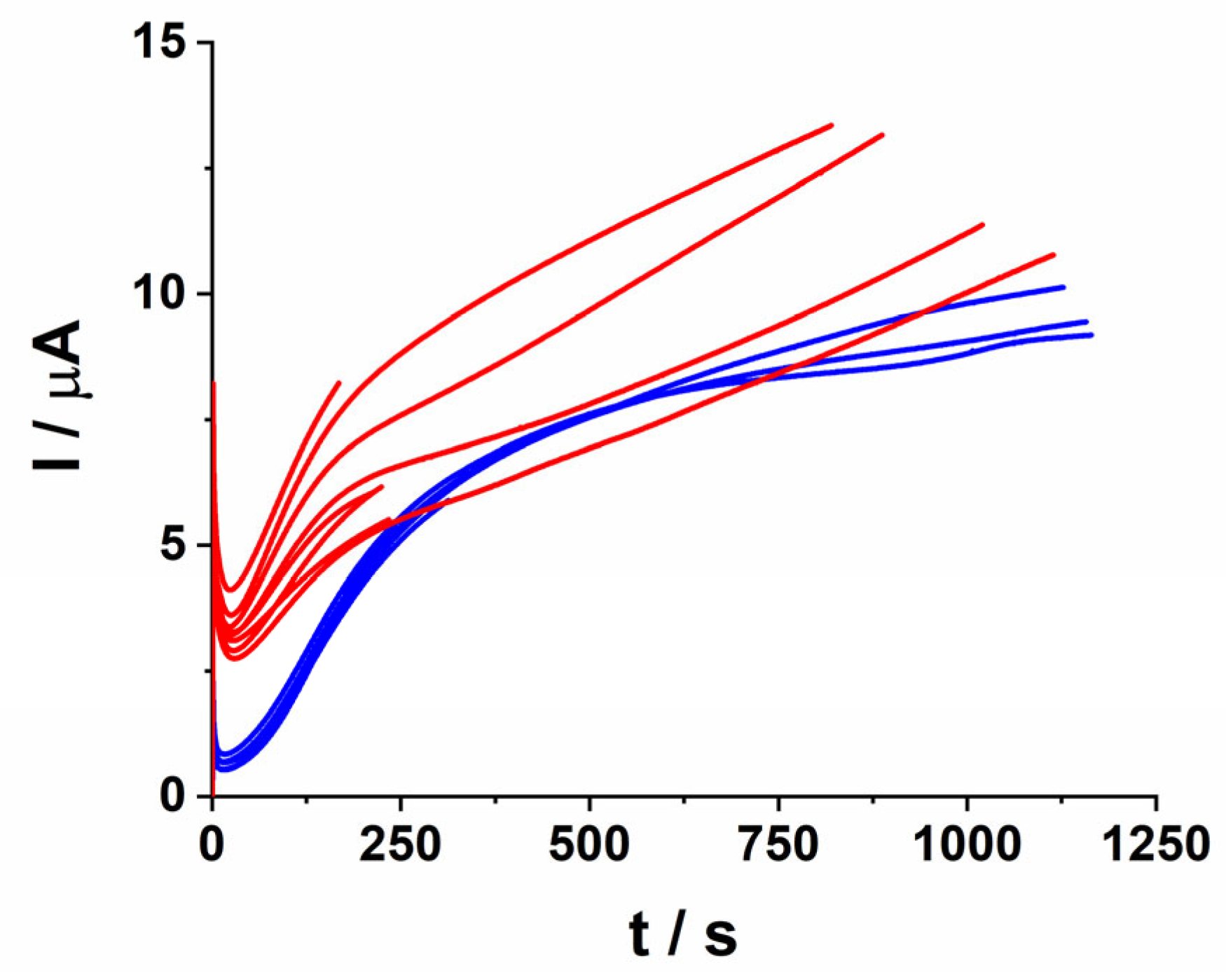
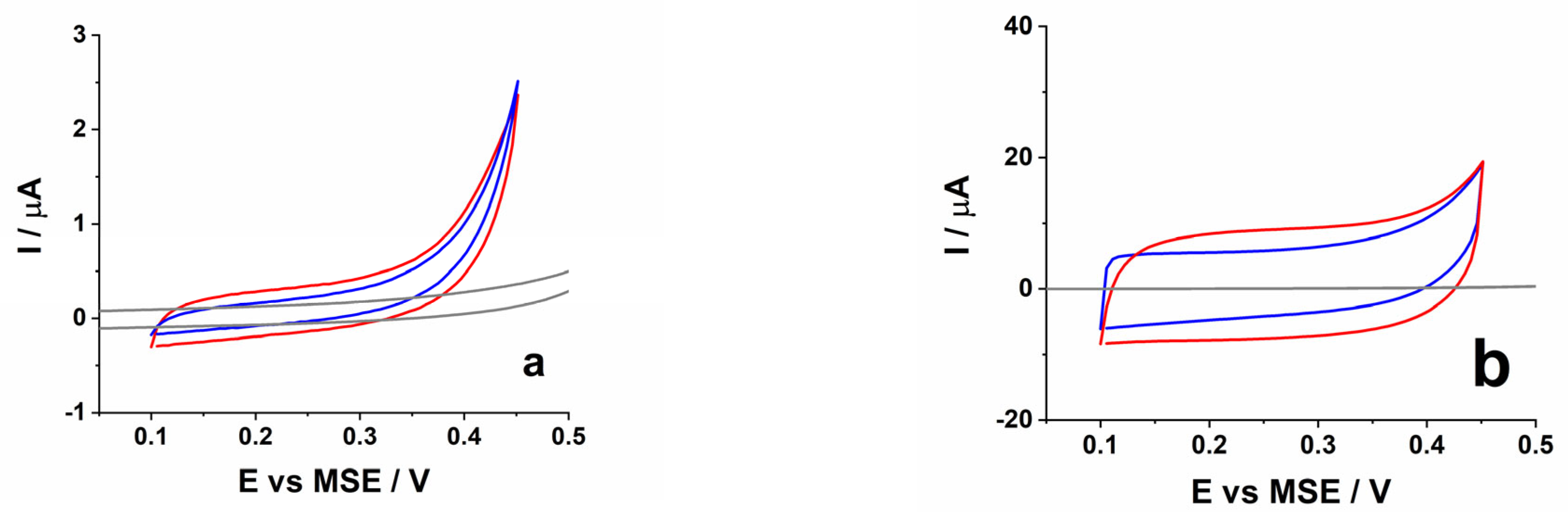
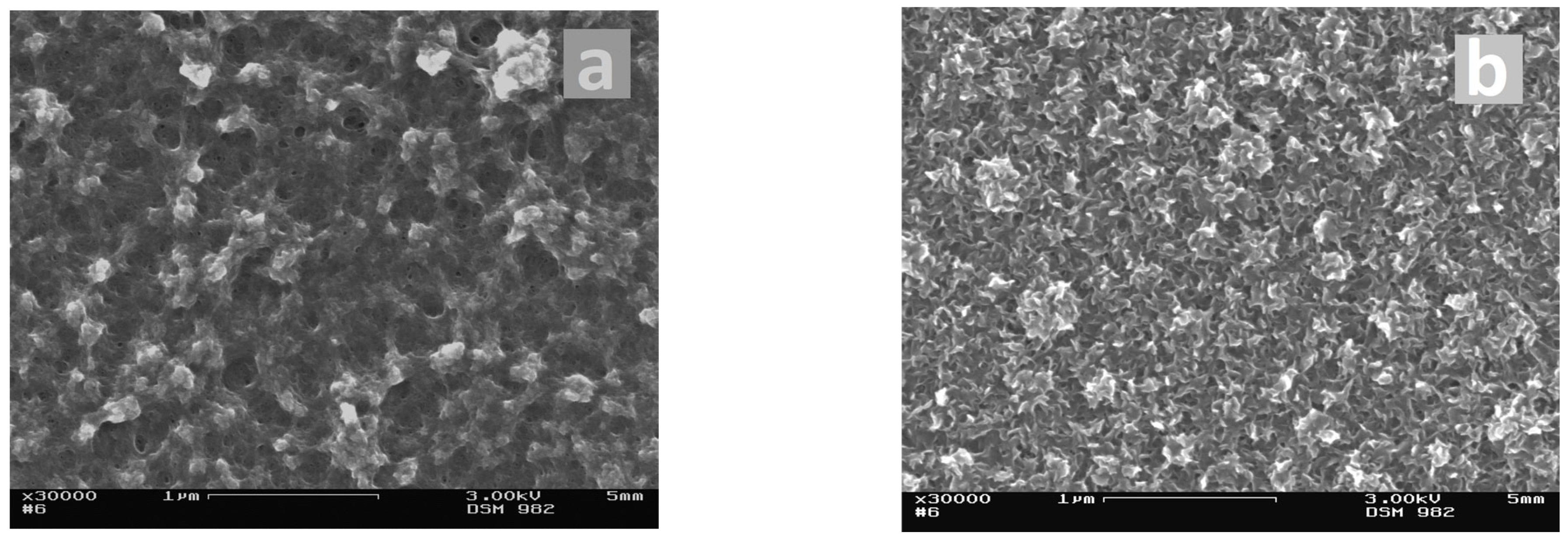
3.1. Electrochemical Synthesis of the PEDOT Coatings

Figure 1 shows several polymerization curves of PEDOT obtained in the presence of both types of counterions. In general, polymerization in the presence of SDS is faster but also shows a larger data discrepancy. Bearing in mind that the GCE is mechanically polished before polymerization, this effect should relate to the role of the initial GCE surface state for the PEDOT-SDS layer’s formation. Polymerization in the presence of PSS is reproducible and not that susceptible to the initial surface state of the glassy carbon.
The voltammetric behavior of both thin and thick layers of SDS- and PSS-doped PEDOT is shown in Figure 2. Twenty voltammetric scans are necessary to establish steady-state behavior after polymerization. For both polymerization charges (Qpoly = 1 mC and Qpoly = 8 mC), PEDOT-PSS shows smaller pseudo-capacitive currents than PEDOT-SDS does, which implies a smaller electroactive surface in the PSS case. SEM pictures (Figure 3) demonstrate the difference in the surface morphology of the two types of PEDOT layers.
In the next series of experiments, the scan rate dependence of the voltammetric response was measured in acetate buffer solution for all PEDOT coatings. Figure 4 presents results obtained for the thick PEDOT-PSS and PEDOT-SDS coatings. The double logarithmic plot (Figure 4c) shows a linearization of the data for the current dependent on the scan rate with the values of the straight line slopes in both cases being close to 1. The latter is also valid for the thin PEDOT layers and indicates usual capacitive behavior without the interference of limiting ionic diffusion inside the polymer coatings.

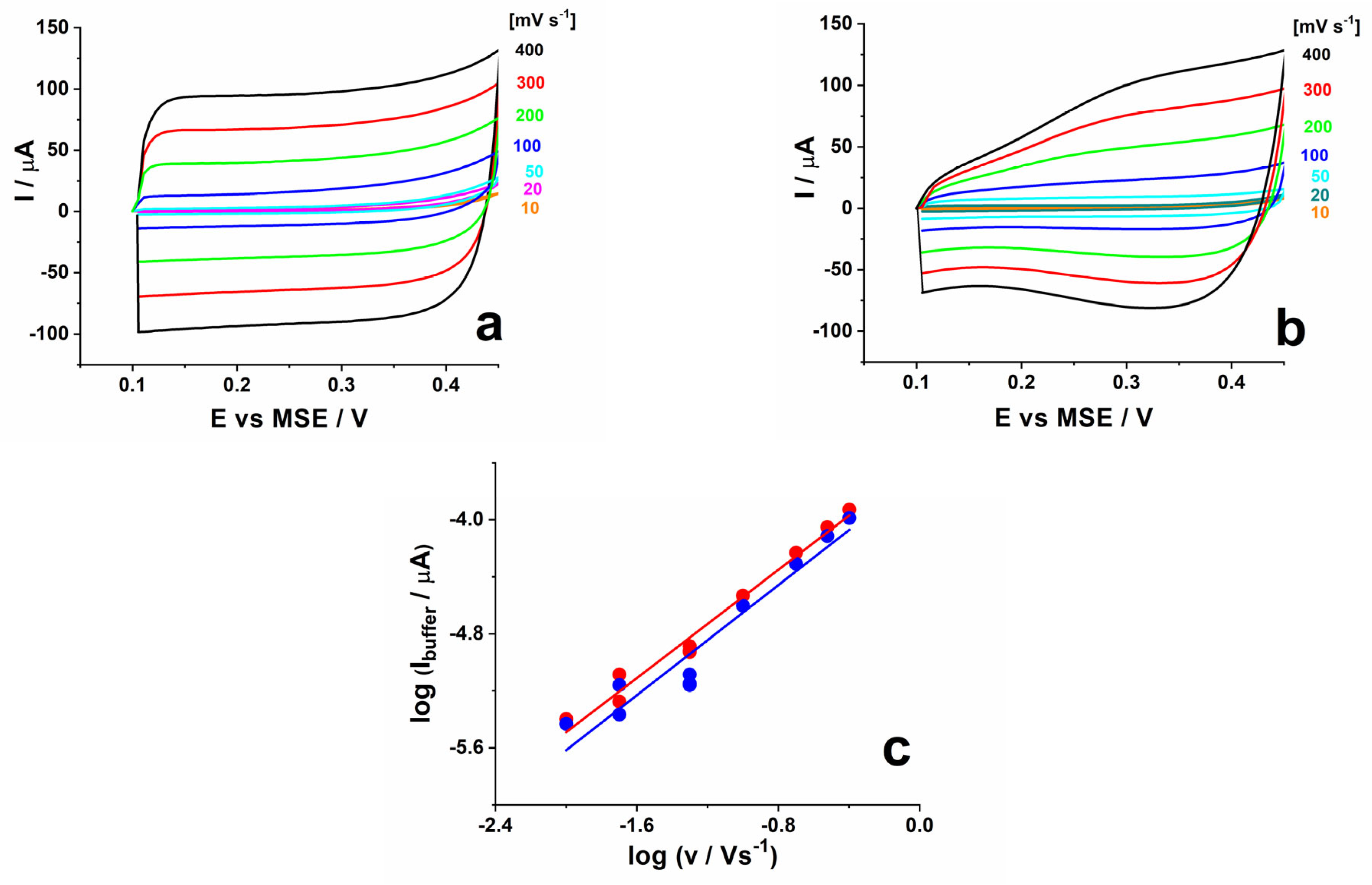
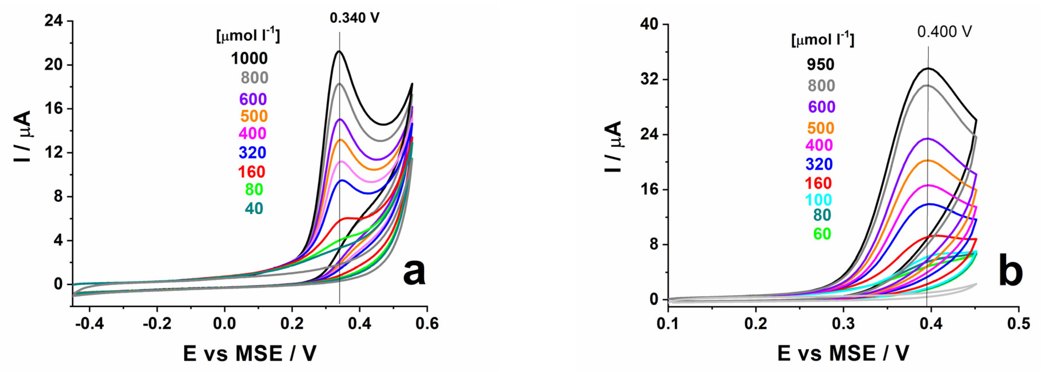
3.2. Voltammetric Measurements in the Presence of ALA

A comparative study of the electrooxidation of ALA on PSS- and SDS-doped PEDOT with the two different thicknesses was first carried out by cyclic voltammetry. Figure 5 shows sets of voltammetric curves measured at different ALA concentrations ranging from 40 µmol L−1 to 1 mmol L−1. In all cases the ALA oxidation peak is well detectable but there are marked differences depending on the doping ions and the thickness of the PEDOT layers. For PEDOT-PSS the ALA oxidation peak is found at less positive potentials whereas for the SDS-doped ones it shifts by about 60 mV in positive direction which implies an inhibited electron transfer in the latter case. Nevertheless, the oxidation peak currents are higher in the SDS case due very probably to the larger electroactive surface area and possible specific analyte/coating interactions (see below).
The comparison between thick and thin PEDOT layers at a constant ALA concentration shows, for both types of counterions, higher peak current values for the thicker coatings that should be related to a higher electroactive surface area of coatings with Qpoly = 8 mC. It is well established that with the thickening of the conducting polymer layers, there is a change in surface morphology, resulting in a rougher and/or more open and porous structure. Both effects would lead to an increase in the effective electroactive surface area of the coatings. It is worth mentioning that for the PEDOT-SDS and the thin PEDOT-PSS layers, the peak potential remains fixed for all concentration measured (Figure 5a,b,d). For the thick PEDOT-PSS layer (Figure 5c), however, a shift in the peak position by about 15 mV in the negative direction is observed. The latter seems to result from the gradual suppression of the PEDOT-PSS intrinsic oxidation currents found at E > 0.35 V.
A voltammetric scan rate study was carried out also in the presence of ALA in the acetate buffer solution. The double-logarithmic plot of the data for the oxidation peak currents vs. scan rate (Figure 6) obtained for the thin PEDOT coatings shows linearization with slopes of the straight lines amounting to 0.57 and 0.65 for PEDOT-PSS and PEDOT-SDS, respectively (Figure 6a). The latter values are close to the expected value (0.5) for a diffusion-limited process. The measurements with the thick PEDOT coatings (Figure 6b, full symbols) show a different result. At the 100 µmol L−1 ALA concentration, the slope of the log(I) vs. log(v) dependence amounts to 1, whereas by increasing the concentration of ALA up to 400 µmol L−1 (Figure 6b, hollow squares), the slope decreases to a value (0.7) closer to the one expected for diffusion. This peculiarity means that for thick PEDOT layers, a linear calibration curve, valid for both low and high concentration ranges of ALA, should not be expected.
Finally, the concentration dependences of the peak currents for the four types of PEDOT coatings are shown in Figure 7. In order to eliminate the contribution of the intrinsic PEDOT-related currents, the data for the ALA oxidation peaks are adjusted by subtracting the corresponding values of the buffer currents of the respective PEDOT coatings. The resulting concentration dependences show higher sensitivities for the thick PEDOT coatings. Larger values of sensitivity are observed for PEDOT-SDS for both thicknesses (see Table 2). The values of LOD obtained by means of the different PEDOT specimens using cyclic voltammetry range from 34 to 50 µmol L−1.

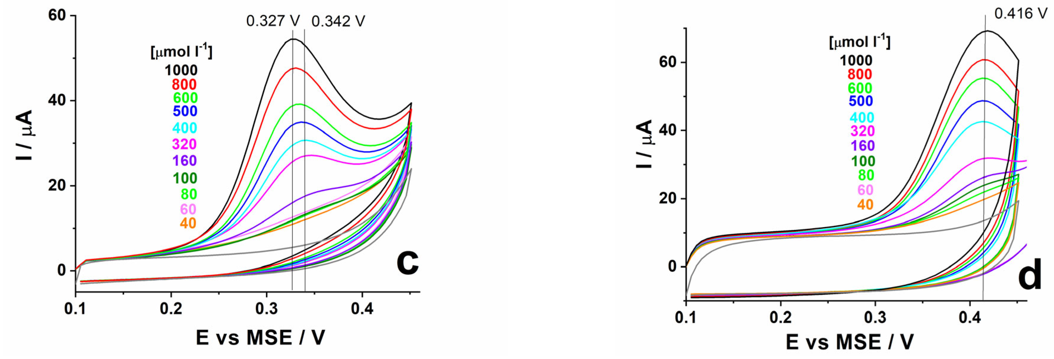
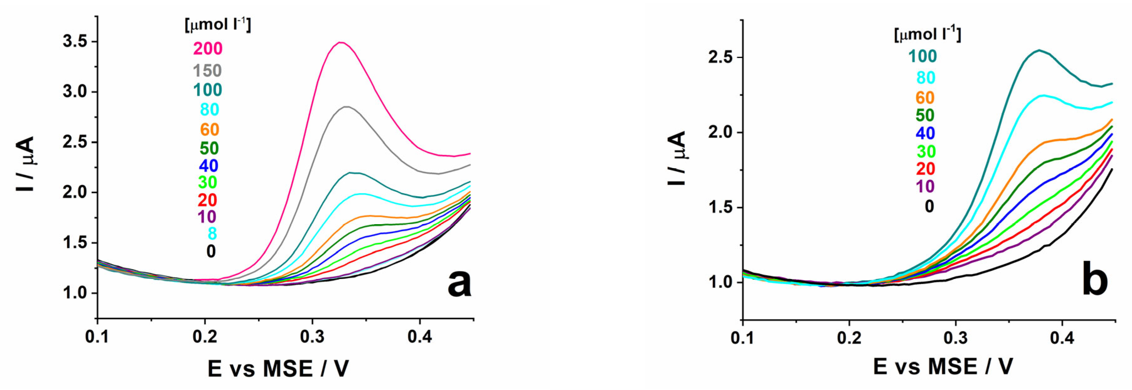
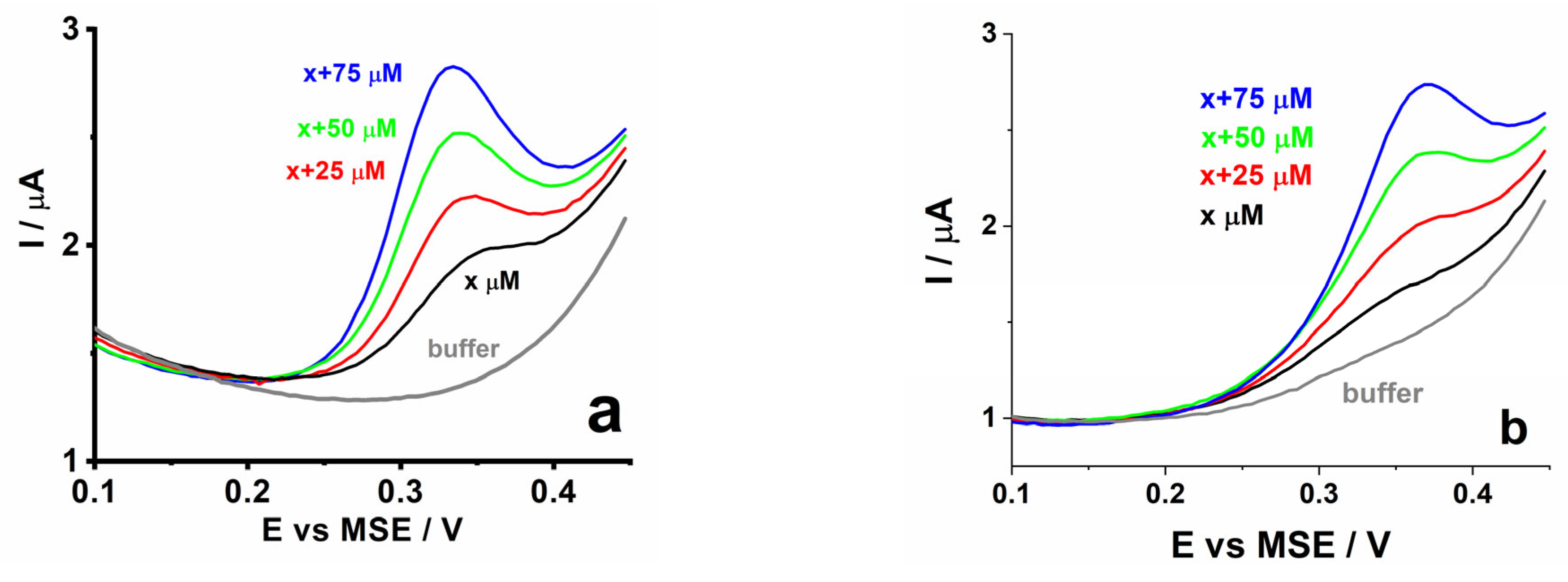
3.3. DPV Measurements in the Presence of ALA

It is well known that the DPV method provides the possibility of increasing the sensitivity of the electroanalytical measurements, and thus allows for electroanalytical detection in a lower concentration range. Figure 8 shows a series of DPV measurements at different ALA concentrations carried out with thin PEDOT-PSS and PEDOT-SDS coatings. The DPV signal is sensitive to ALA concentrations as low as about 10 µmol L−1 for both types of electrodes and close values of the sensitivities are found for both doping anions (Figure 9 and Table 2). The regression lines for both types of PEDOT are y = −2.27 × 10⁻⁸ + 0.0117x (R-square = 0.9959) for PSS and y = −6.42 × 10⁻⁸ + 0.0131x (R-square = 0.9890) for SDS. The DPV method could not be applied for the thick PEDOT coatings, very possibly due to the fact that for these coatings, the limiting factor controlling the ALA oxidation reaction in the low concentration range is not diffusion.
The thin PEDOT coatings were further used for the determination of ALA in real samples. A commercially available food supplement was used for this aim. Via dilution (see Section 2.3) an amount corresponding to a concentration of 50 µmol L−1 was introduced in the buffer solution. Data obtained via the standard addition method are presented in Figure 10. The data for the current obtained in these measurements show a linear dependence on the added concentration with straight lines parallel to the initial calibration curves (Figure 9 open symbols). The intercept from the X-axis results in ALA concentration values corresponding to 49 µmol L−1, i.e., to 98% of the expected value.

4. Conclusions

The present investigation demonstrates the possibility to involve PEDOT in electroanalytical determination of ALA by focusing on the characteristics, thickness and type of counterions that influence the electrooxidation reaction. It is found that by increasing the thickness of PEDOT, irrespective of the counterions, the oxidation currents of ALA increase by more than twice, most probably due to the real surface area of the polymer coatings. An important point in the present investigation is the established difference in the kinetics of the ALA oxidation reaction depending on the thickness of the PEDOT coatings and the concentration of ALA. In fact, voltammetric or DPV peak currents should depend linearly on the analyte concentration, provided that diffusion is limiting the electrochemical reaction in the whole studied concentration interval. This is obviously the case for the thin PEDOT coatings. For the thick PEDOT coatings, however, the enlarged surface area, which is beneficial for increasing the sensitivity of the electroanalytical signal, results simultaneously in the opportunity to experience kinetic limitations, seemingly due to the adsorption of analyte or product species. If adsorption alone is limiting the electrochemical reaction, non-linear calibration curves reaching saturation are observed. In the present case, adsorption is expected to limit the ALA oxidation reaction on thick PEDOT layers and at a low analyte concentration. Nevertheless, at concentrations above 100 µmol L−1, diffusion starts to be operative as far the polymer electroactive surface seems to be saturated with respect to dynamic adsorption/desorption but not blocked with respect to electron transfer and further oxidation events. Thus, the rate-determining steps in the low and high concentration ranges differ and the thick PEDOT coatings could not be properly used at very low ALA concentrations despite the higher sensitivity of the electroanalytical signal. For thin PEDOT layers, however, a large concentration range (40 to 1000 µM L−1) of the linear voltammetric response is established.
Concerning the type of the counter ions in PEDOT, a marked effect is observed in the electrooxidation reaction. In the SDS case, a shift in the ALA oxidation peaks to more positive potentials is established together with larger oxidation currents. Apart from the difference in surface morphology and real electroactive surface area, specific hydrophobic/hydrophilic interactions of the analyte species with the two types of PEDOT coatings should be considered. PSS-doped PEDOT is a more hydrophilic material with significant swelling in aqueous solutions and is also in the origin of the larger thickness at a constant polymerization charge. On the other hand, SDS-doped PEDOT layers are hydrophobic and expected to involve small amounts of solvent molecules in their internal structure. ALA has both a hydrophobic and hydrophilic part but charge transfer should occur in the hydrophobic one. Thus, PEDOT-SDS coatings should provide a better chemical environment for the ALA oxidation reaction. At the same time, a shift in the potential positions of the ALA oxidation peaks depending on the type of counterions provides the opportunity for eventually avoiding the possible overlap of oxidation peaks due to interfering species.
Finally, compared to those obtained in previous electroanalytical studies conducted for ALA determination (Table 1), the present study’s data for the sensitivity, LOD and width of the linear concentration range (Table 2) show the possibility to work in a very large concentration range. The DPV measurements performed with the thin PEDOT layers provide the possibility to work at concentrations even below 10 µmol L−1, and to perform the electroanalytical determination of ALA in real specimens. Concentrations of several µmol L−1 to several tens of µmol L−1 are of practical importance for the determination of ALA in foods [28] and blood plasma [29,30], whereas pharmaceutical formulations and food additives have much larger concentrations [26,27]. Thus, the present study shows that the intrinsic characteristics of PEDOT such as thickness and type of counterions provide a useful instrument for achieving different electroanalytical performance and addressing various tasks concerning the electrochemical determination of ALA.

Author Contributions

Conceptualization, V.T.; methodology, V.T.; investigation, V.K.; analysis of data, V.K. and V.T.; resources, V.T.; writing—original draft preparation, V.T.; writing—review and editing, V.T.; visualization, V.K.; supervision, V.T.; funding acquisition, V.T. All authors have read and agreed to the published version of the manuscript.

Funding

This research was funded by the Bulgarian OP Science and Education for Smart Growth (2014–2020) within National Center of Mechatronics and Clean Technologies (Project BG05M2OP001-1.001-0008-C010) and Bulgarian Ministry of Education and Science within INFRAMAT (National Roadmap for Research Infrastructure).

Institutional Review Board Statement

Not applicable.

Informed Consent Statement

Not applicable.

Data Availability Statement

Data are contained within the article.

Acknowledgments

The technical support of A. Nakova (Institute of Physical Chemistry, Bulgarian Academy of Sciences) is gratefully acknowledged.

Conflicts of Interest

The authors declare no conflict of interest. The funders had no role in the design of the study; in the collection, analyses, or interpretation of data; in the writing of the manuscript; or in the decision to publish the results.

References

  1. Inzelt, G. Conducting Polymers: A New Era in Electrochemistry; Springer: Berlin/Heidelberg, Germany, 2012. [Google Scholar]
  2. Ouyang, J. Application of intrinsically conducting polymers in flexible electronics. SmartMat 2021, 2, 263. [Google Scholar] [CrossRef]
  3. Bobacka, J.; Ivaska, A. Chemical sensors based on conducting polymers. In Electropolymerization. Concepts, Materials, Applications; Cosnier, S., Karyakin, A., Eds.; Wiley: Weinheim, Germany, 2010; pp. 173–188. [Google Scholar]
  4. Tsakova, V. Conducting polymers in electroanalytical medical applications. In Applications of Electrochemistry in Medicine, Modern Aspects of Electrochemistry; Schlesinger, M., Ed.; Springer Science + Business Media: New York, NY, USA, 2013; Volume 56, pp. 283–342. [Google Scholar]
  5. Tezakati, T.; Seifalian, A.; Tan, A.; Seifalian, A.M. Conductive Polymers: Opportunities and Challenges in Biomedical Applications. Chem. Rev. 2018, 118, 6766. [Google Scholar] [CrossRef] [PubMed]
  6. Ramanavicius, S.; Ramanavicius, A. Conducting polymers in the Design of Biosensors and Biofuel Cells (Review). Polymers 2021, 13, 49. [Google Scholar] [CrossRef] [PubMed]
  7. Xu, H.; Zhang, J. A Review on Conducting Polymers and Nanopolymer Composite Coatings for Steel Corrosion Protection. Coatings 2019, 9, 807. [Google Scholar] [CrossRef]
  8. Umoren, S.A.; Solomon, M.M. Protective polymeric films for industrial substrates: A critical review on past and recent applications with conducting polymers and polymer composites/nanocomposites. Progr. Mat. Sci. 2019, 104, 38. [Google Scholar] [CrossRef]
  9. Zhao, Z.; Xia, K.; Hou, Y.; Zhang, Q.; Yead, Z.; Lu, J. Designing flexible, smart and self-sustainable supercapacitors for portable/wearable electronics: From conductive polymers. Chem. Soc. Rev. 2021, 50, 12702–12743. [Google Scholar] [CrossRef] [PubMed]
  10. Groenendaal, L.; Jonas, F.; Freitag, D.; Pielartzik, H.; Reynolds, J.R. Poly(3,4-ethylenedioxythiophene) and its derivatives: Past, present, and future. Adv. Mater. 2000, 12, 481–494. [Google Scholar] [CrossRef]
  11. Elschner, A.; Kirchmeyer, S.; Lovenich, W.; Merker, U.; Reuter, K. PEDOT: Principles and Applications of an Intrinsically Conductive Polymer; CRC Press: Boca Raton, FL, USA, 2010. [Google Scholar]
  12. Nasybulin, E.; Wei, S.; Kymissis, I.; Levon, K. Effect of solubilizing agent on properties of poly(3,4-ethylenedioxythiophene) (PEDOT) electrodeposited from aqueous solution. Electrochim. Acta 2012, 78, 638. [Google Scholar] [CrossRef]
  13. Lyutov, V.; Efimov, I.; Bund, A.; Tsakova, V. Electrochemical polymerization of 3,4-ethylenedioxythiophene in the presence of dodecylsulfate and polysulfonic anions—An acoustic impedance study. Electrochim. Acta 2014, 122, 21. [Google Scholar] [CrossRef]
  14. Lyutov, V.; Gruia, V.; Efimov, I.; Bund, A.; Tsakova, V. An acoustic impedance study of PEDOT layers obtained in aqueous solution. Electrochim. Acta 2016, 190, 285. [Google Scholar] [CrossRef]
  15. Gribkova, O.L.; Iakobson, O.D.; Nekrasov, A.A.; Cabanova, V.A.; Tverskoy, V.A.; Vannikov, A.V. The influence of polyacid nature on poly(3,4-ethylenedioxythiophene) electrosynthesis and its spectroelectrochemical properties. J. Solid State Electrochem. 2016, 20, 2991. [Google Scholar] [CrossRef]
  16. Lyutov, V.; Kabanova, V.; Gribkova, O.; Nekrasov, A.; Tsakova, V. Electrochemically Obtained Polysulfonates Doped Poly(3,4-ethylenedioxythiophene) Films—Effects of the Dopant’s Chain Flexibility and Molecular Weight Studied by Electrochemical, Microgravimetric and XPS Methods. Polymers 2021, 13, 2438. [Google Scholar] [CrossRef] [PubMed]
  17. Gueyea, M.N.; Carella, A.; Faure-Vincent, J.; Demadrillec, R.; Simonato, J.-P. Progress in understanding structure and transport properties of PEDOT-based materials: A critical review. Progr. Mater. Sci. 2020, 108, 100616. [Google Scholar] [CrossRef]
  18. Ziyatdinova, G.K.; Budnikov, G.K.; Pogoreltsev, V.I. Electrochemical determination of lipoic acid. J. Analyt. Chem. 2004, 59, 288. [Google Scholar] [CrossRef]
  19. Corduneanu, O.; Garnett, M.; Oliveira Brett, A.M. Anodic Oxidation of a-Lipoic Acid at a Glassy Carbon Electrode and Its Determination in Dietary Supplements. Analyt. Lett. 2007, 40, 1763. [Google Scholar] [CrossRef]
  20. Skorupa, A.; Michalkiewicz, S. Anodic Oxidation of α-Lipoic Acid on Carbon Electrodes in Acetic Acid—Acetonitrile Solutions, Int. J. Electrochem. Sci. 2019, 14, 5107. [Google Scholar] [CrossRef]
  21. Ferreira, A.P.M.; dos Santos Pereira, L.N.; Santos da Silva, I.; Tanaka, S.M.C.N.; Tanaka, A.A.; Angnes, L. Determination of a-Lipoic acid on a Pyrolytic Graphite Electrode Modified with Cobalt Phthalocyanine. Electroanalysis 2014, 26, 2138. [Google Scholar] [CrossRef]
  22. Stankovic, D.M.; Mehmeti, E.; Kalcher, K. Development of sensitive electroanalytical approach for the quantification of α-lipoic acid using boron doped diamond. Analyt. Sci. 2016, 32, 847. [Google Scholar] [CrossRef]
  23. Ziyatdinova, G.K.; Grigoreva, L.V.; Budnikov, G.K. Electrochemical Determination of Unithiol and Lipoic Acid at Electrodes Modified with Carbon Nanotubes. J. Analyt. Chem. 2009, 64, 185. [Google Scholar] [CrossRef]
  24. Miranda, M.P.; del Rio, R.; del Valle, M.A.; Faundez, M.; Armijo, F. Use of fluorine-doped tin oxide electrodes for lipoic acid determination in dietary supplements. J. Electroanal. Chem. 2012, 668, 1. [Google Scholar] [CrossRef]
  25. Marin, M.; Lete, C.; Manolescu, B.N.; Lupu, S. Electrochemical determination of a-lipoic acid in human serum at platinum electrode. J. Electroanal. Chem. 2014, 729, 128. [Google Scholar] [CrossRef]
  26. Sasikumara, R.; Ranganathan, P.; Chen, S.-M.; Rwei, S.-P. f-MWCNTs-PIN/Ti2O3 nanocomposite: Preparation, characterization and nanomolar detection of α-Lipoic acid in vegetables. Sens. Actuators B 2018, 255, 217. [Google Scholar] [CrossRef]
  27. Ziyatdinova, G.; Antonova, T.; Vorobev, V.; Osin, Y.; Budnikov, H. Selective voltammetric determination of α-lipoic acid on the electrode modified with SnO2 nanoparticles and cetyltriphenylphosphonium bromide. Monatshefte Für Chem.-Hemical Mon. 2019, 150, 401. [Google Scholar] [CrossRef]
  28. Saha, M.; Ahammad, H.; Bhoumik, N.; Shakil, S.; Shavan, M.; Morshed, M.; Hossan, T.; Sarker, S.; Rahman, N.; Rahman, S.; et al. Extraction and estimation of alpha lipoic acid content in different food samples by reverse phase HPLC: Effect of heat treatment. Int. J. Biosci. 2018, 13, 473. [Google Scholar]
  29. Amenta, F.; Traini, E.; Tomassoni, D.; Mignini, F. Pharmacokinetics of Different Formulations of Tioctic (Alpha-Lipoic) Acid in Healthy Volunteers. Clin. Exp. Hypertens. 2008, 30, 767. [Google Scholar] [CrossRef]
  30. Shay, K.; Moreau, R.; Smith, E.; Smith, A.; Hagen, T. Alpha-lipoic acid as a dietary supplement: Molecular mechanisms and therapeutic potential. Biochim. Biophys. Acta 2009, 1790, 1149. [Google Scholar] [CrossRef] [PubMed]
  31. Zanardi, C.; Terzi, F.; Seeber, R. Polythiophenes and polythiophene-based composites in amperometric sensing. Analyt. Bioanalyt. Chem. 2013, 405, 509. [Google Scholar] [CrossRef]
  32. Tsakova, V.; Seeber, R. Conducting polymers in electrochemical sensing: Factors influencing the electroanalytical signal. Analyt. Bioanalyt. Chem. 2016, 408, 7231. [Google Scholar] [CrossRef]
  33. Hui, Y.; Bian, C.; Xia, S.; Tong, J.; Wang, J. Synthesis and electrochemical sensing application of poly(3,4-ethylenedioxythiophene)-based materials: A review. Analyt. Chim. Acta 2018, 1022, 1. [Google Scholar] [CrossRef]
Scheme 1. Oxidation of ALA.
Coatings 13 02014 sch001
Figure 1. Polymerization curves obtained at E = 0.38 V vs. MSE in aqueous solution of 10 mmol L1 of EDOT and 34 mmol L1 of either PSS (blue lines) or SDS (red lines). The current traces registered for short times (200–275 s) correspond to Qpoly = 1 mC.
Coatings 13 02014 g001
Figure 2. Cyclic voltammetry measured in acetate buffer solution at 50 mV s1 for (a) thin (Qpoly = 1 mC) and (b) thick (Qpoly = 8 mC) PEDOT-PSS (blue lines) and PEDOT-SDS (red lines) coatings. The grey lines denote the trace of the un-coated glassy carbon electrode.
Coatings 13 02014 g002
Figure 3. SEM pictures of (a) PEDOT-PSS and (b) PEDOT-SDS coatings. Reprinted with permission from Electrochimica Acta, Vol. 122, p.21, V. Lyutov, I. Efimov, A. Bund, V. Tsakova Electrochemical polymerization of 3,4-ethylenedioxythiophene in the presence of dodecylsulfate and polysulfonic anions—an acoustic impedance study [13], Copyright Elsevier, 2014.
Coatings 13 02014 g003
Figure 4. Scan rate dependence of the intrinsic pseudocapacitive currents of thick (Qpoly = 8 mC) (a) PEDOT-PSS and (b) PEDOT-SDS coatings measured in acetate buffer solution; (c) double logarithmic plot of the data obtained for PEDOT-PSS (blue symbols) and PEDOT-SDS (red symbols).
Coatings 13 02014 g004
Figure 5. Cyclic voltammetry in the presence of different amounts of ALA measured from (a) thin (Qpoly = 1 mC) PEDOT-PSS, (b) thin (Qpoly = 1 mC) PEDOT-SDS, (c) thick (Qpoly = 8 mC) PEDOT-PSS, and (d) thick (Qpoly = 8 mC) PEDOT-SDS coatings. Scan rate: 50 mV s−1. Grey lines denote measurements in buffer solution alone.
Coatings 13 02014 g005aCoatings 13 02014 g005b
Figure 6. Double logarithmic plot of data obtained from the scan rate dependence measured in acetate buffer in the presence of 100 µmol of L−1 ALA for PEDOT-PSS (blue symbols) and PEDOT-SDS (red symbols): (a) thin (Qpoly = 1 mC) and (b) thick (Qpoly = 8 mC) PEDOT coatings. The red squares in the plot (b) are obtained with 400 µmol L−1 of ALA in the solution.
Coatings 13 02014 g006
Figure 7. ALA peak oxidation currents, after the extraction of the corresponding buffer current values, for (a) thin (Qpoly = 1 mC) and (b) thick (Qpoly = 8 mC) PEDOT coatings, PEDOT-PSS (blue symbols), and PEDOT-SDS (red symbols).
Coatings 13 02014 g007
Figure 8. DPV measurements in acetate buffer solution at different concentrations of ALA for thin (Qpoly = 1 mC) (a) PEDOT-PSS and (b) PEDOT-SDS coatings.
Coatings 13 02014 g008
Figure 9. Calibration curves obtained from the DPV concentration dependences from thin (Qpoly = 1 mC) PEDOT-PSS (blue points) and PEDOT-SDS (red points) layers. The blue and red squares show the results from the determination of ALA in a real specimen via the standard addition method using the respective polymer coatings.
Coatings 13 02014 g009
Figure 10. DPV measurements in acetate buffer solution obtained via the standard addition method from thin (Qpoly = 1 mC) (a) PEDOT-PSS and (b) PEDOT-SDS coatings.
Coatings 13 02014 g010
Table 1. Data for the linear range of the electroanalytical response and LOD obtained from different electrode materials.
Working ElectrodeElectrolyteMethodLinear Range
[µM L−1]
LOD
[µM L−1]
Reference
GCEH2SO4LSV
DPV
1.51–173
2.5–75
5.75
1.8
[18]
GCEPBSDPV1–1501.8[19]
MWCNTs/GCEH2SO4LSV26–180 *
210–780
19[23]
Co-phtalocynine/pyrolytic graphitePBSDPV
CA
CV
0.499–19.6
1.9–25
7.3–260
0.0034
0.098
0.25
[21]
B-doped diamondBRB, pH3DPV
CA
0.3–105
0.3–60
0.088
0.06
[22]
F-doped SnO2PBSSWV5–2003.7[24]
PtBRB, pH 4.5DPV10–80013.15[25]
MWCNT/-polyindole/Ti2O3PBSDPV0.39–1100.0012[26]
SnO2 NPs-CTPPB/GCEBRB, pH 4.5DPV0.5–50 *
50–400
0.13
0.43
[27]
* two linear ranges. BRB—Briton–Robinson buffer; CA—chronoamperometry; CTPPB—cetyltriphenylphosphonium bromide; LSV—linear sweep voltammetry; PBS—phosphate-buffered solution; SWV—square wave voltammetry.
Table 2. Data for the sensitivity for ALA oxidation and LOD obtained from different PEDOT coatings.
MethodSpecimenConcentration Range
[µmol L−1]
Sensitivity
[µA µmol−1 cm−2]
LOD
[µmol L−1]
CVPSS, 1 mC40–10000.23350
CVSDS, 1 mC 60–9500.41336
CVPSS, 8 mC100–10000.55034
CVSDS, 8 mC100–7500.87640
DPVPSS, 1 mC8–2000.1466
DPVSDS, 1 mC,10–1000.1637
Disclaimer/Publisher’s Note: The statements, opinions and data contained in all publications are solely those of the individual author(s) and contributor(s) and not of MDPI and/or the editor(s). MDPI and/or the editor(s) disclaim responsibility for any injury to people or property resulting from any ideas, methods, instructions or products referred to in the content.

