Article

Activity of Colloidal Silver Solution against Microorganisms Implicated in Ocular Infections

1
Alfa Intes Industria Terapeutica Splendore (ITS), 80026 Naples, Italy
2
Department of Chemical, Biological, Pharmaceutical and Environmental Sciences, University of Messina, Via Ferdinando Stagno d’Alcontres 31, 98166 Messina, Italy
*
Author to whom correspondence should be addressed.
Coatings 2023, 13(2), 265; https://doi.org/10.3390/coatings13020265
Submission received: 11 December 2022 / Revised: 12 January 2023 / Accepted: 18 January 2023 / Published: 23 January 2023

Abstract

:
Endophthalmitis most likely originates from both planktonic bacteria suspended in the tear film and bacteria adherent to the conjunctiva and the eyelid. This study aimed to expand the research on the effectiveness of a colloidal silver solution (Silverix®) against ocular microorganisms. The activity of Silverix® was evaluated against methicillin-resistant Staphylococcus aureus, S. epidermidis, ofloxacin-resistant Pseudomonas aeruginosa, and Candida albicans strains, previously characterized for their antibiotic resistance and biofilm-forming capabilities. The microbial killing was estimated at various times in the presence and absence of colloidal silver solution against planktonic and biofilm-embedded cells. The results documented the efficacy of Silverix® on planktonic cells of S. aureus and S. epidermidis (2.49–2.87 Log CFU/mL reduction) and P. aeruginosa strains (3–4.35 Log CFU/mL reduction). On the contrary, C. albicans showed mild susceptibility. Regarding early biofilm, the ocular isolates were harder to kill (2–2.6 Log CFU/mL reduction) than the reference strains, whereas a similar decrease (3.1 Log CFU/mL reduction) was estimated for P. aeruginosa strains. The light microscope images of biofilms treated with colloidal solution confirmed the ability of Silverix® to destroy the biofilm.

1. Introduction

Endophthalmitis is an inflammatory condition of the intraocular cavities, usually caused by infection. It most likely originates from both planktonic bacteria suspended in the tear film and bacteria adherent to the conjunctiva and the eyelid [1,2]. The margin of the eyelid can be colonized by microorganisms with a sessile-growth lifestyle able to build biofilm communities with a key role in the development of chronic infections. One of the most common eye infections that usually affects both eyes along the edges of the eyelids is represented by blepharitis. Blepharitis is often a condition difficult to treat, characterized by edema, redness and inflammation of the ciliary edge which affects the lid and its dermis, eyelashes, conjunctiva and the meibomian glands. The etiology is complex, including chronic bacterial infections, infestations with certain parasites such as Demodex, and inflammatory skin conditions such as seborrheic dermatitis. Among various bacterial pathogens that impact blepharitis, emerge Staphylococcus aureus, S. epidermidis, Propionibacterium acnes and Corynebacteria sp.. Recently, microbial biofilms have been implicated in a wide array of ocular diseases, including recurrent or chronic blepharitis leading to dry eye, a pathology particularly widespread and often underestimated [3]. The microbial biofilms are cause of a worsening of the pathological picture with lowered vision and chronic inflammation of connective tissue. Today, blepharitis and dry eye disease are considered a single disease referred to as “Dry Eye Blepharitis Syndrome” (DEBS) [3]. Biofilms have become a clinical and therapeutic problem, as microorganisms embedded in a self-produced polymeric matrix constitute impenetrable microbial communities, plugging the meibomian glands or blocking the lash follicles, less susceptible to conventional treatment than their planktonic counterparts [4]. Furthermore, a biofilm extracellular polymeric substance is composed of DNA, proteins, and polysaccharides; it can also constitute a nutrient source for the growth of Demodex mites. These eyelash mites are parasites that live in or around human hair follicles, in symbiosis with many bacterial species, and are involved in blepharitis with cylindrical dandruff [3]. In light of the above, together with the serious concern raised by the increasing phenomenon of antibiotic resistance, it is necessary to shift toward alternative therapies to achieve better success in infection treatment.
Renewed attention has been focused on silver (Ag) because of its broad-spectrum antimicrobial activity against Gram-positive [5] and Gram-negative [6] organisms, fungi [7], protozoa [8], and some viruses [9]. It is known that Ag interacts with multiple target sites, such as cell membranes and microbial proteins, affecting permeability and respiration and causing cell death [5,6,7,8,9,10,11]. Current studies on the experimental biofilm models suggest that it also interferes with bacterial adhesion, destabilizes the biofilm matrix, and kills the bacteria embedded in the biofilm [12,13,14]. However, silver ions (Ag+) or salts have only limited usefulness as antimicrobial agents. As provided from current literature available on silver, there is still a lot to know about the clinical potential of this element. It is pivotal to take in consideration possible side effects for human health. Today, one of the most common forms of Ag is represented by colloidal silver (CS) in nanoparticles (NPs), as the Ag in nanoform is far better and more biocompatible agent. Nanoparticles have higher antibacterial activity than free Ag+ due to both the physical properties of nanoparticles and the elution of Ag+ [15]. AgNPs have multiple modes of action that lead to cell killing, cause structural and physiological alterations in microbial cell membranes, such as changes in permeability and membrane potential, as well as penetrate into the cell, resulting in binding interactions with proteins and DNA. The AgNPs have a surface/volume ratio much greater than the corresponding bulk material; therefore, interactions with microbial surfaces are facilitated, allowing a better ability to release/produce Ag+/reactive oxygen species [16,17,18]. It should be emphasized that, due to the non-specific nature of these mechanisms, AgNPs do not exert selective pressure on bacteria and have a much lower risk of developing resistance than conventional antibiotics. Silver nanoparticles have been also incorporated into different matrices and formulations, such as gels, coatings, composites, membranes, and thin films, to use in biomedical applications, food production, cosmetics, and numerous household products [15,19,20,21]. A form of gelatin-capped AgNPs has been studied as a promising antimicrobial and antiangiogenic nanotherapeutic for preclinical treatment of bacterial keratitis and eye-related microbial infections [22]. Specifically, the stabilized AgNPs showed efficient dispersion in aqueous media and interaction with S. aureus, improving antibacterial properties [22].
Starting from the preliminary data presented as a poster at the EVER Annual Congress [23], the current study expands the research on the activity of a colloidal silver solution (CST), providing new results on its efficacy against ocular isolates of S. aureus, S. epidermidis, Pseudomonas aeruginosa, and Candida albicans, chosen as representative microorganisms implicated in eye infections. Furthermore, microscopic biofilm analysis were also performed.

2. Materials and Methods

2.1. Materials

CST is used to soak gauzes for periocular hygiene (Silverix®, Alfa Intes already on the Italian market; Ocusilver® available on other markets, Italy) containing the following active ingredients: CS of 20–30 nm (0.001%) as an antimicrobial agent, sodium hyaluronate (0.05%) as a hydrating agent, and glycol extracts of Matricaria chamomilla L. (0.22%) and Euphrasia officinalis L. (0.22%) as soothing and emollient agents. Specifically, the CS used as a raw material has an Ag content of 70% to 80% of Ag dried substance, as required in accordance with the European Pharmacopoeia (EU) for CS for external use. As per chemical and physical properties, it is soluble in water and insoluble in ethanol (96%) and methylene chloride. The appearance of the CST is liquid, with a limpid yellowish color. The pH range is 6.30–8.30, with a characteristic floral scent due to the presence of natural extracts of Matricaria chamomilla and Euphrasia officinalis.

2.2. Microorganisms and Inoculum Preparation

The microbial strains, belonging to the private collection of the Microbiology Section, Department of Chemical, Biological, Pharmaceutical, and Environmental Sciences, University of Messina, and previously characterized for antibiotic resistance [24,25,26,27] and biofilm-forming ability [28,29,30,31], were used. Methicillin-resistant Staphylococcus aureus 815 and S. epidermidis 813 have been selected for their well-characterized biofilm-related properties such as the presence of the icaA/icaD genes evaluated by PCR analysis, slime production evaluated by the Congo red agar assay and ability of forming biofilm on polystyrene surface [24]. Moreover, S. aureus 815 was also characterized for its haemolytic activity and agr system by PCR analysis, properties correlated with a background of biofilm producers [28]. Ofloxacin-resistant P. aeruginosa 1 and C. albicans 4 strains have been selected for their biofilm formation on polystyrene surface [29,30,31]. The following international reference strains from American Type Culture Collection (ATCC) were also included: S. aureus ATCC 6538, S. epidermidis ATCC 35984, P. aeruginosa ATCC 9027, and C. albicans ATCC 10231.
Bacteria were cultured in Muller Hinton broth (MHB) at 37 °C for 24 h, while C. albicans was grown on RPMI-1640 at 37 °C for 48 h. For microbial inocula, the centrifuged cells were standardized in phosphate-buffered saline (PBS), pH 7.4, using turbidimetry absorbance to a concentration of 1 × 10⁶ CFU/mL, approximatively.

2.3. Killing Activity

Aliquots of colloidal solution (1 mL) were dispensed into tubes containing the standardized microbial strain and incubated at 37 °C. After 0, 5, 10, and 15 min, the samples were serially diluted in PBS and seeded on Tryptic Soy Agar (TSA) or Sabouraud agar. All plates were then incubated at 37 °C for 18–24 h up to 48–72 h. CFU was counted [31]. All determinations were performed in triplicate, including the growth controls.

2.4. Effectiveness on Biofilm-Embedded Cells

As reported previously, microbial cultures were grown as biofilms on polystyrene flat-bottomed microtiter plates (Costar; Corning) [32]. Briefly, overnight culture in Tryptic Soy Broth (TSB, P. aeruginosa and C. albicans) or TSB + 1% glucose (TSBG, S. aureus and S. epidermidis) was adjusted to 105 CFU/mL and dispensed individually to 96-well cell culture polystyrene microtiter plates. The plates were incubated at 37 °C for 6 h (early biofilms) and 24 h (late biofilms). After incubation, the planktonic phase was gently removed, and the biofilm was carefully washed twice with sterile PBS. Biofilms were then treated with CST or PBS (control). The effect of CST on cell viability was evaluated after different exposure times of 5, 15, 30, and 120 min. The colloidal solution was removed, the remaining biofilm was resuspended in PBS and the wells were scraped with sterile pipette tips as previously reported [32]. The microbial counts were assessed by plating serial dilutions onto TSA. After an incubation period of 48 h at 37 °C, the CFU were detected. All determinations were performed in triplicate.

2.5. Light Microscopy

Microbial early (6 h) biofilms of S. aureus ATCC 6538, S. epidermidis ATCC 35984, P. aeruginosa ATCC 9027 and C. albicans ATCC 10231 formed on polystyrene flat-bottomed microtiter plates (Costar; Corning), as described above, were washed three times with sterile PBS and treated with CST. Sequentially, PBS was added, serving as the control. After an exposure time of 15 min, the CST was removed; the plates were washed with PBS and stained using 0.4% crystal violet for 10 min [33]. The biofilms were observed with a light microscope X 200 (Leica DMLB).

2.6. Statistical Analysis

ANOVA was employed to evaluate any significant differences between the values obtained with and without the solution. A p-value < 0.05 was considered significant.

3. Results

3.1. Killing Activity

The antimicrobial activity of CST was tested on Gram-positive and Gram-negative bacteria and yeasts. The Log CFU/mL of planktonic microbial cells following the exposure to CST is presented in Table 1. It can be observed that the microbial load decreased as the time exposure increased. After 5 min of exposure, a mild decrease in the bacterial count was observed for both ATCC and ocular isolates of S. aureus and S. epidermidis (1.65–1.92 Log reduction) and P. aeruginosa (1.5–1.95 Log reduction) strains.
Interestingly, after 15 min contact, the CST has proved to be more effective achieving a significant (p < 0.05) load decrease ranging from 2.49 to 2.87 Log CFU/mL (about 99.7%–99.8%) for S. aureus and S. epidermidis strains (both ATCC and ocular isolates) and equal to 3–4.35 Log CFU/mL (99.9%–99.99%) for P. aeruginosa strains ATCC and ocular isolate, respectively. Except for C. albicans ATCC 10231, which showed a decrease of 1.4–2 Log units, the C. albicans ocular isolate was the least susceptible strain.

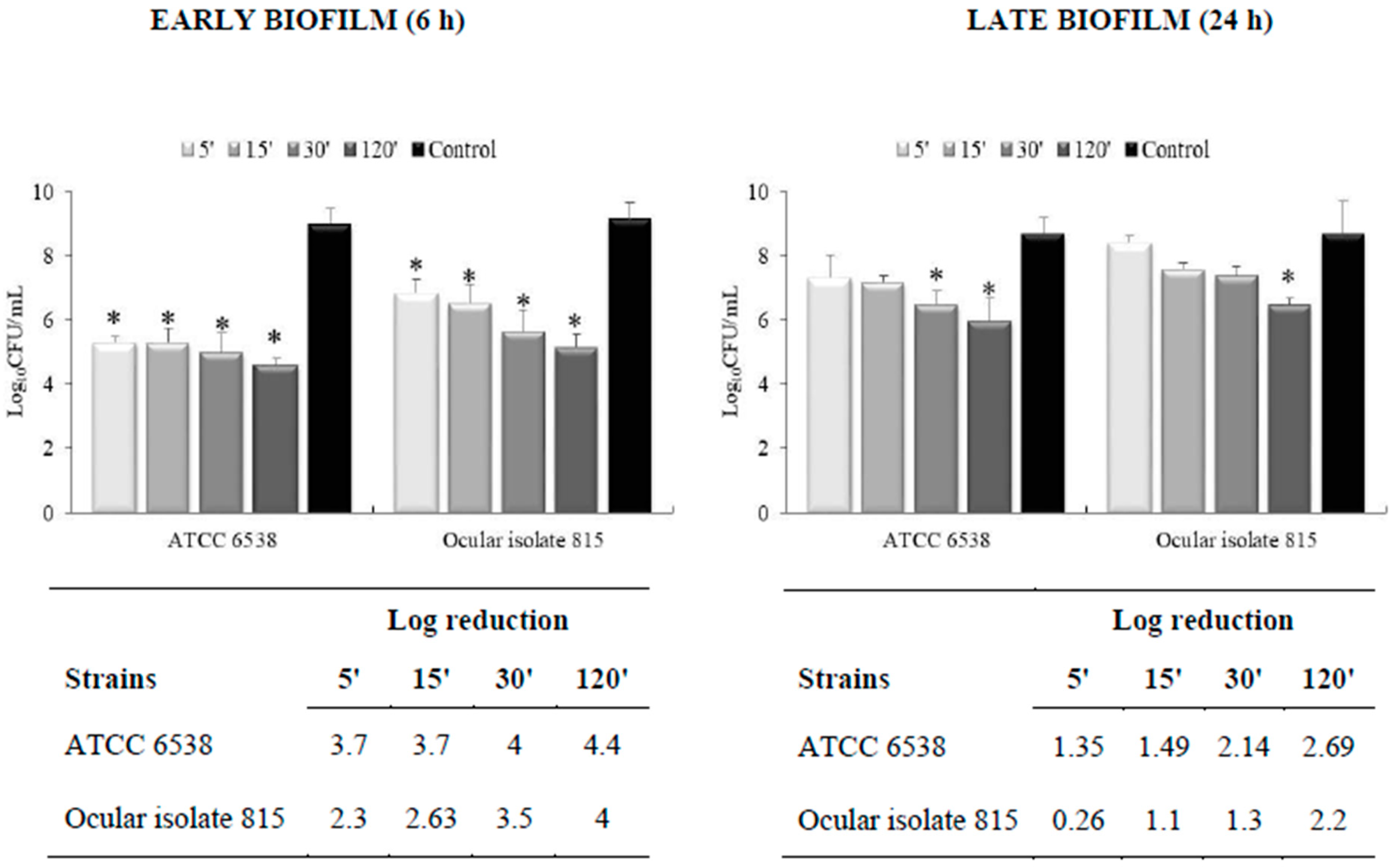
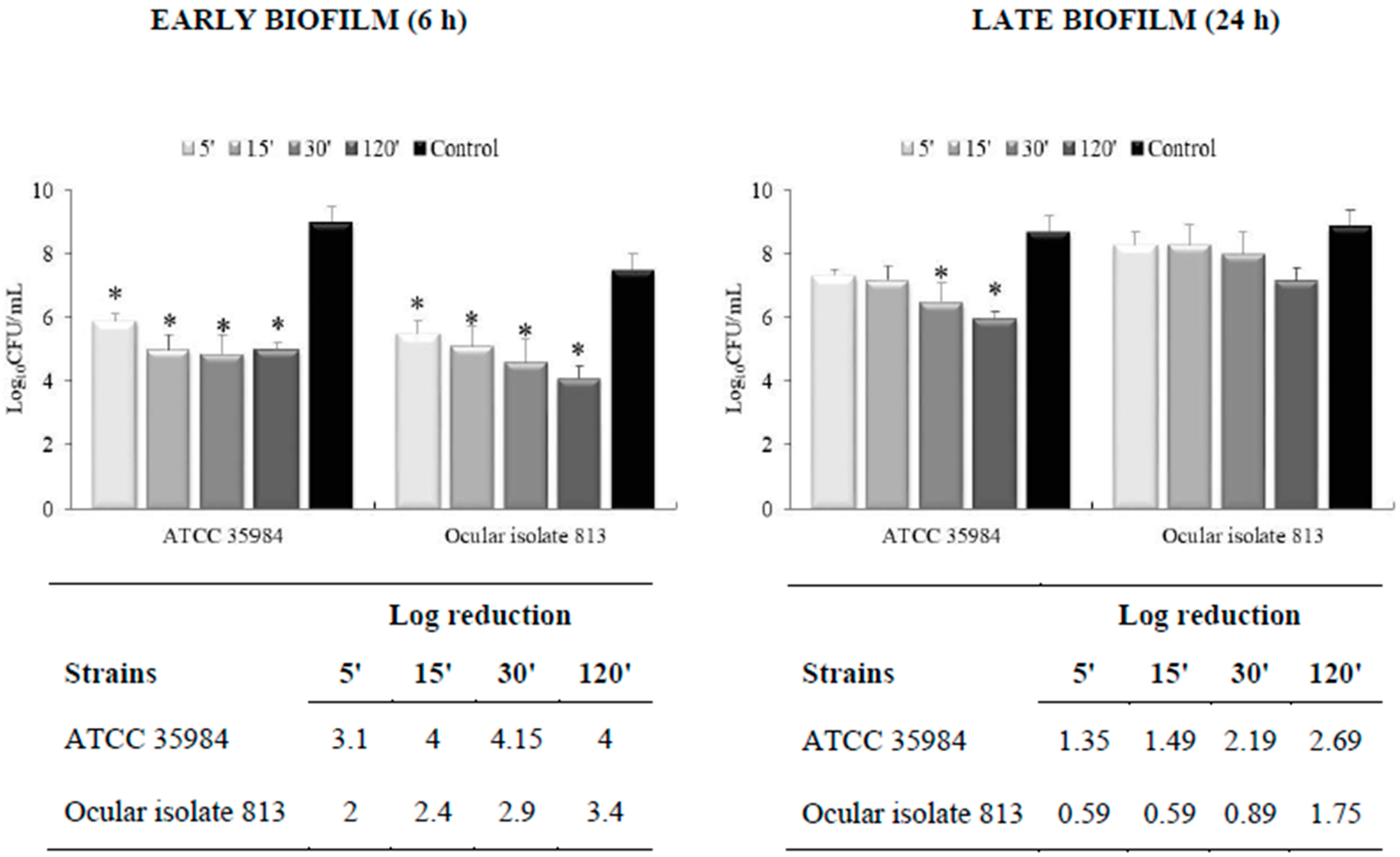
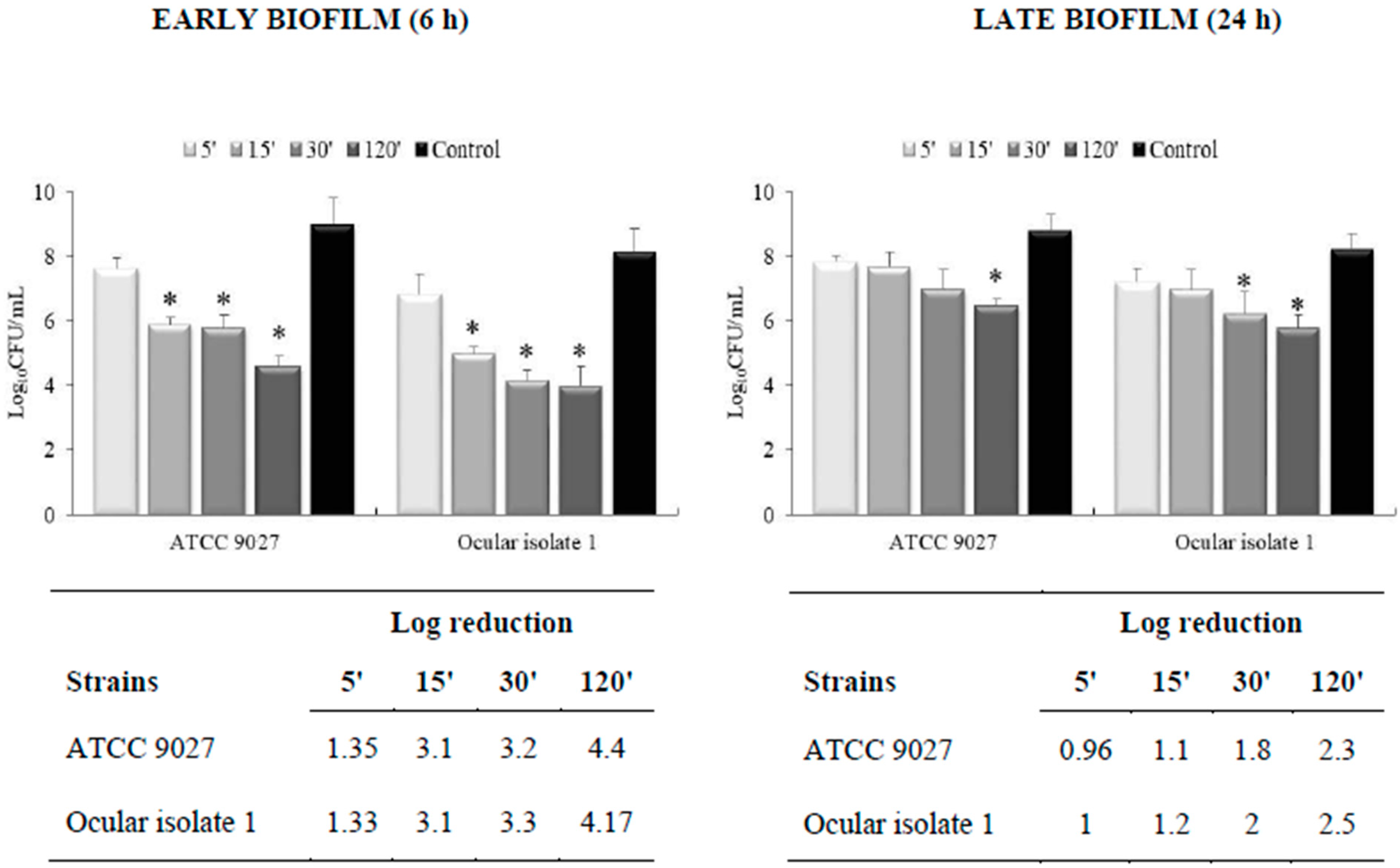
3.2. Effectiveness on Preformed Biofilm

The effect of the colloidal solution on the viability of the cells embedded in early (6 h) and late (24 h)-established biofilms is reported in Figure 1, Figure 2, Figure 3 and Figure 4. The results demonstrated that the inhibitory activity of the solution was more pronounced on early biofilm (Figure 1, Figure 2, Figure 3 and Figure 4) than on late biofilm (Figure 1, Figure 2, Figure 3 and Figure 4). Furthermore, different levels of susceptibility were found between the biofilm formed by ocular isolates and ATCC strains of S. aureus and S. epidermidis. Specifically, after 15 min of contact, a reduction of 2.63 Log for S. aureus and 2.4 Log for S. epidermidis ocular isolates and a reduction of 3.7 Log and 4 Log for S. aureus and S. epidermidis ATCC strains were detected (Figure 1 and Figure 2). Regarding the effect of the colloidal solution on P. aeruginosa biofilm, a reduction of 3.1 Log was displayed for both the ocular isolate and ATCC strain at 15 min of exposure (Figure 3). For C. albicans, the biofilm cell count decreased at a much slower rate. After 120 min, a 3.7 Log reduction was achieved for C. albicans ATCC, while a 1.5 Log reduction was observed for the C. albicans ocular isolate (Figure 4).
In contrast, the late-established biofilms were less susceptible to the colloidal solution than the early biofilms. Discrete inhibition was achieved only after prolonged exposure times (30–120 min) for all microorganisms (Figure 1, Figure 2, Figure 3 and Figure 4).

3.3. Light Microscopy

The results were substantiated by observing early biofilms under a light microscope (Figure 5). The images showed the ability of a colloidal solution to reduce the biofilm of S. aureus, S. epidermidis, and P. aeruginosa. After treatment for 15 min, the biofilm observed in controls (Figure 5a–c) became partially destroyed, and the bottom of the well was only covered by a few cells (Figure 5e–g). The effect was less evident for C. albicans (Figure 5d,h).

4. Discussion

Bacteria are frequently associated with various ocular infections such as conjunctivitis, keratitis, blepharitis and endophthalmitis [34]. Conjunctivitis is the most common ocular infection and if became chronic can also affect the eye lid with potential risk for extra or intraocular infections [34]. Keratitis is a serious eye infection and can also progress to endophthalmitis. Both keratitis and endophthalmitis are potentially devastating ocular infections if not diagnosed and treated early. Effective antimicrobial therapy is the most important approach that should be promptly initiated for treatment of patients afflicted with these eye infections. However, the continue emergence of antimicrobial resistance and the close correlation between biofilm and antibiotic tolerance represent alarming concerns. The identification of alternative strategies effective alone or as adjuvants to common antimicrobials is highly desirable. In recent years, the introduction of AgNPs profoundly impacted clinical settings including the treatment of ocular infections, even those caused by resistant bacterial strains. Nanoparticles are effective in increasing the retention time of active compounds on the ocular surface, protecting them from enzymatic degradation and improving their corneal permeability. Colloidal silver nanoparticles have been demonstrated to be relatively safe when administered to oral mucosa, eye and skin of the animal models for short periods of time [35].
In this study, the CST solution (Silverix®) has been developed according to EU pharmacopoeia for the topical use of silver and it is used to soak gauzes for periocular hygiene. Silverix® is indicated for periocular hygiene of adults and children with ongoing infections such as blepharitis, meibomitis and in patients in treatment with ophthalmic ointments to provide a delicate cleaning while efficiently removing ointments’ residues. Thanks to its unique composition, it can be used also by contact lenses users without any side effects reported to date. Silverix® is also recommended before and after ocular surgery for a cleaning of delicate periocular region. The findings of this study provide the scientific basis for the efficacy of CST against planktonic and sessile microorganisms. The results documented that the activity against planktonic growth was more pronounced against P. aeruginosa than against S. aureus and S. epidermidis. Accordingly, higher activity of AgNPs on E. coli than on S. aureus has been reported [16]. It is known that the antimicrobial activity of AgNPs is influenced by differences in the structure, thickness, and composition of the cell walls of Gram-negative and Gram-positive bacteria. Notably, Gram-negative bacteria possess an inner layer of peptidoglycan less thick than Gram-positive bacteria, and their outer membrane consists of phospholipids, lipopolysaccharides (LPS), lipoproteins, and surface proteins [36]. Therefore, the thinner peptidoglycan layer and the negative charge of LPS that promotes the adhesion of AgNPs may explain the increased susceptibility of Gram-negative bacteria [37]. As exposed above, the activity of AgNPs is also influenced by the dimensions. By decreasing the particle size to nanometer range (between 1 and 100 nm), antibacterial activity of silver can be increased due to a larger surface area-to-mass ratio [38]. The dimensions of the nanoparticles of the colloidal silver solution (Silverix ®) ranged from 20 to 30 nm.
However, it is noteworthy that other components of the colloidal silver solution, such as Matricaria chamomilla L. and Euphrasia officinalis chamomilla extracts, have been studied for their antimicrobial activity [39,40]. Specifically, M. chamomilla-extract-mediated AgNPs exhibited highly effective antimicrobial activity against S. aureus, E. coli, and C. albicans [41]. Theoretically, the components present in the formulation act in combination, causing considerable effects on the cell structural and functional properties.
Interestingly, the colloidal silver solution showed antimicrobial activity against MRSA and ofloxacin-resistant P. aeruginosa 1. These strains are generally less susceptible to topical prophylaxis regimens. Specifically, MRSA is a major public health problem all over the world, frequently associated to serious ocular multi-resistant infections [42]. In accordance, Panáček et al. [43] demonstrated the significant bactericidal potential of colloidal AgNPs against MRSA and Gram-negative bacteria.
The colloidal silver solution showed minimum efficiency against C. albicans compared with bacteria, and this probably stems from the differences between the bacterial and yeast cells. On the other hand, it has been reported that the fungicidal activity of AgNPs is lower than the bactericidal effects [44].
Although growing antibiotic resistance is one of the major causes of treatment failure, the spread of biofilm-forming microorganisms contributes to serious public health threats, including ocular infections [45,46,47,48]. Here, we reported the inhibitory activity of a colloidal solution on preformed microbial biofilms, with a more pronounced effect on early biofilms than late biofilms. Indeed, a significant reduction in the viability of cells embedded in the early biofilm matrix and a disaggregation effect documented in microscopic images were observed. Kalishwaralal et al. [14] described the action of AgNPs on microorganisms organized in biofilms with a detachment of biofilms formed by pathogens causing keratitis, such as P. aeruginosa and S. epidermidis. The Authors suggested that the inhibitory effect of AgNPs on the mature biofilm may be due to the presence of water channels (useful for nutrient transport) which would allow the direct diffusion of AgNPs through the glycocalyx matrix layer, imparting the antimicrobial effect.
Silver nanoparticles have also been assayed for the effect on biofilm formation produced by the P. aeruginosa, P. putida, Shigella flexneri, S. aureus and Streptococcus pneumonia [49,50]. The inhibitory effect of AgNPs was analyzed alone or in combination with antibiotics [50]. Interestingly, AgNPs in combination with antibiotics increased the cell death and increased ROS generation than antibiotics or AgNPs alone. The enhancing effects for ampicillin and vancomycin against Gram-negative and Gram-positive bacteria, respectively suggest that AgNPs can be used as an adjuvant for the treatment of infectious diseases [50]. Although the mechanism of action is not yet known, it is plausible to hypothesize that the components of the colloidal solution may destroy the polar polymeric matrix and predispose the cells to specific interactions with the CST, thus having considerable effects on the structural properties of the biofilm. It is also speculated that the different mechanisms employed by methicillin-susceptible Staphylococcus aureus (MSSA) and MRSA strains for biofilm formation may be involved in the reduced biofilm susceptibility of MRSA ocular isolates. MSSA strains predominantly form biofilms dependent on the polysaccharide intercellular adhesin type, whereas MRSA strains promote the formation of proteinaceous-type biofilms [51].
Effective antimicrobial strategies are needed in many preventive efforts in ophthalmology. The results of this study documented that the colloidal solution is active against microorganisms in both planktonic and biofilm phases and, therefore, could be used to assist in treating periocular surface infections.

Author Contributions

Conceptualization, A.N. and A.R.B.; methodology, A.N., A.R.B., A.M. and M.D.; software, M.D.; validation, A.N.; formal analysis, A.N., A.M. and M.D.; investigation, A.N. and A.M.; data curation, A.N.; writing—original draft preparation, A.N.; writing—review and editing, A.N. and A.R.B.; supervision, A.N. and A.R.B. All authors have read and agreed to the published version of the manuscript.

Funding

This research was supported by Alfa Intes Medical Division ITS (Casoria, Napoli, Italy) with a liberal donation to the University of Messina.

Institutional Review Board Statement

Not applicable.

Informed Consent Statement

Not applicable.

Data Availability Statement

Not applicable.

Acknowledgments

Editorial assistance was provided by Valentina Attanasio (Polistudium SRL, Milan, Italy).

Conflicts of Interest

Author A. Nostro received a liberal donation from Alfa Intes Medical Division ITS (Casoria, Napoli, Italy) for this research. Authors declare that they have no potential conflicts of interest regarding this work. In particular, the author A.R. Blanco declares to be employed in Alfa Intes Medical Division without any personal financial interest. Alfa Intes Medical Division had no role in the design of the study; in the collection, analysis, or interpretation of data; or the writing of the manuscript. Alfa Intes Medical Division approved the design and was allowed to review the manuscript prior to journal submission.

References

  1. Durand, M.L. Bacterial and fungal endophthalmitis. Clin. Microbiol. Rev. 2017, 30, 597–613. [Google Scholar] [CrossRef] [PubMed] [Green Version]
  2. Grzybowski, A.; Brona, P.; Kim, S.J. Microbial flora and resistance in ophthalmology: A review. Graefes Arch. Clin. Exp. Ophthalmol. 2017, 255, 851–862. [Google Scholar] [CrossRef] [PubMed] [Green Version]
  3. Rynerson, J.M.; Perry, H.D. DEBS—A unification theory for dry eye and blepharitis. Clin. Ophthalmol. 2016, 10, 2455–2467. [Google Scholar] [CrossRef] [PubMed] [Green Version]
  4. Olsen, I. Biofilm-specific antibiotic tolerance and resistance. Eur. J. Clin. Microbiol. Infect. Dis. 2015, 34, 77–86. [Google Scholar] [CrossRef] [PubMed]
  5. Jung, W.K.; Koo, H.C.; Kim, K.W.; Shin, S.; Kim, S.H.; Park, Y.H. Antibacterial activity and mechanism of action of the silver ion in Staphylococcus aureus and Escherichia coli. Appl. Environ. Microbiol. 2008, 74, 2171–2178. [Google Scholar] [CrossRef] [Green Version]
  6. Sondi, I.; Salopek-Sondi, B. Silver nanoparticles as antimicrobial agent: A case study on E. coli as a model for Gram-negative bacteria. J. Colloid Interface Sci. 2004, 275, 177–182. [Google Scholar] [CrossRef]
  7. Gajbhiye, M.; Kesharwani, J.; Ingle, A.; Gade, A.; Rai, M. Fungus-mediated synthesis of silver nanoparticles and their activity against pathogenic fungi in combination with fluconazole. Nanomedicine 2009, 5, 382–386. [Google Scholar] [CrossRef]
  8. Hendiger, E.B.; Padzik, M.; Sifaoui, I.; Reyes-Batlle, M.; López-Arencibia, A.; Rizo-Liendo, A.; Bethencourt-Estrella, C.J.; Nicolás-Hernández, D.S.; Chiboub, O.; Rodríguez-Expósito, R.L.; et al. Silver nanoparticles as a novel potential preventive agent against Acanthamoeba keratitis. Pathogens 2020, 9, 350. [Google Scholar] [CrossRef]
  9. Gaikwad, S.; Ingle, A.; Gade, A.; Rai, M.; Falanga, A.; Incoronato, N.; Russo, L.; Galdiero, S.; Galdiero, M. Antiviral activity of mycosynthesized silver nano-particles against herpes simplex virus and human parainfluenza virus type 3. Int. J. Nanomed. 2013, 8, 4303–4314. [Google Scholar]
  10. Maillard, J.Y.; Denyer, S.P. Focus on silver. In European Wound Management Association (EWMA) Position Document: The Role of Topical Antimicrobials in Managing Wound Infection; MEP Ltd.: London, UK, 2006. [Google Scholar]
  11. Russell, A.D.; Hugo, W.B. Antimicrobial activity and action of silver. Prog. Med. Chem. 1994, 31, 351–370. [Google Scholar]
  12. Feng, Q.L.; Wu, J.; Chen, G.Q.; Cui, F.Z.; Kim, T.N.; Kim, J.O. A mechanistic study of the antibacterial effect of silver ions on Escherichia coli and Staphylococcus aureus. J. Biomed. Mater. Res. 2000, 52, 662–668. [Google Scholar] [CrossRef]
  13. Chaw, K.C.; Manimaran, M.; Tay, F.E. Role of silver ions in destabilization of intermolecular adhesion forces measured by atomic force microscopy in Staphylococcus epidermidis biofilms. Antimicrob. Agents Chemother. 2005, 49, 4853–4859. [Google Scholar] [CrossRef] [Green Version]
  14. Kalishwaralal, K.; BarathManiKanth, S.; Pandian, S.R.; Deepak, V.; Gurunathan, S. Silver nanoparticles impede the biofilm formation by Pseudomonas aeruginosa and Staphylococcus epidermidis. Colloids Surf. B Biointerfaces 2010, 79, 340–344. [Google Scholar] [CrossRef]
  15. Richter, K.; Facal, P.; Thomas, N.; Vandecandelaere, I.; Ramezanpour, M.; Cooksley, C.; Prestidge, C.A.; Coenye, T.; Wormald, P.J.; Vreugde, S. Taking the silver bullet colloidal silver particles for the topical treatment of biofilm-related infections. ACS Appl. Mater. Interfaces 2017, 9, 21631–21638. [Google Scholar] [CrossRef]
  16. Marambio-Jones, C.; Hoek, E.M.V. A review of the antibacterial effects of silver nanomaterials and potential implications for human health and the environment. J. Nanopart. Res. 2010, 12, 1531–1551. [Google Scholar] [CrossRef]
  17. Kim, J.S.; Kuk, E.; Yu, K.N.; Kim, J.H.; Park, S.J.; Lee, H.J.; Kim, S.H.; Park, Y.K.; Park, Y.H.; Hwang, C.Y.; et al. Antimicrobial effects of silver nanoparticles. Nanomedicine 2007, 3, 95–101. [Google Scholar] [CrossRef]
  18. Lara, H.H.; Garza-Treviño, E.N.; Ixtepan-Turrent, L.; Singh, D.K. Silver nanoparticles are broad-spectrum bactericidal and virucidal compounds. J. Nanobiotechnol. 2011, 9, 30. [Google Scholar] [CrossRef] [Green Version]
  19. Franci, G.; Falanga, A.; Galdiero, S.; Palomba, L.; Rai, M.; Morelli, G.; Galdiero, M. Silver nanoparticles as potential antibacterial agents. Molecules 2015, 20, 8856–8874. [Google Scholar] [CrossRef] [Green Version]
  20. Porel, S.; Ramakrishna, D.; Hariprasad, E.; Gupta Dutta, A.; Radhakrishnan, T. Polymer thin film with in situ synthesized silver nanoparticles as a potent reusable bactericide. Curr. Sci. 2011, 101, 927–934. [Google Scholar]
  21. Tran, Q.H.; Nguyen, V.Q.; Le, A.T. Silver nanoparticles: Synthesis, properties, toxicology, applications and perspectives. Adv. Nat. Sci. Nanosci. Nanotechnol. 2018, 9, 049501. [Google Scholar] [CrossRef]
  22. Luo, L.J.; Lin, T.Y.; Yao, C.H.; Kuo, P.Y.; Matsusaki, M.; Harroun, S.G.; Huang, C.C.; Lai, J.Y. Dual-functional gelatin-capped silver nanoparticles for antibacterial and antiangiogenic treatment of bacterial keratitis. J. Colloid Interfaces Sci. 2019, 536, 112–126. [Google Scholar] [CrossRef] [PubMed]
  23. Blanco, A.R.; Marino, A.; D’Arrigo, M.; Nostro, A. Efficacy of a colloidal silver-based topical solution on microbial biofilms. In Acta Ophthalmologica; Wiley: Hoboken, NJ, USA, 2018; Volume 96, p. 14. [Google Scholar]
  24. Blanco, A.R.; Sudano-Roccaro, A.; Spoto, G.C.; Nostro, A.; Rusciano, D. Epigallocatechin gallate inhibits biofilm formation by ocular staphylococcal isolates. Antimicrob. Agents Chemother. 2005, 49, 4339–4343. [Google Scholar] [CrossRef] [PubMed] [Green Version]
  25. Marino, A.; Blanco, A.R.; Ginestra, G.; Nostro, A.; Bisignano, G. Ex vivo efficacy of gemifloxacin in experimental keratitis induced by methicillin-resistant Staphylococcus aureus. Int. J. Antimicrob. Agents 2016, 48, 395–400. [Google Scholar] [CrossRef] [PubMed]
  26. Consoli, G.M.L.; Granata, G.; Picciotto, R.; Blanco, A.R.; Geraci, C.; Marino, A.; Nostro, A. Design, synthesis and antibacterial evaluation of a polycationic calix [4]arene derivative alone and in combination with antibiotics. MedChemComm 2017, 9, 160–164. [Google Scholar] [CrossRef]
  27. Blanco, A.R.; Nostro, A.; D’Angelo, V.; D’Arrigo, M.; Mazzone, M.G.; Marino, A. Efficacy of a fixed combination of tetracycline, chloramphenicol, and colistimethate sodium for treatment of Candida albicans keratitis. Investig. Ophthalmol. Vis. Sci. 2017, 58, 4292–4298. [Google Scholar] [CrossRef] [Green Version]
  28. Di Stefano, A.; D’Aurizio, E.; Trubiani, O.; Grande, R.; Di Campli, E.; Di Giulio, M.; Di Bartolomeo, S.; Sozio, P.; Iannitelli, A.; Nostro, A.; et al. Viscoelastic properties of Staphylococcus aureus and Staphylococcus epidermidis mono-microbial biofilms. Microb. Biotechnol. 2009, 2, 634–641. [Google Scholar] [CrossRef] [Green Version]
  29. Nostro, A.; Guerrini, A.; Marino, A.; Tacchini, M.; Di Giulio, M.; Grandini, A.; Akin, M.; Cellini, L.; Bisignano, G.; Saraçoğlu, H.T. In vitro activity of plant extracts against biofilm-producing food-related bacteria. Int. J. Food Microbiol. 2016, 238, 33–39. [Google Scholar] [CrossRef]
  30. Nostro, A.; Marino, A.; Ginestra, G.; Cellini, L.; Di Giulio, M.; Bisignano, G. Effects of adaptation to carvacrol on Staphylococcus aureus in the planktonic and biofilm phases. Biofouling 2017, 33, 470–480. [Google Scholar] [CrossRef]
  31. Scaffaro, R.; Lopresti, F.; D’Arrigo, M.; Marino, A.; Nostro, A. Efficacy of poly(lactic acid)/carvacrol electrospun membranes against Staphylococcus aureus and Candida albicans in single and mixed cultures. Appl. Microbiol. Biotechnol. 2018, 102, 4171–4181. [Google Scholar] [CrossRef]
  32. Nostro, A.; Marino, A.; Blanco, A.R.; Cellini, L.; Di Giulio, M.; Pizzimenti, F.; Roccaro, A.S.; Bisignano, G. In vitro activity of carvacrol against staphylococcal preformed biofilm by liquid and vapour contact. J. Med. Microbiol. 2009, 58, 791–797. [Google Scholar] [CrossRef]
  33. Azeredo, J.; Azevedo, N.F.; Briandet, R.; Cerca, N.; Coenye, T.; Costa, A.R.; Desvaux, M.; Di Bonaventura, G.; Hébraud, M.; Jaglic, Z.; et al. Critical review on biofilm methods. Crit. Rev. Microbiol. 2017, 43, 313–351. [Google Scholar] [CrossRef] [Green Version]
  34. Teweldemedhin, M.; Gebreyesus, H.; Atsbaha, A.H.; Asgedom, S.W.; Saravanan, M. Bacterial profile of ocular infections: A systematic review. BMC Ophthalmol. 2017, 17, 212. [Google Scholar] [CrossRef] [Green Version]
  35. Maneewattanapinyo, P.; Banlunara, W.; Thammacharoen, C.; Ekgasit, S.; Kaewamatawong, T. An evaluation of acute toxicity of colloidal silver nanoparticles. J. Vet. Med. Sci. 2011, 73, 1417–1423. [Google Scholar] [CrossRef] [Green Version]
  36. Nikaido, H.; Vaara, M. Molecular basis of bacterial outer membrane permeability. Microbiol. Rev. 1985, 1, 1–32. [Google Scholar] [CrossRef]
  37. Rai, M.K.; Deshmukh, S.D.; Ingle, A.P.; Gade, A.K. Silver nanoparticles: The powerful nanoweapon against multidrug-resistant bacteria. J. Appl. Microbiol. 2012, 112, 841–852. [Google Scholar] [CrossRef]
  38. Rai, M.; Yadav, A.; Gade, A. Silver nanoparticles as a new generation of antimicrobials. Biotechnol. Adv. 2009, 27, 76–83. [Google Scholar] [CrossRef]
  39. Sharifi-Rad, M.; Nazaruk, J.; Polito, L.; Morais-Braga, M.F.B.; Rocha, J.E.; Coutinho, H.D.M.; Salehi, B.; Tabanelli, G.; Montanari, C.; Del Mar Contreras, M.; et al. Matricaria genus as a source of antimicrobial agents: From farm to pharmacy and food applications. Microbiol. Res. 2018, 215, 76–88. [Google Scholar] [CrossRef]
  40. Singh, H.; Du, J.; Singh, P.; Yi, T.H. Ecofriendly synthesis of silver and gold nanoparticles by Euphrasia officinalis leaf extract and its biomedical applications. Artif. Cells Nanomed. Biotechnol. 2018, 46, 1163–1170. [Google Scholar] [CrossRef] [Green Version]
  41. Dogru, E.; Demirbas, A.; Altinsoy, B.; Duman, F.; Ocsoy, I. Formation of Matricaria chamomilla extract-incorporated Ag nanoparticles and size-dependent enhanced antimicrobial property. J. Photochem. Photobiol. B. 2017, 174, 78–83. [Google Scholar] [CrossRef]
  42. Deguchi, H.; Kitazawa, K.; Kayukawa, K.; Kondoh, E.; Fukumoto, A.; Yamasaki, T.; Kinoshita, S.; Sotozono, C. The trend of resistance to antibiotics for ocular infection of Staphylococcus aureus, coagulase-negative staphylococci, and Corynebacterium compared with 10-years previous: A retrospective observational study. PLoS ONE 2018, 13, e0203705. [Google Scholar] [CrossRef]
  43. Panáček, A.; Libor Kvítek, L.; Prucek, R.; Kolar, M.; Vecerova, R.; Pizúrova, N.; Sharma, V.K.; Nevecna, T.; Zboril, R. Silver colloid nanoparticles: Synthesis, characterization, and their antibacterial activity. J. Phys. Chem. 2006, 110, 16248–16253. [Google Scholar] [CrossRef] [PubMed]
  44. Shehabeldine, A.M.; Salem, S.S.; Ali, O.M.; Abd-Elsalam, K.A.; Elkady, F.M.; Hashem, A.H. Multifunctional silver nanoparticles based on chitosan: Antibacterial, antibiofilm, antifungal, antioxidant, and wound-healing activities. J. Fungi 2022, 8, 612. [Google Scholar] [CrossRef] [PubMed]
  45. Bispo, P.J.; Haas, W.; Gilmore, M.S. Biofilms in infections of the eye. Pathogens 2015, 4, 111–136. [Google Scholar] [CrossRef] [PubMed]
  46. Aslam, B.; Wang, W.; Arshad, M.I.; Khurshid, M.; Muzammil, S.; Rasool, M.H.; Nisar, M.A.; Alvi, R.F.; Aslam, M.A.; Qamar, M.U.; et al. Antibiotic resistance: A rundown of a global crisis. Infect. Drug Resist. 2018, 11, 1645–1658. [Google Scholar] [CrossRef] [PubMed] [Green Version]
  47. Brahma, U.; Sharma, P.; Murthy, S.; Sharma, S.; Chakraborty, S.; Sundarapu Naga Appalaraju, S.N.; Vasundhra Bhandari, V. Decreased expression of femXAB genes and fnbp mediated biofilm pathways in OS-MRSA clinical isolates. Sci. Rep. 2019, 9, 16028. [Google Scholar] [CrossRef] [Green Version]
  48. Heidari, H.; Hadadi, M.; Sedigh Ebrahim-Saraie, H.; Mirzaei, A.; Taji, A.; Hosseini, S.R.; Motamedifar, M. Characterization of virulence factors, antimicrobial resistance patterns and biofilm formation of Pseudomonas aeruginosa and Staphylococcus spp. strains isolated from corneal infection. J. Fr. Ophthalmol. 2018, 41, 823–829. [Google Scholar] [CrossRef]
  49. Fabrega, J.; Renshaw, J.C.; Lead, J.R. Interactions of silver nanoparticles with Pseudomonas putida biofilms. Environ. Sci. Technol. 2009, 43, 9004–9009. [Google Scholar] [CrossRef]
  50. Gurunathan, S.; Han, J.W.; Kwon, D.N.; Kim, J.H. Enhanced antibacterial and anti-biofilm activities of silver nanoparticles against Gram-negative and Gram-positive bacteria. Nanoscale Res. Lett. 2014, 9, 373. [Google Scholar] [CrossRef] [Green Version]
  51. McCarthy, H.; Rudkin, J.K.; Black, N.S.; Gallagher, L.; O’Neill, E.; O’Gara, J.P. Methicillin resistance and the biofilm phenotype in Staphylococcus aureus. Front. Cell Infect. Microbiol. 2015, 5, 1. [Google Scholar] [CrossRef] [Green Version]
Figure 1. Quantitative measurement of S. aureus biofilm-embedded cells after treatment with CST. Data are expressed as means ± SD of three independent experiments carried out in triplicate and relative Log reduction. *, p-value < 0.05 shows the statistical difference between treated groups and control groups.
Coatings 13 00265 g001
Figure 2. Quantitative measurement of S. epidermidis biofilm-embedded cells after treatment with CST. Data are expressed as means ± SD of three independent experiments carried out in triplicate and relative Log reduction. *, p-value < 0.05 shows the statistical difference between treated groups and control groups.
Coatings 13 00265 g002
Figure 3. Quantitative measurement of P. aeruginosa biofilm-embedded cells after treatment with CST. Data are expressed as means ± SD of three independent experiments carried out in triplicate and relative Log reduction. *, p-value < 0.05 shows the statistical difference between treated groups and control groups.
Coatings 13 00265 g003
Figure 4. Quantitative measurement of C. albicans biofilm-embedded cells after treatment with the solution. Data are expressed as means ± SD of three independent experiments carried out in triplicate and relative Log reduction. *, p-value < 0.05 shows the statistical difference between treated groups and control groups.
Coatings 13 00265 g004
Figure 5. Light microscope images of early biofilms. Biofilm control and biofilm after treatment with CST for 15 min of (a,e) S. aureus ATCC 6538, (b,f) S. epidermidis ATCC 35984, (c,g) P. aeruginosa ATCC 9027, (d,h) C. albicans ATCC 10231.
Coatings 13 00265 g005
Table 1. Effectiveness of colloidal solution against microbial strains in planktonic phase. Data are the means of Log CFU/mL ± SD of three independent experiments carried out in triplicate.
StrainsTime (min)
051015
Log CFU/mL
S. aureus 815 (MRSA)6.17 ± 0.314.25 ± 0.334.17 ± 0.353.30 ± 0.12
S. aureus ATCC 65385.28 ± 0.243.48 ± 0.223.08 ± 0.152.52 ± 0.08
S. epidermidis 8136.06 ± 0.224.3 ± 0.184.0 ± 0.143.50 ± 0.10
S. epidermidis ATCC 359845.05 ± 0.253.4 ± 0.152.9 ± 0.102.56 ± 0.12
P. aeruginosa 1 (ofloxacin-resistant)8.0 ± 0.416.5 ± 0.405.5 ± 0.265.0 ± 0.30
P. aeruginosa ATCC 90277.05 ± 0.355.1 ± 0.193.8 ± 0.082.70 ± 0.05
C. albicans 44.25 ± 0.204.14 ± 0.254.0 ± 0.223.6 ± 0.15
C. albicans ATCC 102314.0 ± 0.232.6 ± 0.232.42 ± 0.142.0 ± 0.11
Disclaimer/Publisher’s Note: The statements, opinions and data contained in all publications are solely those of the individual author(s) and contributor(s) and not of MDPI and/or the editor(s). MDPI and/or the editor(s) disclaim responsibility for any injury to people or property resulting from any ideas, methods, instructions or products referred to in the content.

