Article

Using Applied Electrochemistry to Obtain Nanoporous TiO2 Films on Ti6Al4V Implant Alloys and Their Preclinical In Vitro Characterization in Biological Solutions

1
Competences Center: Interfaces-Tribocorrosion-Electrochemical Systems, Dunarea de Jos University of Galati, 47 Domnească Street, RO-800008 Galati, Romania
2
Faculty of Medicine and Pharmacy, Dunarea de Jos University of Galati, 35 Al. I. Cuza Street, RO-800010 Galati, Romania
*
Authors to whom correspondence should be addressed.
Coatings 2023, 13(3), 614; https://doi.org/10.3390/coatings13030614
Submission received: 16 February 2023 / Revised: 7 March 2023 / Accepted: 11 March 2023 / Published: 14 March 2023

Abstract

:
Nanoporous TiO2 film is deposited on grade 5 Ti6Al4V implant alloy by electrochemical oxidation. The nanopores of the film, as highlighted by electron microscopy, have a mean diameter of 58.6 nm, which is measured and calculated from an average value of 10 measurements. The increase in oxygen concentration compared to the untreated alloy, which indicates the oxidation of the titanium alloy surface, is visualized using X-ray spectroscopy coupled to an electron microscope. The beneficial effect of the oxidation and controlled formation of the TiO2 film on the implant alloy is proven by the comparative evaluation of degradation over time through the corrosion of both the untreated alloy and the alloy with an electrochemically formed and controlled TiO2 film in Hank’s solution, which simulates the most corrosive biological fluid, blood. The results show that the electrochemical modification of the grade 5 titanium alloy to form a nanoporous TiO2 surface film using the electrochemical oxidation method confirms the potential of improving the anticorrosive properties of titanium alloys used in implant applications.

1. Introduction

In recent years, a great emphasis has been placed on the applications of materials in biomedical fields [1,2]. Metal and metal oxide biomaterials have been, and are mainly used, to manufacture medical devices for replacing rough tissues such as artificial hip joints, bone plates, and dental implants because they are very reliable in terms of mechanical performance [3,4]. Titanium and its alloys are of high interest as implantable materials due to their superior resistance to corrosion, good mechanical properties, remarkably high specific resistance, low elasticity modulus, and excellent biocompatibility in comparison with other competing biomaterials, such as stainless steel, CoCr alloys, and even nitinol alloys [5,6,7]. The high corrosion resistance of titanium and its alloys is due to the formation of a stable, protective, and adherent passive oxide film on their surface. This film forms instantly when the titanium surface is exposed to air or humidity [8]. The nature, composition, and thickness of the protective oxide layers formed on titanium and titanium alloys depend on the environmental conditions. Usually, the composition of the protective oxide film is based on TiO2, Ti2O3, or TiO [9,10,11,12], the thicknesses of the passive films formed on these materials are about 3–10 nm [9,10,11,12,13,14,15,16], and are made of metal oxides (ceramic films). However, the titanium’s passive state is not completely stable, and in certain corrosive environments, such as human body fluids, it was found that localized breakdown of the passive oxide film occurs at a microscopic scale. Consequently, a strong destructive attack can occur at the surface of the material due to its reaction to the hostile operating environment [17,18]. Any deficiency in the performance of the TiO2 film on the titanium surface is very detrimental to the corrosion behavior as well as other characteristics required for implantation and biocompatibility [17,18]. More than that, the native TiO2 film spontaneously grown on the surface of the titanium and its alloys, does not have strong bioactivity for the osseointegration of the bone, making it necessary to modify the surface of the titanium and its alloys. Therefore, the surface modification of titanium and its alloys is often used to improve their mechanical, biological, and chemical properties [19].
Thus, engineers and scientists searched for various techniques to increase the biocompatibility and the corrosion behavior of metallic implants to better adapt them to the implantation environment [19]. Therefore, surface modification became one of the most attractive branches in the field of biomaterials research. Surface modification techniques are the most viable alternatives to increase corrosion resistance and the properties associated with biomaterial surfaces [18]. Superior behavior to corrosion, improved resistance to wear, better osseointegration, high biocompatibility, and perfect aesthetics are the most important characteristics that can be obtained through surface modification [19]. Anodic oxidation of titanium is considered an effective coating technology for bioimplant surfaces. The creation of biofunctional films of titanium oxide through dielectric breakdown provides improved adhesion of the film to the substrate and helps in obtaining high-quality films with porous or nontubular structures by varying electrolytes, temperature, alloying elements, voltage, current density, and time. Furthermore, the anodic oxidation process can increase the thickness of the native oxide layer on the surfaces of the titanium materials to develop corrosion resistance and decrease the release of metal ions with toxic properties [20,21,22,23,24,25,26,27,28,29,30]. Various studies are reported in the specialized literature that deal with the subject of modifying the surfaces of titanium alloys by various methods to increase corrosion resistance in different environments that simulate the fluids of the human body [20,21,22,23,24,25,26,27,28,29,30,31,32,33,34]. In-depth research to study the effect of time on the degradation by the electrochemical corrosion of untreated grade 5 Ti6Al4V alloy compared to electrochemically oxidized grade 5 Ti6Al4V alloy has not been reported in specialized literature. Additionally, the effect of electrochemically obtained nanoporous TiO2 films on the corrosion resistance of titanium alloys in biological solutions has also not been studied.
The purpose of this research was to produce a nanoporous layer of TiO2 on the surface of grade 5 Ti6Al4V alloy using the electrochemical oxidation method, which imposes a high voltage for a short period of time to make the process of forming the nanoporous titanium oxide film more efficient. The beneficial effect of the oxidation and controlled formation of the TiO2 film on the implant alloy is proven by the comparative evaluation of the degradation over time through the corrosion of both the untreated alloy and the alloy with an electrochemically formed and controlled TiO2 film in Hank’s solution, which simulates the most corrosive biological fluid, blood.

2. Materials and Methods

2.1. The Electrochemical Formation of the Nanoporous TiO2 Surface Layer on Ti6Al4V Grade 5 Alloy

Ti6Al4V grade 5 alloy (Goodfellow SARL, Lille, France) has a chemical composition of N-0.003%, Al-6.01%, C-0.008%, V-3.83%, H-0.002%, Fe-0.083%, O-0.088%, and Ti-balance [26] and is used as a substrate for the growth of the nanoporous film of titanium oxide (TiO2) through the electrochemical oxidation method. To obtain nanoporous layers of titanium oxide on the surface of the grade 5 Ti6Al4V alloy, different electrolytes based on sulfuric acid were tested. The most reproducible results were obtained using a 1 M H2SO4 electrolyte. The electrochemical oxidation process takes place in a classical electrochemical cell composed of two electrodes, where the grade 5 Ti6Al4V plate with an active surface of 10 cm2 is used as the anode and the cathode is a grade 5 Ti6Al4V plate with an active surface of 54 cm2. Before each experiment, the anode and cathode were cleaned with alcohol for 5 min in an ultrasonic bath, then rinsed with distilled water. The electrochemical oxidation process to obtain nanoporous TiO2 film on Ti6Al4V grad 5 alloy take place in an electrochemical cell with specific electrodes as: (i) anode - the titanium alloy to be oxidized, (ii) cathode an inert material that could be another plate of the same titanium alloy having a higher surface area and the oxidizing electrolyte. The two Ti6Al4V grade 5 plates are immersed in 450 mL of H2SO4 1 M electrolyte solution with a pH of 0.41, at a distance of 5 cm from each other. With the help of two electrical contacts, the electrodes are connected to a direct current voltage source TDK LAMBDA GEN 300–8 (0–300V). Several values of the potential or voltage (from 150, 200 and 250 V) were imposed on the electrochemical oxidation process and several oxidation times (0.5 to 3 min) were experimented. The most effective parameters were found to be the voltage of 200 V and the time of 3 min. All electrochemical oxidation tests are done at room temperature 22 ± 1 °C and repeated 6 times to check data reproducibility. Figure 1 presents the electrochemical oxidation process to obtain nanoporous TiO2 film on grade 5 Ti6Al4V alloy showing the electrochemical cell with specific electrodes, the schematic nanoporous TiO2 film obtained on titanium alloy, the measured roughness of the nanoporous TiO2 film approximated as nanopores depth, and the SEM subphase morphology proving the nanoporous surface of TiO2 film obtained and some measurement of nanopore diameter sizes.

2.2. Evaluation of the Corrosion Resistance of the Untreated Ti6Al4V Grade 5 Alloy and the Treated Alloy with the Electrochemically Formed Nanoporous TiO2 Surface Film

A PGZ 100 electrochemical station connected to a PC equipped with VoltaMaster4 software was used to study the corrosion behavior of the untreated grade 5 Ti6Al4V alloy and the electrochemically formed nanoporous TiO2 film. The experimental setup consisted of an electrochemical cell especially made from double-walled glass to maintain the electrolyte at a constant temperature. The cell is composed of three electrodes: (i) the working electrode—WE, which is the untreated titanium alloy or the nanoporous oxide film, (ii) the reference electrode—RE, which is of Ag/AgCl with saturated KCl solution and with a potential constant of +199 mV vs. SHE, and (iii) the auxiliary electrode—AE made of a network of electrochemically inert Pt–Rh material. To carry out the corrosion tests, the untreated grade 5 Ti6Al4V alloy and electrochemically oxidized Ti6Al4V alloy are connected to a copper wire and then insulated with epoxy resin to obtain a well-defined active surface of 4 cm2. Before each corrosion evaluation experiment, both the analyzed sample and the counter electrode were washed with distilled water. The volume of electrolyte, that is Hank’s biological solution, which simulates blood and is used for each experiment, was kept at a constant 250 mL. Hank’s biological solution was prepared with distilled water by dissolving the following reagents: NaCl—8.8 g·L−1, KCl—0.4 g·L−1, CaCl2—0.14 g·L−1, NaHCO3—0.35 g·L−1, Na2HPO4·7H2O—0.06 g·L−1, KH2PO4·7H2O—0.1 g·L−1, MgSO4·7H2O—0.2 g·L−1, and C6H12O6—1 g·L−1. The physicochemical parameters of Hank’s biological solution were measured using a Sension+ multiparameter. The resulting values were pH = 7.41, conductivity = 14.6 mS/cm, and salinity = 8.4 ppt. The tests were performed at human body temperature, 37 ± 1 °C, with the help of a thermostatic solution system. All studied samples were repeated six times to verify the reproducibility of the experimental data. From the moment the sample was immersed in Hank’s biological solution, sequences of electrochemical measurements of open circuit potential (OCP), polarization resistance (Rp), and corrosion rate (Vcorr) were performed over 81 h, to highlight the effect of immersion time on the resistance of the respective surfaces in the biological solution. The experimental protocol for the comparative corrosion investigation is schematized in Figure 2.
The electrochemical measurements were carried out in the following steps:
(1)
At t1 = 0 (immersion): OCP1 lasting 360 min.
(2)
At 360 min: Rp1-Vcorr1, with a duration of 349 min.
(3)
At 709 min: OCP2, with a duration of 720 min (12 h).
(4)
At 1429 min: Rp2-Vcorr2, with a duration of 149 min.
(5)
At 1578 min: OCP3, with a duration of 720 min (12 h).
(6)
At 2298 min: Rp3-Vcorr3, with a duration of 149 min.
(7)
At 2447 min: OCP4, with a duration of 360 min (6 h).
(8)
At 2807 min: Rp4-Vcorr4, with a duration of 149 min.
(9)
At 2956 min: OCP5, with a duration of 360 min (6 h).
(10)
At 3316 min: Rp5-Vcorr5, with a duration of 149 min.
(11)
At 3465 min: OCP6, with a duration of 360 min (6 h).
(12)
At 3825 min: Rp6-Vcorr6, with a duration of 349 min.
(13)
At 4174 min: OCP7, with a duration of 360 min (6 h).
(14)
At 4534 min: Rp7-Vcorr7, with a duration of 349 min.
(15)
Total time: 4534 + 349 = 4883 min (81 h).
The total duration of sample immersion in the biological solution was selected as the average duration of a perturbation process in the human body, which is considered to be approximately 3 days. Each Rp-Vcorr measurement sequence contains 100 value points calculated from as many linear polarization curves, using the Stern Geary equation and Tafel slopes, as shown in Equations (1)–(3):
i corr = B R p
B = b a | b c | 2303 ( b a + b c )
i corr = b a | b c | 2303 ( b a + b c ) / R p
where icorr is the corrosion current density, B is the specific constant for each system—material/environment, ba is the anodic Tafel slope, and bc is the cathodic Tafel slope.

2.3. Characterization of the Untreated Grade 5 Ti6Al4V Alloy and the Treated Alloy with the Electrochemically Formed Nanoporous TiO2 Surface Film

The morphological characterization of the untreated grade 5 Ti6Al4V alloy and the electrochemically formed nanoporous TiO2 film was carried out using an FEI QUANTA 200 scanning electron microscope (ThermoFisscher, Carston, UK). The connection of a dispersed X-ray analyzer to the SEM allowed an elemental (compositional) analysis of the samples to be made, which was studied with the help of the EDAX GENESIS program. Because nanoporous films of titanium oxide are electrical insulators, they were coated with a 5 nm thick layer of gold to prevent them from being charged with electricity. The diameters of the formed nanopores were measured directly from the SEM micrographs.

3. Results

3.1. Morphological and Compositional Analysis of the Untreated Titanium Alloy Surfaces and Electrochemically Formed TiO2 Films through Electron Microscopy (SEM-EDX)

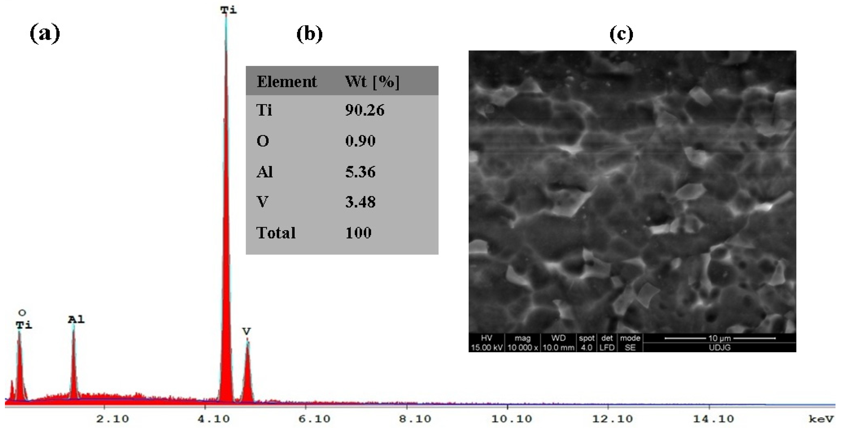
3.1.1. Untreated Ti6Al4V Grade 5 Alloy

The morphological and compositional analysis (SEM-EDX) was carried out using the equipment described in chapter 2.3 of this research work. Figure 3a–c shows the EDX spectrum in layer (a), the EDX elemental analysis in weight percent in layer (b), and the SEM surface morphology of the untreated grade 5 Ti6Al4V alloy in layer (c).
For the EDX analysis of the untreated grade 5 Ti6Al4V alloy implant, Figure 3a,b indicates the presence of the main elements of the studied alloy: Ti, Al, and V, with their mass percentage values Ti = 90.26%, Al = 5.36%, and V = 3.48%. Oxygen, in a percentage of about 0.90 wt%, is present because a thin layer of titanium oxide (TiO2) is natively formed on the surface of the untreated alloy, which is calculated at a percentage of 2.24 wt% TiO2. The percentage of titanium oxide (TiO2) formed on the surface of the grade 5 Ti6Al4V alloy was determined from the transformation of the mass percentage of oxygen from the general analysis of the molecular mass of TiO2 (79.866 g/mol). In Figure 3c, it can be seen from the surface morphology that the surface of the untreated grade 5 Ti6Al4V alloy is relatively smooth and has no surface defects.

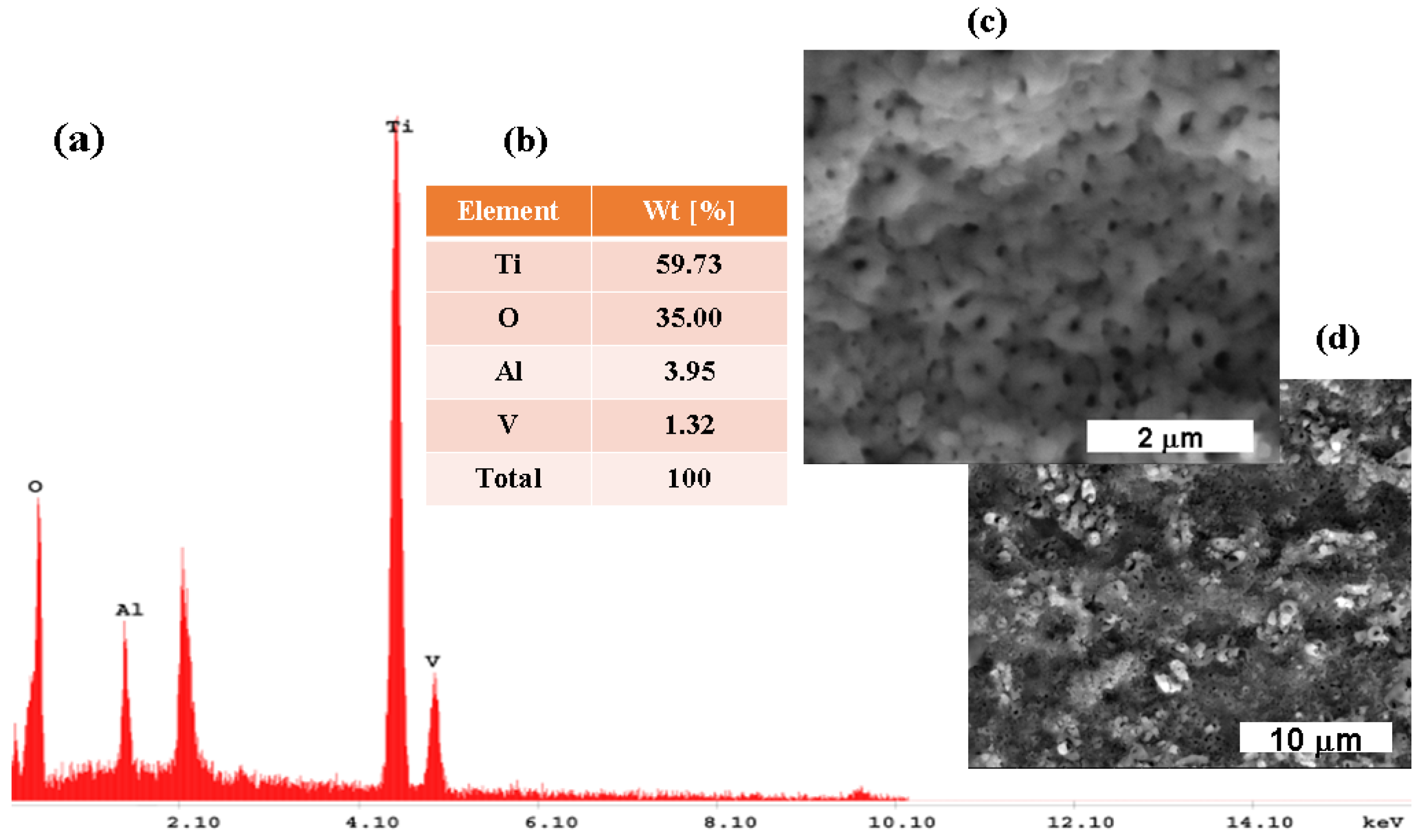
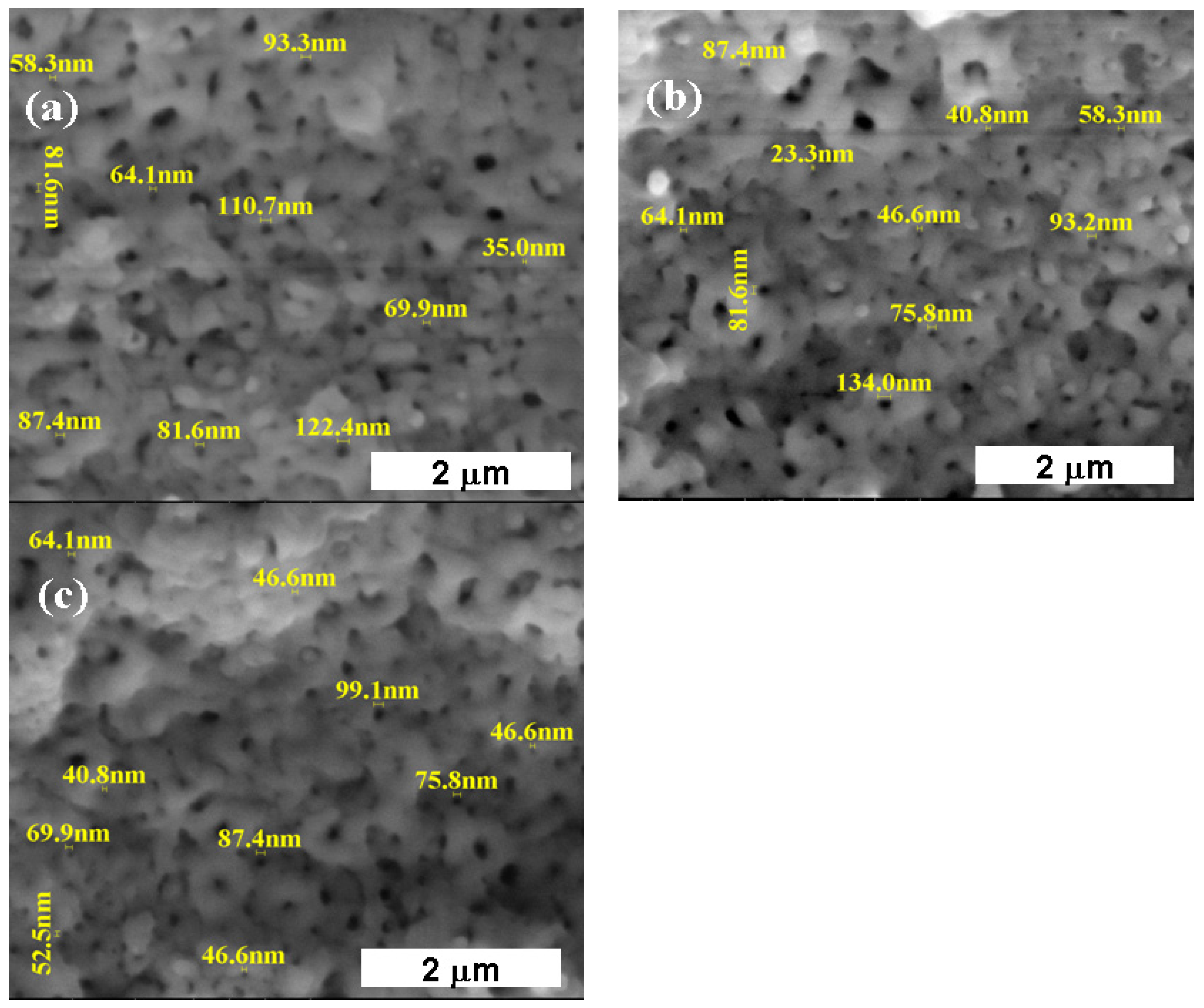
3.1.2. Titanium Oxide Film Electrochemically Formed on the Surface of the Ti6Al4V Alloy

Figure 4a–d shows the EDX spectrum in layer (a), the EDX elemental analysis in weight percent in layer (b), and the SEM surface morphology of the grade 5 Ti6Al4V implant alloy electrochemically oxidized at 200 V and at a time of 3 min in layers (c) and (d). As specified in chapter 2, Materials and Methods, the most effective and relevant values for obtaining titanium oxide films on this type of implant alloy are 200 V for the potential and 3 min for the time, under the conditions of the specified electrochemical cell.
The EDX analysis results shown in Figure 4a show the presence of the main elements observed in the electrochemically oxidized biomaterial studied, i.e., Ti, Al, and V; their mass percentage values are shown in Figure 4b, which are different from the untreated alloy, Ti = 59.64 wt%, Al = 3.95 wt%, V = 1.32 wt%, and oxygen, which increased to about O = 35.00 wt%.
At the same time, it was observed that at the oxidation time of 3 min, there was an increase in the percentage of oxygen to about O = 35.00 wt% compared to the untreated grade 5 Ti6Al4V alloy, which confirms the increase in the titanium oxide film to about TiO2 = 87.35 wt% on the sample’s surface.
From the analysis results shown in Figure 3 and Figure 4, it can be observed that the surface morphology of the grade 5 Ti6Al4V alloy electrochemically oxidized at 200 V for 3 min changes compared to the surface of the untreated grade 5 Ti6Al4V alloy. Before the anodic oxidation process, the surface of the untreated grade 5 Ti6Al4V alloy shows a thin native oxide layer that instantly forms on the surface of the untreated alloy in air. On the other hand, the electrochemically oxidized samples at 200 V show the growth of the titanium oxide film on the surface of the electrochemically oxidized grade 5 Ti6Al4V alloy.
The formation of nanoporous titanium oxide (TiO2) depends on the electrochemical oxidation time of the grade 5 titanium alloy and is exemplified in Figure 5. To support this statement, Figure 5 shows the surface morphologies of the electrochemically oxidized grade 5 Ti6Al4V samples: (a) at 200 V for 1 min, (b) at 200 V for 2 min, and (c) at 200 V for 3 min at a magnitude of 50,000×, with the measurement of the pore diameter size directly from the SEM results.
From the analysis of Figure 5, a decrease in the size of the nanopores can be observed with an increase in the electrochemical oxidation process time. Thus, the average size of the nanopores at 1 min decreases from 80.43 to 58.6 nm at the oxidation time of 3 min.
To better highlight the abovementioned results, Figure 6 presents the comparative evolution of the obtained average TiO2 nanopore diameter values corresponding to grade 5 Ti6Al4V samples electrochemically oxidized at 200 V for 1, 2, and 3 min.
A decrease in the size of the nanopores formed through the anodic oxidation process with increasing time on another alloy has been also reported in the specialized literature by other authors [25].
The size of the nanopores and their uniformity for the three-minute electrochemical oxidation parameters led to the comparative evaluation of the resistance to corrosion degradation of the untreated titanium alloy with titanium oxide (TiO2) film in Hank’s solution, which simulates the corrosive aggressiveness of biological fluids from the human body.

3.2. Evolution of the Open Circuit Potential (OCP) vs. Time in Hank’s Biological Solution for the Untreated Alloy and the Treated Alloy with the Electrochemically Formed Nanoporous TiO2 Surface Film

The open circuit potential (OCP) was measured at different time intervals for the treated and untreated Ti6Al4V alloy in Hank’s solution over an 80 h period. This was carried out to especially monitor the time necessary for the tested surfaces to reach a stationary state since they are intended to be used as implants. The occurrence of passivity is generally due to the formation of a spontaneous passive film on metal surfaces that acts as a kinetic barrier against corrosion, thereby significantly reducing the corrosion rate. Another reason for monitoring the open circuit potential over a longer period was to evaluate the stability and the reactivity of the tested surfaces over time. The chemical properties of the oxide film play an important role in the biocompatibility of implants with surrounding tissues. The chemical interaction of a metallic material with bodily fluids is important for the stability of an implant in the human body [21,22,23,24,25,26,27]. To simulate human body conditions, the corrosion tests were performed in Hank’s physiological solution. The open circuit potential during the immersion period in Hank’s biological solution for the untreated Ti6Al4V alloy (1) and the titanium oxide film electrochemically formed on it (2) for 75.5 h are presented in Figure 7.
From the analysis of Figure 7, it can be observed that in the case of both studied surfaces, the open circuit potential values move toward nobler (more positive) values with increasing immersion period, and the open circuit potential of the electrochemically oxidized surface registers more positive values (nobler) compared with the untreated titanium alloy throughout the whole immersion period. A shift in the open circuit toward more positive values indicates the formation of a passive film, while a decrease in the open circuit toward more negative values indicates breaks in the passive film, the dissolution of the film, or the absence of film formation. Comparatively, the values of the electrochemically oxidized titanium alloy immersed in Hank’s solution were more positive than those of the untreated alloy throughout the whole immersion period.
At the beginning of the immersion period, the value of the open circuit potential for the untreated alloy was E = −222 mV vs. Ag/AgCl, and for the electrochemically oxidized titanium alloy, this value was more positive, E = 113 mV vs. Ag/AgCl. The difference in the open circuit potential between the two surfaces at the start of immersion was ΔE = 335 mV. At the end of the monitoring process, it was observed that the electrochemically oxidized alloy reaches an open circuit potential value of E = 814 mV vs. Ag/AgCl, while the value of the open circuit potential for the untreated alloy is more negative, E = 424 mV vs. Ag/AgCl. The open circuit potential difference between the two surfaces at the end of monitoring is even greater, ΔE = 390 mV. This trend confirms the improvement of the grade 5 Ti6Al4V alloy surface through controlled electrochemical oxidation, which gives it this fine film of nanoporous titanium oxide (TiO2) with qualities of nobler behavior in biological solutions.
For the untreated alloy, it was observed that the value of the open circuit potential shows a continual increase for 30 h after which the value stabilizes at around 424 mV vs. Ag/AgCl. The difference in OCP for the untreated titanium alloy from the beginning of immersion to the end of the 75.6 h immersion period was 646 mV, while for the electrochemically oxidized titanium alloy, the difference between immersion start and finish OCP value, ΔE, was 701 mV. This behavior can be explained by the formation of a more stable and protective thin oxide film on the surface of the electrochemically oxidized alloy, which acts as a protective film to prevent the release of metal ions into the biological environment. The trend toward higher values in the case of different types of materials obtained by electrochemical oxidation compared to different untreated alloys has also been observed by other authors in the specialized literature [21,22,23,24,25,26,27,28,29,30,31,32,33,34,35].

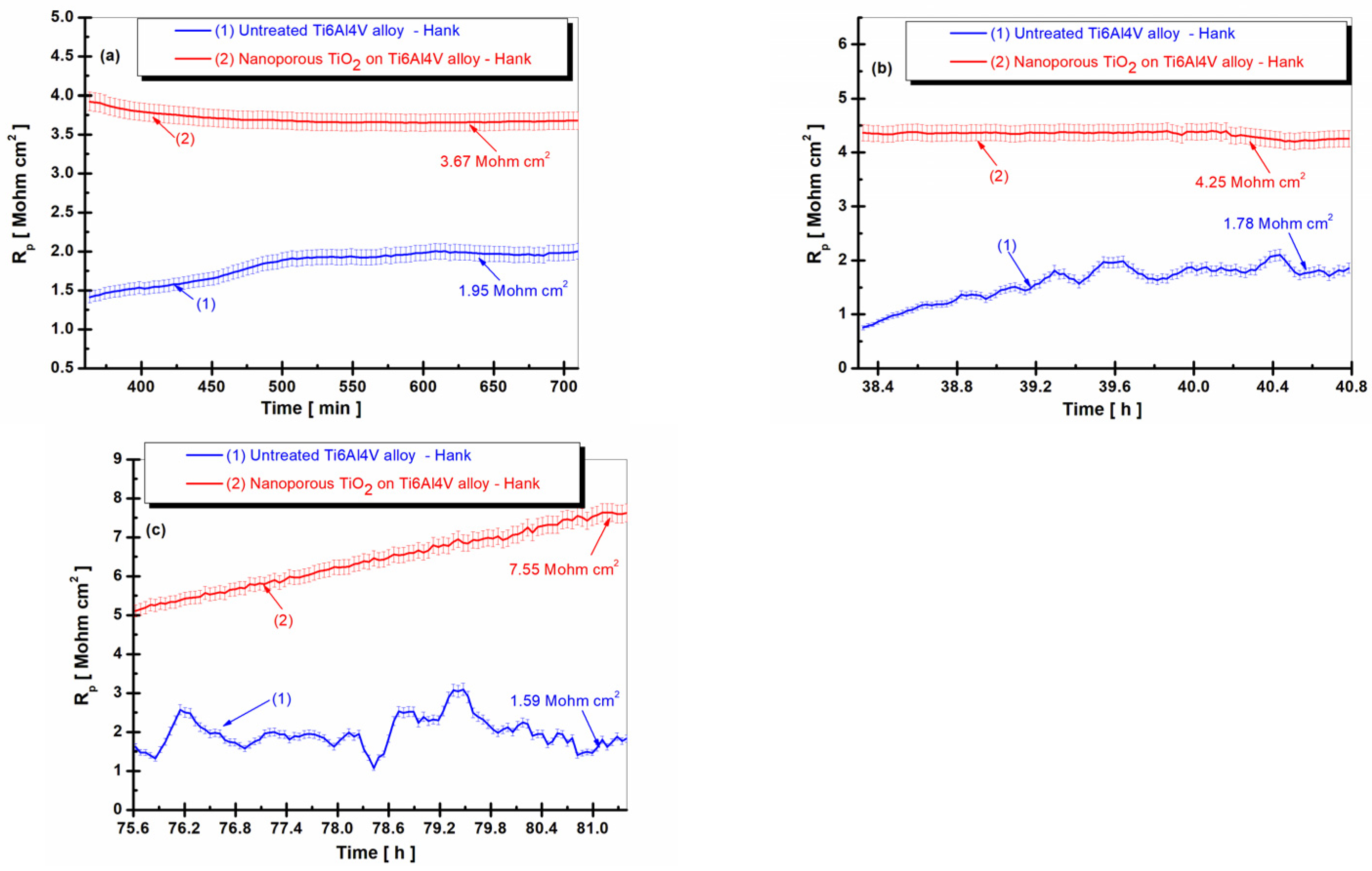
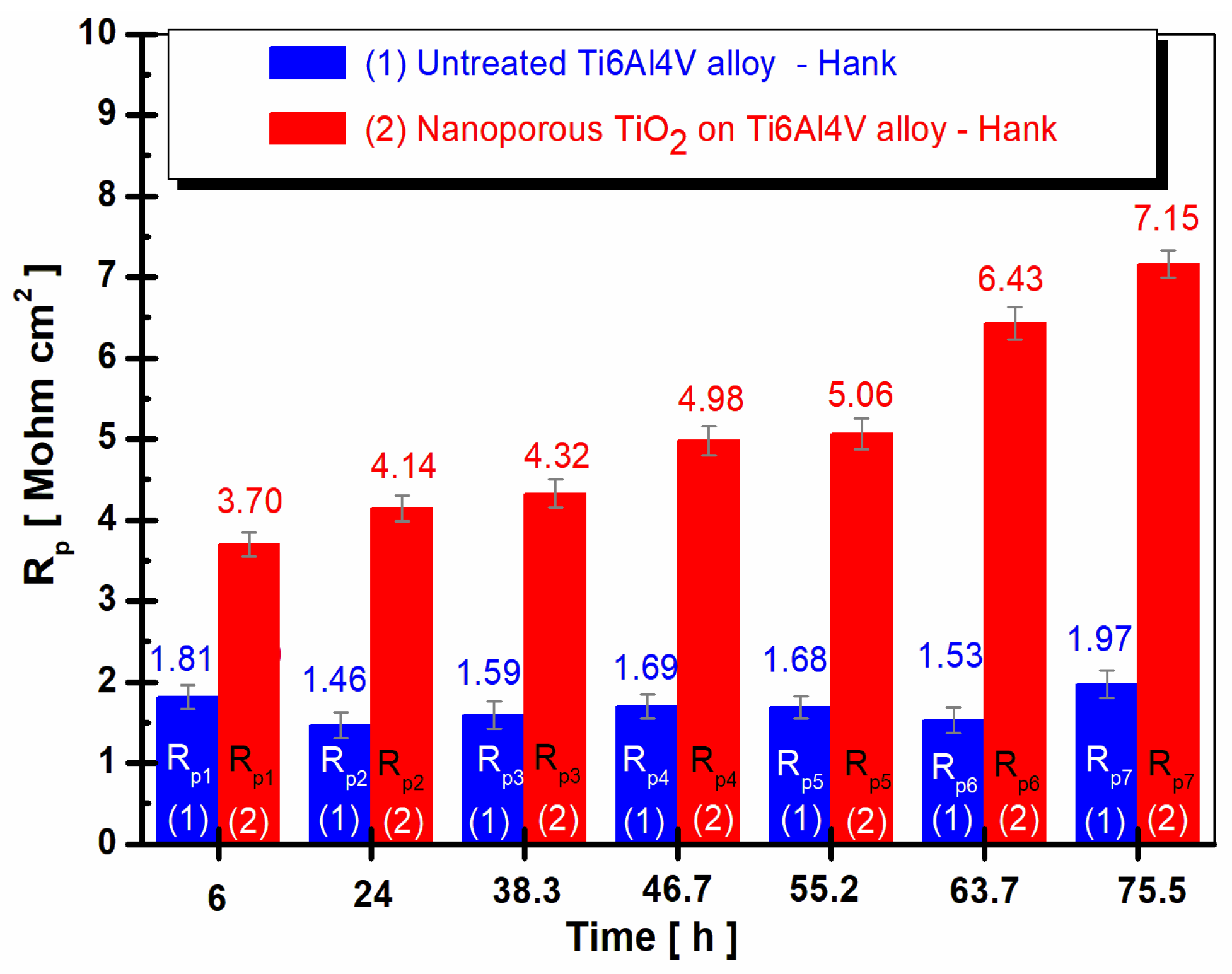
3.3. Evolution of the Polarization Resistance (Rp) vs. Time in Hank’s Biological Solution for the Untreated Alloy and the Treated Alloy with the Electrochemically Formed Nanoporous TiO2 Surface Film

After immersing the untreated and electrochemically oxidized grade 5 titanium alloy samples to form the protective nanoporous film of titanium oxide (TiO2), the open circuit potential was recorded in several sequences for 75.6 h. Between the open circuit potential measurement sequences at the start and end of the experiment, a total of seven recordings of 100 points of polarization resistance were made and calculated from as many linear polarization curves by using the Stern Geary equation and Tafel slopes (as explained in chapter 2, Materials and Methods).
The comparative evolution of the polarization resistance for the untreated grade 5 titanium alloy and for the electrochemically oxidized titanium alloy with the nanoporous film of titanium oxide (TiO2) is presented in Figure 8a–c at three time periods in the total sample immersion period in Hank’s biological solution.
From the analysis of Figure 8, it can be seen that the values of the polarization resistance for the untreated grade 5 Ti6Al4V alloy immersed in Hank’s biological solution (Figure 8(1)) are lower than the values of the polarization resistance recorded for the treated electrochemically controlled oxidized Ti6Al4V alloy (Figure 8(2)) for all measurement periods during immersion. After 6 h of immersion, the polarization resistance (Rp) of the untreated titanium alloy showed a slight increase from immersion and stabilized at a value of 1.95 Mohm·cm2, while the polarization resistance of the electrochemically oxidized titanium alloy showed constant values with a stabilization value of 3.67 Mohm·cm2 at the end of the first measurement period. This is higher than the polarization resistance of the untreated alloy.
After 38 h of immersion, the values of the polarization resistance of the untreated grade 5 titanium alloy show a slight decrease and oscillations in Figure 8b, so that at the end of this measurement period, the Rp value was 1.78 Mohm·cm2, lower than in the first period measurement. The values of the polarization resistance of the electrochemically oxidized grade 5 titanium alloy after 38 h were slightly higher than those in the first measurement period, with an Rp value of 4.25 Mohm·cm2 toward the end of the measurement period. Thus, even after 38 h, the Rp for the electrochemically oxidized titanium alloy is higher than the Rp for the untreated titanium alloy by approximately three times, which confirms the effectiveness of the electrochemical oxidation process in improving the resistance of the implant to the aggressive action of biological fluids in the human body.
After 75.5 h of immersion in Hank’s biological solution, the same trend of the polarization resistance values was maintained for the two surfaces of the immersed untreated grade 5 Ti6Al4V alloy and the electrochemically oxidized grade 5 Ti6Al4V alloy. The value of the polarization resistance (Rp) of the untreated alloy slightly decreases toward the end of the immersion period to Rp = 1.59 Mohm·cm2, while the value of the polarization resistance of the electrochemically oxidized Ti6Al4V alloy slightly increases, reaching a value of Rp = 7.56 Mohm·cm2 at the end of the immersion period, a value that was approximately five times higher than that of the untreated alloy. To better highlight the obtained results, Figure 9 shows the average values of all of the polarization resistance values recorded at different immersion periods for the untreated grade 5 Ti6Al4V alloy compared to the electrochemically oxidized grade 5 Ti6Al4V alloy.
From the analysis of Figure 9, it can be observed that in the case of the electrochemically oxidized grade 5 Ti6Al4V alloy immersed in Hank’s solution, the values of the polarization resistance were higher compared to the untreated grade 5 Ti6Al4V alloy at all studied times. Additionally, in the case of the electrochemically oxidized grade 5 Ti6Al4V alloy, an increase in the values of the polarization resistance was observed with increasing immersion time. At the first measurement, the electrochemically oxidized alloy had an Rp1 value of 3.70 Mohm·cm2; at the end of the 75.5 h of monitoring, the polarization resistance had reached an Rp7 value of 7.15 Mohm·cm2, a behavior that demonstrates the effectiveness of the electrochemical oxidation process of the titanium alloy for improving the resistance of the implant to the aggressive action of biological fluids in the human body.
The increase in the polarization resistance values in the case of the nanoporous TiO2 film electrochemically formed on titanium alloy was due to the presence of the TiO2 layer that provided superior insulating properties through its electrochemically controlled formation.
This behavior has also been observed in the specialized literature by other authors with other biomaterials [20,21,22,23,24,25,26,27,28,29,30,31,32,33,34]. For the electrochemically oxidized grade 5 Ti6Al4V alloy, an increase in the polarization resistance values was observed with increasing immersion time; for the untreated grade 5 Ti6Al4V alloy, it was observed that the polarization resistance values showed a slight decrease after 38 h of immersion in comparison to the value obtained after 360 min, and a slight increase at the end of the immersion period. This behavior was due to the nonuniformity of the natively formed titanium oxide film on the surface of the untreated alloy, which leads to a much faster degradation process of the material in comparison to the electrochemically oxidized titanium alloy having the nanoporous TiO2 film.

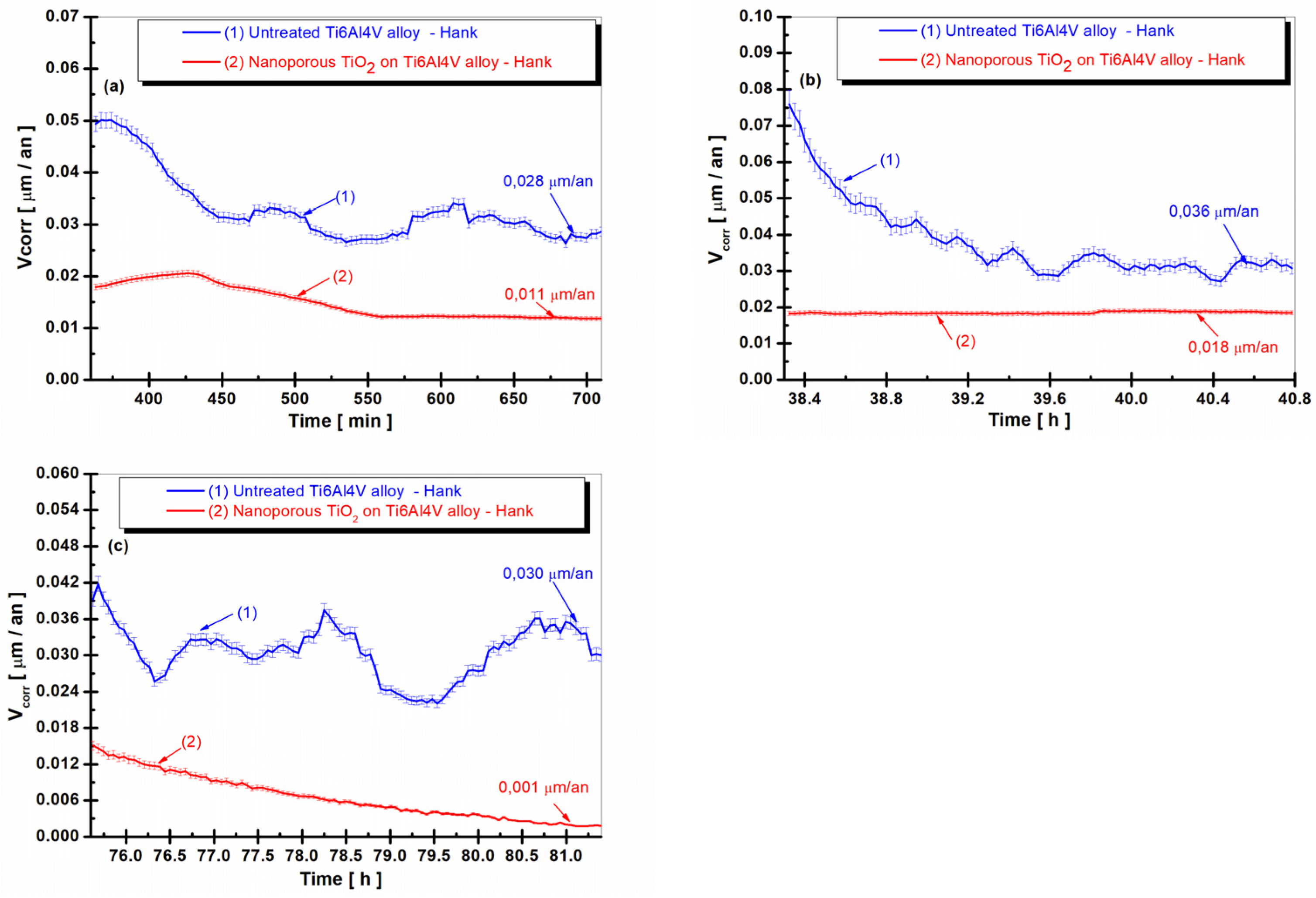
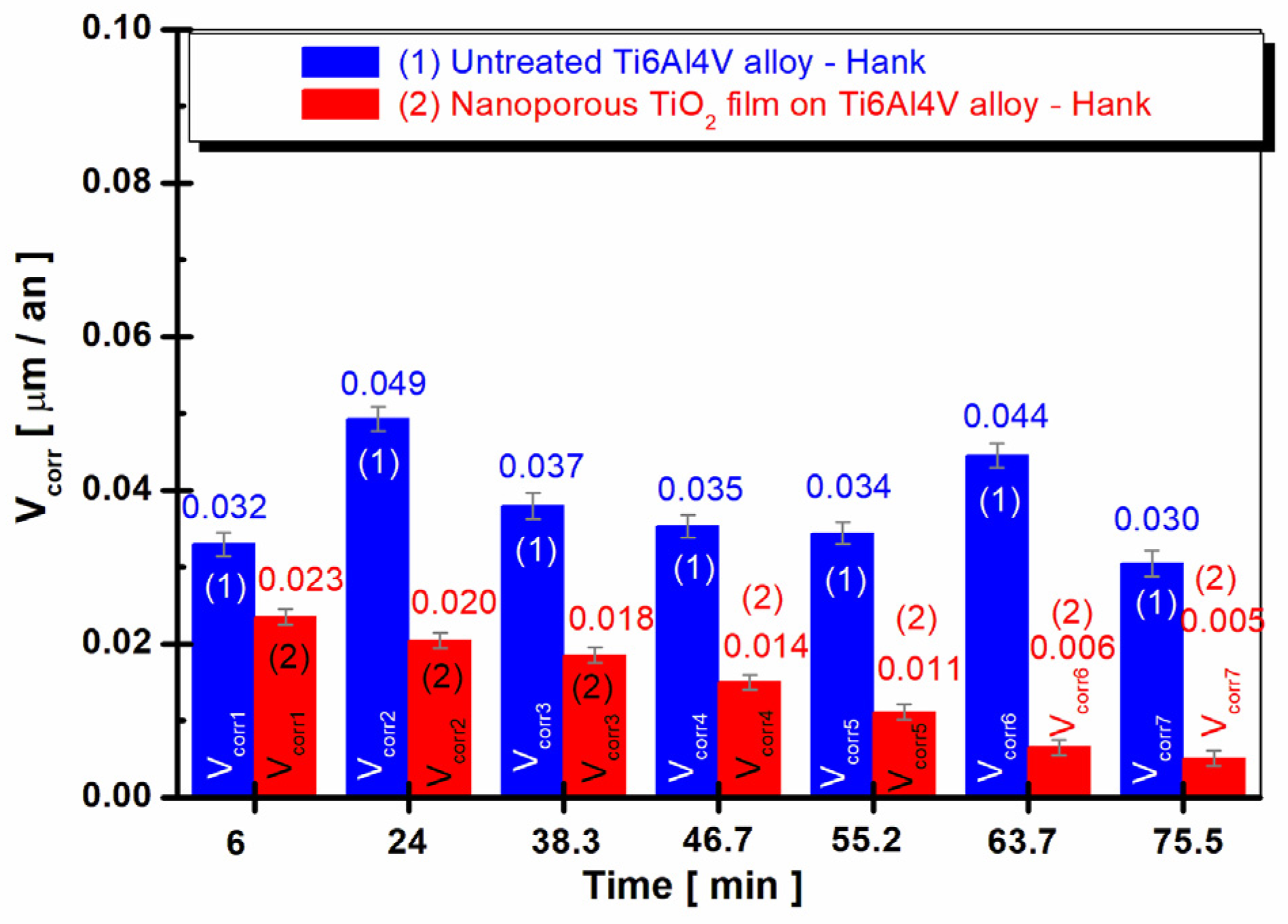
3.4. Time Evolution of the Corrosion Rate (Vcorr) in Hank’s Biological Solution of the Untreated Alloy and the Treated Alloy with the Electrochemically Formed Nanoporous TiO2 Surface Film

Polarization resistance is the only corrosion monitoring method that makes it possible to measure corrosion rates (expressed as thickness loss over time) directly, in real time. The corrosion current determined using this method represents the current that occurs at the corrosive metal/medium interface when the metal is immersed in the solution.
The variation in the corrosion rate expressed as a penetration index for the untreated alloy and for the alloy oxidized with titanium oxide (TiO2) film is shown in Figure 10a–c at three times during the sample immersion period in Hank’s biological solution, and in Figure 11 as the mean value across all studied times.
From the analysis of Figure 10, it can be seen that the corrosion rate values (Vcorr) for the untreated grade 5 Ti6Al4V alloy immersed in Hank’s biological solution, Figure 10(1), are higher than the corrosion rate values recorded for the treated grade 5 Ti6Al4V alloy electrochemically oxidized Ti6Al4V alloy, Figure 10(2), for all measurement periods during immersion. Thus, after 6 h of immersion, in Figure 10a, the corrosion rate (Vcorr) of the untreated titanium alloy shows a slight decrease from immersion with stabilization at a value of 0.032 µm/year, while the corrosion rate value for the treated alloy shows constant values with a stabilization value of 0.023 µm/year at the end of the first measurement period, a corrosion rate that was approximately two times lower than for the untreated alloy.
After 38 h of immersion, Figure 10b, the corrosion rate for the untreated grade 5 Ti6Al4V alloy showed an overall slight increase at the end of this measurement period, with a Vcorr value of 0.036 µm/year, higher than at the end of the first measurement period. The corrosion rate value for the electrochemically oxidized grade 5 Ti6Al4V alloy after 38 h was slightly lower than at the end of the first measurement period, with a Vcorr value of 0.018 µm/year. Thus, even after 38 h, the Vcorr for the electrochemically oxidized titanium alloy was lower than the Vcorr for the untreated titanium alloy by approximately two times, which confirms the efficiency of the electrochemical oxidation process of the titanium alloy for improving the resistance of the implant to the aggressive action of biological fluids in the human body.
After 75.5 h of immersion in Hank’s biological solution, the same trend for the corrosion rate values was maintained for the treated and untreated alloy surfaces. The value of the corrosion rate, Vcorr, for the untreated alloy decreased slightly toward the end of the immersion period to Vcorr = 0.030 µm/year, while the value of the corrosion rate of the electrochemically oxidized grade 5 Ti6Al4V alloy slightly decreased, reaching the end of the immersion period with a Vcorr value of 0.001 µm/year, thus being approximately 30 times lower than the untreated Vcorr level.
From the analysis of Figure 11, it can be seen that in the case of the electrochemically oxidized nanoporous TiO2 film-treated alloy immersed in Hank’s blood-simulating solution, the corrosion rate values were lower compared to the untreated grade 5 Ti6Al4V alloy at all studied times. High polarization resistance means a low corrosion rate, a behavior that is desired for all types of biomaterials that are to be inserted into the human body.
For the electrochemically oxidized grade 5 Ti6Al4V alloy, a decrease in the corrosion rate values was observed with increasing immersion time.
If at the first measurement, the electrochemically oxidized alloy has an average value of Vcorr1 = 0.023 µm/year; at the end of the 80 h monitoring period, Vcorr reached an average value of Vcorr7 = 0.005 µm/year. This is a behavior that demonstrates the efficiency of the electrochemical oxidation process of the titanium alloy for improving the resistance of the implant against the aggressive action of the biological fluids in the human body. For the untreated grade 5 Ti6Al4V alloy, it was observed that the average values of the corrosion rate show a slight increase after 38 h of immersion compared to the value obtained after 360 min, and a slight decrease at the end of the immersion period (80 h).

4. Conclusions

Electrochemical analysis methods including open circuit potential (OCP), polarization resistance (Rp), and corrosion rate (Vcorr) vs. time throughout an overall immersion period of 80 h in Hank’s biological solution were used for the comparative investigation into the electrochemical behaviors of untreated grade 5 Ti6Al4V titanium implant alloy and grade 5 Ti6Al4V titanium implant alloy with an electrochemically formed nanoporous titanium oxide surface film.
From the morphological analysis of the samples, with the help of electronic microscopy, it was observed that with increasing anodic oxidation process time, the oxide layer became much more compact and dense compared to shorter process times of controlled oxide layer growth on the titanium alloy.
From the SEM analysis, a change in the surface morphologies is also observed with increasing electrochemical oxidation processing time. At the same time, from the morphologies presented at the magnitude of 50.000×, a decrease in the size of the nanopores formed on the surface of the grade 5 Ti6Al4V alloy was observed.
The nanopore sizes of the titanium oxide film were smaller after electrochemical oxidation for 3 min at 200 V.
From the EDX analyses of the untreated grade 5 Ti6Al4V alloy and the electrochemically oxidized alloy, an increase in the mass percentage of oxygen was observed with increasing electrochemical oxidation process duration, thus proving the formation of the titanium oxide film.
From the evolution of the open circuit potential, it can be seen that in the case of the untreated grade 5 Ti6Al4V alloy, a more negative immersion potential value was recorded compared to the electrochemically oxidized alloy, followed by an increase for 30 h, after which it stabilizes around the value of 424 mV until the end of the measurement period.
For the electrochemically oxidized alloy having the nanoporous oxide surface film, higher potential values were observed from immersion until the end of 75.5 h compared to the untreated alloy. At the same time, in the case of the nanoporous TiO2 film formed electrochemically on the Ti6Al4V alloy, a continuous shift in the open circuit potential toward more positive values was observed until the end of the immersion period. This behavior indicates the efficiency of the electrochemical oxidation process of the titanium alloy for improving the resistance of the implant to the aggressive action of biological fluids in the human body.
From the analysis of the evolution of polarization resistance, it was observed that in the case of the electrochemically oxidized grade 5 Ti6Al4V alloy immersed in Hank’s solution, the values of the polarization resistance were higher than the untreated Ti6Al4V alloy at all studied times during the immersion period. Additionally, in the case of the treated alloy, an increase in polarization resistance was observed with increasing immersion time.

Author Contributions

L.B.: conceptualization, methodology, supervision, software, writing—original draft preparation, and reviewing; A.R.: data curation and investigation; L.B. and V.N.: visualization, investigation, and validation; E.R.A. and L.B.: writing, editing, and artwork. All authors have read and agreed to the published version of the manuscript.

Funding

This research is financially supported by UEFISCDI-PN-III-P4-PCE-2021-0702, project number PCE 102/2022.

Institutional Review Board Statement

Not applicable.

Informed Consent Statement

Not applicable.

Data Availability Statement

Not applicable.

Acknowledgments

The authors would like to express their thanks to UEFISCDI—PN-III-P4-PCE-2021-0702, project number PCE 102/2022 for the funding support; to Jean Pierre Celis from Katholieke University of Leuven, Belgium; and to Pierre Ponthiaux from the Ecole Centrale Paris, France, for valuable scientific advice.

Conflicts of Interest

The authors declare no conflict of interest.

References

  1. Akhavan, O.; Ghaderi, E. Flash photo stimulation of human neural stem cells on graphene/TiO2 heterojunction for differentiation into neurons. Nanoscale 2013, 5, 10316–10326. [Google Scholar] [CrossRef]
  2. Chen, Q.; Thouas, G.A. Metallic implant biomaterials. Mater. Sci. Eng. R 2015, 87, 1–57. [Google Scholar] [CrossRef]
  3. Li, J.; Liu, X. Chemical surface modification of metallic biomaterials. In Surface Coating and Modification of Metallic Biomaterials; Woodhead Publishing: London, UK, 2015; pp. 159–183. [Google Scholar] [CrossRef]
  4. Verissimo, N.C.; Chung, S.; Webster, T.J. New nanoscale surface modifications of metallic biomaterials. In Surface Coating and Modification of Metallic Biomaterials; Woodhead Publishing: London, UK, 2015; pp. 249–273. [Google Scholar] [CrossRef]
  5. Jawed, S.F.; Rabadia, C.D.; Khan, M.A.; Khan, S.J. Effect of Alloying Elements on the Compressive Mechanical Properties of Biomedical Titanium Alloys: A Systematic Review. ACS Omega 2022, 7, 29526–29542. [Google Scholar] [CrossRef] [PubMed]
  6. Jimenez-Marcos, C.; Mirza-Rosca, J.C.; Baltatu, M.S.; Vizureanu, P. Experimental Research on New Developed Titanium Al-loys for Biomedical Applications. Bioengineering 2022, 9, 686. [Google Scholar] [CrossRef] [PubMed]
  7. Baltatu, M.S.; Chiriac-Moruzzi, C.; Vizureanu, P.; Tóth, L.; Novák, J. Effect of Heat Treatment on Some Titanium Alloys Used as Biomaterials. Appl. Sci. 2022, 12, 11241. [Google Scholar] [CrossRef]
  8. Bocchetta, P.; Chen, L.-Y.; Tardelli, J.D.C.; Reis, A.C.d.; Almeraya-Calderón, F.; Leo, P. Passive Layers and Corrosion Resis-tance of Biomedical Ti-6Al-4V and β-Ti Alloys. Coatings 2021, 11, 487. [Google Scholar] [CrossRef]
  9. Donachie, M.J. Titanium: A Technical Guide; ASM International: Almere, The Netherlands, 2000; ISBN 978-0-87170-686-7. [Google Scholar]
  10. Hanawa, T.; Asami, K.; Asaoka, K. Repassivation of titanium and surface oxide film regenerated in simulated bioliquid. J. Bio-Med. Mater. Res. 1998, 40, 530–538. [Google Scholar] [CrossRef]
  11. Brown, S.; Lemons, J. Medical Applications of Titanium and Its Alloys: The Material and Biological Issues; ASTM Special Series Publication: West Conshohocken, PA, USA, 1996; p. 1272. [Google Scholar]
  12. Neoh, K.G.; Hu, X.; Zheng, D.; Tang Kang, E. Balancing osteoblast functions and bacterial adhesion on functionalized titanium surfaces. Biomaterials 2012, 33, 2813–2822. [Google Scholar] [CrossRef]
  13. De Viteri, V.S.; Fuentes, E. Titanium and Titanium Alloys as Biomaterials. Tribol.-Fundam. Adv. 2013, 1, 154–181. [Google Scholar] [CrossRef] [Green Version]
  14. Williams, D. Titanium in Medicine: Material Science, Surface Science, Engineering, Biological Responses and Medical Applications; Brunette, D.M., Tengvall, P., Textor, M., Thompson, P., Eds.; Springer: Berlin/Heidelberg, Germany, 2001; pp. 13–24. [Google Scholar] [CrossRef]
  15. Adya, M.; Alam, M.; Ravindranath, T.; Mubeen, A.; Saluja, B. Corrosion in titanium dental implants: Literature review. J. Indian Prosthodont. Soc. 2005, 5, 126–131. [Google Scholar] [CrossRef]
  16. Chang, L.; Wang, Z.J.; Liu, Y.; Jia, Z.; Liang, S.-X. Additive Manufacturing of Titanium Alloys, Encyclopedia of Materials. Met. Alloy. 2022, 1, 256–274. [Google Scholar] [CrossRef]
  17. Asserghine, A.; Filotás, D.; Nagy, L.; Souto, R.M.; Nagy, G. Do titanium biomaterials get immediately and entirely repassivated? A perspective. NPJ Mater. Degrad. 2022, 6, 57. [Google Scholar] [CrossRef]
  18. Mohammed, M.T.; Khan, Z.A.; Siddiquee, A.N. Surface Modifications of Titanium Materials for developing Corro-sion Behavior in Human Body Environment: A Review. Procedia Mater. Sci. 2014, 6, 1610–1618. [Google Scholar] [CrossRef] [Green Version]
  19. Mardare, E.; Benea, L.; Bounegru, I. Electrochemical modifications of titanium and titanium alloys surface for bio-medical applications–A review. Ann. Dunarea Jos Univ. Galati. Fascicle IX. Metall. Mater. Sci. 2013, 36, 68–78. [Google Scholar]
  20. Hoque, M.E.; Showva, N.-N.; Ahmed, M.; Rashid, A.B.; Sadique, S.E.; El-Bialy, T.; Xu, H. Titanium and titanium alloys in dentistry: Current trends, recent developments, and future prospects. Heliyon 2022, 8, e11300. [Google Scholar] [CrossRef]
  21. Jáquez-Muñoz, J.M.; Gaona-Tiburcio, C.; Chacón-Nava, J.; Cabral-Miramontes, J.; Nieves-Mendoza, D.; Maldonado-Bandala, E.; Delgado, A.D.; Flores-De los Rios, J.P.; Bocchetta, P.; Almeraya-Calderón, F. Electrochemical Corrosion of Titanium and Titanium Alloys Anodized in H2SO4 and H3PO4 Solutions. Coatings 2022, 12, 325. [Google Scholar] [CrossRef]
  22. Prando, D.; Brenna, A.; Pedeferri, M.; Ormellese, M. Enhancement of pure titanium localized corrosion resistance by anodic oxidation. Mater. Corros. 2017, 69, 503–509. [Google Scholar] [CrossRef] [Green Version]
  23. Benea, L.; Celis, J.P. Reactivity of porous titanium oxide film and chitosan layer electrochemically formed on Ti-6Al-4V alloy in biological solution. Surf. Coat. Technol. 2018, 354, 145–152. [Google Scholar] [CrossRef]
  24. Benea, L.; Danaila, E.; Ponthiaux, P. Effect of titania anodic formation and hydroxyapatite electrodeposition on elec-trochemical behaviour of Ti–6Al–4V alloy under fretting conditions for biomedical applications. Corros. Sci. 2015, 91, 262–271. [Google Scholar] [CrossRef]
  25. Benea, L.; Simionescu-Bogatu, N.; Axente, E.R. Electrochemically obtained Al2O3 nanoporous layers with increased anticorrosive properties of aluminum alloy. J. Mater. Res. Technol. 2022, 17, 2636–2647. [Google Scholar] [CrossRef]
  26. Benea, L.; Mardare, E.; Mardare, M.; Celis, P. Preparation of titanium oxide and hydroxyapatite on Ti–6Al–4V alloy surface and electrochemical behavior in bio–simulated fluid solution. Corros. Sci. 2014, 80, 331–338. [Google Scholar] [CrossRef]
  27. Vasilescu, C.; Drob, P.; Vasilescu, E.; Demetrescu, I.; Ionita, D.; Prodana, M.; Drob, S.I. Characterisation and corrosion re-sistance of the electrodeposited hydroxyapatite and bovine serum albumin/hydroxyapatite films on Ti–6Al–4V–1Zr alloy sur-face. Corros. Sci. 2011, 53, 992–999. [Google Scholar] [CrossRef]
  28. Benea, L.; Ravoiu, A.; Celis, J.P. Anticorrosion performance of the electrochemically grown mixed porous oxide films on titanium alloy in biological solution. ACS Biomater. Sci. Eng. 2019, 5, 5925–5934. [Google Scholar] [CrossRef] [PubMed]
  29. Mazzarolo, A.; Curioni, M.; Vicenzo, A.; Skeldon, P.; Thompson, G.E. Anodic growth of titanium oxide: Electrochemical be-haviour and morphological evolution. Electrochim. Acta 2012, 75, 288–295. [Google Scholar] [CrossRef]
  30. Milošev, I.; Blejan, D.; Varvara, S.; Muresan, L.M. Effect of anodic oxidation on the corrosion behavior of Ti-based materials in simulated physiological solution. J. Appl. Electrochem. 2013, 43, 645–658. [Google Scholar] [CrossRef]
  31. Bayrak, Ö.; Ghahramanzadeh Asl, H.; Ak, A. Protein adsorption, cell viability and corrosion properties of Ti6Al4V alloy treated by plasma oxidation and anodic oxidation. Int. J. Miner. Metall. Mater. 2020, 27, 1269–1280. [Google Scholar] [CrossRef]
  32. Uttiya, S.; Contarino, D.; Prandi, S.; Carnasciali, M.M.; Gemme, G.; Mattera, L.; Rolandi, R.; Canepa, M.; Cavalleri, O. Anodic oxidation of titanium in sulphuric acid and phosphoric acid electrolytes. J. Mater. Sci. Nanotechnol. 2014, 1, S106. [Google Scholar] [CrossRef] [Green Version]
  33. Alves, A.C.; Wenger, F.; Ponthiaux, P.; Celis, J.P.; Pinto, A.M.; Rocha, L.A.; Fernandes, J.C.S. Corrosion mechanisms in tita-nium oxide-based films produced by anodic treatment. Electrochim. Acta 2017, 234, 16. [Google Scholar] [CrossRef]
  34. Torres, N.V.; Calderon, J.P.; Valencia, H.M.; Cecenes, R.L.; Cadena, I.R.; Bustos, E.S.; Valdés, C.I.R.; Rodriguez, J.G. Corrosion Resistance of a Plasma-Oxidized Ti6Al4V Alloy for Dental Applications. Coatings 2021, 11, 1136. [Google Scholar] [CrossRef]
  35. Lario, J.; Viera, M.; Vicente, Á.; Igual, A.; Amigó, V. Corrosion behaviour of Ti6Al4V ELI nanotubes for biomedical applications. J. Mater. Res. Technol. 2019, 8, 5548–5556. [Google Scholar] [CrossRef]
Figure 1. Presentation of the electrochemical oxidation process to obtain the nanoporous TiO2 film on grade 5 Ti6Al4V alloy: (a) electrochemical cell showing the electrodes and reactions at the electrodes in the anodizing electrolyte; (b) a schematic of nanoporous TiO2 on a titanium alloy surface; (c) the Ti6Al4V bulk alloy; (d) the measured roughness of TiO2 nanoporous film approximated as nanopore depth; and (e) SEM surface morphology of TiO2 nanoporous film showing the measurements of nanopore diameters.
Coatings 13 00614 g001
Figure 2. Experimental protocol summarizing the sequential electrochemical measurements during the total immersion period in biological solution.
Coatings 13 00614 g002
Figure 3. SEM surface micrographs and EDX analysis of the untreated Ti6Al4V grade 5 alloy implant: (a) EDX spectrum; (b) EDX elemental analysis; and (c) SEM surface morphology.
Coatings 13 00614 g003
Figure 4. SEM surface micrograph and EDX analysis of the treated Ti6Al4V grade 5 implant alloy electrochemically oxidized at a potential of 200 V for 3 min: (a) EDX spectrum; (b) EDX elemental analysis; and (c,d) SEM surface morphology at different magnifications.
Coatings 13 00614 g004
Figure 5. Morphological and dimensional analysis of the titanium oxide (TiO2) film nanopores formed on the surface of the titanium alloy depending on the oxidation time at a potential of 200 V: (a) nanopores formed on Ti6Al4V alloy with an oxidation time of 1 min, (b) nanopores formed on Ti6Al4V alloy with an oxidation time of 2 min, and (c) nanopores formed on Ti6Al4V alloy with an oxidation time of 3 min.
Coatings 13 00614 g005
Figure 6. Average nanopore diameter size of the titanium oxide (TiO2) film produced on the grade 5 Ti6Al4V alloy electrochemically oxidized in H2SO4 solution: (1) at 200 V for 1 min, (2) at 200 V for 2 min, and (3) at 200 V for 3 min.
Coatings 13 00614 g006
Figure 7. Comparative evolution of the open circuit potential during immersion in Hank’s biological solution for 80 h: (1) the untreated grade 5 Ti6Al4V alloy and (2) the treated grade 5 Ti6Al4V alloy with electrochemically formed titanium oxide surface film.
Coatings 13 00614 g007
Figure 8. Comparative evolution of the polarization resistance during sample immersion in Hank’s biological solution: (a) after 6 h (360 min); (b) after 38 h (2298 min); and (c) after 75.5 h (4534 min). (1) untreated Ti6Al4V alloy and (2) the treated alloy with the electrochemically formed nanoporous TiO2 film.
Coatings 13 00614 g008
Figure 9. Comparative evolution of the average values of polarization resistance during the 75.5 h immersion period in Hank’s biological solution: (1) untreated Ti6Al4V grade 5 alloy and (2) the treated Ti6Al4V grade 5 alloy with an electrochemically nanoporous titanium oxide surface film.
Coatings 13 00614 g009
Figure 10. Comparative evolution of the average values for the corrosion rate (Vcorr) during the immersion period in Hank’s biological solution: (a) after 6 h (360 min); (b) after 38 h (2298 min); (c) after 75.5 h (4534 min). (1) the untreated Ti6Al4V alloy and (2) the treated Ti6Al4V alloy with the electrochemically formed nanoporous titanium oxide film.
Coatings 13 00614 g010
Figure 11. Shows the evolution of the average values for the corrosion rate of the two alloys immersed in Hank’s biological solution for the entire immersion period, measured at different times after immersion.
Coatings 13 00614 g011
Disclaimer/Publisher’s Note: The statements, opinions and data contained in all publications are solely those of the individual author(s) and contributor(s) and not of MDPI and/or the editor(s). MDPI and/or the editor(s) disclaim responsibility for any injury to people or property resulting from any ideas, methods, instructions or products referred to in the content.

