Article

A Polymer-Infiltrated Ceramic as Base Adherent in an Experimental Specimen Model to Test the Shear Bond Strength of CAD-CAM Monolithic Ceramics Used in Resin-Bonded Dental Bridges

by
Maria João Calheiros-Lobo
1,2,*,†,‡,
João Mário Calheiros-Lobo
3,‡,
Ricardo Carbas
4,5,‡,
Lucas F. M. da Silva
4,5,‡ and
Teresa Pinho
1,6,*,‡
1
UNIPRO—Oral Pathology and Rehabilitation Research Unit, University Institute of Health Sciences IUCS-CESPU, 4585-116 Gandra, Portugal
2
Conservative Dentistry, Department of Dental Sciences, University Institute of Health Sciences IUCS-CESPU, 4585-116 Gandra, Portugal
3
Dental Prosthetist, Private Prosthesis Laboratory, 4465-127 São Mamede Infesta, Portugal
4
Department of Mechanical Engineering, Faculty of Engineering, University of Porto, 4200-465 Porto, Portugal
5
INEGI—Institute of Science and Innovation in Mechanical and Industrial Engineering, University of Porto, 4200-465 Porto, Portugal
6
Institute for Molecular and Cell Biology (IBMC), Institute of Innovation and Investigation in Health (i3S), University of Porto, 4200-135 Porto, Portugal
*
Authors to whom correspondence should be addressed.
Current address: IUCS-CESPU, Rua Central de Gandra 1317, 4585-116 Gandra, Portugal; Tel.: +351-224-157-100.
These authors contributed equally to this work.
Coatings 2023, 13(7), 1218; https://doi.org/10.3390/coatings13071218
Submission received: 7 June 2023 / Revised: 24 June 2023 / Accepted: 5 July 2023 / Published: 7 July 2023

Abstract

:
Traditional load-to-failure tests fail to recreate clinical failures of all-ceramic restorations. Experimental fabrication, similar to prosthetic laboratory and clinical procedures, best predicts future clinical performance. A hybrid ceramic adherend, mechanically similar to a human tooth, was tested by comparing the shear bond strength (SBS) and fracture mode of four restorative materials adhered with a dual-cure adhesive cement. Surface energy, shear bond strength (SBS), and fracture mode were assessed. Vita Enamic (ENA), Vita Suprinity (SUP), Vita Y-TPZ (Y-ZT), and a nanohybrid composite (RES) (control group) cylinders, adhered with RelyX Ultimate to ENA blocks were assembled in experimental specimens simulating a 3-unit resin-bonded dental bridge. The ENA adherend was ground or treated with 5% hydrofluoric acid for 60 s. Monobond Plus was used as the coupling agent. Mean shear stress (MPa) was calculated for each group. Forest plots by material elaborated after calculating the difference in means and effect size (α = 0.05; 95% CI; Z-value = 1.96) revealed significant differences in the shear force behavior between materials (p < 0.01). RES (69.10 ± 24.58 MPa) > ENA (18.38 ± 8.51 MPa) > SUP (11.44 ± 4.04 MPa) > Y-ZT (18.48 ± 12.12 MPa). Y-ZT and SUP exhibited pre-test failures. SBS was not related to surface energy. The failure mode in the Y-ZT group was material-dependent and exclusively adhesive. ENA is a potential adherend for dental materials SBS tests. In this experimental design, it withstood 103 MPa of adhesive stress before cohesive failure.

1. Introduction

Computer-aided design-computer-aided manufacturing (CAD-CAM) materials are versatile and emerging as the materials of choice for many restorations. However, proper clinical and research-based evidence is required to confirm their success and durability before they can be recommended for patient care [1,2,3]. Scientific evidence is mainly based on laboratory tests. It has long been suggested [4] that traditional fracture tests of single-unit all-ceramic prostheses are inappropriate because they do not mimic the failure mechanisms observed in retrieved failed clinical specimens. Current evidence suggests as best predictors of future clinical performance are tests with full anatomy restoration design, interproximal wall-length variations, core shape and veneer thickness similar to clinical design, fabrication procedures following laboratory and clinical procedures, comparison of support structures present in the clinical context (e.g., implant- vs. dentin-supported), and fatigue load assessment in water with sliding contacts [5]. In addition, in vitro studies frequently do not simulate bruxism scenarios that often occur in vivo [6].
The high innovation rate of CAD-CAM materials and technology requires good knowledge for optimal and successful clinical use [7]. In a clinical, laboratory, or centralized environment, workflow options are endless, and the variety of technologies is vast, with increased levels of communication, predictability, productivity, efficiency, and patient care [8]. While subtractive techniques are primarily used for definitive restorative purposes, additive techniques, mainly used for treatment planning or temporary devices, offer potential material savings and are beneficial when creating complex geometries, with the disadvantages of high cost, time-consuming post-processing, low flexural strength, and lack of long-term clinical research essential to facilitate the translation of its applications from laboratory to clinical setting [9,10,11]. CAD-CAM monolithic ceramics aim to avoid the technical and mechanical issues associated with layered fixed prostheses and apparently have high survival and low complication rates [12,13]. Further randomized controlled trials (RCTs) are needed to evaluate their long-term clinical performance compared to veneered restorations [12]. Experimental in vitro research designs that simulate clinical conditions using a polymer-infiltrated ceramic as a standardized adherent to replace natural teeth can help to understand the behavior of materials and prostheses. To date, few experimental protocols have been transposed directly from laboratory studies to clinical contexts [1]. Laboratory tests performed on natural teeth have inherent biological variability, with implied heterogeneity of results, and confront ethical restrictions. Testing adhesive protocols brought from the clinic to the laboratory and not vice versa can contribute to clarifying the effectiveness of adhesive procedures in the clinical context as a part of rehabilitative treatment. Adhesive restorations rely on bonding systems to form micromechanical bonds with the teeth [14,15]. However, chemical interactions may occur between functional monomers and components, with potential benefits [16,17,18].
Resin-based cement is widely used to adhere to nonmetallic restorations. Bond strength tests are essential to study mechanical performance [16], as mechanical, thermal, and passive hydrolysis may occur in the mouth, resulting in loss of adhesive joint performance [19]. CAD-CAM esthetic materials fall into four main classes: glass-matrix ceramics, polycrystalline ceramics, indirect composites, and hybrid ceramics [20,21].
The CAD-CAM hybrid ceramic Vita Enamic (ENA) (VITA Zahnfabrik, Bad Säckingen, Germany) is based on a dominant ceramic network reinforced with an acrylic polymer network resin [20]. It combines a low flexural modulus with high flexural strength (150–160 MPa), which is expected to increase its ability to withstand loads by undergoing more elastic deformation before failure, similar to the behavior of human teeth [22]. The typical double-network microstructure of the ENA is essential for the micromechanical bonding and performance of the adhesive interface [23] owing to the decrease in crack propagation [24].
Vita Suprinity (SUP) (VITA Zahnfabrik, Bad Säckingen, Germany) is a versatile pre-crystallized zirconia-reinforced lithium silicate ceramic with easy milling and polishing. It has a fine-grained (0.5–0.7 μm) and homogeneous structure, with a consistently high load capacity (flexural strength, crystallized at 420 MPa) [25]. Despite the biocompatibility and mechanical properties of SUP, data are still scarce, often controversial, and limited to short-term observational periods, which require further in vitro/in vivo studies primarily for long-term performance [26]. The polycrystalline ceramic Vita YZ HT (Y-ZT) (VITA Zahnfabrik, Bad Säckingen, Germany) is a tough opaque whitish material [27,28], and its physical and mechanical characteristics have been used as references for new generations (high flexural strength of 1200–1500 MPa) [27,28]. Recent compositions with higher yttria content, while improving zirconia esthetically, sacrifice mechanical performance, making it more susceptible to breakage [29,30,31]. These findings suggest caution when extrapolating results from longevity research focusing on older materials [7] despite promising RCTs results [32]. The bonding ability of zirconia is conditioned by airborne particle abrasion and primers or adhesives containing 10-methacryloxydecyl dihydrogen phosphate (MDP) [1,33].
Retention of restorative materials depends on the quality of the adhesive joint, which determines the bond quality at different interfaces [3]. The interface between the cement and dental tissue is essential, and the connection between the cement and the surface of the restorative material also plays a crucial role [3,34,35]. This process involves adhesion and cohesion [35,36,37], the first between substrates and the second within each substrate. CAD-CAM restorative materials require a multistep bonding procedure, and the specific bonding strategy for each material is determined based on its composition [1,3,20,38,39].
Characterizing the adhesive interface before adhesion, during function, and after failure is helpful for investigations and remains a significant challenge [36]. The surface treatment of each CAD-CAM material and the luting resin affect the adhesion bond strength. Therefore, a specific adhesive cementation protocol is required for each pair of materials to achieve the highest bond strength [1,40,41]. The adhesive strength or efficacy is influenced by the amount of light transmitted through the resin-matrix composite cement, which in turn is influenced by the size, content, microstructure, and shape of the inorganic filler particles.
A decrease in the degree of conversion negatively affects the physical and mechanical properties of resin-matrix composites [42]. Optimal light-curing parameters result in a low release of monomers and minimal toxicity to the dentin-pulp complex, mucosa, or periodontal tissues [34,43,44]. This aspect is pertinent because the release of these monomers must be added to that released from the restoration itself whenever a resin-based CAD-CAM material is used, except for Vita Enamic (ENA) [45], probably because of its particular structure. For these reasons, a dual-cured adhesive cement, RelyX Ultimate (RU) (3M ESPE, Seefeld, Germany) with a 3-steps adhesive strategy under photoactivated polymerization was used to assemble the experimental model [36,46,47]. Advances in adhesive dentistry and technology have expanded the possibility of using resin-bonded bridges (RBB) with alternative preparation designs and materials [48,49].
This study aimed to test a hybrid ceramic as an adherend for shear bond tests in an experimental specimen model. In parallel, the model was used to evaluate the mechanical behavior of four materials, of which three CAD-CAM monolithic ceramics, potential materials to rehabilitate clinical maxillary lateral incisor agenesis situations. The null hypotheses were that the hybrid ceramic was not a mechanically suitable adherend for shear bond tests and that no differences would be found in the mechanical behavior between the CAD-CAM monolithic ceramics.

2. Research Significance

To the best of our knowledge, this experimental model is innovative because it uses an industrially produced material as an adherend from which a uniform composition is expected, unlike what happens with biological materials. Its hybrid constitution gives it a mechanical behavior that is hypothetically similar to that of a human tooth [22]. This makes it a candidate adherend for future adhesive strength tests of dental materials, at least in preliminary studies. This experimental model identified significant differences between the restorative materials. This would overcome the ethical constraints and result biases inherent in the use of biological materials.

3. Materials and Methods

The primary materials used in this study are listed in Table 1.
VITA Enamic hybrid ceramic (ENA) blocks were used as base adherents for mechanical tests. The idealized testing protocol is shown in Figure 1. Adhesive protocols and equipment accessible in a clinical context were used instead of sophisticated equipment or chemically aggressive but efficient adhesive protocols frequently found in the literature [1]. Nevertheless, standardization was guaranteed, and a single clinical expert performed all the procedures.

3.1. Preparation of the Bases Adherend

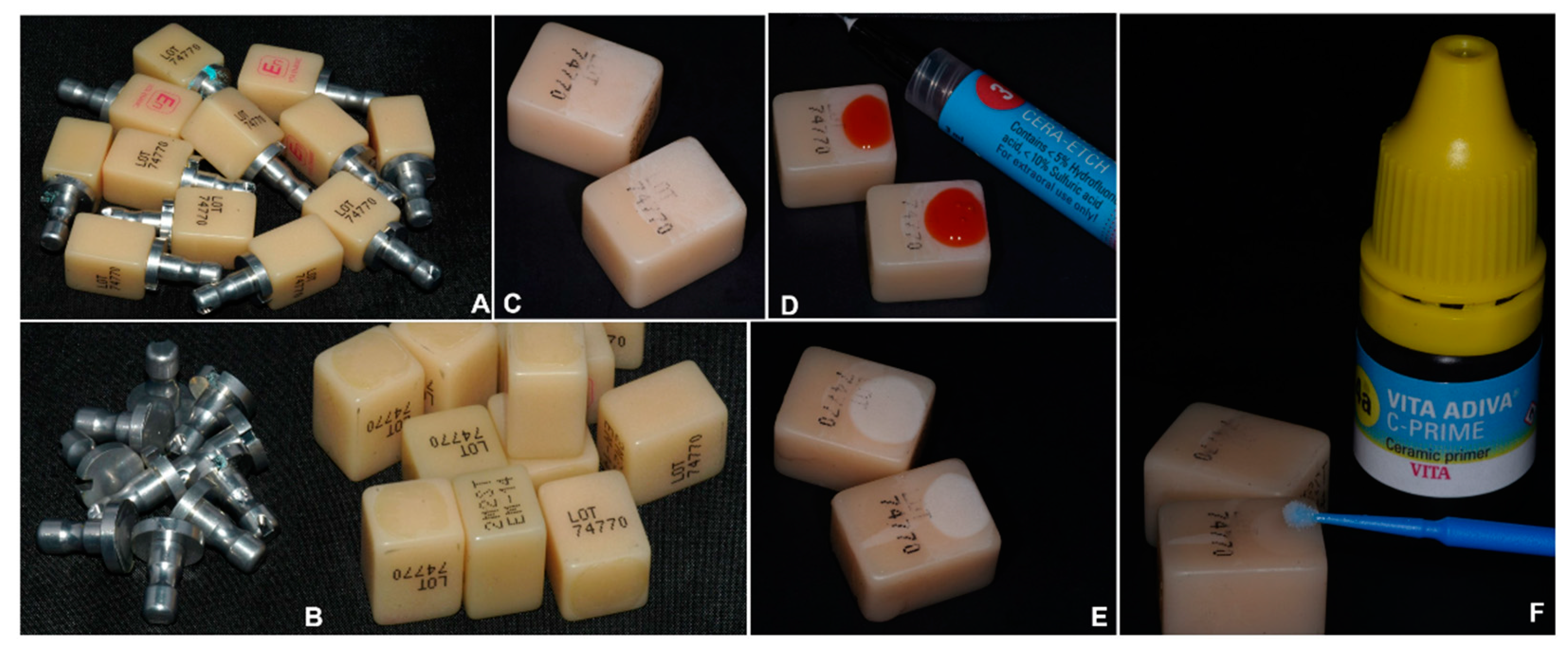
After removing the metallic support pin from the ENA ceramic block (Figure 2A,B), the superficial gloss was removed by dry grinding to simulate the removal of the aprismatic enamel or external fluorohydroxyapatite layer. A coarse finishing disk (Soflex Disc Pop-On, 3M, Saint Paul, MN, USA) mounted in a low-speed handpiece set at 20,000 rpm and attached to the dentist chair at an angle of ±45° with the surface of the block was used. The applied force was driven by hand, as in a clinical setting, by the same restorative dentist (single operator) with >30 years of clinical experience. A new disk was used for each block with eight grinding repetitions. A 20-s oil-free air/water spray removed the debris. The prepared blocks were shuffled to ensure randomization and operator blinding.
The bonding surface of the ENA block was prepared for bonding following the sequence shown in Figure 2C–E and Figure 3. For standardization, only 4.9% (5%) hydrofluoric acid etching gel from the VITA ADIVA kit (HF5) (Vita Zahnfabrik, Bad Säckingen, Germany) was used to etch all acid-sensitive surfaces involved in the study (20 s, SU group; 60 s, ENA group), according to the manufacturer’s instructions. The blocks were etched in pairs to prevent over-etching, and the etching time was controlled using a stopwatch. The treated surfaces were thoroughly cleaned using oil-free water spray for 20 s and then dried using oil-free compressed air for 10 s. The hybrid ceramic was primed with Vita ADIVA C-Primer (Vita Zahnfabrik, Bad Säckingen, Germany) in the control group (Figure 2F), and with a universal silane-containing primer (Monobond Plus, Ivoclar Vivadent) in the other groups [39,50] (Figure 3). Primers were applied using a microbrush and allowed to react for 60 s. If not completely dried after 60 s, air-drying was performed using an oil-free spray.

3.2. Preparation of the Cylinders

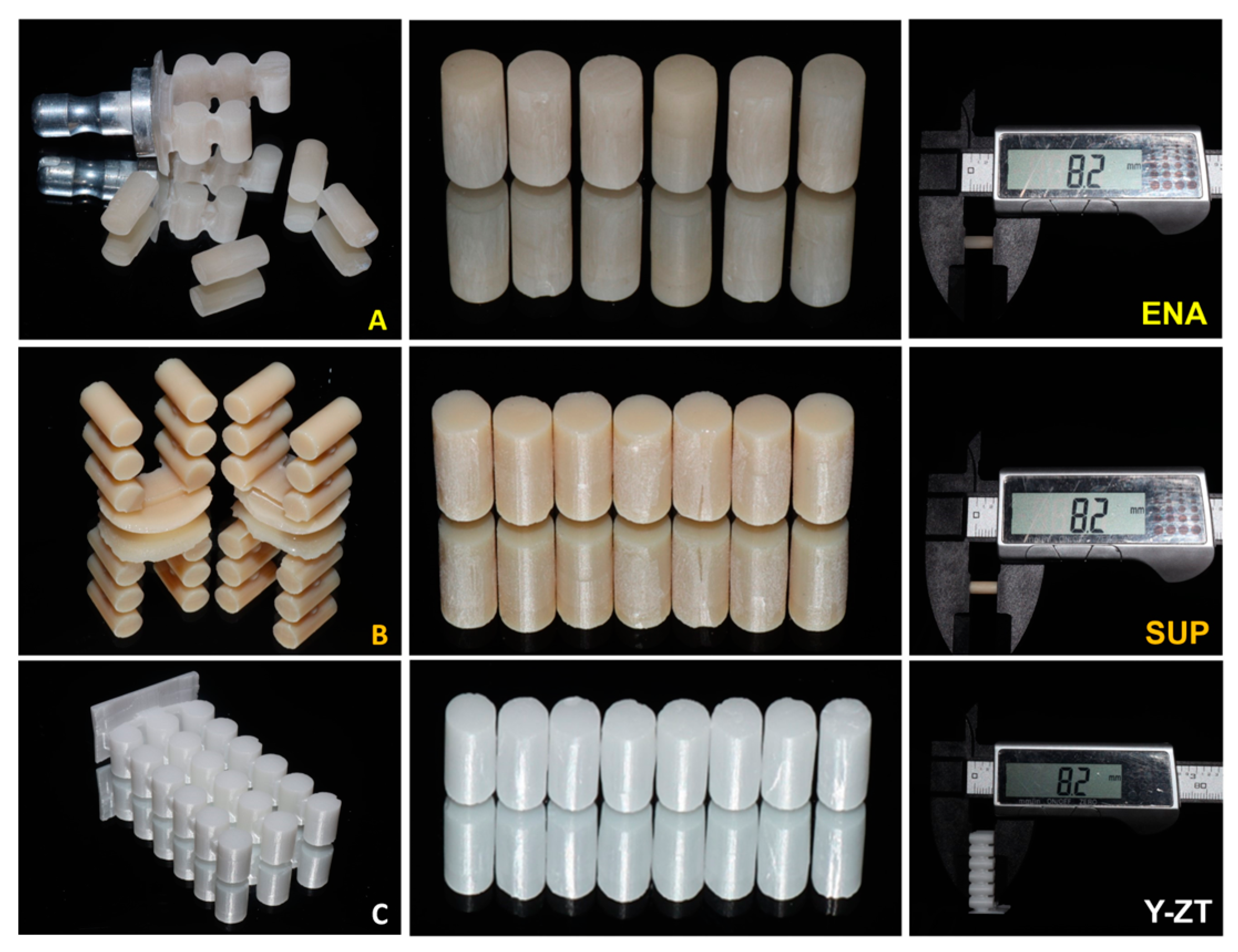
Monolithic ceramic cylinders (base diameter, 3.88 mm; length, 8.2 mm) (Abase = π × r2= 12.19 mm2), designed with EXOCAD software (Exocad GmbH, Darmstadt, Germany) and fabricated using a CAD-CAM inLab milling machine (Dentsply/Sirona, Charlotte, NC, USA), were produced for each ceramic material (n = 18) (Figure 4). Cylinders of the same dimensions, made of a PROCLINIC EXPERT resin-matrix nanohybrid composite (RES) (SDI Limited, Burnston, Australia) (control group), were manually manufactured using a polycarbonate cylinder template. All cylinders were checked at 10× magnification for cracks, surface discontinuities, and air bubbles and voids in the specific case of the manufactured cylinders.
Cylinders considered appropriate for testing were selected (RES, SUP, Y-ZT, n = 5; ENA, n = 6) for bonding with RelyX Ultimate (RU), according to the manufacturer’s instructions (Figure 5). The bonding procedures were performed immediately after each surface-conditioning method to avoid surface contamination.

3.3. Specimens Assembling for Shear Strength Test

To allow standardization during specimen assembly, a silicon mold was prepared to accommodate the blocks and allow the exact height of the cemented cylinder between the blocks (Figure 6A–E). Two blocks sustained in a plastic holder by a metallic pin were inserted into a silicone ice cube mold filled with silicone putty. An extra 0.3 mm space was calculated relative to the cylinder length for easy cylinder insertion after cement application on the tops (Figure 6E).
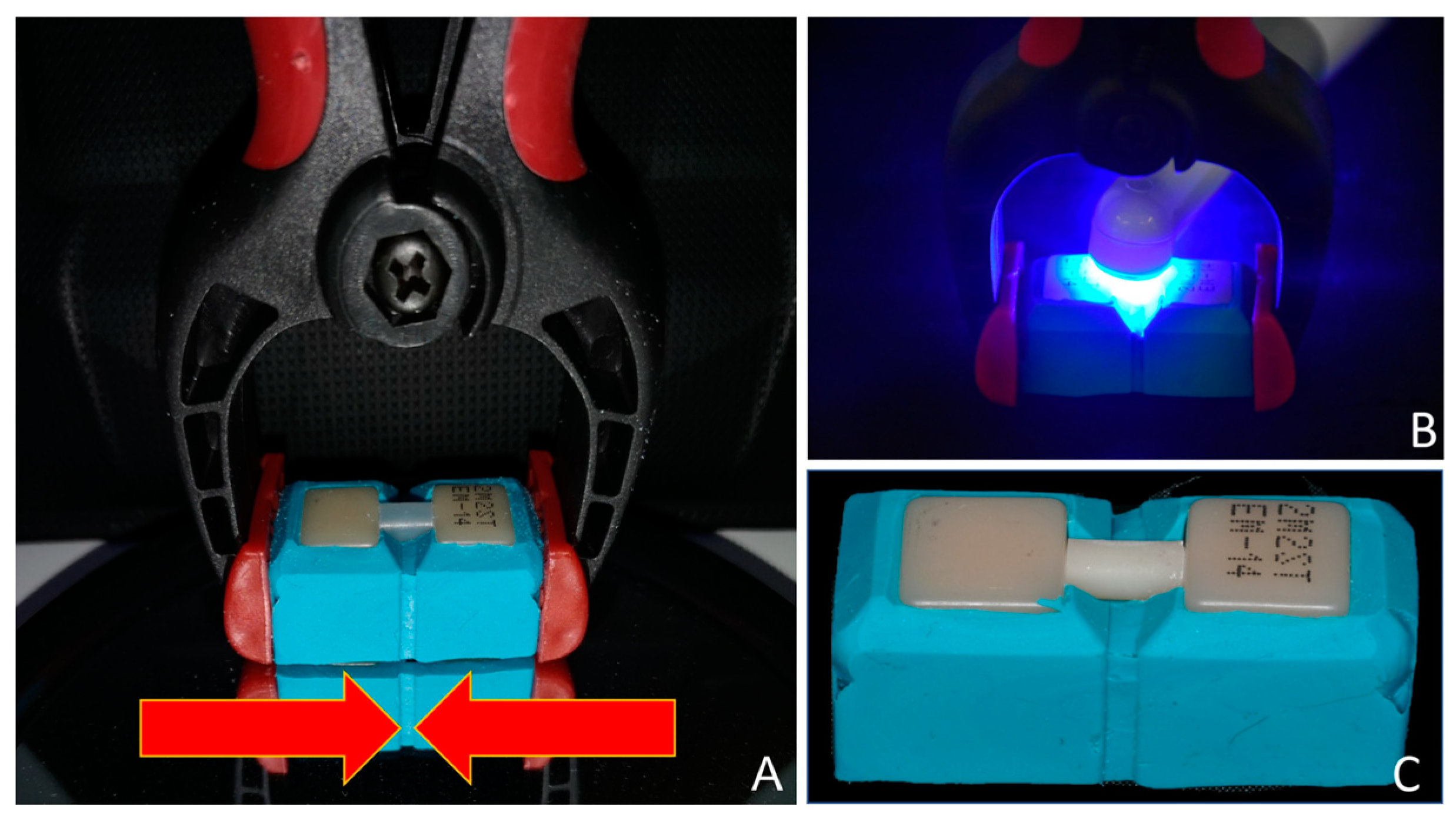
The excess cement was immediately removed using a microbrush. A constant pressure (0.5 kg) was applied during cement polymerization using a pinch spring (Figure 7A–C). The interfaces were light-cured for 10 s through each block and then in the middle of the cylinder for a total of 30 s using an Elipar S10 curing unit (1200 mW/cm2; 3M ESPE) within the accepted procedure [30]. Radiant exposure was ensured by prior calibration of the light-curing device using a radiometer. The compressive force was maintained for 10 min, leaving the material to self-cure.
Any residual cement was removed using a fine-point sickle scaler (SM 11; Hu-Friedy Co., Chicago, IL, USA). The bonded specimens were stored in saline water for 48 h at 37 °C before the SBS test.

3.4. Mechanical Characterization of Adhesive Joints

To avoid bending during testing, CAD-CAM technology creates a polylactic acid (PLA) base using free-design software and a home-mounted 3D printer. This material was selected because of its properties (environment-friendly option, low melting point (can be printed at lower temperatures and with less energy), ease of use, minimal post-processing, good surface finish, and stiff material) [51]. The details are presented in Figure 8.

3.5. In Vitro RBB Materials Adhesive Joint Mechanical Characterization

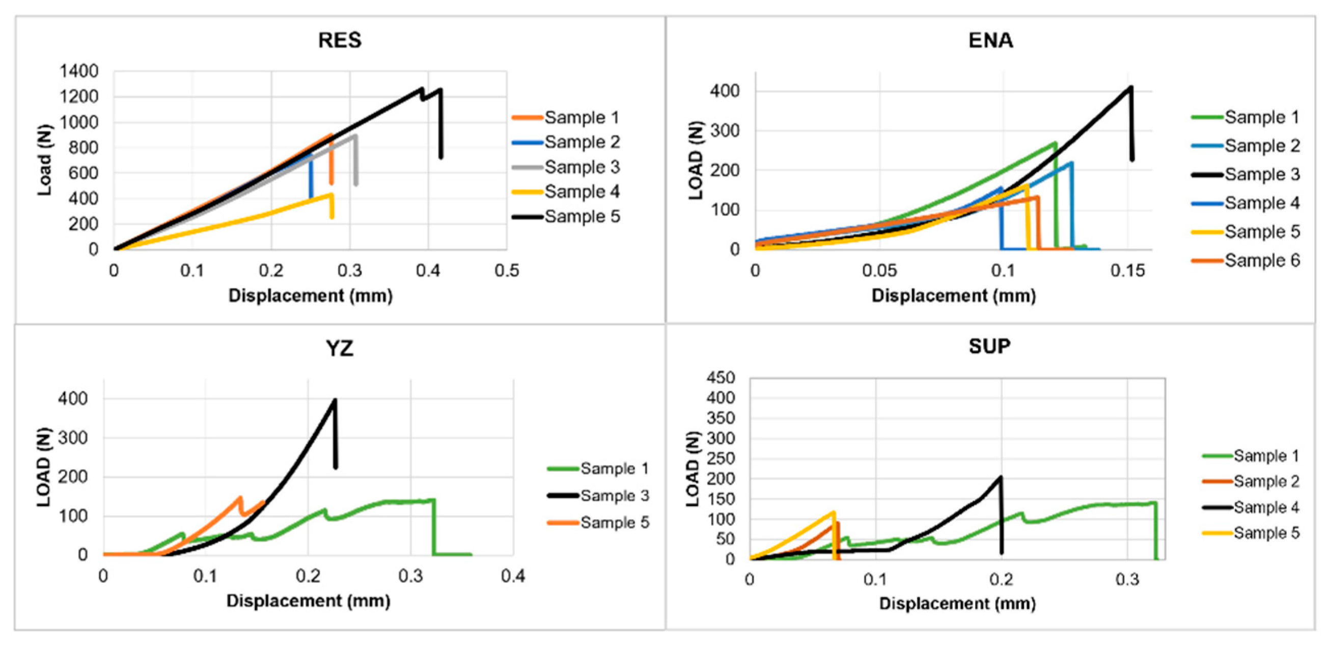
The specimens of the three CAD-CAM monolithic ceramics, potential candidates for RBB to rehabilitate one missing anterior tooth, were mechanically assessed under load displacement of 0.2 mm/min (Instron–Universal tensile machine) (Figure 9). Load-displacement curves were recorded during the mechanical test. The maximum load in the test was used to identify the experimental RBB setting that supported the highest shear stress and the highest shear stress supported by the adherend before cohesive failure.

3.6. Surface Energy Measurements

The surface energy of each CAD-CAM ceramic by treatment surface was measured to be correlated with the shear strength. The most commonly used surface treatments in the literature were chosen to characterize the surface energy of three CAD-CAM ceramics: grinding, hydrofluoric acid (5%), and aluminum oxide sandblasting [1,34,52].
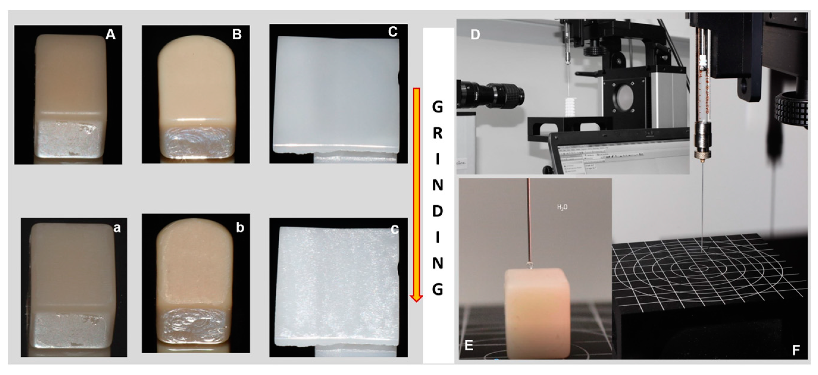
The measurement protocol followed industry-standard methodology [37]. The surface energy (SE) for each surface treatment was calculated based on the mean of three evaluations for each liquid, using a contact angle goniometer (OCA 15, DataPhysics Instruments GmbH, Filderstadt, Germany). Contact angle measurements were performed under ambient conditions using three different liquids: water (polar liquid), ethylene glycol 55% (polar liquid), and n-hexadecane (nonpolar), following the OWRK method [35]. Figure 10 shows the SE determination after block grinding.
For only grinding and hydrofluoric acid, the protocols for surface treatment were the same as in subSection 3.1. (Preparation of the base adherend). For the sandblasting surface treatment, the blocks were air-abraded at 0.20 MPa, for 10 s, with 50 µm alumina (Al2O3) particles. The nozzle was kept perpendicular and as perpendicular as possible to the surfaces of the blocks (angle between 80° and 90°) at a distance of 10 mm. Air abrasion was performed with erratic circular motions to ensure an even application of the AIRSONIC Alu-Oxyd powder (Hager-Werken and AZDENT sandblaster, Duisburg, Germany).

3.7. Adhesive Joint Fractography

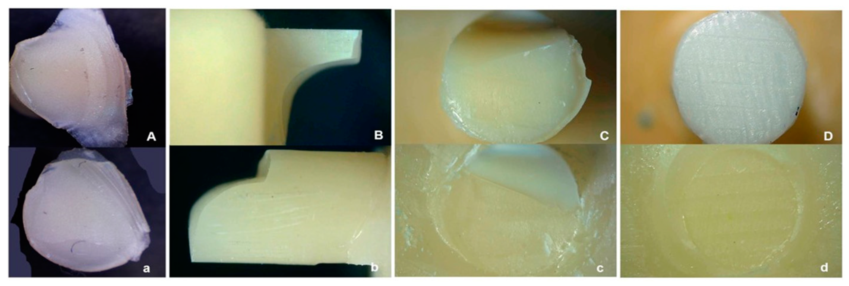
After SBS testing, the failed specimens (block and cylinder) were inspected at a magnification of 50× and 100× using a digital microscope (AmScope Industrial Inspection, Microscope, United Scope LLC, Irvine, CA, USA). For the mode fracture classification, all components were registered as follows: adhesive fracture (bond failure at the interface between the adhesive and restorative material, even if present in small amounts), cohesive fracture in the block (fracture occurring entirely within the block structure), cohesive fracture in the cylinder (fracture occurring entirely within the restorative material), and mixed when the adhesive and cohesive modes coexisted in the central area of any interface.

3.8. Data Analysis

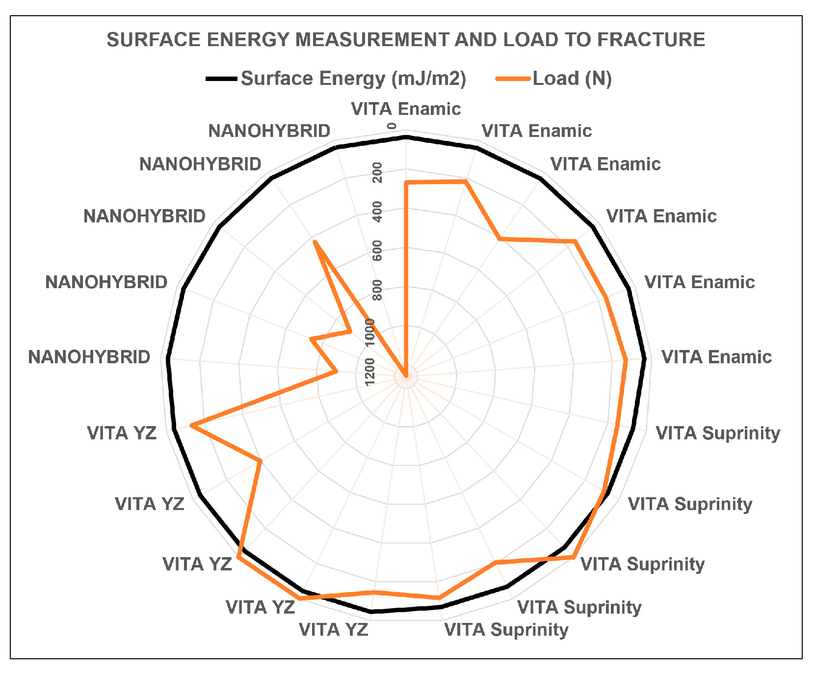
All data were analyzed to verify the achievement of the proposed goals. The mean load to fracture (N) and shear stress (MPa) with standard deviation (SD) were calculated for each group. A meta-analysis was conducted using the type of CAD-CAM restorative material to evaluate the shear stress between materials after calculating the difference between means and effect sizes (random-effects model; α = 0.05; 95% CI; Z-value = 1.96). The failure mode was determined by microscopic observation and was correlated with the maximum load to fracture of the specimens. A radar graph correlates the surface energy with the load to fracture.

4. Results

Mechanical Tests

The mean shear stresses (MPa) calculated for each group are listed in Table 2. RES (69.10 ± 24.58 MPa) > Y-ZT (18.48 ± 12.12 MPa) > ENA (18.38 ± 8.51 MPa) > SUP (11.44 ± 4.04 MPa). The SUP (n = 1) and Y-ZT (n = 2) groups showed pre-test adhesive failure.
Figure 11 shows the behavior of the samples under load, and Figure 12 shows the forest plot of the shear stress by restorative material.
Despite the specimens being assembled using the same procedure, the SUP and Y-ZT groups exhibited inconsistent behavior before and during loading. The adhesion strength was material dependent. Surprisingly, the RES group performed the best, reaching mean values more than four times higher than those of the second-best ENA group.
As shown in Figure 13 and Figure 14, the observation of the fractured specimens reveals the different mechanical behaviors of the assessed ceramics. From the observations in Table 3, failed adhesion is the unique failure mode for the Y-ZT group. The unique failure mode was cohesive in the RES group, either in the cylinder or the base, and sometimes simultaneously. Figure 15 shows a comprehensive schematic of the failure mode.
From the data obtained from the failure mechanism and surface energy of the different materials evaluated (Table 4 and Figure 16), no correlation was found between these parameters, indicating that the intrinsic chemical composition of the restorative material and its interaction with the coupling agent were the main factors affecting the mechanical behavior. Relative to the effect of the surface treatment on the CAD-CAM monolithic ceramics (Figure 17), the three treatments modified the surface of the ENA; the SUP was markedly altered by conditioning with HF 5% for 60 s and only slightly by sandblasting with AL2O3 50 µm, and Y-ZT was unaffected by HF 5%. These findings confirmed the data reported in the literature.
The crossing of microscopy and surface energy data shows that HF 5% is a suitable treatment to prepare the surface of SUP for adhesion if we only consider the microscopic interlocking between the restorative material and adhesive cement. Other materials depend on chemical reactions.

5. Discussion

The main objective of this study was to evaluate the possibility of using a standardized artificial material as a base adherent for the shear bond strength tests of restorative materials. Taking advantage of this objective and because the behavior of this material (ENA) for this purpose was unknown, CAD-CAM ceramics, from which different performances in shear bond strength testing were expected, were tested in parallel to validate the mechanical behavior of the adherend. Based on the results, the null hypothesis that the Vita Enamic hybrid ceramic was not mechanically a suitable adherend for shear bond tests was rejected, as this material withstood load forces up to 1142.89 N corresponding to an adhesive stress of 103.00 MPa. The other null hypothesis, that no differences would be found in the mechanical behavior between the CAD-CAM monolithic ceramics, was also rejected, as significant differences were found (p < 0.01). The meta-analysis conducted relative to different materials revealed substantial heterogeneity of results across groups due to heterogeneity rather than sampling error, with an I2 = 73.59% of total variation and a H2 = 3.79 variance between studies (p < 0.01).
The number of specimens used in this study reflects the surface irregularities due to manufacturing, as more than half of the cylinders were not considered suitable after 10x magnification visual inspection despite the previous calibration of the milling equipment. If the purpose is to manufacture an authentic restoration, this issue should be assessed carefully [53,54] and visual inspection with magnification before delivery should be encouraged. Nevertheless, it was not considered a real constraint for the experimental purpose, as the adherend was a homogeneous industrial material subjected to standardized surface treatment for every experimental condition, with an expected good adhesive if combined with Rely X Ultimate cement [46]. In contrast, the materials used in the cylinders were expected to differ according to the manufacturer’s datasheets [25,55,56].
In this experimental design, a silicone mold was used for stabilization and standardization during the assembly. The base in PLA, because of its physical characteristics [51], ensured stabilization during mechanical tests and no flexion of the specimen, although the specimen was expected to be stable when standing alone. A silane primer was applied to the adherend surface to enhance adhesion by a chemical reaction with the polar component of the ENA structure [50,57].
Rely X Ultimate was selected based on literature [36] and parallel research [58]. The cement was used in a mixed-cure protocol (light-cure for 30 s, followed by self-cure for 10 min). To test the adhesive performance of cement was not an objective of this study. However, a control group photoactivated for only 2–3 s and left in a chemical cure for 6 min (self-adhesive mode) would be interesting to highlight the influence of chemical interactions on the success of the adhesive interface according to the cylinder material, despite the fact that the performance of this type of cement is enhanced by photopolymerization [36].
Vita Enamic was the most accessible material to handle. Vita Suprinity was very brittle in both the pre-sintered and sintered states. Polycrystalline zirconia (Vita Y-ZT) was accessible for milling, but it was almost impossible to separate the cylinders after the block had been sintered, with the destruction of several diamond points in the process. In future studies, we recommend separating cylinders before sintering. Resin-matrix composites (RES) are easy to handle; however, the possibility of including air bubbles in the cylinder upon production was a concern. Given the unexpected performance of the RES cylinder, not having determined the ultimate strength of this material is a limitation of this study, because it would have been interesting to compare it with data relative to the other materials used.
The correlation between adhesive stress and failure mode confirmed that the limitation of experimental Y-ZT RBBs lies in the success of adhesion, which agrees with the results of previous studies [32,59]. In fact, despite being the toughest material, the Y-ZT group, if the failed specimens are excluded, achieved mechanical performance similar to the ENA group (18.48 ± 12.12 MPa and 18.38 ± 8.51 MPa, respectively), which has a toughness about 8 times lower [55,56]. The exclusive adhesive failure in the Y-ZT group, including pre-test failures, reinforces the need for an easily replicable and efficient adhesion protocol when working with this type of material [1], especially in the case of a minimally invasive one-retainer anterior RBB, which does not have additional macromechanical retention [32,59].
Despite several searches of the literature and thousands of articles found related to adhesion, namely relative to zirconia [60], no studies were found that would allow for a comparison of the results of this study with other existing ones. This is due to the lack of comparable adhesive protocols or adherents, which agrees with a recent meta-analysis that identified 686 protocols to adhere 37 different CAD-CAM blocks [1], but also with the fact that, frequently, results are not available in MPa for evidence-based comparisons. Some studies have tested this type of CAD-CAM ceramic or adhesive cement. However, these studies did not use them simultaneously, nor did they evaluate them with a similar experimental setting to that of the current study, as CAD-CAM ceramics are often tested in the form of a one-piece fixed crown subjected to catastrophic fracture or pull-out tests. Other studies have evaluated CAD-CAM ceramics adhered to a cement cylinder or as a block adhered to another block.
This limitation reinforces the importance of this study because the experimental setting approaches a clinical situation of a minimal invasive resin-bonded bridge. It also showed that an industrial material could be used, at least in preliminary tests, as an adherend in shear strength tests of CAD-CAM restorative materials. This type of adherend allows for standardization and overcomes the existing constraints found in the use of biological substrates.

6. Recommendations for Future Research

Considering the potential of the tested adherend, experimental models to evaluate the shear resistance of cement with different adhesive strategies are recommended.
With CAD-CAM materials in rapid evolution, namely, those produced by addition, the use of this type of adherent could facilitate a quick and standardized evaluation of their adhesive strength, allowing easy comparison with existing CAD-CAD monolithic materials, for which there is already some scientific evidence.

7. Conclusions

The VITA Enamic block is a potential base adherent for SBS tests because it resists a shear load of up to 103 MPa (RES sample 5 test) in a cylinder with a double-interface connection design. Significant differences in the mechanical behavior with respect to the shear strength were identified between the tested CAD-CAM ceramics. Under the experimental conditions of this study, the SBS was not related to the surface energy of the substrates, and the failure mode was material-dependent. As a restorative material, ENA is predictable and easy to handle. The SUP was difficult to handle owing to its brittleness in both the pre-sintered and sintered states. The Y-ZT failure mode was always adhesive.
The tested CAD-CAM ceramics have sufficient adhesive strength to be used as resin-bonded bridges for permanent or interim rehabilitation, provided an efficient adhesive protocol is wisely chosen, and the need for short-term removal, equated as Y-ZT, is very difficult to remove by drilling.

Author Contributions

Conceptualization, M.J.C.-L. and J.M.C.-L.; methodology, M.J.C.-L.; software, M.J.C.-L. and J.M.C.-L.; validation, M.J.C.-L., R.C. and L.F.M.d.S.; formal analysis, M.J.C.-L., R.C. and L.F.M.d.S.; investigation, M.J.C.-L. and J.M.C.-L.; resources, M.J.C.-L.; data curation, J.M.C.-L.; writing—original draft preparation, M.J.C.-L.; writing—review and editing, M.J.C.-L.; visualization, M.J.C.-L. and J.M.C.-L.; supervision, T.P.; project administration, M.J.C.-L.; funding acquisition, T.P. All authors have read and agreed to the published version of the manuscript.

Funding

This research was funded by CESPU, CRL (Grant number AlignAgen-GI2-CESPU-2022).

Institutional Review Board Statement

Not applicable.

Informed Consent Statement

Not applicable.

Data Availability Statement

Not applicable.

Conflicts of Interest

The authors declare no conflict of interest.

References

  1. Calheiros-Lobo, M.J.; Carbas, R.; da Silva, L.F.M.; Pinho, T. Impact of in vitro findings on clinical protocols for the adhesion of CAD-CAM blocks: A systematic integrative review and meta-analysis. J. Prosthet. Dent. 2022, S0022-3913(22)00551-0. [Google Scholar] [CrossRef] [PubMed]
  2. Sulaiman, T.A. Materials in digital dentistry-A review. J. Esthet. Restor. Dent. 2020, 32, 171–181. [Google Scholar] [CrossRef]
  3. Raszewski, Z.; Brząkalski, D.; Derpeński, Ł.; Jałbrzykowski, M.; Przekop, R.E. Aspects and Principles of Material Connections in Restorative Dentistry-A Comprehensive Review. Materials 2022, 15, 7131. [Google Scholar] [CrossRef] [PubMed]
  4. Kelly, J.R. Clinically relevant approach to failure testing of all-ceramic restorations. J. Prosthet. Dent. 1999, 81, 652–661. [Google Scholar] [CrossRef]
  5. Rekow, E.D.; Silva, N.R.; Coelho, P.G.; Zhang, Y.; Guess, P.; Thompson, V.P. Performance of dental ceramics: Challenges for improvements. J. Dent. Res. 2011, 90, 937–952. [Google Scholar] [CrossRef] [Green Version]
  6. Baldi, A.; Carossa, M.; Comba, A.; Alovisi, M.; Femiano, F.; Pasqualini, D.; Berutti, E.; Scotti, N. Wear Behaviour of Polymer-Infiltrated Network Ceramics, Lithium Disilicate and Cubic Zirconia against Enamel in a Bruxism-Simulated Scenario. Biomedicines 2022, 10, 1682. [Google Scholar] [CrossRef] [PubMed]
  7. Guth, J.F.; Stawarczyk, B.; Edelhoff, D.; Liebermann, A. Zirconia and its novel compositions: What do clinicians need to know? Quintessence Int. 2019, 50, 512–520. [Google Scholar] [CrossRef]
  8. Fung, L.; Brisebois, P. Implementing Digital Dentistry into Your Esthetic Dental Practice. Dent. Clin. N. Am. 2020, 64, 645–657. [Google Scholar] [CrossRef]
  9. Watanabe, H.; Fellows, C.; An, H. Digital Technologies for Restorative Dentistry. Dent. Clin. N. Am. 2022, 66, 567–590. [Google Scholar] [CrossRef]
  10. Tian, Y.; Chen, C.; Xu, X.; Wang, J.; Hou, X.; Li, K.; Lu, X.; Shi, H.; Lee, E.S.; Jiang, H.B. A Review of 3D Printing in Dentistry: Technologies, Affecting Factors, and Applications. Scanning 2021, 2021, 9950131. [Google Scholar] [CrossRef]
  11. Gad, M.M.; Fouda, S.M. Factors affecting flexural strength of 3D-printed resins: A systematic review. J. Prosthodont. 2023, 32, 96–110. [Google Scholar] [CrossRef]
  12. Mazza, L.C.; Lemos, C.A.A.; Pesqueira, A.A.; Pellizzer, E.P. Survival and complications of monolithic ceramic for tooth-supported fixed dental prostheses: A systematic review and meta-analysis. J. Prosthet. Dent. 2021, 128, 566–574. [Google Scholar] [CrossRef]
  13. Leitão, C.; Fernandes, G.V.O.; Azevedo, L.P.P.; Araújo, F.M.; Donato, H.; Correia, A.R.M. Clinical performance of monolithic CAD/CAM tooth-supported zirconia restorations: Systematic review and meta-analysis. J. Prosthodont. Res. 2022, 66, 374–384. [Google Scholar] [CrossRef] [PubMed]
  14. Leung, G.K.; Wong, A.W.; Chu, C.H.; Yu, O.Y. Update on Dental Luting Materials. Dent. J. 2022, 10, 208. [Google Scholar] [CrossRef] [PubMed]
  15. Manuja, N.; Nagpal, R.; Pandit, I. Dental adhesion: Mechanism, techniques and durability. J. Clin. Pediatr. Dent. 2012, 36, 223–234. [Google Scholar] [CrossRef] [PubMed]
  16. Junior, L.; Baroudi, K.; Barroso, L.; Miranda, M.; Vitti, R.; Brandt, W. Bond strength of self-adhesive resin cement to dentin using different adhesion protocols. J. Clin. Exp. Dent. 2022, 14, e35–e39. [Google Scholar] [CrossRef] [PubMed]
  17. Van Meerbeek, B.; De Munck, J.; Yoshida, Y.; Inoue, S.; Vargas, M.; Vijay, P.; Van Landuyt, K.; Lambrechts, P.; Vanherle, G. Adhesion to enamel and dentin: Current status and future challenges. Oper. Dent. 2003, 28, 215–235. [Google Scholar]
  18. Tosun, B.; Yanıkoğlu, N. Evaluation of the effects of different surface modification methods on the bond strength of high-performance polymers and resin matrix ceramics. Clin. Oral Investig. 2022, 26, 3781–3790. [Google Scholar] [CrossRef]
  19. da Silva, M.A.; Vitti, R.P.; Sinhoreti, M.A.; Consani, R.L.; Silva-Junior, J.G.; Tonholo, J. Effect of alcoholic beverages on surface roughness and microhardness of dental composites. Dent. Mater. J. 2016, 35, 621–626. [Google Scholar] [CrossRef] [Green Version]
  20. Awad, M.M.; Alqahtani, H.; Al-Mudahi, A.; Murayshed, M.S.; Alrahlah, A.; Bhandi, S.H. Adhesive Bonding to Computer-aided Design/Computer-aided Manufacturing Esthetic Dental Materials: An Overview. J. Contemp. Dent. Pract. 2017, 18, 622–626. [Google Scholar] [CrossRef]
  21. Skorulska, A.; Piszko, P.; Rybak, Z.; Szymonowicz, M.; Dobrzyński, M. Review on Polymer, Ceramic and Composite Materials for CAD/CAM Indirect Restorations in Dentistry—Application, Mechanical Characteristics and Comparison. Materials 2021, 14, 1592. [Google Scholar] [CrossRef] [PubMed]
  22. Palacios, T.; Tarancón, S.; Pastor, J.Y. On the Mechanical Properties of Hybrid Dental Materials for CAD/CAM Restorations. Polymers 2022, 14, 3252. [Google Scholar] [CrossRef] [PubMed]
  23. Eldafrawy, M.; Ebroin, M.G.; Gailly, P.A.; Nguyen, J.F.; Sadoun, M.J.; Mainjot, A.K. Bonding to CAD-CAM Composites: An Interfacial Fracture Toughness Approach. J. Dent. Res. 2018, 97, 60–67. [Google Scholar] [CrossRef] [PubMed] [Green Version]
  24. da Silva, P.; Martinelli-Lobo, C.M.; Bottino, M.A.; Melo, R.M.; Valandro, L.F. Bond strength between a polymer-infiltrated ceramic network and a composite for repair: Effect of several ceramic surface treatments. Braz. Oral Res. 2018, 32, e28. [Google Scholar] [CrossRef] [Green Version]
  25. Vita-Zahnfabrik. Vita Suprinity® PC—Working Instructions; VITA Zahnfabrik: Bad Säckingen, Germany, 2019. [Google Scholar]
  26. Zarone, F.; Ruggiero, G.; Leone, R.; Breschi, L.; Leuci, S.; Sorrentino, R. Zirconia-reinforced lithium silicate (ZLS) mechanical and biological properties: A literature review. J. Dent. 2021, 109, 103661. [Google Scholar] [CrossRef] [PubMed]
  27. Kwon, S.J.; Lawson, N.C.; McLaren, E.E.; Nejat, A.H.; Burgess, J.O. Comparison of the mechanical properties of translucent zirconia and lithium disilicate. J. Prosthet. Dent. 2018, 120, 132–137. [Google Scholar] [CrossRef]
  28. Stawarczyk, B.; Keul, C.; Eichberger, M.; Figge, D.; Edelhoff, D.; Lumkemann, N. Three generations of zirconia: From veneered to monolithic. Part I. Quintessence Int. 2017, 48, 369–380. [Google Scholar] [CrossRef]
  29. Fathy, S.M.; Al-Zordk, W.; Grawish, M.E.; Swain, M.V. Flexural strength and translucency characterization of aesthetic monolithic zirconia and relevance to clinical indications: A systematic review. Dent. Mater. 2021, 37, 711–730. [Google Scholar] [CrossRef]
  30. Pereira, G.K.R.; Guilardi, L.F.; Dapieve, K.S.; Kleverlaan, C.J.; Rippe, M.P.; Valandro, L.F. Mechanical reliability, fatigue strength and survival analysis of new polycrystalline translucent zirconia ceramics for monolithic restorations. J. Mech. Behav. Biomed. Mater. 2018, 85, 57–65. [Google Scholar] [CrossRef]
  31. Zhang, Y.; Lawn, B.R. Evaluating dental zirconia. Dent. Mater. 2019, 35, 15–23. [Google Scholar] [CrossRef]
  32. Bilir, H.; Yuzbasioglu, E.; Sayar, G.; Kilinc, D.D.; Bag, H.G.G.; Özcan, M. CAD/CAM single-retainer monolithic zirconia ceramic resin-bonded fixed partial dentures bonded with two different resin cements: Up to 40 months clinical results of a randomized-controlled pilot study. J. Esthet. Restor. Dent. 2022, 34, 1122–1131. [Google Scholar] [CrossRef] [PubMed]
  33. Yoshida, K. Effect of 10-Methacryloyloxydecyl Dihydrogen Phosphate Concentrations in Primers on Bonding Resin Cements to Zirconia. J. Prosthodont. 2021, 30, 356–362. [Google Scholar] [CrossRef]
  34. Heboyan, A.; Vardanyan, A.; Karobari, M.I.; Marya, A.; Avagyan, T.; Tebyaniyan, H.; Mustafa, M.; Rokaya, D.; Avetisyan, A. Dental Luting Cements: An Updated Comprehensive Review. Molecules 2023, 28, 1619. [Google Scholar] [CrossRef]
  35. Marshall, S.J.; Bayne, S.C.; Baier, R.; Tomsia, A.P.; Marshall, G.W. A review of adhesion science. Dent. Mater. 2010, 26, e11–e16. [Google Scholar] [CrossRef]
  36. Calheiros-Lobo, M.J.; Vieira, T.; Carbas, R.; da Silva, L.F.M.; Pinho, T. Effectiveness of Self-Adhesive Resin Luting Cement in CAD-CAM Blocks—A Systematic Review and Meta-Analysis. Materials 2023, 16, 2996. [Google Scholar] [CrossRef]
  37. von Fraunhofer, J.A. Adhesion and cohesion. Int. J. Dent. 2012, 2012, 951324. [Google Scholar] [CrossRef] [PubMed] [Green Version]
  38. Lima, R.B.W.; Silva, A.F.; da Rosa, W.L.O.; Piva, E.; Duarte, R.M.; De Souza, G.M. Bonding Efficacy of Universal Resin Adhesives to Zirconia Substrates: Systematic Review and Meta-Analysis. J. Adhes. Dent. 2023, 25, 51–62. [Google Scholar] [CrossRef] [PubMed]
  39. Fathy, H.; Hamama, H.H.; El-Wassefy, N.; Mahmoud, S.H. Effect of different surface treatments on resin-matrix CAD/CAM ceramics bonding to dentin: In vitro study. BMC Oral. Health 2022, 22, 635. [Google Scholar] [CrossRef]
  40. Peumans, M.; Valjakova, E.B.; De Munck, J.; Mishevska, C.B.; Van Meerbeek, B. Bonding Effectiveness of Luting Composites to Different CAD/CAM Materials. J. Adhes. Dent. 2016, 18, 289–302. [Google Scholar] [CrossRef] [Green Version]
  41. Barutcigil, K.; Barutcigil, Ç.; Kul, E.; Özarslan, M.M.; Buyukkaplan, U.S. Effect of Different Surface Treatments on Bond Strength of Resin Cement to a CAD/CAM Restorative Material. J. Prosthodont. 2019, 28, 71–78. [Google Scholar] [CrossRef]
  42. Fidalgo-Pereira, R.; Carpio, D.; Torres, O.; Carvalho, O.; Silva, F.; Henriques, B.; Özcan, M.; Souza, J.C.M. The influence of inorganic fillers on the light transmission through resin-matrix composites during the light-curing procedure: An integrative review. Clin. Oral. Investig. 2022, 26, 5575–5594. [Google Scholar] [CrossRef]
  43. Diemer, F.; Stark, H.; Helfgen, E.H.; Enkling, N.; Probstmeier, R.; Winter, J.; Kraus, D. In vitro cytotoxicity of different dental resin-cements on human cell lines. J. Mater. Sci. Mater. Med. 2021, 32, 4. [Google Scholar] [CrossRef]
  44. Lopes-Rocha, L.; Ribeiro-Gonçalves, L.; Henriques, B.; Özcan, M.; Tiritan, M.E.; Souza, J.C.M. An integrative review on the toxicity of Bisphenol A (BPA) released from resin composites used in dentistry. J. Biomed. Mater. Res. B Appl. Biomater. 2021, 109, 1942–1952. [Google Scholar] [CrossRef]
  45. Barutcigil, K.; Dundar, A.; Batmaz, S.G.; Yildirim, K.; Barutcugil, C. Do resin-based composite CAD/CAM blocks release monomers? Clin. Oral. Investig. 2021, 25, 329–336. [Google Scholar] [CrossRef] [PubMed]
  46. Hilgemberg, B.; Siqueira, F.S.F.; Cardenas, A.F.M.; Ribeiro, J.L.; Dávila-Sánchez, A.; Sauro, S.; Loguercio, A.D.; Arrais, C.A.G. Effect of Bonding Protocols on the Performance of Luting Agents Applied to CAD-CAM Composites. Materials 2022, 15, 4. [Google Scholar] [CrossRef] [PubMed]
  47. Anuntasainont, M.; Po-Ngam, N.; Lührs, A.K.; Neoh, S.P.; Pongprueksa, P. Fracture resistance of CAD/CAM blocks cemented on dentin using different cementation strategies. J. Prosthodont. Res. 2023, JPR_D_22_00196. [Google Scholar] [CrossRef] [PubMed]
  48. Stylianou, A.; Liu, P.R.; O’Neal, S.J.; Essig, M.E. Restoring Congenitally Missing Maxillary Lateral Incisors Using Zirconia-Based Resin Bonded Prostheses. J. Esthet. Restor. Dent. 2016, 28, 8–17. [Google Scholar] [CrossRef]
  49. Narwani, S.; Yadav, N.S.; Hazari, P.; Saxena, V.; Alzahrani, A.H.; Alamoudi, A.; Zidane, B.; Albar, N.H.M.; Robaian, A.; Kishnani, S.; et al. Comparison of Tensile Bond Strength of Fixed-Fixed Versus Cantilever Single- and Double-Abutted Resin-Bonded Bridges Dental Prosthesis. Materials 2022, 15, 5744. [Google Scholar] [CrossRef]
  50. Fouquet, V.; Lachard, F.; Abdel-Gawad, S.; Dursun, E.; Attal, J.P.; François, P. Shear Bond Strength of a Direct Resin Composite to CAD-CAM Composite Blocks: Relative Contribution of Micromechanical and Chemical Block Surface Treatment. Materials 2022, 15, 5018. [Google Scholar] [CrossRef]
  51. Farah, S.; Anderson, D.G.; Langer, R. Physical and mechanical properties of PLA, and their functions in widespread applications—A comprehensive review. Adv. Drug Deliv. Rev. 2016, 107, 367–392. [Google Scholar] [CrossRef] [Green Version]
  52. Quigley, N.P.; Loo, D.S.S.; Choy, C.; Ha, W.N. Clinical efficacy of methods for bonding to zirconia: A systematic review. J. Prosthet. Dent. 2020, 125, 231–240. [Google Scholar] [CrossRef]
  53. Goujat, A.; Abouelleil, H.; Colon, P.; Jeannin, C.; Pradelle, N.; Seux, D.; Grosgogeat, B. Marginal and internal fit of CAD-CAM inlay/onlay restorations: A systematic review of in vitro studies. J. Prosthet. Dent. 2019, 121, 590–597.e593. [Google Scholar] [CrossRef] [PubMed]
  54. Papadiochou, S.; Pissiotis, A.L. Marginal adaptation and CAD-CAM technology: A systematic review of restorative material and fabrication techniques. J. Prosthet. Dent. 2018, 119, 545–551. [Google Scholar] [CrossRef] [PubMed]
  55. Vita-Zahnfabrik. VITA ENAMIC®—Technical and Scientific Documentation; VITA Zahnfabrik: Bad Säckingen, Germany, 2019. [Google Scholar]
  56. Vita-Zahnfabrik. Vita YZ®—Solutions; VITA Zahnfabrik: Bad Säckingen, Germany, 2019. [Google Scholar]
  57. Yano, H.T.; Ikeda, H.; Nagamatsu, Y.; Masaki, C.; Hosokawa, R.; Shimizu, H. Correlation between microstructure of CAD/CAM composites and the silanization effect on adhesive bonding. J. Mech. Behav. Biomed. Mater. 2020, 101, 103441. [Google Scholar] [CrossRef] [PubMed]
  58. Calheiros-Lobo, M.J.; Carbas, R.; da Silva, L.F.M.; Pinho, T. Effect of the Modulation of the Adhesive Interface between a CAD-CAM Hybrid Ceramic adherend and Three Luting Cements on Shear Bond Strength: In Vitro Study. J. Adhes. Dent. 2023, 16, 2996. [Google Scholar]
  59. Al-Wahadni, A.; Dkmak, M.S.F.; Almohammed, S.; Hatamleh, M.M.; Tabanjah, A.A. Fracture strength of anterior cantilever resin bonded fixed partial dentures fabricated from high translucency zirconia with different intaglio surface treatments. J. Prosthodont. 2023; Epub ahead of print. [Google Scholar] [CrossRef]
  60. Mutlu-Sağesen, H.L.E.; Sağesen, E.A.; Özcan, M. Bibliometric analysis of zirconia publications between 1980 and 2021: Global productivity and publication trends. J. Prosthodont. Res. 2023; Online ahead of print. [Google Scholar] [CrossRef]
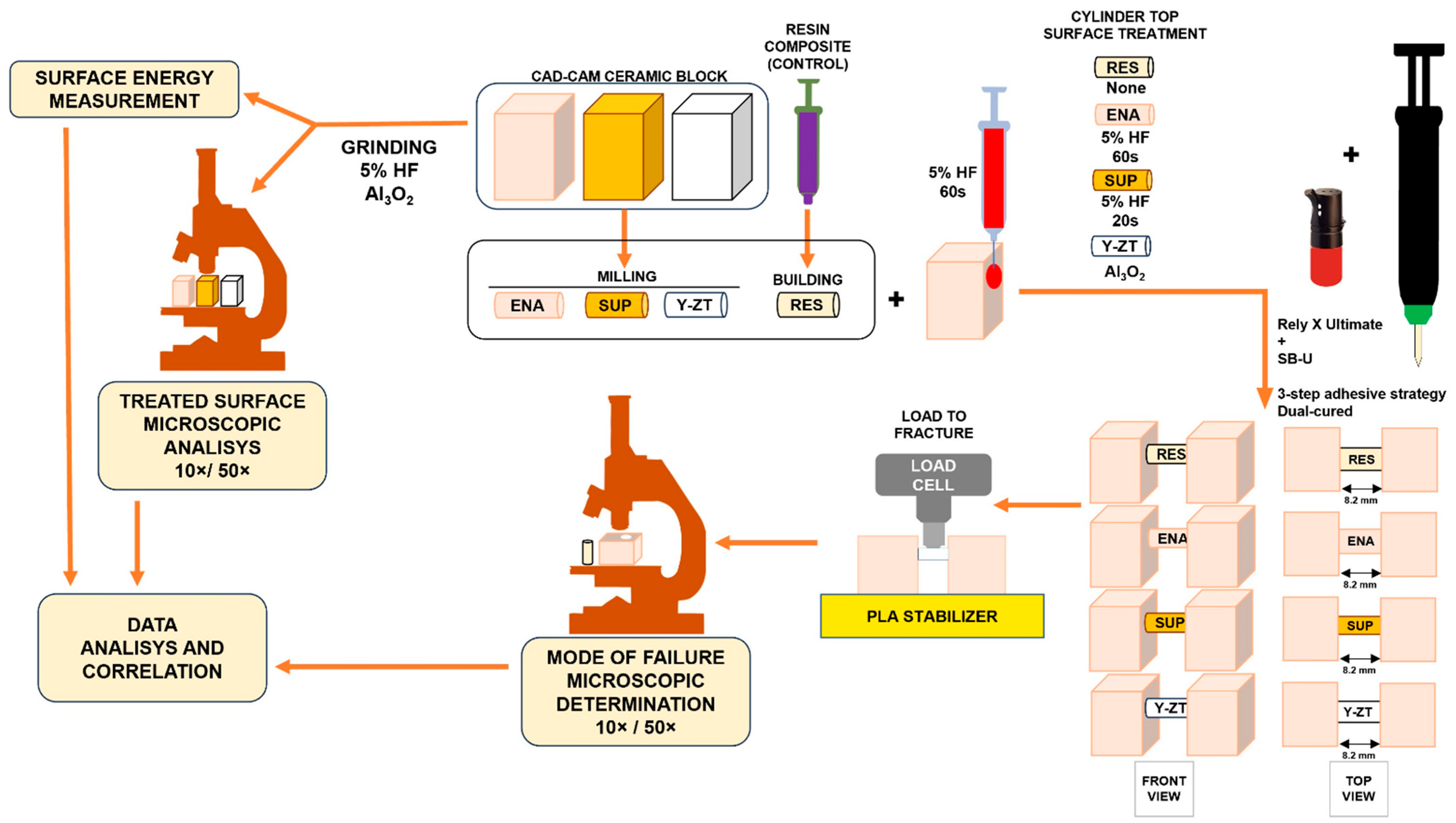
Figure 1. Comprehensive scheme of the experimental protocol workflow. ENA, Vita Enamic; HF, hydrofluoric acid; PLA, polylactic acid; SB-U, Scotchbond Universal adhesive; SUP, Vita Suprinity; Y-ZT, Vita zirconia.
Coatings 13 01218 g001
Figure 2. Vita Enamic blocks and surface treatment in the control group (RES). (A) before and (B) after support pin removal; (C) immediately after grinding; (D) during the hydrofluoric acid conditioning; (E) whitish conditioned and dried surface; (F) during the ceramic primer application, as recommended by the manufacturer.
Coatings 13 01218 g002
Figure 3. Sequence of the protocol for preparing the specimen bases for the groups ENA, SUP, and Y-ZT.
Coatings 13 01218 g003
Figure 4. Fabrication and calibration of the CAD-CAM monolithic ceramic cylinders. (A) immediately after milling, (B) after being cut and regularized, and (C) after testing length calibration.
Coatings 13 01218 g004
Figure 5. Cylinders and blocks in preparation for adhesion. (A) cylinders being conditioned; (B) unconditioned (3 on the left) and HF5 conditioned cylinders (3 on the right); (C) cylinders immersed in Monobond Plus for 60 s; (D) cylinders protected from daylight after adhesive system application; (E) adhesive system application on the blocks.
Coatings 13 01218 g005
Figure 6. Steps for silicone mold production. (A) silicone after setting; (B) making the groove to fit the cylinder. Performed with a bladed round drill mounted on a handpiece at low speed; (C) detail of the groove definition; (D) polycarbonate cylinder template accommodated in the groove for calibration; (E) confirming the intended length between the blocks.
Coatings 13 01218 g006
Figure 7. Constant compression for component adaptation. (A) during the 10 min; (B) detail of the photopolymerization step through the cylinder; (C) after polymerization and before mold removal.
Coatings 13 01218 g007
Figure 8. Steps of the polylactic acid (PLA) base design and fabrication. (A) design steps; (B) PLA coil; (C) PLA base in the printing process; (D) PLA base just printed.
Coatings 13 01218 g008
Figure 9. Specimen mounted on the polylactic acid (PLA) base ready for mechanical testing. (A) specimen inserted in the PLA base; (B) specimen being positioned on the platform; (C) basic scheme of the test; and (D) initial contact with the specimen and data registration.
Coatings 13 01218 g009
Figure 10. Substrates and equipment used for surface energy determination. (A,a) Enamic; (B,b) Suprinity; (C,c) Y-ZT blocks, as provided and after grinding; (D) Y-ZT block positioned for measurements; (E) 1 µL of water dropping on a ground Enamic block; and (F) detail of the reference platform and injection system.
Coatings 13 01218 g010
Figure 11. Graphic representation of specimen behavior under load of the control (RES), ENA, SUP, and Y-ZT groups.
Coatings 13 01218 g011
Figure 12. Forest plot of shear stress by restorative material.
Coatings 13 01218 g012
Figure 13. Fractured specimens after loading to fracture by shear forces grouped by type of material.
Coatings 13 01218 g013
Figure 14. Examples of the mode of failure. (A,a) cohesive in the base adherend (RES_1); (B,b) cohesive in the cylinder (ENA_5); (C,c) mixed (SUP_4); (D,d) adhesive (Y-ZT_2).
Coatings 13 01218 g014
Figure 15. Comprehensive scheme of the mode of failure.
Coatings 13 01218 g015
Figure 16. Radar graphic with compared mechanical performance related to the highest measured surface energy by type of CAD-CAM monolithic ceramic.
Coatings 13 01218 g016
Figure 17. Microscopy observation (50× and 100× ampliation) of the CAD-CAM ceramics after different surface treatments as provided by the manufacturer, ground by coarse disk, 5% hydrofluoric acid for 60 s (HF 5%), aluminum oxide blasting (Al2O3 50 µm). The red cross identifies a null effect.
Coatings 13 01218 g017
Table 1. General description of the materials used in this study, their compositions, and manufacturers.
MaterialNameCodeCompositionManufacturer
CAD-CAM CeramicsVITA
Enamic
ENA86% feldspar ceramic: SiO2 58%–63%, Al2O3 20%–23%, Na2O9–11%, K2O4–6% by weight, 14% polymer by weight: TEGDMA, UDMAVITA Zahnfabrik,
Bad Säckingen, Germany
VITA
Suprinity
SUPZirconium oxide 8–12, silicon dioxide 56%–64%, lithium oxide
15%–21%, various > 10% by weight
VITA Zahnfabrik,
Bad Säckingen, Germany
VITA
3Y-TPZ
Y-ZTZirconia reinforced with 3% YitriaVITA Zahnfabrik,
Bad Säckingen, Germany
Resin-matrix restorative compositePROCLINIC
EXPERT Nano Hybrid composite
RES22.5% weight, multifunctional methacrylic ester; 77.5% weight, inorganic filler (40 nm–1.5 microns).SDI Limited, Burnston, AUS
Resin-matrix
composite cement
RelyX
Ultimate
RUMDP phosphate monomer, dimethacrylate resins, HEMA,
Vitrebond™ copolymer filler, ethanol, water, initiators, silane
3M Oral Care, St. Paul, MN, USA
Etching agentVITA ADIVA
Cera Etch
HF5Hydrofluoric acid 5%VITA Zahnfabrik,
Bad Säck ingen, Germany
Ceramic primerMonobond PlusMB50%–100% ethanol, disulfit methacrylate, ≤2.5% phosphoric acid
di methacrylate, ≤2.5% 3-trimethoxysilylpropyl methacrylate
Ivoclar Vivadent AG,
Schaan, Liechtenstein
VITA ADIVA
C Primer
CPSolution of methacrylsilanes in ethanolVITA Zahnfabrik,
Bad Säck ingen, Germany
Adhesive systemScotchbond Universal
adhesive
SB-UMDP, Bis-GMA, phosphate monomer, dimethacrylate resins, HEMA, methacrylate-modified polyalkenoic acid copolymer,
filler, ethanol, water, initiators, silane-treated silica
3M Oral Care, St. Paul, MN, USA
Table 2. Shear strength by mean and standard deviation in Newtons and MPa.
Groups Failure LoadShear Stress
nMean (N)SD (N)Mean (MPa)SD (MPa)
Resin-matrix Composite5843.07299.8269.1024.58
Rely X UltimateVITA Enamic6224.27103.8218.388.51
VITA Suprinity5139.5648.9911.444.02
VITA Y-ZT5225.40147.8818.4812.12
SD, standard deviation; N, newtons; MPa, megapascals.
Table 3. Mode of failure observed in adhesive interfaces between the block and cylinder.
GROUPType of Failure
Interface 1Interface 2Base 1Base 2CylinderMIX
ADCADCCCC
VITA Enamicx xx
VITA Enamic x
VITA Enamic x
VITA Enamic x
VITA Enamicx xx
VITA Enamic x
Interface 1Interface 2Base 1Base 2Cylinder
ADCADCCCC
VITA Suprinity x xx
VITA Suprinity x xx
VITA Suprinityx x
VITA Suprinity x xx
VITA Suprinityx
Interface 1Interface 2Base 1Base 2Cylinder
ADCADCCCC
VITA Y-ZT x
VITA Y-ZT x
VITA Y-ZT x
VITA Y-ZTx
VITA Y-ZT x
Interface 1Interface 2Base 1Base 2Cylinder
ADCADCCCC
Nanohybrid Resin xxx
Nanohybrid Resin x
Nanohybrid Resin xxx
Nanohybrid Resin x
Nanohybrid Resin x
AD, adhesive failure; C, cohesive; Mix, mixed failures.
Table 4. Surface energy of the tested CAD-CAM monolithic ceramics.
Only Grinding
ENAMICSUPRINITYY-ZT
Contact angle (°)0.00.00.0
45.9–41.021.5–21.637.5–38.2
773–72.744.0–39.058.0–57.2
Surface Energy (mJ/m2)37.254.544.1
HF 5% conditioning—60 s
ENAMICSUPRINITYY-ZT
Contact angle (°)0.00.00.0
23.6–22.80.050.1–48.6
86.6–85.30.057.4–54.6
Surface Energy (mJ/m2)37.268.643.2
Sandblasting AL2O3 50 µm
ENAMICSUPRINITYY-ZT
Contact angle (°)0.00.00.0
15.0–9.00.044.0–42.5
60.7–550.060.0–59.0
Surface Energy (mJ/m2)46.968.642.4
Disclaimer/Publisher’s Note: The statements, opinions and data contained in all publications are solely those of the individual author(s) and contributor(s) and not of MDPI and/or the editor(s). MDPI and/or the editor(s) disclaim responsibility for any injury to people or property resulting from any ideas, methods, instructions or products referred to in the content.