Review

Advances of Sulfonated Hyaluronic Acid in Biomaterials and Coatings—A Review

1
School of Materials Science and Engineering, Zhengzhou University, Zhengzhou 450001, China
2
School of Life Science, Zhengzhou University, Zhengzhou 450001, China
*
Authors to whom correspondence should be addressed.
Coatings 2023, 13(8), 1345; https://doi.org/10.3390/coatings13081345
Submission received: 9 June 2023 / Revised: 20 July 2023 / Accepted: 28 July 2023 / Published: 31 July 2023
(This article belongs to the Special Issue Advanced Alloy Degradation and Implants)

Abstract

:
Hyaluronic acid (HA) is a non-sulfated glycosaminoglycan (GAG) that is a versatile material whose biological, chemical, and physical characteristics can be deeply tuned to modifications. However, HA is easy to decompose by hyaluronidase in vivo, and this process will reduce its structure and function stability during application. The sulfonation of HA can improve its stability under the action of hyaluronidase. Sulfated hyaluronic acid (S-HA) can be synthesized by many methods, and it shows significantly slower degradation by hyaluronidase compared with HA. In addition, negatively charged S-HA has other advantages such as anti-adhesive activity, anti-inflammatory, macromolecules by electrostatic interactions, stable site absorption of positively charged molecules, and enhancement of growth factor binding ability. It has numerous applications in medical (anti-aging, inflammation, tissue regeneration, cancer therapy, wound healing, and drug delivery) and cosmetics as biomaterials and coatings. In this article, the advances of S-HA for potential application of biomaterials and biomedical coatings will be reviewed and comprehensively discussed.

1. Introduction

Hyaluronic acid (HA) is a non-sulfated glycosaminoglycan (GAG) [1,2]. It is a versatile material whose biological, chemical, and physical characteristics can be deeply tuned to modifications [3]. Chemically, HA is a negatively linear polysaccharide. Its disaccharide repeating unit contained N-acetyl-D-glucosamine and D-glucuronic acid. It exists in various parts of the human body; thus, it is a natural molecule that can be regenerated and decomposed in vivo [4,5]. Numerous studies show that HA and its derivatives with different molecular weights have different effects on cells, CD44 targeting, anti-aging, and retention [6]. HA is widely used to prepare nano-gels for drug delivery and scaffold for tissue engineering, and it is used in cosmetics, wound healing, ophthalmology and cancer treatment [7,8,9]. HA has good lubricating and anti-inflammatory properties [10]. HA has a large molecular weight that affects its penetration into the skin and its natural activity. A considerable amount of modification to HA was proposed to improve mechanical properties, viscosity, chemical or enzymatic stability, and biocompatibility [11]. It is reported that intra-articular injections of an HA solution give pain relief and enhance joint motion in osteoarthritis (OA) patients [12]. However, retention and localization of injected HA in joints are normally poor because of the non-crosslinked nature of injectants and degradation by enzymes. The main drawback of HA is that it is easy to decompose by hyaluronidase in vivo [13]. Furthermore, due to the lack of negatively charged sulfate groups, HA cannot bind protein with high affinity and with the presence of hyaluronidase (HAase) in humans, which is involved in the fast degradation of HA by cleaving the structure of β-1, 4-glycosidic bonds [14], but the sulfonation of HA can improve its stability under the action of HAase [15]. In addition, Feng et al. reported that adding sulfur to the medicine will endow them with stronger biological activities, including antiviral, antibacterial, anti-allergic, antimalarial, and cytotoxic properties [16]. Olivito et al. also demonstrated that synthesizing medicine into unsaturated disulfides will enhance its anticancer activity [17].

1.1. Hyaluronidase Action

Hyaluronidase (HAase) is a class of enzymes that is capable of degrading HA [18]. Hyaluronidases are endoglycosidases that can depolymerize HA and create many effects in tissues [19]. They reduce the lubricating quality of HA; they decrease the normal high viscosity of HA and act as a “spreading factor” that can enable the diffusion of many substances subcutaneously injected such as antiviral vaccines and dyes [20]. It is reported that the action of these proteins was discovered before and categorized the HAase into three groups. With respect to its mechanism of action, every group is different from each other [21]. The first group is the mammalian HAase (testis tube). They are endo-β-nacetlyhexosaminidases that degrade the β-1, 4 glycosidic linkages of HA. The second group is the hookworm/leech HAase. They contained endo-β-D glucuronidases. These HAase degrade the β-1,3 glycosidic bond, resulting in tetra- and hexasaccharides [22]. These enzymes only degrade HA and remain inert toward other GAGs as compared with mammalian glycosidase. The third group is the microbial HAase [23]. They are classified as hyaluroate lyases. They are different from other HAase because they do not use hydrolysis, but at β-1, 4 glycosidic linkages, β-elimination reaction happens [24].
It is reported that HAase is involved in the spread of toxins/infection, cancer progression, and ovum fertilization. It cleaves internal β-N-acetyl-D-glucosaminidic linkages in HA. Complete digestion of hyaluronic acid by HAase produces tetrasaccharides, and limited digestion produces angiogenic HA fragments [25]. In humans, six HAase genes group into two tightly linked triplets on chromosomes 3p21.3 (HYAL-1, HYAL-2, and HYAL-3) and 7q31.3 (HYAL-4, HYALPI, and PH20). HYAL-1 HAase is present in human urine and serum, and PH20 is the testicular HAase [26]. It is present on the sperm cell surface and is required for penetration through the follicle cell layer. However, it has been revealed that HYAL-1 is a major tumor-derived HAase that is expressed in the bladder [27], neck, prostate, and head cancer cells [12].

1.2. Effect of S-HA on Hyaluronidase Activity and S-HA Synthetic Techniques

It is reported that sulfation of HA inhibits both testicular and urinary HAases. In a previous study, the effect of sulfonated hyaluronic acid (S-HA) on the activity of HAYAL-1, bee, testicular, and Streptomyces HAases was tested [28]. The study showed that various numbers of sulfation on S-HA affect the ability to inhibit HAase activity. Previously, it was reported that S-HA inhibits urinary HAase activity through noncompetitive and competitive mechanisms. Since S-HA is more effective as an uncompetitive inhibitor than as a competitive inhibitor, it would be more effective in vivo because its efficacy will be liberated of HA [29] concentration present in tissue fluid and target tissues. This is very important for designing anti-HAase treatment for cancer because in several tumor tissues, HA concentration is high. HAase inhibitors exhibiting varied inhibition may prove to be effective in inhibiting HAase activity in many pathophysiologic circumstances [30].
Sulfation is one of the most common modifications of HA, which includes adding sulfate groups to its structure. This modification can enhance the biological activity of HA and change its chemical and physical properties. These numerous methods for sulfating of HA include chemical sulfation, enzymatic sulfation, radiation-induced sulfation, and plasma-induced sulfation [31]. It is reported that in enzymatic sulfation, sulfotransferases are enzymes that are used to transfer the sulfate group to HA. This method is highly specific and allows for the degree and location of sulfation. In the radiation-induced sulfation method, ionizing radiation is carried out to introduce the sulfate group into the HA molecule [32]. This method is highly efficient but can result in the degradation of the HA molecule if not properly controlled [13]. In the plasma-induced sulfation method, plasma is used to introduce the sulfate group into the HA molecule. Moreover, in the chemical sulfation method, sulfur trioxide-pyridine is used to introduce the sulfate group into the HA molecule. This method is highly efficient and also prevents HA from fast degradation [15].
Chemically, S-HA can be synthesized by the modification of hydroxyl groups [33]. S-HA (sulfated) shows significantly slower degradation by HAase as compared with HA (non-sulfated) [34]. In addition, negatively charged S-HA has other advantages such as anti-adhesive activity, anti-inflammatory, macromolecules by electrostatic interactions, stable site absorption of positively charged molecules, and enhancement of growth factor binding ability [35]. Cell adhesion, growth on scaffold surface, and viability can be enhanced by absorption of proteinaceous growth factors of polycationic biomolecules [36]. It prevents cartilage decomposition; osteoarthritis (OA) [37] is categorized by metabolic variations and progressive structure in joint tissues, synovial membrane inflammation, and subchondral sclerosis of the bone [27]. Previous research has shown that sulfation of HA can prevent the degradation of HA. The effect of sulfation on the enzymatic degradation of HA was analytically investigated [38]. Moreover, the study of the regeneration of injured tissues, cell-based repair of scaffold materials, and mainly articular cartilage has been carried out for HA-based hydrogels [29]. Keeping a suitable dosage of transforming growth factor (TGF) in the hMSC-laden hydrogels is important for the stability of subsequent cartilage maturation and chondrogenic phenotype. However, hyaluronic acid hydrogels have limited ability to hold laden proteinaceous growth factors due to the deficiency of the affinity of HA toward these growth factors. S-HA is recognized as a sequester proteinaceous growth factor through electrostatic interaction. It was verified that hydrogels composed of S-HA showed a reduced release rate of the encapsulated stromal-cell-derived factor (SDF). Consequently, in hydrogel, retention of TGF can be improved by sulfated HA hydrogels, thus suppressing the hypertrophy of the encapsulated hMSCs and enhancing chondrogenesis. The summary scheme of sulfation methods, functional advantages, and action pathways of S-HA is displayed in Figure 1.
In previous studies, HA macromeres were prepared with various sulfation levels to evaluate the influence of sulfation on HA degradation by HAase treatment. Their results showed that sulfate groups were implanted on the HA backbone, which productively reduced the degradation level of HA in a prescribed amount. The growth factor binding capacity of HA-based hydrogels can be improved by sulfation. Furthermore, studies showed that in sulfated HA hydrogels, chondrogenic differentiation of hMSCs has been summarized [39]. In vivo and in vitro hydrogel sulfation overpowers the chondrogenic hypertrophy of hMSCs and encourages chondrogenesis. Moreover, these sulfation results of osteoarthritis (OA) animal model experiments showed that the sulfated HA hydrogels are able to relieve the OA signs in damaged animal knee joints.
It is also reported that a shortcoming of HA is high-molecular-weight HA polymers [40], which can be easily cleaved by HAase to form low-molecular-weight fragments, and they can promote migration and proliferation of a tumor [13]. In order to prevent these disadvantages and cleavage by HAase, S-HA was synthesized by presenting sulfation to the –OH groups of HA; thereby, invasion of tumor cells, motility, and inhibiting proliferation was prevented [41]. Furthermore, some researchers proved that S-HA can effectively be reduced in angiogenesis, which can be used to treat solid tumors, retinitis pigmentosa, and wet age-related macular degeneration (wet-AMD) [42]. S-HA has numerous applications such as arthrology, cancer therapy, wound healing, atherosclerosis, tissues regeneration, drug delivery, urology, ophthalmology, pneumology, arthrology, and rhinology [11].
Before 1990, some researchers reported that S-HA was obtained with 3 S O 3 groups for each disaccharide unit [43]. Mainly, S-HA was synthetized by a sulfur trioxide pyridine complex with HA. It was also synthesized by changing hydroxyl groups into RO-SO3H with a sulfur pyridine complex in dimethylformamide and synthesized by sulfonation with amidation/oxidation process, and sulfonate groups (R-SO3H) are combined into the hyaluronic acid backbone because they prevent enzyme activity by steric hindrance [34]. In spite of advanced functionalization, some examples that deal with HA sulfation and sulfonation need the use of toxic solvents [13] and reactants as well as laborious techniques for water organic purification and careful solvent exchange [14]. Synthesis of different types of S-HAs with various sulfation degrees and different molecular weights such as 17 K Dalton, 150 K Dalton, and 1000 K Dalton was reported [44]. The sulfation degrees were controlled by adding various molar ratios of sulfur trioxide (SO3) to the pyridine (C5H5N) complex per repeating unit of HA [13]. The 1:1 ratio of moles of the sulfur trioxide pyridine complex to moles of HA repeat units was reported in S-HA-1 (least sulfated); a 2:1 ratio of moles of the sulfur trioxide pyridine complex to moles of HA repeat units was reported in S-HA-2; a 5:1 ratio of moles of the sulfur trioxide pyridine complex to moles of HA repeat units was reported in S-HA-3; and an 8:1 ratio of moles of the sulfur trioxide pyridine complex to moles of HA repeat units was reported in S-HA-4 (most sulfated). The C-6 position of sulfation is the most reactive hydroxyl in HA [45]. S-HA usually exists in the form of sodium salts in solution; Figure 2 shows the structural diagram of S-HA sodium salt.

2. Selective Binding Characteristics of S-HA

As a synthetic sulfated polysaccharide, S-HA has the capacity to block the binding of P-selection and, in vivo, the cobra venom factor, which was shown to inhibit P-selection-dependent infiltration of leukocytes in acute lung damage. By inhibiting P-selection-dependent leukocyte infiltration, S-HA exhibits therapeutic and preventive effects in glomeruli [35]. In vitro, S-HA has showed good inhibitory impacts on the binding of L-selection and P-selection. Moreover, S-HA can be produced at low cost due to a huge amount of HA availability from a streptococcal culture medium [46]. For clinical use, S-HA is the best candidate of blocking agents. S-HA is a hyaluronidase (HAase) inhibitor. A previous study showed that S-HA is a strong inhibitor of prostate cancer. It blocked the motility, proliferation, Du145, LNCaP-AI, attack of LNCaP and LAPC-4 prostate cells, and phospho-Bad. It inhibited Akt signaling, nuclear factor kB (NFkB) activation, AR activity, and VEGF expression. Due to their low pKa ~ 1.7, sulfate or sulfonated groups have low pH in anionic forms, while the carboxylic group is completely protonated [47]. Along the HA chain, negatively sulfate or sulfonated groups have shown direct effect on few biological processes such as anti-adhesive activity, anti-inflammatory, and growth on scaffold surfaces [32].
One of the main selective binding properties of S-HA is its high affinity for different cytokines and growth factors, including fibroblast growth factor (FBG), vascular endothelial growth factor (VEGF), and transforming growth factor-beta (TGF-β) [48]. These growth factors perform important roles in wound healing, tissue repair and angiogenesis, and S-HA capability to selectively bind to, and sequestering these factors can improve their bioactivity and support tissue regeneration [49].
Another selective binding property of S-HA is its capability to interact with cell surface receptors, such as RHAMM and CD44. For cell migration and cell adhesion, CD44 is used, and it is called surface receptor. It is also used for HA. Sulfation of HA increases its binding affinity to CD44, and this interaction can control cell signaling pathways and promote cellular responses such as differentiation and proliferation [50].
S-HA was produced by the O-sulfation of HA [32]. Many years ago, it showed to be inhibited with both testicular and urinary HAases. It was shown for cell adhesion, fibroblast motility [51], astrocytes, gene expression in keratinocytes, and proliferation of osteoblasts [25]. Many studies have showed the antitumor activities of S-HA [52]. Sulfated HA supports the variation in osteoblasts from human bone marrow stromal cells. Furthermore, sulfated HA has been recommended to promote basic fibroblast growth factor (bFGF) signaling. Sulfated HA shows that it has the ability to maintain the homogeneous state of hips cells by enabling bFGF signaling [15].
In addition to growth factors and cell surface receptors, S-HA can also selectively bind to extracellular matrix (ECM) components such as fibronectin and collagen [53]. It can interact with these ECM components through its sulfate groups and cell ECM interaction, which are important for tissue organization [54]. The scheme of selective binding characteristics of S-HA is displayed in Figure 3.

3. Applications of Sulfonated Hyaluronic Acid (S-HA)

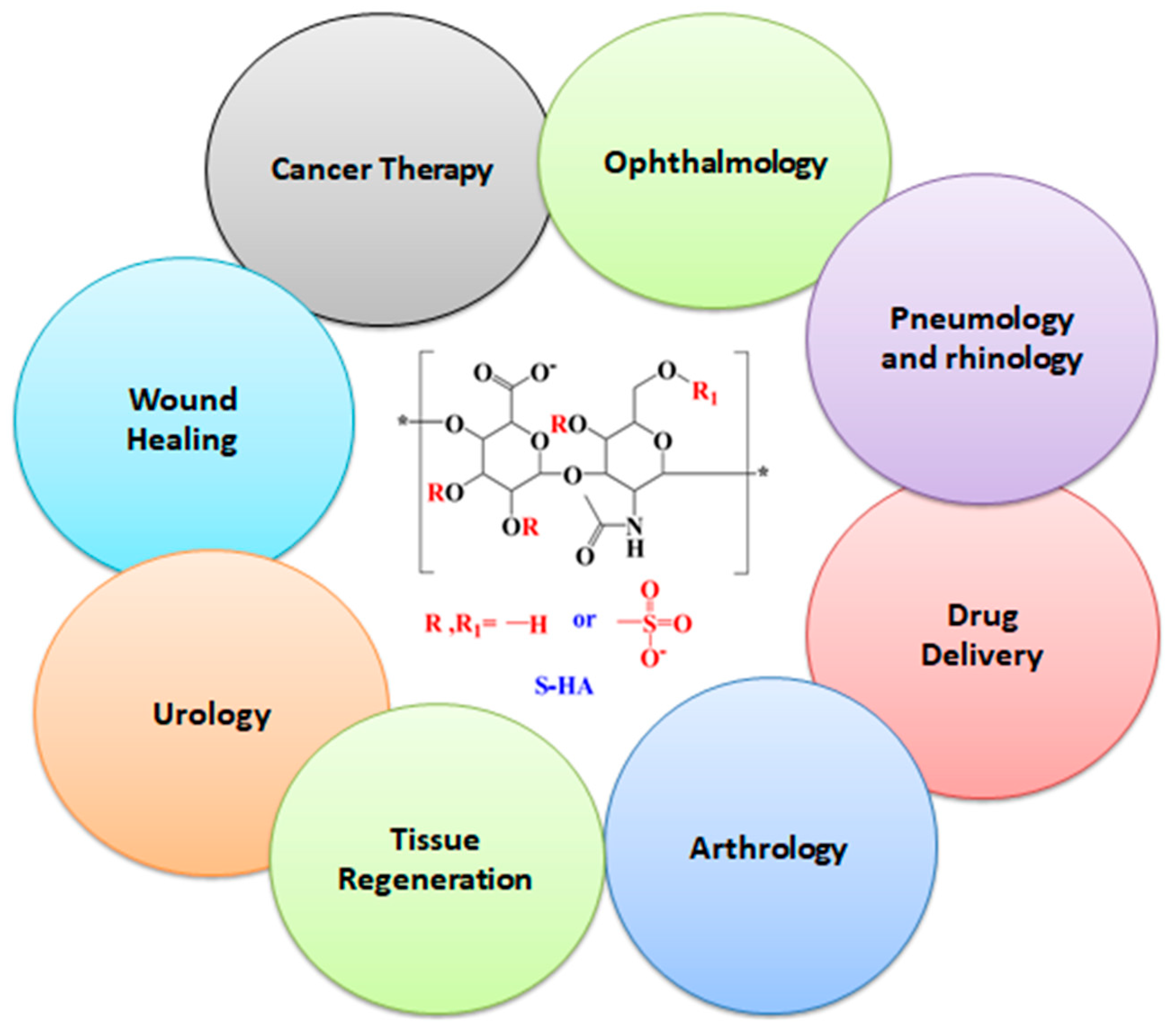
S-HA is a more established form of hyaluronic acid that is designed to last longer in the body, providing more sustainable results [1]. It is usually injected into the skin using a fine needle, and the technique is relatively rapid and painless. The results are visible immediately, and there is little to no downtime after the procedure. On the basis of biocompatibility, biological action, physiochemical, and safety profile, S-HA has numerous applications such as arthrology, cancer therapy [55], wound healing, atherosclerosis, tissue regeneration, drug delivery, urology, ophthalmology, pneumology, arthrology, and rhinology [56] (Figure 4).

3.1. Drug Delivery

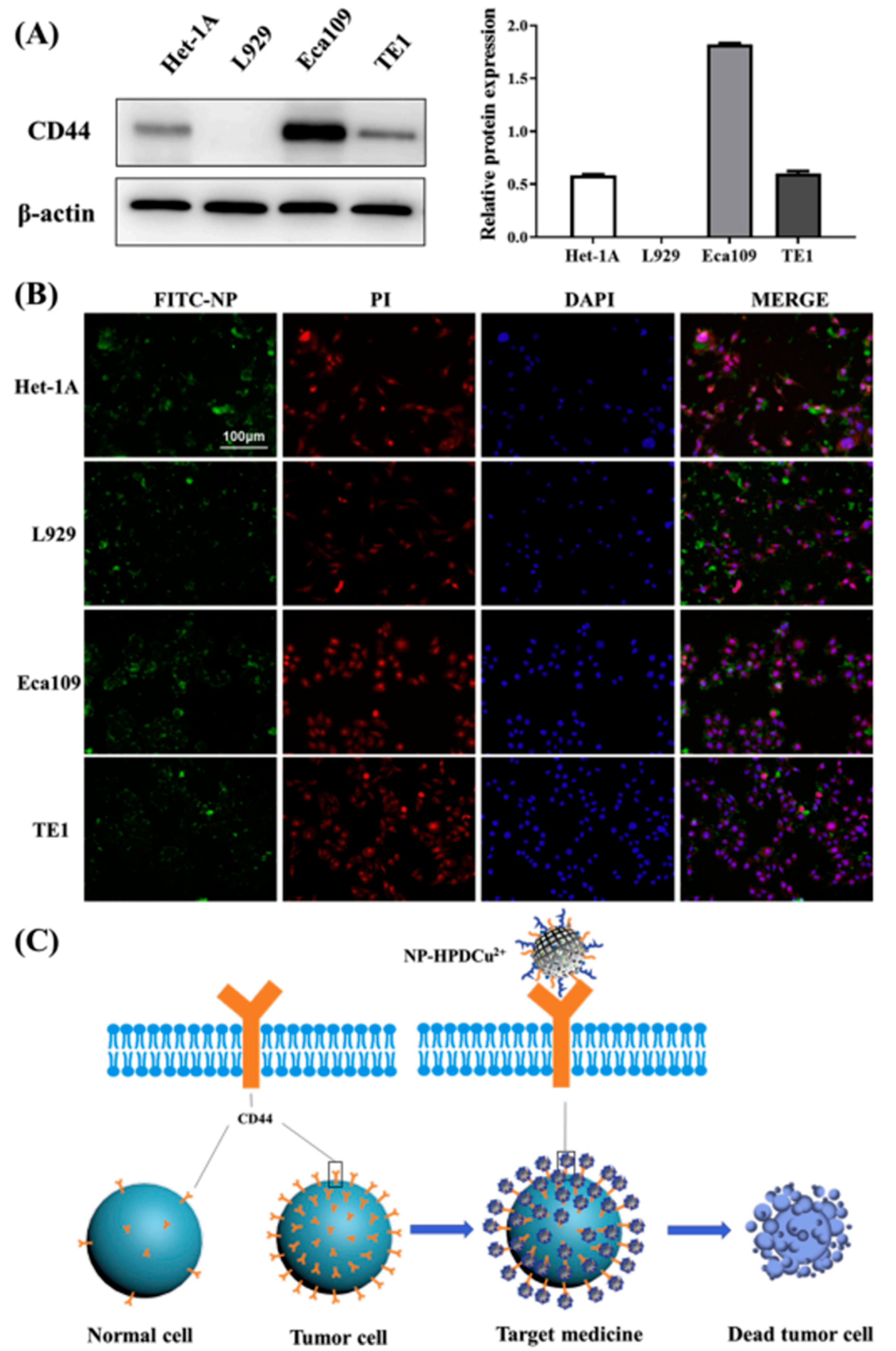
In a drug delivery system, S-HA has taken a growing amount of attention. It has very outstanding tendencies such as biodegradability [57], biocompatibility [58], extremely low toxicity [59], and non-immunogenicity. It has been extensively useful in nano-sized carriers. The surface modification of S-HA can help to improve its drug-loading capacity [60], stability, and controlled release properties, which are important factors for the effective delivery of drug to targeted tissues. To preserve the cellular structure and component of ECM, S-HA is used. Due to its negative charge, it can interact with protein in the ECM [61]. S-HA is used as a reducing and stabilizing agent in drug delivery systems [56]. It is used for prodrug development. For target drug delivery, S-HA and nanoparticles are prepared with other substances. The solubility of drug molecules can be improved by S-HA conjugates during targeted drug delivery in tumor or cancer cells. Due to outstanding hydrophilicity [62] and volume, it can be used to inhibit the unwanted interactions with plasma proteins and cells. Furthermore, due to its hydrophilicity and biocompatibility, it has been used in the progress of nano-carriers to improve target drug delivery for intravenous administration. HA hydrogel is the usually used format for effective drug delivery [63,64], while another usual application of HA for drug delivery is the nanoparticle carrier, which benefits the specific CD44 interaction and nano-effect (Figure 5) [65,66]. Sulfonation will continue to be retained or even strengthen these advantages of HA, and its biggest advantage is to inhibit the degradation effect of HAase on HA and improve molecular stability.

3.2. Tissue Engineering

Tissue engineering is a very outstanding biomedical application of S-HA. Engineering and biomedical methods are used to find novel biomaterials. These biomaterials are used to replace the injured organ and tissue or to enhance, repair, and maintain its damaged functions. For examples in nerve regeneration, conductive nerve scaffold was used to enhance nerve tissue engineering.
In bone tissue engineering, S-HA can also be used as scaffold materials [67]. The surface modification of S-HA can help improve its mechanical properties, biocompatibility, and bioactivity, which are the most important factors for the successful regeneration of bone tissue [68]. In bladder tissue engineering, collagen-derived hydrogels were used. Furthermore, the basis of bioactivity and biocompatibility of biomaterials can be decided for tissue engineering [69].
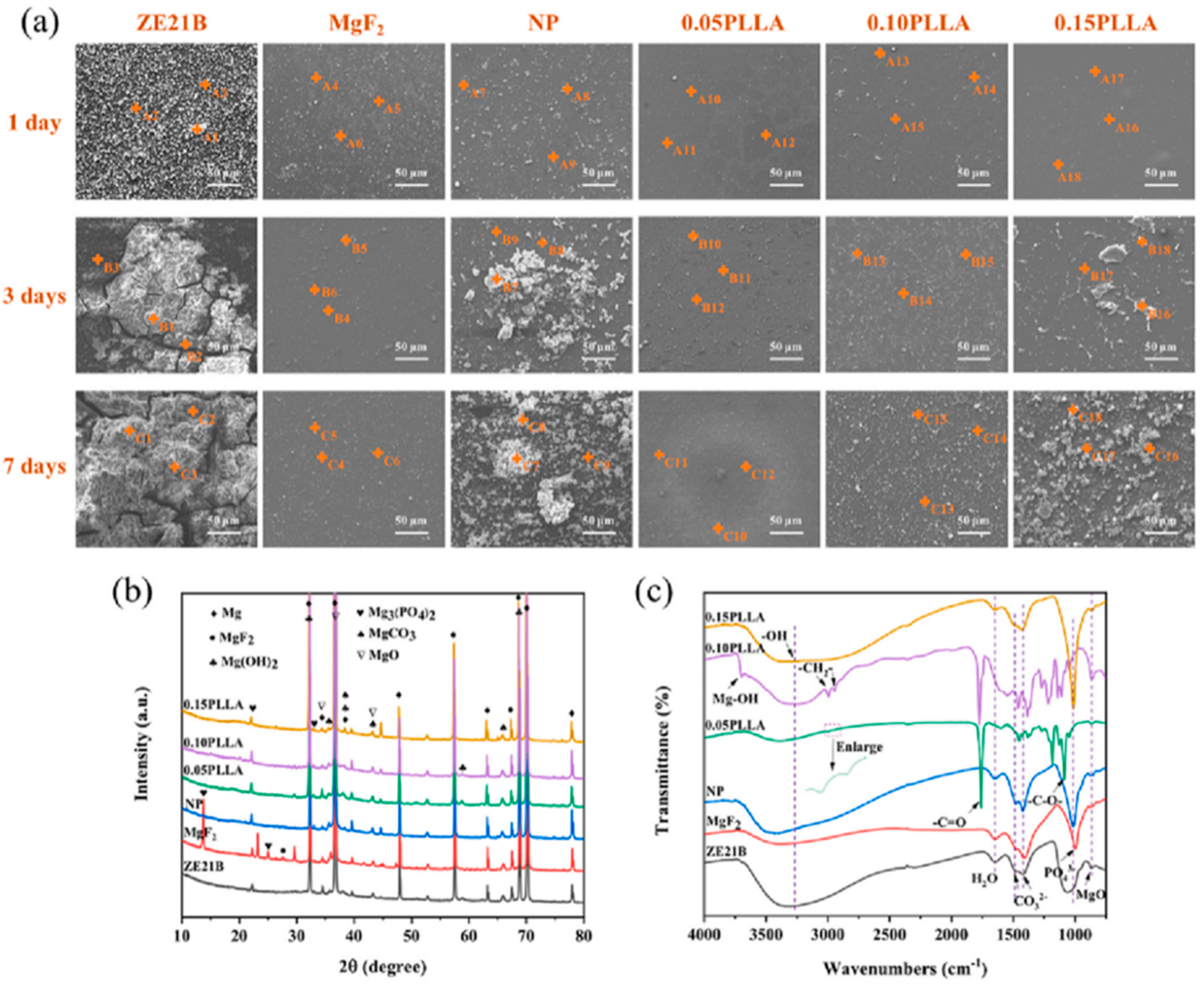
Surface modification of cardiovascular implants with S-HA is a feasible strategy to enhance the biocompatibility and stability compared with HA coatings [14]. Sulfonic groups or sulfur content plays an important role in enhancing coating functionality [70]. The use of sulfonated proteoglycan such as S-HA as a coating is mainly inspired by the design and research of chondroitin sulfate coating [71]. This sulfonated molecule endows stainless-steel vascular stent with more powerful anticoagulant, antiproliferative, anti-inflammatory, and pro-endothelialization promoting functions, thus making the vascular patency better [72]. On the basis of S-HA, corresponding nanoparticles can be prepared. S-HA nanoparticles have a smaller particle size and better zeta potential and dispersion coefficient range than HA nanoparticles, which proved that the prepared coatings have stronger functions [41]. Recently, we designed a composite coating with S-HA nanoparticles on the magnesium (Mg) alloy surface for cardiovascular application (Figure 6), and our data indicated that this composite coating significantly improved the corrosion resistance and biocompatibility of the Mg alloy (Figure 7) [73]. The composite coating even showed stronger pro-endothelialization and anti-hyperplasia functions compared with the rapamycin coating prepared by the same method, which suggests a better substitute of rapamycin by S-HA nanoparticles. Carbon quantum dots (CDs) have broad prospects in the field of biomedical diagnosis and treatment [74]. We combine CDs with S-HA to form nanoparticles, which not only endow cardiovascular implant coatings with good biocompatibility but also endow nanoparticles and their composite coatings with novel tracing properties due to the spontaneous fluorescence function of CDs, thereby obtaining diagnostic and therapeutic functions. We have applied for List of Chinese inventions patent for relevant parameters, and the specific method and results will be published in the near future.

3.3. Treatment for Osteoarthritis

Sulfonated hyaluronic acid (S-HA) is most beneficial for the treatment of osteoarthritis. Osteoarthritis is mainly cartilage degradation, inflammation of synovial membrane and joint tissues, and subchondral sclerosis. S-HA is used to prevent and slow down joint degradation [75]. It is used to reduce pain and increases joint mobility. S-HA has been investigated as a potential treatment for osteoarthritis [52], a degenerative joint disease characterized by the breakdown of cartilage and inflammation [76]. Studies have shown that S-HA can have several beneficial effects on osteoarthritic joints. For example, S-HA can reduce inflammation by inhibiting the production of inflammatory cytokines and enzymes. S-HA can also promote the regeneration of cartilage by stimulating the proliferation and differentiation of chondrocytes, the cells that produce cartilage. S-HA injections have been shown to reduce joint pain [77] and inflammation in people with osteoarthritis [78]. It works by cushioning the joints and providing lubrication, which can reduce friction and improve mobility.
One study investigated the use of S-HA as a treatment for knee osteoarthritis [79]. The study found that S-HA injections into the knee joint improved pain and function in patients with osteoarthritis, and that the effects were sustained for up to six months after treatment [4]. Overall, these studies suggest that S-HA has potential as a treatment for osteoarthritis by reducing inflammation and promoting cartilage regeneration. However, more research is needed to fully understand the mechanisms underlying these effects and to determine the optimal conditions for using S-HA in osteoarthritis therapies [80].
One important aspect noticed in the treatment of osteoarthritis is the anti-bacterial function. Thus, how to give consideration to both anti-bacterial and anti-inflammatory Dwifungsi is the key problem of S-HA in the treatment of osteoarthritis. Metal-organic frame structures (MOFs) are effective materials for anti-bacterial and anti-inflammation, even drug delivery. Therefore, combining MOFs and S-HA will endow the treatment of osteoarthritis with stronger anti-bacterial and anti-inflammatory functions. In addition, the S-HA hydrogels or nanoparticles can be prepared as the ink of bioprinting technology, which can also be applied for the precise treatment of osteoarthritis [81,82].

3.4. Treatment for Inflammatory Diseases

Sulfated hyaluronic acid (S-HA) plays an important role in various selection inhibitors that are important for inflammatory diseases. Many selection inhibitors are produced for human inflammatory disease treatment. For example, Efomycin M is proved as a new selection inhibitor, which reduces skin inflammation [83]. The therapeutic and protective effect of TBC-1269 showed a new blocker for a renal ischemia reperfusion injury model. In vitro S-HA exhibits L-selection and P-selection binding with interfaces. Moreover, in vivo S-HA enhanced the rat progressive mesangial proliferative glomerulonephritis, but these properties were not detected by HA [84]. That is why due to inhibitory effects on binding, S-HA was used for the treatment of inflammatory diseases. S-HA is a very beneficial material for the treatment of crescentic glomerulonephritis. Studies demonstrated that in renal tissues, leukocytes have infiltrated, and they create difficulty in the pathogenesis of different glomerulonephritis. An early event in the process of leukocyte infiltration is categorized by selection-mediated leukocyte rolling on the endothelial surface. By inhibiting P-selection-dependent leukocyte infiltration, S-HA exhibits therapeutic and preventive effects in glomeruli.
S-HA injections have also been used to treat rheumatoid arthritis. They can help reduce joint pain and inflammation [85] and improve joint function. It is reported that S-HA is used as potential treatment of inflammatory bowel disease (IBD). It may help to reduce inflammation in the gut and improve symptoms such as abdominal pain and diarrhea. S-HA has shown to reduce inflammation in the skin in people with psoriasis and anti-inflammatory effects. For the potential treatment of asthma patients, S-HA has proved to reduce inflammation and improve breathing.

3.5. Wound Healing

HA is a natural substance found in the body that plays an important role in tissue repair and wound healing [86]. S-HA is a stabilized form of HA that has been modified to enhance its durability and stability. In recent years, S-HA has been used as a wound-healing agent due to its ability to enhance the body’s natural healing process. S-HA can help to reduce inflammation, stimulate collagen production, and promote cell proliferation; all of these factors are important in wound healing [72]. S-HA can be used to treat a variety of wounds [87], including burns [8], surgical incisions, and chronic ulcers [88]. It can be applied as dressing, directly injected into the wound, and it can be topically administered. Previous studies showed that S-HA is used in the healing process and enhances the overall quality of wound healing. It showed the reduction in risk of scarring and infection.
Overall, S-HA is a promising wound-healing agent that has the potential to improve outcomes for patients with a variety of wounds. However, further research is needed to fully understand its effectiveness and optimal use in wound healing.

4. Summary and Perspective

Compared with hyaluronic acid (HA), sulfonated hyaluronic acid (S-HA) presents stronger stability for hyaluronidase, which proved that S-HA will show better in vivo performance as biomedical materials and coating materials. Obtaining benefit from the sulfonic acid group, S-HA has better functions on anticoagulation, antihyperplasia, anti-inflammation, and pro-endothelialization. Even the S-HA nanoparticles may show smaller size and cytocompatibility than HA nanoparticles in equivalent preparation conditions. S-HA hydrogels also presented better drug-controlled delivery compared with HA due to higher stability. S-HA can be combined with more new materials such as CDs, MOFs, etc. to achieve more compatibility functions. It can also be applied as materials of novel technologies, such as the ink of the bioprinting for wider application of biomaterials and coatings. In the future, S-HA will play more important roles in the biomaterial fields as nanoparticles, hydrogels, coatings, and other composite formats.

Author Contributions

Conceptualization, J.-A.L. and K.Z.; writing—original draft preparation, M.I., A.Y., and A.A.; writing—review and editing, J.-A.L. and K.Z.; supervision, J.-A.L.; project administration, J.-A.L.; funding acquisition, J.-A.L. and K.Z. All authors have read and agreed to the published version of the manuscript.

Funding

This research was funded by the National Natural Science Foundation of China, grant number U2004164; the Key Scientific and Technological Research Projects in Henan Province, grant numbers 232102311155 and 232102230106; and the Natural Science Foundation of the Jiangsu Higher Education Institutions of China, grant number 21KJB430013.

Institutional Review Board Statement

Not applicable.

Informed Consent Statement

Not applicable.

Data Availability Statement

Not applicable.

Acknowledgments

Figure 5 reproduced from Ref. [66] with permission from Elsevier B.V. (License Number: 5592850568872); Figure 6 and Figure 7 reproduced from Ref. [73] with permission from Elsevier B.V. (License Number: 5592940983813).

Conflicts of Interest

The authors declare no conflict of interest.

References

  1. Larrañeta, E.; Henry, M.; Irwin, N.J.; Trotter, J.; Perminova, A.A.; Donnelly, R.F. Synthesis and characterization of hyaluronic acid hydrogels crosslinked using a solvent-free process for potential biomedical applications. Carbohydr. Polym. 2018, 181, 1194–1205. [Google Scholar] [CrossRef] [PubMed] [Green Version]
  2. Angello, J.C.; Hauschka, S.D. Hyaluronic acid synthesis and turnover by myotubes in culture. Dev. Biol. 1979, 73, 322–337. [Google Scholar] [CrossRef] [PubMed]
  3. Feng, Q.; Lin, S.; Zhang, K.; Dong, C.; Wu, T.; Huang, H.; Bian, L. Sulfated hyaluronic acid hydrogels with retarded degradation and enhanced growth factor retention promote hMSC chondrogenesis and articular cartilage integrity with reduced hypertrophy. Acta Biomater. 2017, 53, 329–342. [Google Scholar] [CrossRef] [PubMed]
  4. Schuurmans, C.C.L.; Mihajlovic, M.; Hiemstra, C.; Ito, K.; Hennink, W.E.; Vermonden, T. Hyaluronic acid and chondroitin sulfate (meth)acrylate-based hydrogels for tissue engineering: Synthesis, characteristics and pre-clinical evaluation. Biomaterials 2021, 268, 120602. [Google Scholar] [CrossRef]
  5. Della Sala, F.; Fabozzi, A.; di Gennaro, M.; Nuzzo, S.; Makvandi, P.; Solimando, N.; Borzacchiello, A. Advances in Hyaluronic-Acid-Based (Nano)Devices for Cancer Therapy. Macromol. Biosci. 2022, 22, 2100304. [Google Scholar] [CrossRef]
  6. Ogawa, D.; Shikata, K.; Matsuda, M.; Akima, K.; Iwahashi, M.; Okada, S.; Makino, H. Sulfated hyaluronic acid, a potential selectin inhibitor, ameliorates experimentally induced crescentic glomerulonephritis. Nephron Exp. Nephrol. 2005, 99, 26–32. [Google Scholar] [CrossRef]
  7. Li, J.; Wang, S.; Sheng, Y.; Liu, C.; Xue, Z.; Tong, P.; Guan, S. Designing HA/PEI nanoparticle composite coating on biodegradable Mg–Zn–Y-Nd alloy to direct cardiovascular cells fate. Smart Mater. Med. 2021, 2, 124–133. [Google Scholar] [CrossRef]
  8. Magnani, A.; Lamponi, S.; Consumi, M.; Barbucci, R. Biological performance of two materials based on sulfated hyaluronic acid and polyurethane. J. Mater. Chem. 1999, 9, 2393–2398. [Google Scholar] [CrossRef]
  9. Yu, Y.; Zhu, S.J.; Dong, H.T.; Zhang, X.Q.; Li, J.; Guan, S.K. A novel MgF2/PDA/S-HA coating on the bio-degradable ZE21B alloy for better multi-functions on cardiovascular application. J. Magnes. Alloy. 2023, 11, 480–492. [Google Scholar] [CrossRef]
  10. Bhattacharya, D.S.; Svechkarev, D.; Bapat, A.; Patil, P.; Hollingsworth, M.A.; Mohs, A.M. Sulfation Modulates the Targeting Properties of Hyaluronic Acid to P-Selectin and CD44. ACS Biomater. Sci. Eng. 2020, 6, 3585–3598. [Google Scholar] [CrossRef]
  11. Benitez, A.; Yates, T.J.; Lopez, L.E.; Cerwinka, W.H.; Bakkar, A.; Lokeshwar, V.B. Targeting hyaluronidase for cancer therapy: Antitumor activity of sulfated hyaluronic acid in prostate cancer cells. Cancer Res. 2011, 71, 4085–4095. [Google Scholar] [CrossRef] [Green Version]
  12. Buhren, B.A.; Schrumpf, H.; Bölke, E.; Kammers, K.; Gerber, P.A. Standardized in vitro analysis of the degradability of hyaluronic acid fillers by hyaluronidase. Eur. J. Med. Res. 2018, 23, 37. [Google Scholar] [CrossRef] [PubMed]
  13. Sturabotti, E.; Consalvi, S.; Tucciarone, L.; Macrì, E.; Di Lisio, V.; Francolini, I.; Martinelli, A. Synthesis of Novel Hyaluronic Acid Sulfonated Hydrogels Using Safe Reactants: A Chemical and Biological Characterization. Gels 2022, 8, 480. [Google Scholar] [CrossRef] [PubMed]
  14. Yu, Y.; Zhu, S.; Hou, Y.; Li, J.; Guan, S. Sulfur Contents in Sulfonated Hyaluronic Acid Direct the Cardiovascular Cells Fate. ACS Appl. Mater. Interfaces 2020, 12, 46827–46836. [Google Scholar] [CrossRef]
  15. Miura, T.; Yuasa, N.; Ota, H.; Habu, M.; Kawano, M.; Nakayama, F.; Nishihara, S. Highly sulfated hyaluronic acid maintains human induced pluripotent stem cells under feeder-free and bFGF-free conditions. Biochem. Biophys. Res. Commun. 2019, 518, 506–512. [Google Scholar] [CrossRef]
  16. Feng, M.; Tang, B.; Liang, S.H.; Jiang, X. Sulfur Containing Scaffolds in Drugs: Synthesis and Application in Medicinal Chemistry. Curr Top. Med. Chem. 2016, 16, 1200–1216. [Google Scholar] [CrossRef] [Green Version]
  17. Olivito, F.; Amodio, N.; Di Gioia, M.L.; Nardi, M.; Oliverio, M.; Juli, G.; Tassone, P.; Procopio, A. Synthesis and preliminary evaluation of the anti-cancer activity on A549 lung cancer cells of a series of unsaturated disulfides. MedChemComm 2018, 10, 116–119. [Google Scholar] [CrossRef]
  18. Rzany, B.; Becker-Wegerich, P.; Bachmann, F.; Erdmann, R.; Wollina, U. Hyaluronidase in the correction of hyaluronic acid-based fillers: A review and a recommendation for use. J. Cosmet. Dermatol. 2009, 8, 317–323. [Google Scholar] [CrossRef] [PubMed] [Green Version]
  19. Stern, R.; Jedrzejas, M.J. Hyaluronidases: Their genomics, structures, and mechanisms of action. Chem. Rev. 2006, 106, 818–839. [Google Scholar] [CrossRef] [PubMed] [Green Version]
  20. Cen, L.; Neoh, K.G.; Li, Y.; Kang, E.T. Assessment of in vitro bioactivity of hyaluronic acid and sulfated hyaluronic acid functionalized electroactive polymer. Biomacromolecules 2004, 5, 2238–2246. [Google Scholar] [CrossRef]
  21. Jones, D.; Tezel, A.; Borrell, M. In vitro resistance to degradation of hyaluronic acid dermal fillers by ovine testicular hyaluronidase. Dermatol. Surg. 2010, 36, 804–809. [Google Scholar] [CrossRef]
  22. Hintze, V.; Miron, A.; Moeller, S.; Schnabelrauch, M.; Wiesmann, H.P.; Worch, H.; Scharnweber, D. Sulfated hyaluronan and chondroitin sulfate derivatives interact differently with human transforming growth factor-β1 (TGF-β1). Acta Biomater. 2012, 8, 2144–2152. [Google Scholar] [CrossRef] [PubMed]
  23. Cavallini, M.; Gazzola, R.; Metalla, M.; Vaienti, L. The role of hyaluronidase in the treatment of complications from hyaluronic acid dermal fillers. Aesthetic Surg. J. 2013, 33, 1167–1174. [Google Scholar] [CrossRef] [Green Version]
  24. Chung, C.; Beecham, M.; Mauck, R.L.; Burdick, J.A. The influence of degradation characteristics of hyaluronic acid hydrogels on in vitro neocartilage formation by mesenchymal stem cells. Biomaterials 2009, 30, 4287–4296. [Google Scholar] [CrossRef] [Green Version]
  25. Isoyama, T.; Thwaites, D.M.; Selzer, G.; Carey, R.I.; Barbucci, R.; Lokeshwar, V.B. Differential selectivity of hyaluronidase inhibitors toward acidic and basic hyaluronidases. Glycobiology 2006, 16, 11–21. [Google Scholar] [CrossRef] [PubMed] [Green Version]
  26. Schulz, M.C.; Korn, P.; Stadlinger, B.; Range, U.; Möller, S.; Becher, J.; Hintze, V. Coating with artificial matrices from collagen and sulfated hyaluronan influences the osseointegration of dental implants. J. Mater. Sci. Mater. Med. 2014, 25, 247–258. [Google Scholar] [CrossRef]
  27. Jordan, A.R.; Lokeshwar, S.D.; Lopez, L.E.; Hennig, M.; Chipollini, J.; Yates, T.; Lokeshwar, V.B. Antitumor activity of sulfated hyaluronic acid fragments in pre-clinical models of bladder cancer. Oncotarget 2017, 8, 24262–24274. [Google Scholar] [CrossRef] [Green Version]
  28. Kunze, R.; Rösler, M.; Möller, S.; Schnabelrauch, M.; Riemer, T.; Hempel, U.; Dieter, P. Sulfated hyaluronan derivatives reduce the proliferation rate of primary rat calvarial osteoblasts. Glycoconj. J. 2010, 27, 151–158. [Google Scholar] [CrossRef]
  29. Bayer, I.S. Hyaluronic acid and controlled release: A review. Molecules 2020, 25, 2649. [Google Scholar] [CrossRef]
  30. Kim, D.W.; Yoon, E.S.; Ji, Y.H.; Park, S.H.; Il Lee, B.; Dhong, E.S. Vascular complications of hyaluronic acid fillers and the role of hyaluronidase in management. J. Plast. Reconstr. Aesthetic Surg. 2011, 64, 1590–1595. [Google Scholar] [CrossRef]
  31. Li, J.; Qiao, M.; Ji, Y.; Lin, L.; Zhang, X.; Linhardt, R.J. Chemical, enzymatic and biological synthesis of hyaluronic acids. Int. J. Biol. Macromol. 2020, 152, 199–206. [Google Scholar] [CrossRef] [PubMed]
  32. Miura, T.; Kawano, M.; Takahashi, K.; Yuasa, N.; Habu, M.; Kimura, F.; Nakayama, F. High-Sulfated Hyaluronic Acid Ameliorates Radiation-Induced Intestinal Damage Without Blood Anticoagulation. Adv. Radiat. Oncol. 2022, 7, 100900. [Google Scholar] [CrossRef] [PubMed]
  33. McCarty, M.F.; Russell, A.L.; Seed, M.P. Sulfated glycosaminsglycan and glucosamine may synergize in promoting synovial hyaluronic acid synthesis. Med. Hypotheses 2000, 54, 798–802. [Google Scholar] [CrossRef]
  34. Lim, D.K.; Wylie, R.G.; Langer, R.; Kohane, D.S. Selective binding of C-6 OH sulfated hyaluronic acid to the angiogenic isoform of VEGF165. Biomaterials 2016, 77, 130–138. [Google Scholar] [CrossRef] [Green Version]
  35. Matsuda, M.; Shikata, K.; Shimizu, F.; Suzuki, Y.; Miyasaka, M.; Kawachi, H.; Makino, H. Therapeutic effect of sulphated hyaluronic acid, a potential selectin-blocking agent, on experimental progressive mesangial proliferative glomerulonephritis. J. Pathol. 2002, 198, 407–414. [Google Scholar] [CrossRef]
  36. Vilela, C.A.; Correia, C.; Oliveira, J.M.; Sousa, R.A.; Espregueira-Mendes, J.; Reis, R.L. Cartilage Repair Using Hydrogels: A Critical Review of in Vivo Experimental Designs. ACS Biomater. Sci. Eng. 2015, 1, 726–739. [Google Scholar] [CrossRef]
  37. Muzzarelli, R.A.; Greco, A.F.; Busilacchi, A.; Sollazzo, V.; Gigante, A. Chitosan, hyaluronan and chondroitin sulfate in tissue engineering for cartilage regeneration: A review. Carbohydr. Polym. 2012, 89, 723–739. [Google Scholar] [CrossRef]
  38. Henson, F.M.D.; Getgood, A.M.J.; Caborn, D.M.; Wayne McIlwraith, C.W.; Rushton, N. Effect of a solution of hyaluronic acid-chondroitin sulfate-N-acetyl glucosamine on the repair response of cartilage to single-impact load damage. Am. J. Vet. Res. 2012, 73, 306–312. [Google Scholar] [CrossRef] [PubMed]
  39. Subramaniam, S.; Fang, Y.H.; Sivasubramanian, S.; Lin, F.H.; Lin, C.P. Hydroxyapatite-calcium sulfate-hyaluronic acid composite encapsulated with collagenase as bone substitute for alveolar bone regeneration. Biomaterials 2016, 74, 99–108. [Google Scholar] [CrossRef]
  40. Hou, Y.; Zhang, X.; Li, J.; Wang, L.; Guan, S. A multi-functional MgF2/polydopamine/hyaluronan-astaxanthin coating on the biodegradable ZE21B alloy with better corrosion resistance and biocompatibility for cardiovascular application. J. Magnes. Alloy. 2022, in press. [CrossRef]
  41. Xue, Z.; Sun, X.; Li, H.; Iqbal, M.; Hou, Y.; Jin, Z.; Li, J. Response of cardiovascular environment to sulfonated hyaluronic acid with higher sulfur content. Colloids Surf. B Biointerfaces 2023, 22, 113046. [Google Scholar] [CrossRef] [PubMed]
  42. Möller, S.; Schmidtke, M.; Weiss, D.; Schiller, J.; Pawlik, K.; Wutzler, P.; Schnabelrauch, M. Synthesis and antiherpetic activity of carboxymethylated and sulfated hyaluronan derivatives. Carbohydr. Polym. 2012, 90, 608–615. [Google Scholar] [CrossRef] [PubMed]
  43. Albanese, A.; Lamponi, S.; Barbucci, R. Blood-interaction performance of differently sulphated hyaluronic acids. Thromb. Res. 1996, 81, 383–395. [Google Scholar]
  44. Yeom, J.; Bhang, S.H.; Kim, B.S.; Seo, M.S.; Hwang, E.J.; Cho, I.H.; Hahn, S.K. Effect of cross-linking reagents for hyaluronic acid hydrogel dermal fillers on tissue augmentation and regeneration. Bioconjug. Chem. 2010, 21, 240–247. [Google Scholar] [CrossRef] [PubMed]
  45. Sall, I.; Férard, G. Comparison of the sensitivity of 11 crosslinked hyaluronic acid gels to bovine testis hyaluronidase. Polym. Degrad. Stab. 2007, 92, 915–919. [Google Scholar] [CrossRef]
  46. Payan, E.; Jouzeau, J.Y.; Lapicque, F.; Muller, N.; Netter, P. Hyaluronidase degradation of hyaluronic acid from different sources: Influence of the hydrolysis conditions on the production and the relative proportions of tetra- and hexasaccharide produced. Int. J. Biochem. 1993, 25, 325–329. [Google Scholar] [CrossRef]
  47. Yao, W.; Chen, M.; Dou, X.; Jin, H.; Zhang, X.; Zhu, Y.; Li, Z. Unravel a neuroactive sHA sulfation pattern with neurogenesis activity by a library of defined oligosaccharides. Eur. J. Med. Chem. 2019, 163, 583–596. [Google Scholar] [CrossRef]
  48. Vogel, S.; Ullm, F.; Müller, C.D.; Pompe, T.; Hempel, U. Impact of binding mode of low-sulfated hyaluronan to 3D collagen matrices on its osteoinductive effect for human bone marrow stromal cells. Biol. Chem. 2021, 402, 1465–1478. [Google Scholar] [CrossRef]
  49. Zerbinati, N.; Mocchi, R.; Galadari, H.; Maccario, C.; Maggi, M.; Rauso, R.; Sommatis, S. In vitro evaluation of the biological availability of hyaluronic acid polyethylene glycols-cross-linked hydrogels to bovine testes hyaluronidase. Biomed Res. Int. 2019, 2019, 3196723. [Google Scholar] [CrossRef] [Green Version]
  50. Felz, S.; Neu, T.R.; van Loosdrecht, M.C.M.; Lin, Y. Aerobic granular sludge contains Hyaluronic acid-like and sulfated glycosaminoglycans-like polymers. Water Res. 2020, 169, 115291. [Google Scholar] [CrossRef]
  51. Hamilton, D.W.; Riehle, M.O.; Rappuoli, R.; Monaghan, W.; Barbucci, R.; Curtis, A.S.G. The response of primary articular chondrocytes to micrometric surface topography and sulphated hyaluronic acid-based matrices. Cell Biol. Int. 2005, 29, 605–615. [Google Scholar] [CrossRef] [PubMed]
  52. Magnani, A.; Lamponi, S.; Rappuoli, R.; Barbucci, R. Sulphated Hyaluronic Acids: A Chemical and Biological Characterisation. Polym. Int. 1998, 46, 225–240. [Google Scholar] [CrossRef]
  53. Koehler, L.; Ruiz-Gómez, G.; Balamurugan, K.; Rother, S.; Freyse, J.; Möller, S.; Hintze, V. Dual Action of Sulfated Hyaluronan on Angiogenic Processes in Relation to Vascular Endothelial Growth Factor-A. Sci. Rep. 2019, 9, 18143. [Google Scholar] [CrossRef] [PubMed] [Green Version]
  54. Satish, L.; Santra, S.; Tsurkan, M.V.; Werner, C.; Jana, M.; Sahoo, H. Conformational changes of GDNF-derived peptide induced by heparin, heparan sulfate, and sulfated hyaluronic acid—Analysis by circular dichroism spectroscopy and molecular dynamics simulation. Int. J. Biol. Macromol. 2021, 82, 2144–2150. [Google Scholar] [CrossRef] [PubMed]
  55. Burdick, J.A.; Prestwich, G.D. Hyaluronic acid hydrogels for biomedical applications. Adv. Mater. 2011, 23, 41–56. [Google Scholar] [CrossRef]
  56. Yasin, A.; Ren, Y.; Li, J.; Sheng, Y.; Cao, C.; Zhang, K. Advances in Hyaluronic Acid for Biomedical Applications. Front. Bioeng. Biotechnol. 2022, 10, 910290. [Google Scholar] [CrossRef]
  57. Manfredi, C.; Spirito, L.; Calace, F.P.; Balsamo, R.; Terribile, M.; Stizzo, M.; Arcaniolo, D. Oral Preparation of Hyaluronic Acid, Chondroitin Sulfate, Curcumin, and Quercetin (Ialuril® Soft Gels) for the Prevention of LUTS after Intravesical Chemotherapy. Pathophysiology 2022, 29, 365–373. [Google Scholar] [CrossRef]
  58. Kurczewska, J. Recent Reports on Polysaccharide-Based Materials for Drug Delivery. Polymers 2022, 14, 4189. [Google Scholar] [CrossRef]
  59. Schuiringa, G.H.; Mihajlovic, M.; van Donkelaar, C.C.; Vermonden, T.; Ito, K. Creating a Functional Biomimetic Cartilage Implant Using Hydrogels Based on Methacrylated Chondroitin Sulfate and Hyaluronic Acid. Gels 2022, 8, 7. [Google Scholar] [CrossRef]
  60. Vázquez, I.; Rodríguez-Amado, J.A.; Montemayor, M.I.; Fraguas, J.; Del González, M.P.; Murado, M.A. Chondroitin sulfate, hyaluronic acid and chitin/chitosan production using marine waste sources: Characteristics, applications and eco-friendly processes: A review. Mar. Drugs 2013, 11, 747–774. [Google Scholar] [CrossRef] [Green Version]
  61. Nagira, T.; Nagahata-Ishiguro, M.; Tsuchiya, T. Effects of sulfated hyaluronan on keratinocyte differentiation and Wnt and Notch gene expression. Biomaterials 2007, 28, 844–850. [Google Scholar] [CrossRef]
  62. Amhare, A.F.; Lei, J.; Deng, H.; Lv, Y.; Han, J.; Zhang, L. Biomedical application of chondroitin sulfate with nanoparticles in drug delivery systems: Systematic review. J. Drug Target. 2021, 29, 259–268. [Google Scholar] [CrossRef]
  63. Cui, L.; Li, J.K.; Guan, S.; Zhang, K.X.; Zhang, K.; Li, J.A. Injectable Multifunctional CMC/HA-DA Hydrogel for Repairing Skin Injury. Mater. Today Bio 2022, 14, 100257. [Google Scholar]
  64. Wei, S.; Li, J.; He, H.; Shu, C.; Dardik, A.; Bai, H. A three-layered hydrogel patch with hierarchy releasing of PLGA nanoparticle drugs decrease neointimal hyperplasia. Smart Mater. Med. 2022, 3, 139–147. [Google Scholar] [CrossRef]
  65. Lei, C.; Liu, X.R.; Chen, Q.B.; Li, Y.; Zhou, J.L.; Zhou, L.Y.; Zou, T. Hyaluronic acid and albumin based nanoparticles for drug delivery. J. Control. Release 2021, 331, 416–433. [Google Scholar] [CrossRef]
  66. Xu, R.; Zhang, K.; Liang, J.; Gao, F.; Li, J.; Guan, F. Hyaluronic acid/polyethyleneimine nanoparticles loaded with copper ion and disulfiram for esophageal cancer. Carbohydr. Polym. 2021, 261, 117846. [Google Scholar]
  67. Rother, S.; Galiazzo, V.D.; Kilian, D.; Fiebig, K.M.; Becher, J.; Moeller, S.; Hintze, V. Hyaluronan/Collagen Hydrogels with Sulfated Hyaluronan for Improved Repair of Vascularized Tissue Tune the Binding of Proteins and Promote Endothelial Cell Growth. Macromol. Biosci. 2017, 17, 1700154. [Google Scholar]
  68. Schmidt, J.R.; Vogel, S.; Moeller, S.; Kalkhof, S.; Schubert, K.; von Bergen, M.; Hempel, U. Sulfated hyaluronic acid and dexamethasone possess a synergistic potential in the differentiation of osteoblasts from human bone marrow stromal cells. J. Cell. Biochem. 2019, 120, 8706–8722. [Google Scholar]
  69. Vogel, S.; Arnoldini, S.; Möller, S.; Schnabelrauch, M.; Hempel, U. Sulfated hyaluronan alters fibronectin matrix assembly and promotes osteogenic differentiation of human bone marrow stromal cells. Sci. Rep. 2016, 6, 36418. [Google Scholar]
  70. Yao, S.; Cui, J.; Chen, S.; Zhou, X.; Li, J.; Zhang, K. Extracellular matrix coatings on cardiovascular materials—A review. Coatings 2022, 12, 1039. [Google Scholar]
  71. Zou, D.; Li, J.; Kou, F.; Luo, X.; Yang, P. Reveal crucial subtype of natural chondroitin sulfate on the functionalized coatings for cardiovascular implants. J. Mater. Sci. Technol. 2021, 91, 67–77. [Google Scholar] [CrossRef]
  72. Li, J.; Li, W.; Zou, D.; Kou, F.; Hou, Y.; Yasin, A.; Zhang, K. Comparison of conjugating chondroitin sulfate A and B on amine-rich surface: For deeper understanding on directing cardiovascular cells fate. Compos. Part B Eng. 2022, 228, 109430. [Google Scholar] [CrossRef]
  73. Tong, P.; Chen, L.; Sun, X.; Li, H.; Feng, Y.; Li, J.; Guan, S. Surface modification of biodegradable magnesium alloy with Poly (L-lactic acid) and Sulfonated hyaluronic acid nanoparticles for cardiovascular application. Int. J. Biol. Macromol. 2023, 237, 124191. [Google Scholar] [CrossRef]
  74. Chai, Y.; Feng, Y.; Zhang, K.; Li, J. Preparation of fluorescent carbon dots composites and their potential applications in biomedicine and drug delivery—A review. Pharmaceutics 2022, 14, 2482. [Google Scholar] [CrossRef]
  75. Cohen, M.M.; Altman, R.D.; Hollstrom, R.; Hollstrom, C.; Sun, C.; Gipson, B. Safety and efficacy of intra-articular sodium hyaluronate (Hyalgan®) in a randomized, double-blind study for osteoarthritis of the ankle. Foot Ankle Int. 2008, 29, 657–663. [Google Scholar] [CrossRef]
  76. Chen, G.; Ito, Y.; Imanishi, Y.; Magnani, A.; Lamponi, S.; Barbucci, R. Photoimmobilization of sulfated hyaluronic acid for antithrombogenicity. Bioconjug. Chem. 1997, 8, 730–734. [Google Scholar] [CrossRef]
  77. Shankland, W.E. The effects of glucosamine and chondroitin sulfate on osteoarthritis of the TMJ: A preliminary report of 50 patients. Cranio—J. Craniomandib. Sleep Pract. 1998, 16, 230–235. [Google Scholar] [CrossRef]
  78. Hempel, U.; Möller, S.; Noack, C.; Hintze, V.; Scharnweber, D.; Schnabelrauch, M.; Dieter, P. Sulfated hyaluronan/collagen I matrices enhance the osteogenic differentiation of human mesenchymal stromal cells in vitro even in the absence of dexamethasone. Acta Biomater. 2012, 8, 4064–4072. [Google Scholar] [CrossRef]
  79. Vangsness, C.T.; Spiker, W.; Erickson, J. A Review of Evidence-Based Medicine for Glucosamine and Chondroitin Sulfate Use in Knee Osteoarthritis. Arthrosc.—J. Arthrosc. Relat. Surg. 2009, 25, 86–94. [Google Scholar] [CrossRef]
  80. Zhang, Z.Q.; Yang, Y.X.; Li, J.A.; Zeng, R.C.; Guan, S.K. Advances in coatings on magnesium alloys for cardiovascular stents—A review. Bioact. Mater. 2021, 6, 4729–4757. [Google Scholar] [CrossRef]
  81. Hou, Y.C.; Cui, X.; Qin, Z.; Su, C.; Zhang, G.; Tang, J.N.; Li, J.A.; Zhang, J.Y. Three-dimensional bioprinting of artificial blood vessel: Process, bio-inks and challenges. Int. J. Bioprinting 2023, 9, 740. [Google Scholar]
  82. Xie, M.; Su, J.; Zhou, S.; Li, J.; Zhang, K. Application of Hydrogels as Three-dimensional Bioprinting ink for Tissue engineering. Gels 2023, 9, 88. [Google Scholar] [CrossRef] [PubMed]
  83. Abi Zeid Daou, C.; Bassim, M. Hyaluronic acid in otology: Its uses, advantages and drawbacks—A review. Am. J. Otolaryngol.—Head Neck Med. Surg. 2020, 41, 102375. [Google Scholar] [CrossRef] [PubMed]
  84. Hong, C.I.; Jung, E.G.; Han, K.I.; Kim, Y.H.; Lee, S.H.; Lee, H.S.; Han, M.D. Structural Characteristics and Anti-inflammatory Activities of Chemically Sulfated-hyaluronic Acid from Streptococcus dysgalactiae. J. Life Sci. 2016, 26, 545–554. [Google Scholar] [CrossRef] [Green Version]
  85. Lebaudy, E.; Fournel, S.; Lavalle, P.; Vrana, N.E.; Gribova, V. Recent Advances in Antiinflammatory Material Design. Adv. Healthc. Mater. 2021, 10, 2001373. [Google Scholar] [CrossRef]
  86. Graça, M.F.P.; Miguel, S.P.; Cabral, C.S.D.; Correia, I.J. Hyaluronic acid—Based wound dressings: A review. Carbohydr. Polym. 2020, 241, 116364. [Google Scholar] [CrossRef]
  87. Nishikawa, H.; Mori, I.; Umemoto, J. Influences of sulfated glycosaminoglycans on biosynthesis of hyaluronic acid in rabbit knee synovial membrane. Arch. Biochem. Biophys. 1985, 240, 146–153. [Google Scholar] [CrossRef]
  88. Oe, M.; Tashiro, T.; Yoshida, H.; Nishiyama, H.; Masuda, Y.; Maruyama, K.; Fukui, N. Oral hyaluronan relieves knee pain: A review. Nutr. J. 2016, 15, 11. [Google Scholar] [CrossRef] [Green Version]
Figure 1. The scheme of sulfation methods, functional advantages, and action pathways of S-HA.
Coatings 13 01345 g001
Figure 2. Sodium salt of S-HA.
Coatings 13 01345 g002
Figure 3. Selective binding characteristics of S-HA.
Coatings 13 01345 g003
Figure 4. Applications of sulfonated hyaluronic acid (S-HA).
Coatings 13 01345 g004
Figure 5. (A) Western blot of CD44 expressed on the Het-1A, L929, Eca109, and TE1 (mean ± SD, n = 3). (B) Fluorescence images of Het-1A, L929, Eca109, and TE1 stained with FITC labeled HA nanoparticles with drugs (FITC-NP, green color), PI (apoptosis marker, red color), and DAPI (nucleus marker, blue color). (C) Mechanism diagram of targeted killing tumor cells by HA nanoparticles with drugs [66].
Coatings 13 01345 g005
Figure 6. Scheme of preparing PLLA/NP@S-HA coating (S-HA nanoparticle composite coating) on the ZE21B (Mg alloy) surface [73].
Coatings 13 01345 g006
Figure 7. (a) SEM images of all samples after soaking in Hanks’ solution for 1 day, 3 days, and 7 days; (b) XRD spectrum; (c) FTIR spectrum of samples after soaking in Hanks’ solution for 200 h [73].
Coatings 13 01345 g007
Disclaimer/Publisher’s Note: The statements, opinions and data contained in all publications are solely those of the individual author(s) and contributor(s) and not of MDPI and/or the editor(s). MDPI and/or the editor(s) disclaim responsibility for any injury to people or property resulting from any ideas, methods, instructions or products referred to in the content.