Article

Evaluation of Shielding Performance of Gamma Ray Shielding Tungsten Polymer Composite with LBL-Type Layered Structure

1
Department of Medical Informatics, Keimyung University, 1095 Dalgubeol-daero, Daegu 42601, Republic of Korea
2
Department of Biomedical Engineering, Keimyung University, 1095 Dalgubeol-daero, Daegu 42601, Republic of Korea
Coatings 2024, 14(1), 36; https://doi.org/10.3390/coatings14010036
Submission received: 23 November 2023 / Revised: 26 December 2023 / Accepted: 26 December 2023 / Published: 27 December 2023

Abstract

:
Lead has conventionally been the primary material for shielding radioactive isotopes in medical contexts. In response to environmental concerns, our study proposes an eco-friendly alternative—a gamma ray shielding material utilizing tungsten. Unlike prior research, in our study, the shielding performance through a laminated structure is evaluated, employing a randomly stacked arrangement of tungsten particles. The shielding product was developed by electrospinning a tungsten and polyurethane polymer mixture, with precise control over the radiation speed and time. The irregular stacking of tungsten particles is expected to reduce incident radiation intensity through scattering and absorption. Radiation shielding experiments on isotopes (99mTc, 18F, and 131I) compared our material to standard lead at varying distances. For 99mTc, at a 0.1 m distance, our 1.0 mm thick material exhibits a shielding performance of 67.54%, surpassing that of a 0.25 mm lead plate (58.95%) and matching that of a 0.50 mm plate (69.24%). These findings demonstrate the promising potential of our tungsten-based material in nuclear medicine, proving its efficacy as a shield for radioactive isotopes. Our research introduces an eco-friendly alternative to lead-based shielding in medical settings, showcasing the effectiveness of our tungsten-based material in reducing incident radiation intensity. The demonstrated outcomes position it as a viable option for enhancing safety in nuclear medicine applications.

1. Introduction

Radiopharmaceuticals administered in medical settings play a crucial role in diagnosing and treating diseases directly within a patient’s body [1]. The radiation emitted by radioisotopes used in nuclear medicine, comparable to X-rays for medical imaging, penetrates the human body to generate diagnostic images [2,3]. Consequently, both patients and healthcare providers are exposed to radiation during examinations and treatments, necessitating precautions to minimize non-essential radiation exposure that may be detrimental to medical practice [4].
Traditionally, gamma rays generated by radioactive isotopes were shielded using lead, following the same principles as X-ray defense. The effectiveness of shielding is typically regulated by the thickness, employing a standard lead-equivalent amount [5]. The prevalent use of lead in gamma ray shielding products within existing medical institutions’ nuclear medicine departments is attributed to the uncorrected distance to the nuclide within the gamma ray usage range, coupled with the significant impacts from scattering rays [6].
Shielding at specific distances during the process of injecting radiopharmaceuticals into the human body is a particularly challenging issue, which exposes medical personnel and patients to considerable radiation. Recognizing this, the International Commission on Radiological Protection (ICRP) recommended the adoption of syringe shields to reduce radiation exposure to the hands of medical staff when administering radionuclides in nuclear medicine [7,8].
While lead has historically dominated gamma ray shielding products, a contemporary shift toward eco-friendly alternatives such as tungsten, bismuth, and tin can be observed. However, considering factors such as product processability, thickness, and weight, this transition must be executed without impeding medical practices [9]. For gamma ray shields, whose shielding performance can be adjusted based on their thickness, exploring process technologies to reduce the thickness while maintaining equivalent shielding performance to lead is imperative for enhancing user comfort.
Generally, to diminish the intensity of incident radiation, it is essential to increase the thickness of shielding products [10]. As the path length of radiation expands, the probability of interactions between scattering and absorption increases, resulting in a difference between the incident and transmitted intensities [11]. Ultimately, the composition and density of the material within the shield influences the efficacy of radiation shielding products [12,13]. Additionally, the atomic crystal structure affects the cross-sectional area of particle collisions [14]. Therefore, emphasis should be placed on density controlled by thickness to improve the shielding performance. Furthermore, achieving increased density necessitates the careful consideration of the arrangement and structure of the particles constituting the shielding material.
In this study, we focus on the layer-by-layer (LBL) laminated structure and explored an economical process technology [15]. With an aim to implement this LBL technology, we propose a nanofiber-laminated-coating form through electrospinning. The electrospinning process allows for stacking effects through repeated random scanning [16]. Therefore, multilayer technology, involving layers completed in the same pattern, is anticipated to effectively respond to irregular incident radiation shielding, particularly if the metal particles to be shielded exhibit irregular and random shapes. The shielding material comprises a single piece of tungsten, and polyurethane is used to create the nanofibers. The thickness units of the shielding products are manufactured. Additionally, we seek to compare and verify the shielding performance with 0.25 mm and 0.50 mm lead equivalents, which are commonly used as fabrics for shielding suits in the nuclear medicine departments of medical institutions. The shielding performance is evaluated at distances of 30, 50, and 100 cm, considering the most frequently used nuclides in medical institutions (99mTc, 18F, and 131I) [17].
This study aims to contribute to minimizing unnecessary radiation exposure for medical staff and patients in medical institutions by developing diverse tools for radiation shielding. Crucially, our focus is on creating eco-friendly shielding materials and process technologies based on alternatives to heavy metals and harmful elements like lead. Here, we present a process technology for density control using tungsten, an eco-friendly shielding material, and evaluate the gamma ray shielding performance of prototypes manufactured through this process [18,19,20]. Through these efforts, various gamma ray shielding products are explored to foster the establishment of a secure medical environment that protects medical practitioners.

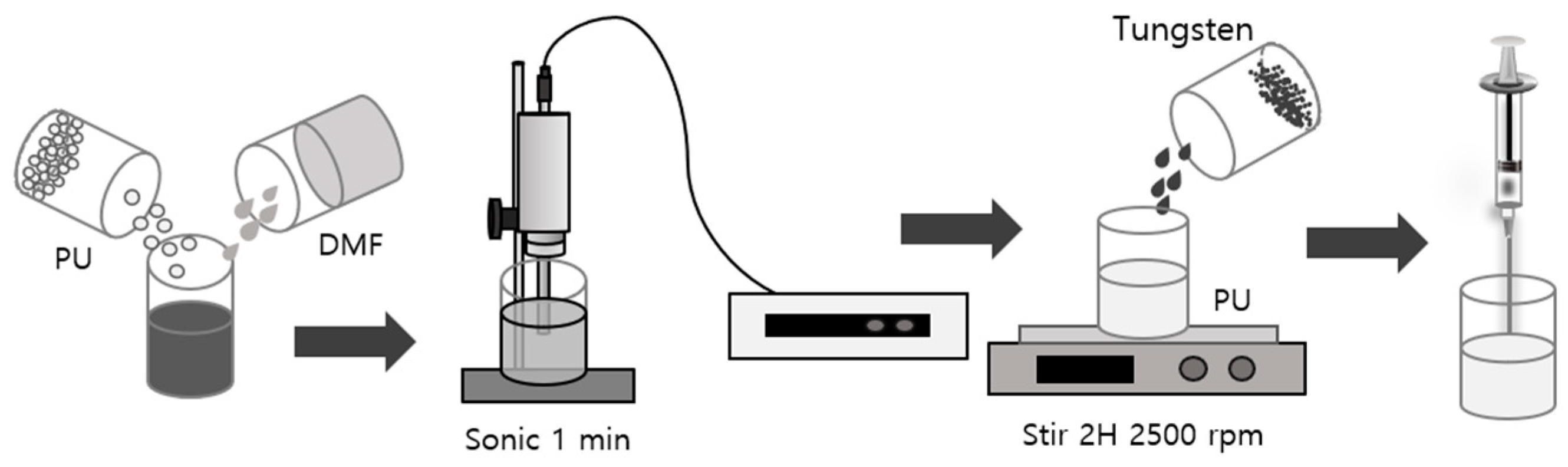
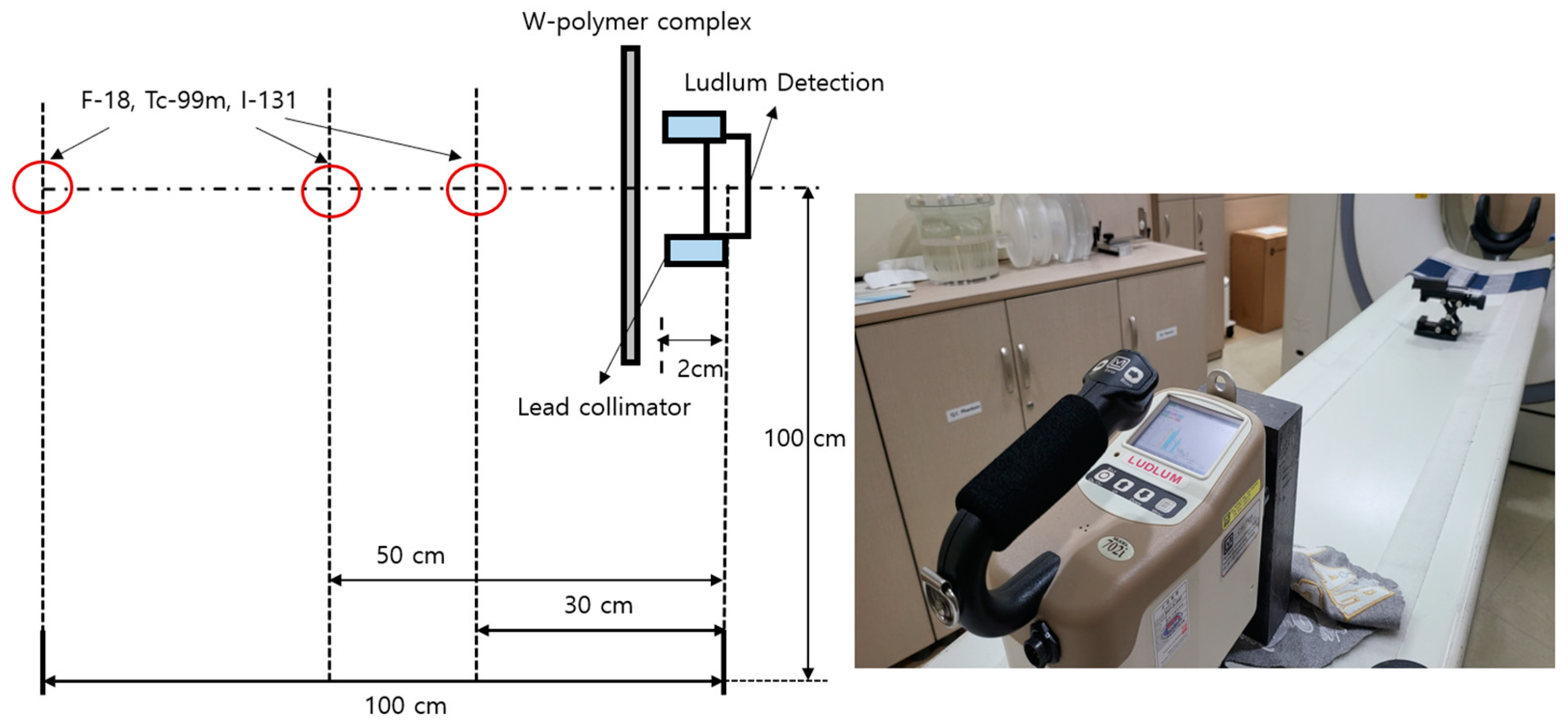
2. Materials and Methods
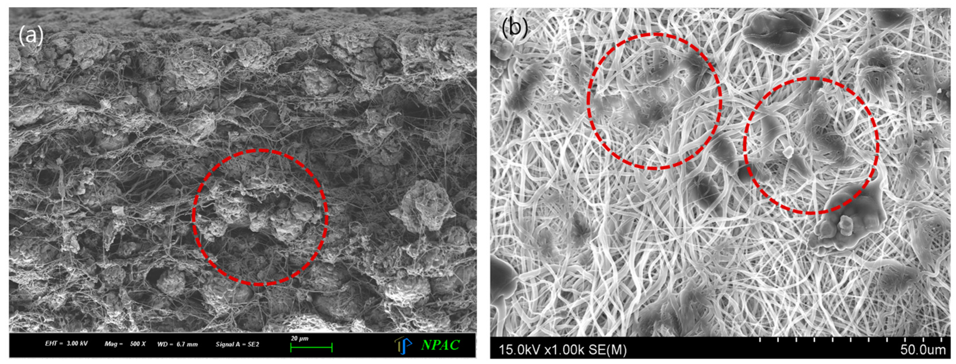
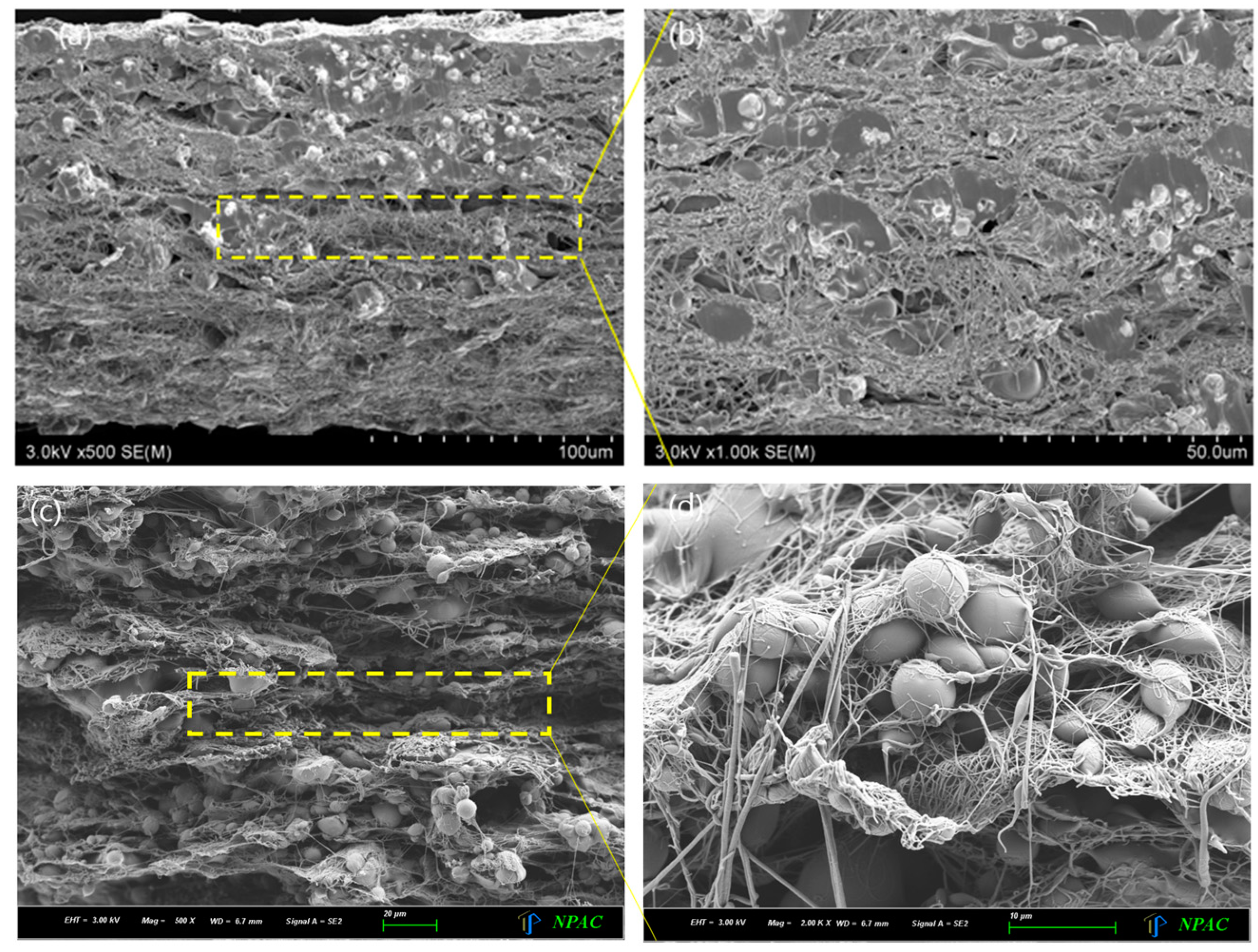
When gamma rays, originating from radioactive isotopes, traverse the sample thickness χ , the incident intensity undergoes attenuation in accordance with the Beer–Lambert law, as illustrated in Equation (1) [21].
I = I 0   exp ( μ x ) ,
Here, Ι 0 and Ι denote the intensities of incident and transmitted radiation, respectively, while χ represents the thickness of the shielding products; μ denotes the linear attenuation coefficient. The attenuation coefficient, when passing through the interior of the shield, is computed as the mass attenuation coefficient ( μ m ) in Equation (2) [22]. This coefficient encapsulates the mass attenuation properties of the entire mixture within the shielding products. In Equation (3), ω i refers to weight and is linked to the atoms representing the probability of interaction with radiation [23].
μ m = μ ρ ρ = μ n ρ ,
μ n = i ω i μ ρ i .
Increasing density within the same area entails accumulating a greater number of atoms in a smaller volume [24]. Within the shielding products, the particle structure can be altered by temperature and pressure, influencing the bonding method between the shield material and polymer. Changes in the internal structure establish a proportional relationship between the linear attenuation coefficient and density. Consequently, enhancing shielding performance involves increasing the linear attenuation coefficient by augmenting the density inside the shielding products [25].
In this study, a method was selected to elevate density by narrowing the gap between particles by configuring the particle array structure of the shielding products in a laminated manner. A shielding body was manufactured by researching a method to reduce volume by randomly arranging particles through electrospinning. The shielding material utilized for gamma ray protection was crushed tungsten powder (W; 99.9%, <4 µm) dried in an oven at 60 °C for 24 h. Polyurethane (PU; P-7195A, Mw 100,000–150,000) served as the polymer, and N,N-dimethylformamide (DMF, 99.5%) acted as the solvent. The spinning solution essential for electrospinning was prepared. To prepare a 15 wt% PU solution, 100 mL of PU chips and DMF were added and stirred for 24 h with a stirrer (SM3000D, Global lab, Pocheon-si, Republic of Korea) to achieve complete dissolution, and bubbles were naturally removed for over 6 h. Tungsten (85 wt%) was placed in DMF and dispersed using an ultrasonic grinder (JAC ultrasonic 5020, Kodo, Gwangju, Republic of Korea). Subsequently, 15 wt% PU solution was added to the completed dispersion, and, after mixing with a motor drill (KI-550K, Gyeyang, Incheon, Republic of Korea) at 2500 rpm, the solution was prepared, as illustrated in Figure 1.
For electrospinning, the high-voltage generator (CPS-60K02VIT, Chungpa EMT Co.; Bucheon, Republic of Korea) utilized to apply voltage, as depicted in Figure 2, has a range of 10 mA and 0–50 kV [26], with a fixed voltage of 10 kV maintained throughout this experiment. The spinning speed was adjusted to achieve a flow rate of 1.0 mL/h using a syringe pump (KDS100, SD Scientific Inc.; Holliston, MA, USA), responsible for dispensing the spinning solution. Indoor conditions play a critical role in electrospinning [27]. Notably, temperature and relative humidity significantly impact electrostatic force, prompting the study to be conducted under constant conditions of 22–25 °C temperature and 25–30% relative humidity. The final spraying conditions for the shielding material are outlined in Table 1, with the distance between the needle and collection plate set at 13–15 cm. The radiation dose of the spinning solution was 1 mL at 1 h intervals, totaling 10 mL. The composite material prepared during the spinning process exhibited improved dispersion, as the spinning time post-stirring is shorter due to the weight of the tungsten particles.
The shielding products derived from the final radiation shielding polymer composite underwent three 10 s post-treatment processes using a heating press (DHP-2, Dad Heung Science, Daegeon, Republic of Korea) at a temperature and pressure of 40 °C and 3000 psi, respectively, for volume reduction. In this study, a total of four shields of 0.3, 0.5, and 1.0 were manufactured and completed with thickness control based on 0.1 mm. This allowed the shields to have the desired shielding thickness, similar to that of lead, which was presented based on 0.1 mm and can be obtained through one-time electrospinning.
A field-emission scanning electron microscopy (FESEM; field-emission scanning electron microscope, S-4800, Hitachi, Tokyo, Japan) was used to examine the internal tungsten dispersion degree and arrangement structure of the prepared polymer composite [28]. To assess the shielding performance of the polymer composite shield, the radioisotope and the measuring instrument (Ludlum, Mo.702i, 2013, Sweetwater, TX, USA) were aligned on the same line. The measuring device was positioned 100 cm, 50 cm, and 30 cm from each radioisotope, at a height of 100 cm from the floor. The polymer composite was placed at a distance of 2 cm from the measuring device, as illustrated in Figure 3. Additionally, for comparative performance evaluation, standard lead plates (Pb; 99.9%, China) with lead content of 0.25 or 0.50 mm were employed.
The shielding performance of the polymer composite was assessed by comparing the pre- and post-penetration doses of 99mTc, 18F, and 131I using radioisotopes. The evaluation involved measuring the change in transmitted dose ( D l u ) with and without the fabricated shielding products. The shielding rate was calculated using Equation (4) [29]. The shielding performance at varying distances was assessed 10 times, and the average value was reported. The characteristics of the radioisotope sources employed in this experiment are outlined in Table 2.
R a d i a t i o n   s h i e l d i n g = 1 D l u   w i t h   s h i e l d i n g D l u   w i t h o u t   s h i e l d i n g   ×   100 ( % ) .

3. Results

The physical properties of the laminated shielding products created through the electrospinning process are detailed in Table 3. The thickness was set based on the 0.1 mm parameter established in the fundamental electrospinning, and it was introduced in a randomized manner into the laminated structure to allow for thickness control through the injected radiation amount. The total weight was 0.781 kg/m2, the density was 2.415 g/cm3, and the tensile strength was measured to be 15.1 MPa.
The effectiveness of shielding relies on the proper dispersion of tungsten metal particles within the polymer, as evident from the density. As illustrated in Figure 4, tungsten particles that agglomerated are visible in (a), with an enlarged image provided in (b). In Figure 4b, it can be observed that the metal particles are securely positioned between the nanofibers, contributing to a well-organized internal structure of the shielding products, arranged at regular intervals.
In a nuclear medicine laboratory within a medical institution, while direct gamma ray shielding for workers remains crucial, the significance of clothing shields made from lightweight materials with a high scattering ray shielding effect is more pronounced for medical professionals. This aspect is instrumental in ensuring the uninterrupted activity of medical practitioners in their clinical duties. Therefore, the arrangement of tungsten particles, playing a primary role in shielding, and the thickness of the shielding products emerge as important factors. This can be visually confirmed in Figure 5b, which is an enlarged image of the inner part of Figure 5a. Therefore, this model appears to have a high density because its internal structure is densely formed through heat compression. Figure 5c presents an enlarged image of Figure 5b. In Figure 5d, this image is further enlarged, which clearly demonstrates that tungsten particles are entangled in the polymer.
Gamma radiation shielding tests were conducted for radioactive isotopes (99mTc, 18F, and 131I) using the shielding products, and the confirmed shielding performance is presented in Table 4, Table 5 and Table 6. Two thicknesses of lead shielding suits commonly used in medical institutions, 0.25 mm and 0.50 mm, were also evaluated for comparative performance with the same shielding products.
For radioactive isotope 99mTc, at a distance of 0.1 m, the shielding rate was 67.54% for the 1.0 mm thick shield. In comparison, the shielding performance for a lead equivalent of 0.5 mmPb was 69.24%, indicating nearly equivalent performance as the 0.5 mm lead thickness. However, at close range, the 0.5 mm lead exhibited a shielding rate of 94.78%, whereas the 1.0 mm shielding products showed that of 75.42%, a difference of approximately 19.36%. This suggests that lead is more effective for shielding at close range.
For radioactive isotope 18F, the shielding performance was notably low. The 1.0 mm thick shield demonstrated a shielding rate of 29.12% at a distance of 0.1 m, while the shielding rates for 0.25 mm and 0.50 mm lead were 35.62% and 60.25%, respectively, indicating a significant difference.
In the case of radioactive isotope 131I, the 1.0 mm thick shield exhibited a shielding rate of 9.21%, while the 0.50 mm thick shield showed a rate of 23.62%, a difference of 14.41%. Although it did not yet reach the same level of shielding performance as lead, the possibility of achieving a similar performance to that of lead is evident, given the adjustability of the thickness.

4. Discussion

Radiation shielding is of the utmost importance in healthcare facilities to protect the well-being of workers, necessitating tools for direct defense [30]. Nuclear medicine tests and treatments using radiation use various types of radioactive isotopes, and, when the isotopes decay, they emit radiation [31]. Since the emitted radiation is unique to each isotope and beyond the control of healthcare workers, additional protection is essential, even when wearing standardized shielding fabrics. Furthermore, radioisotopes used for testing and treatment possess high energy and penetrating power, presenting challenges for shielding due to their non-directional nature. Nuclear medicine workers may be exposed to radiation while handling isotopes used for testing or treatment [32].
This study focuses on developing and validating lightweight radiation shielding products with thin film conditions to ensure the well-being of radiation workers in medical institutions. While previous studies primarily used Monte Carlo simulations for verification [33], this study emphasizes the direct measurement of transmitted doses through detectors, considering the most commonly used nuclides in medical institutions.
Radiation protection has been designed with lead-based shielding, and this has become the absolute standard. As in this study, eco-friendly materials can only be designed for shielding if they present standard lead-equivalent values that satisfy absolute standards [34]. However, lead is a heavy metal and is harmful to the human body; therefore, it is being replaced with eco-friendly shielding materials in medical institutions.
The most common shielding tool for the defense of medical personnel in medical institutions is the Apron. The main materials used are lead and eco-friendly materials such as tungsten, bismuth, barium sulfate, antimony, and tin [35,36]. The important conditions for the Apron are flexibility and weight, to ensure the mobility of medical personnel through thin and light materials. Polymer composites satisfy these conditions, and the shielding performance is determined by the mixed structure of polymers and metal particles. Therefore, existing manufacturing process technologies focus on mixing models of shielding and polymer materials, such as mixing and stirring [37]. The materials used herein primarily utilize tungsten, which is advantageous in terms of thickness and weight control. Eco-friendly radiation shielding of the body has been presented in many previous studies, but studies on the shielding performance of the same thickness are insufficient [38].
The shielding body should be manufactured considering the arrangement and structure of the metal particles and their relationship with the polymer, rather than the polymer itself. Therefore, various processing technologies such as heat, pressure, particle size, and dispersion methods are applied in processing technology, and, recently, nanofibers and graphene materials have been used [39]. In this study, we propose a method to increase shielding performance by applying the electrospinning of nanofibers to lower the thickness and reduce the gap between particles. These changes affect the overall radiation industry and are expected to contribute to the development of research on radiation shielding materials.
Radiation shielding suits in medical institutions have limitations in satisfying lightweight requirements, and, therefore, ergonomic materials and lightweight designs are being researched. However, this issue has not yet been adequately addressed, and changes in the shielding materials that are used are, therefore, important. Existing shielding suits weigh between 2.98 and 3.21 kg, demonstrating a weight variation of approximately 30%. In particular, if the thickness of these suits can be adjusted, the effective use of shielding clothing fabric may be achieved. The thickness of the fabric may be reduced by increasing its density using shielding fabric manufacturing technology. These methods include mixing shielding materials and reorganizing them according to particle size, chemical methods to reduce porosity, and physical methods that apply compression. We are currently conducting research investigating structural design methods to increase shielding material affinity with polymer materials, but it has become apparent that the aforementioned problem cannot be solved using a single process technology.
The range that does not interfere with the medical practice of workers is at least a thickness of less than 3.0 mm, and a shield having thickness less than 1.0 mm is possible to manufacture; the same effect can be achieved with multiple layers of thickness. Radioisotopes widely used in medical institutions include 18F at 511 keV, which is used in PET imaging. The shielding performance was evaluated for 99mTc at 140 keV, which is most commonly used in imaging tests, and for 131I at 364 keV, which is widely used in thyroid tumor treatment [40]. Therefore, in the shielding performance evaluation, the shielding performance was 67.54% for 99mTc, 29.12% for 18F, and 9.21% for 131I at 0.1 m, based on the 1.0 mm thickness of the shielding material. When compared with a 0.5 mm thick lead plate, the shielding performance of the 1.0 mm thick shielding material was still lower by approximately 20%; however, when compared with a 0.25 mm thick lead plate, it showed excellent shielding performance. The layered structure presented in this study is also related to the dispersion of the shielding material and serves to evenly disperse tungsten through a repeated radiation process to solve the problem of sheets and films that do not evenly disperse. Therefore, the LBL method can be described as a method that adds multiple layers of film. LBL increases the collision area of the radiation photons’ incident on the laminated structure. To increase the density of the shielding material in the same area, the shielding material particles must be evenly distributed, and, as shown in this experiment, the laminated structure is widely spread in a small space.
Reducing the size of the particles reduces the spacing between particles and increases their density. This directly affects the radiation shielding performance. One way to reduce the size of the particles is to nanize them through nanofabrication processes, which can increase the density of the final shielding material. However, these processes results in increased costs, thereby limiting their economic feasibility.
Significant research on shielding processing methods using tungsten, an eco-friendly material, is being conducted, but many problems must be resolved before their commercialization, owing to economic feasibility. Therefore, economic feasibility can be achieved if a mechanical system that can inject large amounts of electrospinning, as in this study, is designed. This problem can be solved with a large amount of radiation solution and an injection system. Medical institutions need radiation shielding materials made of eco-friendly materials, so, if the thickness and weight issues are resolved, it can lead to a good product and contribute to lowering the radiation exposure dose. The present study contributes in this direction. The limitations of this study include manufacturing prototypes using eco-friendly materials other than tungsten. Moreover, testing could not be conducted, and the products were not produced using a large-scale process technology. We believe that the establishment of a process technology will be sufficient to solve this problem.

5. Conclusions

A substitute for lead shielding, capable of protecting against radiation from radioactive isotopes in medical institutions, was created by forming a polymer composite with an LBL structure and incorporating tungsten particles as the shielding material. The irregular random structure was achieved through electrospinning process technology to manufacture a radiation shield. The resulting shield exhibited a shielding performance of 67.24% for 99mTc with a thickness of 1.0 mm and 69.24% with a 0.5 mm lead plate. While this performance is currently not on par with that of lead, it was observed that the LBL structure shield possesses a notable radiation shielding effect.

Funding

This research was supported by the Basic Science Research Program through the National Research Foundation of Korea (NRF) funded by the Ministry of Education (2020R1I1A3070451).

Institutional Review Board Statement

Not applicable.

Informed Consent Statement

Not applicable.

Data Availability Statement

Data are contained within the article.

Conflicts of Interest

The author declares no conflict of interest.

References

  1. Ramamoorthy, N. Impact of Nuclear Medicine and Radiopharmaceuticals on Health-Care Delivery: Advances, Lessons, and Need for an Objective Value-Matrix. Indian J. Nucl. Med. 2018, 33, 273–276. [Google Scholar] [CrossRef]
  2. Payolla, F.B.; Massabni, A.C.; Orvig, C. Radiopharmaceuticals for Diagnosis in Nuclear Medicine: A Short Review. Eclética Quim. 2019, 44, 11–19. [Google Scholar] [CrossRef]
  3. Rajkhowa, S.; Sarma, J.; Das, A.R. Chapter 15—Radiological Contaminants in Water: Pollution, Health Risk, and Treatment. In Contamination of Water; Ahamad, A., Siddiqui, S.I., Singh, P., Eds.; Academic Press: Cambridge, MA, USA, 2021; pp. 217–236. ISBN 9780128240588. [Google Scholar] [CrossRef]
  4. Chida, K. What Are Useful Methods to Reduce Occupational Radiation Exposure among Radiological Medical Workers, Especially for Interventional Radiology Personnel? Radiol. Phys. Technol. 2022, 15, 101–115. [Google Scholar] [CrossRef]
  5. Elsafi, M.; El-Nahal, M.A.; Sayyed, M.I.; Saleh, I.H.; Abbas, M.I. Effect of Bulk and Nanoparticle Bi2O3 on Attenuation Capability of Radiation Shielding Glass. Ceram. Int. 2021, 47, 19651–19658. [Google Scholar] [CrossRef]
  6. Al Hamrashdi, H.; Cheneler, D.; Monk, S.D. A Fast and Portable Imager for Neutron and Gamma Emitting Radionuclides. Nucl. Instrum. Methods Phys. Res. Sect. A 2020, 953, 163253. [Google Scholar] [CrossRef]
  7. Jang, D.-G.; Park, C.-W.; Park, E.-T. Scattering Measurement of Syringe Shield Used in PET/CT. J. Radiolog. Sci. Technol. Kor. Soc. Radiolog. Sci. 2020, 43, 375–382. [Google Scholar] [CrossRef]
  8. Park, M.; Kwon, T.E.; Ha, W.H.; Kim, C.H.; Park, S.; Jin, Y.W. Counting Efficiencies Determined by Monte Carlo Methods for In Vivo Measurement of 131I Activity in Thyroid. Health Phys. 2019, 117, 388–395. [Google Scholar] [CrossRef]
  9. Rani, N.; Vermani, Y.K.; Singh, T. Gamma Radiation Shielding Properties of Some Bi-Sn-Zn Alloys. J. Radiol. Prot. 2020, 40, 296–310. [Google Scholar] [CrossRef]
  10. Arif Sazali, M.; Alang, M.; Hamzah, K. A Review on Multilayer Radiation Shielding. In IOP Conference Series: Materials Science and Engineering; IOP Publishing: Bristol, UK, 2019; Volume 555, p. 012008. [Google Scholar] [CrossRef]
  11. Helliwell, J.R.; Moore, P.R.; Papiz, M.Z.; Smith, J.M.A. Measurement of Absorption Curves for Protein Single Crystals on the Oscillation Camera with Time Decaying Incident-Beam Intensity and Variable-Wavelength Synchrotron X-Radiation. J. Appl. Crystallogr. 1984, 17, 417–419. [Google Scholar] [CrossRef]
  12. More, C.V.; Alsayed, Z.; Badawi, M.S.; Thabet, A.A.; Pawar, P.P. Polymeric Composite Materials for Radiation Shielding: A Review. Environ. Chem. Lett. 2021, 19, 2057–2090. [Google Scholar] [CrossRef]
  13. Khan, N.S. Study of two dimensional boundary layer flow of a thin film second grade fluid with variable thermo-physical properties in three dimensions space. Filomat 2019, 33, 5387–5405. [Google Scholar] [CrossRef]
  14. Kadyrzhanov, K.K.; Shlimas, D.I.; Kozlovskiy, A.L.; Zdorovets, M.V. Research of the Shielding Effect and Radiation Resistance of Composite CuBi2O4 Films as well as Their Practical Applications. J. Mater. Sci. Mater. Electron. 2020, 31, 11729–11740. [Google Scholar] [CrossRef]
  15. Zhang, X.; Xu, Y.; Zhang, X.; Wu, H.; Shen, J.; Chen, R.; Xiong, Y.; Li, J.; Guo, S. Progress on the Layer-by-Layer Assembly of Multilayered Polymer Composites: Strategy, Structural Control and Applications. Prog. Polym. Sci. 2019, 89, 76–107. [Google Scholar] [CrossRef]
  16. Švára, D.; Kopřivová, B.; Picek, T.; Mikeš, P.; Kluk, A.; Šoóš, M. The Impact of the Lamination Pressure on the Properties of Electrospinned Nanofibrous Films, European. J. Pharm. Sci. 2022, 173, 106170. [Google Scholar] [CrossRef]
  17. Kijima, K.; Krisanachinda, A.; Pasawang, P.; Hanaoka, K.; Monzen, H.; Nishimura, Y. Shielding efficiency of novel tungsten rubber against radionuclides of 99mTc, 131I, 18F and 68Ga. Radiat. Phys. Chem. 2020, 172, 108755. [Google Scholar] [CrossRef]
  18. Osman, A. Study on gamma ray shielding performance of concretes doped with natural sepiolite mineral. Radiochimica Acta. 2018, 106, 1009–1016. [Google Scholar] [CrossRef]
  19. Liang, C.; Gu, Z.; Zhang, Y.; Ma, Z.; Qiu, H.; Gu, J. Structural Design Strategies of Polymer Matrix Composites for Electromagnetic Interference Shielding: A Review. Nano-Micro Lett. 2021, 13, 181. [Google Scholar] [CrossRef]
  20. Kim, S.-C. Process Technology for Development and Performance Improvement of Medical Radiation Shield Made of Eco-Friendly Oyster Shell Powder. Appl. Sci. 2022, 12, 968. [Google Scholar] [CrossRef]
  21. Oshina, I.; Spigulis, J. Beer–Lambert law for optical tissue diagnostics: Current state of the art and the main limitations. J. Biomed. Opt. 2021, 26, 100901. [Google Scholar] [CrossRef]
  22. Şakar, E.; Özpolat, Ö.F.; Alım, B.; Sayyed, M.I.; Kurudirek, M. Phy-X/PSD: Development of a user friendly online software for calculation of parameters relevant to radiation shielding and dosimetry. Radiat. Phys. Chem. 2020, 166, 179. [Google Scholar] [CrossRef]
  23. Aldhuhaibat, M.J.R.; Amana, M.S.; Jubier, N.J.; Salim, A.A. Improved gamma radiation shielding traits of epoxy composites: Evaluation of mass attenuation coefficient, effective atomic and electron number. Radiat. Phys. Chem. 2021, 179, 109183. [Google Scholar] [CrossRef]
  24. Guo, H.; Chen, Y.; Li, Y.; Zhou, W.; Xu, W.; Pang, L.; Fan, X.; Jiang, S. Electrospun fibrous materials and their applications for electromagnetic interference shielding: A review. Compos. Part A Appl. Sci. Manuf. 2021, 143, 106309. [Google Scholar] [CrossRef]
  25. Yasmin, S.; Barua, B.S.; Khandaker, M.U.; Chowdhury, F.-U.-Z.; Rashid, M.A.; Bradley, D.A.; Olatunji, M.A.; Kamal, M. Studies of ionizing radiation shielding effectiveness of silica-based commercial glasses used in Bangladeshi dwellings. Results Phys. 2018, 9, 541–549. [Google Scholar] [CrossRef]
  26. Hou, J.; Park, C.; Jang, W.; Byun, H. Facile fabrication and characterization of aliphatic polyketone (PK) micro/nano fiber membranes via electrospinning and a post treatment process. RSC Adv. 2021, 11, 678–683. [Google Scholar] [CrossRef] [PubMed]
  27. Bashyal, S.; Seo, J.-E.; Keum, T.; Noh, G.; Lamichhane, S.; Lee, S. Development, Characterization, and Ex Vivo Assessment of Elastic Liposomes for Enhancing the Buccal Delivery of Insulin. Pharmaceutics 2021, 13, 565. [Google Scholar] [CrossRef]
  28. Liang, X.; Gao, G.; Wu, G. Statistical analysis on hollow and core-shell structured vanadium oxide microspheres as cathode materials for Lithium ion batteries. Data Brief. 2018, 18, 719–722. [Google Scholar] [CrossRef]
  29. Kim, S.C. Construction of a medical radiation-shielding environment by analyzing the weaving characteristics and shielding performance of shielding fibers using X-ray-impermeable materials. Appl. Sci. 2021, 11, 1705. [Google Scholar] [CrossRef]
  30. Boice, J., Jr.; Dauer, L.T.; Kase, K.R.; Mettler, F.A., Jr.; Vetter, R.J. Evolution of radiation protection for medical workers. Brit. J. Radiol. 2020, 93, 20200282. [Google Scholar] [CrossRef]
  31. AbuAlRoos, N.J.; Baharul Amin, N.A.; Zainon, R. Conventional and New Lead-Free Radiation Shielding Materials for Radiation Protection in Nuclear Medicine: A Review. Radiat. Phys. Chem. 2019, 165, 108439. [Google Scholar] [CrossRef]
  32. Klaassen, N.J.M.; Arntz, M.J.; Arranja, G.A.; Roosen, J.; Nijsen, J.F.W. The various therapeutic applications of the medical isotope holmium-166: A narrative review. EJNMMI Radiopharm. Chem. 2019, 4, 19. [Google Scholar] [CrossRef]
  33. Kumar, A.; Gaikwad, D.K.; Obaid, S.S.; Tekin, H.O.; Agar, O.; Sayyed, M.I. Experimental studies and Monte Carlo simulations on gamma ray shielding competence of (30 + x)PbO10WO3 10Na2O − 10MgO − (40 − x)B2O3 glasses. Prog. Nucl. Energy 2020, 119, 103047. [Google Scholar] [CrossRef]
  34. Kharrati, H.; Agrebi, A.; Karaoui, M.-K. Monte Carlo Simulation of X-ray Buildup Factors of Lead and Its Applications in Shielding of Diagnostic X-ray Facilities. Med. Phys. 2007, 34, 1398–1404. [Google Scholar] [CrossRef] [PubMed]
  35. AbuAlRoos, N.A.; Azman, M.N.; Amin, N.A.B.; Zainon, R. Tungsten-based material as promising new lead-free gamma radiation shielding material in nuclear medicine. Phys. Medica 2020, 78, 48–57. [Google Scholar] [CrossRef] [PubMed]
  36. Jafari, A.; Mousavi, S.H.; Mohsenizadeh, S.A.; Khoshfetrat, M.; Arefizadeh, R. Investigation of the Efficiency and Quality of Lightweight Gowns with Multi-Layered Nanoparticles Compositions of Bismuth, Tungsten, Barium, and Copper. J. Biomed. Phys. Eng. 2023, 13, 55–64. [Google Scholar] [CrossRef] [PubMed]
  37. Kim, S.-C. Performance Evaluation According to Polymer Encapsulation Characteristics of Eco-Friendly Plastic Gamma-Ray Shield. Coatings 2022, 12, 1621. [Google Scholar] [CrossRef]
  38. Zhao, M.; Wang, C.; Xie, J.; Ji, C.; Gu, Z. Eco-friendly and Scalable Synthesis of Fullerenols with High Free Radical Scavenging Ability for Skin Radioprotection. Small 2021, 17, 202102035. [Google Scholar] [CrossRef]
  39. Laurenzi, S.; de Zanet, G.; Santonicola, M.G. Numerical investigation of radiation shielding properties of polyethylene-based nanocomposite materials in different space environments. Acta Astronaut. 2020, 170, 530–538. [Google Scholar] [CrossRef]
  40. Parvaresh, R.; Jalili, M.; Haghparast, A.; Khoshgard, K.; Eivazi, M.T.; Ghorbani, M. Evaluations for Determination of Optimum Shields in Nuclear Medicine. J. Biomed. Phys. Eng. 2020, 10, 651–658. [Google Scholar] [CrossRef]
Figure 1. Tungsten spinning solution manufacturing method.
Coatings 14 00036 g001
Figure 2. Schematic of electrospinning of polymer composite.
Coatings 14 00036 g002
Figure 3. Schematic of the shielding performance evaluation method for polymer composites.
Coatings 14 00036 g003
Figure 4. Tungsten metal particles and polymer composite reproduced by electrospinning. (b) An enlarged image of the red circle in (a), and the metal particles in the red circle can be confirmed in (b).
Coatings 14 00036 g004
Figure 5. Enlarged cross-sectional views of the polymer composite before and after pressing: (a) image showing the entire cross-section after pressing, (b) partial enlargement of the cross-section, (c) image showing the cross-section before pressing, and (d) partial enlargement of the cross-section exhibiting the composite morphology of the tungsten metal particles and polymer.
Coatings 14 00036 g005
Table 1. Electrospinning conditions of polymer composites.
Humidity
(%)
Temperature
(°C)
Voltage
(kV)
TCD
(cm)
Rate
(mL/h)
Duration
(h)
Needle Size (Gauge)
25~3022~251013~151.01023
Table 2. Characteristics of radioisotope sources.
DistinctionRadionuclide (Unit: keV)
99mTc131I18F
Half-life6.01 h8.04 day109.9 min
Emitting
energy
γ
emission
140.5 (0.89)364 (0.81)511.0 (1.94)
18.4 (0.04)
18.3 (0.02)
β max emission-191 (0.89)633.5 (0.97)
Table 3. Physical characteristics of tungsten polymer composite.
Weight (kg/m2)Tungsten Weight
(kg/m2)
Thickness (mm)Density (g/cm3)Tensile Strength (MPa)
0.781 ± 0.0200.452 ± 0.0120.100 ± 0.0152.415 ± 0.00015.1
Table 4. Transmission dose and shielding rate of 99mTc.
Thickness
(mm)
Dose Rate (mR/h)Shielding Rate (%)
0.03 m0.05 m0.10 m0.03 m0.05 m0.10 m
015.2476.2111.290000
0.113.8695.5581.1209.0410.5113.11
0.25 mmPb1.5225.7200.53090.0279.0758.95
0.310.2994.4441.01432.4528.4521.34
0.56.3522.9480.65158.3452.5449.47
0.50 mmPb0.7961.3270.39794.7878.6369.24
1.03.7481.8480.41875.4270.2467.54
Table 5. Transmission dose and shielding rate of 18F.
Thickness
(mm)
Dose Rate (mR/h)Shielding Rate (%)
0.03 m0.05 m0.10 m0.03 m0.05 m0.10 m
055.21420.7414.956000
0.151.21719.4924.7097.246.024.98
0.25 mmPb21.7879.9833.19160.5451.8735.62
0.340.90316.2964.10825.9221.4317.12
0.538.32415.5123.87930.5925.2121.74
0.50 mmPb15.3276.6291.97072.2468.0460.25
1.035.55813.8473.51235.6033.2429.12
Table 6. Transmission dose and shielding rate of 131I.
Thickness
(mm)
Dose Rate (mR/h)Shielding Rate (%)
0.03 m0.05 m0.10 m0.03 m0.05 m0.10 m
010.4214.9871.248000
0.110.3224.9261.2090.951.223.12
0.25 mmPb9.3544.5891.20610.247.983.36
0.39.9024.7621.1964.984.524.14
0.59.8874.6931.1585.125.897.24
0.50 mmPb6.2313.2310.95340.2135.2123.62
1.09.5754.5651.1338.128.479.21
Disclaimer/Publisher’s Note: The statements, opinions and data contained in all publications are solely those of the individual author(s) and contributor(s) and not of MDPI and/or the editor(s). MDPI and/or the editor(s) disclaim responsibility for any injury to people or property resulting from any ideas, methods, instructions or products referred to in the content.

