Article

Enhanced Anti-Corrosion and Biological Performance of Plasma-Sprayed Nb/ZrO2/HA Coatings on ZK60 Mg Alloy

by
Xiaofeng Wan
1,
Siyi Fang
1,
Shouwei Xu
1,
Lu Yu
1,
Jingling Zhou
1,
Shuangqing Qian
1,
Fenglai Huang
2 and
Chunhui Ma
1,*
1
School of Mechanical Engineering, Nantong University, Nantong 226019, China
2
Wuhan Second Ship Design and Research Institute, Wuhan 430205, China
*
Author to whom correspondence should be addressed.
Coatings 2024, 14(10), 1282; https://doi.org/10.3390/coatings14101282
Submission received: 29 August 2024 / Revised: 29 September 2024 / Accepted: 6 October 2024 / Published: 8 October 2024
(This article belongs to the Special Issue Advanced Corrosion Protection through Coatings and Surface Rebuilding)

Abstract

:
Niobium (Nb) and zirconium dioxide (ZrO2) were doped into hydroxyapatite (HA) to fabricate HA-based composite coatings prepared on a ZK60 magnesium alloy by plasma spraying technology to improve anti-corrosion and biocompatibility for clinical applications. The results revealed that the Nb-enriched coating exhibits fewer cracks and pores with a flat surface due to the decreased temperature gradient during spraying, and small needle-like structures can fill the cracks and pores in the ZrO2-contained coating, resulting in a more uniform and dense surface. Compared to coatings with only niobium or zirconium dioxide, the ZrO2/Nb/HA composite coating significantly enhanced the mechanical properties and corrosion resistance of the magnesium alloys. Among all the specimens, the ZrO2/HA coating and ZrO2/Nb/HA coating revealed high surface hardness values (327.73 HV and 293.80 HV, respectively). However, the higher hardness value made the ZrO2/HA coating fragile and more likely to crack, while the ZrO2/Nb/HA coating avoided this shortcoming and exhibited a more comprehensive performance. During immersion tests, the ZrO2/Nb/HA coating exhibited a gradual pH increase and minimal mass loss, and the cytocompatibility test demonstrated promising cellular activity.

1. Introduction

The rising incidence of fractures and bone injuries, driven by aging populations, traffic accidents, and sports-related injuries, and the growing need for joint arthroplasty, has heightened the demand for advanced bone reconstruction materials [1,2,3,4]. Magnesium (Mg) and its alloys, due to their low density, high specific strength, elastic modulus closely resembling natural bone, and similar yield strength, have been gaining attention as potential medical materials, also due to their excellent biocompatibility, biodegradability, and minimal adverse effects on the human body [4,5,6,7]. Despite these advantages, magnesium alloys tend to react with substances in the humoral environment, leading to poor corrosion resistance and biological performance, and thus restricting their broader clinical applications [8,9,10]. Various methods such as surface alloying and cladding [11,12,13,14], electroplating [15,16,17], and plasma spraying [18,19,20], have been explored to enhance the corrosion resistance of magnesium and its alloys. Among these methods, the plasma spraying method stood out for its excellent coating performance, cost-efficiency, and ability to produce thick coatings [21,22]. Li indicated that the introduction of APTES (3-aminopropyltriethoxy) into a CaP/PLA composite coating by plasma spraying technology can effectively improve its corrosion resistance in body fluid [23]. Yugeswaran found that different percentages of zirconia addition significantly reduced the surface porosity and improved both the hardness and interfacial bonding strength of hydroxyapatite (HA) coatings through plasma spraying [24]. Hussain experimented with the influence of different process parameters on plasma-sprayed HA coatings, and the results showed that the surface roughness and porosity of coatings decreased with increasing synthesis temperature, and the samples presented superior wear resistance and a desirable adhesion strength when treated at 900 °C [25]. Singh fabricated niobium-reinforced HA composite coatings on a Mg alloy ZK60 surface by plasma spraying. The investigation revealed that niobium addition had a pronounced effect on the microhardness and wettability of the HA-based coatings, and the composite coatings were more effective at anti-corrosion than pure HA coatings and exhibited no adverse effects on the erythrocytes and hemolysis rate [26]. Singh also prepared plasma-sprayed niobium-reinforced HA coatings on a cobalt–chromium alloy. Their findings revealed that the 30 wt.%Nb/HA composite coating exhibited superior microhardness and corrosion resistance compared to other coatings, and the cell cytotoxicity test demonstrated a significantly better cell proliferation than the CoCr alloy [27].
As previously mentioned, these studies demonstrated the versatility and effectiveness of plasma spraying in enhancing the properties of HA-based coatings, especially the biological performance with the addition of zirconia or niobium. However, there was a lack of data concerning microstructure and biological performance changes reinforced by the combination of both substances. In the present paper, a series of plasma-sprayed HA coatings with zirconia and niobium were prepared, and their microstructures, surface mechanical properties, corrosion resistance, and cytocompatibility were also investigated.

2. Materials and Methods

2.1. Materials and Processing

The matrix material used in this work was a ZK60 magnesium alloy (Nanjing Yunhai Special Metals Co., Ltd, Nanjing, China), which was cut into 10 mm × 10 mm × 15 mm blocks. Subsequently, the specimens were ground by 400-2000# sandpapers. After being cleaned in deionized water, the samples were subjected to grit blasting using brown corundum particles with 0.2 to 0.6 MPa Sandblasting pressure to create rough surfaces. After sandblasting, the specimens were preheated in a box furnace at 423 K for 0.5 h before plasma spraying. The powder mix compositions were pure HA, 20Nb (wt.%, same as below) + HA(HN), 20ZrO2 + HA(HZ), and 10Nb + 10ZrO2 + HA(HN1Z1), respectively. Specific plasma spraying parameters in the present work were as follows: (a) the spray power was 33 kW, (b) the spray distance was 80 mm, (c) the primary gas (Ar) flow rate was 40 L·min−1, and (d) the secondary gas (H2) flow rate was 10 L·min−1.

2.2. Characterization

The coating microstructures were observed on a GeminiSEM300 scanning electron microscope (Carl Zeiss AG, Oberkochen, Germany) with energy dispersive spectroscopy (EDS). The phase composition analysis of the surface coatings and powders was performed by XRD with a Rigaku D/Max 2500 V diffractometer (Tokyo, Japan) with Cu Kα radiation. Surface roughness and hardness testing were performed on a JB-4C surface roughness tester (Shanghai Taiming Optical Instrument Co., Ltd., Shanghai, China) and TMVS-1 microhardness tester (Beijing TIME High Technology Ltd., Beijing, China), separately. Each specimen was measured three times, and the average value was taken as the final result. For the wettability analysis, the contact angle between the water droplet and the sample surface was measured by a OCA15EC contact angle measuring instrument (Beijing Aodelino Instrument Co., Ltd., Beijing, China), and the average value of the measured results from 5 different positions on the surface was taken as the final result.

2.3. Investigation of Corrosion Resistance

A corrosion immersion test was conducted in simulated body fluid (SBF, Phygene Life Sciences Co., Ltd., Fuzhou, China) using the hanging inverted method for the total test duration of 20 days. The specimens were hung with the coating surface downward in a beaker filled with SBF solution, and the height of the specimens was adjusted to make sure that the coating surface was completely immersed in the solution. Then, the whole beaker was placed in a thermostat at the temperature of 37 °C. Prior to the experiment, the specimens’ surfaces were cleaned and dried, and three measurements were taken for each specimen to obtain an average weight. Three replicate measurements were taken after the experiment to obtain an average weight. Electrochemical measurements were carried out using a CHI660D electrochemistry workstation in SBF at room temperature. Each of the specimens was sealed with epoxy resin, except for an exposed surface of 10 mm × 10 mm submitted to the electrochemical tests in a three-electrode cell. A platinum sheet was used as an auxiliary electrode, a saturated calomel electrode (SCE) with a standard electrode potential of 0.2412 V (vs. SHE) was used as a reference electrode, and testing samples were used as working electrodes.

2.4. Cytocompatibility Evaluation

The cytocompatibility test was conducted using rat bone marrow mesenchymal stem cells (BMSCs, Nanjing herbal source Biotechnology Co., Ltd, Nanjing, China) cultured for 1–5 days on different coatings. Initially, the BMSCs were thawed. After that, the BMSC suspension was slowly added to a 25 mL cell culture flask and placed into an incubator at 37 °C with 5% CO2 concentration for cultivation. Before the analysis, the samples were autoclaved and treated in a blast drying oven for 24 h. After that, the treated samples were placed in 24-well plates and 900 µL of cell culture solution was added to each well for pre-culturing. The cell mother liquor was configured according to the experimental requirements. The 24-well plates containing the pre-culture coating were taken out of the incubator, and after removing the culture solution, 100 µL of BMSC suspension with a density of 1 × 10⁴ cells/well was added and supplemented with 900 µL of fresh culture solution in each well for incubation. The proliferation of BMSCs on different specimens was detected using a CCK8 colorimetric assay. The culture solution was removed after 1, 3, and 5 days of incubation, respectively, and the dead cells were eliminated by washing with PBS solution three times. Subsequently, 300 µL of cell culture solution and 15 µL of CCK8 solution were added to each well. After 24 h of incubation in the incubator, 100 µL of the solution was sequentially added to the 96-well plate, and the absorbance value was measured by a microplate reader at 450 nm. All experiments were repeated 3–5 times under the same experimental conditions to ensure accuracy of the results.

3. Results and Discussion

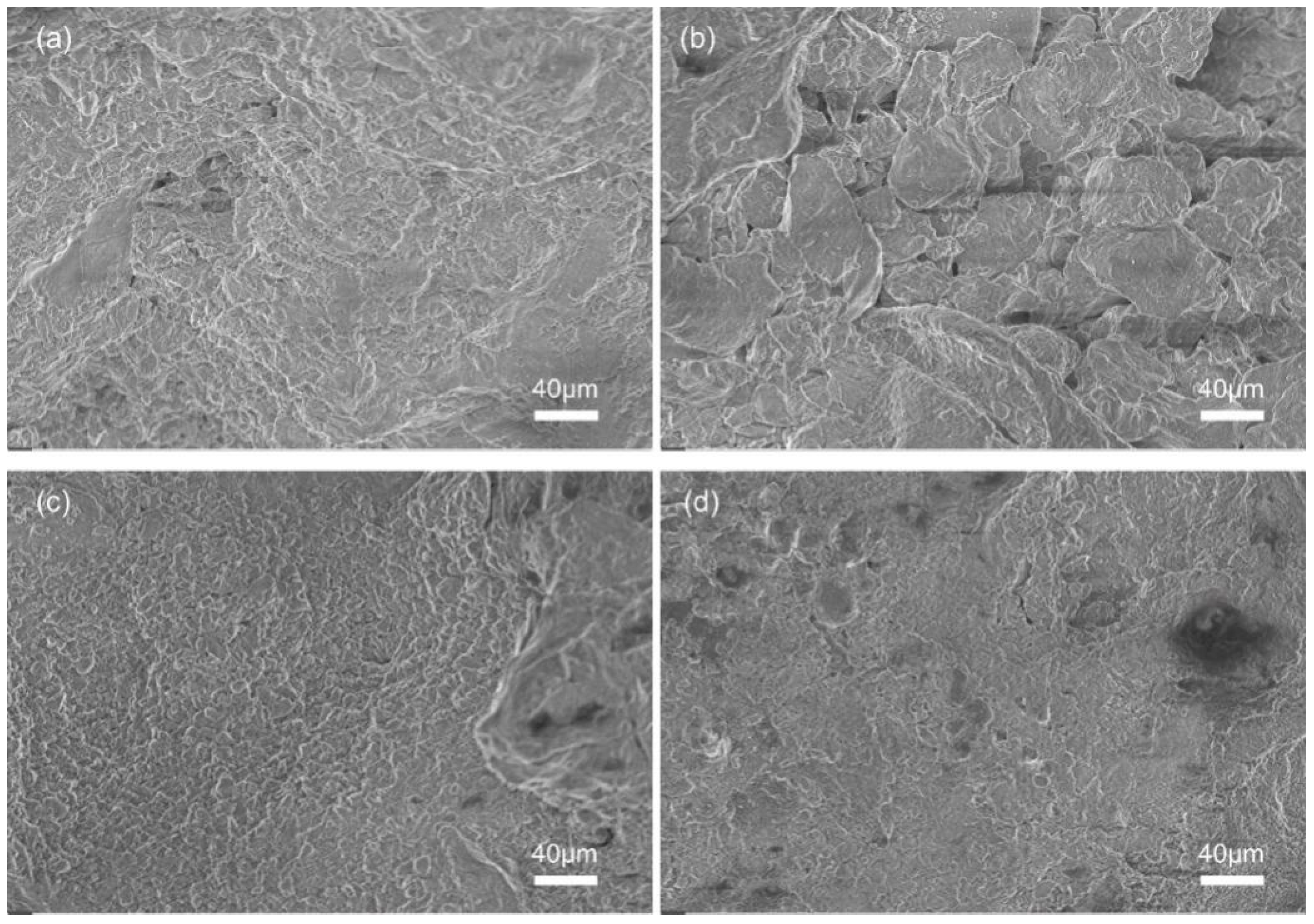
3.1. Microscopic Morphology of the Coatings

Figure 1 shows scanning electron micrographs of various sample surfaces, revealing significant differences in surface morphology characteristics. Figure 1a presents the surface of the HA coating, exhibiting spheroidized and fine particles, irregularly shaped pores and cracks, and a slight quantity of accumulated splats. As shown in Figure 1b, the incomplete melting of particles and pores on the surface of the HN coating decreased. This change might be because the addition of Nb helped to slow down the cooling rate of the surrounding microstructures and reduced the generation of surface cracks and pores. It can be observed from Figure 1c,d that the surface of the HZ coating was uniformly and densely stacked in a flat shape without obvious cracks and pores, which were more prominent on the surface of HN1Z1. More importantly, a large number of needle-like structures were found on the surfaces of HZ and HN1Z1 coatings, as shown in Figure 1e,f. These fine needle-like structures could fill the cracks and pores of the coating surface, which was the key factor to improve the surface quality of the coating. In addition, zirconia has a small diameter and is easily crushed into smaller particles under the action of plasma flame flow, which filled in the cracks and pores generated by incomplete overlap during the coating–stacking process [28] and made the coating surface more uniform and dense; moreover, some of the zirconia underwent phase transformation subjected to the stress field at the crack tip. The shear strain and volume expansion resulted from the phase transformation could suppress the generation of microcracks in the coatings and prevent crack propagation at the surface and interior area of the coating. The convex surface of the needle-like structure could also increase the contact area of the composite coating, providing more adhesive surfaces for the growth of new bone cells.
Figure 2 presents cross-sectional micrographs of the as-deposited coatings. As shown in Figure 2, the specimens’ cross-sections were encapsulated with epoxy, and the average coating thickness was approximately 40~80 µm. The coatings were overlaid and stacked on the substrate, and the bonding interfaces were generally continuous, which revealed a few irregular shapes with a slightly serrated border, indicating a superior bonding property between the coatings and the substrate.

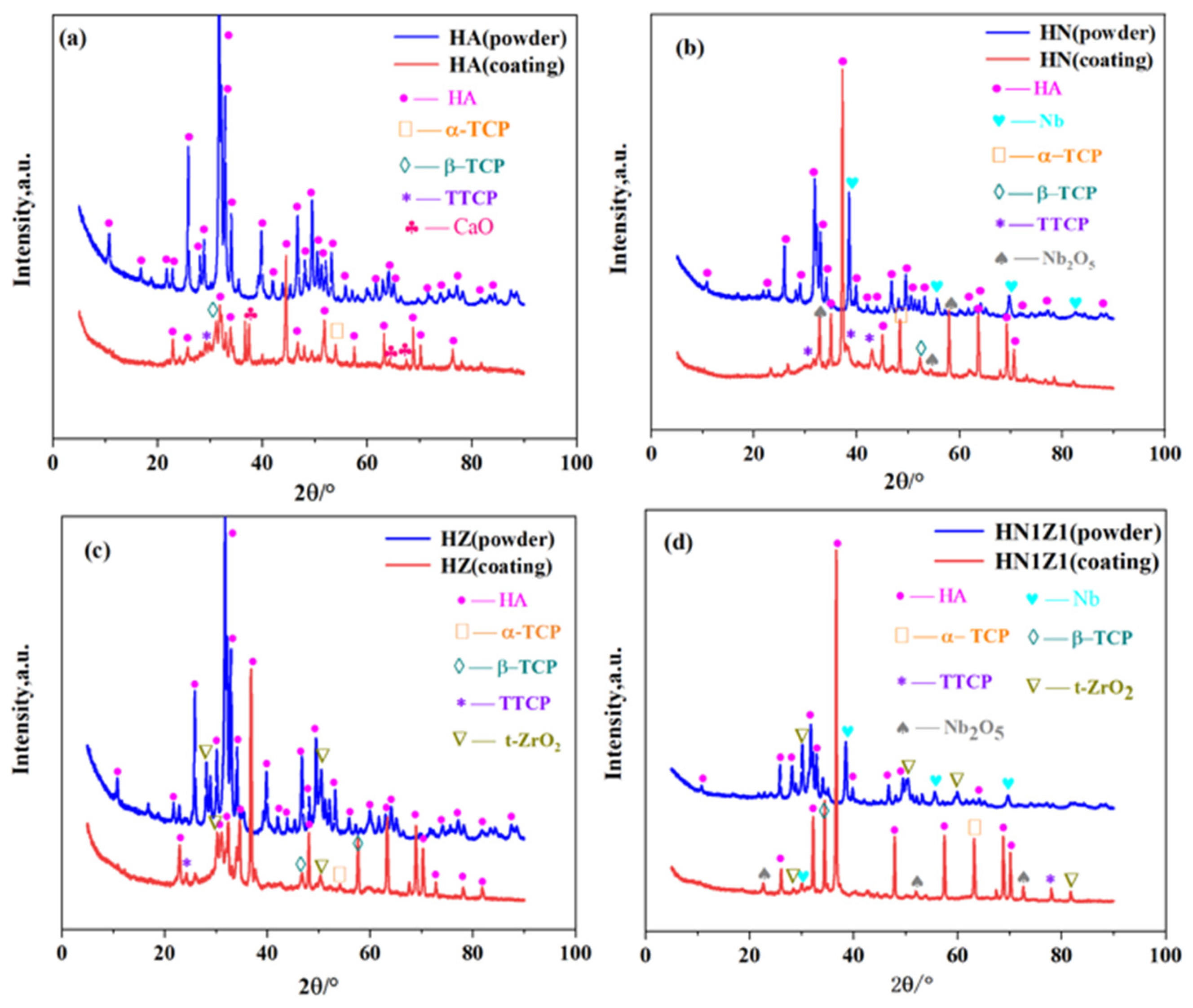
3.2. Phase Analysis of the Coatings

Figure 3 presents the X-ray diffraction patterns of the HA, HN, HZ, and HN1Z1 powders and their coatings. From the results of Figure 3a, it can be seen that the HA powder was single-phase and no other impurity peak except the characteristic peaks of HA phase was detected. The main phase of the HA coating was HA, and the coating also contained tricalcium phosphate (TCP), tetracalcium phosphate (TTCP), and calcium oxide (CaO) phases. The presence of TCP and TTCP phases could be attributed to the HA powder being exposed to high temperatures [29], which caused the heating decomposition of the HA powder, and the appearance of CaO phases could potentially have resulted from the further decomposition of some TTCP phases. Figure 3b reveals that the principal constituents in HN powder are HA and Nb, and no other impurity phase was observed. After spraying, new decomposition phases from the heated HA could be detected, and the diffraction peaks from the niobium oxide (Nb2O5) phases were also identified. As displayed in Figure 3c, the HZ powder mainly contained HA and t-ZrO2 phases, and it could be observed that the t-ZrO2 phase and the decomposition phases generated from HA were presented in the HZ coating. Figure 3d exhibits that the constitutions of the HN1Z1 coating were mainly composed of HA, the t-ZrO2 phase, and partially decomposition phases of HA. Additionally, signals from the Nb2O5 phase were also identified, which signified sufficiently melted and well-deposited raw particles in the coating. Previous studies established that the Nb2O5 phase in the coating would improve the protective performance and biological activity of the surface of HA-based coatings [17,30].

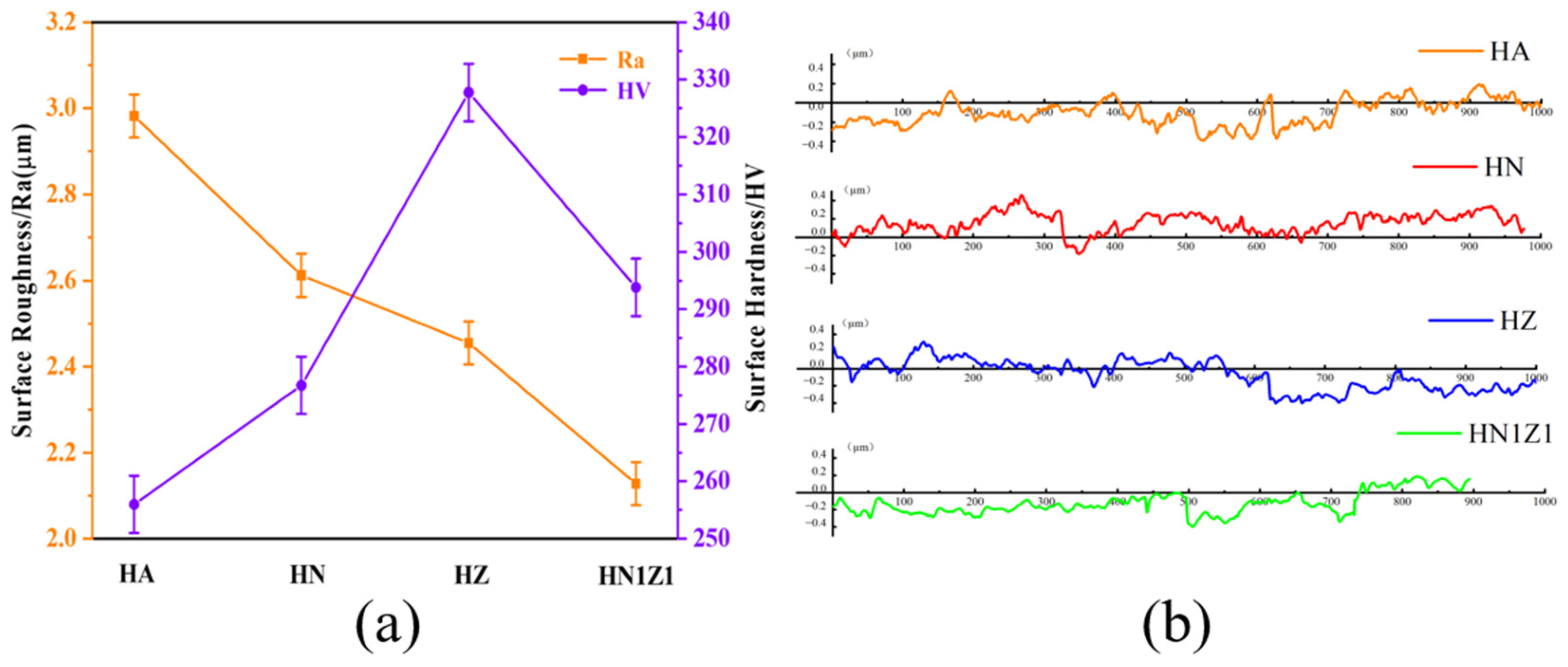
3.3. Hardness and Roughness

A Vickers microhardness test conducted on the surface is commonly used to evaluate coating quality and its functional effectiveness. It can be clearly observed from Figure 4 that the pure HA coating had the lowest surface hardness value. Compared to the HA coating (255.99 HV), the HN coating (276.73 HV) showed an improvement of around 8.1% in surface hardness. It should be noted that the hardness of the HZ coating and the HN1Z1 coating increased to a high level (327.73 HV and 293.80 HV, respectively), which was attributed to the dispersed distribution of zirconia with a relatively high value of hardness (756~786 HV) in the coatings [31]. Although the HZ coating revealed a higher surface hardness value, it was fragile and likely to crack when squeezed by stress, due to more addition of zirconia. Meanwhile, HN1Z1 exhibited a more comprehensive performance.
Cell adhesion and growth were significantly affected by surface roughness. Figure 4 exhibits that the surface roughness values of the HA (Ra, 2.982 µm) and HN1Z1 coatings (Ra, 2.128 µm) were the highest and lowest, respectively. Incompletely melted particles and a large number of cracks and pores were the reason for the uneven surface of the HA coating. The HN coating (Ra, 2.612 µm) had a relatively low roughness value in comparison to the HA coating, which may be attributed to the fact that the addition of Nb resulted in the reduction in the incompletely melted particles. After the addition of the ZrO2 to the coatings, the surface roughness values were further decreased for both HZ (Ra, 2.455 µm) and HN1Z1 coatings, which was due to the small ZrO2 particles with intensive needle-like structures filling the cracks and pores on the coating surface, resulting in a more homogeneous and denser coating. This trend was even more pronounced in HN1Z1, indicating a synergistic effect between Nb and ZrO2.

3.4. Wettability Analysis of Coatings

The wettability of the surface is a critical property that impacts the biological response of an implant when it first interacts with cells [26]. The contact angles of SBF were measured to research the hydrophobicity performance of the different samples, and the evaluation results are presented in Figure 5. The results illustrate that the surface of the uncoated ZK60 substrate exhibits a hydrophobic state. As shown in Figure 5, the hydrophilicity of the HA coating surface is superior, which could be attributed to the enrichment of a large number of hydroxyl groups and phosphate groups on the surface of the coating [32]. Moreover, the rough and porous structure of the HA coating surface facilitates fluid absorption. The contact angle value of the HN coating surface measures 80 ± 5°, indicating an approximately 23.1% decrease compared to the contact angle value of the uncoated ZK60 substrate surface. The contact angle values of the HZ and HN1Z1 coatings are substantially reduced by the addition of ZrO2 to the coatings. Compared to the HZ coating, the increment in the contact angle observed in the HN1Z1 coating may be attributed to the fact that the surface of the HN1Z1 coating has fewer cracks and pores, and the coating is denser and smoother. The experimental results demonstrate that the contact angles of the HZ and HN1Z1 coatings are distributed within a suitable range of 40° to 60°. Additionally, the convex surface of the needle-like structure also increased the contact area of the composite coating.

3.5. Immersion Test

The immersion test was used to measure the weight loss of different samples. The weight loss rate is calculated by measuring the weight of the specimens before immersion and after immersion for 20 days, while the pH value is measured by extracting the corrosion solution at different time points. Figure 6 displays the rate of weight loss and the pH value of the different samples during the 20-day immersion period. From the pH change curves of the soaking solution, it can be seen that the pH value of each sample after soaking increases with time and shows a common characteristic of significant growth in the early stage and slower growth in the later stage. The weight loss rate of the uncoated ZK60 substrate is 14.5% after 20 days of immersion, which is higher than that of all the coated samples, indicating severe corrosion degradation. During the initial immersion period, the pH values of the HA, HN, and HZ coating samples have little difference. However, with prolonged immersion time, the increase in pH value of the HZ coating gradually slows down, compared to the HA and HN coatings. Furthermore, the HZ coating experiences relatively less mass loss after immersion, which can be attributed to the addition of ZrO2 filling the pores and cracks on the coating surface, thus preventing the ingress of corrosive ions and effectively protecting the substrate. It can be clearly seen that the pH value and weight loss rate of the HN1Z1 coating were lower than those of other coatings, indicating that its corrosion degradation was relatively slow. Figure 7 shows the surface corrosion morphology of the HZ and HN1Z1 coatings after soaking in SBF for 1 and 3 days. Compared with the HZ coating, the HN1Z1 coating exhibits fewer small-sized corrosion cracks, and the corroded surface appears relatively flat. This indicates that the HN1Z1 coating has better corrosion resistance. The result indicates that the simultaneous addition of Nb and ZrO2 to the HA coating provides better protection for the ZK60 Mg alloy.

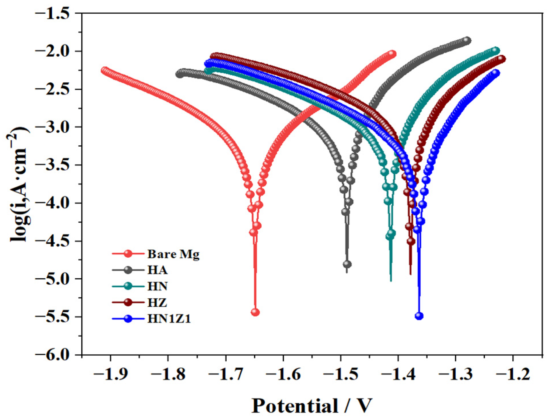
3.6. Electrochemical Testing of Coatings

Potentiodynamic polarization curves offer a more realistic evaluation of the corrosion behavior of biomaterials. Figure 8 represents the potentiodynamic polarization curve of the different samples. Ecorr and Icorr were extracted from the polarization curves using the Tafel extrapolation method, as exhibited in Table 1. The outcomes suggested that the Icorr value of the uncoated ZK60 substrate (1.030 × 10⁻² A·cm−2) was the highest, followed by the HA coating (1.169 × 10⁻³ A·cm−2), the HN coating (1.148 × 10⁻³ A·cm−2), and the HZ coating (1.134 × 10⁻³ A·cm−2) samples. The microcracks and micropores of the HA coating might be the explanation for its poorer corrosion protective effect compared to the HN coating. Compared to the HN coating surface, the HZ coating surface presents the preeminent corrosion resistance with an Ecorr value of −1.379 V. This is due to the fact that a large number of fine ZrO2 particles can fill the cracks and pores in the coating, making the surface smoother and denser, and effectively reducing the channels of corrosive media to the substrate surface. With the simultaneous addition of the Nb and ZrO2 elements to the coating, the corrosion potential of the HN1Z1 coating was −1.364 V, and the corrosion current density was 5.109 × 10⁻⁴ A·cm−2, indicating that the surface possessed the highest corrosion resistance. The lower surface roughness and the synergistic effect of the Nb and ZrO2 elements may be the reason for the enhancement of the corrosion protective effect.

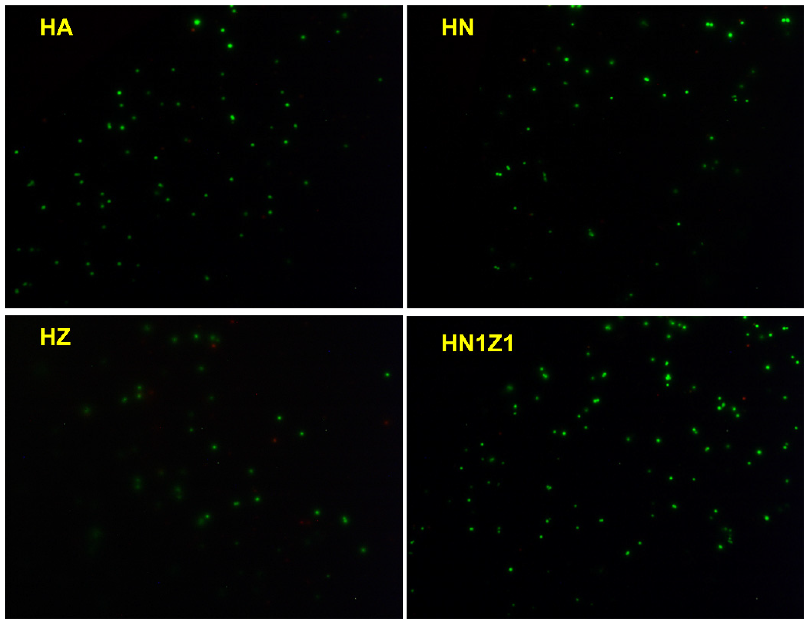
3.7. Cytocompatibility Evaluation of Coatings

Figure 9 shows the cell viability of the uncoated ZK60 substrate and the coated samples during the same cultivation period of BMSCs. The results indicate that the surface cell viability of the coated samples is significantly higher than that of the uncoated magnesium alloy during the 1st, 3rd, and 5th days of cultivation, which suggests that the uncoated magnesium samples are more cytotoxic as compared to the other coated samples. Furthermore, the cell viability of all samples drops as the cultivation time increases, which could be attributed to the occurrence of dissolution in all samples during the cultivation process, leading to an increase in the concentrations of Mg2+ in the solution. Previous studies have shown that an appropriate concentration of Mg2+ is beneficial for cell proliferation [33]. However, when the concentration is too high, it can affect the osmotic pressure of cells and cause changes in the pH environment of the solution, which is not conducive to cell proliferation and growth.
Figure 10 shows the fluorescence microscopy images of BMSCs cultivated for 5 days on the different coated samples. It can be observed from the figure that the plasma-sprayed HN1Z1 coating sample exhibits an increase in the number of cells on its surface and demonstrates a favorable growth state compared to the other samples. The above results reveal that the HN1Z1 coating surface exhibits good cell viability, indicating its facilitation of the attachment and growth of cells at subsequent stages.

4. Conclusions

Plasma spray technology was utilized to deposit various coatings—HA, HN, HZ, and HN1Z1—on a ZK60 magnesium alloy. The HA-coated surface exhibited numerous incompletely melted particles and irregularly shaped pores and cracks. In contrast, the HN coating showed fewer such particles and had a relatively flatter appearance, and the HZ coating further improved upon this, presenting a flat and smooth surface with significantly reduced cracks and pores. The HN1Z1 coating was the most advanced, being dense, uniform, and noticeably smoother. Interestingly, ZrO2 maintained its tetragonal phase during spraying, which is beneficial for phase transformation and the suppression of crack propagation in later stages. Compared to the pure HA coating, the HN, HZ, and HN1Z1 coatings demonstrated reductions in surface roughness of 12.4%, 17.7%, and 28.6%, respectively, and increases in surface hardness of 8.1%, 21.8%, and 12.9%, respectively. All coatings showed good hydrophilic properties. Electrochemical tests revealed that the HN1Z1 coating exhibited the highest corrosion resistance, followed by HZ, with HN showing improved corrosion resistance compared to HA, albeit to a lesser extent. During immersion tests, the composite coating exhibited a gradual pH increase and minimal mass loss. The cytocompatibility test demonstrated that the HN1Z1 coating possesses promising cellular activity. These results suggest that the addition of Nb and ZrO2 to HA coatings has a synergistic effect. Incorporating Nb/ZrO2 into HA coatings could potentially enhance the corrosion resistance and cytocompatibility of Mg alloy implants, offering promising implications for future biomedical applications.

Author Contributions

Conceptualization, X.W. and S.F.; Methodology, S.X. and L.Y.; Software, S.F. and S.X.; Validation, S.X. and L.Y.; Formal analysis, J.Z. and S.Q.; Investigation, S.Q. and F.H.; Resources, J.Z. and F.H.; Data curation, L.Y. and C.M.; Writing—original draft, X.W., S.F. and F.H.; Writing—review & editing, S.F. and S.Q.; Visualization, L.Y. and C.M.; Supervision, X.W. and C.M. All authors have read and agreed to the published version of the manuscript.

Funding

The Test Centre of Nantong University; the Large Instruments Open Foundation of Nantong University, grant number KFJN2428; Science and Technology Project of Nantong City, grant number JC2023001.

Institutional Review Board Statement

Not applicable.

Informed Consent Statement

Not applicable.

Data Availability Statement

All data generated or analyzed during this study are available from the corresponding author on reasonable request.

Acknowledgments

The authors are grateful for the Test Centre of Nantong University, the Large Instruments Open Foundation of Nantong University (KFJN2428), and Science and Technology Project of Nantong City (JC2023001).

Conflicts of Interest

The authors declare that they have no competing financial interests or personal relationships that could have appeared to influence the work reported in this paper.

References

  1. Chiara, G.; Letizia, F.; Lorenzo, F.; Edoardo, S.; Diego, S.; Stefano, S.; Eriberto, B.; Barbara, Z. Nanostructured Biomaterials for Tissue Engineered Bone Tissue Reconstruction. Int. J. Mol. Sci. 2012, 13, 737–757. [Google Scholar] [CrossRef] [PubMed]
  2. Choudhury, D.; Vrbka, M.; Marnat, A.B.; Stavness, I.; Roy, C.K.; Mootanah, R.; Krupka, I. The impact of surface and geometry on coefficient of friction of artificial hip joints. J. Mech. Behav. Biomed. Mater. 2017, 72, 192–199. [Google Scholar] [CrossRef] [PubMed]
  3. Nomura, N. Artificial organs: Recent progress in metals and ceramics. J. Artif. Organs. 2010, 13, 10–12. [Google Scholar] [CrossRef] [PubMed]
  4. Zhang, T.; Wang, W.; Liu, J.; Wang, L.Q.; Tang, Y.J.; Wang, K.S. A review on magnesium alloys for biomedical applications. Front. Bioeng. Biotechnol. 2022, 10, 25. [Google Scholar] [CrossRef] [PubMed]
  5. Chen, J.X.; Tan, L.L.; Yu, X.M.; Etim, I.P.; Ibrahim, M.; Yang, K. Mechanical properties of magnesium alloys for medical application: A review. J. Mech. Behav. Biomed. Mater. 2018, 87, 68–79. [Google Scholar] [CrossRef]
  6. Dong, J.H.; Lin, T.; Shao, H.P.; Wang, H.; Wang, X.T.; Song, K.; Li, Q.H. Advances in degradation behavior of biomedical magnesium alloys: A review. J. Alloys Compd. 2022, 908, 16. [Google Scholar] [CrossRef]
  7. Ramalingam, V.V.; Ramasamy, P.; Kovukkal, M.D.; Myilsamy, G. Research and Development in Magnesium Alloys for Industrial and Biomedical Applications: A Review. Met. Mater.-Int. 2020, 26, 409–430. [Google Scholar] [CrossRef]
  8. Emelyanenko, K.A.; Chulkova, E.V.; Semiletov, A.M.; Domantovsky, A.G.; Palacheva, V.V.; Emelyanenko, A.M.; Boinovich, L.B. The Potential of the Superhydrophobic State to Protect Magnesium Alloy against Corrosion. Coatings 2022, 12, 13. [Google Scholar] [CrossRef]
  9. Liu, H.G.; Cao, F.Y.; Song, G.L.; Zheng, D.J.; Shi, Z.M.; Dargusch, M.S.; Atrens, A. Review of the atmospheric corrosion of magnesium alloys. J. Mater. Sci. Technol. 2019, 35, 2003–2016. [Google Scholar] [CrossRef]
  10. Witte, F. The history of biodegradable magnesium implants: A review. Acta Biomater. 2010, 6, 1680–1692. [Google Scholar] [CrossRef]
  11. Najafi, S.; Sheikhani, A.; Sabbaghian, M.; Nagy, P.; Fekete, K.; Gubicza, J. Modification of the Tensile Performance of an Extruded ZK60 Magnesium Alloy with the Addition of Rare Earth Elements. Materials 2023, 16, 13. [Google Scholar] [CrossRef] [PubMed]
  12. Wu, G.H.; Wang, C.L.; Sun, M.; Ding, W.J. Recent developments and applications on high-performance cast magnesium rare-earth alloys. J. Magnes. Alloy. 2021, 9, 1–20. [Google Scholar] [CrossRef]
  13. Xun, Q.W.; Liu, Y.H.; Pan, Z.R.; Wu, Y. Microstructure and properties of plasma cladding Ni-based alloy coated on 40Cr Surface. Surf. Topogr.-Metrol. Prop. 2023, 11, 16. [Google Scholar]
  14. Yao, X.Y.; Tang, J.C.; Zhou, Y.H.; Atrens, A.; Dargusch, M.S.; Wiese, B.; Ebel, T.; Yan, M. Surface modification of biomedical Mg-Ca and Mg-Zn-Ca alloys using selective laser melting: Corrosion behaviour, microhardness and biocompatibility. J. Magnes. Alloy. 2021, 9, 2155–2168. [Google Scholar] [CrossRef]
  15. Lee, C.Y.; Lee, J.L.; Jian, S.Y.; Chen, C.A.; Aktug, S.L.; Ger, M.D. The effect of fluoride on the formation of an electroless Ni-P plating film on MAO-coated AZ31B magnesium alloy. J. Mater. Res. Technol JMRT 2022, 19, 542–556. [Google Scholar] [CrossRef]
  16. Chen, Q.; Zheng, Y.; Dong, S.; Chen, X.B.; Dong, J. Effects of fluoride ions as electrolyte additives for a PEO/Ni-P composite coating onto Mg alloy AZ31B. Surf. Coat. Technol. 2021, 417, 11. [Google Scholar] [CrossRef]
  17. Pauline, S.A.; Rajendran, N. Biomimetic novel nanoporous niobium oxide coating for orthopaedic applications. Appl. Surf. Sci. 2014, 290, 448–457. [Google Scholar] [CrossRef]
  18. Morks, M.E. Fabrication and characterization of plasma-sprayed HA/SiO2 coatings for biomedical application. J. Mech. Behav. Biomed. Mater. 2008, 1, 105–111. [Google Scholar] [CrossRef]
  19. Zhu, L.; Zhang, N.N.; Bolot, R.; Planche, M.P.; Liao, H.L.; Coddet, C. Very low pressure plasma sprayed yttria-stabilized zirconia coating using a low-energy plasma gun. Appl. Phys. A Mater. Sci. Process. 2011, 105, 991–996. [Google Scholar] [CrossRef]
  20. Hasan, M.F.; Wang, J.; Berndt, C. Determination of the Mechanical Properties of Plasma-Sprayed Hydroxyapatite Coatings Using the Knoop Indentation Technique. J. Therm. Spray Technol. 2015, 24, 865–877. [Google Scholar] [CrossRef]
  21. Singh, B.; Singh, G.; Sidhu, B.S. Analysis of Corrosion Behavior and Surface Properties of Plasma-Sprayed HA/Ta Coating on CoCr Alloy. J. Therm. Spray Technol. 2018, 27, 1401–1413. [Google Scholar] [CrossRef]
  22. Bansal, P.; Singh, G.; Sidhu, H.S. Investigation of surface properties and corrosion behavior of plasma sprayed HA/ZnO coatings prepared on AZ31 Mg alloy. Surf. Coat. Technol. 2020, 401, 9. [Google Scholar] [CrossRef]
  23. Li, B.X.; Niu, J.L.; Liu, H.Y.; Li, G.Y. Fabrication and corrosion property of novel 3-aminopropyltriethoxy-modified calcium phosphate/poly(lactic acid) composite coating on AZ60 Mg alloy. Appl. Phys. A Mater. Sci. Process. 2018, 124, 13. [Google Scholar] [CrossRef]
  24. Yugeswaran, S.; Yoganand, C.P.; Kobayashi, A.; Paraskevopoulos, K.M.; Subramanian, B. Mechanical properties, electrochemical corrosion and in-vitro bioactivity of yttria stabilized zirconia reinforced hydroxyapatite coatings prepared by gas tunnel type plasma spraying. J. Mech. Behav. Biomed. Mater. 2012, 9, 22–33. [Google Scholar] [CrossRef]
  25. Hussain, S.; Shah, Z.A.; Sabiruddin, K.; Keshri, A.K. Characterization and tribological behaviour of Indian clam seashell-derived hydroxyapatite coating applied on titanium alloy by plasma spray technique. J. Mech. Behav. Biomed. Mater. 2023, 137, 15. [Google Scholar] [CrossRef]
  26. Singh, B.; Singh, G.; Sidhu, B.S.; Bhatia, N. In-vitro assessment of HA-Nb coating on Mg alloy ZK60 for biomedical applications. Mater. Chem. Phys. 2019, 231, 138–149. [Google Scholar] [CrossRef]
  27. Singh, B.; Singh, G.; Sidhu, B.S. Investigation of the in vitro corrosion behavior and biocompatibility of niobium (Nb)-reinforced hydroxyapatite (HA) coating on CoCr alloy for medical implants. J. Mater. Res. 2019, 34, 1678–1691. [Google Scholar] [CrossRef]
  28. Chen, M.; Lu, C.; Yu, J. Improvement in performance of MgO–CaO refractories by addition of nano-sized ZrO2. J. Eur. Ceram. Soc. 2007, 27, 4633–4638. [Google Scholar] [CrossRef]
  29. Hung, K.Y.; Lai, H.C.; Yang, Y.C.; Feng, H.-P. Characterization of hydroxyapatite (HA) sputtering targets by APS methods. Coatings 2017, 7, 197. [Google Scholar] [CrossRef]
  30. Ardelean, H.; Frateur, I.; Marcus, P. Corrosion protection of magnesium alloys by cerium, zirconium and niobium-based conversion coatings. Corrosion Sci. 2008, 50, 1907–1918. [Google Scholar] [CrossRef]
  31. Lv, M.; Lv, W.J.; Chen, H.Z.; Zheng, F.; Liu, J.; Kong, F.D.; Liu, S.L.; Wang, L.T. Biotribological properties of nano zirconium dioxide and hydroxyapatite-reinforced polyetheretherketone (HA/ZrO2/PEEK) biocomposites. Iran. Polym. J. 2021, 30, 1127–1136. [Google Scholar] [CrossRef]
  32. Komarova, E.G.; Sharkeev, Y.P.; Sedelnikova, M.B.; Prymak, O.; Epple, M.; Litvinova, L.S.; Shupletsova, V.V.; Malashchenko, V.V.; Yurova, K.A.; Dzyuman, A.N.; et al. Zn-or Cu-containing CaP-based coatings formed by micro-arc oxidation on titanium and Ti-40Nb alloy: Part II—Wettability and biological performance. Materials 2020, 13, 4366. [Google Scholar] [CrossRef] [PubMed]
  33. Li, Z.Q.; Zheng, X.X.; Wang, Y.X.; Tao, T.Y.; Wang, Z.L.; Yuan, L.; Han, B. The Biomimetics of Mg2+-Concentration-Resolved Microenvironment for Bone and Cartilage Repairing Materials Design. Biomimetics 2022, 7, 21. [Google Scholar] [CrossRef] [PubMed]
Figure 1. SEM micrographs of the surface of as-deposited (a) HA; (b) HN; (c) HZ; and (d) HN1Z1 coatings; (e,f) are the magnified images of the blue dash circle areas in (c,d).
Coatings 14 01282 g001
Figure 2. SEM micrographs of the cross-section of the sprayed coatings (a) HA; (b) HN; (c) HZ; and (d) HN1Z1.
Coatings 14 01282 g002
Figure 3. X-ray diffraction patterns of as-deposited (a) HA; (b) HN; (c) HZ; and (d) HN1Z1 coatings and their powders.
Coatings 14 01282 g003
Figure 4. (a) Surface microhardness and roughness of the different samples; (b) surface contour curves of the samples.
Coatings 14 01282 g004
Figure 5. The droplet profile and contact angles of SBF droplets on the sample surface.
Coatings 14 01282 g005
Figure 6. (a) pH value variation; (b) weight loss rate of the as-prepared samples during 20 days of immersion in SBF.
Coatings 14 01282 g006
Figure 7. SEM micrographs of samples after immersion in SBF solution at different time points: (a) HZ-1 day; (b) HZ-3 days; (c) HN1Z1-1 day; (d) HN1Z1-3 days.
Coatings 14 01282 g007
Figure 8. Polarization curves of different samples.
Coatings 14 01282 g008
Figure 9. The cell viability of all samples in the cultivation of BMSCs.
Coatings 14 01282 g009
Figure 10. Fluorescent microscope images of BMSCs cultured for 5 days on different coating samples.
Coatings 14 01282 g010
Table 1. Fitting results from the polarization curves.
Sample E c o r r (V) I c o r r (A·cm−2)
Mg−1.6491.030 × 10−2
HA−1.4901.169 × 10−3
HN−1.4131.148 × 10−3
HZ−1.3791.134 × 10−3
HN1Z1−1.3645.109 × 10−4
Disclaimer/Publisher’s Note: The statements, opinions and data contained in all publications are solely those of the individual author(s) and contributor(s) and not of MDPI and/or the editor(s). MDPI and/or the editor(s) disclaim responsibility for any injury to people or property resulting from any ideas, methods, instructions or products referred to in the content.

