Article

Enhanced Osteogenic Differentiation Based on Combining Pulp Stem Cells with Ultralong Hydroxyapatite Nanowires and Cellulose Fibers

1
Department of Stomatology, Xinhua Hospital Affiliated to Shanghai Jiaotong University School of Medicine, Kongjiang Road 1665, Shanghai 200092, China
2
Department of Gerontology, Xinhua Hospital Affiliated to Shanghai Jiaotong University School of Medicine, Kongjiang Road 1665, Shanghai 200092, China
*
Authors to whom correspondence should be addressed.
These authors contributed equally to this work.
Coatings 2024, 14(2), 190; https://doi.org/10.3390/coatings14020190
Submission received: 23 December 2023 / Revised: 24 January 2024 / Accepted: 30 January 2024 / Published: 1 February 2024
(This article belongs to the Section Surface Coatings for Biomedicine and Bioengineering)

Abstract

:
Previous studies have confirmed the excellent biocompatibility, osteogenic properties, and angiogenic ability of hydroxyapatite (HAP), as well as the good osteoblast differentiation ability of dental pulp stem cells. We hypothesized that combining dental pulp stem cells with ultralong hydroxyapatite nanowires and cellulose fibers could more effectively promote osteoblast differentiation, making it a potential biomaterial for enhancing bone wound healing. Therefore, based on the optimal ratio of ultralong hydroxyapatite nanowires and cellulose fibers (HAPNW/CF) determined in previous studies, we added human dental pulp stem cells (hDPSCs) to investigate whether this combination can accelerate cell osteogenic differentiation. hDPSCs were introduced into HAPNW/CF scaffolds, and in vitro experiments revealed that: (1) HAPNW/CF scaffolds exhibited no cytotoxicity toward hDPSCs; (2) HAPNW/CF scaffolds enhanced alkaline phosphatase staining activity, an early marker of osteogenic differentiation, and significantly upregulated the expression level of osteogenic-related proteins; (3) co-culturing with hDPSCs in HAPNW/CF scaffolds significantly increased the expression of angiogenesis-related factors compared to hDPSCs alone when tested using human umbilical vein endothelial cells (hUVECs). Our study demonstrates that combining hDPSCs with HAPNW/CF can enhance osteogenic differentiation more effectively, potentially through increased secretion of angiogenesis-related factors promoting osteoblast differentiation.

1. Introduction

The repair and reconstruction of critical osseous defects induced by tumors, trauma, and accidents remain a significant clinical challenge [1]. Autologous bone grafting is considered the gold standard due to its safety and effectiveness; however, its clinical application is limited by issues such as infection, donor site scarcity, and complications at the donor site [2]. In recent decades, with the continuous advancement of tissue engineering materials, patients with large segmental bone defects have the potential to receive safer, more effective, and cost-efficient treatments [3,4,5]. An ideal bone tissue engineering scaffold should possess characteristics similar to the extracellular matrix of natural bone and provide a three-dimensional space for cell survival. It should also induce cellular bioactivity to facilitate sufficient nutrient supply to cells and enhance their retention at the implantation site [6,7]. Moreover, as the biomaterial gradually degrades over time, migrating bone cells can continuously proliferate and differentiate toward repairing bone tissue defects [8].
With the emergence of tissue engineering, stem cells have gained widespread utilization as seed cells in regenerative medicine [9,10]. Currently, commonly employed seed cell types include mesenchymal stem cells (MSCs) [11], embryonic stem cells (ESCs) [12], and adult or embryonic neural stem cells (NSCs) [13]. Among these options, mesenchymal stem cells are a type of pluripotent stem cells characterized by their capacity for self-renewal and multi-lineage differentiation. These attributes render them highly applicable in fields such as cell therapy, drug screening, and disease modeling. Consequently, they are highly sought-after “seed cells” within the field of tissue engineering [14,15]. Gronthos successfully isolated dental pulp stem cells (DPSCs) from the third molar’s pulp in 2000, marking a significant milestone in adult stem cell research. [16]. Human dental pulp stem cells (hDPSCs) represent a subset of mesenchymal stem cells derived from dental tissues with potential for the purpose of self-renewal and differentiation into diverse cell lineages, encompassing osteoblasts, adipocytes, chondrocytes, odontoblast-like-cells, and neuronal-like-cells [17,18]. The ease of isolating hDPSCs from extracted teeth provides an abundant cellular source for tissue engineering purposes. Due to their low-risk profile, hDPSCs possess advantages over other types of stem cells. These inherent advantages position hDPSCs at the forefront of dentin repair, tooth regeneration, and bone tissue engineering research [19,20]. Furthermore, research has demonstrated that hDPSCs exhibit promise for craniofacial bone reconstruction encompassing skull bones, mandibular bones, and other related structures [21,22]. Therefore, hDPSCs were chosen as the preferred cell source in our study.
Hydroxyapatite (HAP) is the primary inorganic constituent of human bones and teeth, exhibiting exceptional biocompatibility, bioactivity, osteoinductivity, and biodegradability [23,24]. Upon implantation into bone defects, HAP does not form a fibrous tissue interface with the surrounding bone tissue; instead, it develops a carbonate apatite layer on its surface within the body. Consequently, HAP holds immense potential as a hopeful biomaterial for bone repair applications [25]. In the field of biomedical engineering, HAP finds extensive utilization in bone defect repair, including HAP-based bioactive glass composites, HAP-graphene composites, and HAP-polymer composites [26]. The incorporation of metal compounds with HAP can enhance osteogenesis and angiogenesis processes [27]. To improve the early stability and biocompatibility of metal implants effectively, it is a common practice to employ HAP coatings on their surfaces to leverage the advantages offered by both materials for enhanced success rates [28,29]. Moreover, HAP biomaterials with diverse morphologies and pore structures are widely employed as filling materials or delivery carriers for bone defects in bone tissue engineering studies [30].
Unfortunately, traditional HAP materials exhibit high brittleness and poor ductility when subjected to mechanical loads, resulting in suboptimal mechanical properties [31]. Scaffolds composed solely of these biomaterials fail to meet the mechanical requirements of the relevance of bone tissue engineering in clinical applications. In the present study, we prepared a novel porous and stable biopaper using a simple vacuum filtration method, where ultralong HAP nanowires were interwoven with each other and with cellulose fibers [32]. Unlike traditional brittle HAP materials, the hierarchical architecture in the biopaper imparts exceptional performance to the material. The ultralong HAP nanowires demonstrate remarkable flexibility and can be bent at large angles without fracturing [31]. Furthermore, the as-prepared biopaper based on ultralong hydroxyapatite nanowires and cellulose fibers (HAPNW/CF) exhibits excellent thermal stability, elevated levels of porosity and specific surface area, hydrophilicity, tensile strength as well as a porous network structure that provides an optimal environment for bone formation and ingrowth.
However, apart from bone formation, the successful implantation of bone biomaterials heavily relies on the establishment of a well-functioning vascular network as blood vessel formation directly impacts the osteogenesis of implants [33,34]. HAP is not only a common material for bone defect repair but also exhibits excellent potential in promoting angiogenesis [35]. Our previous research findings [36] demonstrated that highly porous aerogels based on ultralong hydroxyapatite nanowires not only induce new bone formation but also facilitate vascularization in the area of bone defects, indicating HAP’s capability to promote blood vessel generation. Furthermore, the sustained release of Ca2+ ions from the HAPNW/CF biopaper promotes neovascularization while simultaneously influencing osteogenesis by affecting the structure of blood clots at fracture sites.
In this study, as shown in Figure 1, we conducted a systematic investigation of the application of HAPNW/CF biomaterial paper in bone regeneration. In vitro studies demonstrated that HAPNW/CF biomaterial paper exhibited superior performance in terms of proliferation, adhesion, and osteogenic differentiation of hDPSCs while also promoting angiogenesis. The results revealed promising prospects for its application in repairing large segmental bone defects, highlighting its significant scientific and clinical value.

2. Materials and Methods

2.1. Preparation of the HAPNW/CF Biopaper

The HAPNW/CF biopaper was prepared according to the method reported in our previous study [32]. In brief, the mixture was prepared by combining 135 mL of deionized water, 60 mL of methanol, and 105 mL of oleic acid. Then, the above mixture was gradually supplemented with an aqueous solution of NaOH (10.500 g in 150 mL), CaCl2 (3.330 g in 120 mL), and NaH2PO4·2H2O (9.360 g in 180 mL). After solvothermal treatment of the mixture at 185 °C for 25 h, the resulting product was sequentially washed twice with ethanol and deionized water, and then HAPNW dispersions were obtained. Then, when the weight ratio of HAPNWs to CFs was 80:20, cellulose fibers (CFs) were added to the HAPNW dispersion. The dispersion was imported into the filter paper, and the HAPNW/CF biopaper was formed by drying at 95 °C for 10 min using the vacuum filtration method.

2.2. In Vitro Cellular Researches

2.2.1. Isolation and Culture of hDPSCs

Healthy impacted third molars were extracted from 10 individuals aged 18 to 25 years from the Department of Stomatology, Xinhua Hospital, affiliated with Shanghai Jiaotong University School of Medicine. None of the selected objects in this research had systemic disease, a history of radiotherapy, or smoking. All tooth extractions in the research were authorized by the Ethics Committee of Xinhua Hospital Affiliated to Shanghai Jiaotong University School of Medicine. The endodontic tissues were gently isolated from the crown and root and digested in a solution containing 3 mg/mL of collagenase I (Sigma, St. Louis, MO, USA) and 4 mg/mL of dispase (Sigma, USA) for 0.5 h at 37 °C, as described by Jia Q et al. [37]. Then, hDPSCs were cultured in α-modified Eagle medium (α-MEM, HyClone, Logan, UT, USA) containing: (1) 10% fetal bovine serum (FBS, Gibco, Grand Island, NE, USA); (2) 100 μg/mL penicillin (HyClone, USA); and (3) 100 mg/mL streptomycin (HyClone, USA), in 5% carbon dioxide at 37 °C. The culture medium was refreshed every 2 to 3 days. After reaching 70% to 80% confluence, hDPSCs were incubated using 0.05% trypsin (Gibco, USA) for a duration of 1 min to facilitate cell isolation. hDPSCs between passages 3–4 were used for biological experiments.

2.2.2. Identification, Osteogenic and Adipogenic Differentiation of hDPSCs

Cell surface markers of hDPSCs were analyzed through flow cytometry as described previously [38]. In briefly, 5 × 10⁶ cells at the third passage were resuspended in cold phosphate buffer (PBS, HyClone, USA) and subsequently incubated with the following antibodies: CD34-FITC, CD45-PE, CD90-APC, CD105-APC and CD146-FITC (Abcam, Cambridge, UK). All processes were performed away from light at 4 °C for 1 h. Following incubation, the expression profiles were analyzed using flow cytometry (Thermo, Waltham, MA, USA). For osteogenic differentiation, the hDPSCs were cultured in an osteogenic medium (OM) at the third passage containing 10 ng/mL dexamethasone, 10 mmol/L beta-glycerophosphate and 50 mg/mL ascorbic acid (Sigma, USA) for 14 days, and changing the culture medium every 3 days. Control samples were cultured in α-MEM supplemented with 10% FBS with no supplements. After 14 days of culture, the calcium nodules were measured by dyeing with alizarin red (Sigma, USA). The adipogenic medium contained 10 mg/mL insulin, 200 mmol/L indomethacin, 1 mmol/L dexamethasone, and 0.05 mmol/L methylisobutylxan-thine. The hDPSCs were cultured in the adipogenic medium and α-MEM medium for 14 days, respectively. Oil red O staining was used to observe the lipid droplets.

2.2.3. Determination of Cell Adhesion Activity

A total of 3 × 10⁴ hDPSCs were seeded per well on the HAPNW/CF biopaper (2 mm × 2 mm) in 24-well plates and cultured for 1, 4, and 24 h. After performing three rounds of PBS washing at room temperature for each time point, the cells were fixed with 4% paraformaldehyde (PFA) for a duration of 10 min and subsequently subjected to three washes with PBS after the fixation solution was sucked out. The cells were then permeabilized with 0.1% (v/v) Triton X-100 and blocked in bovine serum albumin under light-free conditions. The Actin-Tracker Green working solution (Sigma-Aldrich Trading Co., Shanghai, China) was used to stain F-actin in the dark for 30 min. Subsequently, the cells were immersed with DAPI (Sigma, USA) for nuclear staining for 5 min. The relevant images were observed under a fluorescence microscope.

2.2.4. Cell Proliferation by CCK-8 Assay

The proliferation activity of hDPSCs on HAPNW/CF biopaper was detected by the cell counting kit-8 (CCK-8) assay (Beyotime, Shanghai, China). The hDPSCs were cultured on HAPNW/CF biopaper in 24-well plates at a density of 1 × 10⁴ cells per well. When cultured for 1, 4, and 7 days, the original medium in the pore plate was sucked away, and the cells were thoroughly rinsed with PBS three consecutive times. Subsequently, 200 μL of CCK-8 working liquid (the ratio of CCK-8 liquid to complete medium was 1:10) was added to each 24-well plate and incubated in a cell incubator at 37 °C for 2 h away from light. Then, 100 μL liquid was absorbed into the 96-well plate per well, and the absorbance value was determined at 450 nm using a microplate reader.

2.2.5. ALP Staining and ALP Activity Assay

For ALP staining, the hDPSCs (2 × 10⁴ cells per well) were cultured on HAPNW/CF biopaper in 24-well plates. When two time points arrived (day 7 and day 14), the cells were fixed with 4% paraformaldehyde for thirty minutes and then repetitively rinsed with phosphate-buffered saline (PBS) for three cycles, each lasting five minutes. After preparing the BCIP/NBT staining solution (Beyotime, China) in accordance with the manufacturer’s provided instructions for reagent usage, the appropriate amount of the staining solution was taken to cover the cells and stain for 30 min without light before taking photos.

2.2.6. Live–Dead Staining

The cell viability and proliferation on HAPNW/CF biopaper were assessed using the LIVE/DEAD™ assay kit (Thermo, USA) at 3 days of culture. Briefly, the hDPSCs (1 × 10⁴ cells per well) were seeded on HAPNW/CF biopaper in a 6-well plate for 3 days as described above. Before starting cell staining, the cells were rinsed 3 times with PBS. After the cells were completely covered with the appropriate amount of Calcin-AM /PI solution, they were incubated for 30 min away from light. Images of living and dead cells were observed using a fluorescence microscope, and the number of living and dead cells was calculated separately using Image J software 6.0.

2.2.7. Western Blot

After seeding hDPSCs on HAPNW/CF biopaper for 7 days, the cells were washed with phosphate-buffered saline (PBS) three times and subsequently trypsinized using 0.25% trypsin-EDTA solution. hDPSCs were lysed in 100 uL RIPA lysis buffer and harvested by centrifugation at 12,000 rpm for 10 min. As mentioned above, the total protein content was determined by a BCA protein detection kit, and the isolation of 10 mg of protein from each sample was achieved through the utilization of sodium dodecyl sulfate-polyacrylamide gel electrophoresis (SDS-PAGE), followed by transfer to 0.22 μm pore-sized polyvinylidene fluoride (PVDF) membrane. Following a 60-min incubation at room temperature in TBST buffer containing 5% nonfat dry milk solution to block, the membranes were subsequently incubated overnight with anti-Col-1 (Abcam, UK), anti-RUNX2 (CST, Boston, MA, USA), anti-OCN (CST, USA), anti-Enos (Abcam, UK), anti-AKT (Abcam, UK), anti-VEGF (Abcam, UK), anti-GAPDH (Boster, Wuhan, China) in the blocking buffer at 4 °C. After performing three washes with TBST, incubate the samples at room temperature for 1 h with a properly diluted secondary antibody, followed by detection using an enhanced chemiluminescence detection system. Image J software was used to analyze the gray values of different bands, and GAPDH bands were used as the internal reference to calculate the relative expression levels of each group of proteins for subsequent statistical analysis. Each group of experiments was repeated three times.

2.3. Statistical Analysis

All assays were repeated at least three times. All quantitative data were expressed as mean ± standard deviation (SD), and the t-test was used for statistical analyses by SPSS20.0. p < 0.05 was considered a significant difference.

3. Results

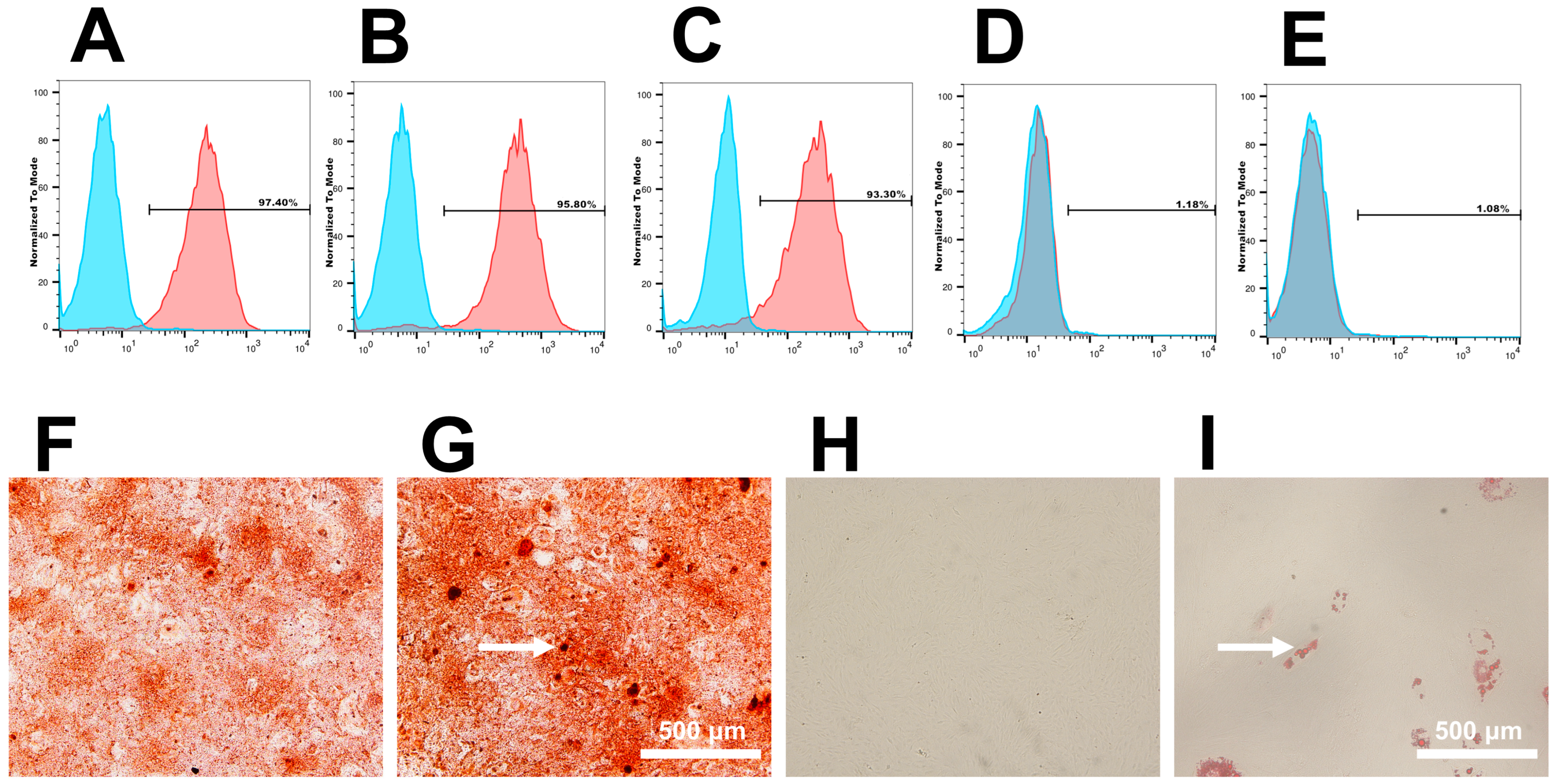
3.1. Cell Culture and Identification

The hDPSCs used in this study were obtained and cultured from dental pulp tissues successfully. To identify hDPSCs, the immunophenotype of the cells was detected using flow cytometer analytic method, which suggested that hDPSCs were both positive for MSC-specific surface markers (CD90 (97.40%); CD105 (95.80%); and CD146 (93.30%)) and negative for hematopoietic cell markers (CD34 (1.18%) and CD45 (1.08%)) (Figure 2A–E). Staining with alizarin red (Figure 2F,G) and oil red O (Figure 2H,I) confirmed multipotency of hDPSCs after 14 days of induction.

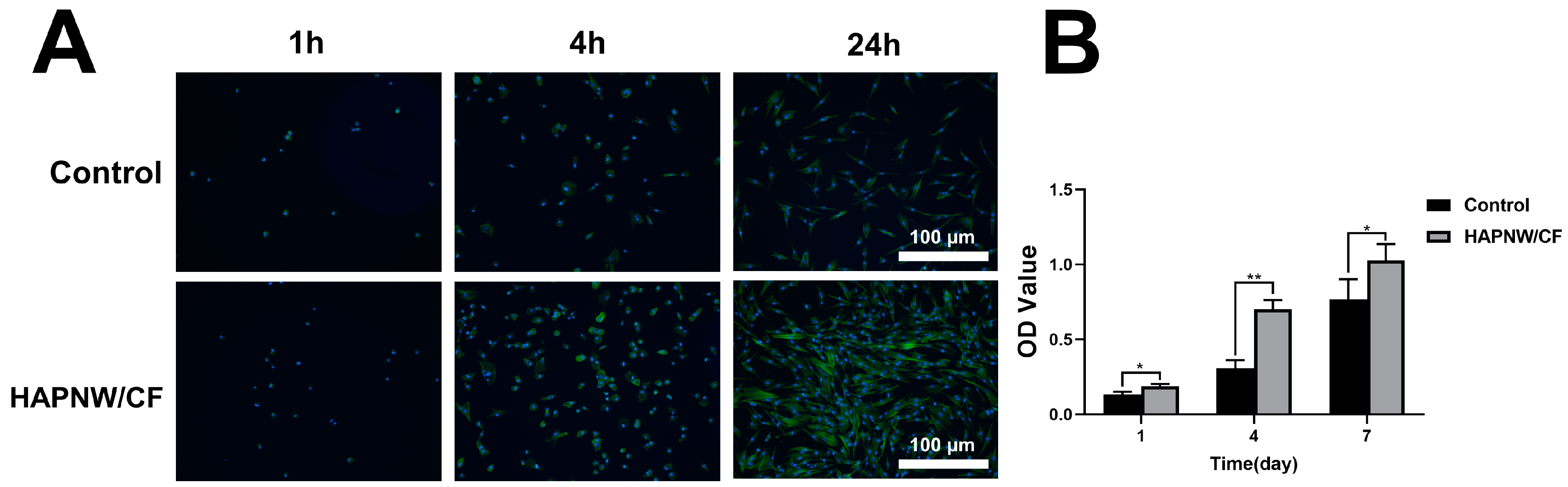
3.2. Cell Viability, Proliferation, and Adhesion Activity of hDPSCs on the HAPNW/CF Biopaper

The hDPSCs were cultured on blank culture dish (control group) and HAPNW/CF biopaper, respectively. Subsequently, the viability of cells on the HAPNW/CF biopaper was assessed. The immunofluorescence staining images of living/dead cells (Figure 3A) and live cell ratios (Figure 3B) after culturing for 3 days clearly showed that there was no significant difference between the numbers of live cells on the HAPNW/CF biopaper of the control group and the nHA group. The result showed that HAPNW/CF biopaper exhibits excellent safety. Cell adhesion serves as the initial stage in mediating the interaction between cells and the surface of biological materials, which not only allows cells to maintain structural stability and morphology but also mediates signaling and regulation between cells.
As shown in Figure 4A, the morphology of the cells by immunofluorescence staining was observed under a fluorescence microscope after seeding to the control and HAPNW/CF groups. After being cultured for 1, 4, and 24 h, compared with the control group, the HAPNW/CF group showed significantly increased cell density, better cell spreading morphology, and more pronounced lopodia extension, especially at 24 h. The images showed that the HAPNW/CF biopaper could provide a more favorable microenvironment for hDPSCs adhesion and thus provide assistance for the subsequent proliferation and differentiation of hDPSCs. Cell proliferation is essential to evaluate the biocompatibility of materials. The CCK-8 assay results showed that the proliferative activity of hDPSCs in the nHA group was obviously higher than that of the control group after being cultured for 1, 4, and 7 days (Figure 4B).

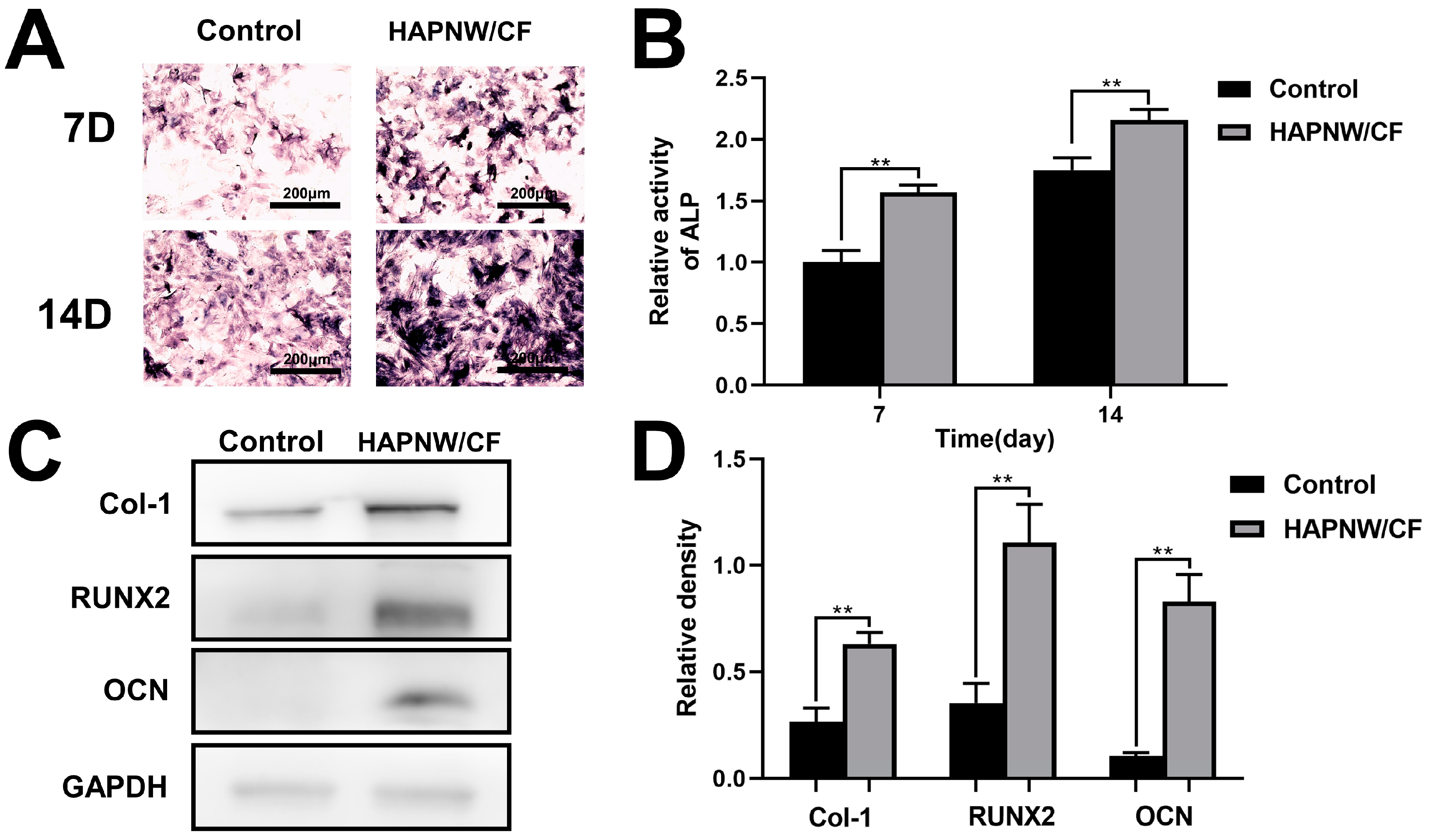
3.3. In Vitro Pro-Osteogenic and Pro-Angiogenic Effects

The hDPSCs represent a subtype of mesenchymal stem cells with multi-directional differentiation and self-renewal ability, which can proliferate and differentiate into osteoblasts under specific environmental and inducing conditions. As widely acknowledged, ALP serves as an early indicator of osteogenic differentiation in hDPSCs, which is qualitatively and quantitatively detected to analyze the osteogenic differentiation of hDPSCs on the surface of the HAPNW/CF biopaper. After being cultured for 7 and 14 days (Figure 5A), compared with the control group, the intensity of ALP staining in the HAPNW/CF group was significantly deeper. In addition, the ALP activity in the HAPNW/CF group was significantly higher than that in the control group (Figure 5B), proving a significant enhancement in osteogenic differentiation upon the utilization of HAPNW/CF biopaper.
Several bone formation-related proteins and transcription factors, such as Col-1, OCN, and RUNX2, play important roles during the process of osteogenic differentiation. Therefore, the effects of HAPNW/CF biopaper on the formation of Col-1, OCN and RUNX2 were analyzed by western blotting. As shown in Figure 5C,D, the hDPSCs cultured on the HAPNW/CF biopaper showed higher expression of Col-1,OCN, and RUNX2 after 7 days’ culture compared with the blank control group. In a word, the HAPNW/CF biopaper has excellent osteogenic properties and accelerates the initiation of osteogenesis.
In addition, the angiogenesis performance and proliferation of human umbilical vein endothelial cells (hUVECs) on the HAPNW/CF biopaper were investigated. As shown in western blot images (Figure 6A,B), the hUVECs on the HAPNW/CF biopaper expressed more angiogenic factors (eNOS, AKT, and VEGF), which were beneficial to neovascularization. The proliferation of cells is a crucial physiological function exhibited by living cells, and promoting cell proliferation is also an important index to evaluate the performance of biomaterials. After being cultured on the HAPNW/CF biopaper for 1, 4, and 7 days, the CCK-8 results showed that compared with the control group, the proliferative activity of hUVECs in the HAPNW/CF group was significantly higher (Figure 6C). Based on the experiment results, the HAPNW/CF biopaper effectively promoted the angiogenesis of hUVECs in vitro.

4. Discussion

Reconstructing bone defects presents a significant challenge for orthopedic and maxillofacial surgeons, particularly in cases of severe bone loss [39,40]. To overcome the limitations associated with traditional bone grafts (autografts and allografts), it is imperative to conduct research and develop bone tissue engineering scaffolds that possess exceptional osteoconductivity and osteoinductivity [3,25,41]. Hydroxyapatite has garnered considerable attention due to its biomimetic structure and chemical composition resembling natural bone. It can effectively stimulate new bone in areas affected by bone defects while also facilitating vascularization to some extent [36]. Therefore, we have developed a novel porous HAP nanowire/plant fiber composite paper known as HAPNW/CF biopaper and further investigated its role in promoting the formation of bone tissue. The HAPNW/CF biopaper exhibits high hydrophilicity, an elevated surface area-to-volume ratio, as well as the porous structure and high porosity inherent to HAP nanomaterials. These characteristics provide more adhesive sites for cells and proteins, thereby facilitating the adhesion and proliferation of osteoblasts. In vitro experiments demonstrate that the HAPNW/CF biopaper not only demonstrates the ability of adhesion, proliferation, and osteogenic differentiation of hDPSCs but also significantly influences the proliferation of hUVECs as well as the secretion of vascular endothelial growth factor.
The immunophenotypic characteristics of hDPSCs exhibited the expression of mesenchymal stem cell markers [42,43]. The staining results of Alizarin Red and Oil Red demonstrated the osteogenic and adipogenic differentiation potential of hDPSCs, demonstrating their multipotent differentiation potential. Dental pulp tissue is a soft tissue containing nerves, blood vessels, and lymphatics inside the pulp cavity. hDPSCs isolated from dental pulp tissue possess the ability to multipotently differentiate and self-renew and can be induced to form osteoblasts under certain conditions [44]. Numerous studies have shown that hDPSCs can form new bone tissue in the area of bone defects, effectively promoting new bone formation and providing assistance for bone defect repair [45,46]. Therefore, hDPSCs are considered necessary seed cells for bone regeneration and possess osteogenic potential [47,48]. Petridis et al. [49] transplanted dental pulp stem cells combined with extracellular matrix scaffolds into a critical cranial bone defect rat model, and the results showed that it effectively promoted rat skull healing. d’Aquino et al. [50] isolated and extracted stem cells derived from the dental pulp of third molars of humans, expanded them, adhered them to collagen sponge scaffolds, and filled them into bone defects in the autogenous mandible caused by bone resorption.
The live–dead staining results showed that, in comparison to the control group, HAPNW/CF biomaterials did not display cell toxicity, indicating excellent biocompatibility of HAPNW/CF biomaterials. Additionally, due to the relatively coarse surface morphology of the HAPNW/CF biomaterial scaffold, it was beneficial for cell adhesion and proliferation. Immunofluorescence staining results showed that cells could adhere to the surface of HAPNW/CF biomaterials early on, and the adhesiveness of hDPSCs on the HAPNW/CF biomaterial surface was superior to the control group. This was because the HAPNW/CF biomaterials provided more attachment sites for hDPSCs due to their higher specific surface area, which further promoted early adhesion of hDPSCs on the composite membrane surface. Cell viability was examined through CCK-8 experiments at different time points, and it was found that HAPNW/CF biomaterials promoted the proliferation of hDPSCs. Therefore, the highly biocompatible HAPNW/CF biomaterial scaffold provided a conducive and advantageous milieu for the proliferation and adhesion of hDPSCs, implying that HAPNW/CF biomaterials could have a positive influence on cell behavior. Similar to other research, many researchers have also found that hydroxyapatite can effectively promote the proliferation and adhesion of hDPSCs [51,52].
Hydroxyapatite nanowire/cellulose fiber (HAPNW/CF) biopaper is believed to possess the capability to promote preosteoblast differentiation [53]. The influence of HAPNW/CF biomaterial on hDPSCs osteogenic differentiation was investigated by assessing the expression of various osteogenic proteins. The staining and activity experiments of alkaline phosphatase (ALP) were conducted on day 7 and day 14. Compared to the control group, HAPNW/CF biomaterial induced higher ALP expression in hDPSCs. ALP is a specific protein enzyme secreted by cells, and its activity level reflects the early stage of osteogenic differentiation in osteoblasts [54]. Western blot experiments were also performed, demonstrating that HAPNW/CF biomaterial significantly promoted hDPSCs’ osteogenic differentiation and the expression of osteogenic proteins, including Col-1, RUNX2, and OCN. The behavior and fate of stem cells are influenced by their microenvironment. The arrangement of ultralong bundles composed of HAP nanowires within the HAPNW/CF biomaterial may exert certain effects on cell orientation and connectivity. Additionally, material degradation characteristics contribute to its biocompatibility as well. In bone tissue engineering, new bone formation often coincides with scaffold material degradation [55]. Degradation of HAPNW/CF biomaterial enables the continuous release of Ca2+ and PO43-ions, which stimulate hDPSCs’ osteogenic differentiation [56].
In vitro studies have demonstrated that HAPNW/CF biomaterial promotes angiogenesis. CCK-8 experiments were conducted on both the control group and HAPNW/CF biomaterial group at 1, 2, and 3 days. Results revealed that HAPNW/CF biomaterial significantly enhanced the proliferation rate among hUVECs. This indicates that the presence of HAPNW/CF biomaterial moderately improves cellular activity in hUVECs as compared to control group conditions. Furthermore, in comparison to the control group, the HAPNW/CF biomaterial also exhibited enhanced expression of angiogenesis-related proteins, including endothelial nitric oxide synthase (eNOS), AKT, and vascular endothelial growth factor (VEGF). VEGF is a highly specific angiogenic factor that stimulates increased vascular permeability, migration, and proliferation of endothelial cells, as well as angiogenesis itself [57,58]. This can be attributed to the continuous release of Ca2+ by the HAPNW/CF biomaterial, which promotes angiogenesis and facilitates blood vessel formation [59]. Angiogenesis remains a significant challenge in large-scale bone transplantation within bone tissue engineering. Synthetic scaffolds capable of promoting both bone and blood vessel formation are considered an effective solution [34,60].
In summary, the HAPNW/CF biomaterial scaffold provides a migratory pathway and reparative space for osteoblasts to address bone defects while demonstrating confirmed osteogenic potential through in vitro experiments. Moreover, owing to its exceptional biocompatibility, tissue compatibility, favorable bone conductivity properties as well as heightened biological activity levels; HAPNW/CF biomaterial emerges as an exceedingly promising candidate for bone defect repair.

5. Conclusions

In this study, we successfully fabricated ultralong hydroxyapatite nanowire-based biopaper. Subsequently, we assessed the in vitro cellular response of HAPNW/CF biopaper. The results demonstrated that HAPNW/CF biopaper promoted adhesion, proliferation, and osteogenic differentiation of human dental pulp stem cells. Furthermore, it enhanced cell viability and expression of angiogenesis-related proteins in human umbilical vein endothelial cells. The HAPNW/CF biopaper, owing to its osteoinductive property, creates a conducive milieu for bone tissue regeneration and exhibits immense potential for clinical applications.

Author Contributions

Conceptualization, K.L. and W.L.; methodology, K.L.; data curation, W.L., C.Y. and L.Y.; writing—original draft preparation, K.L., W.L., Y.Z., L.H., J.G. and C.H.; writing—review and editing, J.D. and Y.L.; project administration, Y.L.; funding acquisition, C.H., J.D. and Y.L. All authors have read and agreed to the published version of the manuscript.

Funding

The present study was financially supported by the National Natural Science Foundation of China (No. 12074253 and 12174289) and Xinhua Hospital affiliated to Shanghai Jiaotong University School of Medicine Rolling Fund for Scientific Research and Development (GD202112).

Institutional Review Board Statement

The experimental protocol employed in this study was granted approval by the Ethics Committee of Xinhua Hospital affiliated to Shanghai Jiao Tong University School of Medicine (approval number: XHEC-D-2022-149).

Informed Consent Statement

The concept is not applicable in this context.

Data Availability Statement

No new data were created or analyzed in this study. Data sharing is not applicable to this article.

Conflicts of Interest

The authors declare no conflicts of interest.

References

  1. Zhang, M.; Liu, J.; Zhu, T.; Le, H.; Wang, X.; Guo, J.; Liu, G.; Ding, J. Functional Macromolecular Adhesives for Bone Fracture Healing. ACS Appl. Mater. Interfaces 2022, 14, 1–19. [Google Scholar] [PubMed]
  2. Majidinia, M.; Sadeghpour, A.; Yousefi, B. The Roles of Signaling Pathways in Bone Repair and Regeneration. J. Cell. Physiol. 2018, 233, 2937–2948. [Google Scholar] [PubMed]
  3. Bharadwaz, A.; Jayasuriya, A.C. Recent Trends in the Application of Widely Used Natural and Synthetic Polymer Nanocomposites in Bone Tissue Regeneration. Mater. Sci. Eng. C 2020, 110, 110698. [Google Scholar]
  4. Zhang, J.; Tong, D.; Song, H.; Ruan, R.; Sun, Y.; Lin, Y.; Wang, J.; Hou, L.; Dai, J.; Ding, J.; et al. Osteoimmunity-Regulating Biomimetically Hierarchical Scaffold for Augmented Bone Regeneration. Adv. Mater. 2022, 34, 2202044. [Google Scholar]
  5. Schätzlein, E.; Kicker, C.; Söhling, N.; Ritz, U.; Neijhoft, J.; Henrich, D.; Frank, J.; Marzi, I.; Blaeser, A. 3D-Printed PLA-Bioglass Scaffolds with Controllable Calcium Release and MSC Adhesion for Bone Tissue Engineering. Polymers 2022, 14, 238. [Google Scholar]
  6. Zhang, Y.; Wang, C.; Fu, L.; Ye, S.; Wang, M.; Zhou, Y. Fabrication and Application of Novel Porous Scaffold in Situ-Loaded Graphene Oxide and Osteogenic Peptide by Cryogenic 3D Printing for Repairing Critical-Sized Bone Defect. Molecules 2019, 24, 1669. [Google Scholar]
  7. Freeman, F.; Browe, D.; Nulty, J.; Von Euw, S.; Grayson, W.; Kelly, D. Biofabrication of Multiscale Bone Extracellular Matrix Scaffolds for Bone Tissue Engineering. Eur. Cells Mater. 2019, 38, 168–187. [Google Scholar]
  8. Zhao, D.; Wang, X.; Cheng, B.; Yin, M.; Hou, Z.; Li, X.; Liu, K.; Tie, C.; Yin, M. Degradation-Kinetics-Controllable and Tissue-Regeneration-Matchable Photocross-Linked Alginate Hydrogels for Bone Repair. ACS Appl. Mater. Interfaces 2022, 14, 21886–22190. [Google Scholar]
  9. Bacakova, L.; Zarubova, J.; Travnickova, M.; Musilkova, J.; Pajorova, J.; Slepicka, P.; Kasalkova, N.S.; Svorcik, V.; Kolska, Z.; Motarjemi, H.; et al. Stem Cells: Their Source, Potency and Use in Regenerative Therapies with Focus on Adipose-Derived Stem Cells—A Review. Biotechnol. Adv. 2018, 36, 1111–1126. [Google Scholar]
  10. Cao, D.; Martinez, J.G.; Anada, R.; Hara, E.S.; Kamioka, H.; Jager, E.W.H. Electrochemical Control of Bone Microstructure on Electroactive Surfaces for Modulation of Stem Cells and Bone Tissue Engineering. Sci. Technol. Adv. Mater. 2023, 24, 2183710. [Google Scholar]
  11. Joshi, S.; Allabun, S.; Ojo, S.; Alqahtani, M.S.; Shukla, P.K.; Abbas, M.; Wechtaisong, C.; Almohiy, H.M. Enhanced Drug Delivery System Using Mesenchymal Stem Cells and Membrane-Coated Nanoparticles. Molecules 2023, 28, 2130. [Google Scholar]
  12. Golchin, A.; Chatziparasidou, A.; Ranjbarvan, P.; Niknam, Z.; Ardeshirylajimi, A. Embryonic Stem Cells in Clinical Trials: Current Overview of Developments and Challenges. In Cell Biology and Translational Medicine, Volume 11; Advances in Experimental Medicine and Biology; Turksen, K., Ed.; Springer International Publishing: Cham, Switzerland, 2020; Volume 1312, pp. 19–37. [Google Scholar]
  13. Guan, S.; Wang, Y.; Xie, F.; Wang, S.; Xu, W.; Xu, J.; Sun, C. Carboxymethyl Chitosan and Gelatin Hydrogel Scaffolds Incorporated with Conductive PEDOT Nanoparticles for Improved Neural Stem Cell Proliferation and Neuronal Differentiation. Molecules 2022, 27, 8326. [Google Scholar]
  14. Zhang, L.; Ma, X.-J.-N.; Fei, Y.-Y.; Han, H.-T.; Xu, J.; Cheng, L.; Li, X. Stem Cell Therapy in Liver Regeneration: Focus on Mesenchymal Stem Cells and Induced Pluripotent Stem Cells. Pharmacol. Ther. 2022, 232, 108004. [Google Scholar] [PubMed]
  15. Sarsenova, M.; Issabekova, A.; Abisheva, S.; Rutskaya-Moroshan, K.; Ogay, V.; Saparov, A. Mesenchymal Stem Cell-Based Therapy for Rheumatoid Arthritis. Int. J. Mol. Sci. 2021, 22, 11592. [Google Scholar] [PubMed]
  16. Gronthos, S.; Mankani, M.; Brahim, J.; Robey, P.G.; Shi, S. Postnatal Human Dental Pulp Stem Cells (DPSCs) in vitro and in vivo. Proc. Natl. Acad. Sci. USA 2000, 97, 13625–13630. [Google Scholar]
  17. Hilkens, P.; Gervois, P.; Fanton, Y.; Vanormelingen, J.; Martens, W.; Struys, T.; Politis, C.; Lambrichts, I.; Bronckaers, A. Effect of Isolation Methodology on Stem Cell Properties and Multilineage Differentiation Potential of Human Dental Pulp Stem Cells. Cell Tissue Res. 2013, 353, 65–78. [Google Scholar] [PubMed]
  18. Kim, B.-C.; Bae, H.; Kwon, I.-K.; Lee, E.-J.; Park, J.-H.; Khademhosseini, A.; Hwang, Y.-S. Osteoblastic/Cementoblastic and Neural Differentiation of Dental Stem Cells and Their Applications to Tissue Engineering and Regenerative Medicine. Tissue Eng. Part B Rev. 2012, 18, 235–244. [Google Scholar] [PubMed]
  19. Lee, J.-H.; Lee, D.-S.; Choung, H.-W.; Shon, W.-J.; Seo, B.-M.; Lee, E.-H.; Cho, J.-Y.; Park, J.-C. Odontogenic Differentiation of Human Dental Pulp Stem Cells Induced by Preameloblast-Derived Factors. Biomaterials 2011, 32, 9696–9706. [Google Scholar] [PubMed]
  20. Sloan, A.; Smith, A. Stem Cells and the Dental Pulp: Potential Roles in Dentine Regeneration and Repair. Oral Dis. 2007, 13, 151–157. [Google Scholar] [PubMed]
  21. Stuepp, R.T.; Barros Delben, P.; Modolo, F.; Trentin, A.G.; Garcez, R.C.; Biz, M.T. Human Dental Pulp Stem Cells in Rat Mandibular Bone Defects. Cells Tissues Organs 2019, 207, 138–148. [Google Scholar]
  22. Song, F.; Sun, H.; Huang, L.; Fu, D.; Huang, C. The Role of Pannexin3-Modified Human Dental Pulp-Derived Mesenchymal Stromal Cells in Repairing Rat Cranial Critical-Sized Bone Defects. Cell Physiol. Biochem. 2017, 44, 2174–2188. [Google Scholar]
  23. Sun, T.-W.; Zhu, Y.-J.; Chen, F. Hydroxyapatite Nanowire/Collagen Elastic Porous Nanocomposite and Its Enhanced Performance in Bone Defect Repair. RSC Adv. 2018, 8, 26218–26229. [Google Scholar]
  24. Fernandes, M.H.; Alves, M.M.; Cebotarenco, M.; Ribeiro, I.A.C.; Grenho, L.; Gomes, P.S.; Carmezim, M.J.; Santos, C.F. Citrate Zinc Hydroxyapatite Nanorods with Enhanced Cytocompatibility and Osteogenesis for Bone Regeneration. Mater. Sci. Eng. C 2020, 115, 111147. [Google Scholar]
  25. Kashiwada, H.; Shimizu, Y.; Sano, Y.; Yamauchi, K.; Guang, H.; Kumamoto, H.; Unuma, H.; Zhu, Y.-J. In vivo Behaviors of Highly Flexible Paper Consisting of Ultralong Hydroxyapatite Nanowires. J. Biomed. Mater. Res. Part B 2021, 109, 1611–1621. [Google Scholar]
  26. George, S.M.; Nayak, C.; Singh, I.; Balani, K. Multifunctional Hydroxyapatite Composites for Orthopedic Applications: A Review. ACS Biomater. Sci. Eng. 2022, 8, 3162–3186. [Google Scholar]
  27. Cheng, D.; Ding, R.; Jin, X.; Lu, Y.; Bao, W.; Zhao, Y.; Chen, S.; Shen, C.; Yang, Q.; Wang, Y. Strontium Ion-Functionalized Nano-Hydroxyapatite/Chitosan Composite Microspheres Promote Osteogenesis and Angiogenesis for Bone Regeneration. ACS Appl. Mater. Interfaces 2023, 15, 19951–19965. [Google Scholar]
  28. Maimaiti, B.; Zhang, N.; Yan, L.; Luo, J.; Xie, C.; Wang, Y.; Ma, C.; Ye, T. Stable ZnO-Doped Hydroxyapatite Nanocoating for Anti-Infection and Osteogenic on Titanium. Colloids Surf. B Biointerfaces 2020, 186, 110731. [Google Scholar]
  29. Tenkumo, T.; Vanegas Sáenz, J.R.; Takada, Y.; Takahashi, M.; Rotan, O.; Sokolova, V.; Epple, M.; Sasaki, K. Gene Transfection of Human Mesenchymal Stem Cells with a Nano-hydroxyapatite–Collagen Scaffold Containing DNA-functionalized Calcium Phosphate Nanoparticles. Genes Cells 2016, 21, 682–695. [Google Scholar]
  30. Munir, M.U.; Salman, S.; Javed, I.; Bukhari, S.N.A.; Ahmad, N.; Shad, N.A.; Aziz, F. Nano-Hydroxyapatite as a Delivery System: Overview and Advancements. Artif. Cells Nanomed. Biotechnol. 2021, 49, 717–727. [Google Scholar]
  31. Zhu, Y.J.; Lu, B.Q. Deformable Biomaterials Based on Ultralong Hydroxyapatite Nanowires. ACS Biomater. Sci. Eng. 2019, 5, 4951–4961. [Google Scholar]
  32. Gao, J.; Hao, L.-S.; Ning, B.-B.; Zhu, Y.-K.; Guan, J.-B.; Ren, H.-W.; Yu, H.-P.; Zhu, Y.-J.; Duan, J.-L. Biopaper Based on Ultralong Hydroxyapatite Nanowires and Cellulose Fibers Promotes Skin Wound Healing by Inducing Angiogenesis. Coatings 2022, 12, 479. [Google Scholar]
  33. Wang, F.; Qian, H.; Kong, L.; Wang, W.; Wang, X.; Xu, Z.; Chai, Y.; Xu, J.; Kang, Q. Accelerated Bone Regeneration by Astragaloside IV through Stimulating the Coupling of Osteogenesis and Angiogenesis. Int. J. Biol. Sci. 2021, 17, 1821–1836. [Google Scholar]
  34. Diomede, F.; Marconi, G.D.; Fonticoli, L.; Pizzicanella, J.; Merciaro, I.; Bramanti, P.; Mazzon, E.; Trubiani, O. Functional Relationship between Osteogenesis and Angiogenesis in Tissue Regeneration. Int. J. Mol. Sci. 2020, 21, 3242. [Google Scholar]
  35. Ji, X.; Yuan, X.; Ma, L.; Bi, B.; Zhu, H.; Lei, Z.; Liu, W.; Pu, H.; Jiang, J.; Jiang, X.; et al. Mesenchymal Stem Cell-Loaded Thermosensitive Hydroxypropyl Chitin Hydrogel Combined with a Three-Dimensional-Printed Poly(ε-Caprolactone) /Nano-Hydroxyapatite Scaffold to Repair Bone Defects via Osteogenesis, Angiogenesis and Immunomodulation. Theranostics 2020, 10, 725–740. [Google Scholar] [PubMed]
  36. Huang, G.-J.; Yu, H.-P.; Wang, X.-L.; Ning, B.-B.; Gao, J.; Shi, Y.-Q.; Zhu, Y.-J.; Duan, J.-L. Highly Porous and Elastic Aerogel Based on Ultralong Hydroxyapatite Nanowires for High-Performance Bone Regeneration and Neovascularization. J. Mater. Chem. B 2021, 9, 1277–1287. [Google Scholar]
  37. Jia, Q.; Chen, X.; Jiang, W.; Wang, W.; Guo, B.; Ni, L. The Regulatory Effects of Long Noncoding RNA-ANCR on Dental Tissue-Derived Stem Cells. Stem Cells Int. 2016, 2016, 3146805. [Google Scholar] [PubMed]
  38. Zeng, L.; Sun, S.; Han, D.; Liu, Y.; Liu, H.; Feng, H.; Wang, Y. Long Non-Coding RNA H19/SAHH Axis Epigenetically Regulates Odontogenic Differentiation of Human Dental Pulp Stem Cells. Cell Signal. 2018, 52, 65–73. [Google Scholar]
  39. Krasny, M.; Krasny, K.; Fiedor, P.; Zadurska, M.; Kamiński, A. Long-Term Outcomes of the Use of Allogeneic, Radiation-Sterilised Bone Blocks in Reconstruction of the Atrophied Alveolar Ridge in the Maxilla and Mandible. Cell Tissue Bank 2015, 16, 631–638. [Google Scholar] [PubMed]
  40. Hofmann, A.; Gorbulev, S.; Guehring, T.; Schulz, A.P.; Schupfner, R.; Raschke, M.; Huber-Wagner, S.; Rommens, P.M.; CERTiFy Study Group. Autologous Iliac Bone Graft Compared with Biphasic Hydroxyapatite and Calcium Sulfate Cement for the Treatment of Bone Defects in Tibial Plateau Fractures: A Prospective, Randomized, Open-Label, Multicenter Study. J. Bone Jt. Surg. 2020, 102, 179–193. [Google Scholar]
  41. Moshaverinia, A.; Ansari, S.; Chen, C.; Xu, X.; Akiyama, K.; Snead, M.L.; Zadeh, H.H.; Shi, S. Co-Encapsulation of Anti-BMP2 Monoclonal Antibody and Mesenchymal Stem Cells in Alginate Microspheres for Bone Tissue Engineering. Biomaterials 2013, 34, 6572–6579. [Google Scholar]
  42. Chen, Z.; Zhang, K.; Qiu, W.; Luo, Y.; Pan, Y.; Li, J.; Yang, Y.; Wu, B.; Fang, F. Genome-Wide Identification of Long Noncoding RNAs and Their Competing Endogenous RNA Networks Involved in the Odontogenic Differentiation of Human Dental Pulp Stem Cells. Stem Cell Res. Ther. 2020, 11, 114. [Google Scholar]
  43. Bao, M.; Liu, G.; Song, J.; Gao, Y. Long Non-Coding RNA MALAT1 Promotes Odontogenic Differentiation of Human Dental Pulp Stem Cells by Impairing microRNA-140-5p-Dependent Downregulation of GIT2. Cell Tissue Res. 2020, 382, 487–498. [Google Scholar]
  44. Agriesti, F.; Landini, F.; Tamma, M.; Pacelli, C.; Mazzoccoli, C.; Calice, G.; Ruggieri, V.; Capitanio, G.; Mori, G.; Piccoli, C.; et al. Bioenergetic Profile and Redox Tone Modulate in Vitro Osteogenesis of Human Dental Pulp Stem Cells: New Perspectives for Bone Regeneration and Repair. Stem Cell Res. Ther. 2023, 14, 215. [Google Scholar]
  45. Ferrarotti, F.; Romano, F.; Gamba, M.N.; Quirico, A.; Giraudi, M.; Audagna, M.; Aimetti, M. Human Intrabony Defect Regeneration with Micrografts Containing Dental Pulp Stem Cells: A Randomized Controlled Clinical Trial. J. Clin. Periodontol. 2018, 45, 841–850. [Google Scholar]
  46. Li, J.; Du, H.; Ji, X.; Chen, Y.; Li, Y.; Heng, B.C.; Xu, J. ETV2 Promotes Osteogenic Differentiation of Human Dental Pulp Stem Cells through the ERK/MAPK and PI3K-Akt Signaling Pathways. Stem Cell Res. Ther. 2022, 13, 495. [Google Scholar]
  47. Huang, C.-C.; Narayanan, R.; Alapati, S.; Ravindran, S. Exosomes as Biomimetic Tools for Stem Cell Differentiation: Applications in Dental Pulp Tissue Regeneration. Biomaterials 2016, 111, 103–115. [Google Scholar]
  48. Jin, Q.; Yuan, K.; Lin, W.; Niu, C.; Ma, R.; Huang, Z. Comparative Characterization of Mesenchymal Stem Cells from Human Dental Pulp and Adipose Tissue for Bone Regeneration Potential. Artif. Cells Nanomed. Biotechnol. 2019, 47, 1577–1584. [Google Scholar]
  49. Petridis, X.; Diamanti, E.; Trigas, G.C.; Kalyvas, D.; Kitraki, E. Bone Regeneration in Critical-Size Calvarial Defects Using Human Dental Pulp Cells in an Extracellular Matrix-Based Scaffold. J. Cranio-Maxillofac. Surg. 2015, 43, 483–490. [Google Scholar]
  50. d’Aquino, R.; De Rosa, A.; Lanza, V.; Tirino, V.; Laino, L.; Graziano, A.; Desiderio, V.; Laino, G.; Papaccio, G. Human Mandible Bone Defect Repair by the Grafting of Dental Pulp Stem/Progenitor Cells and Collagen Sponge Biocomplexes. Eur. Cells Mater. 2009, 18, 75–83. [Google Scholar]
  51. Zheng, P.; Yao, Q.; Mao, F.; Liu, N.; Xu, Y.; Wei, B.; Wang, L. Adhesion, Proliferation and Osteogenic Differentiation of Mesenchymal Stem Cells in 3D Printed Poly-ε-Caprolactone/Hydroxyapatite Scaffolds Combined with Bone Marrow Clots. Mol. Med. Rep. 2017, 16, 5078–5084. [Google Scholar]
  52. Chen, Y.; Zhang, F.; Fu, Q.; Liu, Y.; Wang, Z.; Qi, N. In Vitro Proliferation and Osteogenic Differentiation of Human Dental Pulp Stem Cells in Injectable Thermo-Sensitive Chitosan/β-Glycerophosphate/Hydroxyapatite Hydrogel. J. Biomater. Appl. 2016, 31, 317–327. [Google Scholar]
  53. Wang, B.; Li, Y.; Wang, S.; Jia, F.; Bian, A.; Wang, K.; Xie, L.; Yan, K.; Qiao, H.; Lin, H.; et al. Electrodeposited Dopamine/Strontium-Doped Hydroxyapatite Composite Coating on Pure Zinc for Anti-Corrosion, Antimicrobial and Osteogenesis. Mater. Sci. Eng. C 2021, 129, 112387. [Google Scholar]
  54. Lee, Y.-C.; Chan, Y.-H.; Hsieh, S.-C.; Lew, W.-Z.; Feng, S.-W. Comparing the Osteogenic Potentials and Bone Regeneration Capacities of Bone Marrow and Dental Pulp Mesenchymal Stem Cells in a Rabbit Calvarial Bone Defect Model. Int. J. Mol. Sci. 2019, 20, 5015. [Google Scholar]
  55. Lin, J.H.C.; Kuo, K.H.; Ding, S.J.; Ju, C.P. Surface Reaction of Stoichiometric and Calcium-Deficient Hydroxyapatite in Simulated Body Fluid. J. Mater. Sci. Mater. Med. 2001, 12, 731–741. [Google Scholar]
  56. Kang, H.-J.; Makkar, P.; Padalhin, A.R.; Lee, G.-H.; Im, S.-B.; Lee, B.-T. Comparative Study on Biodegradation and Biocompatibility of Multichannel Calcium Phosphate Based Bone Substitutes. Mater. Sci. Eng. C 2020, 110, 110694. [Google Scholar]
  57. Liu, Y.; Long, L.; Zhang, F.; Hu, X.; Zhang, J.; Hu, C.; Wang, Y.; Xu, J. Microneedle-Mediated Vascular Endothelial Growth Factor Delivery Promotes Angiogenesis and Functional Recovery after Stroke. J. Control. Release 2021, 338, 610–622. [Google Scholar] [PubMed]
  58. Abdulkadir, S.; Li, C.; Jiang, W.; Zhao, X.; Sang, P.; Wei, L.; Hu, Y.; Li, Q.; Cai, J. Modulating Angiogenesis by Proteomimetics of Vascular Endothelial Growth Factor. J. Am. Chem. Soc. 2022, 144, 270–281. [Google Scholar] [PubMed]
  59. Wood, W. Wound Healing: Calcium Flashes Illuminate Early Events. Curr. Biol. 2012, 22, R14–R16. [Google Scholar]
  60. Zhang, X.; Huang, P.; Jiang, G.; Zhang, M.; Yu, F.; Dong, X.; Wang, L.; Chen, Y.; Zhang, W.; Qi, Y.; et al. A Novel Magnesium Ion-Incorporating Dual-Crosslinked Hydrogel to Improve Bone Scaffold-Mediated Osteogenesis and Angiogenesis. Mater. Sci. Eng. C 2021, 121, 111868. [Google Scholar]
Figure 1. A concise schematic diagram illustrating the preparation, angiogenesis, and osteogenesis processes of HAPNW/CF biopaper.
Coatings 14 00190 g001
Figure 2. Cell identification, osteogenic and adipogenesis differentiation of hDPSCs. Cell surface markers: (A) CD90 (97.40%); (B) CD105 (95.80%); (C) CD146 (93.30%); (D) CD73 (95.50%) and (E) CD34 (1.18%). Alizarin red staining was used to test mineralized tissue formation of hDPSCs in the control group (F) and experimental group (G) on days 14 post-osteoinduction. As shown by the arrow, calcium nodes were generated in the experimental group (scale bar: 500 μm). Oil red O staining was used to test adipogenic differentiation of hDPSCs in the control group (H) and experimental group (I) for 14 days (scale bar: 500 μm). As shown by the arrow, lipid droplets were generated in the experimental group (scale bar: 500 μm).
Coatings 14 00190 g002
Figure 3. Live–dead staining on the HAPNW/CF sample and blank control sample. (A) Live–dead staining images of hDPSCs cultured on scaffold for 3 days (scale bar: 500 μm). (B) Live cell ratio of hDPSCs in live–dead staining results.
Coatings 14 00190 g003
Figure 4. Cell adhesion and proliferation on the blank control sample and the HAPNW/CF sample. (A) Fluorescence images of hDPSCs cultured for 24 h (scale bar: 100 μm). (B) The proliferation of hDPSCs cultured for 1, 4, and 7 days (* p < 0.05, ** p < 0.01, n = 3).
Coatings 14 00190 g004
Figure 5. Promotion effects of the HAPNW/CF sample on osteogenic differentiation of hDPSCs. (A) ALP staining (scale bar: 200 μm) and (B) ALP activity of hDPSCs incubated on various samples for 7 and 14 days (** p < 0.01, n = 3). (C,D) Western blotting analysis of bone formation-related proteins of hDPSCs cultured for 7 days, including Col-1, RUNX2, and OCN. GAPDH was selected as the internal reference. (** p < 0.01, n = 3).
Coatings 14 00190 g005
Figure 6. Assessment of in vitro cell proliferation and angiogenesis of the HAPNW/CF sample and blank control sample (A,B). Western blotting analysis of protein expression (eNOS, AKT, and VEGF) of hUVECs cultured for 7 days. (C) The proliferation of hUVECs cultured for 1, 4, and 7 days (* p < 0.05, ** p < 0.01, n = 3).
Coatings 14 00190 g006
Disclaimer/Publisher’s Note: The statements, opinions and data contained in all publications are solely those of the individual author(s) and contributor(s) and not of MDPI and/or the editor(s). MDPI and/or the editor(s) disclaim responsibility for any injury to people or property resulting from any ideas, methods, instructions or products referred to in the content.

