Article

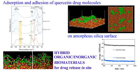
Hybrid Organic–Inorganic Biomaterials as Drug Delivery Systems: A Molecular Dynamics Study of Quercetin Adsorption on Amorphous Silica Surfaces

by
Giuseppina Raffaini
1,*,
Pasqualina Pirozzi
1,
Michelina Catauro
2,* and
Antonio D’Angelo
2
1
Department of Chemistry, Materials, and Chemical Engineering ‘‘Giulio Natta’’, Politecnico di Milano, Piazza L. da Vinci 32, 20131 Milan, Italy
2
Department of Engineering, University of Campania “Luigi Vanvitelli”, Via Roma 29, 813031 Aversa, Italy
*
Authors to whom correspondence should be addressed.
Coatings 2024, 14(2), 234; https://doi.org/10.3390/coatings14020234
Submission received: 28 December 2023 / Revised: 14 February 2024 / Accepted: 15 February 2024 / Published: 17 February 2024

Abstract

:
Many important drugs in pharmaceutical applications are poorly soluble. Solubilization, which is diffusion through biological barriers, and the control of local administration are crucial steps for bioavailability and to avoid cytotoxic effects. Hybrid organic/inorganic biomaterials can incorporate drugs for in situ release after implantation. Molecular Mechanics (MM) and Molecular Dynamics (MD) simulations are useful tools for investigating intermolecular interactions between drug and biomaterial surfaces at the atomistic level for these applications. This work studies quercetin, a flavonoid drug important for its anti-inflammatory, antioxidant, and anticancer properties, and the amorphous SiO2 surface using a simulation protocol proposed in previous work related to ketoprofen drugs. After adsorption on the amorphous silica surface, the adsorption process of quercetin drug molecules at two different drug concentrations near a hydrated and then dried silica surface is investigated. Interestingly, these theoretical results are compared with experimental data obtained via Fourier Transform Infrared Spectroscopy (FT–IR) spectra related to quercetin molecules homogenously entrapped in a silica matrix obtained via the Sol–Gel method. Favorable H– bonds and some π–π interactions among drug molecules are crucial surface interactions for the new generation of biocompatible materials capable of incorporating anti-inflammatory agents for release into the human body.

Graphical Abstract

1. Introduction

The use of antibiotics has achieved profound success in combatting numerous bacterial infections, but antibiotic resistance due to the long-term use of antibiotics is a rapidly emerging problem [1,2,3,4,5]. Implant-associated infections are not easy to diagnose and very difficult to treat. Local antimicrobial treatment aimed at enhancing the concentration of the drug at the site of infection while avoiding systemic exposure is becoming established [6,7,8,9]. Therapeutic drug monitoring based on blood analysis can help alleviate the emergence of bacterial antimicrobial resistance and effectively reduce the risk of toxic drug concentrations in the blood of the patients. The concentrations at the site of infection using wearable sensors are crucial. Locally targeted treatment of bone and joint infections using carriers containing antibiotics is a common practice today [10,11,12,13,14,15,16,17,18,19]. Organic/inorganic silica-based biomaterials capable of encapsulating the drug for in situ release after implantation also represent an increasingly attractive field for a good response to antibiotic resistance [20,21,22,23,24].
To overcome the difficulty of therapeutic drugs penetrating the blood–brain barrier, nanoparticles can be used as drug transport carriers and are also known as nanoparticle-based drug delivery systems [25,26,27,28,29,30,31,32]. Nanoparticle-based drug delivery systems must be biocompatible, stable, and biodegradable and are not expected to be cytotoxic or, in general, provoke an immune response. Every single NP formulation must be proofed with respect to its cytotoxicity, biocompatibility, dose dependency, and so on [33,34]. In recent years, carbon nanomaterials, such as graphene, carbon nanotubes, and carbon dots, have gained widespread interest in neuroscience as promising and outstanding agents for various biomedical applications [35,36,37,38,39]. Inorganic materials often used to form nanoparticle systems include iron, gold, silica, silver, titanium, and zinc, which are also used in biomaterial applications [40,41,42,43,44,45,46,47], and, in particular, mesoporous silica nanoparticles (MSNs) have recently received much interest because of their potential benefits in drug delivery [48,49,50,51,52]. The surface properties of MSNs need to be modified or fine-tuned to improve the biocompatibility of these new and versatile drug delivery systems [53,54,55,56].
Mesoporous silica nanoparticles are interesting materials capable of improving the bioavailability of drugs thanks to the adsorption, adhesion, and dispersion of poorly soluble drug molecules in a homogeneous arrangement on the external surface of the specific organic/inorganic biomaterial [57,58,59,60,61,62,63,64]. The Sol–Gel method is an interesting technique that is useful for preparing micro- or nanoporous silica-based materials with different percentages of drugs [65]. Thanks to spectroscopic techniques, it is possible to obtain useful information if drug molecules interact with the solid surface [66,67,68]. In a previous study, the molecules of the drug ketoprofen, a poorly soluble inflammatory drug [69], were encapsulated in a silica matrix via the Sol–Gel method [70,71,72,73,74,75,76]. After FT-IR experiments [77] related to drug-loaded silica-based biomaterials, the adsorption of the drug ketoprofen on amorphous SiO2 surfaces was confirmed considering two different concentrations, with two different release kinetics faster at relatively higher concentrations. Using theoretical Molecular Mechanics (MM) and Molecular Dynamics (MD) methods and a specific simulation protocol proposed for the adsorption process on the hydrated and dried surface [70,77], favorable non-covalent interactions at the external surface are confirmed. In particular, the H– bonds among ketoprofen drug molecules and hydrated silica surface are due to the important role of water molecules in the initial adsorption that forms an extensive network of hydrogen bonds (H– bonds) with the drug and the surface. These drug/surface H– bonds formed are also present in the dried surface, as indicated by FTIR spectra [77].
Using MM/MD methods, the adsorption and adhesion of drugs on carriers that are useful for their solubilization, transport, and release in a biological environment have been studied with interesting comparisons with experimental data [78,79,80,81,82,83,84,85,86,87,88,89,90,91,92,93,94,95,96,97,98]. In particular, the adsorption process and the possible release mechanism of piroxicam, a non-steroidal anti-inflammatory drug, adsorbed on the β-cyclodextrin nanosponge model [95] were studied. The dispersion of the hydrophobic curcumin, an important anticancer drug, has been theoretically studied, both in new pH-dependent carriers proposed by professors Ferruti and Ranucci [96] and well dispersed in nano aggregates formed by amphiphilic cyclodextrins proposed by Mazzaglia [97], and was theoretically investigated. All these theoretical results agree with the experimental data [92,93,94,95]. Possible new classes of hydrophobic drug carriers that can adsorb on the chiral DNA structure are currently being studied [99,100]. Interestingly, using MM/MD methods, the possible slower or faster release of drugs adhered and dispersed in nano aggregates or on a solid silica surface for their solubilization may be related to the strength of intermolecular interaction [95,98] as in the present work. The procedure proposed in this theoretical study of the adsorption of quercetin on amorphous silica following the experimental procedure on hydrated and then dried surfaces [77] allows for a better description and understanding of the data obtained via FT–IR on these new inorganic/organic materials [101,102,103,104]. This study aims to understand the ability of silica-based biomaterials to incorporate quercetin (Q), an anti-inflammatory agent, to be released into the human body in situ after implantation.

2. Materials and Methods

2.1. Materials

For the Sol–Gel route, tetraethyl orthosilicate reagent grade (TEOS, (C2H5O)4Si) was used as a precursor, and pure ethanol (99.8% (EtOH)) as the main solvent. MilliQ water and nitric acid (HNO3, 65%) were added to control the hydrolysis rate reaction [105,106]. Pure quercetin (Q, 95%, code Q4951-100G) was added as a drug, while potassium bromide 99.0% (KBr) was used for FTIR analyses. All these reagents were provided by Sigma-Aldrich (Darmstadt, Germany).

2.2. Methods

2.2.1. SiO2 and SiO2/Quercetin (5 and 15 wt.%) Sol–Gel Synthesis

To prepare the silica (SiO2) matrix, TEOS and EtOH were mixed under continuous magnetic stirring. After the solution was formed, MilliQ water and HNO3 were added to start the hydrolysis reactions. The synthesis followed the fixed molecular ratios TEOS:EtOH:H2O:HNO3 = 1:6:4:0.6. In regard to the hybrid SiO2/Q(5 and 15 wt.%), two EtOH-based solutions containing 5 and 15 wt.% (with respect to SiO2 content) of quercetin were added drop by drop to the TEOS/EtOH solution, while keeping the above-mentioned ratios, before MilliQ water and HNO3 addition. After gelation occurrences, the three wet gels were dried at 50 °C for 24 h.

2.2.2. Fourier Transform Infrared Analysis

FT–IR spectra were acquired using the Prestige21 Shimadzu system (Shimadzu, Milan, Italy). This system is equipped with a DTGS KBr (deuterated triglycine sulfate with potassium bromide windows) detector. The analyses were performed with KBr disk-based method [107] (2 mg of sample and 198 mg of KBr) in the range of 400–4000 cm−1, with a number of scans equal to 60 and a resolution of 2 cm−1.

2.2.3. Molecular Mechanics and Dynamics Methods

This theoretical study is based on the Molecular Mechanics (MM) and Molecular Dynamics (MD) methods, which describe, at an atomistic level, both the quercetin molecule and an amorphous SiO2 surface, adopting the same simulation protocol proposed in previous work related to the adsorption process of drug ketoprofen on silica surface [77]. All simulations were carried out using the Materials Studio package (version 7.0, BIOVIA, Accelrys Inc., San Diego, CA, USA) [108] and the CVFF force field [109].
Initially, the quercetin structure was optimized after MD run, lasting 2 ns and with energy minimization. Then, the amorphous SiO2 surface was modeled using the amorphous structure reported in Materials Studio Template considering a film thickness of approximately 15 Å and therefore considering a simulation box of size equal to (85.53 × 85.53 × 250.0) Å. As in previous work, the silica surface was considered fixed in all calculations [77]. After studying the structure of the quercetin drug molecule and preparing the model of the solid silica surface, the adsorption process of the single quercetin molecule and these drug molecules at both small and, at higher concentrations, on the hydrated and dried silica surface was investigated. This proposed simulation protocol allows us to mimic the same experimental procedure before the FT–IR experiments [77]. Starting from the optimized geometries obtained at the end of MD run related to the adsorption process on the hydrated surface and drug molecules in contact with silica surface, the water molecules are removed, and this new dried system is studied.
All simulations are carried out using periodic boundary conditions in the NVT ensemble, considering a constant temperature equal to 300 K controlled through the Berendsen thermostat. All energy minimizations were carried out using the Conjugate Gradient algorithm up to an energy gradient lower than 4 × 10⁻³ kJ mol−1 Å−1. After an initial energy minimization, MD runs, and final geometry optimizations, the adsorption at small and larger concentrations was studied, considering the hydrated surface and, finally, the dried surface. The integration of the dynamical equations was carried out with the Verlet algorithm using a time step of 1 fs. After energy minimization, MD runs, and optimization of the final geometry assumed by the system at equilibrium achieved in vacuo, the intermolecular interactions are investigated between quercetin molecules and the dried surface.

3. Results and Discussion

The results of the adsorption process of a single quercetin molecule on the amorphous silica surface will be discussed in Section 3.1, of quercetin molecules at small concentrations (14 drug molecules) in Section 3.2, at higher concentrations (28 drug molecules) in Section 3.3, and then comparison with FT–IR spectra in Section 3.4. The chemical structure and the optimized geometry of quercetin calculated after a 2 ns MD run and energy minimization are shown in Figure 1. It is interesting to note that for the ketoprofen molecule studied in previous work (chemical formula C16H14O3, 2-(3-Benzoylphenyl) propanoic acid), the volume occupied by this single drug molecule in the optimized geometry is equal to 241 Å3, and its surface area is 255 Å2. Regarding the quercetin molecule (C15H10O7, 3,3′,4′,5,7-pentahydroxyflavone), its occupied volume in the final optimized studied geometry after the MD run equals 239 Å3, and the surface area is 254 Å2. This fact explains two important aspects: first, the quercetin drug molecule can form a larger number of H– bonds due to its chemical structure with respect to the ketoprofen molecule (see Figure 1 in reference [77]), and these two drug molecules studied in a concentration that avoids total surface coverage are reasonably studied considering the same number of molecules near the solid surface for a good comparison with the FT–IR experimental data. In the next section, the results will be presented and discussed.

3.1. Adsorption of a Single Quercetin Molecule on the Amorphous SiO2 Surface

The adsorption process of the single quercetin molecule on the SiO2 surface in vacuo is carried out by considering the drug molecule in four different initial orientations near the solid surface using a simulation protocol proposed in previous work [77,81]. In two initial geometries, the quercetin is perpendicular to the surface, and in the other two geometries, it is parallel to the surface, facing all the aromatic rings and the –OH group to the amorphous silica surface in order to better characterize all the possible initial geometries of interaction and their stability after the MD run. Figure 2 shows the four initial geometries in panels (a), (b), (c), and (d) and the optimized geometries after four MD runs lasting 1 ns, respectively. The most stable geometry is represented in panel (h) in Figure 2. The number of quercetin intramolecular H– bonds and intermolecular H– bonds formed between the drug and silica surface in all final optimized geometries in Figure 2 is reported in Table 1.
The intermolecular interactions characterizing the adsorption of the quercetin molecule and the silica surface after the MD run are due to H– bonds formed in each of the four geometries (see blue dashed line in Figure 2) between the drug and the solid surface. These H– bonds form between the hydroxyl groups of the quercetin molecule and the hydroxyl group of the silica surface and also between the carbonylic group of the quercetin molecule and the hydroxyl group of the SiO2 matrix (–C=Oquercetin ⋯ H–O–SiO2 surface). These weak intermolecular interactions allow the adhesion of quercetin onto the silica matrix. Interestingly, for the drug ketoprofen, starting from several initial trial arrangements, the mobility on the silica surface was so high that the process of adsorption and desorption of a single drug molecule was too fast, and the drug was an average distance from the surface.
During all four MD runs, the drug molecule shows high mobility near the amorphous surface due to weak intermolecular interactions. Due to its chemical structure compared to the newly studied ketoprofen drug molecule, the single quercetin molecule can form more H– bonds with the silica surface, so an adsorption–desorption process occurs. After the adsorption process, the desorption process can also take place thanks to the kinetic energy of quercetin at the simulation temperature, which can overcome the weak intermolecular interaction. During the MD run at room temperature, after desorption of the quercetin and random motion in the simulation cell, the molecule, as it approaches the silica surface, adsorbs and remains adhered, forming more stable H– bonds. To study the adsorption–desorption process that occurs during an MD run in vacuo at a temperature equal to 300 K, the best-fit plane defined by all silica atoms was characterized (see the green plane in panel (a) of Figure 3), and the distance between the center of mass (c.o.m.) of the quercetin molecule and this silica best-fit plane during the MD run lasting 1 ns has been calculated, as reported in Figure 3, which more precisely starts from the geometry reported in panel (d) in Figure 2.

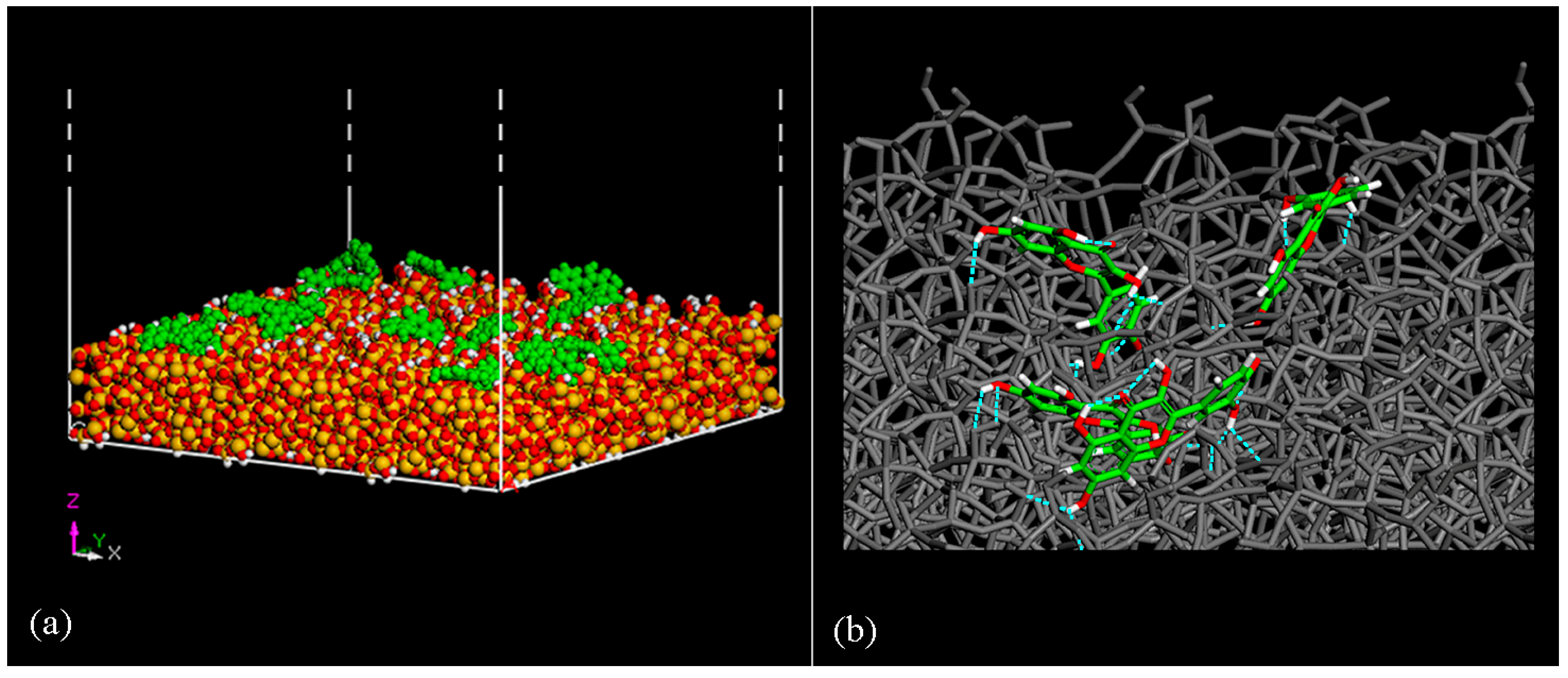
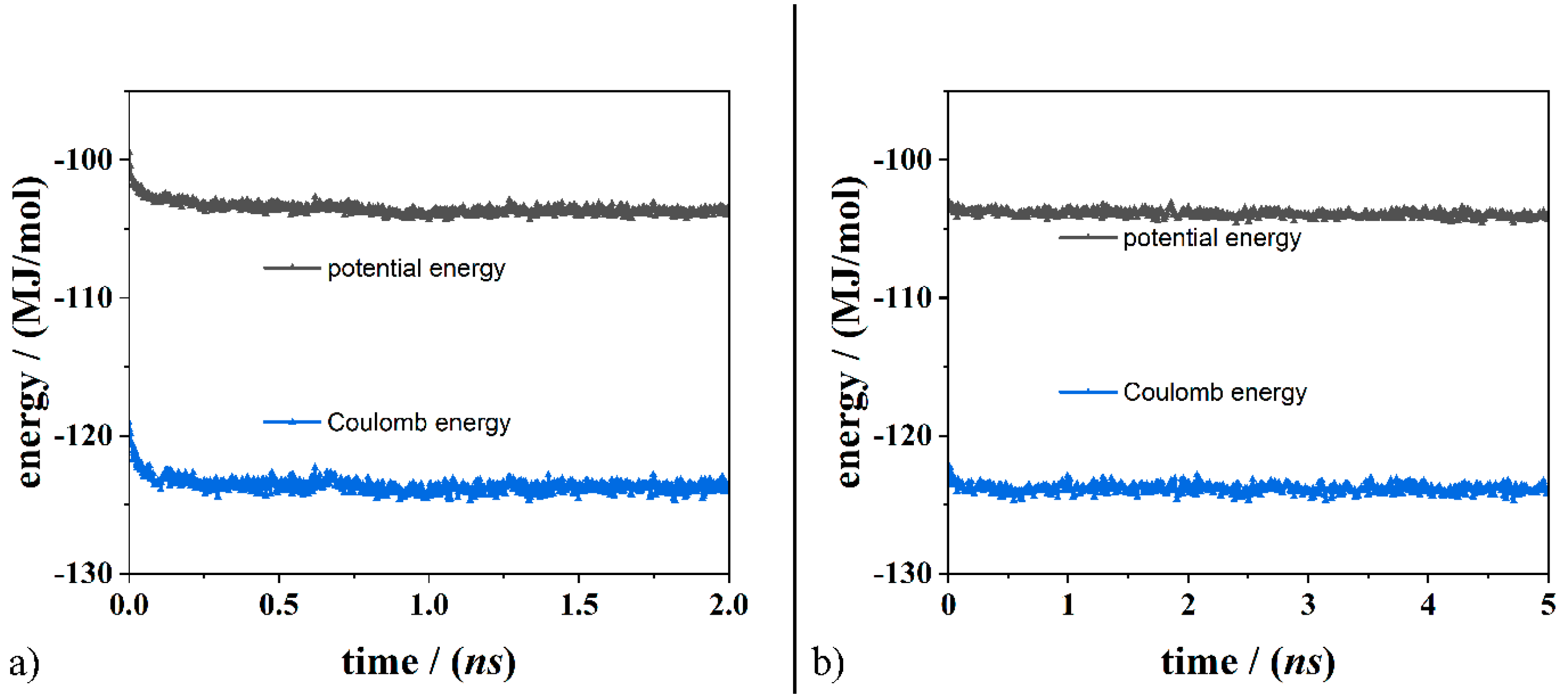
3.2. Quercetin Molecules on the Amorphous SiO2 Surface: Small Drug Concentration

The adsorption process on the silica surface of quercetin molecules at small drug concentrations was performed considering 14 quercetin molecules as in previous work related to ketoprofen drugs. MM and MD simulations were performed on hydrated silica and then dried SiO2 surfaces, mimicking the same experimental procedure for FT–IR data.
Initially, the adsorption of 14 quercetin drug molecules near the hydrated SiO2 surface was investigated. After energy minimization, MD runs lasting 2 ns, a layer of water molecules, and quercetin drug molecules were homogeneously adsorbed on the amorphous SiO2 surface. Starting from this last optimized geometry, a further MD run lasting 3 ns has been performed to verify the equilibrium state achieved by the system. The potential energy and Coulombic components calculated during the MD run are reported in Figure 4, and their running average values are calculated during the MD run. Interestingly, the fast decrease in the potential energy is due to the important contribution of electrostatic energy, as found by Nejad et al. [110,111] regarding the adsorption of insulin on polar crystalline SiO2 (cristobalite) and in a previous study on the adsorption process of ketoprofen drug molecules on an amorphous silica surface with silanol groups exposed from the solid surface. The stability of the system is the same also after the additional MD run lasting 3 ns starting from the optimized geometry calculated after the first MD run lasting 2 ns. The van der Waals contributions fluctuate around an average value during two MD runs, as found in the ketoprofen molecule on this SiO2 surface (see Figure S1, Supplementary Materials). The mobility of 14 drug molecules adsorbed on a hydrated silica surface is lower than the mobility of a single quercetin molecule, indicating that the water molecules act as an important connection with the silica surface and drug molecules due to the network of H– bonds with the –OH groups exposed from the silica surface and in the chemical structure of the drug quercetin.
The molecules show limited mobility on the surface due to the favorable interaction with the silica surface in the presence of water molecules. The distance between the center of mass of two different quercetin molecules and the silica best-fit plane was calculated during the first MD run lasting 2 ns. In panel (c) of Figure 4, the length distribution of the quercetin molecule indicated as molecule one varies from 7.7 to 11.2 Å, while the one of the molecule indicated as molecule two varies from 10.1 to 11.6 Å. The first drug molecule displays major mobility on the hydrated amorphous SiO2 surface. The same study related to two different quercetin molecules after the second MD run lasting 3 ns shows how the molecule adheres well to the silica matrix after thermalization and reaching the equilibrium state. For the second MD run, two other different quercetin molecules were considered, called molecules three and four, as reported in panel (d) in Figure 4. The c.o.m. of each molecule was calculated together with their distance from the silica best-fit plane. The length distribution, in this case, is more limited for molecule three; in fact, it varies from 7.7 Å to 9.1 Å, while the values related to molecule four vary from 10 Å to 11.6 Å, therefore showing low mobility on the surface.
Interestingly, after the MD run lasting 2 ns, optimization of the conformation assumed by the system when the equilibrium state was achieved (see Figure S2, Supplementary Materials), the equilibrium state achieved after the second MD run lasting 3 ns is very similar to that found at 2 ns. This suggests the possibility of producing these organic/inorganic drug/loaded biomaterials at room temperature by placing the sample prepared via the Sol–Gel method in a freezer, and when this is brought back to room temperature for use, the system reaches the same state of equilibrium.
After studying the adsorption process on the hydrated amorphous SiO2 surface, the dried surface is investigated, deleting water molecules in order to mimic the drying process for the sample synthesized and experimentally prepared via the Sol–Gel method before FT–IR spectra. After initial energy minimization, an MD run in vacuo mimicked the dried surface, lasting 5 ns, and a final geometry optimization of the system at an equilibrium state was performed. The potential energy and its components obtained at the end of the dynamic simulation were investigated (see panel (a) and panel (b) of Figure 5). As observed in the hydrated sample, even in vacuo, the adsorption process is mainly driven by the Coulomb energy, while the van der Waals energy contribution is less relevant.
Figure 6 shows the final optimized geometry at the end of the MD run lasting 5 ns on the dried silica surface. A homogeneous dispersion of drug molecules is observed (see panel (a) in Figure 6), and the H– bonds between quercetin molecules and the SiO2 surface are in a blue dashed line. The ability to form H– bonds with silica matrix for all single quercetin molecules is very high considering the number of –OH groups per molecule. These aspects will be discussed with the information on FT–IR spectra in Section 3.4.
Interestingly, in the final geometry optimized after 5 ns (2 ns and subsequently 3 ns) reported in Figure S1 related to 14 quercetin molecules adsorbed on the hydrated silica surface, the number of intermolecular H– bonds between drugs and the solid surface is 29, approximately two H– bonds per molecule. In the final geometry optimized after 10 ns in vacuo shown in panel (a) of Figure 6, considering 14 quercetin molecules on the dried silica surface, the number of intermolecular H– bonds between the drugs and the SiO2 surface is 38, approximately three H– bonds per molecule. Without the water molecule that hydrates the adsorbed drugs, the quercetin molecules form a greater number of H– bonds with the silica surface. Due to their chemical structure, quercetin molecules always form intramolecular H– bonds: on the hydrated surface, they are 17; on the dried SiO2 surface, they are 16. In practice, the number of intramolecular H– bonds does not vary in the presence or absence of water molecules when a small drug concentration is investigated.

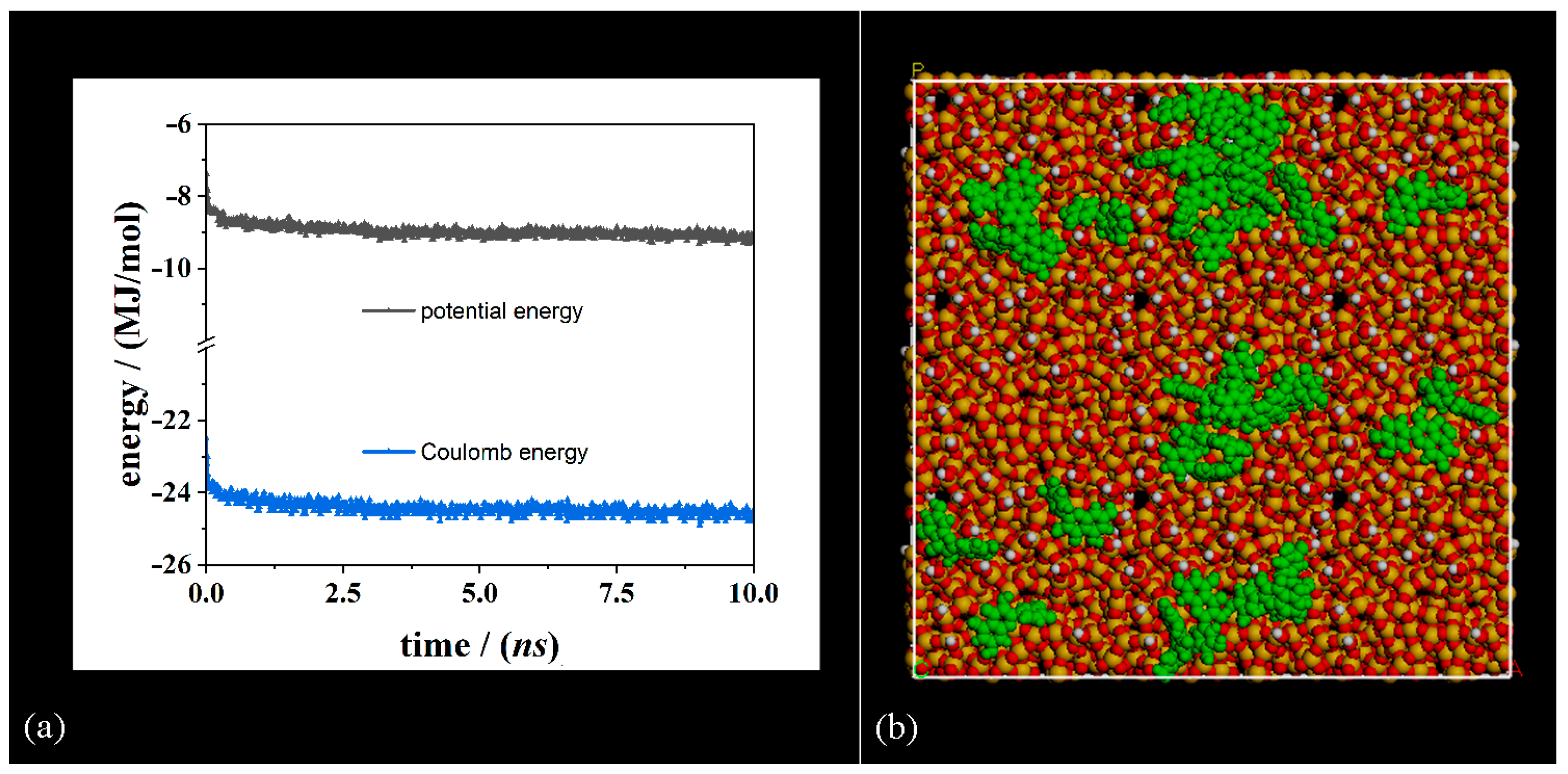
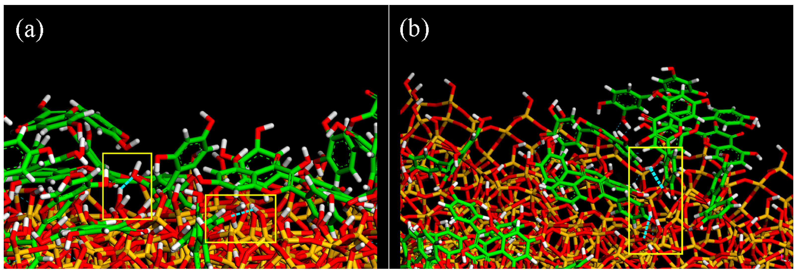
3.3. Quercetin Molecules on the Amorphous SiO2 Surface: Higher Drug Concentration

The adsorption process of quercetin on an amorphous SiO2 surface at higher concentrations, considering 28 drug molecules, was studied following the same approach explained in Section 3.2. First, adsorption on the hydrated silica surface was performed, and then, it was put on a dried surface.
Panel (a) in Figure 7 shows the potential energy, Coulomb contributions, and their running average values calculated during the first MD run lasting 2 ns related to 28 quercetin molecules near the hydrated silica surface. Panel (b) in Figure 7 displays the same energy contributions calculated during the second MD run lasting 5 ns performed starting from the final optimized geometry obtained at the end of the first MD run. The fast decrease in potential energy is again due to the significant contribution of electrostatic energy, as previously identified in Section 3.2. The van der Waals contributions fluctuate around an average value, as reported in Figure S3 (Supplementary Materials).
In both cases, a homogenous dispersion of quercetin molecule at the end of the MD run is observed, as well as a reduced mobility of quercetin on the surface due to not only the H– bonds with the silica surface but also to hydrophobic interaction with the neighboring drug molecule at larger concentrations. While drug molecules are uniformly dispersed during the first dynamics simulation on the hydrated surface, they tend to aggregate in the second MD run. It is still interesting to note that after the MD run lasting 2 ns, the optimization of the conformation assumed by the system upon reaching the equilibrium state, the equilibrium state achieved after the second MD run lasting 5 ns, and energy minimization (see Figure S4, Supplementary Materials) are very similar. Once again, this suggests the possibility of producing these organic/inorganic drug-loaded biomaterials at room temperature, placing the sample obtained via the Sol–Gel method in a freezer, and when it is brought back to room temperature, the system reaches the same equilibrium state.
After studying the adsorption process on a hydrated SiO2 surface, using the same procedure used at a small drug concentration [77], the interaction on the dried silica surface loaded by drug molecules is studied in vacuo. Also, in this case, the potential energy shows an initial rapid decrease as for the Coulomb energy, and then it fluctuates around an average value calculated during an MD run at room temperature lasting 10 ns, as reported in panel (a) of Figure 8.
Panel (b) in Figure 8 shows the dispersion of quercetin molecules adsorbed on the amorphous silica surface. Interestingly, H– bonds are formed between different quercetin molecules and between drug molecules and silica surfaces, as reported in Figure 9. At higher concentrations, some hydrophobic interactions among quercetin molecules take place.
A few molecules far from the dried silica surface do not form H– bonds with the surface but interact with another hydrophobic molecule thanks to van der Waals interactions. The other drug molecules statistically establish at least two H– bonds with the bridging oxygen atoms in Si–O–Si groups of the silica matrix, thus confirming a good adhesion to the biomaterial surface. Molecules that form hydrogen bridge bonds with the surface will likely be released more slowly, while those slightly further from the surface that interact with weak interactions with other drug molecules will be released more slowly. Previous work involving ketoprofen molecules indicated a faster release at the higher concentration, probably because, in this case, some molecules not in direct contact with the silica surface and, therefore, interacting less intensely with it, were released more quickly [77].
In the final geometry optimized after 7 ns (2 ns and subsequently 5 ns) reported in Figure S4 related to 28 quercetin molecules adsorbed on the hydrated silica surface, the number of intermolecular H– bonds between drug molecules and the amorphous SiO2 surface is 45, approximately 1.61 H– bonds per molecule. In the final geometry optimized after 10 ns in vacuo shown in panel (b) of Figure 8, considering 28 quercetin molecules on the dried silica surface, the number of intermolecular drug/surface H– bonds is 49, approximately 1.75 H– bonds per molecule. Without the water molecule that hydrates the adsorbed drugs and the solid surface, quercetin molecules at larger concentrations form approximately the same number of intermolecular H– bonds with the silica surface. Interestingly, at larger concentrations, the interaction between quercetin molecules and quercetin molecules occurs: in fact, the number of intermolecular H– bonds between quercetin molecules is 77, then approximately 2.75 per molecule in water and only 7 in vacuo, indicating the tendency of this hydrophobic drug to form aggregates in aqueous environments, otherwise maximizing the intermolecular interaction with dried SiO2 surface.
These theoretical results related to the formation of H– bonds between quercetin molecules and the silica surface, and at larger concentrations among quercetin molecules, are in good agreement with the experimental data obtained by the FT–IR analysis, as discussed in Section 3.4, related to two different percentages considered in the experiments.

3.4. FT–IR Spectra

The theoretical results obtained during this work and presented in the previous sections are compared to the experimental data obtained by the Fourier Transform Infrared analysis performed by Catauro et al. [101]. Two different percentages of the loaded quercetin, 5 and 15 wt.%, were entrapped in a SiO2 matrix synthesized via the Sol–Gel route (see detailed information regarding the synthesis procedure, the scanning electron microscopy images, and UV-visible study in [112,113,114]). Figure 10 reports the FT–IR spectra of SiO2, SiO2/Que 5 and 15 wt.%, and pure quercetin, respectively. In the SiO2 spectrum, the bands at 3600–3200 cm–1 are due to the –OH stretching of water adsorbed on the matrix surface and the silanol groups, while the signal at 1640 cm–1 is assigned to H–OH bending vibration [103]. The main absorption band at 1090 cm–1 with a typical shoulder at 1200 cm–1 is due to the asymmetric stretching of Si–O–Si [101,102], whereas the absorption band at 470 cm–1 is due to the Si–OH bending [73]. The spectrum of quercetin shows the C–H vibrations in the range of 2985–2857 cm–1 [74], the C–O vibration of the aryl ketone group at 1739 cm−1 [103,104], and the –OH vibration of the –OH of the phenolic group. These signals are also present in the hybrid spectra of SiO2/Q5 and 15 wt.%, together with signals from the SiO2 matrix. Interestingly, an increase in the quercetin amount results in an increment in the intensity of the absorption bands of the organic drug. Moreover, the co-presence of C–O and phenolic –OH absorption bands in the hybrid spectra, as well as the presence of silanol –OH groups, suggest the formation of an H–bond between the organic and the inorganic phases.
Considering all explanations related to the FT–IR spectra, it is interesting to note that the peak of the silanol –OH group in the pure silica matrix is less intense in the two hybrid compounds (SiO2/Q5 and Q15 wt.%) due to H– bonds formed between the quercetin molecules and the silica surface, as reported in Figure 10.
The peak related to the stretching of quercetin –OH at 1378 cm−1 due to the intramolecular H– bonds of the quercetin molecule is still present in the two hybrid compounds, but it is less evident since quercetin molecules are also involved in forming H– bonds with both other drug molecules and the surface, as in theoretical results (see Figure 10).
The adsorption of quercetin molecules on the silica surface is confirmed by the vibration of the C–H group (2985–2857 cm−1) of the drug molecule (see yellow band in Figure 10). In the spectrum of pure quercetin, the band at 1378 cm−1 confirms that the absorption process is due to the hydroxyl groups. Again, in the spectrum of pure quercetin (Q), the C=O group is involved in the formation of two intramolecular H– bonds that characterize the single drug molecule. In the hybrid compounds (SiO2/Q5 and SiO2/Q15 wt.%), the C=O groups form acceptor H– bonds with other quercetin molecules and with the surface. The peak related to the C=O group (highlighted in green in Figure 10) confirms the favorable adhesion of quercetin both with other drug molecules and with the atoms of the surface.
The consistency between computational and experimental data confirms the favorable adhesion process of quercetin drug molecules on the biomaterial surface, as in previous work related to hydrophobic ketoprofen drugs [77] and the same amorphous silica surface. The important difference is the larger number of H– bonds per quercetin molecule due to a larger number of –OH groups coming from this drug.

4. Conclusions

Silica-based hybrid organic/inorganic materials synthesized via the Sol–Gel method are useful biomaterials for in situ drug delivery systems after implantation. The adsorption process on these materials with a specific amount of loaded drug is due to favorable intermolecular interactions between the drug and surface. In this theoretical work, the adsorption of the hydrophobic quercetin drug molecules on the amorphous SiO2 surface confirms the previously proposed simulation protocol relating to ketoprofen drug molecules [77]. Intermolecular interactions play a key role in the adsorption process thanks to possible H– bonds between the drug and the SiO2 surface and possible intramolecular H– bonds and π–π interactions among hydrophobic drug molecules at larger hydrophobic drug concentrations [94,95]. The silanol groups and the oxygen atoms in Si–O–Si groups exposed from the amorphous silica surface to the environment form H– bonds with the adsorbed drug molecules. Interestingly, the water molecules on the hydrated surface aid the initial adhesion process on the silica matrix in a network of H– bonds with drug molecules and silanol groups exposed from the silica surface. At higher concentrations, quercetin molecules form aggregates adsorbed on the solid surface in an aqueous environment; on the dried surface, they maximize intermolecular drug/surface and drug/drug interactions.
This adhesion with reduced mobility related to the number and strength of intermolecular forces influences the kinetics of release and cytotoxic effects. Moreover, these simulations suggest that when the equilibrium state was achieved on the hydrated and dried surface, the geometry of the system with the drug homogeneously dispersed on the SiO2 surface was the same after placing the obtained sample in the freezer (minimization of energy at T = 0 K) and bringing it back to room temperature because the system reaches a similar state of equilibrium with very small variations in geometry and potential energy.
By comparing these theoretical results with the FTIR spectra, it is possible to confirm the adhesion of the quercetin molecules on the silica surface. At larger hydrophobic drug concentrations, some drug aggregation processes can take place. The time-dependent release for a greater amount of quercetin in the silica matrix causes a faster release, probably because some of the drug molecules farthest from the surface cannot form hydrogen bonds with the silica matrix.
Biomaterials with tailored functionalities that influence their bioactivity with the addition of biological agents, such as drugs [77,101,102,103,104], proteins or protein fragments, peptides [108,109,110,111,112,113,114,115,116,117,118,119,120,121,122], and metallic ions, play a crucial role in manifesting the prevention of bacterial infections, inflammatory responses, cytotoxic effects, and coagulation mechanisms [31,32,46]. This new approach that combines Sol–Gel methods and MM/MD simulations at the atomistic level in this current field of hybrid organic/inorganic biomaterials can improve our knowledge of biomaterials tailored for more specific applications and specific drug concentrations, avoiding unwanted side effects.

Supplementary Materials

The following supporting information can be downloaded at https://www.mdpi.com/article/10.3390/coatings14020234/s1. Video S1. Animation files of the adsorption process of single quercetin molecules on the amorphous silica surface during the four MD runs in vacuo lasting 2 ns (side view and top view). Figure S1. Van der Waals energy contribution calculated during two MD simulations lasting 2 ns and 3 ns, respectively, related to 14 quercetin molecules adsorbed on hydrated silica surface, as discussed in Section 3.2. Figure S2. Top view of the optimized geometry of the hydrated system at small drug concentration after the first MD run lasting 2 ns and energy minimization discussed in Section 3.2. Figure S3. Van der Waals energy contribution calculated during two MD simulations lasting 2 ns and 5 ns, respectively, related to 28 quercetin molecules adsorbed on hydrated silica surface, as discussed in Section 3.3. Figure S4. Top view of the optimized geometry of the hydrated system at large drug concentration after the second MD run lasting 5 ns and energy minimization, as discussed in Section 3.3.

Author Contributions

Conceptualization, G.R. and M.C.; methodology, G.R. and M.C.; software, G.R.; validation, G.R., P.P., M.C. and A.D.; formal analysis, G.R., P.P., M.C. and A.D.; investigation, G.R. and P.P.; resources, G.R.; data curation, G.R., P.P., M.C. and A.D.; writing—original draft preparation, G.R., P.P., M.C. and A.D.; writing—review and editing, G.R. and M.C.; supervision, G.R. and M.C.; project administration, G.R.; funding acquisition, G.R. All authors have read and agreed to the published version of the manuscript.

Funding

G.R. gratefully acknowledges ICSC—Centro Nazionale di Ricerca in High Performance Computing, Big Data, and Quantum Computing funded by European Union—NextGenerationEU.

Institutional Review Board Statement

Not applicable.

Informed Consent Statement

Not applicable.

Data Availability Statement

The data presented in this study are available on request from the corresponding author.

Acknowledgments

The authors want to thank the PRIN 2022 PNRR project #P2022S4TK2 GLASS-based TREAtments for Sustainable Upcycling of inorganic RESidues. G.R. and P.P. gratefully acknowledge Paolo Carta of IT of the Department of Chemistry, Materials, and Chemical Engineering “Giulio Natta” of the Politecnico di Milano for useful technical support.

Conflicts of Interest

The authors declare no conflicts of interest.

Abbreviations

MSNMesoporous silica nanoparticle
MMMolecular Mechanics
MD Molecular Dynamics
NVT ensembleNumber of particles, Volume, and Temperature is constant
c.o.m.center of mass

References

  1. Gao, W.; Chen, Y.; Zhang, Y.; Zhang, Q.; Zhang, L. Nanoparticle-based local antimicrobial drug delivery. Adv. Drug Deliv. Rev. 2018, 127, 46–57. [Google Scholar] [CrossRef] [PubMed]
  2. Willing, B.P.; Russell, S.L.; Finlay, B.B. Shifting the balance: Antibiotic effects on host-microbiota mutualism. Nat. Rev. Microbiol. 2011, 9, 233–243. [Google Scholar] [CrossRef] [PubMed]
  3. Xie, M.; Gao, M.; Yun, Y.; Malmsten, M.; Rotello, V.M.; Zboril, R.; Akhavan, O.; Kraskouski, A.; Amalraj, J.; Cai, X.; et al. Antibacterial Nanomaterials: Mechanisms, Impacts on Antimicrobial Resistance and Design Principles. Angew. Chem. Int. Ed. 2023, 62, e202217345. [Google Scholar] [CrossRef] [PubMed]
  4. Martinez-Perez, D.; Guarch-Pérez, C.; Purbayanto, M.A.K.; Choinska, E.; Riool, M.; Zaat, S.A.J.; Swieszkowski, W. 3D-printed dual drug delivery nanoparticle-loaded hydrogels to combat antibiotic-resistant bacteria. Int. J. Bioprint. 2023, 9, 64–79. [Google Scholar] [CrossRef] [PubMed]
  5. Aminov, R.I. A brief history of the antibiotic era: Lessons learned and challenges for the future. Front. Microbiol. 2010, 1, 134. [Google Scholar] [CrossRef] [PubMed]
  6. Xuan, J.; Feng, W.; Wang, J.; Wang, R.; Zhang, B.; Bo, L.; Chen, Z.-S.; Yang, H.; Sun, L. Antimicrobial peptides for combating drug-resistant bacterial infections. Drug Resist. Updates 2023, 68, 100954. [Google Scholar] [CrossRef] [PubMed]
  7. Brasier, N.; Ates, H.C.; Sempionatto, J.R.; Cotta, M.O.; Widmer, A.F.; Eckstein, J.; Goldhahn, J.; Roberts, J.A.; Gao, W.; Dincer, C. A three-level model for therapeutic drug monitoring of antimicrobials at the site of infection. Lancet Infect. Dis. 2023, 23, E445–E453. [Google Scholar] [CrossRef]
  8. Schwach-Abdellaoui, K.; Vivien-Castioni, N.; Gurny, R. Local delivery of antimicrobial agents for the treatment of periodontal diseases. Eur. J. Pharm. Biopharm. 2000, 50, 83–99. [Google Scholar] [CrossRef]
  9. Myhrman, E.; Håkansson, J.; Lindgren, K.; Björn, C.; Sjöstrand, V.; Mahlapuu, M. The novel antimicrobial peptide PXL150 in the local treatment of skin and soft tissue infections. Appl. Microbiol. Biotechnol. 2013, 97, 3085–3096. [Google Scholar] [CrossRef]
  10. Huang, J.; Sebastian, S.; Collin, M.; Tägil, M.; Lidgren, L.; Raina, D.B. A calcium sulphate/hydroxyapatite ceramic biomaterial carrier for local delivery of tobramycin in bone infections: Analysis of rheology, drug release and antimicrobial efficacy. Ceram. Int. 2023, 49, 33725–33734. [Google Scholar] [CrossRef]
  11. Caplin, J.D.; García, A.J. Implantable antimicrobial biomaterials for local drug delivery in bone infection models. Acta Biomater. 2019, 93, 2–11. [Google Scholar] [CrossRef] [PubMed]
  12. Mulazzi, M.; Campodoni, E.; Bassi, G.; Montesi, M.; Panseri, S.; Bonvicini, F.; Gentilomi, G.A.; Tampieri, A.; Sandri, M. Medicated Hydroxyapatite/Collagen Hybrid Scaffolds for Bone Regeneration and Local Antimicrobial Therapy to Prevent Bone Infections. Pharmaceutics 2021, 13, 1090. [Google Scholar] [CrossRef] [PubMed]
  13. Young, B.C.; Dudareva, M.; Vicentine, M.P.; Hotchen, A.J.; Ferguson, J.; McNally, M. Microbial Persistence, Replacement and Local Antimicrobial Therapy in Recurrent Bone and Joint Infection. Antibiotics 2023, 12, 708. [Google Scholar] [CrossRef] [PubMed]
  14. Sebastian, S.; Tandberg, F.; Liu, Y.; Raina, D.B.; Tägil, M.; Collin, M.; Lidgren, L. Extended local release and improved bacterial eradication by adding rifampicin to a biphasic ceramic carrier containing gentamicin or vancomycin. Bone Jt. Res. 2022, 11, 787–802. [Google Scholar] [CrossRef] [PubMed]
  15. Nishio Ayre, W.; Birchall, J.C.; Evans, S.L.; Denyer, S.P. A novel liposomal drug delivery system for PMMA bone cements. J. Biomed. Mater. Res. B Appl. Biomater. 2016, 104, 1510–1524. [Google Scholar] [CrossRef]
  16. Levack, A.E.; Turajane, K.; Yang, X.; Miller, A.O.; Carli, A.V.; Bostrom, M.P.; Wellman, D.S. Thermal Stability and in Vitro Elution Kinetics of Alternative Antibiotics in Polymethylmethacrylate (PMMA) Bone Cement. J. Bone Jt. Surg. 2021, 103, 1694–1704. [Google Scholar] [CrossRef]
  17. Lukaviciute, L.; Karciauskaite, J.; Grigoraviciute, I.; Vasiliauskiene, D.; Sokol, D.; Kareiva, A. Calcium Hydroxyapatite Coatings: Low-Temperature Synthesis and Investigation of Antibacterial Properties. Coatings 2023, 13, 1991. [Google Scholar] [CrossRef]
  18. Longo, R.; Raimondo, M.; Vertuccio, L.; Ciardulli, M.C.; Sirignano, M.; Mariconda, A.; Della Porta, G.; Guadagno, L. Bottom-Up Strategy to Forecast the Drug Location and Release Kinetics in Antitumoral Electrospun Drug Delivery Systems. Int. J. Mol. Sci. 2023, 24, 1507. [Google Scholar] [CrossRef]
  19. Guadagno, L.; Raimondo, M.; Vertuccio, L.; Lamparelli, E.P.; Ciardulli, M.C.; Longo, P.; Mariconda, A.; Della Porta, G.; Longo, R. Electrospun Membranes Designed for Burst Release of New Gold-Complexes Inducing Apoptosis of Melanoma Cells. Int. J. Mol. Sci. 2022, 23, 7147. [Google Scholar] [CrossRef]
  20. Liu, L.; Li, S.; Yang, K.; Chen, Z.; Li, Q.; Zheng, L.; Wu, Z.; Zhang, X.; Su, L.; Wu, Y.; et al. Drug-Free Antimicrobial Nanomotor for Precise Treatment of Multidrug-Resistant Bacterial Infections. Nano Lett. 2023, 23, 3929–3938. [Google Scholar] [CrossRef]
  21. Talat, A.; Khan, A.U. Artificial intelligence as a smart approach to develop antimicrobial drug molecules: A paradigm to combat drug-resistant infections. Drug Discov. Today 2023, 28, 103491. [Google Scholar] [CrossRef]
  22. Qin, X.; Zheng, J.; Yang, X.; Gong, W.; Luo, L.P.; Ji, L. Bioactivity of a gelatin/organic-inorganic hybrid biomaterial fibre enhanced by metronidazole release. Mater. Lett. 2022, 325, 132803. [Google Scholar] [CrossRef]
  23. Doraiswamy, A.; Jin, C.; Narayan, R.J.; Mageswaran, P.; Mente, P.; Modi, R.; Auyeung, R.; Chrisey, D.B.; Ovsianikov, A.; Chichkov, B. Two photon induced polymerization of organic-inorganic hybrid biomaterials for microstructured medical devices. Acta Biomater. 2006, 2, 267–275. [Google Scholar] [CrossRef]
  24. Andrade, G.; Barbosa-Stancioli, E.F.; Mansur, A.A.P.; Vasconcelos, W.L.; Mansur, H.S. Design of novel hybrid organic-inorganic nanostructured biomaterials for immunoassay applications. Biomed. Mater. 2006, 1, 221–234. [Google Scholar] [CrossRef]
  25. Duan, L.; Li, X.; Ji, R.; Hao, Z.; Kong, M.; Wen, X.; Guan, F.; Ma, S. Nanoparticle-Based Drug Delivery Systems: An Inspiring Therapeutic Strategy for Neurodegenerative Diseases. Polymers 2023, 15, 2196. [Google Scholar] [CrossRef]
  26. Chen, Y.; Wu, X.; Li, J.; Jiang, Y.; Xu, K.; Su, J. Bone-Targeted Nanoparticle Drug Delivery System: An Emerging Strategy for Bone-Related Disease. Front. Pharmacol. 2022, 13, 909408. [Google Scholar] [CrossRef]
  27. Li, H.; Yang, Y.-G.; Sun, T. Nanoparticle-Based Drug Delivery Systems for Induction of Tolerance and Treatment of Autoimmune Diseases. Front. Bioeng. Biotechnol. 2022, 19, 889291. [Google Scholar] [CrossRef] [PubMed]
  28. Wohlfart, S.; Gelperina, S.; Kreuter, J. Transport of drugs across the blood-brain barrier by nanoparticles. J. Control. Release 2012, 161, 264–273. [Google Scholar] [CrossRef] [PubMed]
  29. Michaelis, K.; Hoffmann, M.M.; Dreis, S.; Herbert, E.; Alyautdin, R.N.; Michaelis, M.; Kreuter, J.; Langer, K. Covalent linkage of apolipoprotein E to albumin nanoparticles strongly enhances drug transport into the brain. J. Pharmacol. Exp. Ther. 2006, 317, 1246–1253. [Google Scholar] [CrossRef]
  30. Kreuter, J.; Hekmatara, T.; Dreis, S.; Vogel, T.; Gelperina, S.; Langer, K. Covalent attachment of apolipoprotein A-I and apolipoprotein B-100 to albumin nanoparticles enables drug transport into the brain. J. Control. Release 2007, 118, 54–58. [Google Scholar] [CrossRef]
  31. Peetla, C.; Vijayaraghavalu, S.; Labhasetwar, V. Biophysics of cell membrane lipids in cancer drug resistance: Implications for drug transport and drug delivery with nanoparticles. Adv. Drug Deliv. Rev. 2013, 65, 1686–1698. [Google Scholar] [CrossRef]
  32. Partlow, K.C.; Lanza, G.M.; Wickline, S.A. Exploiting lipid raft transport with membrane targeted nanoparticles: A strategy for cytosolic drug delivery. Biomaterials 2008, 29, 3367–3375. [Google Scholar] [CrossRef]
  33. Ossola, B.; Kääriäinen, T.M.; Raasmaja, A.; Männistö, P.T. Time-dependent protective and harmful effects of quercetin on 6-OHDA-induced toxicity in neuronal SH-SY5Y cells. Toxicology 2008, 250, 1–8. [Google Scholar] [CrossRef] [PubMed]
  34. Chena, R.; Lin, J.; Hong, J.; Han, D.; Zhang, A.D.; Lan, R.; Fu, L.; Wu, Z.; Lin, J.; Zhang, W.; et al. Potential toxicity of quercetin: The repression of mitochondrial copy number via decreased POLG expression and excessive TFAM expression in irradiated murine bone marrow. Toxicol. Rep. 2014, 1, 450–458. [Google Scholar] [CrossRef] [PubMed]
  35. Baldrighi, M.; Trusel, M.; Tonini, R.; Giordani, S. Carbon Nanomaterials Interfacing with Neurons: An In vivo Perspective. Front. Neurosci. 2016, 10, 250. [Google Scholar] [CrossRef] [PubMed]
  36. Bianco, A.; Kostarelos, K.; Prato, M. Applications of carbon nanotubes in drug delivery. Curr. Opin. Chem. Biol. 2005, 9, 674–679. [Google Scholar] [CrossRef] [PubMed]
  37. Ashrafizadeh, M.; Mohammadinejad, R.; Kailasa, S.K.; Ahmadi, Z.; Afshar, E.G.; Pardakhty, A. Carbon dots as versatile nanoarchitectures for the treatment of neurological disorders and their theranostic applications: A review. Adv. Colloid Interface Sci. 2020, 278, 102123. [Google Scholar] [CrossRef] [PubMed]
  38. Singh, R.K.; Knowles, J.C.; Kim, H.-W. Advances in nanoparticle development for improved therapeutics delivery: Nanoscale topographical aspect. J. Tissue Eng. 2019, 10, 2041731419877528. [Google Scholar] [CrossRef]
  39. Khan, S.; Dunphy, A.; Anike, M.S.; Belperain, S.; Patel, K.; Chiu, N.H.L.; Jia, Z. Recent Advances in Carbon Nanodots: A Promising Nanomaterial for Biomedical Applications. Int. J. Mol. Sci. 2021, 22, 6786. [Google Scholar] [CrossRef]
  40. Vimbela, G.V.; Ngo, S.M.; Fraze, C.; Yang, L.; Stout, D.A. Antibacterial properties and toxicity from metallic nanomaterials. Int. J. Nanomed. 2017, 12, 3941. [Google Scholar] [CrossRef]
  41. Lee, H.P.; Gaharwar, A.K. Light-Responsive Inorganic Biomaterials for Biomedical Applications. Adv. Sci. 2020, 7, 2000863. [Google Scholar] [CrossRef]
  42. Fattahi, P.; Yang, G.; Kim, G.; Abidian, M.R. A Review of Organic and Inorganic Biomaterials for Neural Interfaces. Adv. Mater. 2014, 26, 1846–1885. [Google Scholar] [CrossRef]
  43. Zheng, H.; Huang, Z.H.; Che, S. Mesostructured chitosan-silica hybrid as a biodegradable carrier for a pH-responsive drug delivery system. Dalton Trans. 2012, 41, 5038–5044. [Google Scholar] [CrossRef]
  44. Peveler, W.J.; Bear, J.C.; Southern, P.; Parkin, I.P. Organic-inorganic hybrid materials: Nanoparticle containing organogels with myriad applications. ChemComm 2014, 50, 14418–14420. [Google Scholar] [CrossRef]
  45. Wang, T.; Leng, B.; Che, R.C.; Shao, Z. Biomimetic Synthesis of Multilayered Aragonite Aggregates Using Alginate as Crystal Growth Modifier. Langmuir 2010, 26, 13385–13392. [Google Scholar] [CrossRef]
  46. Zhou, L.; O’Brien, P. Mesocrystals: A New Class of Solid Materials. Small 2008, 4, 1566–1574. [Google Scholar] [CrossRef] [PubMed]
  47. Gao, Y.; Nagai, M.; Seo, W.S.; Koumoto, K. Template-free self-assembly of a nanoporous TiO2 thin film. J. Am. Ceram. Soc. 2007, 90, 831–837. [Google Scholar] [CrossRef]
  48. Vallet-Regí, M.; Schüth, F.; Lozano, D.; Colilla, M.; Manzano, M. Engineering mesoporous silica nanoparticles for drug delivery: Where are we after two decades? Chem. Soc. Rev. 2022, 51, 5365–5451. [Google Scholar] [CrossRef]
  49. Xu, B.; Li, S.; Shi, R. Multifunctional mesoporous silica nanoparticles for biomedical applications. Signal Transduct. Target Ther. 2023, 8, 435. [Google Scholar] [CrossRef] [PubMed]
  50. Narayan, R.; Nayak, U.Y.; Raichur, A.M.; Garg, S. Mesoporous Silica Nanoparticles: A Comprehensive Review on Synthesis and Recent Advances. Pharmaceutics 2018, 10, 118. [Google Scholar] [CrossRef] [PubMed]
  51. Moghadam, M.E.; Sadeghi, M.; Mansouri-Torshizi, H.; Saidifar, M. High cancer selectivity and improving drug release from mesoporous silica nanoparticles in the presence of human serum albumin in cisplatin, carboplatin, oxaliplatin, and oxalipalladium treatment. Eur. J. Pharm. Sci. 2023, 187, 106477. [Google Scholar] [CrossRef] [PubMed]
  52. Kuang, Y.; Zhai, J.; Xiao, Q.; Zhao, S.; Li, C. Polysaccharide/mesoporous silica nanoparticle-based drug delivery systems: A review. Int. J. Biol. Macromol. 2021, 193, 457–473. [Google Scholar] [CrossRef] [PubMed]
  53. Ibrahim, A.H.; Smatt, J.-H.; Govardhanam, N.P.; Ibrahim, H.M.; Ismael, H.R.; Afouna, M.I.; Samy, A.M.; Rosenholm, J.M. Formulation and optimization of drug-loaded mesoporous silica nanoparticle-based tablets to improve the dissolution rate of the poorly water-soluble drug silymarin. Eur. J. Pharm. Sci. 2020, 142, 105103. [Google Scholar] [CrossRef]
  54. He, H.; Xiao, H.; Kuang, H.; Xie, Z.; Chen, X.; Jing, X.; Huang, Y. Synthesis of mesoporous silica nanoparticle-oxaliplatin conjugates for improved anticancer drug delivery. Colloids Surf. B 2014, 117, 75–81. [Google Scholar] [CrossRef]
  55. Miao, Y.; Feng, Y.; Bai, J.; Liu, Z.; Zhao, X.J. Optimized mesoporous silica nanoparticle-based drug delivery system with removable manganese oxide gatekeeper for controlled delivery of doxorubicin. Colloid Interface Sci. 2021, 592, 227–236. [Google Scholar] [CrossRef]
  56. Ahmed, H.; Gomte, S.S.; Prathyusha, E.; Prabakaran, A.; Agrawal, M.; Alexander, A. Biomedical applications of mesoporous silica nanoparticles as a drug delivery carrier. J. Drug Deliv. Sci. Technol. 2022, 76, 103729. [Google Scholar] [CrossRef]
  57. Tourne-Peteilh, C.; Begu, S.; Lerner, D.A.; Galarneau, A.; Lafont, U.; Devoisselle, J.-M. Sol-gel one-pot synthesis in soft conditions of mesoporous silica materials ready for drug delivery system. J. Solgel Sci. Technol. 2012, 61, 455–462. [Google Scholar] [CrossRef]
  58. Alhendal, A.; Rashad, M.; Husain, A.; Seyyal, E.; Mouffouk, F.; Makhseed, S. Efficient sol-gel immobilization of microporous polymer on silica-based adsorbent for the enrichment of non-steroidal anti-inflammatory drugs. Microporous Mesoporous Mater. 2022, 344, 112152. [Google Scholar] [CrossRef]
  59. Rani, R.; Malik, P.; Dhania, S.; Mukherjee, T.K. Recent Advances in Mesoporous Silica Nanoparticle-Mediated Drug Delivery for Breast Cancer Treatment. Pharmaceutics 2023, 15, 227. [Google Scholar] [CrossRef]
  60. Niu, N.; He, F.; Ma, P.; Gai, S.; Yang, G.; Qu, F.; Wang, Y.; Xu, J.; Yang, P. Up-Conversion Nanoparticle Assembled Mesoporous Silica Composites: Synthesis, Plasmon-Enhanced Luminescence, and Near-Infrared Light Triggered Drug Release. ACS Appl. Mater. Interfaces 2014, 6, 3250–3262. [Google Scholar] [CrossRef]
  61. Stephen, S.; Gorain, B.; Choudhury, H.; Chatterjee, B. Exploring the role of mesoporous silica nanoparticle in the development of novel drug delivery systems. Drug Deliv. Transl. Res. 2022, 12, 105–123. [Google Scholar] [CrossRef]
  62. Feng, Y.; Li, N.-X.; Yin, H.-L.; Chen, T.-Y.; Yang, Q.; Wu, M. Thermo-and pH-responsive, Lipid-coated, Mesoporous Silica Nanoparticle-based Dual Drug Delivery System to Improve the Antitumor Effect of Hydrophobic Drugs. Mol. Pharm. 2019, 16, 422–436. [Google Scholar] [CrossRef]
  63. Li, X.; He, Q.; Shi, J. Global Gene Expression Analysis of Cellular Death Mechanisms Induced by Mesoporous Silica Nanoparticle-Based Drug Delivery System. ACS Nano 2014, 8, 1309–1320. [Google Scholar] [CrossRef]
  64. Li, H.; Zhang, J.Z.; Tang, Q.D.M.; Hu, J.; Yang, D. Reduction-responsive drug delivery based on mesoporous silica nanoparticle core with crosslinked poly(acrylic acid) shell. Mater. Sci. Eng. C 2013, 33, 3426–3431. [Google Scholar] [CrossRef]
  65. Brinker, C.J.; Scherer, G.W. Sol-Gel Science: The Physics and Chemistry of Sol-Gel Processing; Elsevier Inc.: Amsterdam, The Netherlands, 1990; ISBN 978-0-08-057103-4. [Google Scholar] [CrossRef]
  66. Kozina, A.; Espinoza, K.A.; Ortiz-Islas, E.; Rivero, I.A. Effect of Thermal Treatment of Sol-Gel Titania Reservoirs on Release Kinetics of Phenytoin. J. Am. Ceram. Soc. 2012, 95, 2022–2027. [Google Scholar] [CrossRef]
  67. Ding, Y.; Li, W.; Correia, A.; Yang, Y.Y.; Zheng, K.; Liu, D.; Schubert, D.W.; Boccaccini, A.R.; Santos, H.A.; Roether, J.A. Electrospun Polyhydroxybutyrate/Poly(ε-caprolactone)/Sol-Gel-Derived Silica Hybrid Scaffolds with Drug Releasing Function for Bone Tissue Engineering Applications. ACS Appl. Mater. Interfaces 2018, 10, 14540–14548. [Google Scholar] [CrossRef]
  68. Moral, A.; Borrull, F.; Furton, K.G.; Kabir, A.; Fontanals, N.; Marce, R.M. Development of sol-gel silica-based mixed-mode zwitterionic sorbents for determining drugs in environmental water samples. J. Chromatogr. A 2022, 1676, 463237. [Google Scholar] [CrossRef]
  69. Vueba, M.L.; Pina, M.E.; Veiga, F.; Sousa, J.J.; Batista de Carvalho, L.A.E. Conformational study of ketoprofen by combined DFT calculations and Raman spectroscopy. J. Pharm. 2006, 307, 56–65. [Google Scholar] [CrossRef] [PubMed]
  70. Catauro, M.; Melisi, D.; Curcio, A.; Rimoli, M.G. Sol-gel processing of anti-inflammatory entrapment in silica, release kinetics, and bioactivity. J. Biomed. Mater. Res. A 2008, 87A, 843–849. [Google Scholar] [CrossRef] [PubMed]
  71. Al-Oweini, R.; El-Rassy, H. Synthesis and Characterization by FTIR Spectroscopy of Silica Aerogels Prepared Using Several Si(OR)4 and R′′Si(OR′)3 Precursors. J. Mol. Struct. 2009, 919, 140–145. [Google Scholar] [CrossRef]
  72. Capeletti, L.B.; Zimnoch, J.H. Fourier Transform Infrared and Raman Characterization of Silica-Based Materials. In Applications of Molecular Spectroscopy to Current Research in the Chemical and Biological Sciences; Stauffer, M.T., Ed.; InTech: Greensburg, PA, USA, 2016; ISBN 978-953-51-2680-5. [Google Scholar]
  73. Almeida, R.M.; Pantano, C.G. Structural Investigation of Silica Gel Films by Infrared Spectroscopy. J. Appl. Phys. 1990, 68, 4225–4232. [Google Scholar] [CrossRef]
  74. Silverstein, R.M.; Bassler, G.C. Spectrometric Identification of Organic Compounds. J. Chem. Educ. 1962, 39, 546. [Google Scholar] [CrossRef]
  75. Danks, A.E.; Hall, S.R.; Schnepp, Z. The evolution of ‘sol-gel’ chemistry as a technique for materials synthesis. Mater. Horiz. 2016, 3, 91–112. [Google Scholar] [CrossRef]
  76. Deshmukh, K.; Kovárík, T.; Krenek, T.; Docheva, D.; Stich, T.; Pola, J. Recent advances and future perspectives of sol-gel derived porous bioactive glasses: A review. RSC Adv. 2020, 10, 33782–33835. [Google Scholar] [CrossRef]
  77. Raffaini, G.; Catauro, M. Surface Interactions between Ketoprofen and Silica-Based Biomaterials as Drug Delivery System Synthesized via Sol-Gel: A Molecular Dynamics Study. Materials 2022, 15, 2759. [Google Scholar] [CrossRef]
  78. Raffaini, G.; Elli, S.; Ganazzoli, F. Computer simulation of bulk mechanical properties and surface hydration of biomaterials. J. Biomed. Mater. Res. A 2006, 77A, 618–626. [Google Scholar] [CrossRef]
  79. Raffaini, G.; Ganazzoli, F. Surface Hydration of Polymeric (Bio)Materials: A Molecular Dynamics Simulation Study. J. Biomed. Mater. Res. Part A 2010, 92, 1382–1391. [Google Scholar] [CrossRef]
  80. Mantero, S.; Piuri, D.; Montevecchi, F.M.; Vesentini, S.; Ganazzoli, F.; Raffaini, G. Albumin adsorption onto pyrolytic carbon: A molecular mechanics approach. J. Biomed. Mater. Res. 2002, 59, 329–339. [Google Scholar] [CrossRef] [PubMed]
  81. Raffaini, G.; Ganazzoli, F. Adsorption of charged albumin subdomains on a graphite surface. J. Biomed. Mater. Res. A 2006, 76A, 638–645. [Google Scholar] [CrossRef] [PubMed]
  82. Mücksch, C.; Urbassek, H.M. Molecular Dynamics Simulation of Free and Forced BSA Adsorption on a Hydrophobic Graphite Surface. Langmuir 2011, 27, 12938–12943. [Google Scholar] [CrossRef] [PubMed]
  83. Zhang, L.; Sun, Y. Molecular simulation of adsorption and its implications to protein chromatography: A review. Biochem. Eng. J. 2010, 48, 408–415. [Google Scholar] [CrossRef]
  84. Barinov, N.A.; Prokhorov, V.V.; Dubrovin, E.V.; Klinov, D.V. AFM visualization at a single-molecule level of denaturated states of proteins on graphite. Colloids Surf. B-Biointerfaces 2016, 146, 777–784. [Google Scholar] [CrossRef] [PubMed]
  85. De Leo, F.; Magistrato, A.; Bonifazi, D. Interfacing proteins with graphitic nanomaterials: From spontaneous attraction to tailored assemblies. Chem. Soc. Rev. 2015, 44, 6916–6953. [Google Scholar] [CrossRef] [PubMed]
  86. Ozboyaci, M.; Kokh, D.B.; Corni, S.; Wade, R.C. Modeling and simulation of protein-surface interactions: Achievements and challenges. Q. Rev. Biophys. 2016, 49, e4. [Google Scholar] [CrossRef]
  87. Karajanagi, S.S.; Yang, H.C.; Asuri, P.; Sellitto, E.; Dordick, J.S.; Kane, R.S. Protein-assisted solubilization of single-walled carbon nanotubes. Langmuir 2006, 22, 1392–1395. [Google Scholar] [CrossRef]
  88. Yang, J.; Shojaei, S.; Shojaei, S. Removal of drug and dye from aqueous solutions by graphene oxide: Adsorption studies and chemometrics methods. npj Clean Water 2022, 5, 5. [Google Scholar] [CrossRef]
  89. Xu, P.; Cao, J.; Yin, C.; Wang, L.; Wu, L. Quantum chemical study on the adsorption of megazol drug on the pristine BC3 nanosheet. Supramol. Chem. 2021, 33, 63–69. [Google Scholar] [CrossRef]
  90. Yadav, S.; Asthana, A.; Singh, A.K.; Chakraborty, R.; Vidya, S.S.; Susan, M.A.; Carabineiro, S.A.C. Adsorption of cationic dyes, drugs and metal from aqueous solutions using a polymer composite of magnetic/β-cyclodextrin/activated charcoal/Na alginate: Isotherm, kinetics and regeneration studies. J. Hazard. Mater. 2021, 409, 124840. [Google Scholar] [CrossRef]
  91. Kerdpol, K.; Nutho, B.; Krusong, K.; Poo-arporn, R.P.; Rungrotmongkol, T.; Hannongbua, S. Encapsulation of α-tocopherol in large-ring cyclodextrin containing 26 α-D-glucopyranose units: A molecular dynamics study. J. Mol. Liq. 2021, 339, 116802. [Google Scholar] [CrossRef]
  92. Sherje, A.P.; Dravyakar, B.R.; Kadam, D.; Jadhav, M. Cyclodextrin-based nanosponges: A critical review. Carbohydr. Polym. 2017, 173, 37–49. [Google Scholar] [CrossRef]
  93. Shukla, S.; Jakowski, J.; Kadian, S.; Narayan, R.J. Computational approaches to delivery of anticancer drugs with multidimensional nanomaterials. Comput. Struct. Biotechnol. J. 2023, 21, 4149–4158. [Google Scholar] [CrossRef]
  94. Raffaini, G.; Elli, S.; Ganazzoli, F.; Catauro, M. Inclusion Complexes Between β-cyclodextrin and the Anticancer Drug 5-Fluorouracil for its Solubilization: A Molecular Dynamics Study at Different Stoichiometries. Macromol. Symp. 2022, 404, 2100305. [Google Scholar] [CrossRef]
  95. Raffaini, G.; Ganazzoli, F. Understanding Surface Interaction and Inclusion Complexes between Piroxicam and Native or Crosslinked beta-Cyclodextrins: The Role of Drug Concentration. Molecules 2020, 25, 2848. [Google Scholar] [CrossRef]
  96. Treccani, S.; Alongi, J.; Manfredi, A.; Ferruti, P.; Cavalli, R.; Raffaini, G.; Ranucci, E. L-Arginine-Derived Polyamidoamine Oligomers Bearing at Both Ends β-Cyclodextrin Units as pH-Sensitive Curcumin Carriers. Polymers 2022, 14, 3193. [Google Scholar] [CrossRef] [PubMed]
  97. Zagami, R.; Barattucci, A.; Scolaro, L.M.; Viale, M.; Raffaini, G.; Bonaccorsi, P.M.; Mazzaglia, A. Curcumin/amphiphilic cyclodextrin nanoassemblies: Theoretical and spectroscopic studies to address their debut in anticancer therapy. J. Mol. Liq. 2023, 389, 122841. [Google Scholar] [CrossRef]
  98. Pivato, R.V.; Rossi, F.; Ferro, M.; Castiglione, F.; Trotta, F.; Mele, A. beta-Cyclodextrin Nanosponge Hydrogels as Drug Delivery Nanoarchitectonics for Multistep Drug Release Kinetics. ACS Appl. Polym. Mater. 2021, 3, 6562–6571. [Google Scholar] [CrossRef]
  99. Dignam, J.D.; Qu, X.G.; Ren, J.; Chaires, J.B. Daunomycin binding to detergent micelles: A model system for evaluating the hydrophobic contribution to drug-DNA interactions. J. Phys. Chem. B 2007, 111, 11576–11584. [Google Scholar] [CrossRef] [PubMed]
  100. Raffaini, G. Adsorption and Self-Aggregation of Chiral [5]-Aza[6]helicenes on DNA Architecture: A Molecular Dynamics Study. J. Phys. Chem. B 2023, 127, 8285–8295. [Google Scholar] [CrossRef] [PubMed]
  101. Catauro, M.; Blanco, I.; Clarke, R.J.; Fiorentino, M.; D’Angelo, A.; Raffaini, G. Sol–Gel Synthesis, Thermal Characterization, Surface Interactions, and Release Kinetics of Silica/Drug Hybrid System. Macromol. Symp. 2022, 404, 2100262. [Google Scholar] [CrossRef]
  102. Catauro, M.; Papale, F.; Bollino, F.; Piccolella, S.; Marciano, S.; Nocera, P.; Pacifico, S. Silica/Quercetin Sol– Gel Hybrids as Antioxidant Dental Implant Materials. Sci. Technol. Adv. Mater. 2015, 16, 035001. [Google Scholar] [CrossRef] [PubMed]
  103. Blanco, I.; Latteri, A.; Cicala, G.; D’Angelo, A.; Viola, V.; Arconati, V.; Catauro, M. Antibacterial and Chemical Characterization of Silica-Quercetin-PEG Hybrid Materials Synthesized by Sol–Gel Route. Molecules 2022, 27, 979. [Google Scholar] [CrossRef] [PubMed]
  104. Petrelli, V.; Dell’Anna, M.M.; Mastrorilli, P.; Viola, V.; Catauro, M.; D’Angelo, A. Synthesis by Sol–Gel Route of Organic–Inorganic Hybrid Material: Chemical Characterization and In Vitro Release Study. Appl. Sci. 2023, 13, 8410. [Google Scholar] [CrossRef]
  105. Bokov, D.; Jalil, A.T.; Chupradit, S.; Suksatan, W.; Ansari, M.J.; Shewael, I.H.; Valiev, G.H.; Kianfar, E. Nanomaterial by Sol-Gel Method: Synthesis and Application. Adv. Mater. Sci. Eng. 2021, 2021, 5102014. [Google Scholar] [CrossRef]
  106. Chakraborty, P.K.K.; Adhikari, J.; Saha, P. Variation of the properties of sol–gel synthesized bioactive glass 45S5 in organic and inorganic acid catalysts. Mater. Adv. 2021, 2, 413–425. [Google Scholar] [CrossRef]
  107. Alzahrani, F.A.; Sorathiya, V. Phase change material and MXene composited refractive index sensor for a wide range of sensing applications at visible and infrared wavelength spectrum. Optik 2023, 272, 170242. [Google Scholar] [CrossRef]
  108. Materials Studio BIOVIA. Accelrys Inc. InsightII 2000; Accelrys Inc.: San Diego, CA, USA, 2000; Available online: http://www.accelrys.com (accessed on 4 April 2021).
  109. Dauber-Osguthorpe, P.; Roberts, V.A.; Osguthorpe, D.J.; Wolff, J.; Genest, M.; Hagler, A.T. Structure and energetics of ligand binding to proteins: Escherichia coli dihydrofolate reductase-trimethoprim, a drug-receptor system. Proteins Struct. Funct. Genet. 1988, 4, 31–47. [Google Scholar] [CrossRef] [PubMed]
  110. Nejad, M.A.; Mücksch, C.; Urbassek, H.M. Insulin adsorption on crystalline SiO2: Comparison between polar and nonpolar surfaces using accelerated molecular-dynamics simulations. Chem. Phys. Lett. 2017, 670, 77–83. [Google Scholar] [CrossRef]
  111. Nejad, M.A.; Urbassek, H.M. Diffusion of cisplatin molecules in silica nanopores: Molecular dynamics study of a targeted drug delivery system. J. Mol. Graph. Model. 2019, 86, 228–234. [Google Scholar] [CrossRef]
  112. Catauro, M.; D’Angelo, A.; Fiorentino, M.; Giullifa, G.; Risoluti, R.; Vecchio Ciprioti, S. Thermal behavior, morphology and antibacterial properties study of silica/quercetin nanocomposite materials prepared by sol–gel route. J Therm. Anal. Calorim. 2022, 147, 5337–5350. [Google Scholar] [CrossRef]
  113. Catauro, M.; Bollino, F.; Papale, F.; Gallicchio, M.; Pacifico, S. Influence of the polymer amount on bioactivity and biocompatibility of SiO2/PEG hybrid materials synthesized by sol–gel technique. Mater. Sci. Eng. Part C 2015, 48, 548–555. [Google Scholar] [CrossRef]
  114. Catauro, M.; Bollino, F.; Papale, F.; Piccolella, S.; Pacifico, S. Sol–gel synthesis and characterization of SiO2/PCL hybrid materials containing quercetin as new materials for antioxidant implants. Mater. Sci. Eng. Part C 2016, 58, 945–952. [Google Scholar] [CrossRef]
  115. Raffaini, G. Surface Chemistry, Crystal Structure, Size and Topography Role in the Albumin Adsorption Process on TiO2 Anatase Crystallographic Faces and Its 3D-Nanocrystal: A Molecular Dynamics Study. Coatings 2021, 11, 420. [Google Scholar] [CrossRef]
  116. Shen, J.-W.; Wu, T.; Wang, Q.; Pan, H.H. Molecular simulation of protein adsorption and desorption on hydroxyapatite surfaces. Biomaterials 2008, 29, 513–532. [Google Scholar] [CrossRef]
  117. Utesch, T.; Daminelli, G.; Mroginski, M.A. Molecular Dynamics Simulations of the Adsorption of Bone Morphogenetic Protein-2 on Surfaces with Medical Relevance. Langmuir 2011, 27, 13144–13153. [Google Scholar] [CrossRef]
  118. Chong, W.L.; Chupradit, K.; Chin, S.P.; Khoo, M.M.; Khor, S.M.; Tayapiwatana, C.; Nimmanpipug, P.; Thongkum, W.; Lee, V.S. Protein-Protein Interactions: Insight from Molecular Dynamics Simulations and Nanoparticle Tracking Analysis. Molecules 2021, 26, 5696. [Google Scholar] [CrossRef]
  119. Martin, J.; Frezza, E. A dynamical view of protein-protein complexes: Studies by molecular dynamics simulations. Front. Mol. Biosci. 2022, 9, 970109. [Google Scholar] [CrossRef] [PubMed]
  120. Carravetta, V.; Monti, S. Peptide−TiO2 Surface Interaction in Solution by Ab Initio and Molecular Dynamics Simulations. J. Phys. Chem. B 2006, 110, 6160–6169. [Google Scholar] [CrossRef]
  121. Roussel, G.; Caudano, Y.; Matagne, A.; Sansom, M.S.; Perpète, E.A.; Michaux, C. Peptide-surfactant interactions: A combined spectroscopic and molecular dynamics simulation approach. Spectrochim. Acta Part A Mol. Biomol. Spectrosc. 2018, 190, 464–470. [Google Scholar] [CrossRef] [PubMed]
  122. Davis, C.H.; Berkowitz, M.L. Interaction Between Amyloid-β (1–42) Peptide and Phospholipid Bilayers: A Molecular Dynamics Study. Biophys. J. 2009, 96, 785–797. [Google Scholar] [CrossRef] [PubMed]
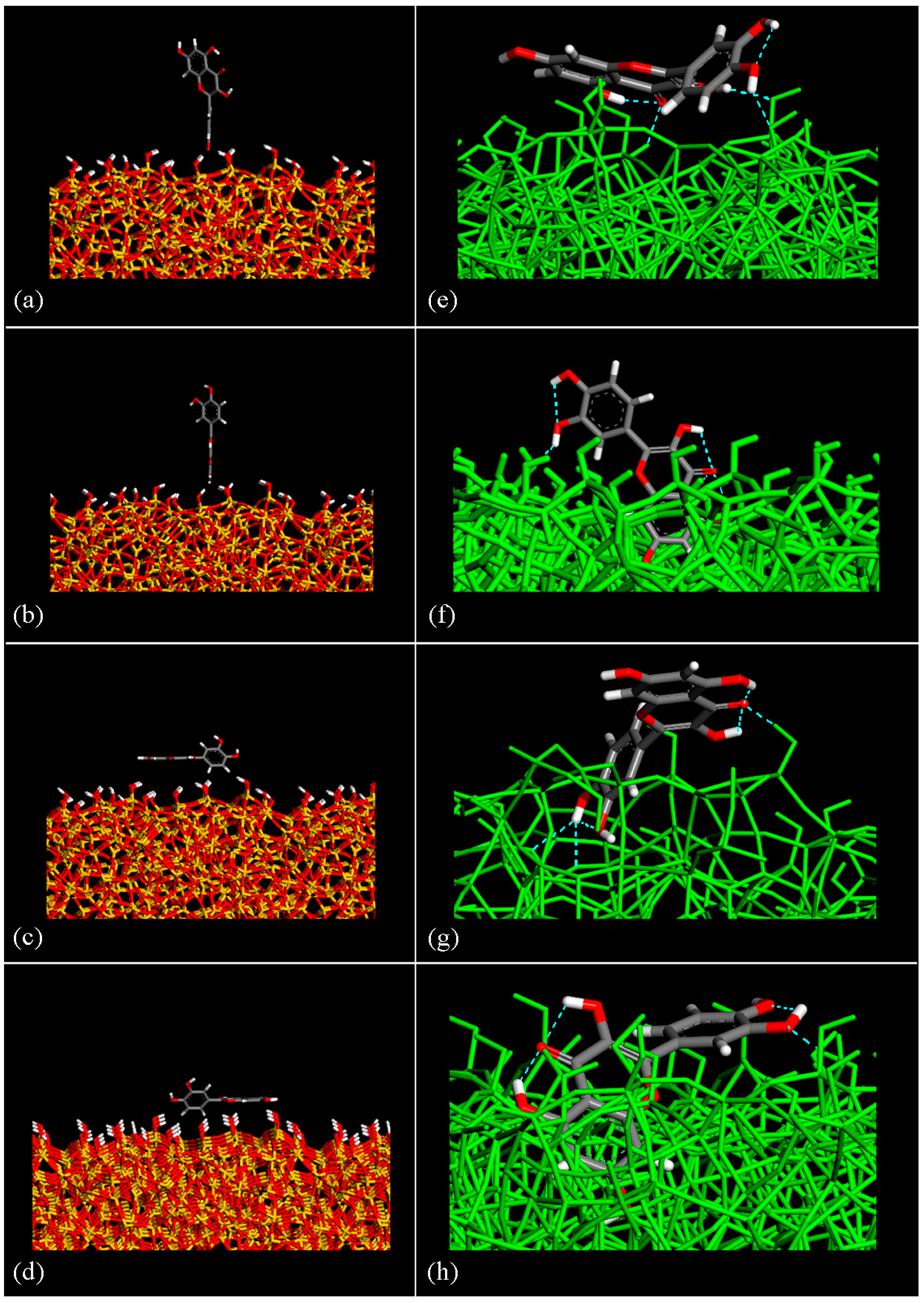
Figure 1. Chemical structure of the quercetin drug molecule (see panel (a)) and its optimized geometry after MM and MD run (see panel (b)). Color code: carbon atoms are grey, oxygen atoms are red, and hydrogen atoms are white. The three intramolecular H–bonds are represented with a blue dashed line.
Coatings 14 00234 g001
Figure 2. Four different initial geometries (panel (ad)) with the respective geometries of the energy minima calculated after MD run (panel (eh)), respectively. H– bonds are represented with a blue dashed line.
Coatings 14 00234 g002
Figure 3. Side view of the best-fit plane of the SiO2 surface (see green plane in panel (a)). Distance between the center of the mass (c.o.m.) of quercetin molecule and the silica best-fit plane calculated during MD run lasting 1 ns starting from the geometry reported in panel (b) in Figure 2.
Coatings 14 00234 g003
Figure 4. Potential energy and Coulomb contributions calculated in the adsorption process of quercetin molecules at small concentrations (14 drug molecules) during the first MD run lasting 2 ns (panel (a)) and the second MD run lasting 3 ns (panel (b)) on hydrated silica surface. Panel (c) and panel (d) show the distribution of distances between the c.o.m. relating to four different quercetin molecules relative to the silica best-fit plane calculated during MD runs lasting 2 ns (see panel (c)) and 3 ns (see panel (d)), respectively.
Coatings 14 00234 g004
Figure 5. Panel (a) shows the potential energy and Coulomb contributions calculated in the adsorption process in vacuo of quercetin molecules at small concentrations (14 drug molecules); panel (b) shows the van der Waals contribution calculated during the same MD run lasting 5 ns.
Coatings 14 00234 g005
Figure 6. Optimized geometry after MD run of 14 quercetin drug molecules adsorbed on dried amorphous SiO2 surface; in particular, the side view is in panel (a) with drug molecules in green, the silicon atoms in yellow, the oxygen atoms in red, and hydrogen atoms in white. In panel (b), a particular number of quercetin molecules adsorbed are shown with all silica surface atoms in grey, quercetin carbon atoms in green, oxygen atoms in red, and hydrogen atoms in white. H– bonds are in dashed blue lines.
Coatings 14 00234 g006
Figure 7. Potential energy and Coulomb contributions calculated in the adsorption process of quercetin molecules at higher concentrations (28 drug molecules) during the first MD run lasting 2 ns (see panel (a)) and the second MD run lasting 5 ns on hydrated silica surface (see panel (b)).
Coatings 14 00234 g007
Figure 8. Panel (a) shows the potential energy and Coulomb contributions calculated in the adsorption process of quercetin molecules at higher concentrations (28 drug molecules) during the first MD run lasting 10 ns on dried silica surface. Panel (b) shows the top view of the optimized final geometry.
Coatings 14 00234 g008
Figure 9. Panel (a) shows the H– bonds between oxygen atoms in quercetin –OH groups and hydrogen atoms in silanol –OH groups. Panel (b) shows the H– bonds between the hydrogen atoms of quercetin –OH groups and oxygen atoms in Si–O–Si of the silica surface. The yellow boxes highlight where these H– bonds are in the figure.
Coatings 14 00234 g009
Figure 10. FTIR spectra of quercetin, SiO2/quercetin (5 and 15 wt.%) hybrid materials, and pure SiO2.
Coatings 14 00234 g010
Table 1. Information on the number of quercetin intramolecular H– bonds and intermolecular H– bonds formed between the drug and silica surface in all final optimized geometries (e), (f), (g), (h), respectively, in Figure 2 is reported.
Geometries in Figure 2Intramolecular H– BondsIntermolecular H– Bonds
Geometry (e)23
Geometry (f)32
Geometry (g)33
Geometry (h)32
Disclaimer/Publisher’s Note: The statements, opinions and data contained in all publications are solely those of the individual author(s) and contributor(s) and not of MDPI and/or the editor(s). MDPI and/or the editor(s) disclaim responsibility for any injury to people or property resulting from any ideas, methods, instructions or products referred to in the content.

