Article

Novel Polyamide/Chitosan Nanofibers Containing Glucose Oxidase and Rosemary Extract: Fabrication and Antimicrobial Functionality

by
Ghazaleh Chizari Fard
1,2,3,*,
Mazeyar Parvinzadeh Gashti
4,5,*,
Seyed Ahmad Dehdast
1,2,
Mohammad Shabani
1,
Ehsan Zarinabadi
3,
Negin Seifi
1 and
Ali Berenjian
2,6
1
Department of Biochemistry, School of Medicine, Iran University of Medical Sciences, Tehran 14496-14535, Iran
2
“Pajoohesh BAMA” Knowledge Enterprise Co., Tehran 15847-43311, Iran
3
Clothing and Fabric Design Department, Art Faculty, Imam Javad University College, Yazd 89158-73763, Iran
4
GTI Chemical Solutions, Inc., Wellford, SC 29385, USA
5
InsectaPel, LLC, Wellford, SC 29585, USA
6
Department Textile, Islamic Azad University, South Tehran Branch, Tehran 15847-43311, Iran
*
Authors to whom correspondence should be addressed.
Coatings 2024, 14(4), 411; https://doi.org/10.3390/coatings14040411
Submission received: 29 December 2023 / Revised: 11 March 2024 / Accepted: 26 March 2024 / Published: 29 March 2024
(This article belongs to the Special Issue Fabrication and Properties of Bio-Coatings and Their Applications)

Abstract

:
In recent years, the synthesis of nanofibers using plant extracts and bioactive materials has been extensively studied and recognized as a suitable and efficient method applicable in the food packaging field. In this research, an antimicrobial material was introduced by the immobilization of glucose oxidase (GOx) in Nylon–Ag masterbatch/chitosan/Rosmarinus officinalis extract nanofiber via electrospinning technology. Nylon–Ag masterbatch/chitosan/Rosmarinus officinalis composite nanofibrous membranes with an average diameter of 207 ± 18 nm were successfully prepared using the electrospinning technique. The chemical properties of membranes were analyzed by Fourier transform infrared spectroscopy (FTIR) and the morphological characterization of nanofibers was evaluated with field emission scanning electron microscopy (FE-SEM). Moreover, enzymatic activity of GOx was determined by the Carmine method. FTIR results showed the successful incorporation of glucose oxidase and Rosmarinus officinalis into the nanofiber composite. Immobilized GOx showed high (79.5%) enzymatic activity in the optimum sample. The Rosmarinus officinalis, glucose oxidase-incorporated Nylon–Ag masterbatch/chitosan nanofibrous exhibited excellent antimicrobial activity on both gram-negative bacterium Escherichia coli (97.5%) and gram-positive bacterium Staphylococcus aureus (99.5%). The antibacterial and antioxidant Nylon–Ag masterbatch/chitosan/Rosmarinus officinalis/GOx nanofibrous membrane showed higher potential, compared to the control sample, to be used as food packaging by improving the shelf life and maintaining the quality of food stuffs. Therefore, this research recommends it as a promising candidate for food preservation applications.

Graphical Abstract

1. Introduction

Over the last decade, food industries have mainly encountered key challenges related to microbial infection and oxygenation [1]. The “shelf life” definition has become considerably important for food products to preserve their desired physical, chemical, and microbiological properties. Shelf life is the length of time from food production to storage and packaging under certain conditions with approved guidelines. Maintaining the prolonged shelf life and quality of foods during storage and transportation is the main objective of food packaging. Indeed, several other objectives must be met by food manufacturers, including preserving the inherent features of the foods, protecting against exposed environmental situations, making sure appropriate storage and transportation conditions are in place and, most significantly of all, ensuring the quality of the packaging system [2].
Recent reports show that the packaging industry contributes almost 2% of the Gross National Products (GNP) for developed countries and is expected to grow globally in the next 5 years [3]. It is important to note that, in the last decade, researchers have been working on different methods to develop new packaging technologies and materials, aiming to extend the shelf life of food products and also ensure their safety and cost efficiency [4,5,6,7,8]. Recently, electrospinning techniques have been extensively acknowledged in the production of nanostructured food packaging materials. Surface functionalization of electrospun nanofibers has been one of the major strategies used to produce active and sensory food packages [4].
In the past few years, the majority of electrospun nanofibers have been developed with biopolymers. In addition to acting as a strong barrier, enabling food preservation against humidity and oxidation, antimicrobial active nanoparticles are also generally integrated into nanofibers to prevent different bacterial transmissions [5,6,7,8]. Accordingly, the antibacterial electrospun nanofibers can inhibit or kill pathogenic microorganisms, thereby preventing bacterial diseases and infections [9]. More recently electrospun nanofibers have been deposited on the packaging sheets to produce bilayer or multilayered packaging products [10,11].
On the other hand, recent research demonstrated that active packaging systems containing immobilized antimicrobial enzymes open an avenue for prolonging the shelf life of non-sterile foods. Beyond the anti-bacterial requirements, the application of enzymes could be evolved in the processing of food packages [12,13,14,15,16,17,18,19,20]. In this context, glucose oxidase (GOx) has been widely considered in glucose monitoring, food preservation, and wound treatment applications [21]. This oxidoreductase enzyme is generally generated by Aspergillus niger and Penicillium spp. cultures. Moreover, this enzyme is categorized as a 140–160 kDa dimeric protein. It is worth mentioning that flavin adenine dinucleotides (FAD) are bounded by non-covalent interactions in each monomer unit. Researchers stated that GOx acts as a catalyst in the oxidation process of β-D-glucose units and converts them to D-glucono-δ-lactone and H2O2. Also, GOx has shown antimicrobial properties, which is related to the generated H2O2 molecules during enzymatic reactions. On the other hand, this enzyme may reduce the pH after development of D-gluconic acids due to the interaction of D-glucono-δ-lactone and H2O. This can be another reason for the growth of microorganisms in active food packaging. Recent investigations showed that GOx has excellent antibacterial properties against both gram-negative and gram-positive bacteria [22,23].
Enzymes can be physically confined or immobilized on a wide variety of substrates for retention of their functionality and catalytic activities. Many techniques have been explored for this purpose, including adsorption, ionic and covalent bonding, encapsulation, electrospinning, and graft co-polymerization [24,25]. Among them, the electrospinning method has recently attracted more attention due to several advantages, which follow:
  • Maintaining a high surface area for enzymes or bioactive components for bonding [26];
  • Providing a high mass transfer rate, from an electrospun substrate to the active sites of an enzyme;
  • Representing a highly durable performance for multiple usages;
  • Simple recovering from a reaction solution when used as biocatalyst supports [27,28].
In the past two decades researchers have been working on chitosan (CS), as a promising carbohydrate biopolymer with both natural and broad spectrum antibacterial activities [29,30]. It was demonstrated that rosemary extract is one of the best antioxidant candidates in food preservation [31,32]. Rosmarinus officinalis leaves contain phenolic compounds and diterpenes that have been used in film packaging [33]. Gómez-Estaca et al. [34] evaluated antioxidant properties of gelatin sheets after the addition of rosemary aqueous extract. They found that gelatin can change the polyphenols release, due to different intermolecular bindings; Farghal et.al [35] treated polyester sheets with rosemary extract for fish packaging applications.
Several studies have been conducted of the fabrication of different electrospun nanofibers for the food packaging industry [36,37] However, very few researchers have utilized Rosmarinus officinalis extract and glucose oxidase in electrospun mats for these applications [38,39]. Moreover, previous studies of Nylon-6/chitosan nanofibers have shown efficient antibacterial effect and sustainable drug release, and Nylon–Ag nanofibers showed higher antibacterial efficacy, compared to nylon nanofibers, against Gram-negative microorganisms [40,41]. Therefore, in this study we electrospun Nylon–Ag masterbatch/chitosan/Rosmarinus officinalis extract nano-fibrous composites for the immobilization of glucose oxidase. Different characteristics of the resultant nanofibers were evaluated, including morphological, chemical, and biological properties, by FESEM, FTIR, and bacterial culture tests. Interesting antibacterial activity and deoxidization ability were observed in the nanofiber composites.

2. Materials and Methods

2.1. Materials

Nylon–Ag masterbatch (MBN-Ag) was obtained from Persian Polymer Novin Co.-Tehran, Iran. Chitosan (CS) medium weight (Sigma-Aldrich, MO, USA, Deacetyliertes Chitin, Poly (D-glucosamin, CAS-Nummer: 9012-76-4), acetic acid (Merck, Darmstadt, Germany), and glucose oxidase (GOx) were provided by Sigma. Co., and a Rosmarinus officinalis (RO) plant was obtained from Kerman Climate Co. in Tehran, Iran.

2.2. Extraction

Rosmarinus officinalis (RO) (700 g) were extracted using an ethanol/distilled water mixture (80/20) at room temperature, using the maceration method. The process was continued for 72 h (three replications), using a 6 L solvent. Then, we mixed resultant extracts, followed by paper filtration. The resultant material was dried at 40 °C under vacuum conditions.

2.3. Fabrication of MBN-Ag/CS Nanofibers Containing RO and GOx

At first, different amounts of MBN-Ag (7 wt%) and CS (5 wt%) were dissolved in acetic acid for spinning purposes. In order to generate different concentrations, ratios of 80:20, 70:30, 60:40, 50:50, 40:60, 30:70, and 20:80 (MBN-Ag/CS) were used, respectively. Next, different mixtures were stirred using a magnetic stirrer for 12 h to receive homogenous solutions. Finally, prepared mixtures were electrospun under a fixed electrical field of 18 kV and the feeding rate of 1.2 mL/h. We adjusted the distance between the tip of the needle and collector to 16 cm. Produced nanofibers were finally collected onto an aluminum (Al) foil. The electro-spinning machine was received from Fanavaran Nano-Meghyas Co. (Tehran, Iran).
We selected the optimal ratio of two components (MBN-Ag/CS) after an initial evaluation of the morphology and the uniformity of produced nanofibers. Then, different amounts of GOx (0.01, 0.03, 0.05, 0.07 mg) were mixed to the optimum solution, and they were stirred for 2 h, and electrospun under the above conditions. At the next step, 1.5% of RO extract was added to the MBN-Ag/CS/GOx solutions. The electrospinning procedure was then conducted, as previously reported. Finally, all nanofibers were put in an oven at 70 °C for 3 h to completely remove the solvent.

2.4. Design of Experiments

In this study, design expert software (V 11.0.3) was used to design experiments. Response surface methodology (RSM) was utilized, using Box–Behnken design (BBD), and the results are illustrated in Table 1. Experiments were also repeated to create the software’s confidence limit for minimizing the error.
As is illustrated in Table 1, on the basis of the production method, the amount of preservation (%) in the prepared packages was zero for runs 3, 11, 16, 25, 29, 35, 39, and 44. This was due to the fact that nanofibers cannot be formed in the MBN-Ag/CS nanofiber composite sample (30/70). The maximum preservation was observed for run No. 26 (MBN-Ag/CS nanofiber composite samples (30/70)). It consisted of dissolved MBN-Ag (8% w/w), dissolved CS (2% w/w), RO (1.5% w/w) and GOx (0.05 gr). We expected that the predicted optimum level in the final product, with 99% preservation, could be achieved by BBD experimental design.
According to the designed RSM in Table 2, the test validity was less than 0.05 (0.0001). We should express that there was a significant difference between the impact of variables on the preservation (%) values. Moreover, the F-variable displayed the most value (147.54) for CS/MBN-Ag composite, and the maximum impact of preservation was observed for this variable. In addition, P-variable affected (less than 0.05%) the RO and MBN-Ag/CS composites. This result stated that these variables had a significant impact on preservation (%) values. Notably, MBN-Ag and CS had a significant impact in the quadratic model, depending on their combination. We concluded that the impact of these components could be further measured by reviewing the 3D charts and the execution model. The mathematical model of the designed experiment can be seen by using Equation (1):
Conserve Percent: 79.9659 + (−0.924529) × [MBN-Ag] + 2.8125 × [CS] + (−40.638) × [MBN-Ag/CS] + 12.2884 × [RO] + 3.41336 × [GOx] + (−2.75) × [MBN-Ag] [CS] + (−1.80188) × [MBN-Ag] [MBN-Ag/CS] + 10.1535 × [MBN-Ag] [RO] + 1.90346 × [MBN-Ag] [GOx] + 0.75 × [CS] [MBN-Ag/CS] + (−2.5) × [CS] [RO] + (−4.75) × [CS] [GOx] + (−10.9035) × [MBN-Ag/CS] [RO] + 0.0965449 × [MBN-Ag/CS] [GOx] + 3.80115 × [RO] [GOx] + (−13.0159) × [MBN-Ag]2 + (−12.5332) × [CS]2 + (−25.0992) × [MBN-Ag/CS]2 + (−18.4328) × [RO]2 + (−12.9328) × [GOx]2
Although some moderate scatter was expected from the produced data, the normal probability diagram shows how the data followed a normal distribution at the central line (Figure 1A). According to the specified and curved patterns (S-shape), we could achieve an accurate analysis by transfer function on the dependent variables or the model response. Therefore, from a statistical point of view, these results further illustrated that our experimental design was acceptable and represented a normal condition.
The Box–Cox diagram was used as a valuable tool to recognize the optimized power transmission function to exert on the response. The lowest point in the Box–Cox diagram represented the best condition for Lambda (λ), in which the lowest residual sum of squares in a converted model could be created. When the maximum to the minimum ratio of the response value was greater than 3, the power function can be potentially improved. Regarding Figure 1B, the difference between the minimum and maximum of the response value was 3, and no more improvement was shown in the experimental model. Moreover, a 95% confidence level was achieved in the diagram by simulating the created mathematical and computational connections between all variables.

2.5. Chemical Analysis by FTIR Spectroscopy

The FTIR spectra of nanofibers were assessed using FTIR spectroscopy (ThermoNicolet NEXUS 870 FTIR from Nicolet Instrument Corp., 5225 Verona Rd., Madison, WI, USA).

2.6. Morphological Analysis by FESEM

The surface properties of nanofibers were measured by the FESEM tool (LEO1455VP, Cambridge, UK).

2.7. Antibacterial Test

Staphylococcus aureus (S. aureus), an American Culture No. 6538 type, as a gram-positive bacterium, and Escherichia coli (E. coli), an American Culture Collection No. 11303 in Luria-Bertania (LB), were cultivated at 37 °C and a density of about 2 × 10⁵ Cells/mL. Then, the bacteria were used for the antibacterial test. Antibacterial activity of MBN-Ag/CS/RO (60/40/3%), MBN-Ag/CS (60/40), MBN-Ag/CS/GOx (60/40/0.05), and MBN-Ag/CS/GOx/RO (60/40/0.05/3%) were evaluated by the colony counting method (100 mL of dilute bacterial suspension was prepared and was quickly placed in LB medium). We incubated plates at 37 °C for 24 h, and the colonies were finally counted. It should be noted that the MBN-Ag/CS was considered a control sample. The growth inhibition zone and colony numbers were determined after three repeated experiments.

2.8. The Glucose Oxidase Activity Determination

The activity of glucose oxidase was examined by the Carmine method [42]. For this purpose, 1 mL of glucose oxidase (1 mg/mL) and 4 mL of glucose oxidase solution (0.2 mol/L) were poured into a test tube at 37 °C for 10 min to react. This enzyme solution served as a substrate. Acetic acid-sodium acetate mixture was used as a medium and the Indigo Carmine was utilized as a color indicator. We placed the testing vial in a boiling water bath for 30 min, and the absorbance at 615 nm wave number was measured with a distilled water reference. The activity of each GOx unit (U/mL) is defined based on the amount of GOx producing 1 mg of hydrogen peroxide per minute. The GOx activity of the prepared sample (U/mg) was calculated from Equation (2):
% G O x   a c t i v i t y   U m g = G O x   a c t i v i t y   U m L w e i g h t s a m p l e   m g m L × 100
W e i g h t s a m p l e   ~   t h e   w e i g h t   o f   t h e   s a m p l e   ( m g )

2.9. Food Packaging Experiments

After the physicochemical studies of nanofibers and antibacterial activity investigation, the prepared nanofibers were used for packaging evaluation. At first, strawberries without contamination and visible damage were selected and each group of strawberries was placed inside the nanofiber mats separately and at different times (0, 5, 10, and 15 days); the degree of contamination and the retention of different groups were then evaluated with macroscopic imaging.

3. Results and Discussion

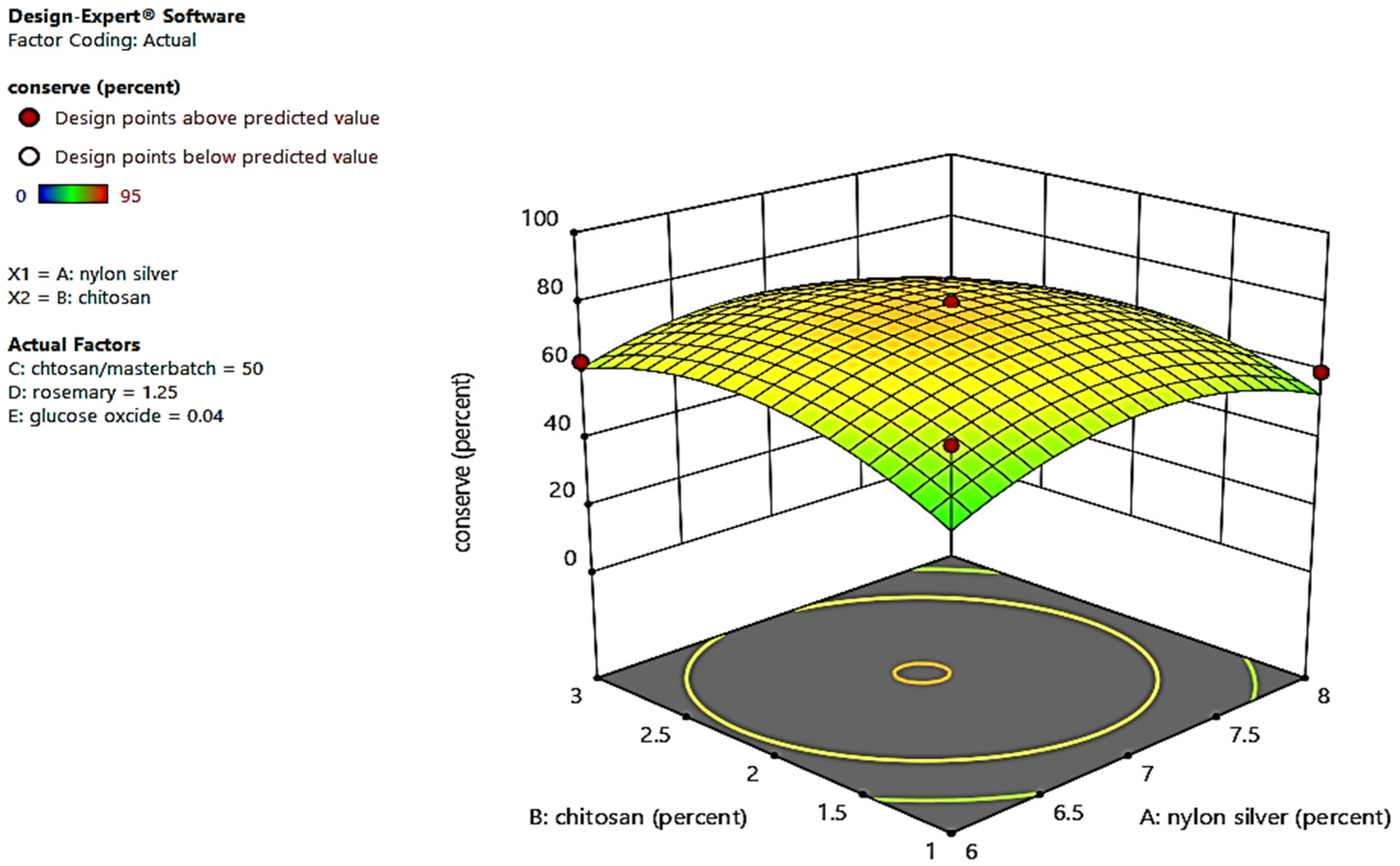
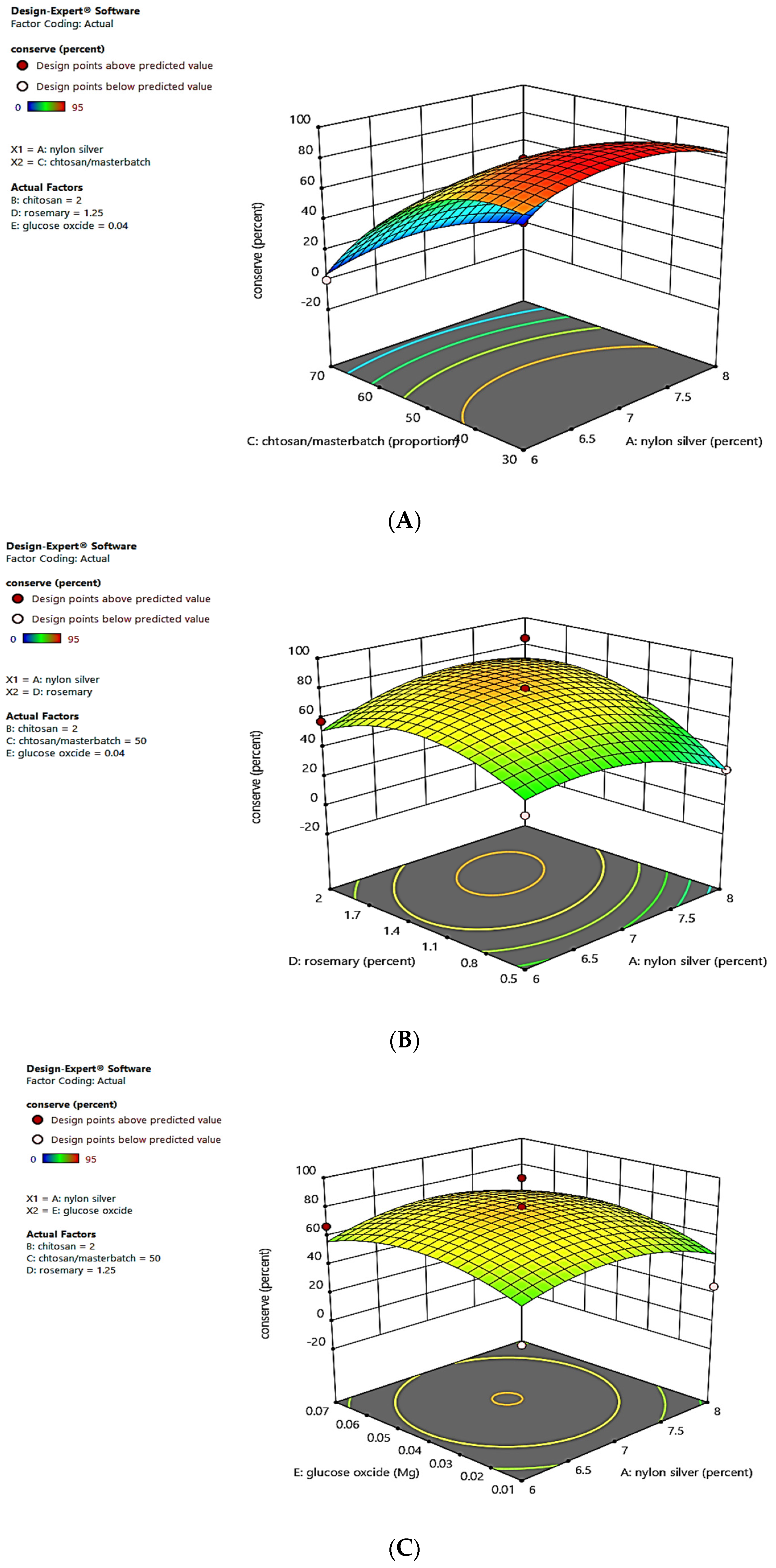
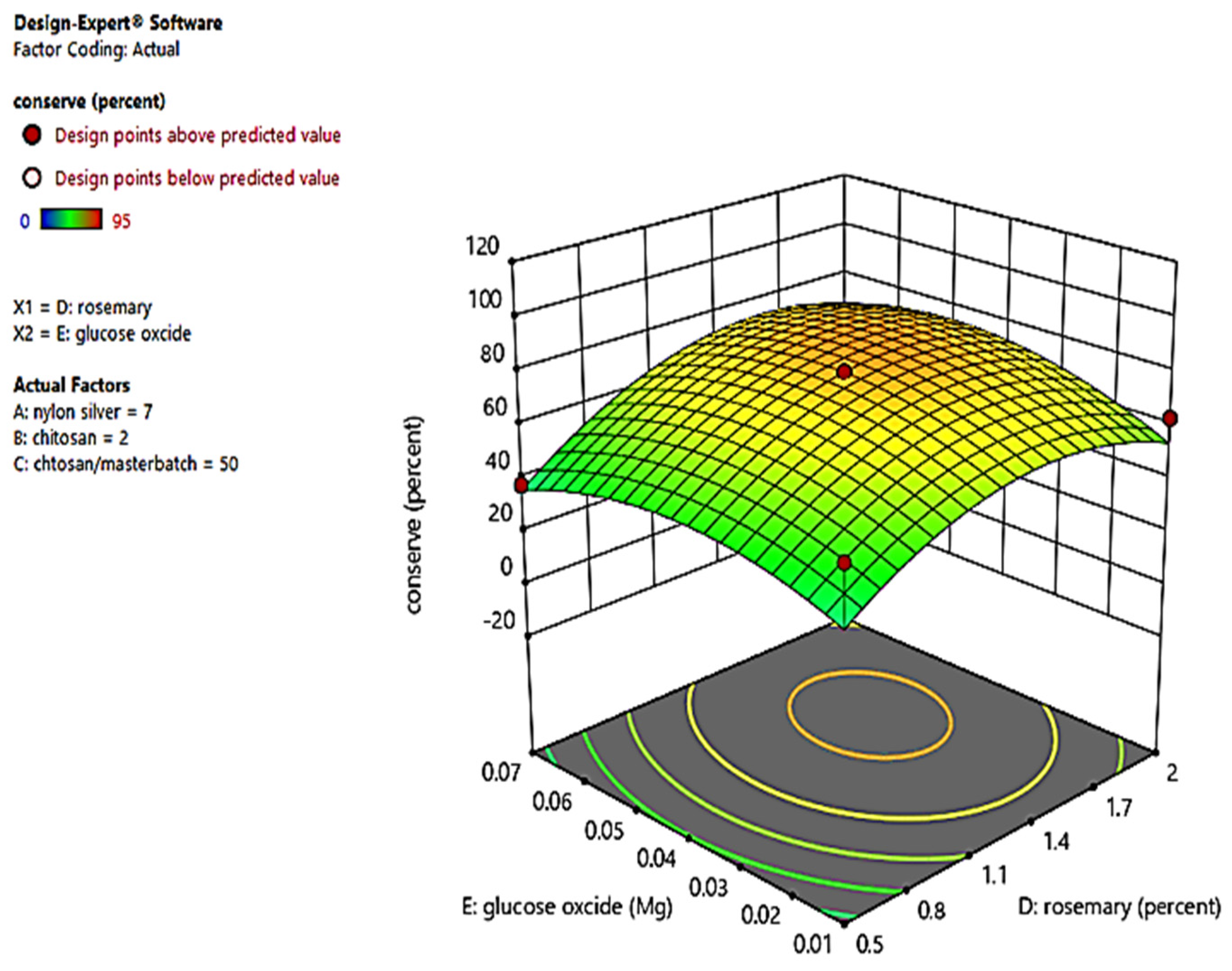
3.1. Identifying the Impact of Variables on Preservation (%) Values

We evaluated the impact of different variables in the experiments using the extracted 3D diagrams from the software (Figure 2, Figure 3, Figure 4, Figure 5 and Figure 6). In this regard, the relation between each variable and their impact on the preservation (%) values were individually assessed. Moreover, a combination of materials with their optimum (%) values (refer to optimization) was representable by the maximum preservation (%).
According to Figure 2, the maximum preservation was about 80% in the middle of the diagram (determined by the orange color). In other words, about 80% of preservation in the experiments could be achieved by 2% and 7% densities of CS and MBN-Ag, respectively. This measurement applied regardless of other variables and materials in the formulation, and only the impacts of CS and MBN-Ag densities were considered as variables.
On the other hand, we only considered the MBN-Ag solution (%) and the ratio of mixed MBN-Ag and CS solutions as variables in the 3D diagram in Figure 3A. For the MBN-Ag/CS nanofiber composite (65/35), the highest preservation value was 90% (this percentage is determined on the top of the diagram, and is indicated in red). Regarding Figure 3B, 1.25% and 1.5% RO represented the maximum impact of preservation (%) in variables (the orange circle) when mixed with 8% MBN-Ag solution. Furthermore, preservation (%) values were increased from 22% to 74% after increasing the amount of GOx from 0.01 to 0.05 gr in 8% MBN-Ag solution (Figure 2C). However, we should express that preservation (%) values were decreased after increasing the amount of GOx from 0.05 to 0.07 gr in MBN-Ag (%) solution.
As can be seen in Figure 4A, the maximum preservation (85%) was achieved in the comparison of two variables: 2.5% CS solution and the ratio of mixed MBN-Ag and CS solutions (70:30). It should be noted from Figure 4B that the minimum preservation (%) was obtained for the sample containing 2% CS and 0.8% RO. However, the preservation (%) was increased from 85% to 89% (orange area of the diagram) by increasing the amount of RO to 1.5% at a fixed CS %. According to Figure 3C, the sample containing 0.05 gr GOx and 2% CS represented the highest preservation (%) value.
Regarding the results shown in Figure 5A, the MBN-Ag/CS (70/30) sample containing 2% RO displayed a 99% preservation value. Also, the MBN-Ag/CS (70/30) sample containing 0.07 mg GOx indicated the maximum preservation capability (85%), as can be seen in Figure 5B.
As is illustrated in Figure 6, the maximum preservation (%) resulted from a sample containing 1.5% RO and 0.05 mg GOx (orange region). We extracted the predicted optimum values from different variables in Figure 1, Figure 2, Figure 3, Figure 4 and Figure 5, and the results are represented in Table 3.

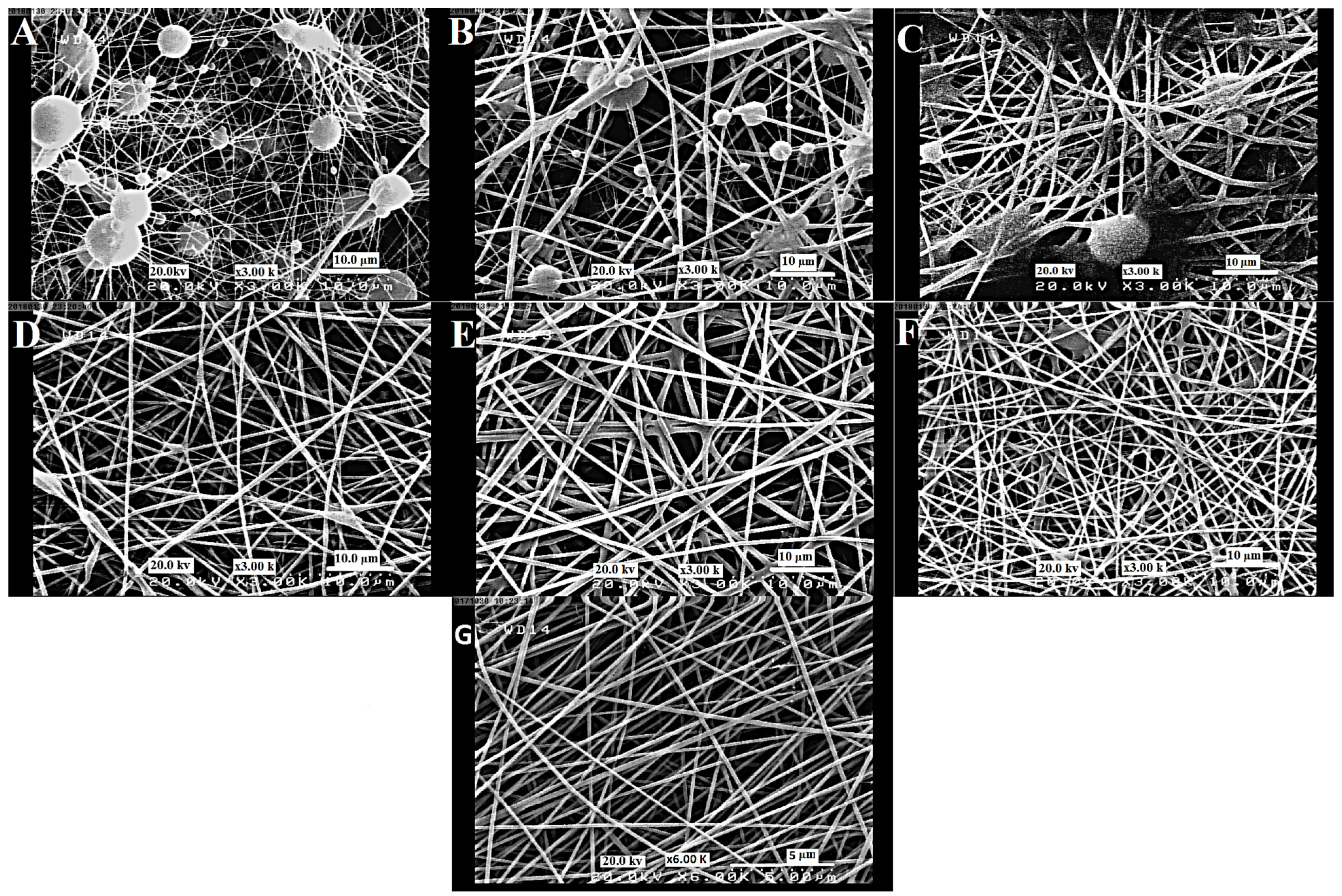
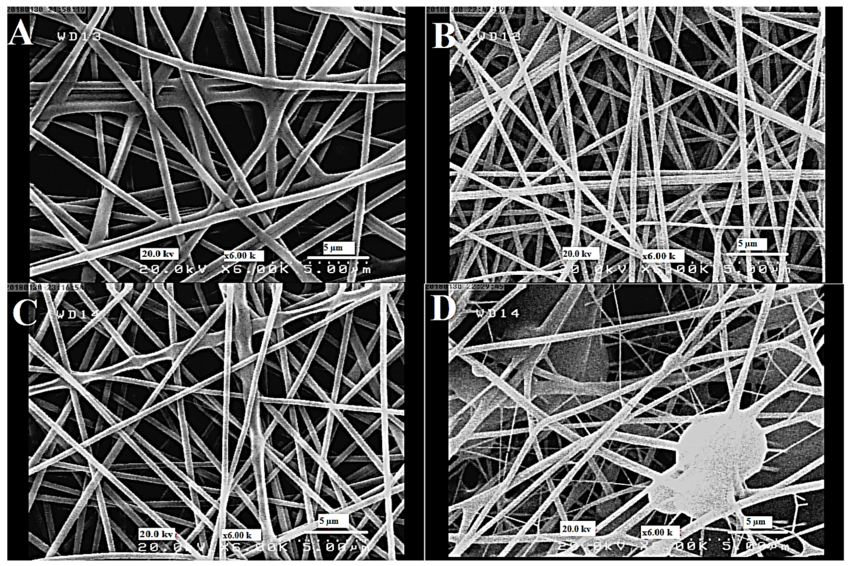
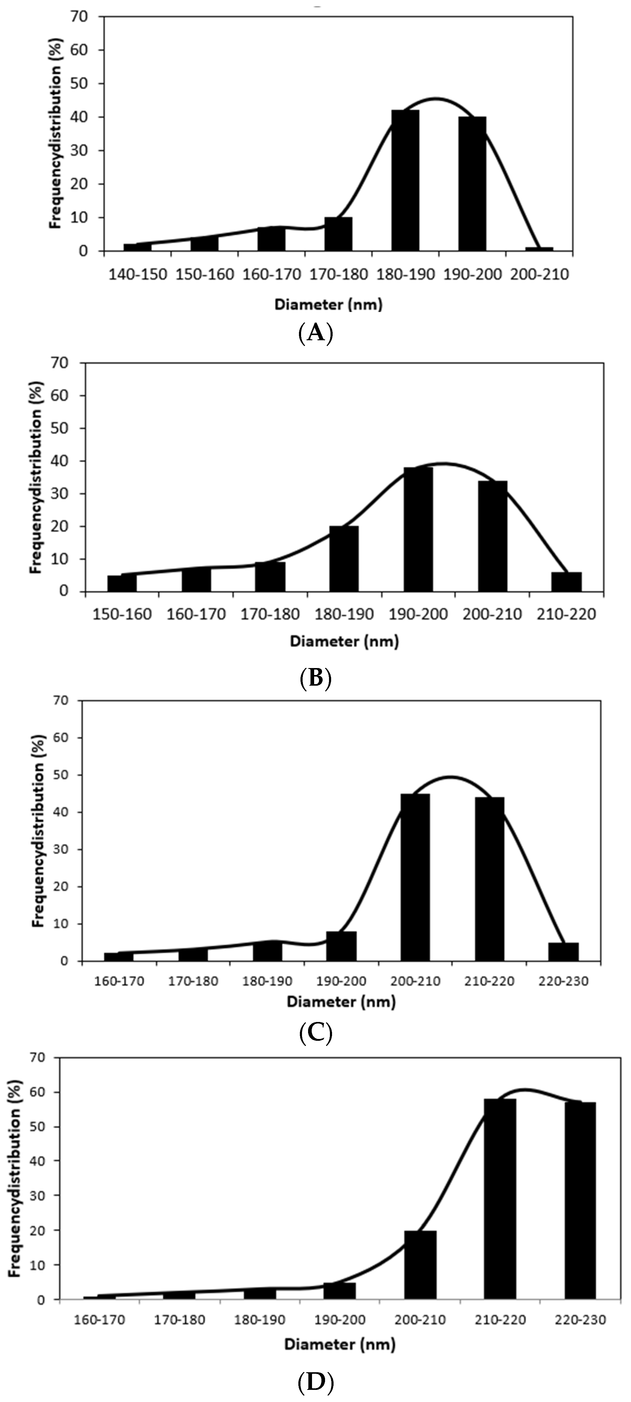
3.2. The Morphology of Nanofibers

FESEM images of fabricated MBN-Ag/CS and MBN-Ag/CS/GOx nanofiber composites are shown in Figure 7 and Figure 8. It should be noted that the spinning solutions for MBN-Ag/CS with 80:20 and 70:30 ratios did not form continuous nanofiber composites, due to Low CS content. It was observed that those samples containing 60% and 50% CS had several structural defects and beads. Notably, we found a fluctuation in the nanofiber diameters that was due to these defects. By decreasing the CS content to 20 and 30 wt% in solutions, uniform nanofiber composites with a smooth surface were generated. In addition, a reduction in CS content in the nanofiber composites resulted in a decrease in the average diameter of nanofiber composites. The average diameters of the MBN-Ag/CS nanofiber composites were 170, 185, and 197 nm when the ratio of MBN-Ag to CS were 80:20, 70:30, and 60:40, respectively. Zhang et al. studied the complex interactions in the electrospun nylon-6/CS complex nanofibers. They found that the addition of cationic and anionic polyelectrolytes to the electrospinning solution could generally increase the conductivity of the polymer mixture thus leading to the generation of thinner nanofibers. Changes in the diameter and uniformity of MBN-Ag/CS nanofiber composites could also result from the changes in the charge density and viscosity of MBN-Ag and CS mixed solutions at different ratios [43]. Similar results were also reported elsewhere [44].
We used MBN-Ag/CS solution with a ratio of 70:30 as a reference and different amounts of GOx were added to this solution, including 0.01, 0.03, 0.05, and 0.07 mg. The FESEM images of the resultant nanofiber composites are shown in Figure 8. It was observed that the degree of unevenness and defects in nanofiber composites depends on the contribution of the GOx. The more GOx content is used in nanofiber composites, the lower the uniformity in fabricated samples. This phenomenon could be attributed to the interaction between the amine and carboxyl groups of GOx on the one hand, and functional groups of the nylon and chitosan chains on the other hand. These interactions were mostly based on ionic and hydrogen bonding, which could lead to rising in the viscosity of the solutions and the mean diameter of the nanofiber. As a result, more deficiencies in nanofiber morphology could be observed [45,46]. The average diameter of nanofibers was increased from 198 nm (MBN-Ag/CS/GOx nanofiber composite containing 0.01 g of GOx) to 207, 215, and 225 nm for samples containing 0.03, 0.05, and 0.07 mg of GOx, respectively (Figure 9).
In contrast to the changes by glucose oxidase (GOx), the FESEM revealed no morphological changes in MBN-Ag/CS/GOx nanofiber composites due to the addition of RO (1 and 1.5 wt%) (Figure 10), however, it should be noted that the spinning solution for MBN-Ag/CS/GOx with 2 wt% of RO did not form continuous nanofiber composites due to high RO content.

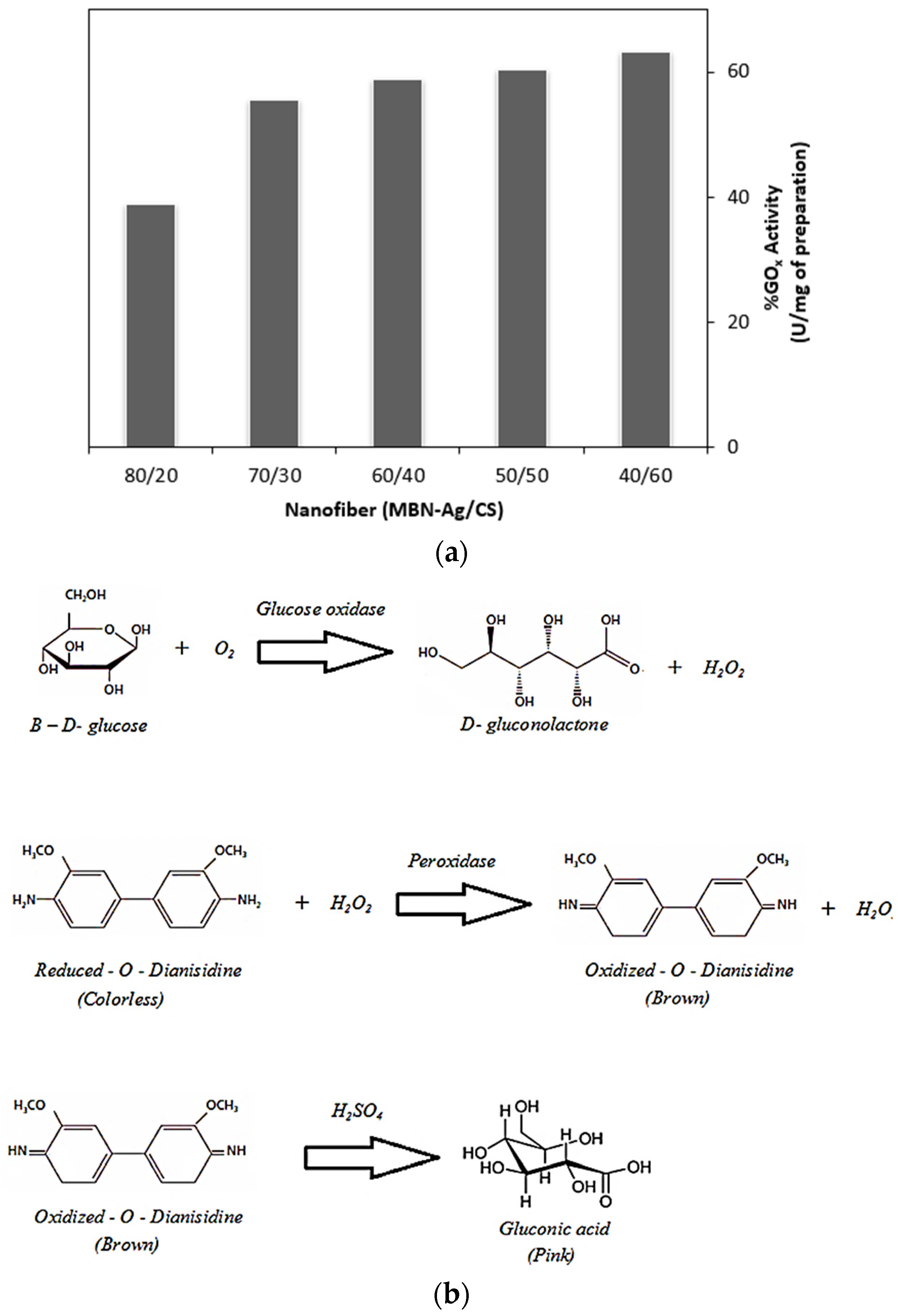
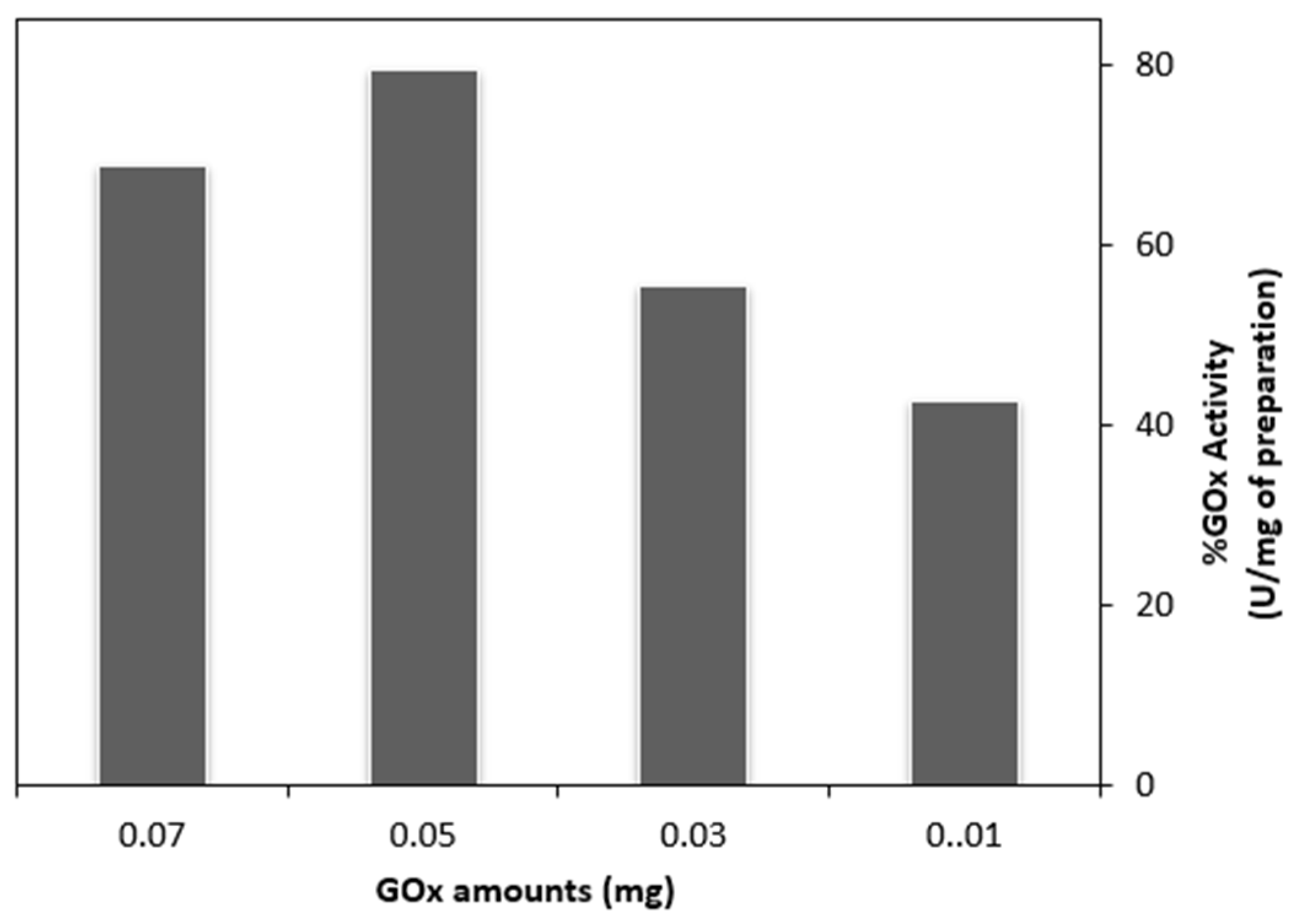
3.3. The Inhibitory Effect of Silver on Glucose Oxidase

In general, the activity of GOx as an enzyme varies based on certain circumstances, such as pH and the existence of different inhibitors. Previous studies suggested silver and heavy metals as the inhibitors for the GOx activity [47]. To this end, we examined the enzymatic activity of the MBN-Ag/CS nanofiber composites (ratios at 40:60, 50:50, 60:40, 70:30, and 80:20) containing 0.03 mg of GOx, and the results are presented in Figure 11. It can be seen that the GOx activity was decreased by increasing the ratio of MBN-Ag to CS. In this study, MBN-Ag material contained silver particles, as received from the manufacturer. The more MBN-Ag content in the MBN-Ag/CS nanofiber composites, the lower the achieved GOx activity. However, the decreasing trend in the GOx activity was not so significant for MBN-Ag/CS nanofiber composites with 40:60, 50:50, and 60:40 ratios. On the other hand, we obtained the lowest GOx activity for nanofiber composites with ratios of 70:30 and 80:20. For further studies, MBN-Ag/CS nanofiber composite with a ratio of 70:30 was selected as an optimum sample. The mechanism for the GOx retention effect is also illustrated in Figure 9.
The effect of different concentrations of GOx (0.01, 0.03, 0.05, and 0.07 mg) in the resultant MBN-Ag/CS/GOx nanofiber composites (MBN-Ag/CS at a ratio of 70:30) is shown in Figure 12. The results revealed that the retention ability of nanofiber composites was increased with any increase in the enzyme content (up to 0.05 mg). However, we observed a reverse effect on the enzymatic activity in the nanofiber composite containing 0.07 mg GOx. As a result, MBN-Ag/CS/GOx nanofiber composite with 0.05 GOx content was selected as an optimum sample due to the highest enzymatic activity (79.5%).
Ge et al. previously immobilized glucose oxidase in PVA/CS/tea extract electrospun nanofibers. They found that the enzyme solution ratio should be at 3:7 to achieve the highest activity. In addition, electrospun nanofibers with GOx showed higher activity, in comparison with casting films from the same solution. Their study proposed that electrospun nanofiber composites are the best candidates for food preservation applications [48].

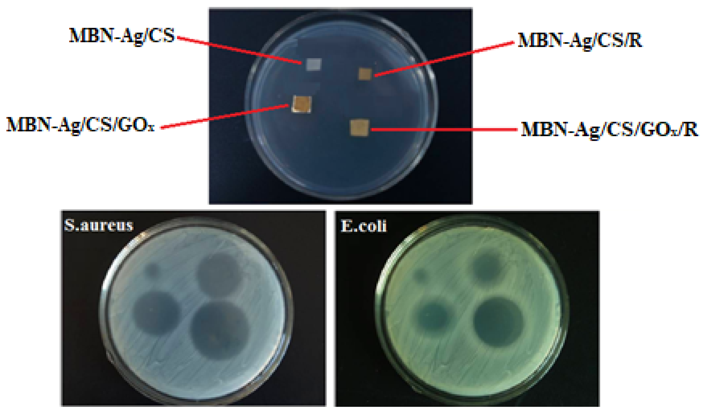
3.4. Antibacterial Studies
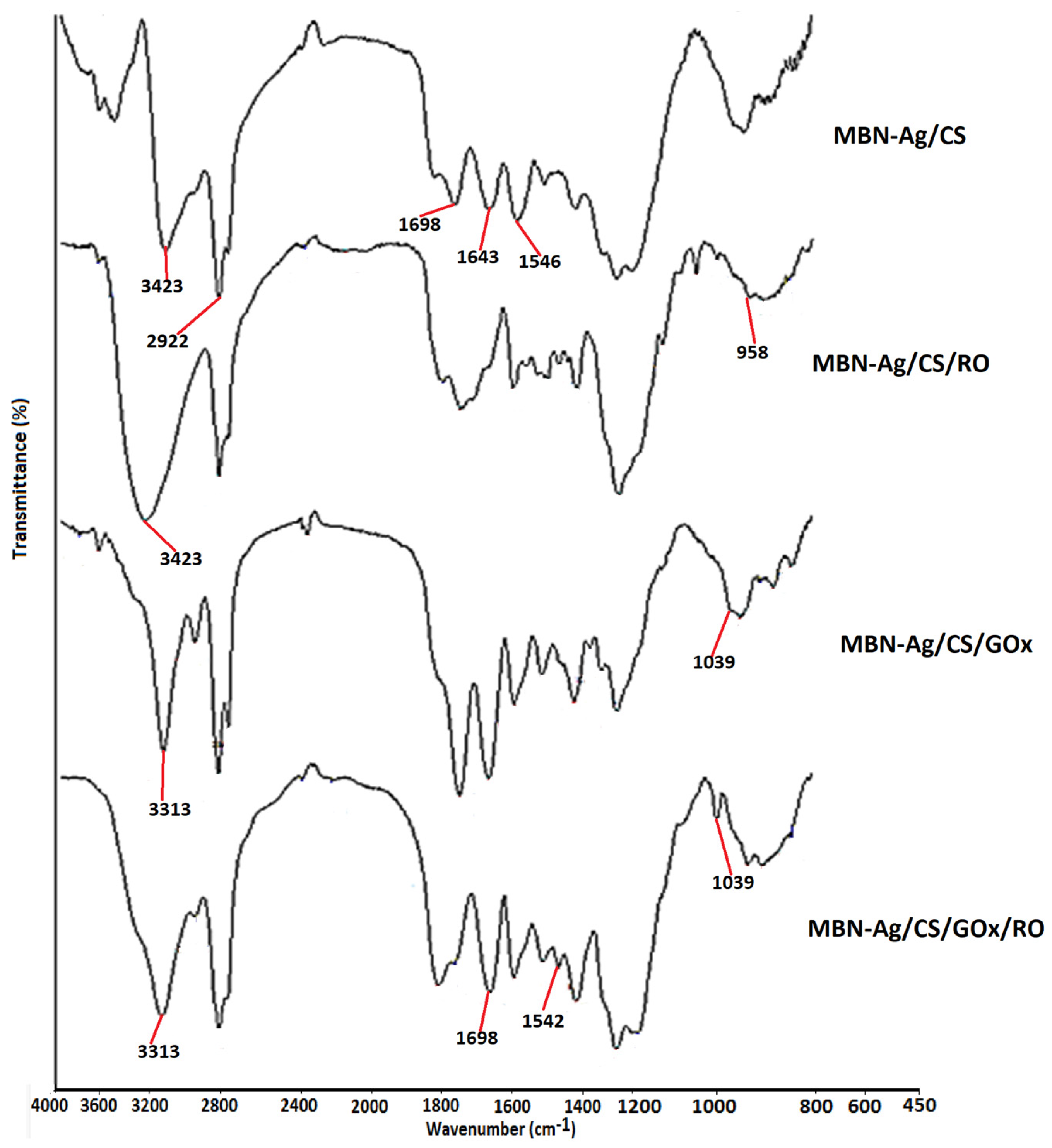
We assessed the inhibition zone size for different nanofiber composites produced in this study and the results are displayed in Figure 13. As can be seen, MBN-Ag/CS nanofibers showed stronger antibacterial activity against S. aureus,in comparison with E. coli. This was due to the fact that the inhibition zone was bigger for S. aureus. We should note that electrospun MBN-Ag/CS/RO, MBN-Ag/CS/GOx, and MBN-Ag/CS/GOx/RO nanofibrous composites could eliminate S. aureus and E. coli bacterial growth. By adding the rosemary to an electrospun membrane, it was indeed found that the antibacterial activities of RO, including nanofiber composites, were increased, due to the existence of 1,8 cinede, α-pinene, camphor, borneol, and verbenone in RO [28,29]. Previous studies also suggested the synergetic, antagonist, and additive effects between different natural reagents in RO could lead to strong antibacterial properties [49]. In general, essential oils exhibit a major antibacterial mechanism after interaction with bacterial cells: Due to their hydrophobic properties, they interact with lipids in the bacterial membranes, leading to cell disruption [50,51]. We recently demonstrated similar antibacterial properties from different bio-nanocomposites for biomedical applications [52,53,54,55].
It is concluded that the addition of the GOx led to an increase in the antibacterial activity of the resultant nanofiber composites, which could be due to the production of hydrogen peroxide as an antimicrobial agent. Hydrogen peroxide is a robust oxidizing agent used as a disinfectant agent. This chemical reagent can eliminate germs by forming radicals, which can subsequently interact with lipid membranes, DNA, and other important cellular constituents of micro-organisms [56]. In the presence of MBN-Ag/CS/GOx/RO nanofibers composite, S. aureus (99.5%) and E. coli (97.5%>) bacterial cells were mostly destroyed, due to the presence of the GOx and RO. Antibacterial disc diffusion assay showed a zone of inhibition against E. coli and S. aureus. A small inhibitory zone (4 ± 0.1 mm) was observed around the MBN-Ag/CS nanofiber mat against S. aureus. Also, the inhibition zone reached to 11 ± 0.2 mm, 13 ± 0.1 mm, 17.0 ± 0.1 mm against S. aureus for MBN-Ag/CS/R, MBN-Ag/CS/GOx, and MBN-Ag/CS/GOx/R, respectively. Moreover, a 3 ± 0.3 inhibitory zone around the MBN-Ag/CS mat against E. coli was observed. Also, the inhibition zone reached to 8 ± 1 mm, 10 ± 1 mm, 15.0 ± 0.5 mm for MBN-Ag/CS/R, MBN-Ag/CS/GOx, and MBN-Ag/CS/GOx/R against E. coli, respectively. The corresponding results from this quaternary nanofiber composite denoted the most promising inhibitory results against tested gram-positive and negative bacteria. Therefore, our fabricated MBN-Ag/CS/GOx/RO nanofibers composite is a promising candidate for the food packaging industry.

3.5. FTIR Analysis

The FTIR spectra of various electrospun nanofiber composite membranes are shown in Figure 14. As can be seen in the FTIR spectrum for MBN-Ag/CS nanofiber composite, the stretching and bending vibrations of the NH bonds (in the primary amines) appeared at 3423 and 1546 cm−1, respectively. Also, the stretching vibrations of C=O groups were assigned at 1698 cm−1. The absorption band at 1643 cm−1 is attributed to the stretching vibrations of NH groups (in the secondary amine) [57]. The stretching and bending vibrations of CH groups (including CH3, CH2, and CH) appeared at 2922 cm−1, and a range of 1475–1307 cm−1. After the inclusion of RO in nanofiber composites, the absorption band at 3423 cm−1 was broadened and the corresponding intensity was increased, due to vibration of hydroxyl groups in RO and the overlapping between OH and NH groups. The absorption band at 958 cm−1 is related to OH bending vibrations in carboxylic acid groups [58,59,60,61,62]. For MBN-Ag/CS/GOx/RO nanofiber composite, the absorption band at 3313 cm−1 was increased, due to the detection of amine and hydroxyl groups. We should highlight that the absorption peak at 1039 cm−1 was related to the vibrations of the cysteine groups in the GOx. Also, the intensity of the absorption bands at 1542 and 1698 cm−1 was increased, due to the presence of amine and carboxyl groups in the chemical structure of MBN-Ag/CS/GOx/RO quaternary nanofiber composite.

3.6. Application of Electrospun Nanofibers in Food Packaging

Strawberry is a fruit that is very sensitive to ethylene gas, and it can quickly ripen. For this reason, it is usually stored in cold temperatures. To evaluate the applicability of our developed electrospun nanofibers in the food industry, we used them for strawberry packing. The GOx activity of different samples was investigated, and the results are presented in Figure 15. The results showed that MBN-Ag/CS/GOx/RO nanofiber composite had the highest activity (90.1%), with excellent preservation properties.
In this study, strawberries were stored in different packages fabricated from our nanofiber composites at 21 °C for 15 days. We also investigated different properties of packages, including weight loss, appearance features, and decay of strawberries in different time periods. We observed that the weight loss for the control package was 43.1% on the 15th day. However, the result of the weight loss for the packages fabricated from MBN-Ag/CS/RO, MBN-Ag/CS/GOx, and MBN-Ag/CS/GOx/RO nanofiber composites were 23.8%, 20.3%, and 5.73%, respectively. Additionally, the control package lost its glossy appearance over time, and strawberries were rotten and molded in this sample. This is probably due to the destruction of fruit cell walls and, consequently, loss of water due to wall breakage [62]. Although strawberries packed with nanofibers were later rotten, MBN-Ag/CS/RO nanofiber composite safely stored them on the 15th day without any mildew. Results indicated that our fabricated nanofiber composites are excellent candidates for storing strawberries. It should be concluded that MBN-Ag/CS/GOx/RO nanofiber composites not only represent anti-bacterial properties that are a key factor for food packaging but are also able to preserve the appearance, taste, and health of fruits.

4. Conclusions

Composite membranes of MBN-Ag/CS/RO/GOx with antibacterial features against both gram-positive and gram-negative bacteria were prepared by electrospinning technology. Our studies demonstrated that GOx and RO were successfully incorporated into composite nanofibers. FESEM images showed that MBN-Ag/CS nanofiber composite (70:30) exhibited the most uniform morphology, in comparison to other samples. Due to the addition of GOx to the MBN-Ag/CS nanofiber composite structure, some non-uniformity and imperfection was observed in the final sample. Interestingly, after 15 days of cold storage, the strawberries’ firmness, appearance, and sensory evaluation, which are important quality factors from both postharvest and consumer viewpoints, in MBN-Ag/CS/GOx/RO nanofiber composites were acceptable and had higher scores (90.1%), compared to the control. In conclusion, the present study demonstrated the benefits of incorporating GOx and RO into MBN-Ag/CS nanofibers, which could play a significant role in the active packaging and preservation of strawberry fruits.

Author Contributions

Conceptualization, M.P.G. and G.C.F.; methodology, G.C.F.; software, E.Z.; validation, S.A.D., M.P.G. and G.C.F.; formal analysis, S.A.D., G.C.F. and N.S.; investigation, M.S.; data curation, A.B.; writing—original draft preparation, G.C.F.; writing—review and editing, M.P.G.; supervision, G.C.F.; project administration, G.C.F.; funding acquisition, G.C.F. All authors have read and agreed to the published version of the manuscript.

Funding

This research received no external funding.

Institutional Review Board Statement

Not applicable.

Informed Consent Statement

Not applicable.

Data Availability Statement

All data generated or analyzed during this study are included in this published article.

Conflicts of Interest

Ghazaleh Chizari Fard, Seyed Ahmad Dehdast, and Ali Berenjian were employed by the company “Pajoohesh BAMA” Knowledge Enterprise Co. Mazeyar Parvinzadeh Gashti was employed by the company GTI Chemical Solutions, Inc. and InsectaPel, LLC. The remaining authors declare that the research was conducted in the absence of any commercial or financial relationships that could be construed as a potential conflict of interest.

References

  1. Wu, X.; Liu, Z.; He, S.; Liu, J.; Shao, W. Development of an edible food packaging gelatin/zein based nanofiber film for the shelf-life extension of strawberries. Food Chem. 2023, 426, 136652. [Google Scholar] [CrossRef] [PubMed]
  2. Robertson, G.L. Food Packaging. In Encyclopedia of Agriculture and Food Systems; Van Alfen, N.K., Ed.; Academic Press: Oxford, UK, 2014; pp. 232–249. [Google Scholar]
  3. Robertson, G.L. Food Packaging: Princible and Practice, 2nd ed.; Routledge: London, UK, 2006. [Google Scholar]
  4. Muthu Kumar, T.S.; Senthil Kumar, K.; Rajini, N.; Siengchin, S.; Ayrilmis, N.; Varada Rajulu, A. A Comprehensive Review of Electrospun Nanofibers: Food and Packaging Perspective. Compos. Part B Eng. 2019, 175, 107074. [Google Scholar] [CrossRef]
  5. Ansarifar, E.; Moradinezhad, F. Encapsulation of thyme essential oil using electrospun zein fiber for strawberry preservation. Chem. Biol. Technol. Agric. 2022, 9, 2. [Google Scholar] [CrossRef]
  6. Wang, F.; Xie, C.; Tang, H.; Hao, W.; Wu, J.; Sun, Y.; Sun, J.; Liu, Y.; Jiang, L. Development, characterization and application of intelligent/active packaging of chitosan/chitin nanofibers films containing eggplant anthocyanins. Food Hydrocoll. 2023, 139, 108496. [Google Scholar] [CrossRef]
  7. Yildirim, S.; Röcker, B.; Pettersen, M.K.; Nilsen-Nygaard, J.; Ayhan, Z.; Rutkaite, R.; Radusin, T.; Suminska, P.; Marcos, B.; Coma, V. Active Packaging Applications for Food. Compr. Rev. Food Sci. Food Saf. 2018, 17, 165. [Google Scholar] [CrossRef]
  8. Zhong, T.; Oporto, G.S.; Jaczynski, J. 19—Antimicrobial Food Packaging with Cellulose-Copper Nanoparticles Embedded in Thermoplastic Resins. In Food Preservation; Grumezescu, A.M., Ed.; Academic Press: Cambridge, MA, USA, 2017; pp. 671–702. [Google Scholar]
  9. Dilbraiz, M.A.; Ahmed, N.; Javid, M.T.; Zada, A.; Wazir, H.U.; Ahmed, S. Varied Morphological Study of Albite Nanomaterials at Low Temperature with Co-effect of Single Walled Nanotubes and Graphene Oxide for Kevlar Fabric Strength. Mater. Innov. 2022, 2, 1–14. [Google Scholar] [CrossRef]
  10. MohammadAzmin, S.N.H.; Hayat, N.A.B.M.; Nor, M.S.M. Development and Characterization of Food Packaging Bioplastic Film From Cocoa Pod Husk Cellulose Incorporated With Sugarcane Bagasse Fibre. J. Bioresour. Bioprod. 2020, 5, 248–255. [Google Scholar]
  11. Cherpinski, A.; Gozutok, M.; Sasmazel, H.T.; Torres-Giner, S.; Lagaron, J.M. Electrospun Oxygen Scavenging Films of Poly(3-Hydroxybutyrate) Containing Palladium Nanoparticles for Active Packaging Applications. Nanomaterials 2018, 8, 469. [Google Scholar] [CrossRef] [PubMed]
  12. Zhang, Y.; Min, T.; Zhao, Y.; Cheng, C.; Yin, H.; Yue, J. The developments and trends of electrospinning active food packaging: A review and bibliometrics analysis. Food Control. 2024, 160, 110291. [Google Scholar] [CrossRef]
  13. Panda, P.K.; Park, K.; Seo, J. Development of poly (vinyl alcohol)/regenerated chitosan blend film with superior barrier, antioxidant, and antibacterial properties. Prog. Org. Coat. 2023, 183, 107749. [Google Scholar] [CrossRef]
  14. Barbiroli, A.; Bonomi, F.; Capretti, G.; Iametti, S.; Manzoni, M.; Piergiovanni, L.; Rollini, M. Antimicrobial Activity of Lysozyme and Lactoferrin Incorporated in Cellulose-Based Food Packaging. Food Control. 2012, 26, 387. [Google Scholar] [CrossRef]
  15. Lian, Z.X.; Ma, Z.S.; Wei, J.; Liu, H. Preparation and Characterization of Immobilized Lysozyme and Evaluation of Its Application in Edible Coatings. Process Biochem. 2012, 47, 201. [Google Scholar] [CrossRef]
  16. Ge, L.; Zhao, Y.S.; Mo, T.; Li, J.R.; Li, P. Immobilization of Glucose Oxidase in Electrospun Nanofibrous Membranes for Food Preservation. Food Control. 2012, 26, 188. [Google Scholar] [CrossRef]
  17. Bai, R.; Shao, H.; Chang, H.; Wang, H.; Ding, X.; Cao, W.; Cao, Y.; Lin, T. Novel piezoelectric properties of electrospun polyamide-imide nanofiber membranes. J. Mater. Chem. A 2023, 11, 26230. [Google Scholar] [CrossRef]
  18. Cowan, D.A.; Fernandez-Lafuente, R. Enhancing the Functional Properties of Thermophilic Enzymes by Chemical Modification and Immobilization. Enzym. Microb. Technol. 2011, 49, 326. [Google Scholar] [CrossRef] [PubMed]
  19. Rebroš, M.; Rosenberg, M.; Mlichova, Z.; Krištofíková, Ľ. Hydrolysis of Sucrose by Invertase Entrapped in Polyvinyl Alcohol Hydrogel Capsules. Food Chem. 2007, 102, 784. [Google Scholar] [CrossRef]
  20. Naidu, A.S. Natural Food Antimicrobial Systems; CRC Press: Boca Raton, FL, USA, 2000. [Google Scholar]
  21. Khatami, S.H.; Vakili, O.; Ahmadi, N.; Fard, E.S.; Mousavi, P.; Khalvati, B.; Maleksabet, A.; Savardashtaki, A.; Taheri-Anganeh, M.; Movahedpour, A. Application of Biopreservatives in Meat Preservation: A Review. Biotechnol. Appl. Biochem. 2021. ahead of print. [Google Scholar]
  22. Bucekova, M.; Jardekova, L.; Juricova, V.; Bugarova, V.; Di Marco, G.; Gismondi, A.; Leonardi, D.; Farkasovska, J.; Godocikova, J.; Laho, M.; et al. Antibacterial Activity of Different Blossom Honeys: New Findings. J. Mol. 2019, 24, 1573. [Google Scholar] [CrossRef] [PubMed]
  23. Jjagwe, J.; Olupot, P.W.; Menya, E.; Kalibbala, H.M. Synthesis and Application of Granular Activated Carbon from Biomass Waste Materials for Water Treatment: A Review. J. Bioresour. Bioprod. 2021, 6, 292–322. [Google Scholar] [CrossRef]
  24. Fernández, A.; Cava, D.; Ocio, M.J.; Lagarón, J.M. Perspectives for Biocatalysts in Food Packaging. Trends Food Sci. Technol. 2008, 19, 198. [Google Scholar] [CrossRef]
  25. Lopez-Rubio, A.; Gavara, R.; Lagaron, J.M. Bioactive Packaging: Turning Foods into Healthier Foods Through Biomaterials. Trends Food Sci. Technol. 2006, 17, 567. [Google Scholar] [CrossRef]
  26. Ognibene, G.; Cristaldi, D.A.; Fiorenza, R.; Blanco, I.; Cicala, G.; Scirè, S.; Fragalà, M.E. Photoactivity of Hierarchically Nanostructured Zno–PES Fibre Mats for Water Treatments. RSC Adv. 2016, 6, 42778–42785. [Google Scholar] [CrossRef]
  27. Ren, G.; Xu, X.; Liu, Q.; Cheng, J.; Yuan, X.; Wu, L.; Wan, Y. Electrospun Poly (Vinyl Alcohol)/Glucose Oxidase Biocomposite Membranes for Biosensor Applications. React. Funct. Polym. 2006, 66, 1559. [Google Scholar] [CrossRef]
  28. Hu, Y.; Jia, J.; Qiao, J.; Ge, C.; Cao, Z. Antimicrobial Activity of Pu-Erh Tea Extracts in Vitro and Its Effects on the Preservation of Cooled Mutton. J. Food Saf. 2010, 30, 177. [Google Scholar] [CrossRef]
  29. Song, D.-H.; Hoa, V.B.; Kim, H.W.; Khang, S.M.; Cho, S.-H.; Ham, J.-S.; Seol, K.-H. Edible Films on Meat and Meat Products. Coatings 2021, 11, 1344. [Google Scholar] [CrossRef]
  30. Magovac, E.; Vončina, B.; Budimir, A.; Jordanov, I.; Grunlan, J.C.; Bischof, S. Environmentally Benign Phytic Acid-Based Nanocoating for Multifunctional Flame-Retardant/Antibacterial Cotton. Fibers 2021, 9, 69. [Google Scholar] [CrossRef]
  31. Bonilla, J.; Sobral, P.J.D.A. Antioxidant and Antimicrobial Properties of Ethanolic Extracts of Guarana, Boldo, Rosemary and Cinnamon. Braz. J. Food Technol. 2017, 20. [Google Scholar] [CrossRef]
  32. López de Dicastillo, C.; Garrido, L.; Alvarado, N.; Romero, J.; Palma, J.L.; Galotto, M.J. Improvement of Polylactide Properties Through Cellulose Nanocrystals Embedded in Poly(Vinyl Alcohol) Electrospun Nanofibers. Nanomaterials 2017, 7, 106. [Google Scholar] [CrossRef] [PubMed]
  33. Zaharescu, T.; Blanco, I. Stabilization Effects of Natural Compounds and Polyhedral Oligomeric Silsesquioxane Nanoparticles on the Accelerated Degradation of Ethylene-Propylene-Diene Monomer. Molecules 2021, 26, 4390. [Google Scholar] [CrossRef] [PubMed]
  34. Gómez-Estaca, J.; Bravo, L.; Gómez-Guillén, M.; Alemán, A.; Montero, P. Antioxidant Properties of Tuna-Skin and Bovine-Hide Gelatin Films Induced by the Addition of Oregano and Rosemary Extracts. Food Chem. 2009, 112, 18–25. [Google Scholar] [CrossRef]
  35. Farghal, H.H.; Karabagias, I.; El Sayed, M.; Kontominas, M.G. Determination of Antioxidant Activity of Surface-Treated PET Films Coated with Rosemary and Clove Extracts. Packag. Technol. Sci. 2017, 30, 799–808. [Google Scholar] [CrossRef]
  36. Kakoria, A.; Sinha-Ray, S. A Review on Biopolymer-Based Fibers via Electrospinning and Solution Blowing and Their Applications. Fibers 2018, 6, 45. [Google Scholar] [CrossRef]
  37. Valerini, D.; Tammaro, L.; Vitali, R.; Rinaldi, A. Sputter-Deposited Ag Nanoparticles on Electrospun Pcl Scaffolds: Morphology, Wettability and Antibacterial Activity. Coatings 2021, 11, 345. [Google Scholar] [CrossRef]
  38. Manilal, A.; Sabu, K.R.; Woldemariam, M.; Aklilu, A.; Biresaw, G.; Yohanes, T.; Seid, M.; Merdekios, B. Antibacterial Activity of Rosmarinus officinalis against Multidrug-Resistant Clinical Isolates and Meat-Borne Pathogens. Hindawi Evid. Based Complement. Altern. Med. 2021, 2021, 6677420. [Google Scholar] [CrossRef] [PubMed]
  39. Dobbenie, D.; Uyttendaele, M.; Debevere, J. Antibacterial Activity of the Glucose Oxidase/Glucose System in Liquid Whole Egg. J. Food Prot. 1995, 58, 273–279. [Google Scholar] [CrossRef] [PubMed]
  40. Keirouz, A.; Radacsi, N.; Ren, Q.; Dommann, A.; Beldi, G.; Maniura-Weber, K.; Rossi, R.M.; Fortunato, G. Nylon-6/chitosan core/shell antimicrobial nanofibers for the prevention of mesh-associated surgical site infection. J. Nanobiotechnol. 2020, 18, 51. [Google Scholar] [CrossRef] [PubMed]
  41. Xu, Z.; Mahalingam, S.; Rohn, J.L.; Ren, G.; Edirisinghe, M. Physio-chemical and antibacterial characteristics of pressure spun nylon nanofibres embedded with functional silver nanoparticles. Mater. Sci. Eng. C 2015, 56, 195–204. [Google Scholar] [CrossRef] [PubMed]
  42. Zhang, H.; Li, S.; Whit, C.J.B.; Ning, X.; Nie, H.; Zhu, L. Studies on Electrospun Nylon-6/Chitosan Complex Nanofiber Interactions. Electrochim. Acta 2009, 54, 5739. [Google Scholar] [CrossRef]
  43. Al-Deyab, S.S.; El-Newehy, M.H.; Nirmala, R.; Abdel-Megeed, A.; Kim, H.Y. Antimicrobial Surfaces for Craniofacial Implants: State of the Art. Korean J. Chem. Eng. 2013, 30, 422. [Google Scholar] [CrossRef]
  44. Jia, Y.T.; Gong, J.; Gu, X.H.; Kim, H.Y.; Dong, J.; Shen, X.Y. Fabrication and Characterization of Poly (Vinyl Alcohol)/Chitosan Blend Nanofibers Produced by Electrospinning Method. Carbohydr. Polym. 2007, 67, 403. [Google Scholar] [CrossRef]
  45. Ramakrishna, S.; Fujihara, K.; Teo, W.E.; Lim, T.C.; Ma, Z. Basics Relevant to Electrospinning; World Scientific Publishing Co., Pte. Ltd.: Singapore, 2005. [Google Scholar]
  46. Tan, S.-H.; Inai, R.; Kotaki, M.; Ramakrishna, S. Systematic Parameter Study for Ultra-Fine Fiber Fabrication via Electrospinning Process. Polymer 2005, 46, 6128–6134. [Google Scholar] [CrossRef]
  47. Leonarta, F.; Lee, C.-K. Nanofibrous Membrane with Encapsulated Glucose Oxidase for Self-Sustained Antimicrobial Applications. Membranes. 2021, 11, 997. [Google Scholar] [CrossRef] [PubMed]
  48. Andrady, A.L. Science and Technology of Polymer Nanofibers; John Wiley&Sons: Hoboken, NJ, USA, 2008. [Google Scholar]
  49. Megelski, S.; Stephens, J.S.; Chase, D.B.; Rabolt, J.F. Micro- and Nanostructured Surface Morphology on Electrospun Polymer Fibers. Macromolecules 2002, 35, 8456–8466. [Google Scholar] [CrossRef]
  50. Jiang, Y.; Wu, N.; Fu, Y.-J.; Wang, W.; Luo, M.; Zhao, C.-J.; Zu, Y.-G.; Liu, X.-L. Chemical Composition and Antimicrobial Activity of The Essential Oil of Rosemary. Environ. Toxicol. Pharmacol. 2011, 32, 63–68. [Google Scholar] [CrossRef] [PubMed]
  51. Nieto, G.; Ros, G.; Castillo, J. Antioxidant and Antimicrobial Properties of Rosemary (Rosmarinus officinalis, L.): A Review. Medicines 2018, 5, 98. [Google Scholar] [CrossRef] [PubMed]
  52. Noralian, Z.; Gashti, M.P.; Moghaddam, M.R.; Tayyeb, H.; Erfanian, I. Ultrasonically Developed Silver/Iota-Carrageenan/Cotton Bionanocomposite as an Efficient Material for Biomedical Applications. Int. J. Biol. Macromol. 2021, 180, 439. [Google Scholar] [CrossRef] [PubMed]
  53. Gashti, M.P.; Dehghan, N. Gel Diffusion-Inspired Biomimetic Calcium Iodate/Gelatin Composite Particles: Structural Characterization and Antibacterial Activity. J. Solid State Chem. 2020, 285, 121262. [Google Scholar] [CrossRef]
  54. Topuz, F.; Uyar, T. Antioxidant, antibacterial and antifungal electrospun nanofibers for food packaging applications. Food Res. Int. 2020, 130, 108927. [Google Scholar] [CrossRef] [PubMed]
  55. Elashnikov, R.; Rimpelová, S.; Vosmanská, V.; Kolská, Z.; Kolářová, K.; Lyutakov, O.; Švorčík, V. Effect of sterilization methods on electrospun cellulose acetate butyrate nanofibers for SH-SY5Y cultivation. React. Funct. Polym. 2019, 143, 104339. [Google Scholar] [CrossRef]
  56. Romano, C.S.; Abadi, K.; Repetto, V.; Vojnov, A.A.; Moreno, S. Synergistic Antioxidant and Antibacterial Activity of Rosemary Plus Butylated Derivatives. Food Chem. 2009, 115, 456. [Google Scholar] [CrossRef]
  57. Klančnik, A.; Piskernik, S.; Jeršek, B.; Možina, S.S. Evaluation of Diffusion and Dilution Methods to Determine the Antibacterial Activity of Plant Extracts. J. Microbiol. Methods 2010, 81, 121–126. [Google Scholar] [CrossRef] [PubMed]
  58. Gashti, M.P.; Shokri, A. Hydrogel-Assisted Low-Temperature Synthesis of Calcium Borate Nanoparticles. J. Aust. Ceram. Soc. 2018, 54, 601–607. [Google Scholar] [CrossRef]
  59. Gashti, M.P.; Helali, M.; Karimi, S. Biomineralization-Inspired Green Synthesis of Zinc Phosphate-Based Nanosheets in Gelatin Hydrogel. Int. J. Appl. Ceram. Technol. 2016, 13, 1069–1073. [Google Scholar] [CrossRef]
  60. Gashti, M.P.; Burgener, M.; Stir, M.; Hulliger, J. Barium Hydrogen Phosphate/Gelatin Composites Versus Gelatin-Free Barium Hydrogen Phosphate: Synthesis and Characterization of Properties. J. Colloid Interface Sci. 2014, 431, 149–156. [Google Scholar] [CrossRef]
  61. Gashti, M.P.; Hegemann, D.; Stir, M.; Hulliger, J. Thin Film Plasma Functionalization of Polyethylene Terephthalate to Induce Bone-Like Hydroxyapatite Nanocrystals. Plasma Process. Polym. 2013, 11, 37–43. [Google Scholar] [CrossRef]
  62. Hong, W.; Li, Q.S.; Jiao, J.; Guo, H.L.; Sun, J.; Liu, J.; Guo, B.; Xing, G.Z. Research on the Preparation of Food-grade Sea Cucumber/Gelatin Nanofiber Membrane with Electrospinning Method. Adv. Mater. Res. 2012, 427, 139–142. [Google Scholar] [CrossRef]
Figure 1. (A) Scatter diagram of the normal distribution experiment, (B) Box–Cox amount of preservation diagram.
Coatings 14 00411 g001
Figure 2. 3D diagram of relations and the impact of different solution densities of CS and MBN-Ag on the preservation (%) values.
Coatings 14 00411 g002
Figure 3. 3D diagram of (A) preservation (%) changes in relation to MBN-Ag (%) and the ratio of mixed MBN-Ag and CS, (B) preservation (%) changes in relation to rosemary (%) and MBN-Ag (%), (C) preservation (%) changes in relation to GOx (%) and MBN-Ag (%).
Coatings 14 00411 g003
Figure 4. 3D diagram of (A) preservation (%) changes in relation to CS (%) and the ratio of mixed MBN-Ag and CS, (B) preservation (%) changes in relation to CS (%) and RS (%), (C) preservation (%) changes in relation to CS (%) and GOx (mg).
Coatings 14 00411 g004
Figure 5. 3D diagram of (A) preservation (%) changes in relation to RO (%) and the ratio of mixed MBN-Ag and CS, (B) preservation (%) changes in relation to GOx (mg) and the ratio of mixed MBN-Ag and CS.
Coatings 14 00411 g005
Figure 6. 3D diagram of preservation (%) changes in relation to RO (%) and GOx (mg).
Coatings 14 00411 g006
Figure 7. The FESEM of image for MBN-Ag/CS nanofiber composites: (A) 30:70, (B) 40:60, (C) 50:50, (D) 60:40, (E) 70:30, (F) 80:20, (G) MBN-Ag/CS/RO (1.5 wt%).
Coatings 14 00411 g007
Figure 8. The FESEM of images for MBN-Ag/CS/GOx nanofiber composites containing different amounts of GOx: (A) nanofiber composites with 0.01 mg of GOx, (B) nanofiber composites with 0.03 mg of GOx, (C) nanofiber composites with 0.05 mg of GOx, (D) nanofiber composites with 0.07 mg of GOx.
Coatings 14 00411 g008
Figure 9. The distribution diameter for MBN-Ag/CS/GOx nanofiber composites containing different amounts of GOx: (A) nanofiber composites with 0.01 mg of GOx, (B) nanofiber composites with 0.03 mg of GOx, (C) nanofiber composites with 0.05 mg of GOx, (D) nanofiber composites with 0.07 mg of GOx.
Coatings 14 00411 g009
Figure 10. The FESEM of images for MBN-Ag/CS/GOx/RO nanofiber composites containing different amounts of RO: (A) nanofiber composites with 1wt% of RO, (B) nanofiber composites with 1.5 wt% of RO.
Coatings 14 00411 g010
Figure 11. (a) GOx activity of different nanofiber composites, (b) the possible mechanism for the GOx retention effect.
Coatings 14 00411 g011
Figure 12. The effect of GOx content on the overall enzymatic activity of MBN-Ag/CS/GOx nanofiber composites.
Coatings 14 00411 g012
Figure 13. The antibacterial properties of different nanofiber composites (MBN-Ag/CS, MBN-Ag/CS/R, MBN-Ag/CS/GOx, and MBN-Ag/CS/GOx/R,) against S. aureus (99.5%) and E. coli using disc diffusion assay.
Coatings 14 00411 g013
Figure 14. The FTIR of different nanofiber samples fabricated in this study.
Coatings 14 00411 g014
Figure 15. (I) The GOx activity of different packages produced with different elecrospun nanofiber composites for strawberry storage, (II) The appearance of strawberry stored at 21 °C for 0, 5, 10 and 15 days: (a) control sample with regular package, (b) MBN-Ag/CS/RO nanofiber composite, (c) MBN-Ag/CS/GOx nanofiber composite, (d) MBN-Ag/CS/GOx/RO nanofiber composite.
Coatings 14 00411 g015
Table 1. Experimental design that was used to evaluate the impact of preservation percentage.
RunMBN-Ag
(wt%)
CS (%)MBN-Ag/CS
(Ratios of wt%)
RO (%)GOx (g)Preservation (%)
171301.250.0491
261501.250.0472
373701.250.040
481501.250.0460
563501.250.0463
672301.250.0790
771500.50.0420
862500.50.0438
972501.250.0480
10723020.0485
1172701.250.070
12715020.0450
1362501.250.0129
1473500.50.0445
1562301.250.0478
1672701.250.010
1772501.250.0480
18735020.0465
1972501.250.0480
2073301.250.0488
2172501.250.0480
2272301.250.0191
2373501.250.0756
24825020.0485
2571701.250.040
2682301.50.0595
2782501.250.0770
28725020.0757
2962701.250.040
30725020.0163
3183501.250.0440
3271501.250.0747
3362501.250.0767
3472501.250.0480
3582701.250.040
3672501.250.0480
3771501.250.0138
3872300.50.0442
39727020.040
4082501.250.0125
4172500.50.0738
4282500.50.0425
4373501.250.0166
4472700.50.040
4572500.50.0159
46625020.0458
Table 2. Preservation (%)- ANOVA test analysis (Quadratic model).
SourceSum of SquaresdfMean SquareF-Valuep-Value
Model38,039.71201901.9910.70<0.0001significant
A (MBN-Ag)13.57113.570.07640.7846
B (CS)126.561126.560.71220.4067
C (MBN-Ag/CS)26,218.88126,218.88147.54<0.0001
D (RO) 2424.0312424.0313.640.0011
E (GOx)187.031187.031.050.3148
AB30.25130.250.17020.6834
AC12.59112.590.07090.7923
AD417.861417.862.350.1377
AE14.69114.690.08260.7761
BC2.2512.250.01270.9113
BD25.00125.000.14070.7108
BE90.25190.250.50790.4827
CD481.881481.882.710.1121
CE0.037810.03780.00020.9885
DE57.88157.880.32570.5733
A21483.1011483.108.350.0079
B21367.2611367.267.690.0103
C25514.9615514.9631.03<0.0001
D22950.6812950.6816.600.0004
E21452.5311452.538.170.0085
Residual4442.7325177.71
Lack of Fit4442.7320222.14
Pure Error0.000050.0000
Cor Total42,482.4345
Table 3. Optimum values of different variables, as extracted from Figure 2, Figure 3, Figure 4, Figure 5 and Figure 6.
MBN-Ag (%)CS (%)CS/MBN-AgR (%)GOx (mg)Conserve (%)Desirability
6.7592.29530.4661.6870.05299.0421.000
7.2912.17530.2221.6280.05296.5571.000
8.0112.00130.0421.5000.050095.4391.000
Disclaimer/Publisher’s Note: The statements, opinions and data contained in all publications are solely those of the individual author(s) and contributor(s) and not of MDPI and/or the editor(s). MDPI and/or the editor(s) disclaim responsibility for any injury to people or property resulting from any ideas, methods, instructions or products referred to in the content.

