Article

Copper Alloying Improves the Microbiologically Influenced Corrosion Resistance of Pipeline Steel

1
Tianjin Key Laboratory for Advanced Mechatronic System Design and Intelligent Control, School of Mechanical Engineering, Tianjin University of Technology, Tianjin 300384, China
2
National Demonstration Center for Experimental Mechanical and Electrical Engineering Education, Institute of Mechanical Engineering, Tianjin University of Technology, Tianjin 300384, China
3
CCCC First Harbor Engineering Co., Ltd., Tianjin 300461, China
4
CNOOC Safety Technology Service Co., Ltd., Zhanjiang Branch, Zhanjiang 524057, China
5
College of New Energy and Materials, China University of Petroleum-Beijing, 18 Fuxue Road, Changping, Beijing 102249, China
6
China National Offshore Oil Corporation (CNOOC) China Ltd., Zhanjiang 524057, China
*
Author to whom correspondence should be addressed.
Coatings 2024, 14(7), 834; https://doi.org/10.3390/coatings14070834
Submission received: 11 May 2024 / Revised: 1 July 2024 / Accepted: 1 July 2024 / Published: 3 July 2024
(This article belongs to the Special Issue Investigation on Structure and Corrosion Resistance of Steels/Alloys)

Abstract

:
Microbiologically influenced corrosion (MIC) has long been a critical issue due to its potential to cause severe damage to equipment and the associated risk of operational failures, leading to significant financial losses. This study investigates the resistance to MIC caused by sulfate-reducing bacteria (SRB) in four types of pipeline steel materials, which are soon to be introduced to the market. Two of these materials have been alloyed with copper during the metallurgical process. The uniform corrosion rates of the copper-alloyed materials were found to be 0.012 ± 0.002 mm/y, 0.060 ± 0.01 mm/y, and 0.010 ± 0.001 mm/y under test conditions of 25 °C, 40 °C, and 60 °C, respectively. In contrast, the unalloyed steels exhibited corrosion rates of 0.370 ± 0.033 mm/y, 0.060 ± 0.01 mm/y, and 0.378 ± 0.032 mm/y, respectively. The data indicate that the copper-alloyed materials demonstrate superior resistance to MIC, as confirmed by corrosion morphology, weight loss measurements, and electrochemical data. These findings suggest that copper alloying can significantly enhance the MIC resistance of steel materials, offering a promising direction for future material development.

1. Introduction

Microbiologically influenced corrosion (MIC) poses significant challenges in various industries, such as oil and gas, marine environments, and wastewater systems. This form of corrosion arises from the activities of microorganisms on metal surfaces, leading to increased corrosion rates and subsequent structural failures. Recent studies elucidate the complex interactions between microorganisms and metal surfaces and the impact of microbial communities on metal corrosion across different media, emphasizing the substantial economic consequences due to damage to industrial equipment and highlighting the necessity for eco-friendly corrosion inhibitors [1,2]. The conditions under which MIC occurs in carbon steel are particularly relevant for the deep geological repositories of nuclear waste, considering the significant changes in microbial communities and corrosion behavior under anaerobic conditions [3]. Microbes have a dual influence on metals, some inducing corrosion and others inhibiting it. This suggests the complexity of the corrosion control strategies [4]. The flow conditions in oil pipelines significantly influence the distribution and activity of corrosion-inducing bacteria [5]. The microbial communities in wastewater systems can expedite corrosion, thus compromising the structural integrity of these infrastructures [6]. Also, researchers have considered the role of nutrients in biofilm formation and the subsequent impact of biofilm on corrosion rates, concluding that nutrient levels are crucial in determining biofilm characteristics and, consequently, the efficacy of biocides [7].
Mechanisms of MIC primarily involve extracellular electron transfer (EET) and the activities of microbial metabolites, which are well elucidated and decently discussed in terms of bioenergetics and bioelectrochemistry [8]. The EET process encompasses direct electron transfer (DET) and mediated electron transfer (MET). DET occurs when specific species of sulfate-reducing bacteria (SRB) and iron-reducing bacteria (IRB) directly transfer electrons to metal surfaces. In contrast, MET involves the utilization of electron shuttles or mediators, which may be produced by the microorganisms or introduced externally. These compounds facilitate the transfer of electrons from microbial cells to the metal surface, enhancing reduction reactions that lead to corrosion. In both aerobic and anaerobic environments, multiple electron carriers play a role in facilitating EET [9,10]. EET processes support microbial metabolism, which directly impacts corrosion rates [11,12]. The MIC of various steels can be explained using this theoretical framework [13,14]. This process is also examined at the interface level using focused ion beam technology [15]. A mechanistic model has been proposed to extensively understand MIC mechanisms, focusing on the role of biofilms and their electrochemical interactions with metal surfaces. This model demonstrates how biofilms can facilitate EET and enhance localized corrosion [16]. Beyond the EET process, the role of microbial metabolites is significant. Many microorganisms involved in MIC produce acidic metabolic byproducts such as acetic acid and sulfuric acid. These acids lower the pH at the metal interface, promoting metal dissolution and enhancing corrosion rates. Additionally, SRB generate hydrogen sulfide, a byproduct that is particularly corrosive to metals such as iron and steel, leading to sulfide stress cracking [17].
Numerous strategies have been proposed to prevent microbiologically influenced corrosion (MIC), including the application of biocides and protective coatings [18]. The other antibacterial strategy is surface modification aiming to reduce bacterial adhesion [19]. The modified surface properties include the roughness [20] and surface charge [21,22]. Recent studies have increasingly focused on alloying elements in steels to enhance their resistance to biocorrosion. Notably, the addition of copper to steel alloys significantly reduces biofilm formation, which is crucial in the initial stages of MIC. Copper’s presence disrupts microbial colonization on metal surfaces, thereby mitigating biocorrosion [23]. Galarce et al. investigate the dynamics of biocorrosion in copper pipes and provide insights into how copper’s interaction with microbial biofilms alters the interfaces between metal and solution, critical for understanding copper’s effectiveness in large-scale industrial piping systems, such as those used in water distribution [24]. Furthermore, Victoria et al. discuss the various mechanisms through which microbial activity induces corrosion and highlight the role of copper in enhancing the corrosion resistance of duplex stainless steel, noting both the intrinsic antimicrobial properties of copper and its effects on alloy microstructure [25]. Experimental evidence supports the MIC resistance imparted by copper in novel pipeline steels, suggesting that copper incorporation significantly enhances resistance to microbial attacks, particularly in challenging environments like oil and gas pipelines where biocorrosion is a prevalent concern [26,27,28].
In this study, four types of steel used in oil and gas pipelines were selected, including one copper-containing steel, to conduct corrosion tests in a microbial environment with fixed SRB concentrations. The MIC behavior of these four materials was investigated based on morphological characterization, corrosion rates, and electrochemical data. The inhibitory effect of copper on MIC was determined. The results of this study are of significant importance for the promotion of materials resistant to MIC.

2. Materials and Methods

2.1. Metal Specimens

The test specimens were sourced from four different carbon steels used in pipeline construction. Due to the principles of confidentiality in business cooperation, these pipeline steels were designated as Steel-1, Steel-2, Steel-3, and Steel-4. Steel-1 and Steel-4 underwent alloying treatment during the metallurgical process, with the addition of Cu (copper) elements. In the immersion test, the specimens were processed into strips of 5 mm × 10 mm × 40 mm. They were sequentially ground using silicon carbide papers with grit sizes of 400#, 600#, 800#, and 1200#. Before testing, each specimen was thoroughly rinsed with distilled water and ethyl alcohol and then sanitized under an ultraviolet (UV) lamp for 30 min. Figure 1 shows the OM images of four substrates: (a) Steel-1, (b) Steel-2, (c) Steel-3, (d) Steel-4. Table 1 is the mass percentage of the content of the four metal matrix components.

2.2. SRB Cultivation and Corrosion Tests

The SRB seed culture utilized in this study was sourced from produced water in an oil field in northwest China. The bacterial culture medium employed was Postgate’s C medium, which had the mass concentrations shown in Table 2. The pH of the medium was adjusted to 7.26. This medium was sterilized by autoclaving at 121 °C for 20 min and allowed to cool to room temperature. Subsequently, the SRB seed culture was inoculated by adding 11 mL of seed culture to 100 mL of bacterial culture medium and incubated for three days, reaching an SRB concentration of 107 cells/mL. The corrosion tests were performed in an anaerobic glove chamber. The prepared specimens were immersed in the cultivation with SRB in anaerobic vials. SRB culture seeds were introduced into a PBS solution in anaerobic vials with a 25 mL volume. The SRB concentration was adjusted to 104 cells/mL. The PBS solution was sterilized using the same method as the culture medium. Vials containing SRB and specimens were placed in incubators set at temperatures of 25 °C, 40 °C, and 60 °C for various durations. The reason for choosing 40 °C and 60 °C as test temperatures is because these two temperatures are the working temperatures in the potential application environment. A temperature of 25 °C is chosen for the purpose of the control test.

2.3. Weight Loss Test and Morphological Characterization

The surfaces of the specimens were examined using a scanning electron microscope (SEM) (FEI Quanta 200F field emission environmental scanning electron microscope) (FEI Quanta 200F, CIQTEK, Hefei, China).and an energy-dispersive spectrometer (EDS) (FEI Quanta 200F spectrometer) (FEI Quanta 200F, CIQTEK, Hefei, China) to determine the morphology and composition of the corrosion products. Subsequently, the corrosion products were removed using a specified rust remover (500 mL HCl + 500 mL deionized water + 3.5 g hexamethylenetetramine). The exposed surfaces of the specimens were then rinsed with distilled water, cleaned with absolute ethanol, and dried in a high-purity nitrogen gas. Additionally, the corrosion rate was calculated using the following Equation (1):
C R = ω × 365 × 1000 A × T × D
where CR is the corrosion rate in mm/y; w is the weight loss in g; A is the exposed surface area in mm2; T is the immersion time in days; and D is the steel density in g/m3.

2.4. Electrochemical Test

All electrochemical measurements were conducted using a workstation (Gamry 600+, Shanghai Chenhua Instrument Co., Ltd., Shanghai, China) in a three-electrode system, where the carbon steel samples were the working electrode, a platinum foil with a large surface area was the counter electrode, and a saturated calomel electrode (SCE) connected to the cell via a salt bridge served as a reference electrode. After the samples were immersed for specified durations, the open circuit potential (OCP) was first monitored. When the OCP reached stabilization, potentiodynamic polarization tests were performed with a scan rate of 0.5 mV/s, scanning from 200 mV below to 200 mV above the OCP. All electrochemical measurements were carried out in the medium and repeated three times to ensure reproducibility.

3. Results

3.1. Uniform Corrosion Rates

The uniform corrosion rates (mm/y) of the samples at 25 °C, 40 °C, and 60 °C, after undergoing corrosion tests for 5, 10, and 20 days in a solution containing 10,000 SRB/mL, are depicted in Figure 2. The graph reveals that at 25 °C, the uniform corrosion rates of Steel-1 and Steel-2 reached their peak on the 5th day, registering 0.16 ± 0.02 mm/y and 0.37 ± 0.03 mm/y, respectively. These rates diminished after 10 days of immersion and subsequently increased after 20 days, following a consistent trend. Notably, throughout the three immersion intervals, Steel-2 consistently exhibited the highest uniform corrosion rate, whereas Steel-4 maintained the lowest, at merely 0.012 ± 0.002 mm/y. At 40 °C, the uniform corrosion rates of the four steels converged, generally stabilizing at 0.060 ± 0.01 mm/y. The disparity in uniform corrosion rates was most pronounced at 60 °C; after 5 days of immersion, Steel-2 recorded the highest rate at 0.378 ± 0.032 mm/y, while Steel-4 maintained the lowest at 0.010 ± 0.001 mm/y. In the initial stage of corrosion, with shorter corrosion durations, small amounts of corrosion loss can lead to higher corrosion rates, as shown in Equation (1). As the corrosion test progresses, with extended exposure times, the corrosion loss tends to decrease. The variations observed at three different temperatures may be attributed to the influence of temperature on the activity of SRB. Across all three temperature conditions, Steel-4 consistently demonstrated the lowest corrosion rates, whereas Steel-2 exhibited the highest overall uniform corrosion rates. Additionally, a comparison between Steel-3 and Steel-4 revealed that after 20 days of immersion across all three temperature conditions, the uniform corrosion rates for Steel-3 were invariably higher than those for Steel-4.

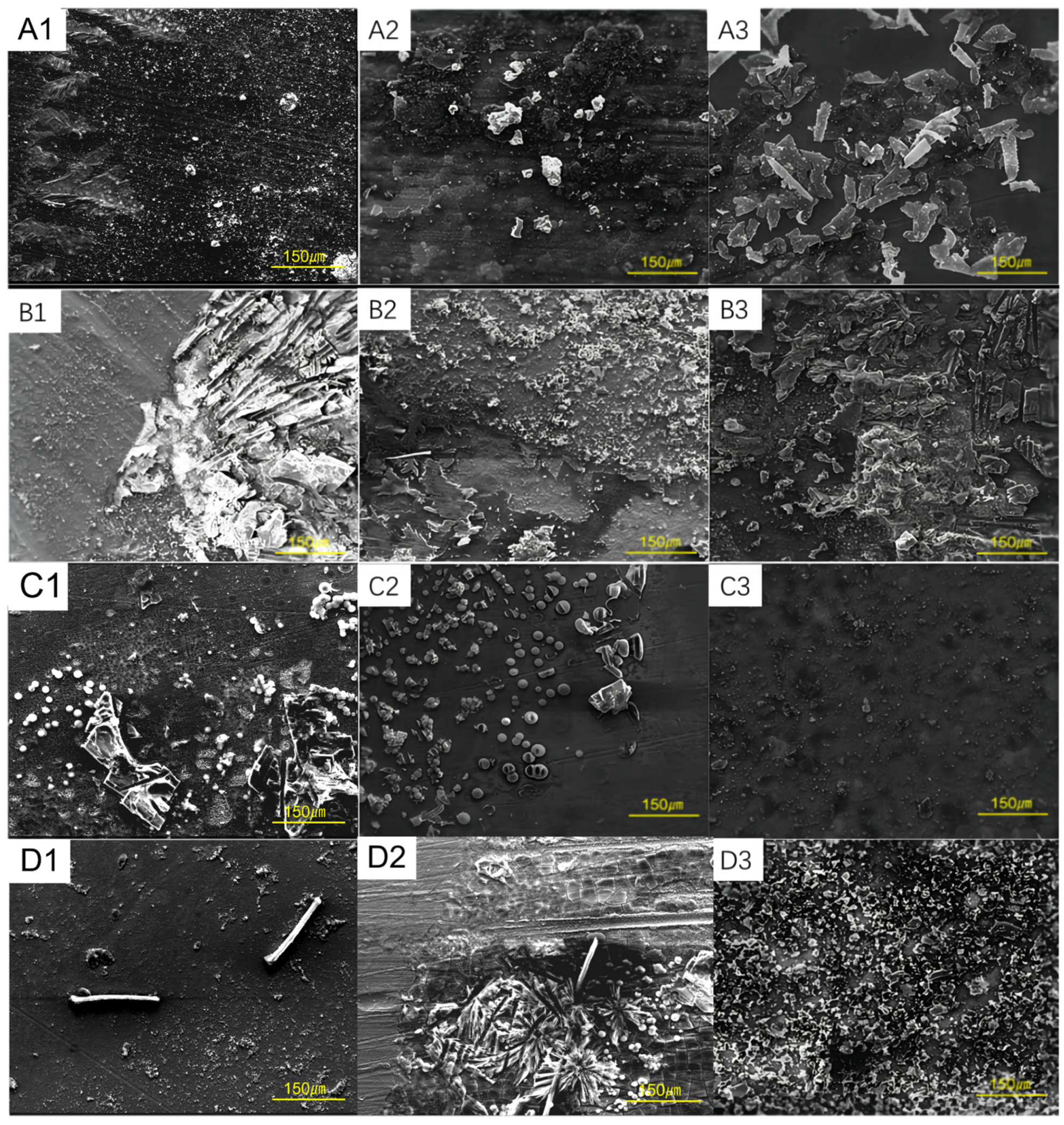
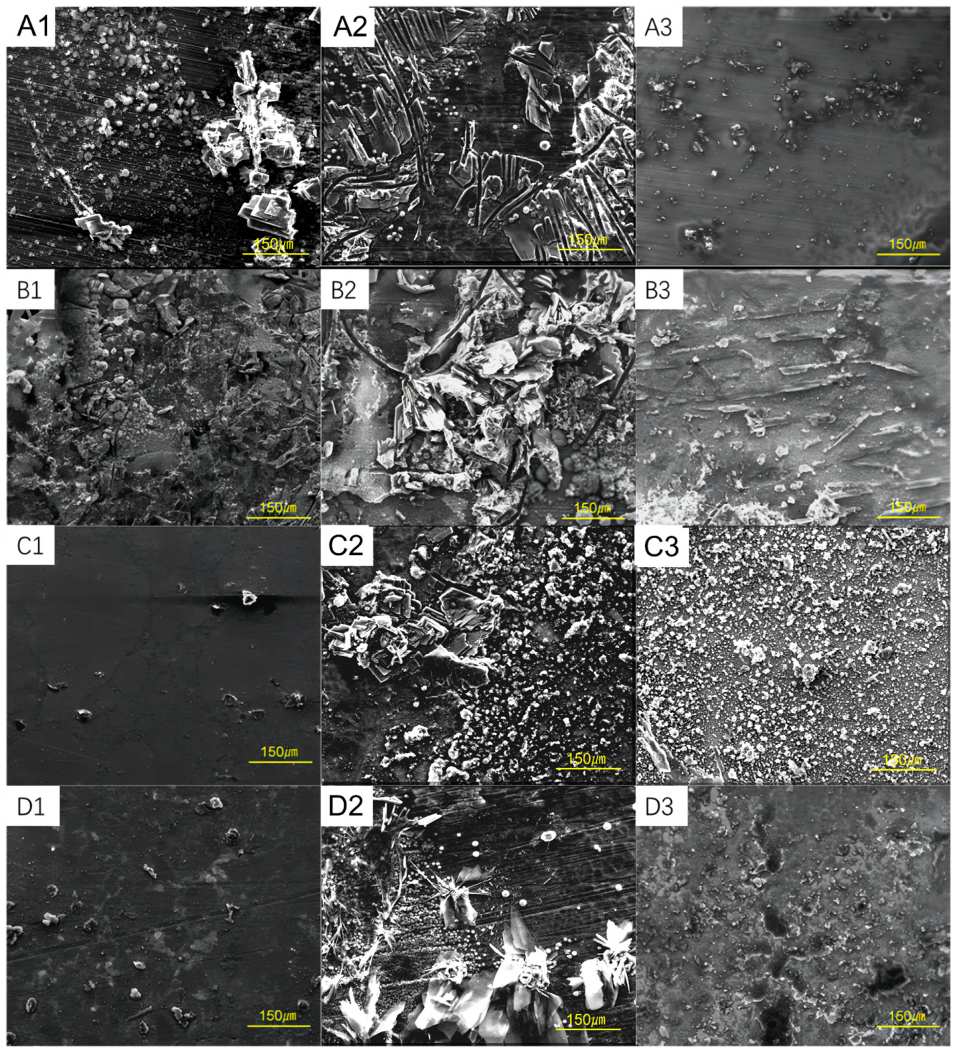
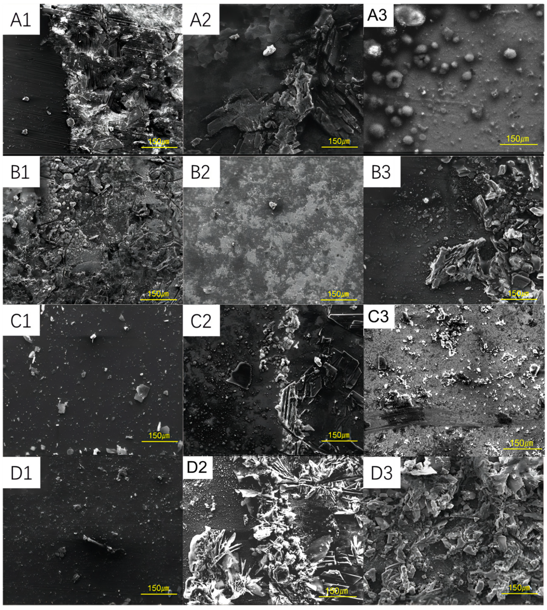
3.2. Corrosion Morphology of Steel Samples at Different Conditions

The surface morphology of specimens observed at 25 °C and the corrosion characteristics of various materials after immersion for different durations are illustrated in Figure 3. Figure 3(A1–A3) depict the SEM images of Steel-1 specimens after immersion periods of 5, 10, and 20 days. Initially, a biofilm presented on the surface after 5 days. By 10 days, loose, granular corrosion products had begun to accumulate on the biofilm’s surface. After 20 days, solidified sulfate-reducing bacteria (SRB) and corrosion products were clearly visible and uniformly dispersed across the specimen’s surface, contributing to corrosion. Figure 3(B1–B3) display the SEM images of the Steel-2 specimens, showing a corrosion morphology similar to that of Steel-1. However, at the 5-day mark, notable scratches and lumpy corrosion products were observable. Figure 3(C1–C3) present the SEM images of the Steel-3 specimens, which initially exhibited a minor presence of granular corrosion products. Over time, these granular products increased and accumulated on the biofilm, with microbial nodules appearing on the surface, thereby exacerbating the corrosion. Figure 3(D1–D3) illustrate the SEM images of the Steel-4 specimens. Compared to the Steel-3 specimens, these showed less corrosion, characterized by fewer corrosion products.
The surface morphology of the specimens, after 5, 10, and 20 days of immersion at 40 °C, for the four different steel types is depicted in Figure 4. Figure 4(A1–A3) illustrate the SEM images of the Steel-1 specimen: After 5 days, a biofilm and dispersed corrosion products were observed on the surface. By the 10th day, an increase in the accumulation of corrosion products on the biofilm was noted. Figure 4(B1–B3) display the SEM images of the Steel-2 specimen. The morphology of biofilm and corrosion products appeared relatively complex throughout the corrosion test. Figure 4(C1–C3) present the SEM images of the Steel-3 specimens. Initially, the surface was covered with a biofilm and exhibited fewer corrosion products, potentially due to the shorter immersion time. As the immersion period was extended, an increase in granular corrosion products was observed, with bacteria evident around these deposits. By the 20th day, the biofilm and corrosion product were well distributed on the steel surface. Figure 4(D1–D3) reveal the SEM images of the Steel-4 specimen. Initially, minimal scratches were observed. In comparison to the Steel-3 specimen, Steel-4 exhibited less severe corrosion, characterized by fewer biofilm and corrosion products on the steel surface.
The surface morphology of specimens after 5, 10, and 20 days of immersion at 60 °C for four types of steel is illustrated in Figure 5. Figure 5(A1–A3) show the SEM images of the Steel-1 specimen. After 5 days of immersion, numerous corrosion products formed on the surface, displaying a discontinuous distribution with holes that allowed corrosive bacteria to access the specimen’s surface, thereby increasing corrosion. By the 20th day, spherical corrosion products were observed on the surface of the biofilm. Figure 5(B1–B3) display the SEM images of the Steel-2 specimen. Initially, the specimen’s surface was covered with biofilm and corrosion products. The porous structure of these products intensified MIC. In the later stages of immersion, the corrosion product significantly increased. Figure 5(C1–D3) show the SEM images of the Steel-3 and Steel-4 specimens, respectively. In the 5-day tests, a lower degree of corrosion was observed with a few corrosion products appearing. As the immersion time progressed, no polishing line was visible on the specimen’s surface; however, an increase in corrosion products was noted.

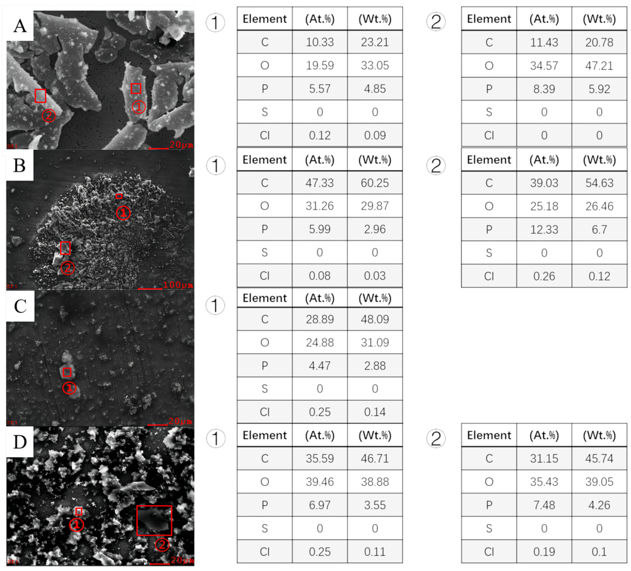
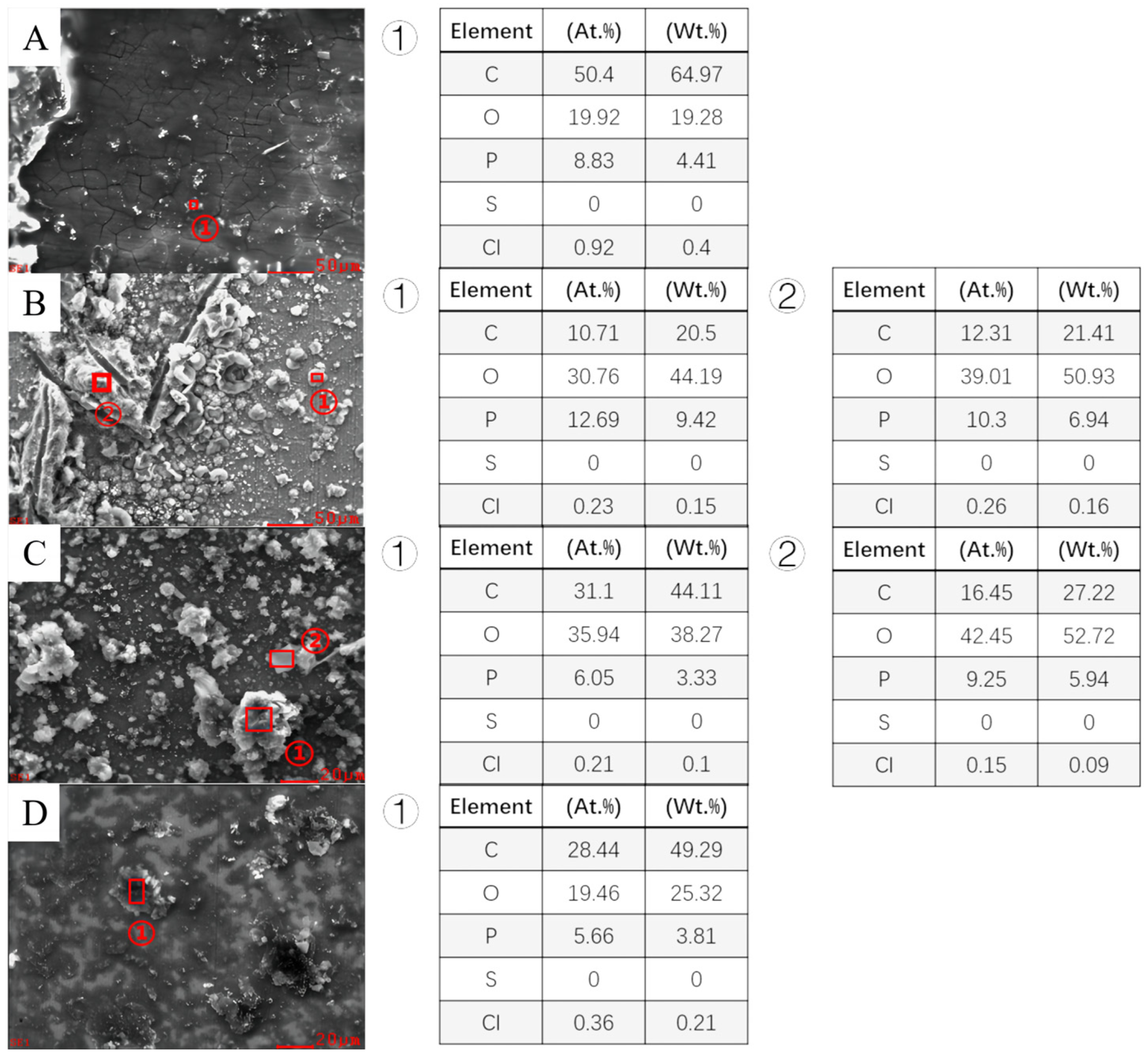
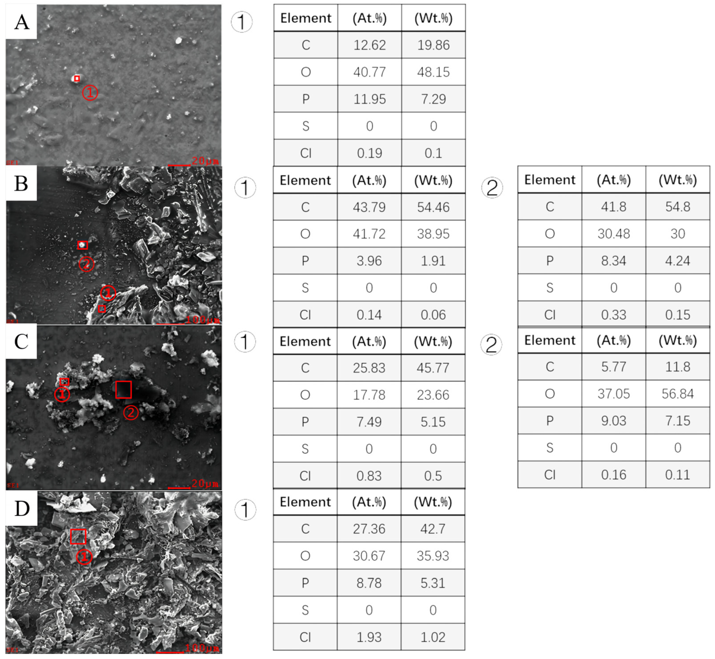
3.3. Analysis of Corrosion Product of Different Steels

Under conditions with an SRB bacteria concentration of 10,000 cells/mL for 20 days, the EDS scanning results for specimens of different materials at 25 °C, 40 °C, and 60 °C are presented in Figure 6, Figure 7 and Figure 8. In the energy spectrum, the elements S, P, C, O, and Cl were selected. Among these elements, S is the main metabolite (HS-) of the anaerobic respiration of SRB. The element P is detected as in the MIC products [8]. The elements C and O are the main components of extracellular polymeric substances (EPSs), which are in the SRB biofilm. In this research, a large amount of elemental element Cl was found at the same time, which was presumed to be due to the composition of the medium in the early stage of SRB growth. It is observed that the atomic and weight ratios of sulfur (S) in all four materials remain zero across different temperatures due to the lack of sufficient nutrients for SRB in this environment. In contrast, the content of chlorine (Cl) in all materials, although consistently higher than that of sulfur, remains at a lower level overall. Regarding the carbon (C) content, in Steel-1, there is an initial increase followed by a decrease with rising temperatures. In Steel-2, the carbon content gradually decreases, though the reduction is slight. Steel-3 exhibits the lowest carbon content at 60 °C. Conversely, the carbon content in Steel-4 shows a gradual decrease overall. The oxygen (O) content reaches its highest level in Steel-1 at 60 °C. In Steel-2 and Steel-3, the oxygen content slightly increases with temperature. However, in Steel-4, the oxygen content initially decreases and then increases. As for the phosphorus (P) content, it increases gradually with temperature in Steel-1 and Steel-3. In Steel-2, phosphorus content initially increases and then decreases, while in Steel-4, it remains relatively unchanged.

3.4. Electrochemical Analysis

Figure 9 and Table 3 present the Tafel fitting data from polarization curves for each parent material after 20 days of immersion in a 10,000 SRB/mL culture solution at various temperatures. According to Figure 9, Steel-1 exhibits the lowest corrosion potential (Ecorr) at 25 °C, which is 153 mV more negative than that of Steel-2, the highest. A more negative Ecorr in the polarization curves signifies a higher corrosion tendency, indicating that Steel-1 is more susceptible to corrosion under these conditions. Furthermore, analysis of the SEM images from Figure 3(A1–D1) reveals that the surface of the Steel-1 specimen develops a biofilm after 5 days of immersion. It is important to note that Ecorr primarily reflects the corrosion tendency of a specimen’s surface and does not directly indicate the rate of corrosion at the time of testing. When comparing the corrosion current density (Icorr) values, Steel-1 shows the lowest at 5.85 μA/cm2, while Steel-3 has the highest at 6.41 μA/cm2. Generally, a lower Icorr in the polarization curves suggests a slower corrosion rate and better corrosion resistance of the material. By comparing the Icorr values of the four steel materials, under the three conditions, Steel-1 exhibits the best corrosion resistance, followed by Steel-4. Steel-2 and Steel-4 do not demonstrate good corrosion resistance, and overall, their performance is inferior compared to the other two steels. One thing that needs to be mentioned is that because Steel-1 is alloyed, even though there is a thermodynamic tendency, the protective effect of the corrosion products causes the kinetics to be hindered. To provide a deep analysis of the SRB corrosion properties for different pipe steels, Steel-1–Steel-4, the breakdown potentials were calculated shown in Table 3. The breakdown potentials of Steel-1 and Steel-4 are all greater than those of Steel-2 and Steel-3 at different temperatures (25 °C, 40 °C, and 60 °C).

4. Discussion

Sulfate-reducing bacteria (SRB) adhering to metal surfaces can transport electrons, produced during the dissolution of the metal anode, to their interior using the extracellular electron transfer (EET) mechanism to obtain energy. In this process, the iron (Fe) matrix serves as the anode, with the adhered SRB acting as the biological cathode. These adhered SRB significantly accelerate the corrosion of steel. It is reported that SRB significantly increase the rate of metal corrosion by utilizing iron directly, especially when coupled with other types of corrosion, such as under-deposit corrosion [29]. In this study, the experimental medium solution, which was sterilized, contained no organic carbon sources; thus, SRB primarily utilized the steel matrix as their energy source over prolonged immersion periods.
The antibacterial properties of copper (Cu) and its alloys have been extensively documented [30,31,32]. The ions Cu2+ and Cu+, released during the corrosion process, are key factors contributing to these antibacterial properties [33,34,35]. In addition to copper, other alloying elements have also been reported to exhibit certain antibacterial properties, such as tin (Sn) [36]. These ions disrupt the integrity of bacterial cell membranes, penetrate these membranes, causing protein coagulation, and lead to enzyme inactivation within the cell, ultimately resulting in the apoptosis of SRB cells [37,38]. Steel-2 does not have special corrosion resistance treatments, such as alloying or improvements at the metallurgical and heat treatment levels; therefore, its corrosion resistance performance is relatively weak. Following alloying treatments, Steel-1 and Steel-4, which incorporate copper elements, show improved antibacterial effects, particularly in long-term corrosion tests. It is safe to conclude that appropriate heat treatment processes further enhance the antibacterial efficacy of these metal materials.

5. Conclusions

In the four tested steels of this research, it was determined that Steel-1 and Steel-4 showed a better MIC resistance than Steel-2 and Steel-3, which exhibited greater susceptibility to corrosion, as indicated by the polarization curves and uniform corrosion rates. This difference in corrosion behavior may be attributed to the addition of copper (Cu) to the specimen. Copper ions are believed to inhibit the metabolic activity of SRB by binding to proteins on the bacterial cell wall and membrane, thereby disrupting normal bacterial metabolism and reducing SRB activity, which in turn decreases the corrosion rate. It is safe to draw the conclusion that with alloying of Cu element during the metallurgical process improves the anti-biocorrosion ability of steels.

Supplementary Materials

The following supporting information can be downloaded at: https://www.mdpi.com/article/10.3390/coatings14070834/s1.

Author Contributions

Conceptualization, Q.L. (Qingjian Liu); Methodology, P.L.; Resources, B.W. and J.S.; Writing—original draft, Y.W.; Writing—review & editing, H.J. and Q.L. (Qingwen Liang). All authors have read and agreed to the published version of the manuscript.

Funding

This study is supported by the application research project on the anti-corrosion of production water pipelines in the Wushi 17-2/23-5 project (No. 70304805).

Institutional Review Board Statement

Not applicable.

Informed Consent Statement

Not applicable.

Data Availability Statement

Data are contained within the article and Supplementary Materials.

Acknowledgments

The authors thank all the people who helped in the completion of this paper and the people who read it and provided advice.

Conflicts of Interest

Pei Li was employed by the company CCCC First Harbor Engineering Co., Ltd., Baihong Wu was employed by the company CNOOC Safety Technology Service Co., Ltd., Qingwen Liang was employed by the company China National Offshore Oil Corporation (CNOOC) China Ltd. The remaining authors declare that the research was conducted in the absence of any commercial or financial relationships that could be construed as a potential conflict of interest.

References

  1. Loto, C.A. Microbiological corrosion: Mechanism, control and impact—A review. Int. J. Adv. Manuf. Technol. 2017, 92, 4241–4252. [Google Scholar] [CrossRef]
  2. Lavanya, M. A Brief Insight into Microbial Corrosion and its Mitigation with Eco-friendly Inhibitors. J. Bio-Tribo-Corros. 2021, 7, 125. [Google Scholar] [CrossRef]
  3. Shrestha, R.; Černoušek, T.; Stoulil, J.; Kovářová, H.; Sihelská, K.; Špánek, R.; Ševců, A.; Steinová, J. Anaerobic microbial corrosion of carbon steel under conditions relevant for deep geological repository of nuclear waste. Sci. Total Environ. 2021, 800, 149539. [Google Scholar] [CrossRef] [PubMed]
  4. Kip, N.; Van Veen, J.A. The dual role of microbes in corrosion. ISME J. 2015, 9, 542–551. [Google Scholar] [CrossRef] [PubMed]
  5. Song, X.; Yang, Y.; Yu, D.; Lan, G.; Wang, Z.; Mou, X. Studies on the impact of fluid flow on the microbial corrosion behavior of product oil pipelines. J. Petrol. Sci. Eng. 2016, 146, 803–812. [Google Scholar] [CrossRef]
  6. Jiang, G.; Zhou, M.; Chiu, T.H.; Sun, X.; Keller, J.; Bond, P.L. Wastewater-Enhanced Microbial Corrosion of Concrete Sewers. Environ. Sci. Technol. 2016, 50, 8084–8092. [Google Scholar] [CrossRef] [PubMed]
  7. Salgar-Chaparro, S.J.; Lepkova, K.; Pojtanabuntoeng, T.; Darwin, A.; Machuca, L.L. Nutrient Level Determines Biofilm Characteristics and Subsequent Impact on Microbial Corrosion and Biocide Effectiveness. Appl. Environ. Microbiol. 2020, 86, e02885-19. [Google Scholar] [CrossRef]
  8. Li, Y.; Xu, D.; Chen, C.; Li, X.; Jia, R.; Zhang, D.; Sand, W.; Wang, F.; Gu, T. Anaerobic microbiologically influenced corrosion mechanisms interpreted using bioenergetics and bioelectrochemistry: A review. J. Mater. Sci. Technol. 2018, 34, 1713–1718. [Google Scholar] [CrossRef]
  9. Little, B.J.; Hinks, J.; Blackwood, D.J. Microbially influenced corrosion: Towards an interdisciplinary perspective on mechanisms. Int. Biodeterior. Biodegrad. 2020, 154, 105062. [Google Scholar] [CrossRef]
  10. Xu, D.; Gu, T.; Lovley, D.R. Microbially mediated metal corrosion. Nat. Rev. Microbiol. 2023, 21, 705–718. [Google Scholar] [CrossRef]
  11. Kato, S. Microbial extracellular electron transfer and its relevance to iron corrosion. Microb. Biotechnol. 2016, 9, 141–148. [Google Scholar] [CrossRef] [PubMed]
  12. Li, Z.; Chang, W.; Cui, T.; Xu, D.; Zhang, D.; Lou, Y.; Qian, H.; Song, H.; Mol, A.; Cao, F.; et al. Adaptive bidirectional extracellular electron transfer during accelerated microbiologically influenced corrosion of stainless steel. Commun. Mater. 2021, 2, 67. [Google Scholar] [CrossRef]
  13. Zheng, Y.; Yang, Y.; Liu, X.; Liu, P.; Li, X.; Zhang, M.; Zhou, E.; Zhao, Z.; Wang, X.; Zhang, Y.; et al. Accelerated corrosion of 316L stainless steel in a simulated oral environment via extracellular electron transfer and acid metabolites of subgingival microbiota. Bioact. Mater. 2024, 35, 56–66. [Google Scholar] [CrossRef]
  14. Zhang, P.; Xu, D.; Li, Y.; Yang, K.; Gu, T. Electron mediators accelerate the microbiologically influenced corrosion of 304 stainless steel by the Desulfovibrio vulgaris biofilm. Bioelectrochemistry 2015, 101, 14–21. [Google Scholar] [CrossRef] [PubMed]
  15. Li, Y.; Feng, S.; Liu, H.; Tian, X.; Xia, Y.; Li, M.; Xu, K.; Yu, H.; Liu, Q.; Chen, C. Bacterial distribution in SRB biofilm affects MIC pitting of carbon steel studied using FIB-SEM. Corros. Sci. 2020, 167, 108512. [Google Scholar] [CrossRef]
  16. Xu, D.; Li, Y.; Gu, T. Mechanistic modeling of biocorrosion caused by biofilms of sulfate reducing bacteria and acid producing bacteria. Bioelectrochemistry 2016, 110, 52–58. [Google Scholar] [CrossRef] [PubMed]
  17. Dou, W.; Jia, R.; Jin, P.; Liu, J.; Chen, S.; Gu, T. Investigation of the mechanism and characteristics of copper corrosion by sulfate reducing bacteria. Corros. Sci. 2018, 144, 237–248. [Google Scholar] [CrossRef]
  18. Chilkoor, G.; Karanam, S.P.; Star, S.; Shrestha, N.; Sani, R.K.; Upadhyayula, V.K.K.; Ghoshal, D.; Koratkar, N.A.; Meyyappan, M.; Gadhamshetty, V. Hexagonal Boron Nitride: The Thinnest Insulating Barrier to Microbial Corrosion. ACS Nano 2018, 12, 2242–2252. [Google Scholar] [CrossRef]
  19. Yang, K.; Shi, J.; Wang, L.; Chen, Y.; Liang, C.; Yang, L.; Wang, L.-N. Bacterial anti-adhesion surface design: Surface patterning, roughness and wettability: A review. J. Mater. Sci. Technol. 2022, 99, 82–100. [Google Scholar] [CrossRef]
  20. Chinnaraj, S.B.; Jayathilake, P.G.; Dawson, J.; Ammar, Y.; Portoles, J.; Jakubovics, N.; Chen, J. Modelling the combined effect of surface roughness and topography on bacterial attachment. J. Mater. Sci. Technol. 2021, 81, 151–161. [Google Scholar] [CrossRef]
  21. Zhao, L.; Chen, R.; Lou, L.; Jing, X.; Liu, Q.; Liu, J.; Yu, J.; Liu, P.; Wang, J. Layer-by-Layer-Assembled antifouling films with surface microtopography inspired by Laminaria japonica. Appl. Surf. Sci. 2020, 511, 145564. [Google Scholar] [CrossRef]
  22. Bao, J.; Li, H.; Xu, Y.; Chen, S.; Wang, Z.; Jiang, C.; Li, H.; Wei, Z.; Sun, S.; Zhao, W.; et al. Multi-functional polyethersulfone nanofibrous membranes with ultra-high adsorption capacity and ultra-fast removal rates for dyes and bacteria. J. Mater. Sci. Technol. 2021, 78, 131–143. [Google Scholar] [CrossRef]
  23. Ogawa, A.; Takakura, K.; Hirai, N.; Kanematsu, H.; Kuroda, D.; Kougo, T.; Sano, K.; Terada, S. Biofilm Formation Plays a Crucial Rule in the Initial Step of Carbon Steel Corrosion in Air and Water Environments. Materials 2020, 13, 923. [Google Scholar] [CrossRef] [PubMed]
  24. Galarce, C.; Fischer, D.; Díez, B.; Vargas, I.T.; Pizarro, G.E. Dynamics of Biocorrosion in Copper Pipes under Actual Drinking Water Conditions. Water 2020, 12, 1036. [Google Scholar] [CrossRef]
  25. Victoria, S.N.; Sharma, A.; Manivannan, R. Metal corrosion induced by microbial activity—Mechanism and control options. J. Indian Chem. Soc. 2021, 98, 100083. [Google Scholar] [CrossRef]
  26. Shi, X.; Yan, W.; Xu, D.; Yan, M.; Yang, C.; Shan, Y.; Yang, K. Microbial corrosion resistance of a novel Cu-bearing pipeline steel. J. Mater. Sci. Technol. 2018, 34, 2480–2491. [Google Scholar] [CrossRef]
  27. Mansour, R.; Elshafei, A. Role of Microorganisms in Corrosion Induction and Prevention. Br. Biotechnol. J. 2016, 14, 1–11. [Google Scholar] [CrossRef]
  28. Yu, H.; Li, Z.; Xia, Y.; Qi, Y.; Li, Y.; Liu, Q.; Chen, C. Effect of copper addition in carbon steel on biocorrosion by sulfate-reducing bacteria in solution. Anti-Corros. Methods Mater. 2021, 68, 302–309. [Google Scholar] [CrossRef]
  29. Udowo, V.; Yan, M.; Liu, F.; Ikeuba, A. Role of Fe oxide in the underdeposit corrosion of pipeline steel in oilfield produced water containing SRB. Mater. Corros. 2023, 1, 118–129. [Google Scholar] [CrossRef]
  30. Nan, L.; Yang, W.; Liu, Y.; Xu, H.; Li, Y.; Lu, M.; Yang, K. Antibacterial Mechanism of Copper-bearing Antibacterial Stainless Steel against E.Coli. J. Mater. Sci. Technol. 2008, 24, 197–201. [Google Scholar]
  31. Li, Y.; Liu, L.; Wan, P.; Zhai, Z.; Mao, Z.; Ouyang, Z.; Yu, D.; Sun, Q.; Tan, L.; Ren, L.; et al. Biodegradable Mg-Cu alloy implants with antibacterial activity for the treatment of osteomyelitis: In vitro and in vivo evaluations. Biomaterials 2016, 106, 250–263. [Google Scholar] [CrossRef] [PubMed]
  32. Grass, G.; Rensing, C.; Solioz, M. Metallic Copper as an Antimicrobial Surface. Appl. Environ. Microbiol. 2011, 77, 1541–1547. [Google Scholar] [CrossRef] [PubMed]
  33. Xia, J.; Yang, C.G.; Xu, D.K.; Sun, D.; Nan, L.; Sun, Z.; Li, Q.; Gu, T.; Yang, K. Laboratory investigation of the microbiologically influenced corrosion (MIC) resistance of a novel Cu-bearing 2205 duplex stainless steel in the presence of an aerobic marine Pseudomonas aeruginosa biofilm. Biofouling 2015, 31, 481. [Google Scholar] [CrossRef] [PubMed]
  34. Sharifahmadian, O.; Salimijazi, H.R.; Fathi, M.H.; Mostaghimi, J.; Pershin, L. Relationship between surface properties and antibacterial behavior of wire arc spray copper coatings. Surf. Coat. Technol. 2013, 233, 74–79. [Google Scholar] [CrossRef]
  35. Zhang, S.; Yang, C.; Ren, G.; Ren, L. Study on behaviour and mechanism of Cu2+ ion release from Cu bearing antibacterial stainless steel. Mater. Technol. 2015, 30, B126–B132. [Google Scholar] [CrossRef]
  36. Ercetin, A.; Özgün, Ö.; Aslantas, K.; Aykutoğlu, G. The microstructure, degradation behavior and cytotoxicity effect of Mg–Sn–Zn alloys in vitro tests. Discov. Appl. Sci. 2020, 2, 173. [Google Scholar] [CrossRef]
  37. Sun, D.; Xu, D.; Yang, C.; Chen, J.; Shahzad, M.B.; Sun, Z.; Zhao, J.; Gu, T.; Yang, K.; Wang, G. Inhibition of Staphylococcus aureus biofilm by a copper-bearing 317L-Cu stainless steel and its corrosion resistance. Mater. Sci. Eng. C 2016, 69, 744–750. [Google Scholar] [CrossRef]
  38. Warnes, S.L.; Keevil, C.W. Mechanism of Copper Surface Toxicity in Vancomycin-Resistant Enterococci following Wet or Dry Surface Contact. Appl. Environ. Microbiol. 2011, 77, 6049–6059. [Google Scholar] [CrossRef]
Figure 1. OM light microscopy of four substrates: (a) Steel-1, (b) Steel-2, (c) Steel-3, (d) Steel-4.
Coatings 14 00834 g001
Figure 2. Uniform corrosion rates of each sample after 5, 10, and 20 days of corrosion test in a solution with 10,000 SRB/mL at different temperatures: (a) 25 °C, (b) 40 °C, and (c) 60 °C.
Coatings 14 00834 g002
Figure 3. SEM scans of specimens of different materials after corrosion for 5, 10, and 20 days at 40 °C and SRB concentration of 10,000 cells/mL: (A1) Steel-1, 5 d; (B1) Steel-2, 5 d; (C1) Steel-3, 5 d; (D1) Steel-4, 5 d; (A2) Steel-1, 10 d; (B2) Steel-2, 10 d; (C2) Steel-3, 10 d; (D2) Steel-4, 10 d; (A3) Steel-1, 20 d; (B3) Steel-2, 20 d; (C3) Steel-3, 20 d; (D3) Steel-4, 20 d.
Coatings 14 00834 g003
Figure 4. SEM scans of specimens of different materials after corrosion for 5, 10, and 20 days at 40 °C and SRB concentration of 10,000 cells/mL: (A1) Steel-1, 5 d; (B1) Steel-2, 5 d; (C1) Steel-3, 5 d; (D1) Steel-4, 5 d; (A2) Steel-1, 10 d; (B2) Steel-2, 40 10 d; (C2) Steel-3, 10 d; (D2) Steel-4, 10 d; (A3) Steel-1, 20 d; (B3) Steel-2, 20 d; (C3) Steel-3, 20 d; (D3) Steel-4, 20 d.
Coatings 14 00834 g004
Figure 5. SEM scans of specimens of different materials after corrosion for 5, 10, and 20 days at 40 °C and SRB concentration of 10,000 cells/mL: (A1) Steel-1, 5 d; (B1) Steel-2, 5 d; (C1) Steel-3, 5 d; (D1) Steel-4, 5 d; (A2) Steel-1, 10 d; (B2) Steel-2, 10 d; (C2) Steel-3, 10 d; (D2) Steel-4, 10 d; (A3) Steel-1, 20 d; (B3) Steel-2, 20 d; (C3) Steel-3, 20 d; (D3) Steel-4, 20 d.
Coatings 14 00834 g005
Figure 6. EDS results of specimens of different materials after corrosion with SRB bacteria concentration 10,000/mL at 25 °C for 20 days: (A) Steel-1, (B) Steel-2, (C) Steel-3, (D) Steel-4.
Coatings 14 00834 g006
Figure 7. EDS results of specimens of different materials after corrosion at 40 °C for 20 days at a concentration of 10,000 SRB cells/mL: (A) Steel-1, (B) Steel-2, (C) Steel-3, (D) Steel-4.
Coatings 14 00834 g007
Figure 8. EDS results of specimens of different materials after corrosion at 60 °C for 20 days at a concentration of 10,000 SRB cells/mL: (A) Steel-1, (B) Steel-2, (C) Steel-3, (D) Steel-4.
Coatings 14 00834 g008
Figure 9. Polarization curves of each parent material after 20 days of immersion in 10,000 SRB/mL culture solution at different temperatures (a) 25 °C, (b) 40 °C, (c) 60 °C.
Coatings 14 00834 g009
Table 1. The mass percentage of composition content of four metal substrates.
CSiMnPSVNiTiCu
Steel-10.160.451.650.020.010.090.050.06
Steel-20.180.451.70.0250.015V + Ni + Ti < 0.15
Steel-30.160.451.650.020.030.090.30.06
Steel-40.160.451.650.020.030.090.30.060.35
Table 2. Composition of Postgate’s C medium.
NameSodium LactateNa2SO4NH4ClYeastKH2PO4Sodium CitrateCaCl2·6H2OMgSO4·7H2OFeSO4·7H2ODeionized Water
Concentration (g/L) and Volume6.04.51.01.00.50.30.060.06Few1 L
Table 3. Fitting results of polarization curves of parent materials after soaking in 10,000 SRB/mL culture solution for 20 days at 25 °C, 40 °C, and 60 °C.
25 °C40 °C60 °C
Ecorr/(V)Icorr/(μA/cm2)Eb(V)Ecorr/(V)Icorr/(μA/cm2)Eb (V)Ecorr/(V)Icorr/(μA/cm2)Eb (V)
Steel-1−0.6565.85−0.294−0.6786.21−0.352−0.5795.67−0.240
Steel-2−0.5036.03−0.239−0.6256.38−0.205−0.5786.65−0.140
Steel-3−0.5826.41−0.241−0.6296.46−0.301−0.3886.31−0.134
Steel-4−0.6206.23−0.327−0.6346.36−0.308−0.6076.12−0.465
Disclaimer/Publisher’s Note: The statements, opinions and data contained in all publications are solely those of the individual author(s) and contributor(s) and not of MDPI and/or the editor(s). MDPI and/or the editor(s) disclaim responsibility for any injury to people or property resulting from any ideas, methods, instructions or products referred to in the content.

