Article

Effects of Compound Use of Two UV Coating Microcapsules on the Physicochemical, Optical, Mechanical, and Self-Healing Performance of Coatings on Fiberboard Surfaces

1
Co-Innovation Center of Efficient Processing and Utilization of Forest Resources, Nanjing Forestry University, Nanjing 210037, China
2
College of Furnishings and Industrial Design, Nanjing Forestry University, Nanjing 210037, China
*
Author to whom correspondence should be addressed.
Coatings 2024, 14(8), 1012; https://doi.org/10.3390/coatings14081012
Submission received: 15 July 2024 / Revised: 25 July 2024 / Accepted: 5 August 2024 / Published: 9 August 2024

Abstract

:
Ultraviolet (UV) coatings are widely used because of their good performance. However, the self-healing performance of UV coatings can be further improved. Microcapsule technology can be used to solve this problem. To investigate the effects of the compound use of two UV coating microcapsules on coatings of a fiberboard surface, three kinds of UV primer microcapsules (1#, 2#, and 3# microcapsules) with different contents were added to a UV primer, and a UV top coating was prepared with UV top coating microcapsules at a consistent ratio. The UV coating was used to coat the fiberboard surface by way of a two-primer and two-top coating method. The results show that as the content of the UV primer microcapsules was increased, the self-healing rates of all three groups of coatings increased and later decreased. The color difference ΔE of coatings with the content of the UV primer microcapsules at 4.0% and top coating microcapsules at 6.0% was 3.59, the gloss was 1.33 GU, the reflectance was 21.17%, the adhesion grade was 2, the hardness was 2H, the impact resistance grade was 5, the roughness was 1.085 μm, and the self-healing rate was 30.21%. Compared with the self-healing rate of the blank control group, the increase in the self-healing rate was 10.07%, and the improvement rate was 50.00%. The comprehensive performance of the coating was better. The results provide a technical reference for the application of the UV coating microcapsules in the UV coating on fiberboard surfaces. Incorporating the self-healing UV coating microcapsules into the UV coatings and applying the UV coating microcapsules on the fiberboard surfaces supports the microcapsule technology of self-healing UV coatings, lays the foundation for extending the service life of furniture while improving the furniture’s quality, and promotes the sustainable development of the coating industry.

1. Introduction

Wooden furniture is a very important type of furniture, but wood is prone to decay and cracking, resulting in a large amount of wooden furniture waste and other wasted wooden materials. The decay of wooden furniture causes huge resource waste and serious environmental pollution [1,2,3,4,5]. Wood is used as a furniture material and occupies an important part in furniture manufacturing and people’s daily use [6,7,8]. Therefore, the protection of wooden furniture should be studied to reduce the rate of waste of wooden furniture [9,10,11]. Currently, there are various modification methods used to protect wood [12,13,14]; for example, coating the surface of wooden furniture is one of the commonly used methods [15,16,17]. There are various types of resins used for preparing ultraviolet (UV) coatings, including unsaturated polyester, polyurethane acrylate, acrylate and methacrylate resins, epoxy oligomers, polyamides, and hyperbranched polymers [18]. When these UV coatings are applied on the surface of wooden furniture, a homogeneous and dense coating can be formed to provide protection, but the coating will inevitably be damaged during the furniture’s long-term use. A rupture of the coating accelerates the wear and tear of furniture and shortens its service lifetime [18,19,20,21,22,23]. Therefore, optimizing the performance of different coatings is critical, and adding microcapsules to the coating is a commonly used method to improve the performance of furniture coatings and expand the functionality of the coating [24,25,26].
However, UV coatings also have certain limitations. For example, for objects with more complex shapes, it is difficult to achieve an all-around curing effect of the UV coatings under the irradiation of UV light due to the existence of blocked areas. Meanwhile, traditional UV coatings generally have problems, such as a higher viscosity and a weaker mobility, and thus need to be adjusted by adding diluents throughout the process of applying them to furniture, which tends to be toxic and may cause harm to human beings [27]. Several photochemical methods, including involving disulfide groups, reversible Diels–Alder reactions, hydrogen bond breaking, reforming, photodimerization, and self-filling systems, have been used to develop self-healing coatings [28]. Although the self-healing efficiency of the UV coatings prepared with microcapsule technology still needs to be improved, the synthesis of self-healing microcapsules and their addition to UV coatings are relatively simple and cost-effective compared to the other methods above for preparing self-healing UV coatings. A basic theory of a self-healing system of a UV coating with the addition of microcapsules is relatively mature. Sow et al. [29] investigated the effects of alumina and silica nanoparticles on the mechanical, optical, and thermal performance of UV waterborne nanocomposite coatings. The results show that the scratch resistance of the nanocomposite coating was significantly improved. It was found that the alumina and silica nanoparticles increased the glass transition temperature of the nanocomposite coating. Finally, by investigating the behavior of the silica nanoparticles, it was proved that the grafting of trialkoxysilanes onto the silica nanoparticles not only improved the dispersion of the silica nanoparticles but also significantly improved the scratch resistance and adhesion of UV waterborne coatings containing the silica nanoparticles, even at a silica nanoparticle content of 1 wt%. Microcapsules can be added to UV coatings to improve the mechanical and chemical performance of the coatings to a certain extent and at the same time allow the UV coating to achieve the desired performance. Wu et al. [30] designed and prepared several multichannel and multifunctional droplet microfluidic devices based on soft lithography for the efficient synthesis of core–shell hydrogel microcapsules for different applications. The core–shell hydrogel microcapsules with gelatin methacryloyl as a core and polyacrylamide as a thin shell were synthesized by using a UV crosslinking method. Another type of core–shell structure microcapsules with gelatin methacryloyl as the core and Ca2+ crosslinked alginate and polyethyleneimine as the shell were constructed using an interfacial polymerization process. The core diameter and a total droplet diameter were flexibly controlled by sculpting. These hydrogel microcapsules were stimuli-responsive and capable of controlled release. Microcapsule shells are usually made of polymer materials and are used to isolate active ingredients by encapsulating different solid, liquid, or gaseous substances [31,32]. The use of microcapsule technology to prepare self-healing UV coating microcapsules and the application of microcapsules to UV coatings are of great practical significance for protecting furniture substrates and promoting sustainable development. Zhang et al. [33] prepared novel amino-functionalized polyurethane microcapsules coated with a self-healing agent (flaxseed oil) and a corrosion inhibitor (benzotriazole) with an average particle size of 1.0 μm by using “intra-emulsion droplet phase separation” combined with photopolymerization. Both microcapsule formation and the modification of the microcapsule shell by amino groups were achieved in a one-pot process. The amino-functionalized polyurethane microcapsules were incorporated into UV-curable resins to prepare the coatings with excellent self-healing and corrosion inhibition properties. The results show that cracks of the UV coating based on the amino-functionalized polyurethane microcapsules were almost completely healed after 30 days of tests, showing a higher self-healing efficiency than the coatings with the unmodified microcapsules. The self-healing microcapsules can be prepared with a core–shell structure and added to the coating. When the coating is subjected to temperature and humidity, the coating accumulates stress and undergoes strain, the microcapsule wall ruptures, and the encapsulated core material flows out, filling microcracks in surrounding space. Due to consistency between the core material and the coating, the microcracks can slowly be healed.
The 1#, 2#, and 3# UV primer microcapsules were selected and added to the UV primer, and the content of UV top coating microcapsules added to the UV top coatings was 6.0%. The UV coatings were coated on the fiberboard surface by a two-primer and two-top coating method. The UV top coating microcapsules used in this test had a good morphology and minimal aggregation. The particle sizes of the microcapsules were between 2 and 12 μm [34]. The effects of different contents of UV primer microcapsules and the fixed content of UV top coating microcapsules on the optical performance, mechanical performance, and self-healing performance of the UV coatings on the fiberboard surface were investigated.

2. Materials and Methods

2.1. Materials

The fiberboard was 50 mm × 50 mm × 5 mm. Table 1 shows the test materials. The UV top coating included polyurethane acrylic resin, 1,6-hexanediol diacrylate, photoinitiator 184 (1-hydroxycyclohexyl phenyl ketone), functional filler, extinction powder, wax powder, defoamer, dispersant, and anti-settling agent, etc. The UV primer included epoxy acrylic resin, polyester acrylic resin, trihydroxy methacrylate, trimethyl methacrylate, leveling agent, photoinitiator 1173 (2-hydroxy-2-methylpropiophenone), defoamer, etc. The coatings both had more than 98.0% solids content. The test instruments are shown in Table 2. A single-lamp curing machine with a UV mercury lamp with a light intensity of 80–120 W/cm2 was employed. The wavelength range of the UV irradiation released was 250–420 nm.

2.2. Preparation Method of Three UV Coating Microcapsules

Table 3 provides formulations for the 1#, 2#, and 3# UV primer microcapsules, as well as the UV top coating microcapsules. The 1# UV primer microcapsules are listed below as an example of preparation, with # indicating the unit of the sample number:
Formulation of wall materials: Following a molar ratio of 3.5:1.0, 10.81 g of 37% formaldehyde solution and 4.80 g of melamine were measured [35]. The deionized water was used to dissolve the formaldehyde, melamine, and triethanolamine. The pH of the system was approximately 9.0. The system was placed in a 60 °C water bath and stirred at 700 rpm. After 20 min of the reaction, the wall materials were prepared and maintained at a constant temperature of 60 °C.
Formulation of the core materials: 0.08 g of Triton X-100 and 0.22 g of Spectra 20 were added to ethanol to form an emulsifier system. After the system was stirred sufficiently, 4.40 g of UV primer was added. The beaker was kept in the 60 °C water bath at 700 rpm. After 70 min, the core materials were prepared.
Formation of the UV primer microcapsules: The wall materials were mixed with the core materials and sonicated for 15 min. The mixture system was then placed in the water bath with a pH of approximately 4.0 following the addition of citric acid monohydrate. After the reaction for 2 h, products were left for 5 d. The products were filtered and dried in an oven at 60 °C to obtain the powder, i.e., the 1# UV primer microcapsules. The 2# and 3# UV primer microcapsules and the UV top coating microcapsules were prepared using the same method.

2.3. Preparation Method of UV Coatings on the Fiberboard Surface

Samples were coated manually by applying two layers of the UV primer and two layers of the UV top coating using the following process:
The samples without any microcapsules were used as a blank control group. The surface was sanded with 800# sandpaper and cleaned of debris before each application of the UV coatings. The 1#, 2#, and 3# UV primer microcapsules were added to the UV primer at 2.0%, 4.0%, 6.0%, 8.0%, and 10.0%. The UV primer containing the microcapsules was applied on the fiberboard surface with a brush. After waiting 1 min for the UV primer to level, the UV primer was cured for 25 s in the single-lamp curing machine and then cooled to room temperature. Then, the sample was prepared for the second UV primer.
The UV top coating was then coated by the method described above. The quantities of coating materials are provided in Table 4. The density of the UV top coating was 1.24 g/cm3, and the density of the UV primer was 1.12 g/cm3. Therefore, the thickness of the UV coating corresponding to the size of the fiberboard surfaces was a total of about 270 μm.

2.4. Tests

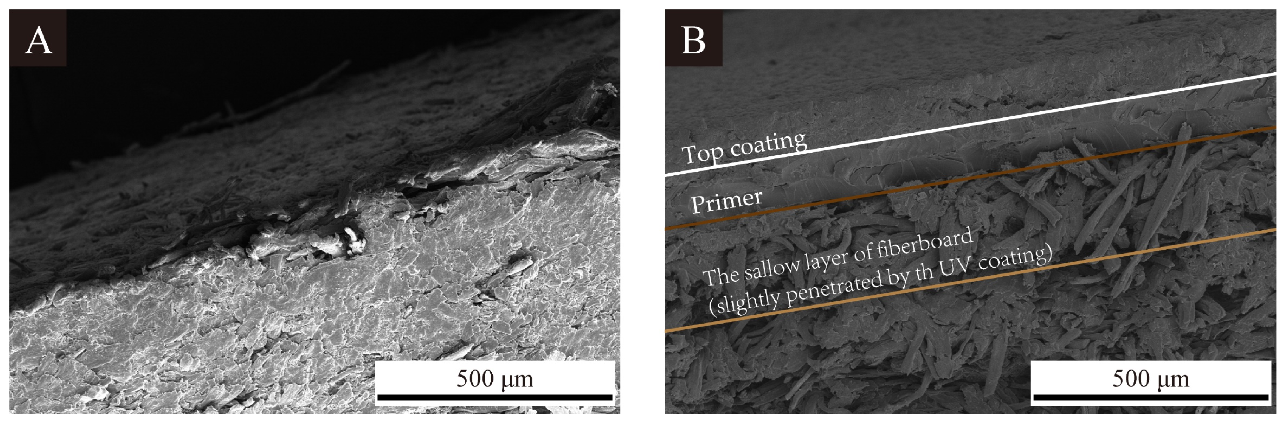
2.4.1. Microscopic Characterization

Scanning electron microscope (SEM): After the fiberboard surface was vertically cut, the finished sample was gold-sprayed. Then, the sample was placed into the instrument for observation after the air pressure inside the instrument stabilized [36,37].

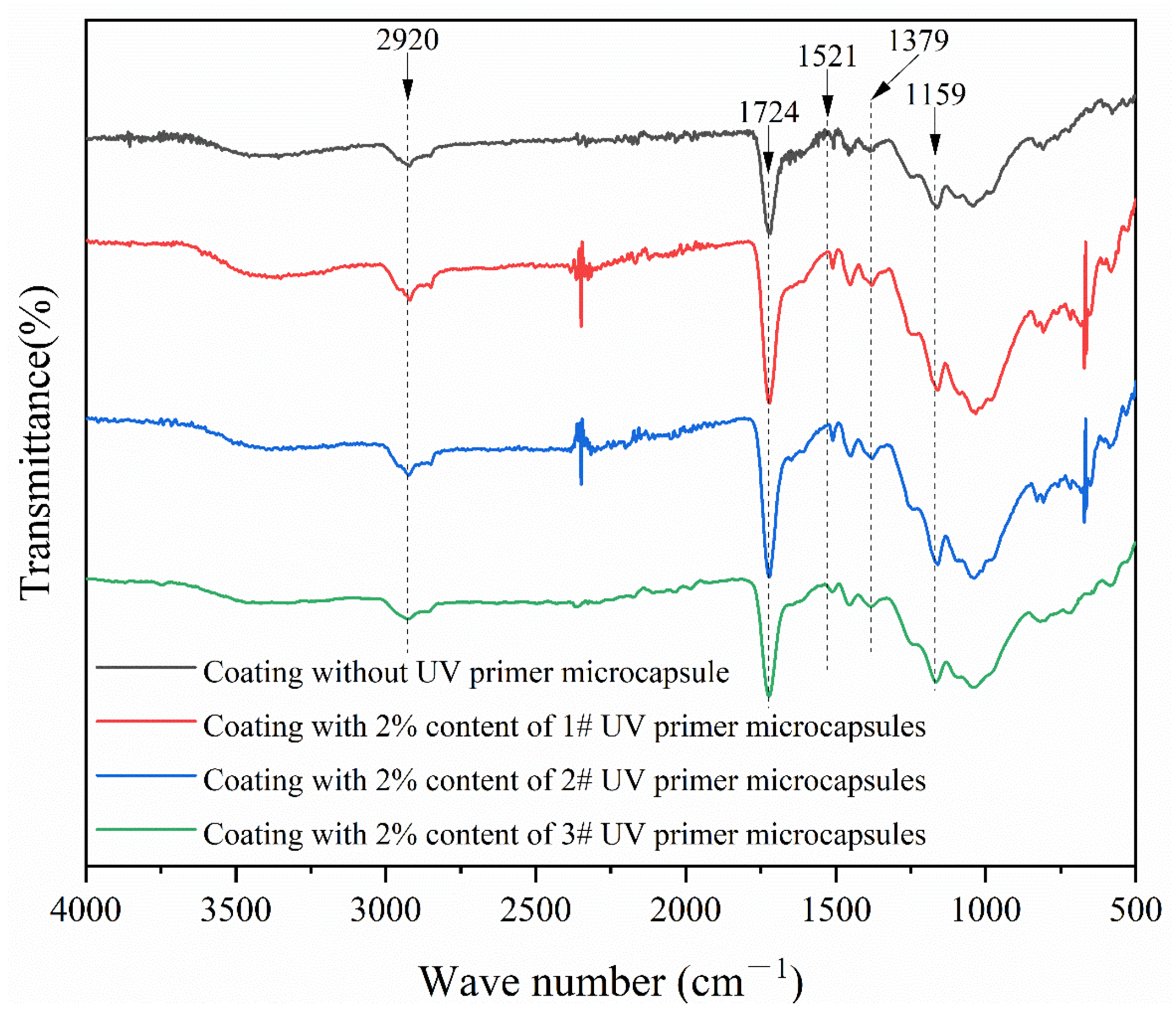
2.4.2. Chemical Composition Test

A small quantity of sample was pressed into a thin sheet based on KBr. The chemical composition of the sample was analyzed by using an infrared spectrometer.

2.4.3. Optical Performance Test

Color difference: Based on GB/T 11186.3-1989 [38], a color difference meter was used to test the samples. The data for the blank control group were recorded as L1, a1, and b1, and the test data for the sample were recorded as L2, a2, and b2. The color difference ΔE was calculated with Formula (1), in which ΔL = L2L1, Δa = a2a1, Δb = b2b1.
ΔE = [(ΔL)2 + (Δa)2 + (Δb)2] 1/2
Gloss: Based on GB/T 4893.6-2013 [39], a gloss of the sample was tested by using a gloss meter. The data were recorded at three incidence angles, 85°, 60°, and 20° [40].
Reflectance: A UV spectrophotometer was used to test the reflectance. The samples were placed in the instrument and reflected beams of different wavelengths emitted by the instrument. The instrument recorded the reflectance data of the samples [41].

2.4.4. Mechanical Performance Test

Impact resistance: Based on GB/T 1732-2020 [42], a steel ball was dropped 5 times above the sample at 50 mm height at different parts. Grades of impact resistance were recorded. The higher the grade, the poorer the impact resistance [43].
Hardness: Based on GB/T 6739-2022 [44], a processed pencil with specific hardness was placed into the instrument for each test without any external force and came into contact with the sample. A fixed weight was placed on the sample, and after the instrument was pushed forward, a scratch was applied to the sample. The hardness of the coating was recorded as the pencil hardness that did not scratch the sample [45].
Adhesion: Based on GB/T 4893.4-2013 [46], a multi-blade cutting tool was used to vertically cut off the coating, and this process then repeated to form a mesh scratch. A cut area was stuck and torn off with clear tape. The test was repeated three times, and average data were obtained to show the adhesion grade of 0–5. The higher the grade, the worse the adhesion.
Roughness: After the sample was placed horizontally, a probe of a roughness meter was adjusted to make contact with a sample surface. The roughness data were exported by recording vertical fluctuations of the probe through a horizontal movement on the sample surface.

2.4.5. Self-Healing Performance Test

A crack was left on the sample surface by using a blade. Using an external light source to illuminate from a fixed angle, the widest crack width was recorded as W1 using corresponding software. One week later, the crack width was measured again at the same location and recorded as W2. The self-healing rate (W) of the sample is calculated in Formula (2).
W = [(W1W2)/W1] × 100%

3. Results and Discussion

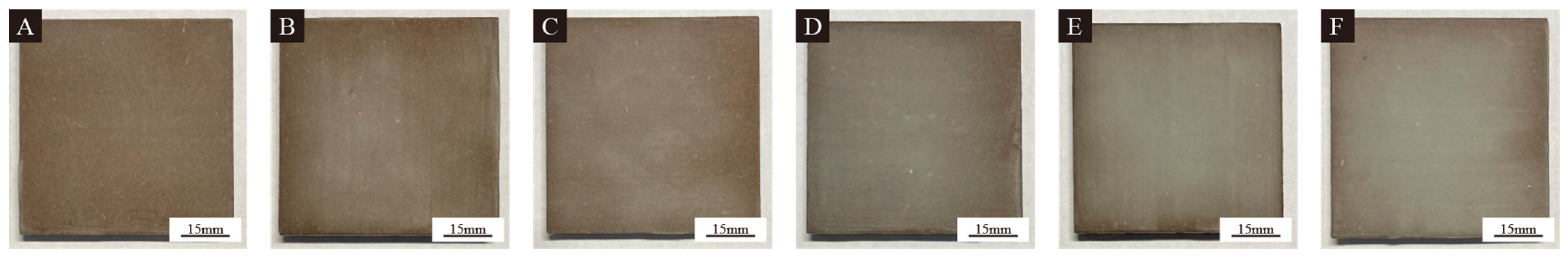
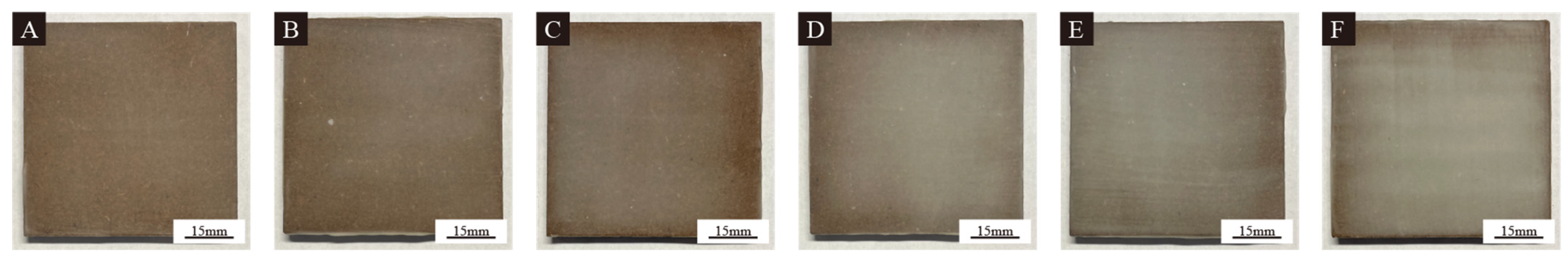
3.1. Macroscopic Analysis

Figure 1, Figure 2 and Figure 3 show surface morphologies of the fiberboards with different contents of the 1#, 2#, and 3# microcapsules in the UV primer, respectively. The color difference of the coating was slight when the content of the UV primer microcapsules was 4.0% or less. When the content of the UV primer microcapsules reached 6.0% and above, the color difference of the coating was obvious and radial, gradually fading from the center to the edges. This is because each layer of the coating was dripped to the center of the fiberboard and then coated outward uniformly. Therefore, in the four-layer process of manual coating, the content of microcapsules at the center of the fiberboard was slightly higher than that at surrounding areas. That resulted in a situation where the coating in the center of the fiberboard was slightly thicker than that at the edges.
SEM images of the fiberboard surfaces without microcapsules in the UV primer and with 2.0% content of 1#, 2#, and 3# microcapsules in the UV primer are shown in Figure 4. The morphology of the samples in Figure 4A,B was similar, with slight undulations on the coating surface and overall flatness. The samples in Figure 4C,D showed granular bumps and more folds on the surface. This is because of the more extensive agglomeration of the 2# and 3# microcapsules. The 2# and 3# microcapsules are difficult to disperse uniformly in the coating, affecting the leveling process of the coatings and resulting in a more uneven coating surface. Figure 5 shows the SEM images of the longitudinal section of the samples. Compared with the blank fiberboard, the UV primer, UV top coating, and the fiberboard were significantly different and bonded well. The UV primer with UV primer microcapsules slightly penetrated the fiberboard surface, which provided better protection for both the fiberboard and the coating.

3.2. Chemical Analysis

Figure 6 shows the infrared spectrum of the coating with and without the UV primer microcapsules in the primer. Characteristic peaks at 1379 cm−1 and 1521 cm−1 are C-N telescopic vibration peaks and N-H bending vibration peaks in the diamine resin. These show that the wall materials exist in the primer, and the chemical composition remains intact. The 1159 cm−1, 1724 cm−1, and 2920 cm−1 peaks are C-O, C=O, and C-H stretching vibration peaks. These three peaks belong to the common characteristic peaks of both the UV primer and the core materials, which include the main components of polyester acrylic resin, trihydroxy methacrylate, and trimethyl methacrylate. These three peaks appeared in all four absorption curves, which shows that the UV primer microcapsules do not influence the curing performance of the UV primer and proves that the UV primer microcapsules exist stably in the UV primer.

3.3. Optical Analysis

3.3.1. Analysis of Color Difference

The color difference of the samples with different contents of 1#, 2#, and 3# microcapsules is shown in Table 5. The L value of the samples with the different contents of UV primer microcapsules increased with the increased content of microcapsules, indicating that the coating on the fiberboard surface gradually became bright, which is because the fiberboard itself is brown, and in the natural lighting conditions, the surface coating would appear to be relatively dark. The UV primer and the microcapsules were white, so with the increase in the number of coated layers and microcapsules, the coating surface became whiter, which made the whole surface brighter, and the L value increased. The b value of the samples decreased with the increase in the microcapsules, indicating that the tan color of the fiberboard itself faded gradually. When the tan color was overlaid with the white color, it reduced the saturation of the coating as a whole. Thus, the coating slowly turned to a lighter brown or even gray color. The color difference of the samples with 1#, 2#, and 3# microcapsules increased gradually with the increase in the microcapsules. The maximum color difference of the coating with 1# microcapsules reached 8.95. The maximum color difference of the coating with 2# microcapsules reached 11.29, and the maximum color difference of the coating with 3# microcapsules reached 9.77. There was not much difference in the color difference of the samples with different contents of UV primer microcapsules, which shows that the 1#, 2#, and 3# microcapsules did not affect the color difference of the coating.

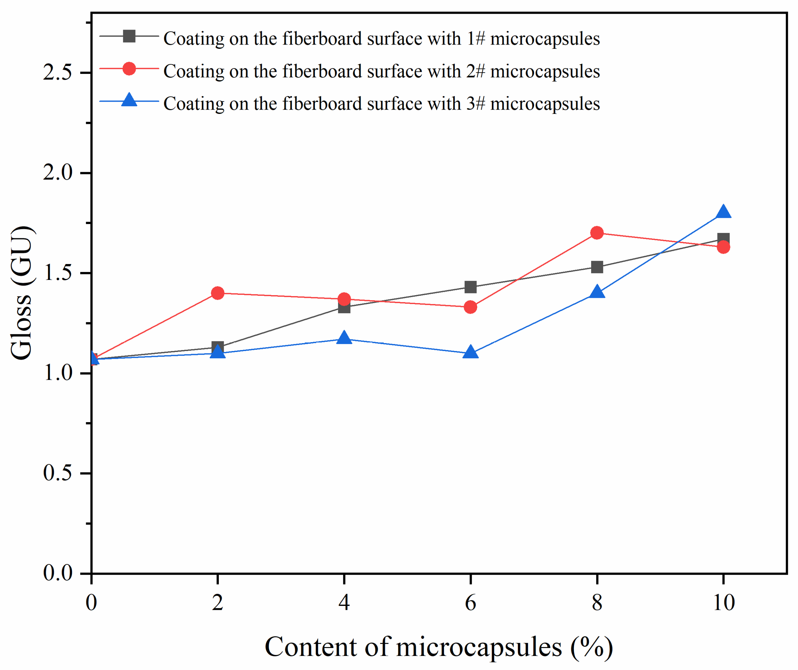
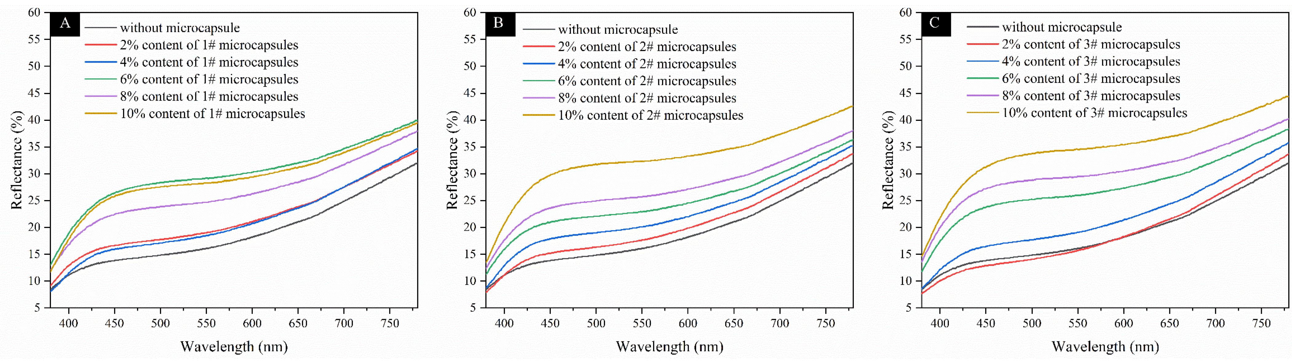
3.3.2. Gloss and Reflectance Analysis

Table 6 and Figure 7 show the changes in gloss and reflectance of the samples with different contents of 1#, 2#, and 3# microcapsules. Figure 8 shows the reflectance of the samples with microcapsules. As the content of UV primer microcapsules increased, the overall trend of the gloss increased. The gloss under the 60° incidence angle changed with a gentler trend, indicating that the reflectance change of the samples was small. This indicates that the effect of the 1#, 2#, and 3# microcapsules on the gloss is relatively weak. The reflectance of the samples was positively increased with the increase in the UV primer microcapsules as a whole. The trend of the reflectance of the samples was similar, and when the content of UV primer microcapsules was 4.0% and below, the change in the reflectance of the coating was small. When the content of the UV primer microcapsules was 6.0% and above, the reflectance increased rapidly. The change in reflectance of the coating on the fiberboard surface with the 2# microcapsules was average. When the content of the UV primer microcapsules was 6.0% and below, the change in reflectance of the coating was small. The coating reflectance with 6.0% content of the 1# UV primer microcapsules reached a maximum of 29.97%. The coating reflectance with the 2# and 3# UV primer microcapsules reached the maximum at 10.0% content, which was 32.83% and 34.74%.
The optical performance of the samples was comprehensively observed. With the increased content of the UV primer microcapsules, the color difference, the gloss, and the reflectance of the samples showed an increasing trend. The trend between the various data was similar, which indicated that the different contents of different microcapsules had small effects on the optical performance of the coating.

3.4. Mechanical Analysis

Table 7 shows the mechanical performance of the samples. The adhesion of samples was negatively correlated with the increased content of the UV primer microcapsules. The adhesion grade of the samples with the 1# and 2# microcapsules was grade 1 at 2.0% content and below and decreased to grade 2 at 4.0% content and above. The adhesion of the samples with the 3# microcapsules was decreased to grade 2 at 2.0% content of UV primer microcapsules, and it was maintained at grade 2. The coating on the fiberboard surface with the 1# and 2# microcapsules had a slower trend of weakening adhesion than that with the 3# microcapsules. The 1# and 2# microcapsules are better dispersed than the 3# microcapsules and have less influence on the coating–fiberboard interface. Therefore, the samples with the 1# and 2# microcapsules showed better adhesion with the same content of the UV primer microcapsules.
The hardness of the samples was positively correlated with the content of the UV primer microcapsules, and the hardness of the samples with the addition of 1# microcapsules at 6.0% content and below was 2H, increased to 3H at 8.0% content, and increased to 4H at 10.0% content. The hardness of the samples with the 2# microcapsules at 8.0% content and below was 2H and increased to 3H at 10.0% content. The hardness of the samples with the 3# microcapsules at 6.0% content and below was 2H and increased to 3H at 8.0% content and above. The samples with the 1# microcapsules showed the most significant increase in hardness, and the maximum hardness achieved was also the highest among the three groups.
The impact resistance of samples increased with the increase in the UV primer microcapsules. The impact resistance of samples with the 1# and 2# microcapsules reached the highest grade 3 at 8.0% and 10.0% content of the UV primer microcapsules. The impact resistance of the UV coating on the fiberboard surface with the 3# microcapsules reached the highest grade 4 at 8.0% content, which indicated that the 1# and 2# microcapsules had a better effect on the impact resistance performance than the 3# microcapsules. This is because the particle size of 1# and 2# microcapsules was larger than the 3# microcapsules. The microcapsules due to their spherical structure provide a better buffer for the coating when subject to the impact of the external force. The external force is evenly dispersed, so the larger particle size provides better protection for the coating, which improves the impact resistance of the coating.
The coating roughness increased with the increased content of the UV primer microcapsules. The roughness of the samples with the 1# and 3# microcapsules reached 2.064 μm and 2.598 μm at 10.0% content. The roughness of the samples with the 2# microcapsules of 6.0% content and below showed a decreasing trend, which was attributed to bumps caused by microcapsules in the UV coatings.
The mechanical performance of the samples was analyzed comprehensively. The microcapsules weakened the adhesion to a certain extent, enhanced the hardness, strengthened the impact resistance, and increased the roughness.

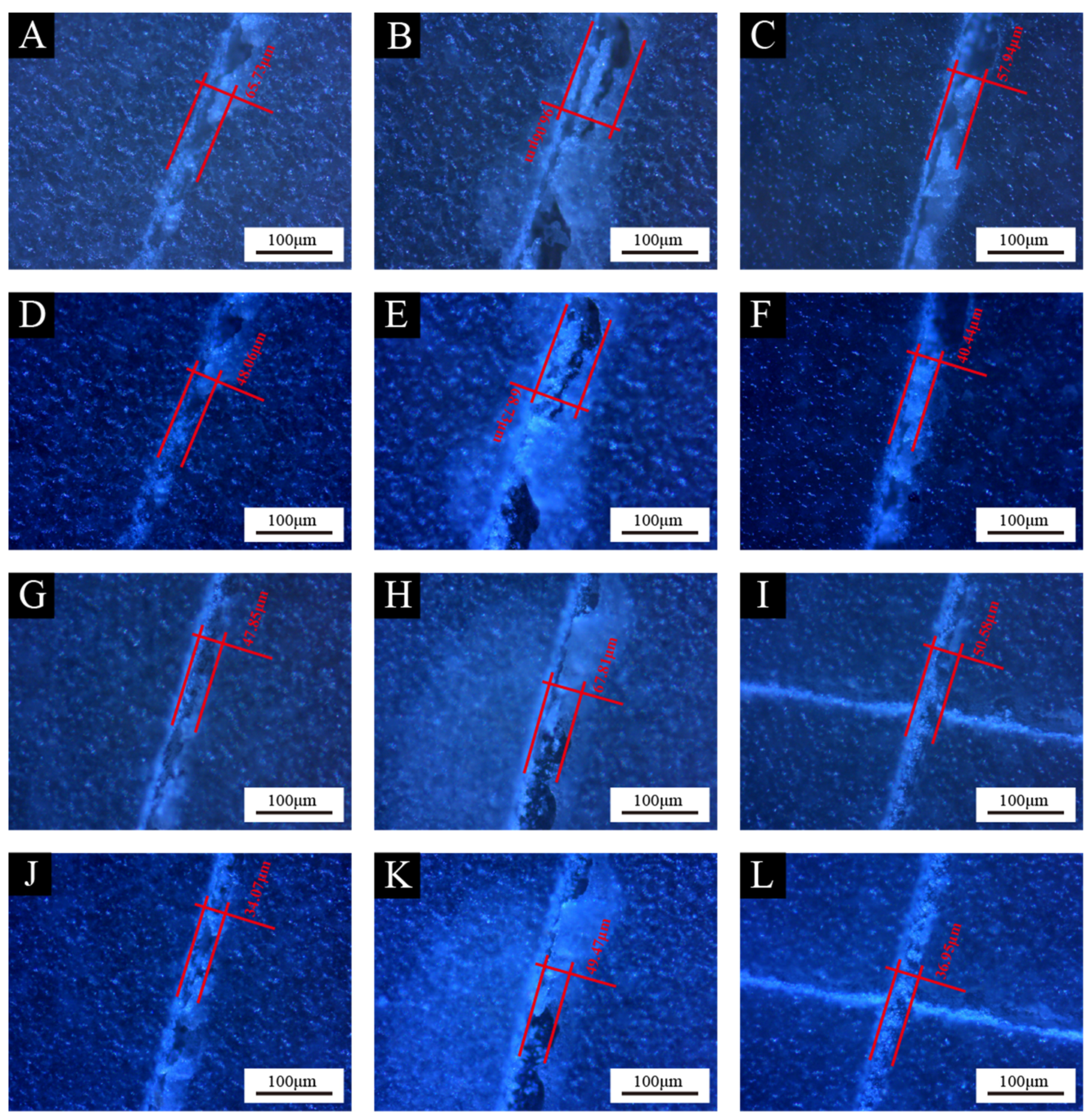
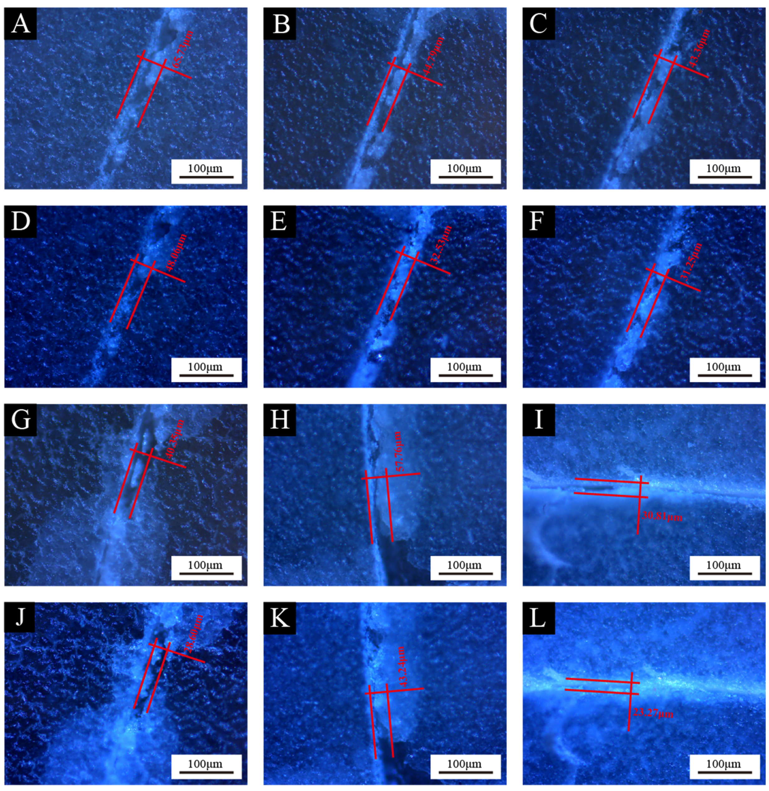
3.5. Self-Healing Performance Analysis

Figure 9, Figure 10 and Figure 11 show the self-healing performance of the samples with different contents of the UV primer microcapsules. The self-healing rates based on Figure 9, Figure 10 and Figure 11 are shown in Table 8. The self-healing rate of the samples with the 1# microcapsules at 4% content was the highest at 30.21%. Compared with the self-healing rate of the blank control group [34], the increase in the self-healing rate was 10.07%, and the improvement rate was 50.00%. The self-healing rate of all samples showed an increasing and then decreasing trend with the increased content of the UV primer microcapsules. Because all the UV top coating contains UV top coating microcapsules of 6.0% content, even if the UV primer microcapsules are not added to the UV primer, the coating itself has a certain degree of self-healing performance. With the appropriate content of the UV primer microcapsules, the self-healing performance of the coating was further enhanced by the UV primer microcapsules and the UV top coating microcapsules. However, when the UV primer microcapsules continued to be added, the four layers of samples accumulated too many microcapsules, resulting in the UV primer core materials and the UV top coating core material outflow being affected. The contact between the UV primer core material and the UV top coating core material also affected the curing effect of the two core materials, which resulted in a decrease in the self-healing rate of the samples, which was even lower than that of the samples with only one type of microcapsule. The self-healing rate of the samples was 26.89% when the UV top coating microcapsules content was 6.0%. Compared to the sample with the highest self-healing rate of 30.21%, the increase was 3.32%. The self-healing rate was improved by the compound use of two UV coating microcapsules.
Because the core material is partially isolated from the outside after being coated, the self-healing efficiency of the coating remains stable before the microcapsules rupture. Therefore, while maintaining the consistency with the surrounding coating matrix after flowing out and curing, the core material has better self-healing performance of the coating than before [47].

4. Conclusions

Three kinds of UV primer microcapsules were added to the UV primer. The UV coatings were applied by using the two-primer and two-top coating method. The effects of the UV primer microcapsules compounded with the UV top coating microcapsules on the microscopic morphology, chemical composition, and overall performance of coatings were investigated. The results showed increasing trends of the color difference, gloss, and reflectance of the samples with the increased content of the UV primer microcapsules. The overall difference in optical performance was not significant. The content of microcapsules weakened the adhesion of the coating to a certain extent, enhanced the hardness of the coating, strengthened the impact resistance of the coating, and increased the roughness of the coating. The increased content of microcapsules caused the self-healing rates of the coating to increase first and then decrease. The sample with 4.0% content of the 1# UV primer microcapsules in the UV primer and 6.0% content of the UV top coating microcapsules in the UV top coating had better comprehensive performance, with a color difference of 3.59, a gloss of 1.33 GU, a reflectance of 21.17%, an adhesion of grade 2, a hardness of 2H, an impact resistance of grade 5 at a 50 mm drop height, a roughness of 1.085 μm, and a self-healing rate of 30.21%. The self-healing rate was 10.07% higher than that of the coating on the fiberboard surface without the microcapsules, with an enhancement rate of 50.00%. The self-healing process is influenced by various factors, and the efficiency can be improved by modifying the wall of the UV coating microcapsules. In response to the scientific problem, it can be expected that a modification design of oriented self-healing microcapsules for microcracks will help improve the self-healing efficiency in the future.

Author Contributions

Conceptualization, methodology, validation, resources, data management, and supervision, Y.Z.; writing—review and editing, Y.X.; formal analysis and investigation, X.Y. All authors have read and agreed to the published version of the manuscript.

Funding

This project was partly supported by Qing Lan Project and the Natural Science Foundation of Jiangsu Province (BK20201386).

Institutional Review Board Statement

Not applicable.

Informed Consent Statement

Not applicable.

Data Availability Statement

Data are contained within the article.

Conflicts of Interest

The authors declare no conflicts of interest.

References

  1. Yang, D.R.; Zhu, J.G. Recycling and Value-added Design of Discarded Wooden Furniture. Bioresources 2021, 16, 6953–6963. [Google Scholar] [CrossRef]
  2. Wang, C.; Zhang, C.Y.; Zhu, Y. Reverse design and additive manufacturing of furniture protective foot covers. Bioresources 2024, 19, 4670–4678. [Google Scholar] [CrossRef]
  3. Zhu, J.G.; Niu, J.Y. Green Material Characteristics Applied to Office Desk Furniture. Bioresources 2022, 17, 2228–2242. [Google Scholar] [CrossRef]
  4. Zhu, H.L.; Luo, W.; Ciesielski, P.N.; Fang, Z.Q.; Zhu, J.Y.; Henriksson, G.; Himmel, M.E.; Hu, L.B. Wood-Derived Materials for Green Electronics, Biological Devices, and Energy Applications. Chem. Rev. 2016, 116, 9305–9374. [Google Scholar] [CrossRef] [PubMed]
  5. Zhang, Z.Y.; Zhu, J.G.; Qi, Q. Research on the Recyclable Design of Wooden Furniture Based on the Recyclability Evaluation. Sustainability 2023, 15, 16758. [Google Scholar] [CrossRef]
  6. Liu, Y.; Hu, W.G.; Kasal, A.; Erdil, Y.Z. The State of the Art of Biomechanics Applied in Ergonomic Furniture Design. Appl. Sci. 2023, 13, 12120. [Google Scholar] [CrossRef]
  7. Hu, W.G.; Luo, M.Y.; Liu, Y.Q.; Xu, W.; Konukcu, A.C. Experimental and numerical studies on the mechanical properties and behaviors of a novel wood dowel reinforced dovetail joint. Eng. Fail. Anal. 2023, 152, 107440. [Google Scholar] [CrossRef]
  8. Hu, W.G.; Luo, M.Y.; Hao, M.M.; Tang, B.; Wan, C. Study on the effects of selected factors on the diagonal tensile strength of oblique corner furniture joints constructed by wood dowel. Forests 2023, 14, 1149. [Google Scholar] [CrossRef]
  9. Wang, C.; Yu, J.H.; Jiang, M.H.; Li, J.Y. Effect of selective enhancement on the bending performance of fused deposition methods 3D-printed PLA models. Bioresources 2024, 19, 2660–2669. [Google Scholar] [CrossRef]
  10. Hu, W.G.; Liu, Y.; Li, S. Characterizing mode I fracture behaviors of wood using compact tension in selected system crack propagation. Forests 2021, 12, 1369. [Google Scholar] [CrossRef]
  11. Hu, W.G.; Fu, W.J.; Zhao, Y. Optimal design of the traditional Chinese wood furniture joint based on experimental and numerical method. Wood Res. 2024, 69, 50–59. [Google Scholar] [CrossRef]
  12. Hu, J.; Liu, Y.; Wang, J.X.; Xu, W. Study of selective modification effect of constructed structural color layers on European beech wood surfaces. Forests 2024, 15, 261. [Google Scholar] [CrossRef]
  13. Tao, M.X.; Liu, X.; Xu, W. Effect of the Vacuum Impregnation Process on Water Absorption and Nail-Holding Power of Silica Sol-Modified Chinese Fir. Forests 2024, 15, 270. [Google Scholar] [CrossRef]
  14. Wang, X.Y.; Liu, X.; Wu, S.S.; Xu, W. The influence of different impregnation factors on mechanical properties of silica sol-modified Populus tomentosa. Wood Fiber Sci. 2024, 56, 65–71. [Google Scholar]
  15. Miklecic, J.; Jirous-Rajkovic, V. Effectiveness of finishes in protecting wood from liquid water and water vapor. J. Build. Eng. 2021, 43, 102621. [Google Scholar] [CrossRef]
  16. Chang, Y.J.; Yan, X.X.; Wu, Z.H. Application and prospect of self-healing microcapsules in surface coating of wood. Colloid Interfac. Sci. 2023, 56, 100736. [Google Scholar] [CrossRef]
  17. Zigon, J.; Kovac, J.; Petric, M. The influence of mechanical, physical and chemical pre-treatment processes of wood surface on the relationships of wood with a waterborne opaque coating. Prog. Org. Coat. 2022, 162, 106574. [Google Scholar] [CrossRef]
  18. Czachor-Jadacka, D.; Pilch-Pitera, B. Progress in development of UV curable powder coatings. Prog. Org. Coat. 2021, 158, 106355. [Google Scholar] [CrossRef]
  19. Nowrouzi, Z.; Mohebby, B.; Petric, M.; Ebrahimi, M. Influence of nanoparticles and olive leaf extract in polyacrylate coating on the weathering performance of thermally modified wood. Eur. J. Wood Wood Prod. 2022, 80, 301–311. [Google Scholar] [CrossRef]
  20. Piao, X.X.; Guo, H.X.; Cao, Y.Z.; Wang, Z.; Jin, C.D. Exploration of multifunctional wood coating based on an interpenetrating network system of rosin-CO2-based polyurethane and mussel bionic rosin-based benzoxazine. J. Mater. Chem. B 2022, 36, 6939–6945. [Google Scholar] [CrossRef]
  21. Hu, W.G.; Wan, H. Comparative study on weathering durability properties of phenol formaldehyde resin modified sweetgum and southern pine specimens. Maderas-Cienc. Tecnol. 2022, 24, 17. [Google Scholar] [CrossRef]
  22. Hu, W.G.; Yu, R.Z. Mechanical and acoustic characteristics of four wood species subjected to bending load. Maderas-Cienc. Tecnol. 2023, 25, 39. [Google Scholar] [CrossRef]
  23. Hu, W.G.; Li, S.; Liu, Y. Vibrational characteristics of four wood species commonly used in wood products. Bioresources 2021, 16, 7101–7111. [Google Scholar] [CrossRef]
  24. Tao, Z.L.; Cui, J.C.; Qiu, H.X.; Yang, J.H.; Gao, S.L.; Li, J. Microcapsule/Silica dual-fillers for self-healing, self-reporting and corrosion protection properties of waterborne epoxy coatings. Prog. Org. Coat. 2021, 159, 106394. [Google Scholar] [CrossRef]
  25. Li, K.K.; Liu, Z.J.; Wang, C.J.; Fan, W.H.; Liu, F.T.; Li, H.Y.; Zhu, Y.J.; Wang, H.Y. Preparation of smart coatings with self-healing and anti-wear properties by embedding PU-fly ash absorbing linseed oil microcapsules. Prog. Org. Coat. 2020, 145, 105668. [Google Scholar] [CrossRef]
  26. da Cunha, A.B.M.; Leal, D.A.; Santos, L.R.L.; Riegel-Vidotti, I.C.; Marino, C.E.B. pH-sensitive microcapsules based on biopolymers for active corrosion protection of carbon steel at different pH. Surf. Coat. Tech. 2020, 402, 126338. [Google Scholar] [CrossRef]
  27. Wang, Q.H.; Thomas, J.; Soucek, M.D. Investigation of UV-curable alkyd coating properties. J. Coat. Technol. Res. 2023, 20, 545–557. [Google Scholar] [CrossRef]
  28. Guo, Z. Research advances in UV-curable self-healing coatings. RSC Adv. 2022, 12, 32429–32439. [Google Scholar] [CrossRef]
  29. Sow, C.; Riedl, B.; Blanchet, P. UV-waterborne polyurethane-acrylate nanocomposite coatings containing alumina and silica nanoparticles for wood: Mechanical, optical, and thermal properties assessment. J. Coat. Technol. Res. 2011, 8, 211–221. [Google Scholar] [CrossRef]
  30. Wu, Q.; Huang, X.; Liu, R.; Yang, X.Z.; Xiao, G.; Jiang, N.; Weitz, D.A.; Song, Y.J. Multichannel Multijunction Droplet Microfluidic Device to Synthesize Hydrogel Microcapsules with Different Core-Shell Structures and Adjustable Core Positions. Langmuir 2023, 40, 1950–1960. [Google Scholar] [CrossRef]
  31. Kim, A.L.; Musin, E.V.; Chebykin, Y.S.; Tikhonenko, S.A. Characterization of Polyallylamine/Polystyrene Sulfonate Polyelectrolyte Microcapsules Formed on Solid Cores: Morphology. Polymers 2024, 16, 1521. [Google Scholar] [CrossRef] [PubMed]
  32. Wang, R.; Kang, Y.J.; Lei, T.X.; Li, S.J.; Zhou, Z.Y.; Xiao, Y. Microcapsules composed of stearic acid core and polyethylene glycol-based shell as a microcapsule phase change material. Int. J. Energy Res. 2021, 45, 9677–9684. [Google Scholar] [CrossRef]
  33. Zhang, L.C.; Chen, Y.X.; Wu, K.Y.; Sun, G.Q.; Liu, R.; Luo, J. One-pot efficient synthesis of amino-functionalized polyurethane capsules via photopolymerization for self-healing anticorrosion coatings. Prog. Org. Coat. 2024, 189, 108348. [Google Scholar] [CrossRef]
  34. Zou, Y.M.; Xia, Y.X.; Yan, X.X. Effect of UV Top Coating Microcapsules on the Coating Properties of Fiberboard Surfaces. Polymers 2024, 16, 2098. [Google Scholar] [CrossRef]
  35. Xia, Y.X.; Yan, X.X. Preparation of UV Topcoat Microcapsules and Their Effect on the Properties of UV Topcoat Paint Film. Polymers 2024, 16, 1410. [Google Scholar] [CrossRef]
  36. Weng, M.Y.; Zhu, Y.T.; Mao, W.G.; Zhou, J.C.; Xu, W. Nano-Silica/Urea-Formaldehyde Resin-Modified Fast-Growing Lumber Performance Study. Forests 2023, 14, 1440. [Google Scholar] [CrossRef]
  37. Hu, J.; Liu, Y.; Xu, W. Influence of Cell Characteristics on the Construction of Structural Color Layers on Wood Surfaces. Forests 2024, 15, 676. [Google Scholar] [CrossRef]
  38. GB/T 11186.3-1989; Methods for Measuring the Colour of Coating Films. Part III: Calculation of Colour Differences. Standardization Administration of the People’s Republic of China: Beijing, China, 1990.
  39. GB/T 4893.6-2013; Test of Surface Coatings of Furniture—Part 6: Determination of Gloss Value. Standardization Administration of the People’s Republic of China: Beijing, China, 2013.
  40. Wang, C.; Zhou, Z.Y. Optical properties and lampshade design applications of PLA 3D printing materials. Bioresources 2023, 18, 1545–1553. [Google Scholar] [CrossRef]
  41. Wang, C.; Yu, J.H.; Jiang, M.H.; Li, J.Y. Effect of slicing parameters on the light transmittance of 3D-printed polyethylene terephthalate glycol products. Bioresources 2024, 19, 500–509. [Google Scholar] [CrossRef]
  42. GB/T 1732-2020; Test of Surface Coatings of Furniture—Part 9: Determination of Impact Resistance of Coating Films. Standardization Administration of the People’s Republic of China: Beijing, China, 2020.
  43. Hu, W.G.; Zhang, J.L. Effect of growth rings on acoustic emission characteristic signals of southern yellow pine wood cracked in mode I. Constr. Build. Mater. 2022, 329, 127092. [Google Scholar] [CrossRef]
  44. GB/T 6739-2022; Paints and Varnishes—Determination of Film Hardness by Pencil Test. Standardization Administration of the People’s Republic of China: Beijing, China, 2022.
  45. Hu, W.G.; Liu, Y.; Konukcu, A.C. Study on withdrawal load resistance of screw in wood-based materials: Experimental and numerical. Wood Mater. Sci. Eng. 2023, 18, 334–343. [Google Scholar] [CrossRef]
  46. GB/T 4893.4-2013; Test of Surface Coatings of Furniture—Part 4: Determination of Adhesion-Cross Cut. Standardization Administration of the People’s Republic of China: Beijing, China, 2013.
  47. Saman, N.M.; Ang, D.T.C.; Shahabudin, N.; Gan, S.N.; Basirun, W.J. UV-curable alkyd coating with self-healing ability. RSC Adv. 2022, 12, 32429–32439. [Google Scholar] [CrossRef]
Figure 1. The surface morphologies of fiberboards with different contents of 1# microcapsules: (A) 0%, (B) 2.0%, (C) 4.0%, (D) 6.0%, (E) 8.0%, and (F) 10.0%.
Coatings 14 01012 g001
Figure 2. The surface morphologies of fiberboards with different contents of 2# microcapsules: (A) 0%, (B) 2.0%, (C) 4.0%, (D) 6.0%, (E) 8.0%, and (F) 10.0%.
Coatings 14 01012 g002
Figure 3. The surface morphologies of fiberboards with different contents of 3# microcapsules: (A) 0%, (B) 2.0%, (C) 4.0%, (D) 6.0%, (E) 8.0%, and (F) 10.0%.
Coatings 14 01012 g003
Figure 4. SEM images of samples: (A) blank control sample, (B) 2.0% of 1# microcapsules, (C) 2.0% of 2# microcapsules, and (D) 2.0% of 3# microcapsules.
Coatings 14 01012 g004
Figure 5. SEM images of the longitudinal section of samples: (A) uncoated fiberboard and (B) 2.0% of 1# microcapsules.
Coatings 14 01012 g005
Figure 6. Infrared spectrum of coating on fiberboard surface.
Coatings 14 01012 g006
Figure 7. Gloss trend of samples at a 60° incidence angle.
Coatings 14 01012 g007
Figure 8. Reflectance of samples with different contents of microcapsules: (A) 1# microcapsules, (B) 2# microcapsules, and (C) 3# microcapsules.
Coatings 14 01012 g008
Figure 9. Cracks on the samples before and after 1 week of self-healing with different contents of 1# microcapsules. Before self-healing: (A) 0%, (B) 2.0%, (C) 4.0%, (G) 6.0%, (H) 8.0%, and (I) 10.0%, and after self-healing: (D) 0%, (E) 2.0%, (F) 4.0%, (J) 6.0%, (K) 8.0%, and (L) 10.0%.
Coatings 14 01012 g009
Figure 10. Cracks on the samples before and after 1 week of self-healing with different contents of 2# microcapsules. Before self-healing: (A) 0%, (B) 2.0%, (C) 4.0%, (G) 6.0%, (H) 8.0%, and (I) 10.0%, and after self-healing: (D) 0%, (E) 2.0%, (F) 4.0%, (J) 6.0%, (K) 8.0%, and (L) 10.0%.
Coatings 14 01012 g010
Figure 11. Cracks on the samples before and after 1 week of self-healing with different contents of 3# microcapsules. Before self-healing: (A) 0%, (B) 2.0%, (C) 4.0%, (G) 6.0%, (H) 8.0%, and (I) 10.0% and after self-healing: (D) 0%, (E) 2.0%, (F) 4.0%, (J) 6.0%, (K) 8.0%, and (L) 10.0%.
Coatings 14 01012 g011
Table 1. Materials.
Table 1. Materials.
MaterialsMolecular Mass (g/mol)CASProducer
Melamine126.12108-78-1Nanjing Houxin Biotechnology Co., Ltd., Nanjing, China
37% formaldehyde30.0350-00-0Nantong Yaoxin Chemical Co., Ltd., Nantong, China
Triethanolamine149.19102-71-6Nanjing Houxin Biotechnology Co., Ltd., Nanjing, China
Span-20346.459133-39-2Nanjing Houxin Biotechnology Co., Ltd., Nanjing, China
Triton X-100646.859002-93-1Shandong Yousuo Chemical Technology Co., Ltd., Linyi, China
Absolute ethanol46.0764-17-5Guangzhou Kema Chemical Technology Co., Ltd., Guangzhou, China
UV top coating--Jiangsu Himonia Technology Co., Ltd., Zhenjiang, China
1-hydroxycyclohexyl phenyl ketone --Jiangsu Himonia Technology Co., Ltd., Zhenjiang, China
UV primer--Jiangsu Himonia Technology Co., Ltd., Zhenjiang, China
2-hydroxy-2-methylpropiophenone --Jiangsu Himonia Technology Co., Ltd., Zhenjiang, China
Citric acid monohydrate210.1395949-29-1Jinan Xiaoshi Chemical Co., Ltd., Jinan, China
Table 2. Test instruments.
Test InstrumentsModelManufacturer
Water bathDF-101SHenan Heming Instrument Equipment Co., Ltd., Zhengzhou, China
Scanning electron microscope (SEM)Zeiss Sigma 300FEI Company, Hillsboro, OR, USA
Infrared spectrometerVERTEX 80VGermany BRUKER Co., Ltd., Karlsruhe, Germany
Color difference instrumentSEGT-JSuzhou Weifu Photoelectric Technology Co., Ltd., Suzhou, China
Gloss meterLS195Shenzhen Linshang Technology Co., Ltd.,
Shenzhen, China
Ultraviolet spectrophotometerU-3900Shanghai Smeo Analytical Instrument Co., Ltd., Shanghai, China
Coating roughness testerSJ-411Shanghai Taiming Optical Instrument Co., Ltd., Shanghai, China
Single-lamp curing machine620#Huzhou Tongxu Machinery Equipment Co., Ltd., Huzhou, China
Table 3. Microcapsule formulation.
Sample
(#)
Triton X-100
(g)
Span 20
(g)
Ethanol
(mL)
UV primer
(g)
UV Top
Coating (g)
Formaldehyde
(g)
Melamine
(g)
Deionized
Water (mL)
10.080.2278.904.40-10.814.8024.00
20.080.2278.904.40-9.834.3621.82
30.080.2278.904.40-9.014.0020.00
UV top coating
microcapsule
0.080.2278.90-4.4013.526.0030.00
Table 4. Quantities of coating materials.
Content of UV Primer Microcapsules (%)Mass of UV Primer Microcapsules (g)Mass of UV Primer (g)Content of UV Top Coating Microcapsules (%)Mass of UV Top Coating Microcapsules (g)Mass of UV Top Coating (g)
000.8006.00.0480.752
2.00.0160.7846.00.0480.752
4.00.0320.7686.00.0480.752
6.00.0480.7526.00.0480.752
8.00.0640.7366.00.0480.752
10.00.0800.7206.00.0480.752
Table 5. The color difference of the samples with different contents of the UV primer microcapsules.
SamplesContent of UV Primer Microcapsules (%)LabΔE
Samples with 1# microcapsules049.972.677.43-
2.049.872.938.300.91
4.046.904.537.433.59
6.055.901.536.776.08
8.057.331.636.337.52
10.058.731.136.538.95
Samples with 2# microcapsules049.972.677.43-
2.049.803.237.400.59
4.053.031.877.103.19
6.045.674.578.634.85
8.057.671.336.837.84
10.061.000.336.9011.29
Samples with 3# microcapsules049.972.677.43-
2.048.074.138.632.68
4.055.502.106.905.59
6.055.572.006.435.73
8.043.735.839.837.39
10.059.570.936.839.77
Table 6. Gloss and reflectance of samples.
SamplesContent of UV Primer
Microcapsules (%)
Gloss (GU)Reflectance (%)
20°60°85°
Samples with 1# microcapsules00.201.073.6018.93
2.00.201.133.7021.57
4.00.301.335.3321.17
6.00.501.435.4329.97
8.00.271.5311.9326.50
10.00.371.6713.3729.20
Samples with 2# microcapsules00.201.073.6018.93
2.00.271.406.3320.38
4.00.331.379.5022.52
6.00.301.338.2024.92
8.00.371.705.2727.29
10.00.471.633.8732.83
Samples with 3# microcapsules00.201.073.6018.93
2.00.201.104.6318.94
4.00.231.175.3721.88
6.00.331.103.3027.46
8.00.371.405.4330.39
10.00.401.8012.0734.74
Table 7. Mechanical performance of the samples.
SamplesContent of
Microcapsules (%)
Adhesion (Grade)HardnessImpact Resistance (Grade)Roughness (μm)
Samples with 1# microcapsules012H51.063
2.012H51.065
4.022H51.085
6.022H41.336
8.023H31.468
10.024H32.064
Samples with 2# microcapsules012H51.063
2.012H50.794
4.022H50.810
6.022H40.956
8.022H41.293
10.023H31.451
Samples with 3# microcapsules012H51.063
2.022H51.179
4.022H51.463
6.022H51.766
8.023H42.139
10.023H42.598
Table 8. Self-healing rates of samples with different contents of microcapsules.
SamplesContent of
Microcapsules (%)
Crack Width
after Scratch (μm)
Crack Width after
1 Week of Self-Healing (μm)
Self-Healing Rates (%)
Samples with 1# microcapsules065.7348.0626.89
2.096.0668.7328.45
4.057.9440.4430.21
6.047.8534.0728.79
8.067.8149.4727.05
10.050.5836.9526.95
Samples with 2# microcapsules065.7348.0626.89
2.056.6340.8627.84
4.082.6657.7930.09
6.076.7154.5328.92
8.085.9263.5226.07
10.058.1943.2425.69
Samples with 3# microcapsules065.7348.0626.89
2.044.7932.5327.38
4.043.3631.2527.92
6.040.3528.6029.12
8.057.7643.2425.13
10.030.8123.2724.46
Disclaimer/Publisher’s Note: The statements, opinions and data contained in all publications are solely those of the individual author(s) and contributor(s) and not of MDPI and/or the editor(s). MDPI and/or the editor(s) disclaim responsibility for any injury to people or property resulting from any ideas, methods, instructions or products referred to in the content.

