Article

Synthesis and Characterization of Titania-Coated Hollow Mesoporous Hydroxyapatite Composites for Photocatalytic Degradation of Methyl Red Dye in Water

State Key Laboratory of Fine Chemicals, School of Chemical Engineering, Dalian University of Technology, Dalian 116024, China
*
Author to whom correspondence should be addressed.
Coatings 2024, 14(8), 921; https://doi.org/10.3390/coatings14080921 (registering DOI)
Submission received: 9 June 2024 / Revised: 18 July 2024 / Accepted: 22 July 2024 / Published: 23 July 2024

Abstract

:
Hollow mesoporous hydroxyapatite (HM-HAP) composites coated with titania are prepared to increase the stability and catalytic performance of titania for azo dyes present in the wastewater system. In this work, HM-HAP particles were first synthesized by a hydrothermal method utilizing the CaCO3 core as a template and then coated with titania to form TiO2/HM-HAP composites. Utilizing SEM, XRD, XPS, BET, FTIR, EDS, UV–vis DRS spectroscopy, and point of zero charge (PZC) analysis, the coating morphological and physicochemical parameters of the produced samples were analyzed. The photocatalytic efficiency of the synthesized coated composites was assessed by the degradation of methyl red (MR) dye in water. The results indicated that TiO2/HM-HAP particles could efficiently photodegrade MR dye in water under UV irradiation. The 20% TiO2/HM-HAP coating exhibited high catalytic performance, and the degradation process was followed by the pseudo-first-order (PFO) kinetic model with a rate constant of 0.033. The effect of pH on the degradation process was also evaluated, and the maximum degradation was observed at pH 6. The analysis of degraded MR dye products was investigated using LC-MS and FTIR analysis. Finally, a good support material, HM-HAP for TiO2 coatings, which provides a large number of active adsorption sites and has catalytic degradation performance for MR dye, was revealed.

Graphical Abstract

1. Introduction

The majority of dyes used in textiles, leather, cosmetics, food processing, ink, and paper are azo dyes, which are categorized by the existence of one or more azo groups in their chemical structure. Furthermore, 15% of the world’s manufactured dyes are lost during synthesis and application as wastes that pose a threat to human health and the environment because of their toxic nature [1]. A high concentration of color, suspended particles, and COD also characterize the polluted water discharged by various industries. Even in minute quantities, the effluents of the dyes are conspicuous and hazardous. These contaminants severely impact water bodies and the plants and animals that depend on them. It is commonly known that methyl red dye is used in textile industries, paper printing, and as a pH indicator in the laboratory. However, it may create problems with the eyes, skin, and digestive tract when swallowed or inhaled [2]. Thus, owing to the health risks posed by dyes to human health, it is of the utmost importance to develop effective methods for their effective removal from wastewater.
Numerous methods have been introduced for the treatment of waste, which include chemicals such as ozonation and chlorination [3,4], physical such as adsorption [5,6] and membrane filtration [7,8], photocatalysis [9,10], and biological treatment methods [11]. Among them, photocatalytic technology provides a simple and inexpensive way to eliminate organic and inorganic contaminants from wastewater, as photocatalytic degradation may break down or mineralize the majority of organic pollutants. Among the many photocatalysts, titanium dioxide (TiO2) is the most widely used because of its non-toxicity, low cost, and high photochemical stability, but the disadvantages of limited pollutant adsorption capacity [12,13,14,15] and significant recombination of photogenerated electron–hole pairs hinder its photocatalytic efficiency [16]. Several approaches have been implemented to improve photocatalytic performance in an effort to address these problems.
Recent research has demonstrated the capability of TiO2-based composites to improve photocatalytic activity. For example, Sukhadeve et al. (2023) revealed the effective degradation of methylene blue dye using Ag-Fe co-doped TiO2 nanoparticles [17]. Furthermore, Ali et al. (2023) reviewed the catalytic activity of Ag- and Zn-doped TiO2 nano-catalysts for the removal of methylene blue and methyl orange dyes [18]. Liza et al. (2024) conducted an additional study that examined the impact of Ag-doping on the morphology, band gap, and photocatalytic activity of TiO2 nanoparticles in the context of textile dye degradation [19]. Moreover, TiO2 has been implemented in a variety of applications beyond wastewater treatment, including antibacterial remedies [20], protective coatings for metal prostheses [21], and self-cleaning surfaces for air purification [22].
Recent publications have indicated that TiO2-supported materials, such as silica, hydroxyapatite (HAP), zeolite, pure natural diatomite, and activated carbon, can provide a large number of active adsorption sites, resulting in rapid mass transport and catalytic processes [23,24,25,26]. Among all these supporting materials, hydroxyapatite (HAP) has been extensively researched because of its mechanical stability, high biocompatibility, non-toxicity, and inexpensive cost [27,28,29,30]. Hydroxyapatite (HAP) is also extensively recognized for its applications in a variety of areas, such as the environmental, biomedical, and industrial sectors [31,32]. HAP’s ecological friendliness is one of its most intriguing features, as it can be produced from waste materials, rendering it a sustainable and environmentally responsible option. Recent research has investigated the potential of HAP, which is derived from sources of waste, such as egg shells, fly ash, and fish bones, to reduce environmental impacts and promote sustainable practices [33,34,35].
Hydroxyapatite (HAP) possesses hydroxyl groups (OH) and adsorbed H2O molecules on its surface that can interact with h+ to create hydroxyl radicals (˙OH), hence enhancing photocatalytic efficiency. Moreover, during the photocatalytic process, the electron phase shift of the PO43− groups on the HAP surface can also result in the formation of ˙O2 radicals [36]. Consequently, it is anticipated that integrating the advantages of HAP and TiO2 will not only increase the capability of TiO2 to absorb contaminants but also reduce the combination of photogenerated electron–hole pairs. Therefore, the use of TiO2 in conjunction with HAP may prove to be an effective wastewater treatment method.
Prior research on TiO2/HAP composites has mainly concentrated on using rod-shaped or quasi-spherical hydroxyapatite (HAP) structures. However, these structures often face difficulties such as particle agglomeration and ineffective TiO2 loading [37,38,39]. These problems might result in a decrease in the number of active sites on the surface, which in turn reduces the effectiveness of the composite in interacting with contaminants. Our research presents a novel method that utilizes a template technique to create hollow mesoporous HAP structures, which are then coated with TiO2. This innovative approach effectively solves the issues of aggregation that are typically seen with traditional HAP forms while also greatly improving the surface area and ease of access to the active areas inside the composite. By employing hollow mesoporous hydroxyapatite (HAP) in this distinct morphology, the effectiveness and durability of the TiO2/HAP composite for photocatalytic and environmental purposes are significantly enhanced. This study is the first known case of employing hollow mesoporous HAP templates for TiO2 coatings. This opens new prospects for improving the efficiency and adaptability of composite materials in sustainable technologies. The unique structure of spherical hollow hydroxyapatite particles not only prevents aggregation but also allows for a greater surface area owing to the hollow interior. This makes them very appropriate for coating TiO2 and conducting investigations on photocatalysis.
In this research work, we prepared HM-HAP particles through a hydrothermal technique using the CaCO3 core as a template. The synthesized HM-HAP was then coated with different amounts of TiO2 to form a TiO2-coated HM-HAP composite. Figure 1 shows a schematic diagram of the TiO2/HM-HAP particle synthesis. The synthesized TiO2/HM-HAP composites exhibited good photocatalytic activity for MR dye removal under UV light. XRD, XPS, BET, SEM, and UV–vis DRS were used to examine the effect of TiO2 coatings on the structural behavior of HM-HAP. The photocatalytic degradation of methyl red (MR) dye under UV irradiation was investigated over the TiO2/HM-HAP composites. Hence, this research paper presents results on a good support material, HM-HAP, for TiO2 coatings, which provides a large number of adsorption sites, resulting in rapid mass transport and catalytic processes, and finally studies the photocatalytic degradation of MR dye.

2. Experimental Methods

2.1. Materials

Sodium carbonate (Na2CO3), calcium nitrate (Ca(NO3)2), disodium hydrogen phosphate dodecahydrate (Na2HPO4·12H2O), potassium hydrogen phthalate (KHP), potassium chloride (KCl), potassium dihydrogen phosphate (KH2PO4), sodium hydroxide (NaOH), and hydrochloric acid (HCl) were purchased from DAMAO. Sodium poly (styrene sulfonate) (PSS, Mw = 70,000), methyl red (MR), and Titanium dioxide (TiO2) were purchased from MACKLIN.

2.2. Synthesis of HM-HAP Particles

A 0.1 M solution of Na2CO3 (20 mL) was quickly mixed with a 0.1 M Ca (NO3)2 solution (20 mL) containing 150 mg of PSS under magnetic stirring (600 rpm). The solution temperature was maintained at 30 °C for about 30 min. The obtained CaCO3 particles, resulting from the quick precipitation procedure, were rinsed three times with deionized water before being collected by centrifugation (7000 rpm, 5 min). The obtained CaCO3 particles were then added to a Na2HPO4 solution (30 mL, 0.8 M). The resultant mixture was then placed in an autoclave of 100 mL capacity and heated at 140 °C for 4 h. After the reaction was complete, the CaCO3 core was removed by adding a few drops of acid to the solution and stirring it for 2 h.

2.3. Synthesis of TiO2/HM-HAP Composite

The obtained materials, HM-HAP (150 mg) and titanium dioxide (TiO2), were mixed in various amounts with butanol and agitated for one hour at 45 °C. The butanol was then evaporated at 60 °C using a rotary evaporator. The sample was then heated for 4 h at 450 °C. The TiO2 was coated onto the HM-HAP at 10%, 20%, 30%, 40%, and 50%.

2.4. Characterization of TiO2/HM-HAP Composite

The material’s surface shape and size were determined using a scanning electron microscope HITACHI-SU8220 (High-Tech corporation, Tokyo, Japan). The X-ray diffraction (XRD) patterns were acquired using a smart lab X-ray diffractometer (XRD, Rigaku corporation, Tokyo, Japan) with Cu Kα (λ = 0.15406 nm) radiation. On a ThermoFisher—6700 (Waltham, MA, USA) FTIR spectrometer, the material’s FTIR spectrum was obtained using the KBr pellet technique. The pore structure and specific surface area of the coated particles were determined using a BET surface area analyzer (BSD Instrument Technology (Beijing, China) Co., Ltd. (BSD-PS (M)) at 77 K, utilizing helium as the carrier gas at liquid nitrogen temperature. The formation of TiO2 coatings on the HM-HAP particles was confirmed via an energy-dispersive X-ray spectrometer-QUANTA 450 (Thermo Fisher Scientific, Waltham, MA, USA). X-ray photoelectron spectroscopy (XPS) data were collected on a Thermo Scientific photoelectron spectrometer—ESCALAB250Xi (Thermo Fisher Scientific, Winsford, UK). UV-vis diffuse reflectance spectra (DRS) of the synthesized materials were measured by a UV-vis spectrophotometer (Lambda 1050+, Perkin Elmer, Shanghai, China) utilizing BaSO4 as a reference material. A spectroscopic study utilizing a UV-vis spectrophotometer carry-100 (Agilent, Kuala Lumpur, Malaysia) was used to evaluate the absorbance of methyl red dye throughout its photocatalytic degradation. The absorbance spectra were recorded between 350 and 750 nm wavelength. The pH measurement was performed using a PHS-3C pH meter (Shanghai Yoke Instrument Co., Ltd., Shanghai, China) with an integrated temperature sensor for temperature regulation. The examination of degradation products was conducted using liquid chromatography–mass spectrometry (LC-MS, G6230, Agilent, Santa Clara, CA, USA). The TOC of before and after decolorization of the dye solution was quantified by an organic element analyzer (UNICUBE, Elementar Analysensysteme GmbH, Langenselbold, Germany).

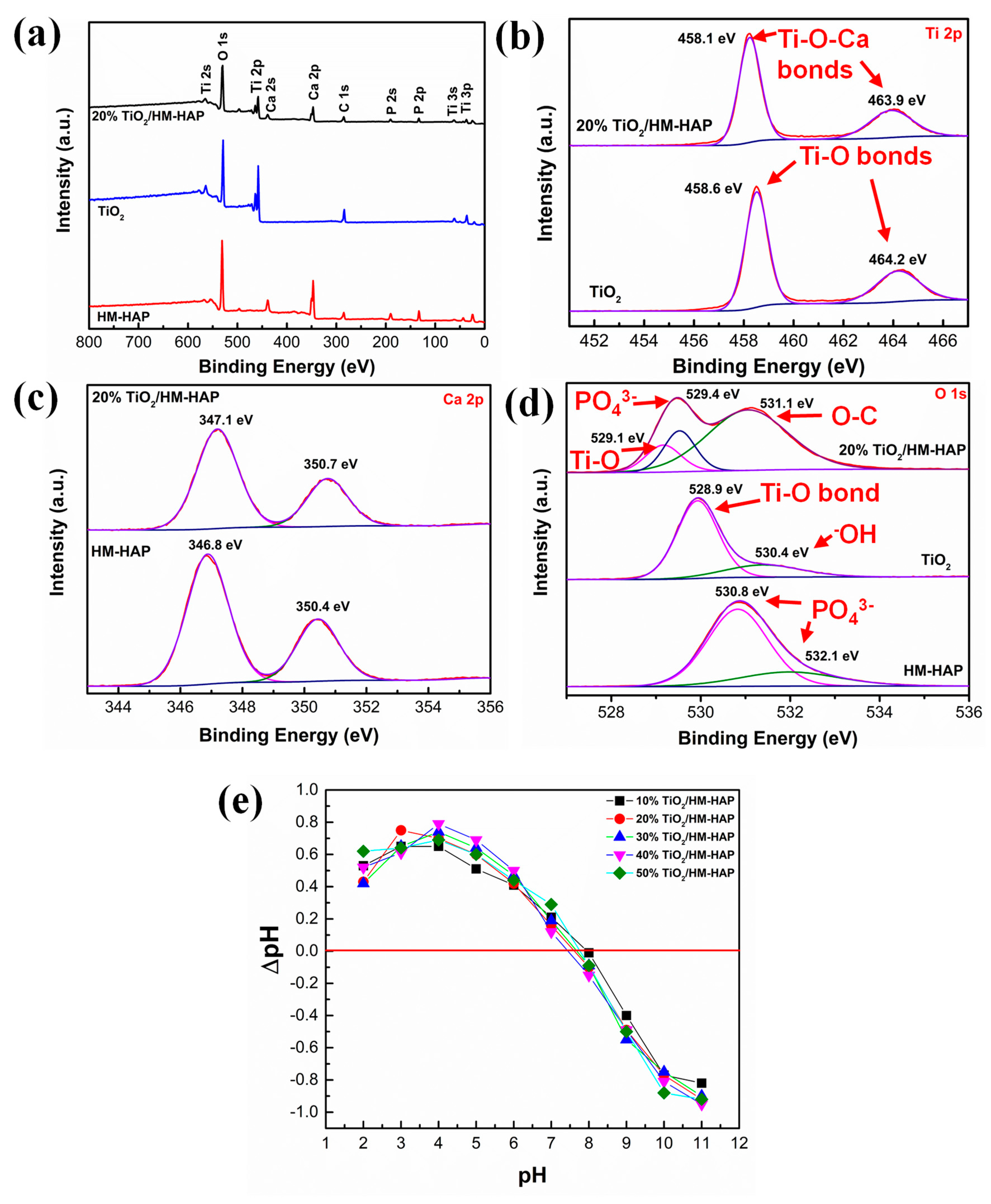
2.5. Point of Zero Charge (PZC)

Measuring PZC aids in predicting the ionic character of a catalyst, which clarifies the interaction process between a dye and the catalyst. The salt addition method was used to measure the PZC point of the formed TiO2-coated HM-HAP composites. Typically, a 0.1 M NaCl solution was prepared, and for each experiment, 50 mL of the solution was taken in a 100 mL beaker. To each beaker, 30 mg of TiO2-coated HM-HAP was added and then stirred for about 3 h. Each solution’s pH was maintained using a mixture of buffers (pH 2 to 12).

2.6. Photocatalytic Studies of MR

The photocatalytic activity of the samples was evaluated by observing the degradation of MR dye as the pollutant target under ultraviolet irradiation. The chemical structure and other characteristics of the employed dye are detailed in Table S1. Initially, 25 mL of MR solution (10 mg/L) and 30 mg of catalyst were vigorously agitated for 30 min in the dark to produce an adsorption/desorption equilibrium. The solution was then agitated at room temperature under UV light irradiation (λ = 254 nm) for several minutes. Periodically, 3 mL of the solution was collected and filtered to eliminate the solid phase. Although this decreased the initial volume, parallel experiments were carried out to ensure uniformity and eliminate potential mistakes resulting from volume reduction. More precisely, 25 mL samples were prepared and subjected to the same circumstances. This enabled the extraction of small portions from various samples at each specific time interval. This method guaranteed that the decrease in volume did not impact the accuracy of the outcomes. After that, the absorbance of the filtrates was measured spectroscopically at 437 nm for MR dye. In order to measure the extent of degradation of the MR dye, a calibration curve was employed, which can be seen in Figure S1 in the Supplementary Information. The calibration curve was generated by measuring the absorbance of a range of MR dye solutions with predetermined concentrations, enabling us to establish a precise relationship between absorbance and dye concentration. Applying this calibration curve, the concentration of MR dye at various irradiation times was established, facilitating the calculation of the photodegradation rate and efficiency.
Photolysis and adsorption experiments were also conducted under the same conditions as the photocatalytic experiment.
The effect of various parameters, such as pH and contact time, on the degradation of MR by the catalysts was also analyzed. The solution pH (2, 4, 6, 8, and 10) was monitored by the addition of sodium hydroxide (NaOH), sodium bicarbonate (NaHCO3), potassium hydrogen phthalate (KHP), potassium chloride (KCl), hydrochloric acid (HCl), and potassium dihydrogen phosphate (KH2PO4).
The percentage degradation of the MR dye was determined using the following relationship [40]:
%   D e g r a d a t i o n = M R 0 M R t M R 0 × 100
where [MR]0 represents the initial concentration and [MR]t represents the concentration at time t of the dye MR.

2.7. Kinetics of MR Degradation

The obtained experimental data were assessed further using the (PFO) pseudo-first-order kinetic model, which is represented by the given expression [41]:
l n M R 0 M R t = k 1 t
where t is the given time, [MR]0 symbolizes the initial concentration, and [MR]t symbolizes the concentration at any time (t) of MR dye. k1 is the rate constant, and its values can be derived from the slope of the graph ln[MR]0/[MR]tvs. reaction time.

3. Results and Discussion

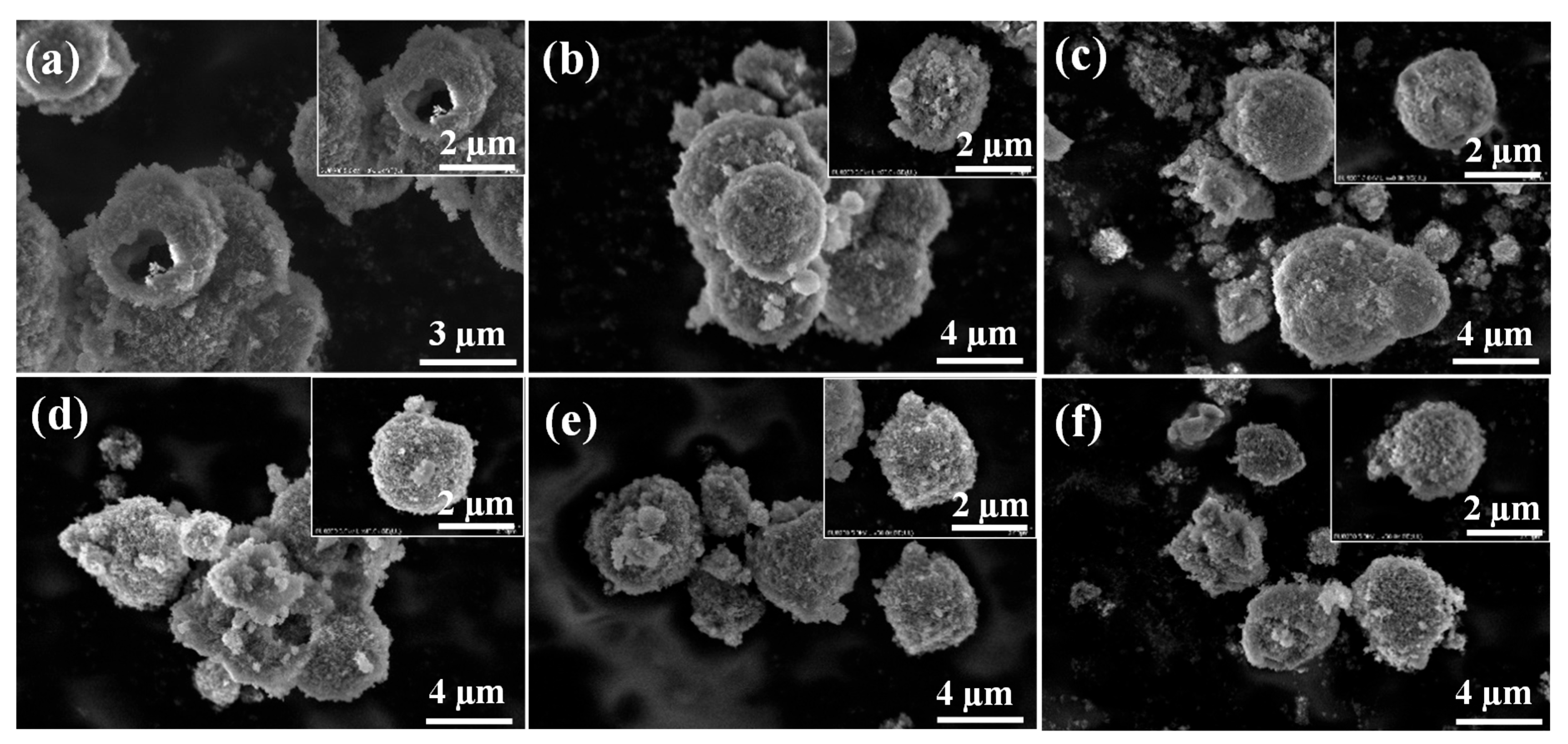
3.1. Synthesis of TiO2/HM-HAP Composite

In this research study, TiO2-coated HM-HAP composites were produced and employed in a photocatalytic study. First, HM-HAP particles were synthesized using a hydrothermal method with PSS-doped vaterite CaCO3 as a hard template. The presence of PSS, a polyelectrolyte with a substantial negative charge, produces negatively charged HM-HAP particles. In addition, PSS (sodium poly (styrene sulfonate)) was used as a crystal growth additive to accelerate the transformation of CaCO3 from calcite to vaterite during the CaCO3 manufacturing process. The reagents Na2CO3 and Ca (NO3)2 were utilized first for the synthesis of CaCO3 by the formation of precipitates. The obtained CaCO3 particles were then added to Na2HPO4, and after the completion of the reaction, the core was removed by an etching process carried out with acetic acid. The synthesized HM-HAP particles were then coated with a range of TiO2 concentrations (10% to 50%) to create a TiO2-coated HM-HAP composite. FTIR and XRD analysis confirmed the synthesis of HM-HAP and TiO2-coated HM-HAP composites.
The FTIR spectra of pure TiO2, HM-HAP, and TiO2-coated HM-HAP composites are shown in Figure 2a. The functional peaks at 3432 cm−1 and 1632 cm−1 are the results of lattice water in the samples [42]. At 634 cm−1, the distinctive broad absorption bands of TiO2 are detected, which can be ascribed to the stretching vibration of Ti–O–Ti bonds [43]. The distinctive bands at 563 and 602 cm−1 for the HM-HAP sample correspond to the bending vibration of PO43−, whereas the band at 1031 cm−1 corresponds to the stretching vibration of PO43−. The characteristic CO32− bands are situated at 1466, 1410, and 874 cm−1 [44,45,46]. The spectra of TiO2-coated HM-HAP (10%–50%) composites show that the characteristic peaks of TiO2 and HM-HAP are well preserved, indicating that the structure transformation of TiO2 and HM-HAP did not change during composite production. This demonstrates the effective synthesis of TiO2-coated HM-HAP composites, which is compatible with the XRD data.
The XRD patterns of TiO2, HM-HAP, and TiO2-coated HM-HAP composites are shown in Figure 2b. The diffraction peaks at 25.2°, 37.1°, 53.9°, 55.8°, 62.7°, 68.5°, and 70.3° correspond to the (101), (004), (105), (211), (204), (116), and (220) planes of anatase-TiO2, respectively (JCPDS no. 21-1272) [47]. In the case of pure HM-HAP, all diffraction peaks and their relative intensities correspond to the standard diffraction data of pure hexagonal phase HAP (JCPDS, 09-0432) [48,49]. Specifically, the peaks at two values of 31.9°, 32.1°, and 33.1° correspond to the (211), (112), and (300) planes of HAP, respectively. These indices confirm the successful synthesis of hydroxyapatite with a well-defined crystalline structure. In addition, the diffraction patterns of the TiO2-coated HM-HAP composites (10%–50%) demonstrate the existence of both anatase TiO2 and hexagonal phase HAP. Thus, the TiO2-coated HM-HAP composites were successfully synthesized using the hydrothermal technique.

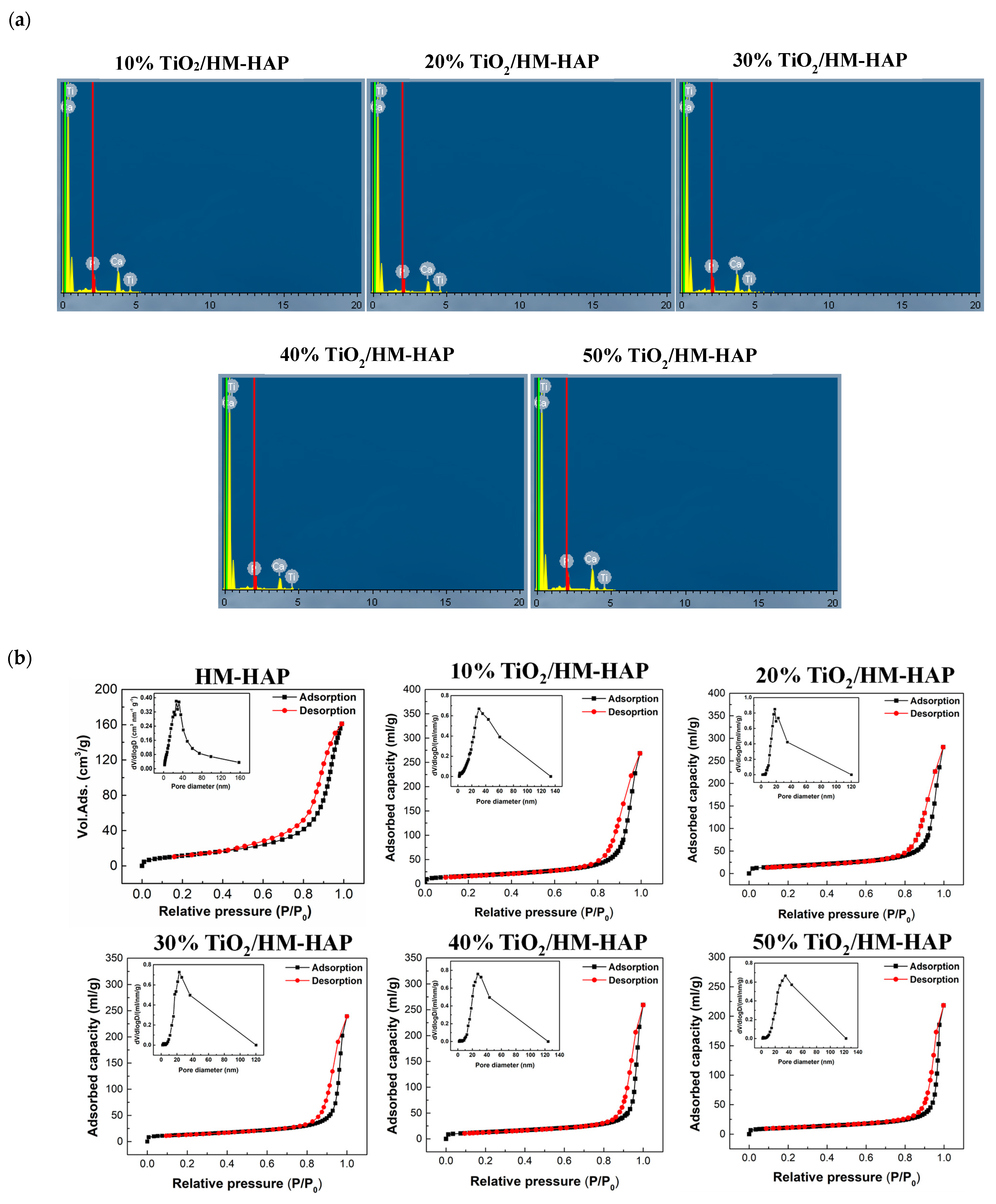
3.2. Characterization of the TiO2/HM-HAP Composite

After confirming that the material was synthesized, we further characterized a series of samples with different Ti contents. Figure 2c depicts the UV–vis DRS spectra of pure TiO2, HM-HAP, and TiO2-coated HM-HAP composites. The band gap energy values for the as-synthesized samples are illustrated in Table S2, and their respective graphs are shown in Figure S2. It can be seen that the absorption edge of pure TiO2 is evidently located around 429 nm, whereas the absorption edge of pure HM-HAP is less than 350 nm. Compared with pure HM-HAP, the TiO2-coated HM-HAP composites exhibit a considerable red shift in the absorption edge. In addition, the absorption edge of the TiO2-coated HM-HAP composites exhibit a little blue shift relative to that of TiO2, indicating that the TiO2-coated HM-HAP composites have a greater oxidation capability than TiO2.
The size and morphology of HM-HAP and TiO2-coated HM-HAP composites were visualized using SEM (Figure 3a–f). The morphology of the particles revealed a spherical shape with a hollow interior. The micrographs indicated that the particle size of HM-HAP was around 1–2 μm. All six formulations of HM-HAP had the same morphology and shape.
Energy-dispersive X-ray spectroscopy (EDS) was used to examine the surface elemental analysis of TiO2-coated HM-HAP composites. The levels of three elements, i.e., Ca, P, and Ti, in the samples, are summarized in Figure 4. The mapping profile illustrates the homogenous surface distribution of the three components. All TiO2-coated HM-HAP composites exhibited a close interaction between these elements. The corresponding EDS spectra in Figure 5a confirmed the existence of the Ti element coming from TiO2, demonstrating the successful formation of a TiO2-coated HM-HAP composite. Hence, the incorporation of TiO2 into HM-HAP particles may therefore be validated.
The pore volume, pore size, and Brunauer–Emmett–Teller (BET) specific surface area acquired from N2 adsorption–desorption for all the TiO2-coated HM-HAP composites are presented in Table 1. Figure 5b depicts the isotherms and corresponding pore size distribution histograms, indicating the mesoporous structure of the TiO2-coated HM-HAP composites. According to the IUPAC system, all the synthesized samples have type IV isotherms, which have an H3 hysteresis loop. This is because particle aggregation makes slit-shaped pores [50]. The BET-specific surface area of 10% TiO2-coated HM-HAP was 56 m2/g and declined as the TiO2 concentration increased, while the corresponding pore diameter increased gradually. A possible explanation for this result is that when the number of TiO2 molecules increases, aggregation may occur, resulting in no greater pore occupancy. Thus, a decreasing trend in the specific surface area occurred. Moreover, the coating of TiO2 onto HAP greatly increases the surface area of the resulting composite material (Table 1). The augmentation in surface area is essential as it offers a greater number of active sites for photocatalytic reactions, hence enhancing the efficacy of the photodegradation process. More precisely, the TiO2-coated HM-HAP composite has a greater surface area in comparison with pure HAP, as specified in Table 1. The improvement can be attributed to the synergistic interplay between TiO2 and HAP, with HAP serving as a supporting framework that hinders the clumping of TiO2 nanoparticles, hence ensuring a large surface area.
The surface chemical composition and chemical states of pure TiO2, HM-HAP, and TiO2-coated HM-HAP composites were investigated using XPS. The XPS survey spectra of TiO2, HM-HAP, and 20% TiO2/HM-HAP coatings are depicted in Figure 6. In Figure 6a, the survey spectra of TiO2-coated HM-HAP reveal the presence of the four elements including Ti, Ca, P, and O. As indicated in the figure, the additional C element peak is mostly produced from adventitious carbon. The XPS peaks in the Ti 2p of TiO2 in Figure 6b, situated at a binding energy of 458.6 eV and 464.2 eV, are ascribed to the Ti-O bonds [51] but in the 20% TiO2/HM-HAP coating, the Ti 2p peaks shift to lower energy levels, 458.1 eV and 463.9 eV, respectively, which may be due to the presence of Ti-O-Ca bonds in the TiO2/HM-HAP coated composite. Likewise, the Ca 2p peaks of HAP shown in Figure 6c are situated at 346.8 eV and 350.4 eV, whereas in the 20% TiO2/HAP coating, the Ca 2p peaks slightly shift towards the higher binding energy, i.e., 347.1 eV and 350.7 eV [52]. Figure 6d displays the high-resolution O 1s spectra that correspond to TiO2, HM-HAP, and TiO2/HM-HAP coated composites, respectively. Ti-O bonds and -OH groups are responsible for the two peaks in TiO2 that are located at 528.9 and 530.4 eV, respectively. In the HM-HAP spectra, the phosphate group (PO43−) and adsorbed water are responsible for the two peaks that are positioned at 530.8 eV and 532.1 eV, respectively [53,54]. In the case of the 20% TiO2/HM-HAP coated composite, the three peaks at 529.1 eV, 529.4 eV, and 531.1 eV are ascribed to the lattice oxygen species Ti-O bonds (TiO2), PO43− (phosphate group), and O-C bond of the CO3−2, respectively [54]. Hence, the analysis indicated that TiO2 particles existed in the formed composition.
Moreover, the charge transfer between HAP and TiO2 was also confirmed by the XPS analysis of the samples. By comparing the XPS data of individual atoms in pure TiO2 and HM-HAP with those in the composite material, significant shifts in binding energy peaks were observed. These shifts in binding energy are compelling evidence of alterations in the chemical environment, suggesting the occurrence of charge transfer between HAP and TiO2. Hence, the shift in peaks not only signifies the formation of the composite material but also provides clear evidence of the charge transfer process taking place between the two components.
Furthermore, the stability of the synthesized photocatalyst can be supported by the fact that hydroxyapatite (HAP) exhibits exceptional stability up to 1000 °C, as supported by rigorous testing through Fourier Transform Infrared (FTIR) analysis and Thermogravimetric Analysis (TGA) [55,56]. Therefore, in the formation of the TiO2 composite with HAP, the inherent stability of HAP was utilized, which inherently imparts durability to the resulting photocatalyst. This stability is not merely asserted but substantiated through meticulous examination, as evidenced in various papers. The FTIR and TGA analysis presented in the literature elucidates the stability of HAP at different temperatures, thereby confirming the stable nature of the synthesized photocatalyst.
PZC is regarded as one of the several methods for determining the kind of surface charge. It is the value at which the surface charge density of a material is equal to zero. Consequently, the PZC points of the TiO2/HM-HAP coated composites were determined. According to Figure 6e, the PZC point of the TiO2/HM-HAP composites (10%–50%) is between 7.5 and 8.1. The detailed PZC values of each composite are shown in Table S3. This signifies that the synthesized composites have a negative charge above this number and a positive charge below it.
In conclusion, the findings of several characterizations revealed that the TiO2/HM-HAP coated composite was successfully formed. The SEM micrographs indicated the spherical morphology of the particles having mesopores, as confirmed by the analysis of N2 adsorption–desorption. The EDS elemental mapping and the corresponding spectra clearly showed the existence of Ti in the composite. Furthermore, the XPS analysis validated the peak corresponding to the lattice oxygen species of TiO2 in the composite of TiO2-coated HM-HAP.

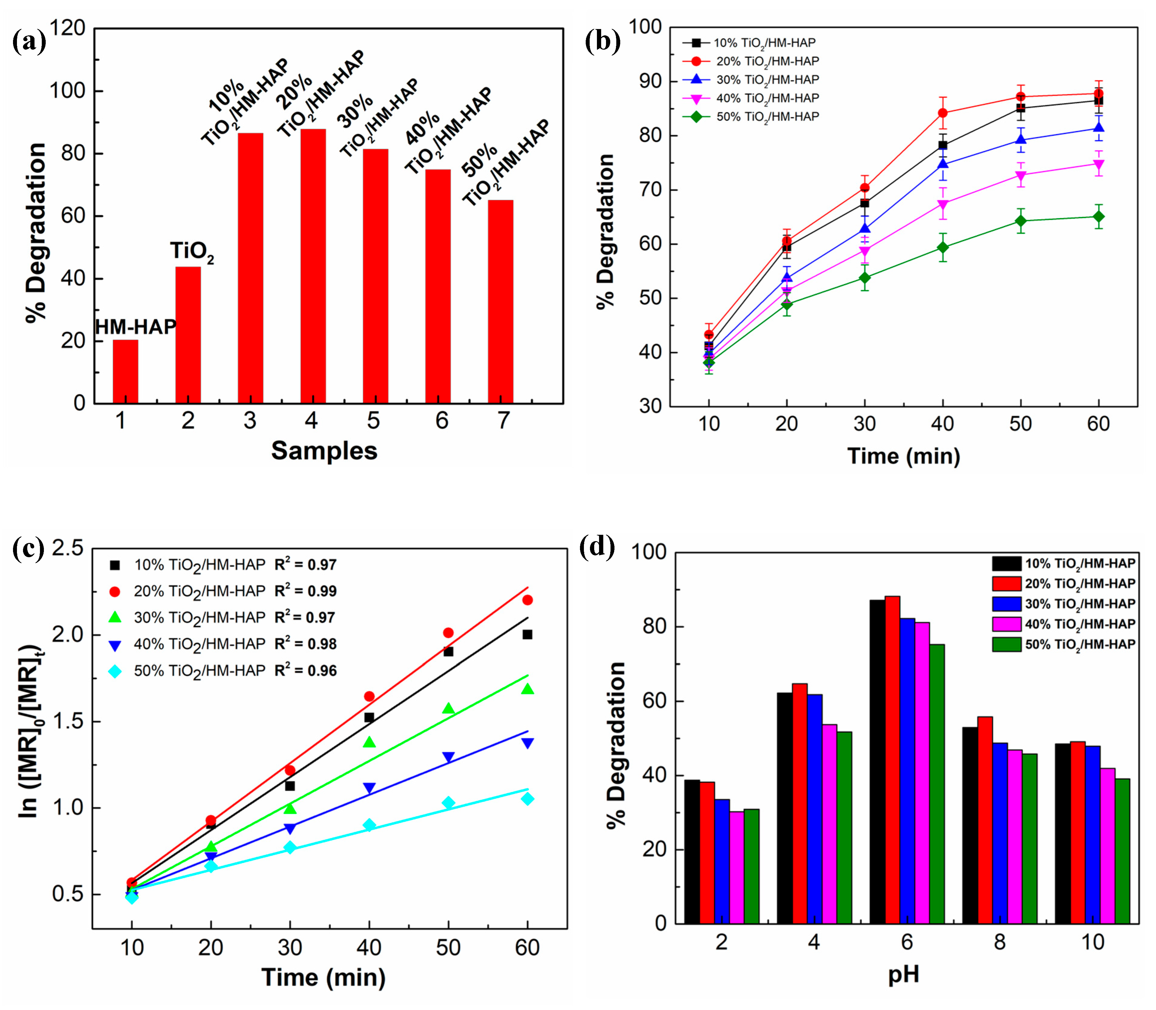
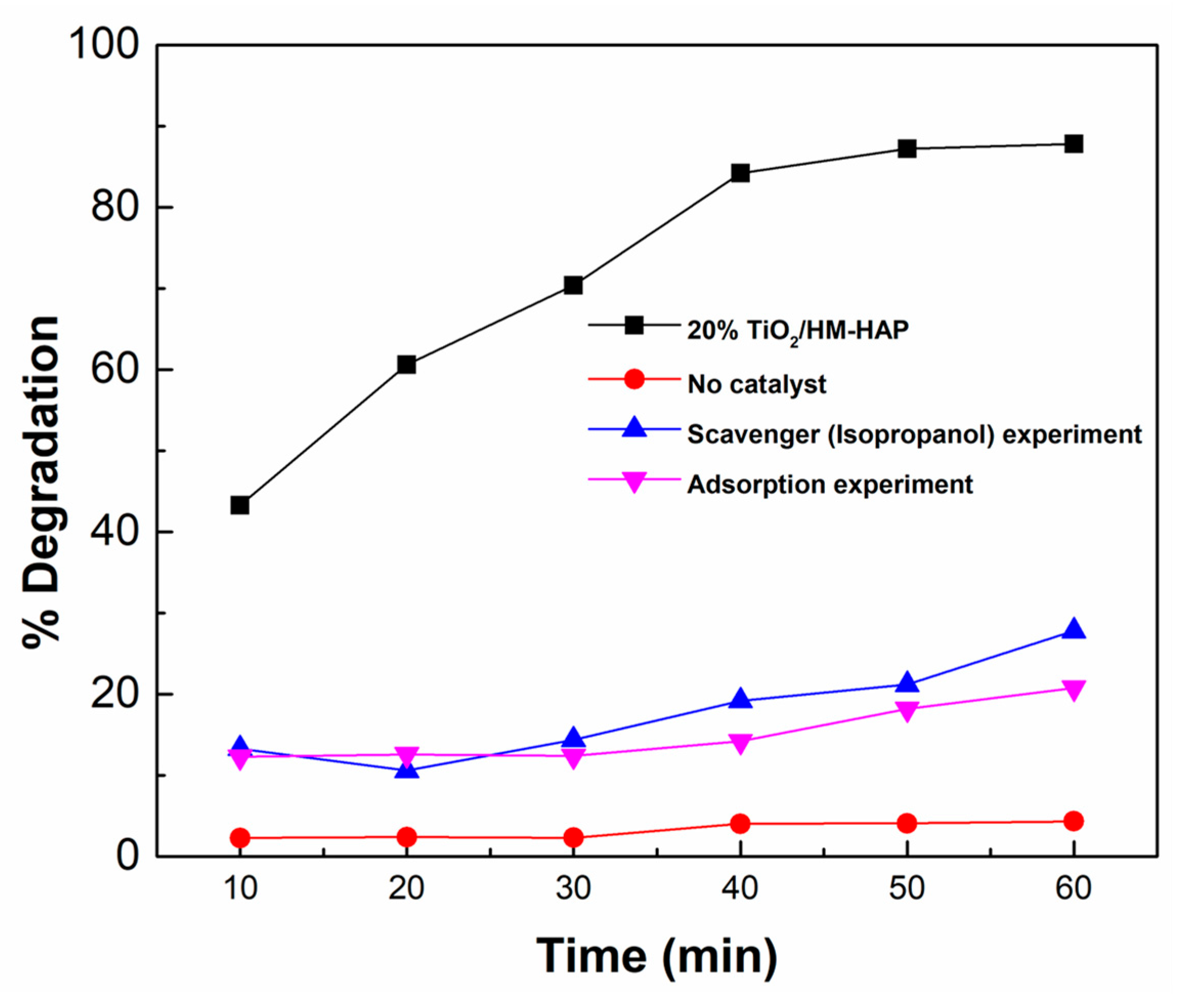
3.3. Evaluation of the Photocatalytic Performance of the Synthesized Composites for the Degradation of MR

The photocatalytic activity of the synthesized TiO2-coated composites was assessed by the photodegradation of MR under UV light irradiation. First, the linearity of MR’s spectroscopic response was measured, and a graph of absorbance against concentration was plotted to demonstrate the conformity of the dye to the Lambert–Beer law (Figure S1). As illustrated in Figure 7a, when catalyzed by a TiO2-coated HM-HAP composite with UV irradiation (60 min), the degradation efficiency increased significantly, and in the presence of the 20% TiO2/HM-HAP coating, up to almost 88% degradation efficiency was achieved. In contrast, pure HM-HAP and TiO2 demonstrated degradation efficiencies of 20% and 42%, respectively (Figure 7a). This enhanced performance can be attributed to the unique properties of HAP, specifically its hydroxyl groups (OH) and adsorbed H2O molecules on the surface. These hydroxyl groups are instrumental in interacting with h+ to generate hydroxyl radicals (˙OH), a highly reactive species known for its effectiveness in photocatalysis. Furthermore, the photocatalytic process involves intriguing electron phase shifts within the PO43− groups on the HAP surface. This phenomenon leads to the formation of ˙O2 radicals, further augmenting the catalytic efficiency of the composite material [57]. Although bare TiO2 exhibits photocatalytic activity, its efficiency is restricted by the rapid recombination of photogenerated electron–hole pairs. Conversely, HM-HAP alone offers adsorption sites but does not possess the catalytic efficiency of TiO2. The TiO2/HM-HAP composite optimizes charge separation and increases the number of active sites for the degradation reaction by integrating the benefits of both materials. Hence, integrating the advantages of HAP and TiO2 not only results in an enhancement in TiO2’s capability to absorb contaminants but also mitigates the recombination of photogenerated electron–hole pairs. This synergy between HAP and TiO2 not only improves the material’s adsorption capacity but also amplifies its overall photocatalytic performance.
However, it can also be seen that as the concentration of TiO2 increased by over 30%, the degradation efficiency declined gradually, demonstrating a decrease in activity. Generally, adsorption and degradation capacities should increase as the TiO2 level rises. However, this was not the situation observed, as HM-HAP with a higher TiO2 concentration exhibited only a little increase in adsorption in comparison with the one with a lower TiO2 concentration. The existence of a large number of catalysts can prevent light from penetrating pores and promote intermolecular collision, thereby reducing photodegradation. In addition, the large number of TiO2 particles could have covered the active sites and inhibited the generation of the active compounds [58]. These disadvantages counteract the enhanced photocatalysis provided by additional TiO2.

3.3.1. Photocatalytic Degradation Kinetics

Figure 7b shows the MR percent degradation versus irradiation time graphs of the synthesized TiO2/HM-HAP coated composites, demonstrating the gradual decomposition of MR. Figure S3 shows the UV spectra of MR at different time intervals. The rates of MR photodegradation were evaluated using a pseudo-first-order kinetic model in order to establish quantitative comparisons. In Figure 7c, it is evident that the pseudo-first-order kinetic model suited the experimental data well, and the R2 values were all above 0.90, indicating that the reactions were compatible with the first-order kinetic. Table 2 displays the values of the rate constant (kMR) and regression coefficient (R2) for the TiO2/HM-HAP coated composites for MR removal. Among the samples, the composite containing the 20% TiO2/HM-HAP coating exhibited the highest degradation rate constant, i.e., 3.3 × 10⁻² min−1. The kMR followed the order of 20% TiO2/HM-HAP > 10%, TiO2/HM-HAP > 30% TiO2/HM-HAP > 40% TiO2/HM-HAP > 50% TiO2/HM-HAP. As previously indicated, this could be because composites with low amounts of TiO2 have many active sites on their surface.
These above observations clearly indicate that 10% TiO2/HM-HAP has remarkable photocatalytic efficiency, even though it has a lower TiO2 concentration compared with 20% TiO2/HM-HAP. This finding indicates that 10% TiO2/HM-HAP has high efficiency as a photocatalyst, attaining a degradation degree (DD) that is almost equivalent to 20% TiO2/HM-HAP while containing half the amount of TiO2. When comparing 10% TiO2/HM-HAP to pure TiO2, it is evident that 10% TiO2/HM-HAP exhibits a substantially greater efficiency per unit of TiO2. Furthermore, the degradation degree (DD) per unit of TiO2 was calculated for each sample and is presented in Supplementary Information Section S1.
The efficiencies of different synthesized composites were compared to identify the most effective and cost-efficient photocatalyst. According to the findings, 20% TiO2/HM-HAP exhibits the highest overall degrading efficiency. However, 10% TiO2/HM-HAP provides a comparable performance with less TiO2. The lower TiO2 content of 10% TiO2/HM-HAP is notably advantageous from a cost and application perspective, as it reduces material costs and increases the potential for large-scale applications.

3.3.2. Effect of pH on the Degradation of MR

The pH of a solution is a crucial variable in the photocatalytic degradation process. The pH affects both the properties of a dye (hydrophobicity, speciation behavior, and water solubility) and the surface charge of a catalyst. Below pH 5.3 (pKa), MR is mostly cationic (i.e., protonated), and above pH 5.3, it is anionic (i.e., unprotonated). It was previously stated that the electrostatic interaction among the material surface, solvent molecules, and dye during photocatalytic degradation is pH-dependent [59]. The influence of solution pH was studied to find the optimal pH range for maximal photocatalytic degradation. While examining the pH range (2, 4, 6, 8, and 10), a blue shift in λmax for MR was observed when the pH of the solution was changed from 4 to 6. No other shifts in the wavelength were observed when the pH was increased further. The UV spectra of MR dye at different pHs are presented in Figure S4.
Figure 7d shows the photocatalytic behavior of the synthesized composite towards the degradation of a solution of MR dye at different pH levels carried at room temperature. In Figure 7d, it is clear that the maximum photocatalytic degradation happened at pH 6. The explanation for this behavior is that the PZC of the TiO2/HM-HAP coated composites is in the range of 7.5–8.1. Thus, the material’s surface is positively charged when the pH is <7.5 and negatively charged when the pH is >8.1. Therefore, the dye was in anionic form, and the surface of the material at pH 6 was positively charged [46,60,61]. Hence, in this condition, the interaction between the two species preferred photocatalytic degradation.

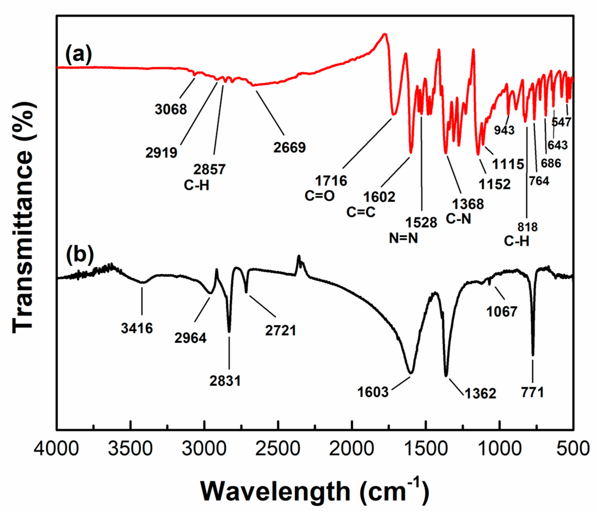
3.3.3. Post-Photodegradation FTIR Analysis and Mineralization Study of MR Dye

The FTIR spectrum of the MR dye is depicted in Figure 8a. The spectrum exhibited prominent peaks at 2919 and 2857 cm−1, which correspond to the stretching vibrations of the C-H for –CH3 groups. The O-H stretch is represented by the band at 3430 cm−1. The apparent band at 1716 cm−1 is the result of the C=O stretching of carbonyl groups. The C–C stretching vibration and N=N vibration of benzene are observed at 1602 cm−1 and 1528 cm−1, respectively. At 1368 and 1152 cm−1, strong bands are ascribed to C–N stretching vibrations. The minor bands detected at 1115, 818, and 764 cm−1 are indicative of C–H stretching vibrations [62,63].
The FTIR analysis of the product obtained subsequent to the degradation of MR is illustrated in Figure 8b. The disappearance of the peak at 1528 cm−1, which was caused by the stretching vibration of N=N, indicates the cleavage of the azo group. The disappearance of the band at 1159 cm−1 signifies a breakdown of C–N stretching vibrations [63,64]. The peak observed at 1067 cm−1 is the result of stretching variations in the C–O bond. Significant variations can be observed by comparing the FTIR spectra of the original dye with those of the degraded dye or its metabolites. Certain methyl red dye peaks were observed to have vanished after degradation, while others re-emerged subsequent to degradation; this observation suggested that the dye had transformed into new compounds or metabolites. FTIR analysis confirmed and contributed to the reduction and elimination of the methyl red dye’s azo linkage. Moreover, an LC-MS [65] analysis of decolorized MR solution was also carried out. The resultant spectra are provided in Figure S5, and the identified degraded products of MR are shown in Figure S6.
As per the results of FTIR, the degradation of MR dye was confirmed. Additionally, the mineralization of MR dye was assessed by monitoring the reduction in total organic carbon (TOC) content following the treatment with the TiO2/HM-HAP coated composite. At a pH of 6, a TOC removal of 35.64% was observed for 10 mg/L MR using the TiO2/HM-HAP coated composite following a 1 h reaction time. Consequently, the TiO2/HM-HAP coated composite was capable of facilitating dye decolorization and mineralization.
Moreover, the results of the photolysis experiment (Figure 9) demonstrated that there was minimal degradation of the MR dye when exposed just to direct UV light. This suggests that direct photolysis has a small impact on the overall degradation process. Furthermore, the findings from the adsorption experiment, presented in Figure 9, demonstrate that the catalyst was responsible for 20% of the overall removal of MR by adsorption. This demonstrates that although adsorption plays a role in the overall process, the main mechanism for removing dye under UV irradiation is definitely photodegradation. HAP possesses a significant number of hydroxyl groups, which contribute to its surface having a primarily negative charge. Methyl red (MR) is a negatively charged dye, and because of the like charges, there is a repulsive force that decreases its ability to stick to the HAP surface. While HAP does have some sites with a positive charge due to calcium ions, the overall negative charge from the many hydroxyl groups is more prominent. The structure of HAP and an explanation of the interaction between HAP and TiO2 in TiO2/HM-HAP composites are further presented in Supplementary Information Section S2.
In addition, an examination was conducted on the impact of an OH scavenger on photocatalysis. Isopropanol was employed as an *OH scavenger for the experiment. The catalyst’s catalytic efficiency decreases when a scavenger is present. This is illustrated using the graph presented in Figure 9. The predominant catalyst in this process is now clearly identified as the *OH radical [66].
In comparison with other recently researched sorbents, the adsorption capability of the Sa-modified hydroxyapatite as synthesized demonstrated promising findings. Zenefar et al. observed that the photodegradation potential of a HAp-TiO2-ZnO photocatalyst for methylene blue (MB) and methyl orange (MO) dye was 95% and 45% after 2 h, respectively [67]. A Hap-TiO2 nanocomposite demonstrated a degradation efficiency of 80% for methyl orange (5 mg/mL), as stated by Sharifat et al. [38]. Furthermore, Anmin et al. reported the degradation efficiency of titanium-substituted HAp for methylene blue (MB) to be 17%–37% under visible light and 39%–50% under UV light [68]. This literature review shows that the TiO2-coated HM-HAP composite is an effective catalyst for the removal of dyes from water.
Combining the outcomes of SEM, DRS, FTIR, N2 adsorption–desorption, and XRD analysis, the enhanced photocatalytic degradation performance of the TiO2/HM-HAP coated composites in comparison with pure TiO2 on the photodegradation of methyl red is associated with the creation of a uniform layer of TiO2 particles on the highly porous structure of HM-HAP containing a large number of hydroxyl (OH) groups. These OH groups interact with the generated holes as Lewis bases, which results in the generation of hydroxyl radicals. The generated OH radicals will then oxidize the adsorbed molecules, hence improving the material’s photocatalytic efficiency. Moreover, the hollow mesoporous structure of HM-HAP promotes the degradation performance of the composite by adsorbing the dye particles on its surface, as observed in Figure 7a. The relatively better catalytic performance of the 20% TiO2/HM-HAP coated composites compared with those with a high concentration of TiO2 is mostly determined by the existence of active sites, surface area, and availability of active substances.

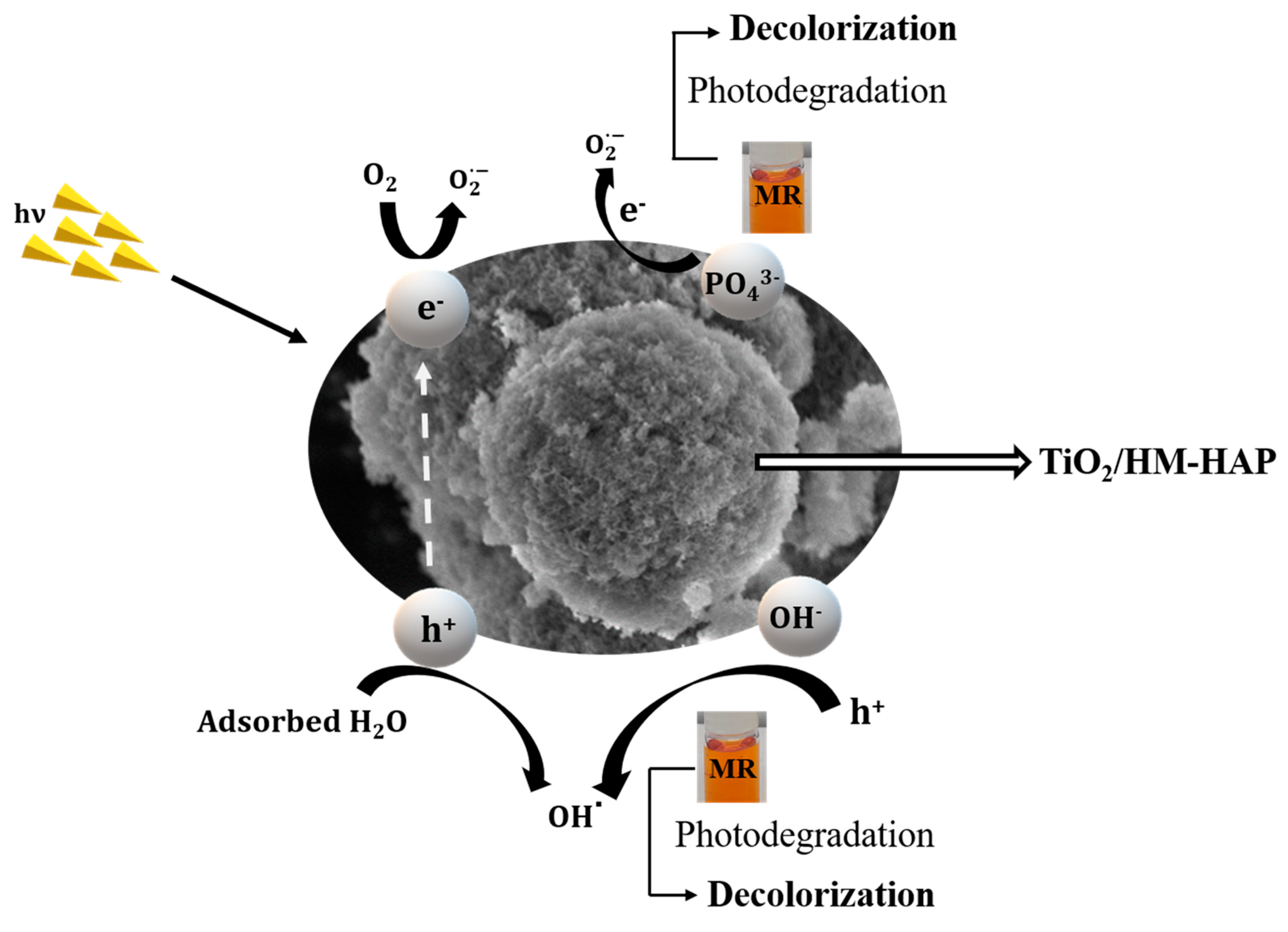
3.4. Photocatalytic Degradation Mechanism of TiO2/HM-HAP Composites on MR Dye

The possible mechanism of TiO2/HM-HAP coated composites for the degradation of MR is shown in Figure 10. Generally, the UV irradiation of TiO2-coated HM-HAP alters the electronic condition of the PO4−3 group on the surface and generates a vacancy on HM-HAP. In the same way, when UV light is applied to TiO2, electrons in the VB (valance band) move to the CB (conduction band), making an equal number of holes in the VB. The reaction is given as:
TiO 2 + h ν e + h +
The reaction of a CB electron with O2 produces superoxide radicals (O2.−), which then oxidizes the organic compounds. The reaction is given as follows:
e + O 2 O 2 .
The VB hole interacts with the hydroxyl anions or the water to generate hydrogen peroxide.
h + + H 2 O OH . + H +
h + + OH OH .
The hydrogen peroxide then breaks apart and releases hydroxyl radicals (OH·), which are powerful oxidizing agents that attack organic molecules that have adsorbed to the composite [37].

4. Conclusions

The present research demonstrated the effective synthesis of TiO2/HM-HAP coated composites by loading the TiO2 at various concentrations, comprising 10%, 20%, 30%, 40%, and 50%, to enhance the degradation efficiency of hazardous dyes present in aqueous solutions and address water pollution issues. Several analysis approaches, including FTIR, XPS, XRD, SEM, and BET, were utilized to analyze the physical properties of all the synthesized TiO2/HM-HAP coated composites. TiO2 was evenly incorporated into the hollow mesoporous HAP particles, with an insignificant effect on the overall structure. The catalytic performance was examined via the degradation of MR in the presence of UV light. We observed that the MR removal ratio of the 20% TiO2/HM-HAP coating was 88%, which was found to be the maximum among the HM-HAP, pure TiO2, and TiO2/HM-HAP coated composites. It was noted that the photocatalytic degradation process was compatible with the pseudo-first-order (PFO) kinetic model. Moreover, it was found that MR could be more easily degraded at pH 6 than in strongly acidic or alkaline environments. This work provides insight into the development of TiO2/HM-HAP coated composites as potential materials for removing organic contaminants from wastewater. This will help with both water treatment and waste management.

Supplementary Materials

The following supporting information can be downloaded at https://www.mdpi.com/article/10.3390/coatings14080921/s1. It includes the details about band gap energy data and other supporting figures.

Author Contributions

Conceptualization, F.S. and W.Q.; Methodology, F.S.; Formal analysis, S.Y. and Y.P.; Investigation, S.Y.; Writing—original draft, F.S.; Project administration, W.Q. All authors have read and agreed to the published version of the manuscript.

Funding

This research was supported by the National Natural Science Foundation of China Grant No. 22378049.

Institutional Review Board Statement

Not applicable.

Informed Consent Statement

Not applicable.

Data Availability Statement

Data is contained within the article or Supplementary Materials.

Conflicts of Interest

The authors declare no conflicts of interest.

References

  1. Badr, Y.; Abd El-Wahed, M.G.; Mahmoud, M.A. Photocatalytic degradation of methyl red dye by silica nanoparticles. J. Hazard. Mater. 2008, 154, 245–253. [Google Scholar] [CrossRef] [PubMed]
  2. Zaheer, Z.; AL-Asfar, A.; Aazam, E.S. Adsorption of methyl red on biogenic Ag@Fe nanocomposite adsorbent: Isotherms, kinetics and mechanisms. J. Mol. Liq. 2019, 283, 287–298. [Google Scholar] [CrossRef]
  3. Slokar, Y.M.; Majcen Le Marechal, A. Methods of decoloration of textile wastewaters. Dye. Pigment. 1998, 37, 335–356. [Google Scholar] [CrossRef]
  4. Gökçen, F.; Özbelge, T.A. Pre-ozonation of aqueous azo dye (Acid Red-151) followed by activated sludge process. Chem. Eng. J. 2006, 123, 109–115. [Google Scholar] [CrossRef]
  5. Khan, A.M.; Shafiq, F.; Khan, S.A.; Ali, S.; Ismail, B.; Hakeem, A.S.; Rahdar, A.; Nazar, M.F.; Sayed, M.; Khan, A.R. Surface modification of colloidal silica particles using cationic surfactant and the resulting adsorption of dyes. J. Mol. Liq. 2019, 274, 673–680. [Google Scholar] [CrossRef]
  6. Ullah, F.; Ji, G.; Irfan, M.; Gao, Y.; Shafiq, F.; Sun, Y.; Ain, Q.U.; Li, A. Adsorption performance and mechanism of cationic and anionic dyes by KOH activated biochar derived from medical waste pyrolysis. Environ. Pollut. 2022, 314, 120271. [Google Scholar] [CrossRef] [PubMed]
  7. Zhao, J.; Liu, H.; Xue, P.; Tian, S.; Sun, S.; Lv, X. Highly-efficient PVDF adsorptive membrane filtration based on chitosan@CNTs-COOH simultaneous removal of anionic and cationic dyes. Carbohydr. Polym. 2021, 274, 118664. [Google Scholar] [CrossRef] [PubMed]
  8. Saulat, H.; Yang, J.; Yan, T.; Raza, W.; Song, W.; He, G. Tungsten incorporated mobil-type eleven zeolite membranes: Facile synthesis and tuneable wettability for highly efficient separation of oil/water mixtures. Chin. J. Chem. Eng. 2023, 60, 242–252. [Google Scholar] [CrossRef]
  9. Badr, Y.; Mahmoud, M.A. Photocatalytic degradation of methyl orange by gold silver nano-core/silica nano-shell. J. Phys. Chem. Solids 2007, 68, 413–419. [Google Scholar] [CrossRef]
  10. Kim, H.; Kim, T.; Gil Lee, D.; Weon Roh, S.; Lee, C. Nitrogen-centered radical-mediated C–H imidation of arenes and heteroarenes via visible light induced photocatalysis. Chem. Commun. 2014, 50, 9273–9276. [Google Scholar] [CrossRef] [PubMed]
  11. Lucas, M.S.; Dias, A.A.; Sampaio, A.; Amaral, C.; Peres, J.A. Degradation of a textile reactive Azo dye by a combined chemical–biological process: Fenton’s reagent-yeast. Water Res. 2007, 41, 1103–1109. [Google Scholar] [CrossRef] [PubMed]
  12. Liu, Y.; Liu, C.Y.; Wei, J.H.; Xiong, R.; Pan, C.X.; Shi, J. Enhanced adsorption and visible-light-induced photocatalytic activity of hydroxyapatite modified Ag–TiO2 powders. Appl. Surf. Sci. 2010, 256, 6390–6394. [Google Scholar] [CrossRef]
  13. Natarajan, T.S.; Lee, J.Y.; Bajaj, H.C.; Jo, W.K.; Tayade, R.J. Synthesis of multiwall carbon nanotubes/TiO2 nanotube composites with enhanced photocatalytic decomposition efficiency. Catal. Today 2017, 282, 13–23. [Google Scholar] [CrossRef]
  14. Wu, H.; Yang, X.; Zhao, S.; Zhai, L.; Wang, G.; Zhang, B.; Qin, Y. Encapsulation of atomically dispersed Pt clusters in porous TiO2 for semi-hydrogenation of phenylacetylene. Chem. Commun. 2022, 58, 1191–1194. [Google Scholar] [CrossRef] [PubMed]
  15. Chen, S.; Fang, S.; Sun, Z.; Li, Z.; Wang, C.; Hu, Y.H. Thin-water-film-enhanced TiO2-based catalyst for CO2 hydrogenation to formic acid. Chem. Commun. 2022, 58, 787–790. [Google Scholar] [CrossRef] [PubMed]
  16. Liu, H.; Lv, T.; Zhu, C.; Zhu, Z. Direct bandgap narrowing of TiO2/MoO3 heterostructure composites for enhanced solar-driven photocatalytic activity. Sol. Energy Mater. Sol. Cells 2016, 153, 1–8. [Google Scholar] [CrossRef]
  17. Sukhadeve, G.K.; Bandewar, H.; Janbandhu, S.Y.; Jayaramaiah, J.R.; Gedam, R.S. Photocatalytic hydrogen production, dye degradation, and antimicrobial activity by Ag-Fe co-doped TiO2 nanoparticles. J. Mol. Liq. 2023, 369, 120948. [Google Scholar] [CrossRef]
  18. Ali, F.; Moin-ud-Din, G.; Iqbal, M.; Nazir, A.; Altaf, I.; Alwadai, N.; Siddiqua, U.H.; Younas, U.; Ali, A.; Kausar, A.; et al. Ag and Zn doped TiO2 nano-catalyst synthesis via a facile green route and their catalytic activity for the remediation of dyes. J. Mater. Res. Technol. 2023, 23, 3626–3637. [Google Scholar] [CrossRef]
  19. Liza, T.Z.; Tusher, M.M.H.; Anwar, F.; Monika, M.F.; Amin, K.F.; Asrafuzzaman, F.N.U. Effect of Ag-doping on morphology, structure, band gap and photocatalytic activity of bio-mediated TiO2 nanoparticles. Results Mater. 2024, 22, 100559. [Google Scholar] [CrossRef]
  20. Poudel, M.B.; Kim, A.A. Silver nanoparticles decorated TiO2 nanoflakes for antibacterial properties. Inorg. Chem. Commun. 2023, 152, 110675. [Google Scholar] [CrossRef]
  21. Cabrera-Rodríguez, O.; Trejo-Valdez, M.D.; Torres-SanMiguel, C.R.; Pérez-Hernández, N.; Bañuelos-Hernández, Á.; Manríquez-Ramírez, M.E.; Hernández-Benítez, J.A.; Rodríguez-Tovar, A.V. Evaluation of the performance of TiO2 thin films doped with silver nanoparticles as a protective coating for metal prostheses. Surf. Coatings Technol. 2023, 458, 129349. [Google Scholar] [CrossRef]
  22. Meroni, D.; Galloni, M.G.; Cionti, C.; Cerrato, G.; Falletta, E.; Bianchi, C.L. Efficient Day-and-Night NO2 Abatement by Polyaniline/TiO2 Nanocomposites. Materials 2023, 16, 1304. [Google Scholar] [CrossRef] [PubMed]
  23. Pal, A.; Jana, T.K.; Chatterjee, K. Silica supported TiO2 nanostructures for highly efficient photocatalytic application under visible light irradiation. Mater. Res. Bull. 2016, 76, 353–357. [Google Scholar] [CrossRef]
  24. Eskandarian, M.R.; Fazli, M.; Rasoulifard, M.H.; Choi, H. Decomposition of organic chemicals by zeolite-TiO2 nanocomposite supported onto low density polyethylene film under UV-LED powered by solar radiation. Appl. Catal. B Environ. 2016, 183, 407–416. [Google Scholar] [CrossRef]
  25. Padmanabhan, S.K.; Pal, S.; Ul Haq, E.; Licciulli, A. Nanocrystalline TiO2–diatomite composite catalysts: Effect of crystallization on the photocatalytic degradation of rhodamine B. Appl. Catal. A Gen. 2014, 485, 157–162. [Google Scholar] [CrossRef]
  26. Zeng, G.; You, H.; Du, M.; Zhang, Y.; Ding, Y.; Xu, C.; Liu, B.; Chen, B.; Pan, X. Enhancement of photocatalytic activity of TiO2 by immobilization on activated carbon for degradation of aquatic naphthalene under sunlight irradiation. Chem. Eng. J. 2021, 412, 128498. [Google Scholar] [CrossRef]
  27. Sans, J.; Arnau, M.; Sanz, V.; Turon, P.; Alemán, C. Hydroxyapatite-based biphasic catalysts with plasticity properties and its potential in carbon dioxide fixation. Chem. Eng. J. 2022, 433, 133512. [Google Scholar] [CrossRef]
  28. Sayed, I.R.; Farhan, A.M.; AlHammadi, A.A.; El-Sayed, M.I.; Abd El-Gaied, I.M.; El-Sherbeeny, A.M.; Al Zoubi, W.; Ko, Y.G.; Abukhadra, M.R. Synthesis of novel nanoporous zinc phosphate/hydroxyapatite nano-rods (ZPh/HPANRs) core/shell for enhanced adsorption of Ni2+ and Co2+ ions: Characterization and application. J. Mol. Liq. 2022, 360, 119527. [Google Scholar] [CrossRef]
  29. Ding, Z.; Han, H.; Fan, Z.; Lu, H.; Sang, Y.; Yao, Y.; Cheng, Q.; Lu, Q.; Kaplan, D.L. Nanoscale Silk-Hydroxyapatite Hydrogels for Injectable Bone Biomaterials. ACS Appl. Mater. Interfaces 2017, 9, 16913–16921. [Google Scholar] [CrossRef] [PubMed]
  30. Gong, M.; Liu, C.; Liu, C.; Wang, L.; Shafiq, F.; Liu, X.; Sun, G.; Song, Q.; Qiao, W. Biomimetic hydroxyapate/polydopamine composites with good biocompatibility and efficiency for uncontrolled bleeding. J. Biomed. Mater. Res. Part B Appl. Biomater. 2021, 109, 1876–1892. [Google Scholar] [CrossRef] [PubMed]
  31. Verma, R.; Mishra, S.R.; Gadore, V.; Ahmaruzzaman, M. Hydroxyapatite-based composites: Excellent materials for environmental remediation and biomedical applications. Adv. Colloid Interface Sci. 2023, 315, 102890. [Google Scholar] [CrossRef] [PubMed]
  32. Amenaghawon, A.N.; Anyalewechi, C.L.; Darmokoesoemo, H.; Kusuma, H.S. Hydroxyapatite-based adsorbents: Applications in sequestering heavy metals and dyes. J. Environ. Manag. 2022, 302, 113989. [Google Scholar] [CrossRef] [PubMed]
  33. Zhang, J.; Yan, B.; Chen, T.; Tu, S.; Li, H.; Yang, Z.; Hao, T.; Chen, C. Piezoelectric hydroxyapatite synthesized from municipal solid waste incineration fly ash and its underlying mechanism for high efficiency in degradation of xanthate. Chem. Eng. J. 2024, 493, 152601. [Google Scholar] [CrossRef]
  34. Kumar Yadav, M.; Hiren Shukla, R.; Prashanth, K.G. A comprehensive review on development of waste derived hydroxyapatite (HAp) for tissue engineering application. Mater. Today Proc. 2023. [Google Scholar] [CrossRef]
  35. Tong, H.; Shi, D.; Cai, H.; Liu, J.; Lv, M.; Gu, L.; Luo, L.; Wang, B. Novel hydroxyapatite (HAP)-assisted hydrothermal solidification of heavy metals in fly ash from MSW incineration: Effect of HAP liquid-precursor and HAP seed crystal derived from eggshell waste. Fuel Process. Technol. 2022, 236, 107400. [Google Scholar] [CrossRef]
  36. Nishikawa, H. Surface changes and radical formation on hydroxyapatite by UV irradiation for inducing photocatalytic activation. J. Mol. Catal. A Chem. 2003, 206, 331–338. [Google Scholar] [CrossRef]
  37. El, A.; Boumanchar, I.; Zbair, M.; Chhiti, Y.; Sahibed-Dine, A.; Bentiss, F.; Bensitel, M. The photocatalytic degradation of methylene bleu over TiO2 catalysts supported on hydroxyapatite. JMES 2017, 8, 1301–1311. Available online: http://www.jmaterenvironsci.com/ (accessed on 7 October 2022).
  38. Sharifat, S.; Zolgharnein, H.; Hamidifalahi, A.; Enayati-Jazi, M.; Hamid, E. Preparation and Characterization of HAp/TiO2 Nanocomposite for Photocatalytic Degradation of Methyl Orange under UV-Irradiation. Adv. Mater. Res. 2014, 829, 594–599. [Google Scholar] [CrossRef]
  39. Yao, J.; Zhang, Y.; Wang, Y.; Chen, M.; Huang, Y.; Cao, J.; Ho, W.; Lee, S.C. Enhanced photocatalytic removal of NO over titania/hydroxyapatite (TiO2/HAp) composites with improved adsorption and charge mobility ability. RSC Adv. 2017, 7, 24683–24689. [Google Scholar] [CrossRef]
  40. Narayan, R.B.; Goutham, R.; Srikanth, B.; Gopinath, K.P. A novel nano-sized calcium hydroxide catalyst prepared from clam shells for the photodegradation of methyl red dye. J. Environ. Chem. Eng. 2018, 6, 3640–3647. [Google Scholar] [CrossRef]
  41. Shan, R.; Lu, L.; Gu, J.; Zhang, Y.; Yuan, H.; Chen, Y.; Luo, B. Photocatalytic degradation of methyl orange by Ag/TiO2/biochar composite catalysts in aqueous solutions. Mater. Sci. Semicond. Process. 2020, 114, 105088. [Google Scholar] [CrossRef]
  42. Xu, Q.; Feng, J.; Li, L.; Xiao, Q.; Wang, J. Hollow ZnFe2O4/TiO2 composites: High-performance and recyclable visible-light photocatalyst. J. Alloys Compd. 2015, 641, 110–118. [Google Scholar] [CrossRef]
  43. Kalaiarasi, S.; Jose, M. Dielectric functionalities of anatase phase titanium dioxide nanocrystals synthesized using water-soluble complexes. Appl. Phys. A 2017, 123, 512. [Google Scholar] [CrossRef]
  44. Wei, J.; Shi, J.; Wu, Q.; Yang, L.; Cao, S. Hollow hydroxyapatite/polyelectrolyte hybrid microparticles with controllable size, wall thickness and drug delivery properties. J. Mater. Chem. B 2015, 3, 8162–8169. [Google Scholar] [CrossRef] [PubMed]
  45. Zhu, X.; Shi, J.; Ma, H.; Chen, R.; Li, J.; Cao, S. Hierarchical hydroxyapatite/polyelectrolyte microcapsules capped with AuNRs for remotely triggered drug delivery. Mater. Sci. Eng. C 2019, 99, 1236–1245. [Google Scholar] [CrossRef] [PubMed]
  46. Shafiq, F.; Liu, C.; Zhou, H.; Chen, H.; Yu, S.; Qiao, W. Adsorption mechanism and synthesis of adjustable hollow hydroxyapatite spheres for efficient wastewater cationic dyes adsorption. Colloids Surfaces A Physicochem. Eng. Asp. 2023, 672, 131713. [Google Scholar] [CrossRef]
  47. Wang, S.; Zhou, S. Photodegradation of methyl orange by photocatalyst of CNTs/P-TiO2 under UV and visible-light irradiation. J. Hazard. Mater. 2011, 185, 77–85. [Google Scholar] [CrossRef] [PubMed]
  48. Xu, W.; Liu, B.; Wang, Y.; Xiao, G.; Chen, X.; Xu, W.; Lu, Y.P. A facile strategy for one-step hydrothermal preparation of porous hydroxyapatite microspheres with core–shell structure. J. Mater. Res. Technol. 2022, 17, 320–328. [Google Scholar] [CrossRef]
  49. Shafiq, F.; Liu, C.; Zhou, H.; Chen, H.; Yu, S.; Qiao, W. Stearic acid-modified hollow hydroxyapatite particles with enhanced hydrophobicity for oil adsorption from oil spills. Chemosphere 2024, 348, 140651. [Google Scholar] [CrossRef] [PubMed]
  50. Guo, Y.P.; Yao, Y.B.; Guo, Y.J.; Ning, C.Q. Hydrothermal fabrication of mesoporous carbonated hydroxyapatite microspheres for a drug delivery system. Microporous Mesoporous Mater. 2012, 155, 245–251. [Google Scholar] [CrossRef]
  51. Li, W.; Tian, Y.; Li, H.; Zhao, C.; Zhang, B.; Zhang, H.; Geng, W.; Zhang, Q. Novel BiOCl/TiO2 hierarchical composites: Synthesis, characterization and application on photocatalysis. Appl. Catal. A Gen. 2016, 516, 81–89. [Google Scholar] [CrossRef]
  52. Abbasi, S.; Bayati, M.R.; Golestani-Fard, F.; Rezaei, H.R.; Zargar, H.R.; Samanipour, F.; Shoaei-Rad, V. Micro arc oxidized HAp–TiO2 nanostructured hybrid layers-part I: Effect of voltage and growth time. Appl. Surf. Sci. 2011, 257, 5944–5949. [Google Scholar] [CrossRef]
  53. Li, K.; Gao, S.; Wang, Q.; Xu, H.; Wang, Z.; Huang, B.; Dai, Y.; Lu, J. In-situ-reduced synthesis of Ti3+ self-doped TiO2/g-C3N4 heterojunctions with high photocatalytic performance under LED light irradiation. ACS Appl. Mater. Interfaces 2015, 7, 9023–9030. [Google Scholar] [CrossRef] [PubMed]
  54. Salarian, M.; Xu, W.Z.; Wang, Z.; Sham, T.K.; Charpentier, P.A. Hydroxyapatite-TiO2-based Nanocomposites Synthesized in Supercritical CO2 for Bone Tissue Engineering: Physical and Mechanical Properties. ACS Appl. Mater. Interfaces 2014, 6, 16918–16931. [Google Scholar] [CrossRef] [PubMed]
  55. Anjaneyulu, U.; Priyadarshini, B.; Arul Xavier Stango, S.; Chellappa, M.; Geetha, M.; Vijayalakshmi, U. Preparation and characterisation of sol–gel-derived hydroxyapatite nanoparticles and its coatings on medical grade Ti-6Al-4V alloy for biomedical applications. Mater. Technol. 2017, 32, 800–814. [Google Scholar] [CrossRef]
  56. Rath, P.C.; Singh, B.P.; Besra, L.; Bhattacharjee, S. Multiwalled Carbon Nanotubes Reinforced Hydroxyapatite-Chitosan Composite Coating on Ti Metal: Corrosion and Mechanical Properties. J. Am. Ceram. Soc. 2012, 95, 2725–2731. [Google Scholar] [CrossRef]
  57. Goto, T.; Cho, S.H.; Ohtsuki, C.; Sekino, T. Selective adsorption of dyes on TiO2-modified hydroxyapatite photocatalysts morphologically controlled by solvothermal synthesis. J. Environ. Chem. Eng. 2021, 9, 105738. [Google Scholar] [CrossRef]
  58. Lin, Y.C.; Fang, Y.P.; Hung, C.F.; Yu, H.P.; Alalaiwe, A.; Wu, Z.Y.; Fang, J.Y. Multifunctional TiO2/SBA-15 mesoporous silica hybrids loaded with organic sunscreens for skin application: The role in photoprotection and pollutant adsorption with reduced sunscreen permeation. Colloids Surf. B Biointerfaces 2021, 202, 111658. [Google Scholar] [CrossRef] [PubMed]
  59. Singh, N.K.; Saha, S.; Pal, A. Methyl red degradation under UV illumination and catalytic action of commercial ZnO: A parametric study. New Pub Balaban 2014, 56, 1066–1076. [Google Scholar] [CrossRef]
  60. Bouzid, T.; Grich, A.; Naboulsi, A.; Regti, A.; Alaoui Tahiri, A.; El Himri, M.; El Haddad, M. Adsorption of Methyl Red on porous activated carbon from agriculture waste: Characterization and response surface methodology optimization. Inorg. Chem. Commun. 2023, 158, 111544. [Google Scholar] [CrossRef]
  61. Amari, A.; Yadav, V.K.; Pathan, S.K.; Singh, B.; Osman, H.; Choudhary, N.; Khedher, K.M.; Basnet, A. Remediation of Methyl Red Dye from Aqueous Solutions by Using Biosorbents Developed from Floral Waste. Adsorpt. Sci. Technol. 2023, 2023, 1532660. [Google Scholar] [CrossRef]
  62. Waghmode, T.R.; Kurade, M.B.; Sapkal, R.T.; Bhosale, C.H.; Jeon, B.H.; Govindwar, S.P. Sequential photocatalysis and biological treatment for the enhanced degradation of the persistent azo dye methyl red. J. Hazard. Mater. 2019, 371, 115–122. [Google Scholar] [CrossRef] [PubMed]
  63. Balu, P.; Asharani, I.V.; Thirumalai, D. Catalytic degradation of hazardous textile dyes by iron oxide nanoparticles prepared from Raphanus sativus leaves’ extract: A greener approach. J. Mater. Sci. Mater. Electron. 2020, 31, 10669–10676. [Google Scholar] [CrossRef]
  64. Ikram, M.; Naeem, M.; Zahoor, M.; Rahim, A.; Hanafiah, M.M.; Oyekanmi, A.A.; Shah, A.B.; Mahnashi, M.H.; Al Ali, A.; Jalal, N.A.; et al. Biodegradation of Azo Dye Methyl Red by Pseudomonas aeruginosa: Optimization of Process Conditions. Int. J. Environ. Res. Public Health 2022, 19, 9962. [Google Scholar] [CrossRef] [PubMed]
  65. Bian, C.; Wang, Y.; Yi, Y.; Shao, S.; Sun, P.; Xiao, Y.; Wang, W.; Dong, X. Enhanced photocatalytic activity of S-doped graphitic carbon nitride hollow microspheres: Synergistic effect, high-concentration antibiotic elimination and antibacterial behavior. J. Colloid Interface Sci. 2023, 643, 256–266. [Google Scholar] [CrossRef]
  66. Shahzad, W.; Badawi, A.K.; Rehan, Z.A.; Khan, A.M.; Khan, R.A.; Shah, F.; Ali, S.; Ismail, B. Enhanced visible light photocatalytic performance of Sr0.3(Ba,Mn)0.7ZrO3 perovskites anchored on graphene oxide. Ceram. Int. 2022, 48, 24979–24988. [Google Scholar] [CrossRef]
  67. Yeasmin, Z.; Alim, A.; Ahmed, S.; Rahman, M.M.; Masum, S.M.; Ghosh, A.K. Synthesis, Characterization and Efficiency of HAp-TiO2-ZnO Composite as a Promising Photocatalytic Material. Trans. Indian Ceram. Soc. 2018, 77, 161–168. [Google Scholar] [CrossRef]
  68. Hu, A.; Li, M.; Chang, C.; Mao, D. Preparation and characterization of a titanium-substituted hydroxyapatite photocatalyst. J. Mol. Catal. A Chem. 2007, 267, 79–85. [Google Scholar] [CrossRef]
Figure 1. Schematic diagram illustrating the synthesis of TiO2/HM-HAP particles.
Coatings 14 00921 g001
Figure 2. FTIR spectra of as-synthesized samples (a), XRD spectra of TiO2, HM-HAP, and TiO2-coated HM-HAP (10%, 20%, 30%, 40%, and 50%) composites (b), and UV–vis DRS spectra of pure TiO2, HM-HAP, and TiO2-coated HM-HAP composites (c).
Coatings 14 00921 g002
Figure 3. SEM micrographs of (a) HM-HAP and (b) 10% TiO2/HM-HAP, (c) 20% TiO2/HM-HAP, (d) 30% TiO2/HM-HAP, (e) 40% TiO2/HM-HAP, and (f) 50% TiO2/HM-HAP coated composites. In (af), the insets depict the high magnifications corresponding to those figures.
Coatings 14 00921 g003
Figure 4. EDS elemental mapping of (a) 10% TiO2/HM-HAP, (b) 20% TiO2/HM-HAP, (c) 30% TiO2/HM-HAP, (d) 40% TiO2/HM-HAP, and (e) 50% TiO2/HM-HAP coated composites.
Coatings 14 00921 g004
Figure 5. EDS spectra (a) and N2 adsorption–desorption isotherms and pore size distribution (b) of the synthesized TiO2/HM-HAP coated composites.
Coatings 14 00921 g005
Figure 6. XPS survey spectra (a) and high-resolution XPS spectra of Ti 2p (b), Ca 2p, (c), O 1s (d), and PZC plot (e) of the synthesized samples.
Coatings 14 00921 g006
Figure 7. Degradation efficiency of MR over the synthesized composites at pH = 6, 25 °C, catalyst = 30 mg, and 60 min of irradiation time (a), time curve of % degradation at pH = 6, and 25 °C temperature (b), degradation kinetics of MR at pH = 6, and 25 °C temperature (c), and effect of pH on the % degradation of MR at 25 °C, and 60 min of irradiation time (d).
Coatings 14 00921 g007
Figure 8. FTIR spectra of MR dye before degradation (a) and after degradation (b).
Coatings 14 00921 g008
Figure 9. Plot representing the effect of a scavenger on the photolysis, adsorption, and photolysis experiment.
Coatings 14 00921 g009
Figure 10. Possible mechanism of TiO2/HM-HAP coated composites under UV irradiation for the photocatalytic degradation of MR.
Coatings 14 00921 g010
Table 1. BET-specific surface area and pore diameter analysis of the synthesized composites.
SampleBET Specific Surface Area (m2/g)Pore Size (nm)
HM-HAP4412.47
10% TiO2/HM-HAP56 27.82
20% TiO2/HM-HAP57 28.19
30% TiO2/HM-HAP47 29.28
40% TiO2/HM-HAP46 31.94
50% TiO2/HM-HAP38 33.43
Table 2. The rate constant values of the photocatalytic degradation of MR.
SampleRate Constant k1 (min−1)R2
10% TiO2/HM-HAP0.0300.97
20% TiO2/HM-HAP0.0330.99
30% TiO2/HM-HAP0.0240.97
40% TiO2/HM-HAP0.0180.98
50% TiO2/HM-HAP0.0110.96
Disclaimer/Publisher’s Note: The statements, opinions and data contained in all publications are solely those of the individual author(s) and contributor(s) and not of MDPI and/or the editor(s). MDPI and/or the editor(s) disclaim responsibility for any injury to people or property resulting from any ideas, methods, instructions or products referred to in the content.

