Review

Zirconia Implants: A Brief Review and Surface Analysis of a Lost Implant

by
Eduardo Borie
1,*,
Eduardo Rosas
2,
Raphael Freitas de Souza
3 and
Fernando José Dias
4
1
CICO Research Centre, Adults Integral Dentistry Department, Dental School, Universidad de La Frontera, Temuco 4811230, Chile
2
Dental Sciences Program, Universidad de La Frontera, Temuco 4811230, Chile
3
Faculty of Dental Medicine and Oral Health Sciences, McGill University, Montreal, QC H3A 0G4, Canada
4
CIBO Research Centre, Adults Integral Dentistry Department, Dental School, Universidad de La Frontera, Temuco 4811230, Chile
*
Author to whom correspondence should be addressed.
Coatings 2024, 14(8), 995; https://doi.org/10.3390/coatings14080995
Submission received: 30 May 2024 / Revised: 10 July 2024 / Accepted: 25 July 2024 / Published: 6 August 2024
(This article belongs to the Special Issue Surface Properties of Implants and Biomedical Devices)

Abstract

:
Zirconia implants have emerged as a valuable alternative for clinical scenarios where aesthetic demands are high, as well as in cases of hypersensitivity to titanium or for patients who refuse metallic objects in their bodies due to personal reasons. However, these implants have undergone various changes in geometry, manufacturing techniques, and surface modifications since the introduction of the first zirconia implants. The present study aims to review the current evidence on zirconia implants, considering the changes they have undergone in recent years. Additionally, it aims to analyze the three-dimensional surface characteristics of a failed zirconia implant using scanning electron microscopy and elemental analysis with energy-dispersive X-ray spectrometry (EDX). A zirconia implant lost three weeks after placement was immediately assessed using VP-SEM equipment and chemically analyzed by EDX using a 410-M detector connected to the microscope. Sparse material depositions were found on all parts of the implant, with a notable concentration in the thread grooves. The elements identified in the sample included zirconium, oxygen, carbon, calcium, and phosphorus. This report demonstrates that the surface of zirconia implants can accumulate elements early in the process of bone matrix neoformation, which is consistent with the initial stage of osseointegration.

1. Introduction

Zirconia implants have arisen as a valuable alternative for clinical scenarios where aesthetic demands are high. Zirconia is a white ceramic material, thus able to better mimic the colors of the dental tissues compared to alloys [1,2]. A major indication for zirconia implants resides in the anterior region of the maxilla, mainly where compromised soft tissues or thin gingival biotypes are present [3]. They are also useful in cases of hypersensitivity to titanium or for patients who refuse metallic objects in their body due to personal opinion [1,4,5].
Compared to titanium, zirconia brings several advantages as a constituent material for dental implants. Aesthetics is possibly the most remarkable asset, as mentioned above. Zirconia implants do not show the same grayish gum line seen in certain situations as metallic implants [3,6]. Additionally, unlike titanium, zirconia is resistant to corrosion and has lower thermal conductivity [7]. Moreover, zirconia is a biocompatible material, being well tolerated by the body and presenting minimal risk of causing allergic reactions [8,9].
Zirconia implants also exhibit several promising mechanical properties, including high flexural strength (900–1200 MPa), favorable fracture strength, and wear resistance. The hardening by transformation from a monoclinic to a tetragonal phase confers excellent mechanical properties to zirconia [10].
The drawbacks of zirconia as an implant material include its elevated brittleness. Despite its high strength, zirconia may be more susceptible to fracture than titanium under certain conditions [11]. Zirconia has a much higher elastic modulus than titanium, which can foster stress shielding [12,13]. In relation to insertion torque, zirconia does not allow the same torque as titanium implants and has a higher risk of fracture. Specifically, the average torques for implant fracture reported for the two-body zirconia implants were 60.7 ± 14.2 N-cm, while the titanium implants sustained average torques of 96.7 ± 13.7 N-cm [7]. The high brittleness also turns explantation into a challenging procedure in cases of biological, mechanical, or iatrogenic failures (e.g., poor positioning). Zirconia implants cannot be removed by a counter-torque system due to the high risk of fracture. In other words, trephination becomes the only viable method, thus raising the invasiveness of the procedure [14]. The mechanical properties of zirconia also limit design possibilities, which may restrict its use under extremely high occlusion loads.
Technical disadvantages of zirconia implants include the demand for a more precise and careful technique due to the brittleness and more delicate insertion process. Manufacturing zirconia implants is a more complex process and requires specialized equipment and technologies [7,11]. These aspects, in turn, enhance the cost of zirconia dental implants compared to titanium implants [7]. Moreover, zirconia implants may not be available in all locations and may have a limited number of prosthetic solutions.
Another major aspect of zirconia dental implants is their relatively short time under use. Titanium implants have a longer history of use and have been widely studied, from in vitro studies to clinical trials. Zirconia implants, on the other hand, lack extensive long-term data regarding their durability and success [5]. Even with less data, these implants have undergone several alterations, rendering the comparison between older and newer systems challenging. For example, zirconia implant designs have evolved from single-body devices to two-piece systems, with a separate abutment connected to the implant proper. That change has been driven by reports of fractured one-piece implants and prosthetic difficulties to compensate for incorrectly positioned implants [11,15,16].
From a biological standpoint, zirconia has performed well according to both in vitro and in vivo research. Studies have shown an osteoconductive nature without cytotoxic or mutagenic effects on bone and fibroblasts [17]. The inflammatory response induced by a zirconia surface is lower than that induced by titanium particles, which suggests greater biocompatibility [9]. Although zirconia is classified as an inert material that does not suffer corrosion or oxidation like titanium, it induces biological responses, likely through the adsorption of blood proteins and platelets, as well as the migration of osteogenic cells [4]. Regarding osseointegration and peri-implant bone healing, studies have reported outcomes with zirconia comparable to those with titanium [7,17,18,19].
Different chemical and physical techniques have been developed to modify the surface of zirconia implants for better osseointegration without influencing their mechanical properties. For example, two treatment types have shown favorable results in terms of osteoblast activity—sandblasting with alumina particles, and sandblasting + etching in a mixture of hydrofluoric acid and sulfuric acid [8,20]. Compared to a standard titanium alloy (sandblasted and etched with acid), both zirconia surface treatments led to a better effect on osteoblast adhesion and proliferation [21].
From a qualitative perspective, titanium implants exhibit higher levels of micro-roughness on their surfaces, whereas zirconia implants display more pronounced macroroughness combined with a submicron granular structure [7,22]. Despite these surface differences, bacterial adhesion on zirconia dental implants tends to be less pronounced compared to titanium, even on modified surfaces [18,23]. This can be attributed to the lower surface energy and wettability of zirconia relative to titanium [23], potentially resulting in a reduced risk of peri-implant inflammatory reactions [24].
Regarding clinical outcomes, short-term studies have shown excellent results in terms of survival rates with zirconia implants. However, prospective long-term studies are still needed to determine the clinical performance of two-piece zirconia implants [5]. One of the main limitations of studies evaluating the success of zirconia implants is their modest follow-up time, as these are novel devices undergoing constant improvement [25]. Therefore, studies describing the reasons for the failure of zirconia implants can help elucidate their clinical performance.
Additionally, research exists on zirconia implant surfaces once they are osseointegrated in animals, where bone–implant contact was analyzed [7]. However, there are no elemental analyses of zirconia implant surfaces in the early stages of osseointegration. Considering all the elements previously discussed in a brief review of the current literature on zirconia implants, we present a case report that aims to analyze the three-dimensional surface characteristics of a failed zirconia implant using scanning electron microscopy, along with elemental analysis conducted using an energy-dispersive X-ray spectrometry (EDX) probe.

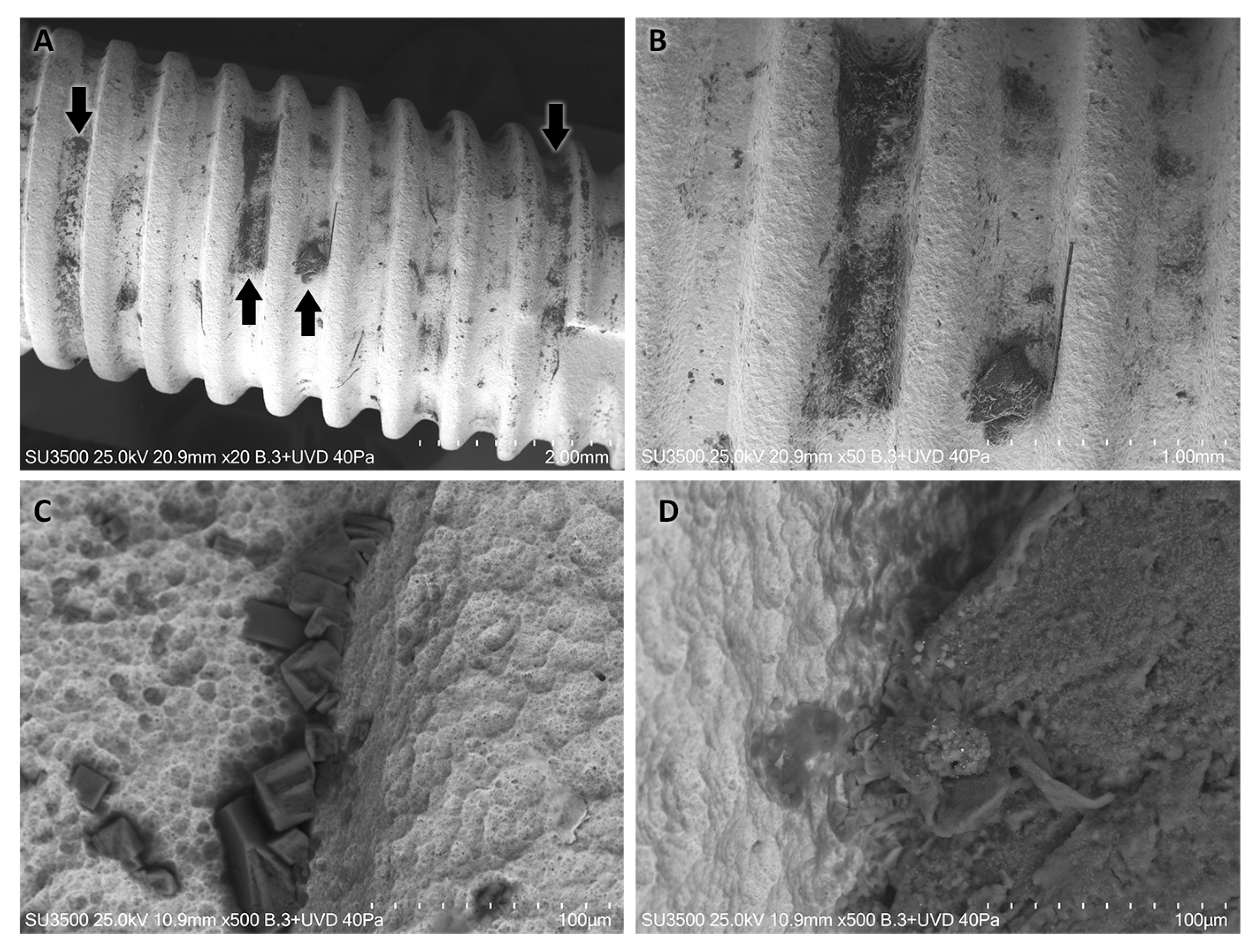
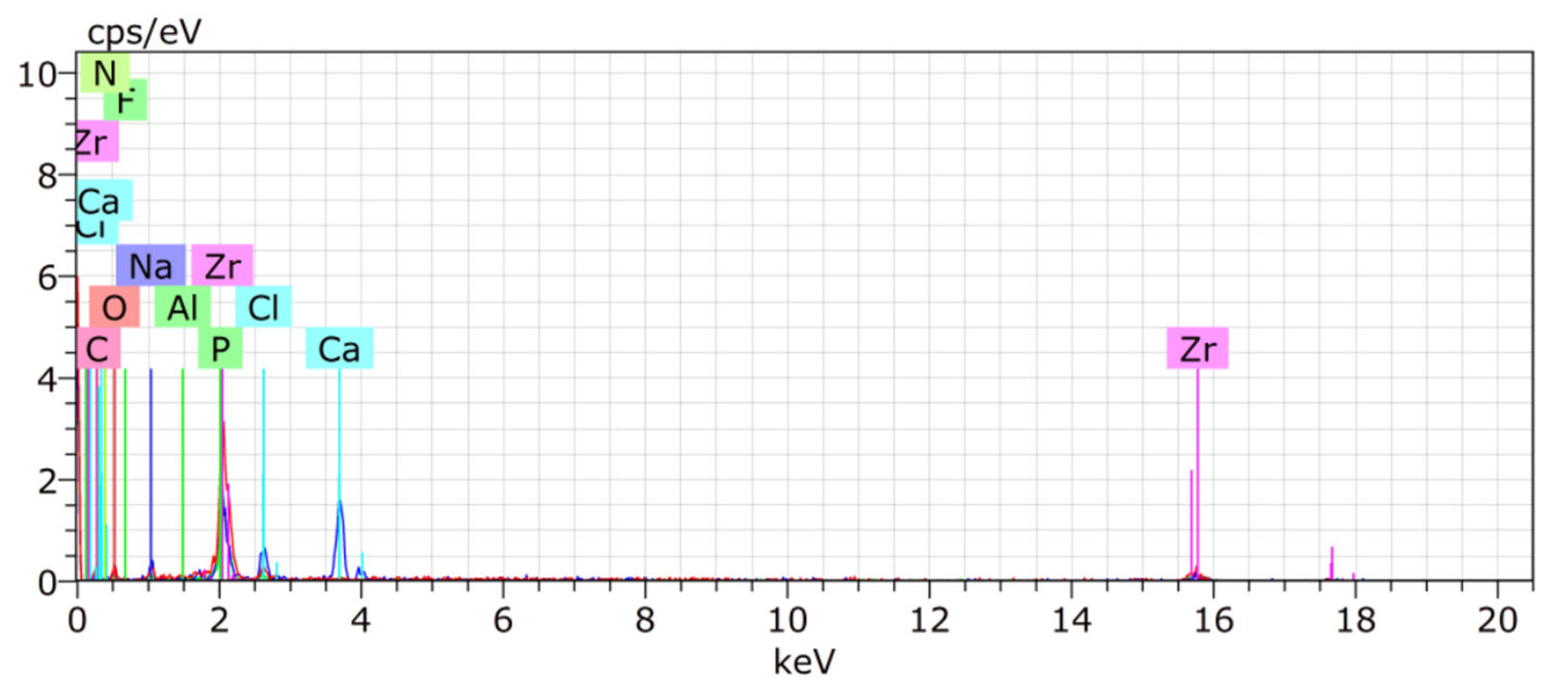
2. Case Report—Surface Characterization and Elemental Analysis of a Lost Zirconia Implant

A 50-year-old male patient with external root resorption in his upper right central incisor (tooth 1.1) requested implant treatment. The tooth had a history of trauma leading to avulsion and replantation (around 10 years ago), as well as root canal treatment associated with an external root resorption with a short root and increased mobility. The individual reported no significant medical condition.
The conditions above plus osseous anatomy allowed the insertion of a zirconia implant immediately after extraction, with a temporary restoration. The device used was a ceramic-injection-molded and double-sintered zirconia implant (Zi 3.75 mm × 13 mm, Neodent, Sao Luis, Brazil), restored with a polymethyl methacrylate crown built on a temporary cylinder, directly connected to the implant. The crown was delivered without occlusal contacts on centric occlusion and excursive mandibular positions. The patient was instructed to avoid biting food or objects with the implant during the first three post-insertion months.
In the third week, the patient overlooked the clinical instruction and used the restored implant to bite directly on hard food, thus leveraging the implant and leading to implant loss. The patient returned for a consultation scheduled at his demand to verify the implant, which felt loose according to him. Clinically, the implant had mobility under percussion and extrusion. The implant was then extracted by reverse torquing without major difficulties (Figure 1A). Then, the implants were rinsed with saline 0.9% for blood removal and washed with distilled water. We avoided other cleaning solutions and contact of instruments on the implant surface to prevent interference of any nature in subsequent element analysis.
Immediately after cleaning, the implant was visualized by VP-SEM equipment, and analyzed chemically by energy-dispersive X-ray spectrometry (EDX) using a 410-M detector (Bruker, Berlin, Germany) at 25 kV connected to the microscope. EDX is a technique that uses X-rays as a source of excitation for analyzing the chemical elements of different structures. This method is based on the principle that the unique atomic structure of each chemical element enables a single peak of electromagnetic emission after excitation [26]. Different areas of the dental implant were analyzed and divided into three sections (thirds T1, T2, T3). Two random measurements of each section were performed using the EDX system, which was set to automatically detect the elements on the surface. Data obtained represent the percentage of weight (wt.%).

3. Results

A panoramic showed the presence of sparse material depositions on all thirds of the implant (Figure 1B). Those depositions were remarkably concentrated in the thread grooves of the dental implant.
Greater magnification revealed a surface with macro- and micro-roughness, where material depositions presented different formats and characteristics. Areas of deposition in the form of an adhered film can be identified; in certain areas, more voluminous structures are suggestive of organic material adhered to the implant. In addition, the presence of crystal-shaped material depositions can be noted, which seems to be embedded mainly in the areas between the implant threads (internal corners) (Figure 2).
SEM imaging did not reveal fractures or irregularities in the structure of the implant. At higher magnifications, only a slightly rough surface was observed. There was no evidence of soft tissue on the implant surface. According to the EDX, the elements found in each sample third were summarized in Table 1. The most observed elements were zirconium (48.23%) and oxygen (16.25%), compatible with the ceramic implant composition. Other elements identified in the specimen surface were carbon (22.63%), calcium (7.97%) and phosphorus (3.34%). Some traces of elements in a lower percentage such as sodium (0.2%), chlorine (0.2%), aluminum (0.01%) and fluorine (0.01%) were observed (Figure 3).

4. Discussion

This case report demonstrates element deposition on a zirconia dental implant at the osseointegration time of 3 weeks. The results of the EDX and microscopy can be related to a process of early bone integration, as disclosed by the apposition of calcium and phosphate on the implanted surface, as well as by a high carbon content. The apparent absence of soft tissue on the implant surface is incompatible with fibro-integration or implant rejection.
The high concentration of carbon observed in the analyzed specimen is consistent with findings reported by Thomé et al. [7]. However, they found high levels of carbon and low levels of other elements, explaining that the surface could have absorbed hydrocarbons from the air atmosphere. We hypothesize that the carbon element identified in our specimen was related to the tissue matrix deposited during the whole process of implant integration to the surrounding bone.
The presence of calcium, phosphorus, and oxygen in the specimen can be attributed to the process of bone apposition on the implant surface, as these elements constitute the main components of the bone mineral matrix. The presence of zirconium is intuitive, as it is a constituent material of the implant itself.
No fracture lines were observed in the sample analyzed, contrasting with findings reported by Koller et al. [27] for many failure cases. However, the mechanical properties of zirconia implants depend on the manufacturing process [11], as well as their surface treatment by sandblasting, milling, and others [28], among other factors. In the past, commercial zirconia implants were produced by subtractive milling, which led to tetragonal-to-monoclinic phase transformation and the generation of compressive stress [29]. When zirconia changes from a tetragonal to a monoclinic phase, it suffers volumetric expansion and subsequent stress, which can lead to dental implant fracture in some cases [10]. One way to avoid these phase changes during manufacturing is the process of ceramic injection molding and sintering, as carried out for the specimen analyzed.
The analyzed zirconia implant by SEM exhibited characteristics of micro-roughness associated with cavities of 10–20 μm and material deposition. The description matches the surface treatment reported by the manufacturer/acid etching [30]. The roughness profile and material depositions concur with the findings of Thomé et al. [7] and indicate good potential for cellular adhesion and osseointegration [31,32].
The literature reports that zirconia implants reach a bone–implant contact similar to titanium [7], although this was not evaluated in this study, and the survival rate of zirconia two-pieced implants ranges between 83% and 99% [33,34,35]. Interestingly, newer studies report higher survival rates, linked to implants that are roughly similar to the specimen tested in this study.
SEM analysis did not identify areas suggestive of soft tissue on the zirconia implant surface, which would be compatible with fibro-integration or implant rejection. The main reason for implant loss was attributed to the patient’s lack of responsibility and the occlusal overload from the temporary restoration. Zhang et al. [11] concluded in their research that factors such as patient responsibility could contribute to clinical failure, in addition to treatment planning, implant surface, and design.
The main limitation of this study is that the analysis was conducted on a single implant from one individual. However, the elemental analysis remains significant as it represents the first clinical case in humans to describe the surface of a two-piece zirconia implant in the early stages of osseointegration.
Further studies performing similar EDX analyses on all two-piece zirconia implants that fail during the osseointegration period are necessary to determine whether the causes are biological, mechanical, or biomechanical, as observed in this case. Additionally, long-term studies are needed to assess the clinical performance of two-piece zirconia implants.

5. Conclusions

The recent improvements and latest findings in zirconia implants are encouraging, indicating that zirconia is a viable metal-free alternative to dental implants. However, the choice between zirconia and titanium implants depends on individual patient needs and responsibility, the location and load-bearing requirements of the implant, anatomical factors, and the clinician’s experience. This report demonstrates that the surface of zirconia implants can accumulate elements early in the process of bone matrix neoformation, which is compatible with the early stage of osseointegration.

Author Contributions

Conceptualization, E.B. and F.J.D.; methodology, F.J.D.; formal analysis, E.R.; investigation, E.B. and E.R.; data curation, F.J.D.; writing—original draft preparation, E.R.; writing—review and editing, E.B., F.J.D. and R.F.d.S.; visualization, E.B.; supervision, E.B. All authors have read and agreed to the published version of the manuscript.

Funding

This research received no external funding.

Institutional Review Board Statement

The study was conducted in accordance with the Declaration of Helsinki, and approved by the SSMO Ethics Committee (30 April 2022).

Informed Consent Statement

Written informed consent has been obtained from the patient to publish this paper.

Data Availability Statement

No new data were created or analyzed in this study.

Conflicts of Interest

The authors declare no conflicts of interest.

References

  1. Stadlinger, B.; Hennig, M.; Eckelt, U.; Kuhlisch, E.; Mai, R. Comparison of zirconia and titanium implants after a short healing period. A pilot study in minipigs. Int. J. Oral Maxillofac. Surg. 2010, 39, 585–592. [Google Scholar] [CrossRef]
  2. Kohal, R.J.; Spies, B.C.; Bauer, A.; Butz, F. One-piece zirconia oral implants for single-tooth replacement: Three-year results from a long-term prospective cohort study. J. Clin. Periodontol. 2018, 45, 114–124. [Google Scholar] [CrossRef] [PubMed]
  3. van Brakel, R.; Noordmans, H.J.; Frenken, J.; de Roode, R.; de Wit, G.C.; Cune, M.S. The effect of zirconia and titanium implant abutments on light reflection of the supporting soft tissues. Clin. Oral Implants Res. 2011, 22, 1172–1178. [Google Scholar] [CrossRef] [PubMed]
  4. Sivaraman, K.; Chopra, A.; Narayan, A.I.; Balakrishnan, D. Is zirconia a viable alternative to titanium for oral implant? A critical review. J. Prosthodont. Res. 2018, 62, 121–133. [Google Scholar] [CrossRef]
  5. Afrashtehfar, K.I.; Del Fabbro, M. Clinical performance of zirconia implants: A meta-review. J. Prosthet. Dent. 2020, 123, 419–426. [Google Scholar] [CrossRef] [PubMed]
  6. Bosshardt, D.D.; Chappuis, V.; Buser, D. Osseointegration of titanium, titanium alloy and zirconia dental implants: Current knowledge and open questions. Periodontol. 2000 2017, 73, 22–40. [Google Scholar] [CrossRef]
  7. Thome, G.; Sandgren, R.; Bernardes, S.; Trojan, L.; Warfving, N.; Bellon, B.; Pippenger, B.E. Osseointegration of a novel injection molded 2-piece ceramic dental implant: A study in minipigs. Clin. Oral Investig. 2021, 25, 603–615. [Google Scholar] [CrossRef]
  8. Gahlert, M.; Gudehus, T.; Eichhorn, S.; Steinhauser, E.; Kniha, H.; Erhardt, W. Biomechanical and histomorphometric comparison between zirconia implants with varying surface textures and a titanium implant in the maxilla of miniature pigs. Clin. Oral Implants Res. 2007, 18, 662–668. [Google Scholar] [CrossRef]
  9. Nishihara, H.; Haro Adanez, M.; Att, W. Current status of zirconia implants in dentistry: Preclinical tests. J. Prosthodont. Res. 2019, 63, 1–14. [Google Scholar] [CrossRef]
  10. Song, X.; Ding, Y.; Zhang, J.; Jiang, C.; Liu, Z.; Lin, C.; Zheng, W.; Zeng, Y. Thermophysical and mechanical properties of cubic, tetragonal and monoclinic ZrO2. J. Mater. Res. Technol. 2023, 23, 648–655. [Google Scholar] [CrossRef]
  11. Zhang, F.; Monzavi, M.; Li, M.; Cokic, S.; Manesh, A.; Nowzari, H.; Vleugels, J.; Van Meerbeek, B. Fracture analysis of one/two-piece clinically failed zirconia dental implants. Dent. Mater. 2022, 38, 1633–1647. [Google Scholar] [CrossRef]
  12. Pessanha-Andrade, M.; Sordi, M.B.; Henriques, B.; Silva, F.S.; Teughels, W.; Souza, J.C.M. Custom-made root-analogue zirconia implants: A scoping review on mechanical and biological benefits. J. Biomed. Mater. Res. B Appl. Biomater. 2018, 106, 2888–2900. [Google Scholar] [CrossRef] [PubMed]
  13. Yin, L.; Nakanishi, Y.; Alao, A.R.; Song, X.F.; Abduo, J.; Zhang, Y. A review of engineered zirconia surfaces in biomedical applications. Procedia CIRP 2017, 65, 284–290. [Google Scholar] [CrossRef] [PubMed]
  14. Leighton, Y.; Miranda, J.; Souza, R.F.D.; Weber, B.; Borie, E. A Novel, Minimally Invasive Method to Retrieve Failed Dental Implants in Elderly Patients. Appl. Sci. 2021, 11, 2766. [Google Scholar] [CrossRef]
  15. Andreiotelli, M.; Wenz, H.J.; Kohal, R.J. Are ceramic implants a viable alternative to titanium implants? A systematic literature review. Clin. Oral Implants Res. 2009, 20 (Suppl. S4), 32–47. [Google Scholar] [CrossRef] [PubMed]
  16. Jodha, K.S.; Salazar Marocho, S.M.; Scherrer, S.S.; Griggs, J.A. Fractal analysis at varying locations of clinically failed zirconia dental implants. Dent. Mater. 2020, 36, 1052–1058. [Google Scholar] [CrossRef] [PubMed]
  17. Scarano, A.; Di Carlo, F.; Quaranta, M.; Piattelli, A. Bone response to zirconia ceramic implants: An experimental study in rabbits. J. Oral Implantol. 2003, 29, 8–12. [Google Scholar] [CrossRef] [PubMed]
  18. Scarano, A.; Piattelli, M.; Caputi, S.; Favero, G.A.; Piattelli, A. Bacterial adhesion on commercially pure titanium and zirconium oxide disks: An in vivo human study. J. Periodontol. 2004, 75, 292–296. [Google Scholar] [CrossRef] [PubMed]
  19. Kohal, R.J.; Weng, D.; Bachle, M.; Strub, J.R. Loaded custom-made zirconia and titanium implants show similar osseointegration: An animal experiment. J. Periodontol. 2004, 75, 1262–1268. [Google Scholar] [CrossRef]
  20. Hempel, U.; Hefti, T.; Kalbacova, M.; Wolf-Brandstetter, C.; Dieter, P.; Schlottig, F. Response of osteoblast-like SAOS-2 cells to zirconia ceramics with different surface topographies. Clin. Oral Implants Res. 2010, 21, 174–181. [Google Scholar] [CrossRef]
  21. Cionca, N.; Hashim, D.; Mombelli, A. Zirconia dental implants: Where are we now, and where are we heading? Periodontol. 2000 2017, 73, 241–258. [Google Scholar] [CrossRef]
  22. Chiou, L.L.; Panariello, B.H.D.; Hamada, Y.; Gregory, R.L.; Blanchard, S.; Duarte, S. Comparison of In Vitro Biofilm Formation on Titanium and Zirconia Implants. Biomed. Res. Int. 2023, 2023, 8728499. [Google Scholar] [CrossRef]
  23. Al-Radha, A.S.; Dymock, D.; Younes, C.; O’Sullivan, D. Surface properties of titanium and zirconia dental implant materials and their effect on bacterial adhesion. J. Dent. 2012, 40, 146–153. [Google Scholar] [CrossRef]
  24. Degidi, M.; Artese, L.; Scarano, A.; Perrotti, V.; Gehrke, P.; Piattelli, A. Inflammatory infiltrate, microvessel density, nitric oxide synthase expression, vascular endothelial growth factor expression, and proliferative activity in peri-implant soft tissues around titanium and zirconium oxide healing caps. J. Periodontol. 2006, 77, 73–80. [Google Scholar] [CrossRef] [PubMed]
  25. Cesar, P.F.; Miranda, R.B.P.; Santos, K.F.; Scherrer, S.S.; Zhang, Y. Recent advances in dental zirconia: 15 years of material and processing evolution. Dent. Mater. 2024, 40, 824–836. [Google Scholar] [CrossRef]
  26. Goldstein, J.I.; Newbury, D.E.; Michael, J.R.; Ritchie, N.W.; Scott, J.H.J.; Joy, D.C. Scanning Electron Microscopy and X-ray Microanalysis, 3rd ed.; Springer: New York, NY, USA, 2017; pp. 209–234. [Google Scholar]
  27. Koller, M.; Steyer, E.; Theisen, K.; Stagnell, S.; Jakse, N.; Payer, M. Two-piece zirconia versus titanium implants after 80 months: Clinical outcomes from a prospective randomized pilot trial. Clin. Oral Implants Res. 2020, 31, 388–396. [Google Scholar] [CrossRef]
  28. Kim, H.K.; Yoo, K.W.; Kim, S.J.; Jung, C.H. Phase Transformations and Subsurface Changes in Three Dental Zirconia Grades after Sandblasting with Various Al2O3 Particle Sizes. Materials 2021, 14, 5321. [Google Scholar] [CrossRef] [PubMed]
  29. Mitra, N.; Vijayan, K.; Pramila Bai, B.N.; Biswas, S.K. Phase transformation introduced by mechanical and chemical surface preparations of tetragonal zirconia polycrystals. J. Am. Ceram. Soc. 1993, 76, 533–535. [Google Scholar] [CrossRef]
  30. Schunemann, F.H.; Galarraga-Vinueza, M.E.; Magini, R.; Fredel, M.; Silva, F.; Souza, J.C.M.; Zhang, Y.; Henriques, B. Zirconia surface modifications for implant dentistry. Mater. Sci. Eng. C Mater. Biol. Appl. 2019, 98, 1294–1305. [Google Scholar] [CrossRef]
  31. Hafezeqoran, A.; Koodaryan, R. Effect of Zirconia Dental Implant Surfaces on Bone Integration: A Systematic Review and Meta-Analysis. Biomed. Res. Int. 2017, 2017, 9246721. [Google Scholar] [CrossRef]
  32. Ding, Q.; Zhang, R.; Zhang, L.; Sun, Y.; Xie, Q. Effects of Different Microstructured Surfaces on the Osseointegration of CAD/CAM Zirconia Dental Implants: An Experimental Study in Rabbits. Int. J. Oral Maxillofac. Implants 2020, 35, 1113–1121. [Google Scholar] [CrossRef] [PubMed]
  33. Jank, S.; Hochgatterer, G. Success Rate of Two-Piece Zirconia Implants: A Retrospective Statistical Analysis. Implant Dent. 2016, 25, 193–198. [Google Scholar] [CrossRef] [PubMed]
  34. Hashim, D.; Cionca, N.; Courvoisier, D.S.; Mombelli, A. A systematic review of the clinical survival of zirconia implants. Clin. Oral Investig. 2016, 20, 1403–1417. [Google Scholar] [CrossRef] [PubMed]
  35. Cionca, N.; Hashim, D.; Mombelli, A. Two-piece zirconia implants supporting all-ceramic crowns: Six-year results of a prospective cohort study. Clin. Oral Implants Res. 2021, 32, 695–701. [Google Scholar] [CrossRef]
Figure 1. (A) Zirconia implant removed from the patient before EDX analysis. (B) Panoramic view of the Zr implant structure (Mag.: ×11).
Coatings 14 00995 g001
Figure 2. Analysis of the structure of the Zr implant under the variable pressure scanning electron microscope. (A) Arrows showing material deposition of implant surface (Mag.: ×11). (B) Presence of material deposition (dark) on the implant structure (clear) with different morphological characteristics (Mag.: ×50). (C) Material in the form of crystals embedded in the structure of the implant (Mag.: ×500). (D) Deposition of more voluminous material suggestive of organic material (Mag.: ×500).
Coatings 14 00995 g002
Figure 3. EDX spectra of the implant regions at 25 kV.
Coatings 14 00995 g003
Table 1. Semiquantitative elemental analysis of each third of the sample analyzed in EDX.
ZrOCCaPNaClAlF
T1141.5524.0324.985.551.470.2---
T1249.2916.3230.37------
T2148.0922.3418.34.442.38----
T2246.1319.5518.81.742.95-0.2--
T3153.8812.3821.4917.744.68----
T3250.4814.1724.19----0.010.01
Mean48.2316.9522.637.973.340.20.20.010.01
SD4.174.614.517.101.35----
Disclaimer/Publisher’s Note: The statements, opinions and data contained in all publications are solely those of the individual author(s) and contributor(s) and not of MDPI and/or the editor(s). MDPI and/or the editor(s) disclaim responsibility for any injury to people or property resulting from any ideas, methods, instructions or products referred to in the content.

