Review

Developments in Dental Implant Surface Modification

Faculty of Science and Technology, Institute of Materials Engineering, University of Silesia in Katowice, 75 Pułku Piechoty 1A, 41-500 Chorzów, Poland
*
Author to whom correspondence should be addressed.
Coatings 2025, 15(1), 109; https://doi.org/10.3390/coatings15010109
Submission received: 28 November 2024 / Revised: 4 January 2025 / Accepted: 17 January 2025 / Published: 19 January 2025
(This article belongs to the Special Issue Ceramic and Metallic Biomaterials. Application in Medical Sciences)

Abstract

:
The development of dental implants has significantly advanced due to technological innovations aimed at improving their performance and patient outcomes. This work presents key factors influencing the success of dental implants, including osseointegration, which is the direct connection between living bone and the implant surface, and the various surface modifications that enhance this process. This review highlights the importance of surface roughness, chemical composition, and the use of bioactive coatings to promote better integration with surrounding bone tissue. Innovations such as nanotechnology, 3D printing, and smart surfaces are paving the way for more effective and personalized dental implant solutions. This review underscores the importance of ongoing research and development to improve success rates, enhance patient comfort, and reduce healing times. It focuses on creating cost-effective, reliable methods that integrate multiple functions, such as combining antibacterial and osteoconductive properties to improve overall implant performance.

1. Introduction

The development of dental implants is driven by technological advancements [1,2,3], patient demand [4,5], aging population [4,6,7,8], cost considerations [9,10,11,12], very high success rates between 90% and 98% [13,14,15], market growth—which is expected to expand at a compound annual growth rate (CAGR) of 9.8% from 2024 to 2030, reaching USD 9.62 billion by 2030 [16]—and patient expectations [17,18,19,20]. These factors collectively underscore the importance of ongoing innovation in the field of dental implants.
Dental implant surface modification is a critical area of research aimed at enhancing the osseointegration and longevity of dental implants. This field has seen significant advancements in recent years, driven by the need to improve the biocompatibility, stability, and overall success rates of dental implants [1,13,14,15,21,22,23,24,25,26,27,28]. A graphical representation of data from a Scopus database search was created to illustrate the trend in publications related to dental implant surface modifications from 1975 to 2024 (Figure 1). The data indicate a timeline of publications spanning 49 years, highlighting the growth in research on this topic over time. The results shown in Figure 1 were the basis for a large analysis of trends in dental implant research, particularly concerning surface modifications, driven by the potential to improve implant performance and patient outcomes.
The analyzed studies highlight the need for more comprehensive long-term studies on dental implant surface modifications [29,30,31,32,33,34,35,36,37,38,39,40,41]. It was found that bioactive modifications, such as collagen-based coatings and combinations with bone morphogenetic protein-2 (BMP-2), aim to enhance osseointegration and implant longevity by improving biological properties for better bone integration [29,34,35,36,37,38,39]. Various biomolecular coatings, including bone morphogenetic proteins, growth factors, peptides, and extracellular matrix molecules, have shown early-stage benefits for bone formation and osseointegration [31,40,41]. The addition of bioactive molecules to titanium surfaces is a novel research area, with promising short-term results, but long-term clinical studies are needed to validate these benefits [31].
The reported results also show that laser modification is rapidly evolving as a physicochemical surface process for dental implants [42,43,44,45,46,47,48]. This technique creates complex surface topographies that enhance osseointegration and reduce bacterial colonization without affecting bulk properties [42,43,44,46]. Key parameters like repetition rate, pulse energy, scanning speed, and smoothness must be controlled to achieve the desired surface characteristics. Laser-modified surfaces promote better osseointegration, minimize bone loss, and allow the direct attachment of periodontal ligaments, acting as a barrier against bacterial invasion [42,43]. Additionally, laser modification improves optical, frictional, and biological properties, making implants more suitable for clinical applications [43]. Recent studies indicate that laser-modified surfaces significantly enhance early peri-implant bone healing and overall implant stability, increasing interest in their clinical use [44,45].
Previous research emphasizes that nanoengineering techniques modify dental implant surfaces at the nano-scale to influence osseointegration by altering surface features for better interaction with bone cells [30,37,49,50,51]. Ongoing research aims to develop new surface treatments, combining physical, chemical, and biological modifications to enhance implant surface properties [51].
This review article provides a comprehensive overview of current advancements and techniques in modifying dental implant surfaces. It details various surface treatments, including acid etching, plasma spraying, and coating applications, and their effects on biocompatibility, osseointegration, and clinical performance. The review updates readers on strategies to improve success rates and healing processes, emphasizing the importance of surface roughness and other properties in enhancing implant–bone interaction and reducing bacterial colonization.

2. Mechanism of the Osseointegration Process

The success of implant treatment highly depends on osseointegration, confirmed by implant immobility and the absence of inflammation [27,31,48,52,53,54,55,56,57,58,59]. Osseointegration is the direct morphological and functional connection between living bone tissue and the implant surface [59]. Key factors of osseointegration include the implantation technique, the primary stabilization of the implant in the embedded bone, bone quality, implant surface type, and loading method. Primary stabilization, influenced by bone density, implant shape, and surface type, is crucial for osseointegration [60,61,62,63].
Increased implant stability can be achieved by increasing the length or diameter of the implant, especially in lower-density bones [64,65,66,67,68,69]. Increasing implant diameter helps prevent bone resorption by reducing stress around the implant neck and better distributing forces on the alveolar bone. Biomechanically, threaded screw implants with various thread pitches optimize force transfer [70]. Implant surface topography also affects stabilization [70]. Rough surfaces with numerous micro-hooks improve anchoring and force distribution compared to smooth surfaces. They facilitate easier force transfer acting on the bone through the prosthetic structure and promote bone growth on the implant surface [71,72,73,74,75,76,77]. Despite the high clinical success of titanium dental implants, the cellular and molecular mechanisms behind osseointegration are not fully understood, mainly due to limited methodological tools in this field [78,79,80,81,82]. Titanium is now seen as an immunomodulatory biomaterial, as its implantation triggers a transient inflammatory state that may activate the immune pathways involved in osseointegration [83,84,85,86,87,88].
Research on osseointegration primarily focuses on surface modifications of titanium endosseous implants to enhance fusion with bone tissue [78,89,90]. In vitro studies examine surface topography, structure, and chemical composition to improve bone cell differentiation and mineralization, but are limited by the intrinsic characteristics of cell cultures, which do not fully simulate in vivo interactions. Preclinical evaluations are often conducted in animals with robust skeletal structures, such as minipigs and dogs, which can mimic human craniofacial bone architecture. However, these models have limitations, including size, weight, and a lack of specific experimental tools, making it challenging to understand the biological basis of osseointegration [66,91,92,93,94,95,96,97,98,99,100,101].
The osseointegration process of a titanium dental implant in the edentulous space between the maxillary right first molar and incisor along the alveolar crest in mice was studied at 3, 7, 14, and 21 days post-implantation [94], as shown in Figure 2. Molecular tests were conducted on 48 one-week-old male wild-type C57Bl/6 mice with an average weight of 25 g. Osseointegration kinetics identified the key elements responsible for cell migration, proliferation, mesenchymal stem cell (MSC) deposition, maturation, angiogenesis, bone formation, and bone remodeling at the implant–bone interface. Biguetti et al. [94] found that osseointegration in C57Bl/6 mice involves multiple stages, including the host’s immune inflammatory response, bone cell differentiation, new bone formation and maturation on the titanium surface, and bone remodeling, which overlap during the process.
Bone healing and osseointegration are complex processes involving inflammation, cell migration and differentiation, matrix formation and mineralization, and remodeling, ultimately leading to stable implant anchorage [94,95,96,97,98,99,100,101,102,103,104,105,106,107,108,109,110,111,112,113,114,115,116,117,118,119,120,121,122,123,124,125,126]. After surgical trauma, bone healing begins with an acute inflammatory reaction, characterized by the release of inflammatory mediators like histamine and prostaglandins, increasing vascular permeability and attracting neutrophils to the site [94,102,103,104]. This phase includes blood clot formation, which serves as a scaffold for healing.
The disruption of blood vessels and bone nutrition leads to local necrosis and inflammation. Micro-cracks from surgery stimulate bone healing by releasing factors like TGF-β, VEGF-B, and various cytokines [94,105,106,107,108,109]. Bone morphogenetic proteins (BMPs) are crucial, inducing the differentiation of cells into osteoblasts and initiating bone formation. A temporary granulation tissue rich in growth factors forms, and the pH around the implant initially decreases but returns to normal [94,110,111,112].
The blood clot organizes into a protein network, forming young connective tissue on the implant surface. Platelet activation influences the expression of growth factors and cytokines, promoting new blood vessel formation, cell migration, and differentiation into osteoblasts [94,113,114,115,116]. Approximately 7 days post-surgery, bone-forming cells secrete an osteoid matrix, which matures and mineralizes over time, replacing the initial repair bone with mature lamellar bone [94].
The remodeling phase involves bone resorption and apposition, strengthening the repair bone and increasing the bone–implant contact (BIC) coefficient [117,118,119]. Osteoclasts and precursor cells become active, regulated by the RANKL/RANK/OPG signaling pathway [94,120,121,122,123]. In challenging cases, stem cells and bone-forming agents like BMPs and platelet-rich plasma (PRP) can accelerate healing and enable immediate implant loading [94,124,125,126].
Surface modifications of dental implants are proposed to accelerate their osseointegration and increase their biological activity [28,127,128,129,130,131,132]. The roughness of the implant surface plays a crucial role in the initial stability and long-term success of the implant. While moderate roughness can enhance osseointegration by promoting better bone–implant contact, excessive roughness can increase the risk of bacterial colonization and subsequent infection.

3. Types of Dental Implant Surface

3.1. Machined Surface

In the 1970s, titanium dental implants with a smooth (machined) surface were introduced to the dental market (Figure 3 [133]). The machined surface of dental implants is characterized by several key features that influence their clinical performance and integration with bone tissue. However, machined surfaces are relatively smooth compared to other types of implant surfaces, and due to the low development of the surface topography, show the lowest degree of osseointegration [129,134,135,136,137,138,139,140]. This smoothness is achieved through mechanical processes such as turning or milling, which result in a uniform and polished surface finish [135,136]. The roughness of machined surfaces is typically low, which can be quantified using parameters such as Ra (average roughness). This low roughness can affect the initial interaction between the implant and bone tissue, influencing the rate and quality of osseointegration [137,138,139]. The arithmetic mean deviation of the roughness profile (Ra) for implants with a machined surface obtained by machining ranges from 0.8 to 1.2 μm. It should be noted that the values of surface roughness parameters like Ra and Rq depend on the measurement length (or area) and the technique used. It is a common misconception to treat these values as “absolute numbers” without specifying the measurement conditions, which can lead to misunderstandings. To ensure accurate interpretation and comparison, always include the length or area over which the roughness parameters were measured and the specific method or equipment used. Different lengths can yield different values, and comparing data obtained with different techniques can lead to incorrect conclusions. Including this essential information when presenting roughness data helps us to understand the context and limitations of the provided values [141].
The machined surface of titanium implants is isometric, anisotropic, and characterized by the presence of parallel grooves with a width of about 10 μm [129]. The thickness of the TiO2 layer on the machined surface is approximately 17 nm. Machined implants have the lowest BIC of approximately 15%, which is associated with an increased risk of implant failure [139]. The wettability of machined surfaces can vary, but they generally exhibit moderate hydrophilicity. This property is important as it affects the initial blood clot formation and subsequent cellular attachment, which are crucial steps in the osseointegration process [138]. While machined surfaces are generally smooth, they can still possess micro- and nano-topographical features that can influence cell behavior and tissue integration. These features, though subtle, can modulate the early biological response to the implant. Machined surfaces are typically made of biocompatible materials such as titanium or titanium alloys, which are known for their good biocompatibility and corrosion resistance. This ensures that the implant does not cause adverse reactions within the body [140]. Machined surfaces have been used in dental implants for many years and have shown good clinical performance. They are often used in situations where a smooth surface is preferred, such as in areas with high aesthetic demands or in patients with specific medical conditions [142].
Examples of machined titanium dental implants include the Brånemark Standard Implants system by Nobel Biocare, the Restore Machined Implants system by Lifecore Dental, and the Machined WINSIX Implants system by BioSAF IN [143,144]. The Brånemark Standard Implants system is known for its parallel-walled design, which is recommended for all bone qualities. The implants feature a machined collar of 0.8 mm for various sizes, such as NP 3.3 and RP 3.75 [143]. The Restore Machined Implants system utilizes machined titanium implants designed to provide a smooth surface that promotes bone integration and stability [144]. The Machined WINSIX Implants system also employs machined titanium implants, focusing on achieving optimal bone-to-implant contact and long-term success [144].

3.2. Titanium Plasma Surface

The first modification of the surface of titanium implants performed in 1976 was their coating with titanium plasma (TPS), produced by the thermal spraying of titanium powder in a plasma stream [145,146]. The TPS surface of dental implants is characterized by several key features that enhance their biological performance and integration with bone tissue. The plasma treatment creates a roughened surface with micro- and nano-topographical features. The roughness of Ra is typically in the range of 20 to 30 μm, which is achieved by depositing a porous titanium coating using plasma spray technology. The thickness of the TPS layer is from 30 to 40 μm. The TPS surface is highly porous, which increases the surface area available for bone tissue interaction. This porosity is crucial for promoting osseointegration, as it allows for better bone ingrowth and mechanical interlocking with the implant [78,145,146,147,148,149]. The TPS surface is characterized by numerous undercuts and intrusions (Figure 4 [148]). Implants covered with a TPS layer increase the BIC coefficient by 6 times in comparison with implants with a machined surface [150]. The increase in the development of the implant surface with the TPS surface improves the implant–bone tissue connection, and the roughness being 10 times greater than in the case of the machined surface causes faster bone resorption around the implant during long-term use. The plasma treatment can improve the wettability of the titanium surface, making it more hydrophilic. This property is beneficial for the initial blood clot formation and subsequent cellular attachment, which are critical steps in the osseointegration process [151]. Titanium and its alloys are inherently biocompatible, and the plasma treatment further enhances this property. The resulting surface is able to bond with osteoblasts, promoting bone formation and integration with the surrounding bone tissue [151]. The plasma-coated surface can improve the mechanical properties of the implant, such as its wear resistance and fatigue strength. This is particularly important for ensuring the long-term stability and durability of the implant [152]. Some plasma treatments can incorporate antibacterial elements, such as silver nanoparticles, which can reduce the risk of infection and improve the overall success rate of the implant [153].
Titanium dental implants with a TPS surface are manufactured by Densply Friadent in the IMZ TPS system [154], Straumann Institute in the Bonefit system [155], Lifecore Dental in the Restore TPS system [156], Nobel Biocare in the Steri-Oss TPS system [157], and Spotimplant in the IMZ Original system [148]. The implants in the IMZ TPS system are designed to mimic the shape of natural tooth roots, which helps in achieving a more natural and secure fit during the implantation process. This system includes various types of implants, such as cylinder and screw implants, which can be used in different clinical scenarios to meet the specific needs of patients. The implants in the IMZ TPS system feature an intermobile element that acts as a stress buffer between the implant and the surrounding bone, which helps in distributing the forces more evenly and reducing the risk of implant failure. The Bonefit system was introduced in 1986 as a two-piece implant system with a smooth transgingival shoulder, which was a significant advancement in soft-tissue-level implant design [155]. The Straumann Research Institute has been involved in the development and continuous improvement of the Bonefit system. The Restore TPS system is a well-documented and FDA-approved dental implant system known for its titanium plasma spray coating, external hexagon connection, and compatibility with various abutments [156]. It has been evaluated positively in long-term studies. The Restore TPS system is a bone-level implant, meaning the implant is placed at the bone level, and the abutment is connected to the implant after the healing period. This system is compatible with various abutments, including temporary abutments with a textured surface for acrylic, available in locking or non-locking options for single and multiple-unit restorations. The Steri-Oss TPS system includes both bone-level and tissue-level implants. These implants can be non-threaded or threaded, and they come in various configurations, such as Hex-Loc Cylindrical Non-threaded and Original Non-threaded. The TPS coating promotes bone growth and integration with the implant, which is crucial for the long-term success of dental implants. Steri-Oss was founded in 1985 and was shut down in 1997. However, the company was acquired by Nobel Biocare in 1998, and the Steri-Oss TPS system continued to be developed and marketed under Nobel Biocare’s brand [157]. The IMZ Original system by Spotimplant is a standard dental implant designed for tissue-level placement [148]. The connection between the implant and the abutment is internal, and it has a non-anti-rotational shape, which means the abutment fits inside the implant without any mechanism to prevent rotation. The implant body is straight and does not have threads. It includes a shock-absorbing intermobile element, which helps in distributing forces and reducing stress on the bone. IMZ Original is a versatile and reliable option for dental implant procedures, suitable for both partially and totally edentulous arches.

3.3. Sandblasted Surface

In a significant group of dental implants, a sandblasted surface is used to enhance osseointegration by increasing surface roughness, promoting better bone-to-implant contact, improving mechanical stability, and reducing the risk of implant failure [28,53,54,74,90,129,134,158,159,160,161,162,163,164,165,166,167,168,169]. During the sandblasting process, the implant surface is bombarded with abrasive particles, most often noble corundum with grain sizes from 25 to 250 μm. It has been shown that the optimal surface roughness of Ra 1–3 μm, which provides the best osseointegration effects, is obtained when sandblasting with Al2O3 particles with a size of 25–75 μm [129,159]. The type and size of the abrasive are important, in addition to the shape of its particles [160]. The highest BIC coefficient is obtained using Al2O3 particles with a sharp shape. After the sandblasting process using noble corundum, a small amount of residual Al2O3 is observed on the titanium surface, which may hinder bone healing. On the other hand, recent progress in dental implant research has shown that negative static charges are generated on the surface of titanium implants with embedded Al2O3 particles, which support the osseointegration process as a result of the selective activation of osteoblasts and the inhibition of fibroblasts [161]. However, the static charges accumulated on the titanium surface during sandblasting disappear over time. The challenge is to find ways to maintain the stability of these charges after quantifying the desired level of negative charges needed to stimulate osteoblast activity in the process of osseointegration around dental implants. Alumina does not dissolve in acidic solutions, such as nitric acid baths used for passivation. However, blasting residues on the surface can be mostly removed when using acids that dissolve titanium, like hydrofluoric acid, after the sandblasting process. A blasting material made from phosphate ceramicis is called Resorbable Blasting Medium (RBM) [162]. This means that any particles left on the surface after blasting can be dissolved by acidic baths, making it easy to remove the blasting residues. In the sandblasting process, TiO2 is used with grain sizes from 10 to 125 μm. Hydroxyapatite (HA), or other forms of calcium phosphate, e.g., tricalcium phosphate (β-tricalcium phosphate) [162], is also used. The increase in the grain size of the abrasive affects the increase in surface roughness. Sandblasted titanium implants are characterized by an irregular, isotropic surface, the development of which is about 34% greater in comparison with the machined surface [163]. The thickness of the TiO2 layer on the surface of sandblasted titanium implants is about 2–5 nm [161,164].
Figure 5a shows a sandblasted dental implant of the Prima Plus 4.1 (RD) model by Lifecore Dental, which features a tapered implant body with threaded features, including reverse buttress, V-shaped, and square threads [165].
The Prima system is a versatile and flexible dental implant system that offers a range of implant body shapes, thread types, and connection options. It includes both one-piece and two-piece implants, making it suitable for various clinical applications. This system’s compatibility with different prosthetic attachments further enhances its utility in dental treatments.
Currently, titanium implants with a surface sandblasted with noble Al2O3 are manufactured by the Polish company Osteoplant Research and Development in the Standard and Hex systems [166], using TiO2 by Astra Tech Dental in the TiOblast system [167,168], and using HA by Lifecore Dental in the Renova [169] and Prima [165] systems.

3.4. Hydroxyapatite Surface

With the development of implant prosthetics, titanium implants with an HA surface covered with a 50 μm thick HA layer and a roughness of Ra of about 8.2–10.2 μm have appeared on the global market [53,54,74,90,134,162,170,171,172,173,174,175,176,177,178,179,180,181]. Anisotropic HA layers on the surface of titanium implants bonding with living bone can be obtained by electrochemical deposition, involving the deposition of HA through an electrochemical process [170,173]; the electrophoretic deposition method, using an electric field to deposit HA particles onto the titanium surface [173,174]; the sol–gel method, where a gel-like precursor is formed and then converted into a solid material [171,172]; pyroprocessing and hydroprocessing, where hydroprocessing involves the deposition of HA on a titanium substrate using a hydrothermal process [175]; and biomimetic deposition, which mimics the natural process of bone formation to deposit HA on the titanium surface, enhancing biocompatibility and osseointegration [176].
Titanium implants covered with an HA layer enable faster implant–bone tissue connection compared to implants with a machined, sandblasted, or TPS surface; however, the high temperature necessary to sinter the ceramic layer with the metal substrate causes changes in the HA phase. These changes lead to the degradation of the HA layer in the body and the reduced biocompatibility of implants [177]. Thermal treatment also causes changes in the titanium structure and a decrease in the bond strength of the implant with the HA layer. Currently, HA layers obtained in amorphous or crystalline form are sought, which will not require sintering with the substrate and will reduce the risk of bone tissue loss during osseointegration [28].
The dental implant type 3i T3 by BIOMET 3i shown in Figure 6a utilizes Discrete Crystalline Deposition (DCD) technology, which is a sophisticated surface modification technique that enhances the osseointegration of dental and medical implants by depositing nanometer-sized HA crystals (Figure 6c) [162]. These features are intended to help retain blood clots along the threaded part of the implant [178]. The discrete nature of the crystal deposition allows for precise control over the surface topography, which can be tailored to specific biomechanical requirements. The DCD process can be combined with other surface modification techniques such as microblasting, acid etching, and anodization to further enhance the implant’s performance.
Another example of titanium dental implants with an HA surface is the IMZ HA system by Densply Friadent, which incorporates an intramobile element (IME) designed to simulate the viscoelasticity of the periodontal ligament, thereby reducing stress on the surrounding bone and improving the long-term outcome of the implant. This system is known for its ability to mimic natural tooth movement and enhance the stability and longevity of dental implants [179]. The Nobel Replace External Hex (Steri-Oss) is a standard dental implant covered with an HA layer produced by Nobel Biocare, featuring a tissue-level implant with an external hexagon connection and a tapered body with buttress threads [181].

3.5. Double-Etched Surface

The double-etched (DE) surface of titanium implants is obtained by two acid etching steps with a mixture of HCl + H2SO4 or HF + HNO3 acids, and is characterized by several key features that enhance their performance and integration with bone tissue [90,134,182,183,184,185,186,187,188,189,190,191]. The DE process creates a micro-rough surface on the titanium implant, which is crucial for improving osseointegration. The Ra of the DE surface is typically in the range of 1–2 μm, which has been shown to enhance bone–implant contact and mechanical interlocking with the surrounding bone [182,183,184,185]. The micro-rough surface generated by the DE process promotes better osseointegration by increasing surface roughness, enhancing cell adhesion and proliferation, mimicking natural bone environments, improving biocompatibility, and accelerating bone formation and healing [183,184,185]. The DE process creates a micro-rough surface with a hierarchical micro- and submicron-scale structure. This increased roughness provides a larger surface area for cell attachment and proliferation, which is crucial for the initial stages of osseointegration. The micro-rough surface has been shown to enhance the adhesion, proliferation, and differentiation of osteoblasts (bone-forming cells). This is because the rough surface mimics the natural extracellular matrix, providing a more favorable environment for cell attachment and growth. The micro-rough surface can also simulate the natural bone environment, which helps in guiding the formation of the bone matrix. This biomimetic property accelerates the bone healing process and promotes a stronger bond between the implant and the bone. The DE process not only increases surface roughness, but also modifies the surface chemistry of the titanium implant. This can enhance the biocompatibility of the implant, reducing the risk of adverse reactions and improving the overall integration with the bone. The micro-rough surface facilitates quicker bone formation and healing by providing an optimal environment for osteoblast activity. This results in a faster and more robust osseointegration process, allowing for earlier loading and functional use of the implant [192]. Interestingly, the BIC coefficient of the DE surface is higher both in comparison to less-rough machined surfaces and rougher surfaces, such as TPS, HA, and sandblasted. This means that the characteristic microstructure of the DE surface has the strongest osteoconductive properties and influences the increased adhesion of bone-forming cells, protein adsorption, and the stimulation of angiogenesis, facilitating the binding of fibrin clots [182].
However, the DE surface is obtained as a result of etching with a mixture of aggressive acids, which can cause fluoride ions to remain in the resulting cavities. Fluoride ions are retained on the surface of a dental implant through a combination of surface adsorption, ion exchange reactions, chemical reactions with surface atoms, surface modifications, and the formation of insoluble fluoride compounds. These processes are facilitated by the chemical reactivity of the surface and the nature of the etching process. Acid etching can modify the surface of the material, making it more reactive. This increased reactivity can facilitate the retention of fluoride ions through enhanced chemical reactions. The etching process can create a more porous surface with a higher surface area, which can trap fluoride ions through physical adsorption and chemical bonding. The retention of fluoride ions on the surface of titanium dental implants during a DE process is generally considered a positive phenomenon due to the resulting enhanced surface properties, improved corrosion resistance, promotion of osseointegration, antibacterial properties, and beneficial surface modification. However, careful control of the process is necessary to avoid potential negative effects associated with excessive fluoride, which can potentially lead to toxicity issues [186].
The DE surface treatment results in the improved mechanical stability of implants. This is particularly beneficial in challenging clinical scenarios where the bone quality is poor, as it helps in achieving better initial and secondary stability [183,185]. Some studies have indicated that the DE surface can also exhibit antibacterial properties, which can help in reducing the risk of infection around the implant. This is an additional benefit that contributes to the overall success of the implant [186]. The DE process creates submicron and nanometer-scale cavities on the surface. These cavities mimic the bone structure, further enhancing the biological response and integration with the bone [184]. Implants with a DE surface have shown excellent clinical success rates, with high survival rates and minimal marginal bone loss over extended periods. This has been observed in long-term clinical evaluations, demonstrating the effectiveness of the DE surface treatment [185].
Figure 7a shows the Osseotite dental implant system developed by Biomet 3i, part of Zimmer Biomet, now belonging to ZimVie [187]. The Osseotite implant features a DE surface that is designed to improve clot/implant attachment, potentially increasing platelet activation and red blood cell adherence. The SEM image of this surface presented in Figure 7b reveals its isotropic nature and the presence of numerous elevations and depressions, which facilitate bone ingrowth [188]. Figure 7c shows an enhanced microscopy image of the Osseotite surface with platelet activation [189].
The Osseotite implant is one of the most well-researched dental implants, with studies showing cumulative success rates of up to 98%. It has demonstrated effectiveness in various clinical scenarios, including immediate loading, and human histologic data confirm enhanced osteoconduction and contact osteogenesis, promoting bone healing and integration. Suitable for single-tooth, multiple-tooth, and full-arch replacements, its design ensures adequate primary stability and appropriate occlusal loading, which are essential for successful integration [187,188,189].
Another example of a titanium dental implant with a DE surface is the Steri-Oss Etched implant by Nobel Biocare [190,191]. The Steri-Oss Etched implant is suitable for various dental applications, including single-tooth replacements, multiple-tooth replacements, and full-arch restorations. During the healing phase, titanium healing abutments are used to prevent soft tissue from closing over the implant and to record the height and position of each healing abutment [191].

3.6. Sandblasted and Etched Surface

The sandblasted, large-grit, acid-etched (SLA) surface is a type of surface treatment commonly used in dental implants to enhance osseointegration [78,90,134,193,194,195,196,197,198,199,200,201,202,203]. SLA is a registered trademark of the Straumann Institute, and the SLA surface was introduced to the market by Straumann in 1997 [204]. The SLA process involves two main steps: large-grit sandblasting followed by acid etching. This combination creates a surface with both macro- and micro-roughness. The sandblasting step uses large particles to create a macro-rough surface with large pores and sharp edges, which increases the surface area for bone contact. In the sandblasting process of the SLA-type surface, typically Al2O3 (250–500 µm) [194], TiO2, or HA are used as abrasives [193,194,195,196,197].
The acid etching step further refines the surface, creating micro-pits and a micro-rough texture that enhances the surface’s ability to retain bone cells and promote bone growth. The resulting surface topography includes large dips, sharp edges, and small micro-pits. This complex structure provides an ideal environment for bone cells to adhere and proliferate [193]. The Ra values typically range around 1.5 µm, which indicates a higher roughness than machined and DE surfaces, and has been shown to improve osseointegration [78,193]. The SLA surface has a thicker oxide layer than machined and DE surfaces, consisting mainly of TiO2, TiO, and Ti2O3 [193]. The developed and differentiated SLA surface has increased protein adsorption, and the BIC coefficient increases compared to the machined surface [159]. The process significantly improves the implant’s surface hydrophilicity, which is crucial for bone cell adhesion and growth. SLA surfaces have been widely used and studied in clinical settings, demonstrating improved implant stability and reduced early failure rates compared to smoother surfaces [198]. The surface characteristics allow for the early loading of implants, which can reduce overall treatment time and improve patient outcomes.
Figure 8a shows a dental implant of the Standard Implants type by the Straumann Institute. This is a type of dental implant with an SLA surface designed for tissue-level placement [198].
Straumann Standard implants are particularly recommended for use in patients whose prosthetic solution will be based on a retention system, such as a bar, ball locks, or clasps. In such a situation, the aesthetics of the soft tissue around the implant are not crucial, and the high neck makes it easier for the patient to maintain hygiene around the implant. The implant collar has an optimized conical shape that allows for the use of a one-step clinical protocol. The implant is placed intragingivally and closed with a healing screw immediately after implantation, which allows avoiding the procedure of re-exposing the implant. The optimal thread shape ensures optimal primary and secondary stability [198]. The SLA surface of dental implants is isotropic and has a characteristic micro-topography, with wide craters of 20–40 µm in diameter (Figure 8b,c [134]). The craters created in the sandblasting process contain microcavities of 0.5–3 µm in diameter and nanofeatures that are formed as a result of etching [78,134]. The Al2O3 sandblasting process and etching with a HCl + H2SO4 mixture are used in the production of Standard Implants, Standard Plus Implants, and Tapered Effect Implants by the Straumann Institute [134,198,200], as well as SPI [201] and DFI [202] systems by Alpha Bio.

3.7. Hydrophilic Surface

The hydrophilic surface (SLActive) is obtained in the same way as the SLA surface, but after the etching process, the implants are dried in a nitrogen atmosphere and then kept in a physiological saline solution to protect from hydrocarbon adsorption [78,90,134,205,206,207,208,209,210,211]. The surface hydrophilicity of SLActive is primarily due to its unique surface treatment process. This process preserves the high surface energy and chemical purity of the implant, resulting in superhydrophilic properties. The hydrophilic nature allows for the rapid attachment of blood and proteins, which accelerates the osseointegration process and reduces the healing time to 3–4 weeks [212]. A clinical study comparing conventional SLA and SLActive implants found that there is generally no significant difference in clinical outcomes between the two types of implants. The cumulative survival rate was 99.4% overall, with 99.1% for SLA implants and 100% for SLActive implants. Additionally, the mean marginal bone resorption for all implants was similar, indicating comparable clinical performance between the two types of implants [213]. In the case of the SLA surface, the water contact angle is 139.90°, which indicates its ultrahydrophobic properties and lack of bioadhesion (Figure 9a) [207]. The reflections visible under a drop of water on the SLA surface result from the presence of air bubbles trapped between the water and the implant surface. The initial water wetting angle for the SLActive surface is 0°, which indicates its superhydrophilic properties and very strong bioadhesion (Figure 9b) [207]. After immersing SLA and SLActive dental implants in water, a meniscus is visible at the water–air–implant interface only in the case of the SLActive surface, due to its superhydrophilicity (Figure 9b) [207].
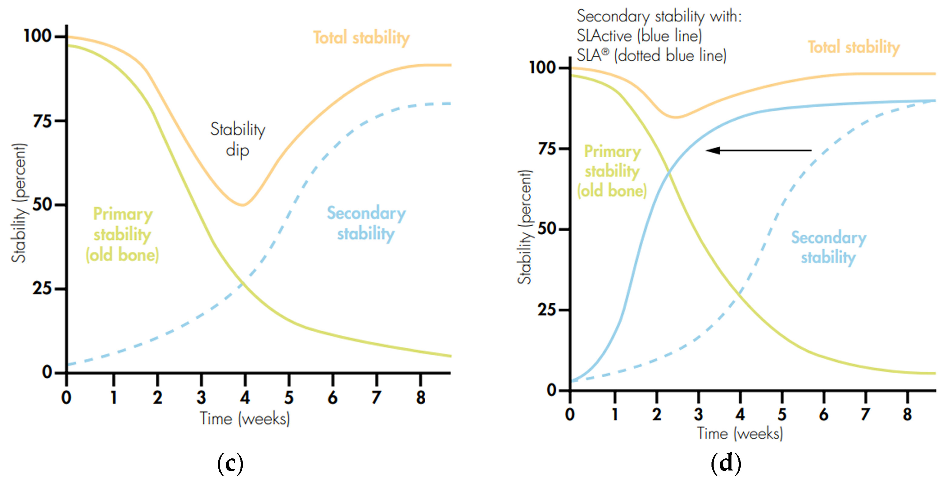
In the osseointegration process, two key factors are crucial for the success of dental implants: primary stability and secondary stability (Figure 9c) [207]. Primary stability refers to the mechanical stability of the implant immediately after placement. It is influenced by the quality of the bone where the implant is inserted and is determined by the specific design features of the implant system, such as thread pitch, the precision of the implant’s dimensions, and accurate sizing, ensuring a snug fit in the bony socket’s corresponding drills. Secondary stability relates to the biological healing process that occurs after the implant is placed. Unlike primary stability, secondary stability is not directly controlled by the clinician. It develops over time as the bone integrates with the implant, which is influenced by various factors, the most critical being the speed of osseointegration. The surface texture and composition of the implant can significantly enhance the rate at which this integration occurs. The combination of primary and secondary stability is known as total stability. It is important to note that a delay in the healing process can lead to a notable decrease in total stability, particularly between weeks 2 and 4 post-implantation. This period, often referred to as the “stability dip,” is critical for the success of the osseointegration process, as compromised stability during this time can affect the long-term success of the implant. Increasing the hydrophilicity of the SLActive surface and maintaining high surface energy until the moment of implantation affects the stability of the implant after implantation (Figure 9d) [207]. The SLActive implant surface is chemically active and reaches maximum secondary stabilization twice as fast as the implant with the SLA surface, which minimizes the risk of implant loss or loosening. The increase in secondary stabilization is due to the fact that in the early stages of bone tissue regeneration, the adsorption of proteins on the SLActive surface subjected to constant moistening is facilitated, which accelerates the process of new bone formation and makes it possible to place a prosthetic reconstruction even on the day of implantation. In the case of other types of surfaces, the placement of the proper crown usually takes place about eight weeks after the procedure, when about 75% of implant anchorage is achieved [207,208]. The non-activated SLA surface is less stable, which makes it susceptible to strong mechanical stress and post-implantation complications, or loosening of the implant during mechanical loading. The chemical composition of the SLActive surface shows more than twice the carbon content of the SLA surface [208]. An increased BIC coefficient has also been shown for the SLActive surface [207,208]. Considering the fact that the SLActive and SLA surfaces do not differ in terms of roughness, the acceleration of osseointegration in the case of the SLActive surface is due to its increased hydrophilicity.
It should be stressed that the wettability of dental implant surfaces is a critical factor that influences their interaction with water or body fluids. This property is not constant and is primarily determined by the surface chemistry of the dental implants, which are most often made of titanium. Titanium and its self-passive oxide layer are high-energy surfaces, inherently making them hydrophilic, similar to sandblasted or acid-etched surfaces. However, the absorption of hydrocarbons from the atmosphere can alter the surface properties, causing them to transition from hydrophilic to hydrophobic. This change in surface wettability can significantly impact the osseointegration process. Hydrophilic surfaces tend to adsorb plasma proteins more rapidly, which promotes clot formation, initiates healing, and supports bone regeneration. Consequently, it is suggested that dental implants be stored under controlled conditions, such as in oxygen-free atmospheres or liquids, to mitigate the biological aging process and maintain their desired surface properties. The wettability of dental implant surfaces is crucial for their performance and integration with the bone. Ensuring that these surfaces remain hydrophilic through proper storage and handling can enhance the overall success of dental implants [207,208].
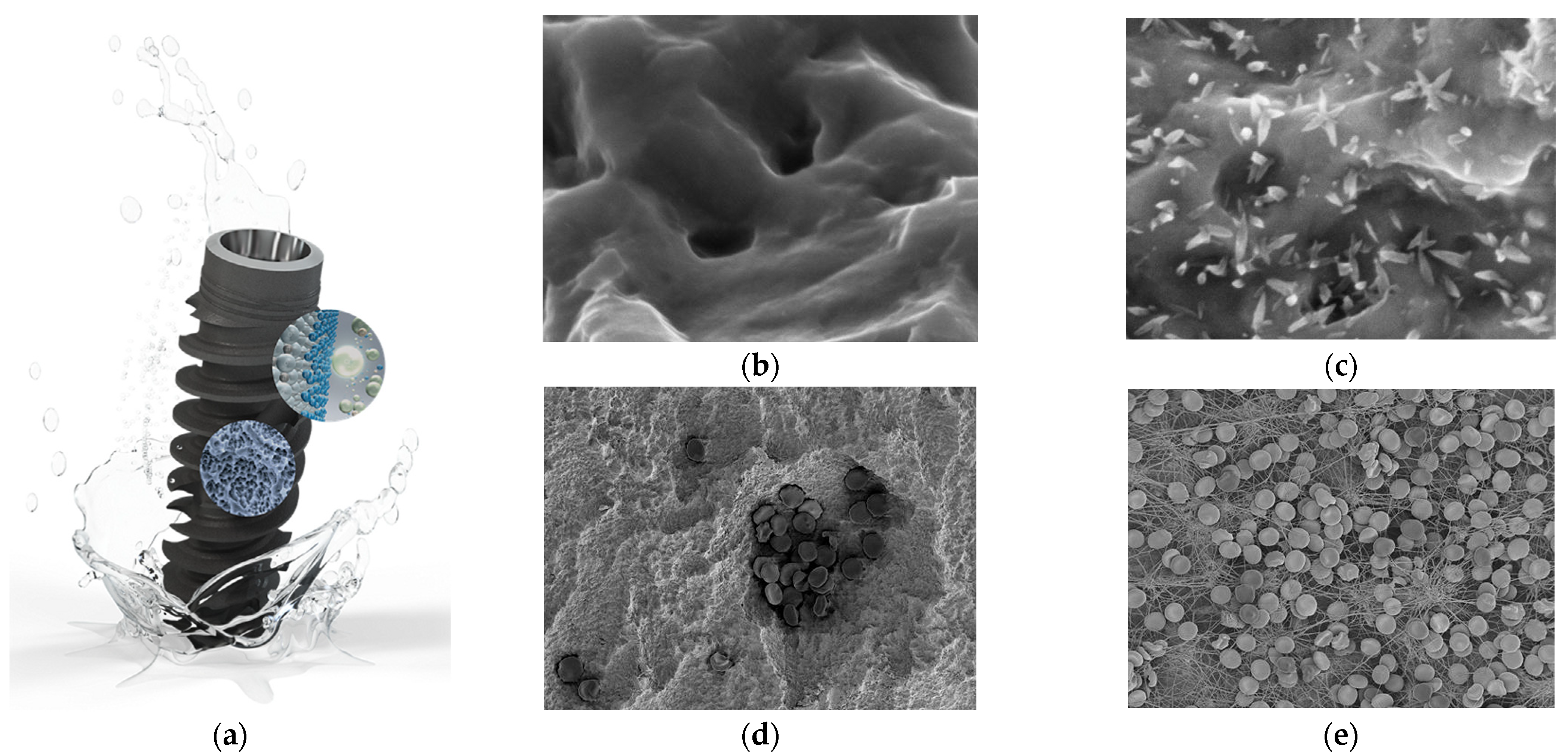
Dental implant systems with SLActive surfaces, such as Bone Level Implants with extensive healing potential, Standard Implants, Standard Plus Implants and Tapered Effect Implants with SLActive surfaces, were introduced to the dental market by the Straumann Institute [207,208,209,210,211]. In the SEM image of the Roxolid® SLActive® surface shown in Figure 10c, clearly shaped nanostructures can be observed, which are not present in the Roxolid® SLA® surface topography visible in Figure 10b [210]. Recent in vitro studies have demonstrated that the enhanced formation of fibrin networks on the Roxolid® SLActive® surface with nanostructures is significant [211]. SEM imaging reveals that after just 15 min of contact with human whole blood, these nanostructures contribute to improved fibrin network development (Figure 10e) [211]. Interestingly, while hydrophilicity has been recognized for its role in promoting osseointegration, it does not fully account for the accelerated integration observed with Roxolid® SLActive® surfaces.
The latest findings indicate that the presence of nanostructures on the Roxolid® SLActive® surface not only enhances fibrin network formation, but also supports the mineralization process of bone cells. This results in more effective early-stage osseointegration compared to the Roxolid® SLActive® surfaces lacking these nanostructures.

3.8. Oxidized Surface

Oxidized (anodized) surfaces are obtained via an electrochemical oxidation process carried out at a constant current density or voltage in the range of 1–300 V in electrolyte solutions, most often based on H2SO4 and H3PO4, with the addition of HF, which increases the current density [21,22,23,78,90,134,214,215,216,217,218,219,220,221,222,223,224,225,226]. The oxidized surface was introduced to the dental market in 2001 as TiUnite [215]. Depending on the electrochemical oxidation parameters, a continuous oxide layer [216], a porous oxide layer [217], or a layer of oxide nanotubes [21,22,23,218] of different thickness, structure, and surface morphology can be produced on the anode surface. The anodizing process is carried out in the optimal temperature range of 18–38 °C [219]. With the increase in the electrolyte temperature, the oxide layer thickens, which increases the resistance of titanium implants to wear and corrosion and accelerates osseointegration in the initial period after implantation [218]. The oxidized surface is rough, with an Ra value above 2 μm, and the thickness of the oxide layer ranges from 1–2 to 7–10 μm [128,220].
The TiUnite Dental Implant by Nobel Biocare is a highly advanced dental implant system with an anodized surface, known for its superior performance and clinical success (Figure 11a) [220]. The characteristic feature of the oxidized surface is isotropy and flowery morphology, with pores of dimensions ranging from 1 to 10 μm, along with smaller pores that have diameters less than 1 micrometer (Figure 11b) [134]. This results in shorter healing times and improved stability [222]. Such a microscopically porous surface enhances bone cell attachment and integration [223,224]. Hemostasis by the newly formed fibrin matrix on the TiUnite surface is shown in Figure 11c [224]. This fibrin matrix plays a crucial role in blood clot formation and subsequent hemostasis. The anodized surface of titanium, which has these specific pore dimensions, significantly enhances thrombogenicity and blood clot formation. The well-structured and spread transitory matrix formed by the fibrin clot facilitates the colonization of osteoprogenitor cells, thereby promoting osseointegration and overall hemostatic efficiency [223,224].
The results of clinical studies indicate a higher degree of osseointegration of implants with an oxidized surface than titanium implants with a machined or TPS surface [134,220,222,224,225]. However, surface treatment via anodizing may cause titanium brittleness. The TiUnite implant has demonstrated a high success rate, with a whole-patient group success rate of 98.2%, including a 100% success rate for implants with an oxidized surface [223,226]. Studies have shown that TiUnite implants have a remarkably low early failure rate and support long-term clinical survival. Early implant- and patient-level survival rates exceed 99% at one year, and the late implant-level survival rate is estimated at 95.1% after 10 years [223]. The TiUnite surface has been proven to support peri-implant health and bone maintenance, with low rates of peri-implantitis and successful soft tissue outcomes [220,223]. This surface is used in various Nobel Biocare implant systems, including the NobelActive and Brånemark System Mk III, making it suitable for different bone types and clinical scenarios [226]. It allows for immediate function of the implant, meaning patients can start using their new teeth soon after the procedure, which is beneficial for both patient comfort and clinical efficiency [220,223]. The TiUnite surface has been extensively researched, with over 465 publications featuring more than 89,500 implants and 22,600 patients, providing a robust body of evidence supporting its efficacy [223].
The latest development in anodizing technology is plasma electrolytic oxidation (PEO), also known as micro arc oxidation (MAO), which is an electrochemical surface treatment process that allows for the production of porous and hard oxide layers on titanium [227,228,229,230]. The PEO method involves high-voltage electrochemical oxidation at voltages of several hundred V, during which plasma microdischarges are generated at the titanium–electrolyte interface in the form of sparks, causing structural changes to the anode surface. Oxide layers obtained by the PEO method are about 1.3 μm thick, and their surface contains pores with a diameter of over 1.3 μm, which indicates a roughness similar to that of a machined surface [230]. Titanium implants with the TiUnite surface—Replace, Perfect, and Direct, manufactured by Nobel Biocare (Kloten, Switzerland)—are available on the dental market [220,225,226]. Oxide layers formed on the anode surface in the PEO process can be additionally enriched with Mg, Ca, S, and P ions, which provides a mechanical and biochemical connection between the implant surface and the bone tissue [231]. An example of a surface with the addition of Ca and P ions is the Biomimetic Advanced Surface developed by Avinent (Barcelona, Spain) for the Coral and Ocean dental systems [232]. These systems are characterized by optimal micro-porosity and macro-roughness, which provide high primary stabilization, and the hydrophilic properties of their surface support the osseointegration process by facilitating the migration of bone-forming and osteogenic cells.

3.9. Biologically Active Surface

The continuous development of technology has led to the creation of titanium implants with a biologically active surface, which are still in the experimental phase [36,42,43,44,45,47,48,78,90,139,150,171,193,215,217,233,234,235,236,237,238,239,240,241,242,243,244]. Biologically active surfaces on dental implants refer to surfaces that have been modified or coated with bioactive materials to enhance their interaction with biological tissues, particularly bone. These surfaces are designed to improve the implant’s integration with the surrounding bone, enhance its mechanical properties, and provide additional functionalities such as antibacterial properties and improved tissue healing. The biologically active surface of titanium implants is achieved through various surface modification techniques that enhance their interaction with bone tissue, improving osseointegration and overall implant performance. Open-pored coatings are designed to improve bone ingrowth and enhance the mechanical strength of the bone–implant interface. The open-pored structure allows for better integration with the surrounding bone tissue [90,150,171,217]. Multilevel micro-pit structures are created through a series of surface treatments, including sandblasting, acid etching, and glow discharge. This process forms a hierarchical microstructure that enhances the biocompatibility and osseointegration of the titanium implant [90,215]. Micro–nano composite structures are created using laser processing and multiple acid etching steps, which improve cell behavior and bone integration [36,42,43,44,45,47,48,193]. The biological modification of titanium implants based on the loading of specific bioactive substances, such as growth factors, peptides, proteins, and drugs, has been proposed [233,234,235,237,238,239,240,241,242,243]. These substances directly interact with bone cells and the surrounding tissues, promoting cell proliferation, differentiation, and mineralization. Methods of biological modification include physical adsorption, nanotechnology, chemical bonding, self-assembly, and nucleic acid-related technologies. These methods ensure the stability of the surface morphology and create a drug delivery system that releases bioactive substances over time [233,234,235]. To address the issue of bacterial adhesion and postoperative infections, antibacterial coatings are applied to titanium implants. These coatings can release antibiotics or other antibacterial agents, reducing the risk of infection and promoting bone healing [78,153,241,242,243]. Osteoconductive coatings are designed to enhance bone growth and integration. They often include materials like hydroxyapatite and bioactive glass, which are known for their osteoconductive properties [28,41,44,170,171,172,173,174,175,176,177,218,245]. Advanced surface treatment as anodization forms a TiO2 nanotube layer on the titanium surface, which improves biocompatibility and antibacterial properties. The TiO2 nanotubes enhance osseointegration and reduce the risk of infection [21,22,23,218]. Additive manufacturing technique allows for the creation of complex surface structures with controlled porosity, which can be tailored to enhance bone integration and mechanical properties [112].
Research is also being conducted on fluoride-enriched surfaces [134,139,244]. An example is the nanotextured surface of the OsseoSpeed implant by Astra Tech, which, thanks to the presence of incorporated fluoride ions, stimulates the bone formation process and accelerates osseointegration, ensuring stable implant placement in the jaw bone (Figure 12a) [244]. The OsseoSpeed dental implant is a high-performance dental implant designed for immediate loading and enhanced osseointegration. The implant features a microthreaded neck that increases the surface area for bone contact, thereby enhancing stability and osseointegration. The implant surface is treated with a unique surface technology that promotes rapid bone growth and integration. This technology is designed to improve the initial stability and long-term success of the implant. The SEM image shown in Figure 12b,c depicts a titanium dental implant that has a surface enriched with fluoride [134]. This unique surface treatment accelerates the osseointegration process, leading to faster healing and a quicker return to normal oral function. The OsseoSpeed dental implant offers a combination of advanced surface technology, immediate loading capability, and versatile design, making it a reliable choice for dental implant procedures.
The use of metal–organic frameworks (MOFs) in dental implant surface modification is a novel approach. MOFs can be used to create bioactive surfaces that improve the stability and biocompatibility of the implants, as well as provide additional functionalities [246,247,248,249]. MOFs can be used to modify the surface of titanium implants to improve osseointegration. This is achieved by creating a more bioactive surface that promotes bone cell adhesion and proliferation. For example, the use of UiO-66/AgNPs nanocomposite coatings has shown promise in enhancing osseointegration and preventing bacterial colonization, which is crucial for the long-term success of dental implants [246,247]. MOFs can incorporate antibacterial agents, such as silver nanoparticles (AgNPs), to create surfaces that resist bacterial colonization. This is particularly important in preventing peri-implantitis, a common complication in dental implantology. The UiO-66/AgNP nanocomposite, for instance, has been shown to have effective antibacterial properties [246]. Certain MOFs, like Bio-MOF-1, are designed with bio-derived constituents that are inherently biofriendly. These materials can enhance the bioactivity of the implant surface, promoting bone regeneration and integration with the surrounding tissue [248]. MOFs can be engineered to act as drug delivery systems, releasing bioactive molecules or antibiotics in a controlled manner. This can help in managing post-surgical infections and promoting healing. The use of MOFs as a confinement matrix for drug delivery systems is an emerging area of research in dental implantology [246,247]. MOFs can be used to create complex surface topographies and roughness at the micro- and nano-scale. This enhances the mechanical interlocking of the implant with the bone, further improving osseointegration. Techniques such as anodization and laser surface treatment can be combined with MOF coatings to achieve these effects [247,249]. MOFs can be tailored to improve the biocompatibility and osteoconductivity of the implant surface. This is achieved by incorporating bioactive materials like hydroxyapatite or other bone-like minerals into the MOF structure, which can mimic the natural bone environment and promote bone growth [247,248]. The field of dental implant surface modification is dynamic and continuously evolving. Advances in bioactive coatings, laser treatments, nanoengineering, and the use of MOFs are contributing to the development of more effective and durable dental implants. These innovations are expected to significantly improve patient outcomes and the overall success rates of dental implant procedures.
The addition of bioactive molecules to the surface of titanium implants induces a response from living tissue [250]. It has been shown that the biological surface supports cell adhesion to the implant surface by absorbing blood and circulating bone tissue [31]. The bioactive surface of titanium dental implants can promote bone formation around the implant, which will result in increased osseointegration in the early stages of healing. However, long-term clinical studies are required due to the fact that the results of in vivo animal studies do not fully reflect the clinical reality of the human body [28].

3.10. Hybrid Surface

Current trends in the development of dental implants with hybrid surfaces concern the search for new ways of modifying the surface of titanium implants, which will affect the acceleration of the osseointegration process and will enable the immediate functional loading of implants [27,31,48,52,53,54,55,56,57,58,59,60,61,62,63,78,79,80,81,82,83,84,85,162,173,194,208,224,237,238]. Considering the fact that too high roughness of the implant surface can cause effects opposite to those intended, surfaces with optimal micro-roughness and appropriate topography, structure, chemical composition, surface charge, wettability, and bioactivity are being developed, which will stimulate bone tissue to grow. The type of titanium implant surface is one of the key factors ensuring the success of the implantation procedure.
A hybrid surface on titanium dental implants refers to a combination of different surface treatments or coatings that aim to enhance various properties of the implant, such as osseointegration, biocompatibility, and resistance to biofilm formation [251,252,253,254,255,256,257,258]. Hybrid surfaces often combine micro- and nano-scale features. For example, a controllable hybrid micro–nano titanium model surface has been developed and contrasted with commercially available nano-featured surfaces, showcasing improved properties for dental implants [92,251]. Hybrid coatings on titanium implants can include antibacterial agents, which help in reducing the risk of infection. These coatings also promote soft tissue attachment through fibroblasts, enhancing the overall integration of the implant with the surrounding tissues [251]. Some hybrid coatings are designed to improve the aesthetic properties and wear resistance of the implant, combining the benefits of titanium and zirconia [251]. The hybrid surface design can influence biofilm formation on dental implants. Studies have evaluated the biofilm formation on hybrid titanium implants with moderately rough and turned surfaces, indicating that the hybrid surface can affect the colonization of bacteria [252]. Implants with hybrid surfaces have been evaluated for their radiological, clinical, and microbiological outcomes, particularly in patients with a history of periodontitis. These studies aim to assess the long-term success and stability of hybrid surface implants [253]. Various surface modification techniques are used to create hybrid surfaces, including chemical treatments, laser irradiation, and plasma spraying. These methods can alter the surface micro/nano-topography and composition, improving hydrophilicity, mechanical properties, and osseointegration [42,254,255,256,257,258].
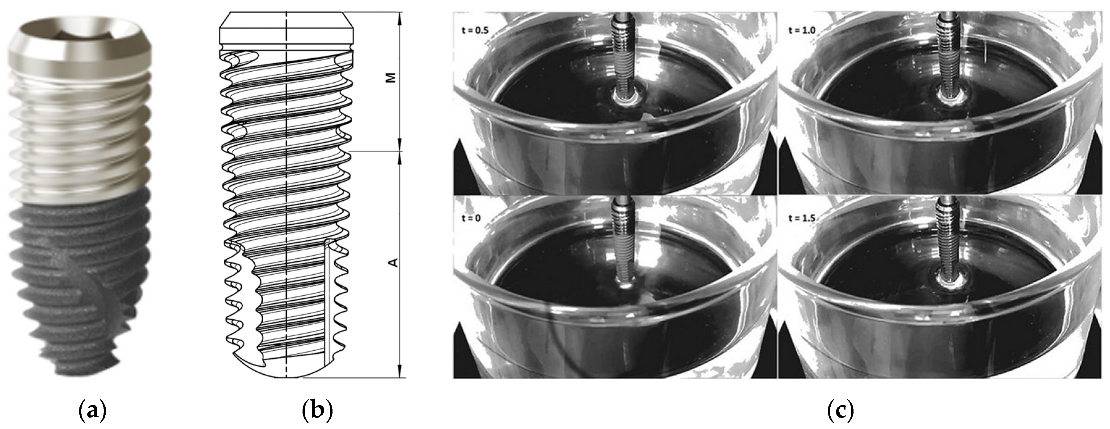
The prototype for a hybrid implant with a micro-rough implant surface in the endosseous portion to promote faster and better osseointegration of the implant during healing and during function, and a smooth, machined surface in the neck/collar region for the transcrestal area with contact to the peri-implant sulcus was developed by Dennis Tarnow in 1993 [258]. His Hybrid Design (HD) is a significant advancement in dental implant technology, focusing on improving aesthetic outcomes and bone integration through the use of different surface textures and materials. HD has been implemented in modern IMAX NHSI hybrid dental implants, which were introduced to the medical market as standard bone-level implants by iRES, founded in Switzerland in 2014 [256], as shown in Figure 13a. The iRES implants are additionally covered with a double layer of hyaluronic acid, which is covalently bonded to the titanium surface and ensures increased hydrophilicity and the absorption of growth factors in the healing phase, as well as their better utilization by extracellular matrix proteins. The bioactive hyaluronic coating also results in a more intensive recruitment, proliferation, and differentiation of osteoblasts, and the long-term provision of the implant–bone connection. The most promising hybrid dental implants are those with machined surfaces where the arithmetic mean deviation of the surface unevenness height from the reference plane (Sa) of 0.5 μm occurs in the coronal part of the implant on 40% of its length, while the rough surface is Sa = 1.9 μm and occupies the remaining 60% (Figure 13a) [256]. The machined surface does not support the growth of bacteria; therefore, its task is to protect the implant from inflammation, resulting from the formation of an aggressive biofilm around the exposed implant neck and from bone tissue loss around the implant (peri-implantitis) (Figure 13b) [257]. The implant surface is most often exposed as a result of non-bacterial disturbance of the biological balance of tissues around the implant. Bacteria attach secondarily when the implant surface is exposed, and this favors their settlement and multiplication. On rough surfaces, bacteria form a bacterial plaque and penetrate deeper into the implant more easily.
Figure 13c presents a sequence of Wilhelmy plate measurements for such a hybrid implant covered with a bioactive hyaluronic acid nanolayer over a 1.5 s interval, with each frame taken every 0.5 s [257]. The implant, connected to a microbalance, was suspended above a beaker rising with ultrapure water. Initially, at t = 0, the implant was dry, showcasing its lower matte micro-rough texture and upper shiny machined finish. Once the apex of the implant contacted the water, capillary action rapidly occurred through the threaded connector, influenced by the implant’s surface chemistry and roughness. After 0.5 s, water rose approximately halfway up the 10.5 mm micro-rough area; by 1 s, the micro-rough section was fully wetted, and at 1.5 s, the capillary rise was advancing toward the implant platform, 16 mm from the water surface. Despite being stored for a year, the implant demonstrated full wettability.
The results of the long-term wettability tests conducted on clinically available hyaluronan-coated titanium implants, which were packaged and sterile, or in their “on the shelf” condition, after one year from production, demonstrate that nanoengineering the implant surface by attaching the hydrophilic hyaluronic acid molecule provides these titanium implants with permanent wettability. This eliminates the need for wet storage, which is currently used to maintain long-term hydrophilic implant surfaces.
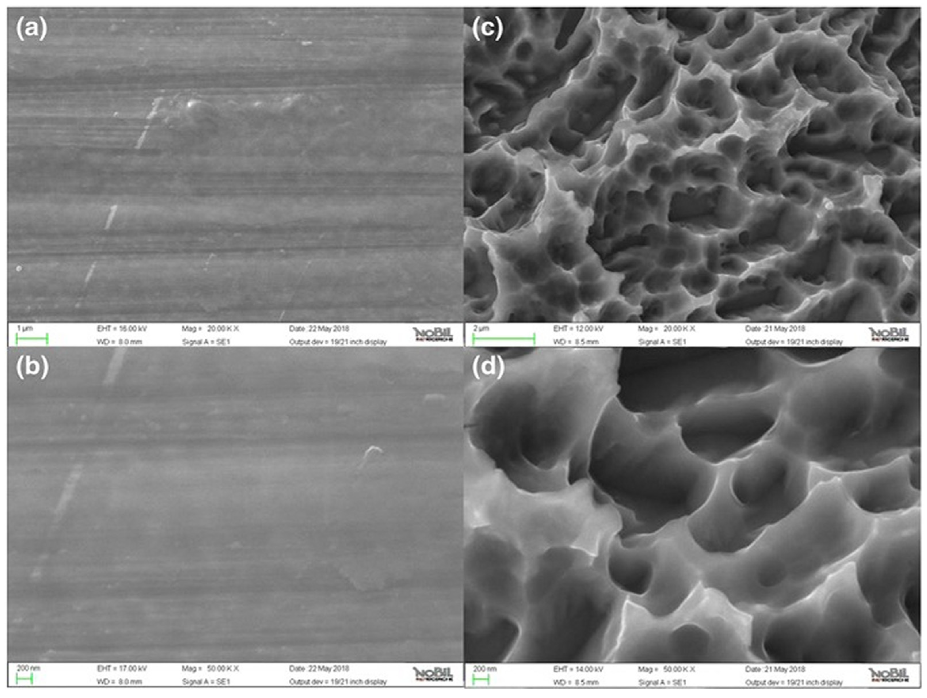
Figure 14 presents SEM images of the hybrid implant surface covered with a bioactive hyaluronic acid nanolayer at magnifications of 20,000× and 50,000×, showing both machined (a,b) and micro-rough (c,d) sections [257]. The SEM images are primarily characterized by the distinct features of each microarchitecture: the machined section (a,b) exhibits parallel grooves, indicative of machining tools, and the micro-rough section (c,d) shows sharp, closely packed peaks, resulting from double acid etching. At this level of magnification, the macro-roughness typical of sandblasted–acid-etched surfaces, caused by blasting, is not visible because the field of view is too small. Consequently, these images do not provide much information about the overlying hyaluronic acid layer, including its presence or homogeneity. Increasing the magnification further would not yield more information, as image contrast cannot offer vertical resolution at the nanometer scale. From an analytical perspective, the introduction of nanoengineered implant surfaces into routine clinical practice necessitates the advancement of analytical methods to the nano-scale.
In the case of developed surfaces, the time of osseointegration is shortened and a balance between the coexisting phenomena of osteogenesis and osteolysis is more easily achieved [78]. Therefore, the challenge is to design implants that are not only mechanically durable and strongly bound to the body, but that also exhibit antibacterial resistance [92].

3.11. Laser-Structured Surface

The laser-structured surface of dental implants is characterized by precise and controlled surface textures, increased roughness, enhanced osseointegration, improved bacterial resistance, material versatility, and high reproducibility [36,42,43,44,45,48,193]. Laser surface modifications can create a contamination-free titanium implant with a thick oxide layer, which promotes better osseointegration compared to conventional methods. This means the implant integrates more effectively with the surrounding bone tissue, leading to the improved stability and longevity of the implant [42,259]. Lasers can create precise and controlled surface textures on dental implants. This is achieved using different types of laser pulses, such as femtosecond, picosecond, and nanosecond pulses, which allow for the creation of micro- and nano-topographies on the implant surface. These textures can mimic the natural bone structure, enhancing the biological response and integration with the bone [260]. The micro- or nano-topography created by laser texturing can provide antimicrobial properties, which help in reducing the risk of infection around the implant. This is particularly important for preventing peri-implantitis, a common complication in dental implantology [47,261].
The controlled surface modifications can also enhance the mechanical properties of the implant, making it more durable and resistant to wear and tear [43]. Several in vivo studies have demonstrated the effectiveness of laser-structured surfaces in improving the biointegration of dental implants. These studies have shown that implants with laser-structured surfaces exhibit better osseointegration and reduced inflammation compared to those with traditionally treated surfaces [262]. Laser technology allows for the customization of implant surfaces, which can be tailored to meet specific clinical needs. This includes the ability to create surfaces that are more conducive to bone growth and integration, as well as surfaces that can be used for specific types of dental implants, such as ceramic implants [263].
Over the past few decades, the application of laser technology in dental implantology has evolved significantly. Techniques such as laser etching (Laser-Lok) have been developed and proven effective in creating a biological seal around the implant, enhancing its stability and longevity [264]. The origins of laser etching can be traced back to the invention of the laser in the early 1960s. The first laser engraver was used in 1978, and since then, the technology has evolved to become a widely used marking method in various industries [265]. Laser-Lok is a proprietary dental implant surface treatment developed by BioHorizons (Birmingham, AL, USA) [266,267].
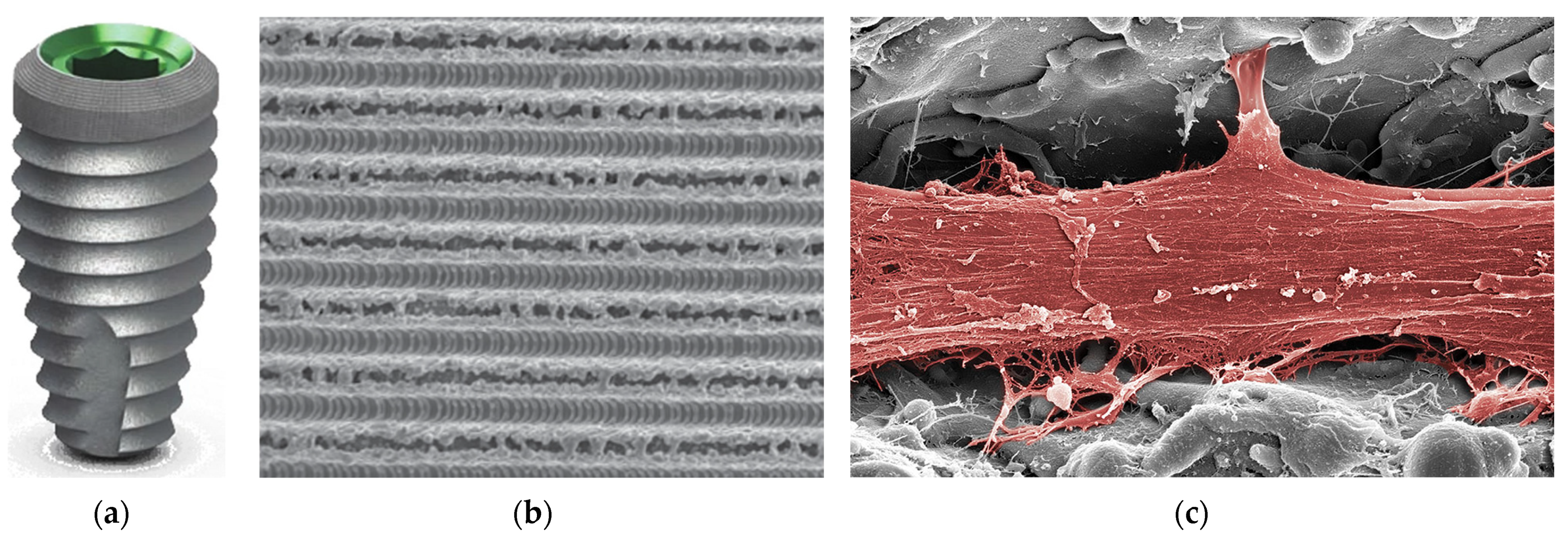
Figure 15a shows the Tapered Internal Plus (4.5) dental implant by BioHorizons, which is a high-quality implant that offers several advantages, including predictable results, enhanced bone maintenance, improved soft tissue health, and ease of use [266]. Its design features include a 45° conical internal hex connection, color-coded platform, anatomically tapered body, and aggressive buttress threads.
The Laser-Lok surface of this dental implant is characterized by uniformly shaped microchannels designed to facilitate and enhance tissue growth (Figure 15b) [267]. A colorized SEM image reveals that the unique Laser-Lok surface has demonstrated its ability to trigger a biological reaction that prevents the downward growth of epithelial tissue and promotes the bonding of connective tissue. This connection creates a biological seal around the implant, which helps safeguard and preserve the health of the crestal bone (Figure 15c) [267].
BioHorizons Laser-Lok technology is a sophisticated surface treatment for dental implants and abutments that enhances the biological integration and mechanical stability of dental implants and abutments, leading to improved clinical outcomes. It consists of precision-engineered, cell-sized microchannels that are laser-machined onto the surface of dental implants and abutments. This technology is designed to enhance osseointegration and promote bone growth around the implant. The microchannels are created using a patented laser ablation process [267].

4. Future Directions

The future of dental implant surface modification is promising, with ongoing research and development aimed at improving the success rates, longevity, and patient satisfaction of dental implant procedures. Advances in materials science, nanotechnology, and bioengineering are driving innovations in implant surface modification.
Nanotechnology has already made significant inroads into dental implant surface modification, with nanostructured surfaces shown to enhance cell adhesion, proliferation, and differentiation. These surfaces can mimic the natural environment of bone, promoting better osseointegration. Researchers are exploring more sophisticated nanostructures, such as nanotubes and nanofibers, which can further optimize the surface properties of implants. These structures can be designed to incorporate bioactive molecules or drugs for controlled release, enhancing osseointegration and reducing the risk of infection.
Three-dimensional printing technology is being used to create customized dental implants with complex, porous structures that promote bone ingrowth. These implants can be designed to match the specific anatomy of the patient, improving fit and osseointegration. Advances in 3D printing materials and techniques will allow for even more precise and personalized implant designs. Researchers are also exploring the integration of nanostructured surfaces and bioactive coatings into 3D-printed implants, creating multifunctional surfaces that optimize osseointegration, mechanical stability, and antibacterial properties.
Bioactive coatings, such as hydroxyapatite and calcium phosphate, are used to enhance the biological response to dental implants. These coatings promote the formation of a calcium phosphate layer on the implant surface, mimicking the natural bone mineral. Researchers are developing more advanced biomimetic coatings that not only promote bone growth, but also mimic the complex structure and composition of natural bone. These coatings can incorporate growth factors, peptides, and other bioactive molecules to stimulate bone formation and reduce healing time.
Smart surfaces that can respond to changes in the local environment are being developed for dental implants. These surfaces can release drugs or bioactive molecules in a controlled manner, targeting specific stages of the healing process or responding to infections. The integration of smart surfaces with advanced drug delivery systems will enable more precise and effective treatment strategies. For example, surfaces that can sense bacterial infections and release antibiotics locally can reduce the risk of implant failure due to infection.
Laser and plasma surface modification techniques are used to create micro- and nano-scale patterns on implant surfaces, enhancing cell attachment and bone integration. Advances in laser and plasma technologies will allow for even more precise and controlled surface modifications. Researchers are exploring the creation of hierarchical structures that combine micro- and nano-scale features to optimize both mechanical interlocking and biological responses.
Infection is a major complication of dental implant surgery. Surface modifications that reduce bacterial adhesion and growth are being developed to minimize this risk. Researchers are exploring new materials and coatings with inherent antibacterial properties, as well as surfaces that can release antibacterial agents in a controlled manner. These surfaces can help prevent biofilm formation and reduce the risk of peri-implantitis.
Advanced imaging techniques, such as computed tomography (CT) and cone beam computed tomography (CBCT), are used to plan dental implant procedures. The integration of advanced imaging with dental implant surface modification will enable more precise and personalized treatment planning. For example, imaging data can be used to design customized implant surfaces that optimize fit and osseointegration based on the patient’s specific anatomy and bone quality.
The future of dental implant surface modification is bright, with ongoing research and development focused on creating smarter, more biocompatible, and personalized implant surfaces. These innovations will not only improve the success rates of dental implant procedures, but also enhance patient comfort and reduce healing times. As these technologies advance, the field of dental implantology will continue to evolve, providing better solutions for tooth replacement and restoration. While many surface modification techniques have shown promise, they often come with high costs or inconsistent results, limiting their widespread clinical application. Ongoing research aims to develop more cost-effective and reliable methods. Future advancements will likely focus on integrating multiple functions into a single surface treatment, such as combining antibacterial and osteoconductive properties to enhance overall implant performance.

5. Conclusions

The future of dental implant surface modification is promising, with ongoing research focused on enhancing success rates, longevity, and patient satisfaction. Advances in materials science and bioengineering are driving innovations that improve osseointegration and reduce the risk of complications such as peri-implantitis. Techniques like laser surface modification and the incorporation of bioactive molecules are expected to play a crucial role in developing implants that not only integrate better with bone, but also resist bacterial colonization. As these technologies evolve, they will likely lead to more reliable and effective dental implant solutions, ultimately improving patient outcomes and satisfaction.
The development of dental implant surface modification techniques has significantly advanced the field of dental implantology. By optimizing the surface properties of implants, clinicians can achieve better osseointegration, improved mechanical stability, and a reduced risk of complications. Ongoing research continues to explore new materials and techniques to further enhance the performance and success of dental implants.
The latest surface modification techniques for dental implants improve patient outcomes by enhancing osseointegration, biocompatibility, and antibacterial properties, while also incorporating drug delivery systems and improving mechanical and durability characteristics. These advancements collectively contribute to higher success rates, reduced infection risks, and the better long-term stability of dental implants.
The most important factor for the success of the osseointegration process is the composition and type of the surface layer of the implants. The surfaces of titanium implants, which affect the biomechanical potential of the implant–bone tissue contact and the rate of protein adsorption, have been modified over the years through various methods. These modifications include physical, chemical, and biological approaches to enhance the surface properties of the implants.

Author Contributions

Conceptualization, B.Ł.; methodology, B.Ł., P.O., D.N. and J.M.; writing—original draft preparation, B.Ł., P.O. and D.N.; writing—review and editing, J.M.; visualization, B.Ł, P.O., D.N. and J.M.; supervision, B.Ł.; project administration, D.N. All authors have read and agreed to the published version of the manuscript.

Funding

This work was created partly within the framework of the Student Research Grant of the European City of Science Katowice, 2024, entitled: “Development of an innovative method for manufacturing porous oxide layers on titanium for applications in regenerative medicine” (University of Katowice, Poland).

Conflicts of Interest

The authors declare no conflicts of interest.

References

  1. Waghmare, G.; Waghmare, K.; Bagde, S.; Deshmukh, M.; Kashyap, D.N.; Shahu, V.T. Materials Evolution in Dental Implantology: A Comprehensive Review. J. Adv. Res. Appl. Mech. 2024, 123, 75–100. [Google Scholar] [CrossRef]
  2. Lanis, A.; Peña-Cardelles, J.F.; Negreiros, W.M.; Hamilton, A.; Gallucci, G.O. Impact of digital technologies on implant surgery in fully edentulous patients: A scoping review. Clin. Oral Implants Res. 2024, 35, 1000–1010. [Google Scholar] [CrossRef] [PubMed]
  3. Shivgotra, R.; Soni, B.; Kaur, M.; Thakur, S. Advancement in Biomaterials in the Form of Implants. In Engineered Biomaterials. Engineering Materials; Malviya, R., Sundram, S., Eds.; Springer: Singapore, 2023. [Google Scholar] [CrossRef]
  4. Nitschke, I.; Krüger, K.; Jockusch, J. Age-related knowledge deficit and attitudes towards oral implants: Survey-based examination of the correlation between patient age and implant therapy awareness. BMC Oral Health 2024, 24, 403. [Google Scholar] [CrossRef] [PubMed]
  5. Zhao, Y.; Kim, B. The Effects of the Expansion of Dental Care Coverage for the Elderly. Healthcare 2024, 12, 1949. [Google Scholar] [CrossRef]
  6. Kim, H.-J.; Sung, I.-Y. Analysis of Dental Prosthetic Treatment in Patients with Cancer Aged 65 Years and Older after Expanded Health Insurance Coverage: A Retrospective Clinical Study. Medicina 2024, 60, 1509. [Google Scholar] [CrossRef]
  7. Gargallo-Albiol, J.; Ortega-Martínez, J.; Salomó-Coll, O.; López-Boado, A.P.; Paternostro-Betancourt, D.; Hernández-Alfaro, F. Mouth opening limitation and influence of age and surgical location for static fully guided dental implant placement: An observational, cross-sectional clinical study. Int. J. Oral Max. Surg. 2024, 53, 526–532. [Google Scholar] [CrossRef]
  8. Dave, M.; Tattar, R.; Patel, N. Medical considerations in the ageing implant patient. Oral Surgery 2024, 17, 59–66. [Google Scholar] [CrossRef]
  9. Karlsson, K.; Derks, J.; Wennström, J.L.; Petzold, M.; Berglundh, T. Health economic aspects of implant-supported restorative therapy. Clin Oral Implants Res. 2022, 33, 221–230. [Google Scholar] [CrossRef]
  10. Brägger, U.; Krenander, P.; Lang, N.P. Economic aspects of single-tooth replacement. Clin Oral Implants Res. 2005, 16, 335–341. [Google Scholar] [CrossRef]
  11. Massa, L.O.; Fraunhofer, J.A. Economics of Dental Implants. In The ADA Practical Guide to Dental Implants; Wiley-Blackwell: Hoboken, NJ, USA, 2021; pp. 139–143. [Google Scholar] [CrossRef]
  12. Losenická, J.; Gajdoš, O.; Kamenský, V. Cost-utility analysis of an implant treatment in dentistry. BMC Oral Health 2021, 21, 433. [Google Scholar] [CrossRef]
  13. Fan, Y.Y.; Li, S.; Cai, Y.J.; Wei, T.; Ye, P. Smoking in relation to early dental implant failure: A systematic review and meta-analysis. J. Dent. 2024, 151, 105396. [Google Scholar] [CrossRef] [PubMed]
  14. Howe, M.S.; Keys, W.; Richards, D. Long-term (10-year) dental implant survival: A systematic review and sensitivity meta-analysis. J. Dent. 2019, 84, 9–21. [Google Scholar] [CrossRef] [PubMed]
  15. French, D.; Ofec, R.; Levin, L. Long term clinical performance of 10 871 dental implants with up to 22 years of follow-up: A cohort study in 4247 patients. Clin. Implant Dent. Relat. Res. 2021, 23, 289–297. [Google Scholar] [CrossRef] [PubMed]
  16. Dental Implants Market Growth & Trends. Available online: https://www.grandviewresearch.com/press-release/global-dental-implants-market (accessed on 28 October 2024).
  17. O’ Dwyer, S.; Riordain, R.N. The patient experience of dental implant surgery: A literature review of pertinent qualitative studies. Ir. J. Med. Sci. 2021, 190, 835–842. [Google Scholar] [CrossRef]
  18. Chakaipa, S.; Prior, S.J.; Pearson, S.; van Dam, P.J. Improving Patient Experience through Meaningful Engagement: The Oral Health Patient’s Journey. Oral 2023, 3, 499–510. [Google Scholar] [CrossRef]
  19. Improve competitive advantage, job satisfaction and the patient experience. Br. Dent. J. 2022, 232, 347. [CrossRef]
  20. Shrivastava, R.; Luxenberg, R.; Sutton, E.; Emami, E. Patients experience and satisfaction with immediate loading of implant-supported overdentures—A qualitative study. J. Dent. 2023, 137, 104644. [Google Scholar] [CrossRef]
  21. Stróż, A.; Dercz, G.; Chmiela, B.; Stróż, D.; Łosiewicz, B. Electrochemical Formation of Second Generation TiO2 Nanotubes on Ti13Nb13Zr Alloy for Biomedical Applications. Acta Phys. Pol. A 2016, 130, 1079–1080. [Google Scholar] [CrossRef]
  22. Smołka, A.; Dercz, G.; Rodak, K.; Łosiewicz, B. Evaluation of corrosion resistance of nanotubular oxide layers on the Ti13Zr13Nb alloy in physiological saline solution. Arch. Metall. Mater. 2015, 60, 2681–2686. [Google Scholar] [CrossRef]
  23. Smołka, A.; Rodak, K.; Dercz, G.; Dudek, K.; Łosiewicz, B. Electrochemical Formation of Self-Organized Nanotubular Oxide Layers on Ti13Zr13Nb Alloy for Biomedical Applications. Acta Phys. Pol. A 2014, 125, 932–935. [Google Scholar] [CrossRef]
  24. Szklarska, M.; Dercz, G.; Rak, J.; Simka, W.; Łosiewicz, B. The influence of passivation type on corrosion resistance of Ti15Mo alloy in simulated body fluids. Arch. Metall. Mater. 2015, 60, 2687–2693. [Google Scholar] [CrossRef]
  25. Łosiewicz, B.; Stróż, A.; Kubisztal, J.; Osak, P.; Zubko, M. EIS and LEIS Study on In Vitro Corrosion Resistance of Anodic Oxide Nanotubes on Ti–13Zr–13Nb Alloy in Saline Solution. Coatings 2023, 13, 875. [Google Scholar] [CrossRef]
  26. Hosseini-Faradonbeh, S.A.; Katoozian, H.R. Biomechanical evaluations of the long-term stability of dental implant using finite element modeling method: A systematic review. J. Adv. Prosthodont. 2022, 14, 182–202. [Google Scholar] [CrossRef] [PubMed]
  27. Xiao, L.; Zhou, Y.J.; Jiang, Y.B.; Tam, M.S.; Cheang, L.H.; Wang, H.J.; Zha, Z.G.; Zheng, X.F. Effect of Diabetes Mellitus on Implant Osseointegration of Titanium Screws: An Animal Experimental Study. Orthop Surg. 2022, 14, 1217–1228. [Google Scholar] [CrossRef]
  28. Osak, P.; Maszybrocka, J.; Kubisztal, J.; Ratajczak, P.; Łosiewicz, B. Long-Term Assessment of the In Vitro Corrosion Resistance of Biomimetic ACP Coatings Electrodeposited from an Acetate Bath. J. Funct. Biomater. 2021, 12, 12. [Google Scholar] [CrossRef]
  29. Shayeb, M.A.; Elfadil, S.; Abutayyem, H.; Shqaidef, A.; Marrapodi, M.M.; Cicciù, M.; Minervini, G. Bioactive surface modifications on dental implants: A systematic review and meta-analysis of osseointegration and longevity. Clin. Oral Investig. 2024, 28, 592. [Google Scholar] [CrossRef]
  30. Canullo, L.; Menini, M.; Pesce, P.; Iacono, R.; Sculean, A.; Del Fabbro, M. Nano-superhydrophilic and bioactive surface in poor bone environment. Part 1: Transition from primary to secondary stability. A controlled clinical trial. Clin. Oral Investig. 2024, 28, 372. [Google Scholar] [CrossRef]
  31. Meng, H.W.; Chien, E.Y.; Chien, H.H. Dental implant bioactive surface modifications and their effects on osseointegration: A review. Biomark. Res. 2016, 4, 24. [Google Scholar] [CrossRef]
  32. López-Valverde, N.; Flores-Fraile, J.; Ramírez, J.M.; Macedo de Sousa, B.; Herrero-Hernández, S.; López-Valverde, A. Bioactive Surfaces vs. Conventional Surfaces in Titanium Dental Implants: A Comparative Systematic Review. J. Clin. Med. 2020, 9, 2047. [Google Scholar] [CrossRef]
  33. Katić, J.; Šarić, A.; Despotović, I.; Matijaković, N.; Petković, M.; Petrović, Ž. Bioactive Coating on Titanium Dental Implants for Improved Anticorrosion Protection: A Combined Experimental and Theoretical Study. Coatings 2019, 9, 612. [Google Scholar] [CrossRef]
  34. Munisamy, S.; Vaidyanathan, T.K.; Vaidyanathan, J. A bone-like precoating strategy for implants: Collagen immobilization and mineralization on pure titanium implant surface. J. Oral Implantol. 2008, 34, 67–75. [Google Scholar] [CrossRef] [PubMed]
  35. de Almeida, T.C.S.; Valverde, T.M.; Martins, T.M.d.M.; Oliveira, F.d.P.; Cunha, P.d.S.; Tavares, M.A.B.; Rodrigues, E.M.; Albergaria, J.D.S.; Vieira, G.M.; Gomes, D.A.; et al. Enhanced osteogenic response by collagen type I coating on surface-modified titanium bone implants. Mater. Today Commun. 2024, 39, 108535. [Google Scholar] [CrossRef]
  36. Belloni, A.; Argentieri, G.; Orilisi, G.; Notarstefano, V.; Giorgini, E.; D’Addazio, G.; Orsini, G.; Caputi, S.; Sinjari, B. New insights on collagen structural organization and spatial distribution around dental implants: A comparison between machined and laser-treated surfaces. J. Transl. Med. 2024, 22, 120. [Google Scholar] [CrossRef] [PubMed]
  37. Erturk, P.A.; Altuntas, S.; Irmak, G.; Buyukserin, F. Bioinspired Collagen/Gelatin Nanopillared Films as a Potential Implant Coating Material. ACS Appl. Bio Mater. 2022, 5, 4913–4921. [Google Scholar] [CrossRef]
  38. Petrović, Ž.; Šarić, A.; Despotović, I.; Katić, J.; Peter, R.; Petravić, M.; Ivanda, M.; Petković, M. Surface Functionalisation of Dental Implants with a Composite Coating of Alendronate and Hydrolysed Collagen: DFT and EIS Studies. Materials 2022, 15, 5127. [Google Scholar] [CrossRef]
  39. O’Neill, L.; Twomey, B.; Tan, F.; O’Donoghue, J.; Junt, J.A. Collagen Coating of Titanium Implants Using Non-thermal Plasma. Plasma Med. 2020, 11, 63–79. [Google Scholar] [CrossRef]
  40. Abdulghafor, M.A.; Mahmood, M.K.; Tassery, H.; Tardivo, D.; Falguiere, A.; Lan, R. Biomimetic Coatings in Implant Dentistry: A Quick Update. J. Funct. Biomater. 2024, 15, 15. [Google Scholar] [CrossRef]
  41. Lee, S.W.; Hahn, B.D.; Kang, T.Y.; Lee, M.J.; Choi, J.Y.; Kim, M.K.; Kim, S.G. Hydroxyapatite and collagen combination-coated dental implants display better bone formation in the peri-implant area than the same combination plus bone morphogenetic protein-2-coated implants, hydroxyapatite only coated implants, and uncoated implants. J. Oral Maxillofac. Surg. 2014, 72, 53–60. [Google Scholar] [CrossRef]
  42. Kolarovszki, B.; Ficsor, S.; Frank, D.; Katona, K.; Soos, B.; Turzo, K. Unlocking the potential: Laser surface modifications for titanium dental implants. Lasers Med. Sci. 2024, 39, 162. [Google Scholar] [CrossRef]
  43. Saran, R.; Ginjupalli, K.; George, S.D.; Chidangil, S.; Unnikrishnan, V.K. LASER as a tool for surface modification of dental biomaterials: A review. Heliyon 2023, 9, e17457. [Google Scholar] [CrossRef]
  44. Santos, A.F.P.; da Silva, R.C.; Hadad, H.; de Jesus, L.K.; Pereira-Silva, M.; Nímia, H.H.; Oliveira, S.H.P.; Guastaldi, A.C.; Queiroz, T.P.; Poli, P.P.; et al. Early Peri-Implant Bone Healing on Laser-Modified Surfaces with and without Hydroxyapatite Coating: An In Vivo Study. Biology 2024, 13, 533. [Google Scholar] [CrossRef] [PubMed]
  45. The Use of Lasers for Dental Implant Surgery. Available online: https://www.deserthillsdental.com/dental-implants-and-laser-dentistry/ (accessed on 28 October 2024).
  46. Papa, S.; Maalouf, M.; Claudel, P.; Sedao, X.; Maio, Y.D.; Hamzeh-Cognasse, H.; Thomas, M.; Guignandon, A.; Dumas, V. Key topographic parameters driving surface adhesion of Porphyromonas gingivalis. Sci. Rep. 2023, 13, 15893. [Google Scholar] [CrossRef] [PubMed]
  47. Fenelon, T.; Bakr, M.; Walsh, L.J.; George, R. Effects of lasers on titanium dental implant surfaces: A narrative review. Laser Dent. Sci. 2022, 6, 153–167. [Google Scholar] [CrossRef]
  48. Luczak, W.; Reiner-Rozman, C.; Muck, M.; Heitz, J.; Mitov, G.; Pfaffeneder, F.; See, C.; Hassel, A.W.; Kleber, C. Laser Treatment of Dental Implants toward an Optimized Osseointegration: Evaluation via Tapping-Mode Atomic Force Microscopy and Scanning Electron Microscopy. Phys. Status Solidi Appl. Mater. Sci. 2023, 220, 2200605. [Google Scholar] [CrossRef]
  49. Alamoudi, A. Nanoengineering and Surface Modifications of Dental Implants. Cureus 2024, 16, e51526. [Google Scholar] [CrossRef]
  50. Gulati, K. Nano-Engineering Solutions for Dental Implant Applications. Nanomaterials 2022, 12, 272. [Google Scholar] [CrossRef]
  51. Nagamoto, K.; Nakanishi, K.; Akasaka, T.; Abe, S.; Yoshihara, K.; Nakamura, M.; Hayashi, H.; Takemoto, S.; Tamura, M.; Kitagawa, Y.; et al. Investigation of a new implant surface modification using phosphorylated pullulan. Front. Bioeng. Biotechnol. 2024, 12, 1378039. [Google Scholar] [CrossRef]
  52. Karthik, K.; Thangaswamy, V. Evaluation of implant success: A review of past and present concepts. J. Pharm. Bioallied. Sci. 2013, 5 (Suppl. S1), S117–S119. [Google Scholar] [CrossRef]
  53. Han, W.; Fang, S.; Zhong, Q.; Qi, S. Influence of Dental Implant Surface Modifications on Osseointegration and Biofilm Attachment. Coatings 2022, 12, 1654. [Google Scholar] [CrossRef]
  54. Kligman, S.; Ren, Z.; Chung, C.-H.; Perillo, M.A.; Chang, Y.-C.; Koo, H.; Zheng, Z.; Li, C. The Impact of Dental Implant Surface Modifications on Osseointegration and Biofilm Formation. J. Clin. Med. 2021, 10, 1641. [Google Scholar] [CrossRef]
  55. Cooper, L.F.; Shirazi, S. Osseointegration—The biological reality of successful dental implant therapy: A narrative review. Front. Oral Maxillofac. Med. 2022, 4, 39. [Google Scholar] [CrossRef]
  56. Lechner, J.; von Baehr, V.; Notter, F.; Schick, F. Osseointegration and osteoimmunology in implantology: Assessment of the immune sustainability of dental implants using advanced sonographic diagnostics: Research and case reports. J. Int. Med. Res. 2024, 52, 3000605231224161. [Google Scholar] [CrossRef] [PubMed]
  57. Simão, B.S., Jr.; Costa, D.D.; Cangussu, M.C.T.; Sotto-Maior, B.S.; Devita, R.L.; de Carvalho, J.J.; da Silva Brum, I. Observational Study on the Success Rate of Osseointegration: A Prospective Analysis of 15,483 Implants in a Public Health Setting. BioMed 2022, 2, 422–430. [Google Scholar] [CrossRef]
  58. Parithimarkalaignan, S.; Padmanabhan, T.V. Osseointegration: An update. J. Indian Prosthodont. Soc. 2013, 13, 2–6. [Google Scholar] [CrossRef] [PubMed]
  59. Branemark, P.I.; Zarb, G.A.; Albrekson, T. Tissue-Integrated Prostheses: Osseointegration in Clinical Dentistry; Quintessence Publishing Company: Batavia, IL, USA, 1985. [Google Scholar]
  60. Gill, T.; Kühl, S.; Rawlinson, S.; Pippenger, B.; Bellon, B.; Shahdad, S. Primary stability and osseointegration comparing a novel tapered design tissue-level implant with a parallel design tissue-level implant. An experimental in vivo study. Clin. Oral Implants Res. 2024, 35, 1114–1127. [Google Scholar] [CrossRef] [PubMed]
  61. Lioubavina-Hack, N.; Lang, N.P.; Karring, T. Significance of primary stability for osseointegration of dental implants. Clin. Oral Implants Res. 2006, 17, 244–250. [Google Scholar] [CrossRef]
  62. Javed, F.; Ahmed, H.B.; Crespi, R.; Romanos, G.E. Role of primary stability for successful osseointegration of dental implants: Factors of influence and evaluation. Interv. Med. Appl. Sci. 2013, 5, 162–167. [Google Scholar] [CrossRef]
  63. Xu, L.; Jacobs, R.; Cao, Y.; Sun, X.; Qin, X. Tissue-engineered bone construct promotes early osseointegration of implants with low primary stability in oversized osteotomy. BMC Oral Health 2024, 24, 69. [Google Scholar] [CrossRef]
  64. Lee, J.; Lim, Y.-J.; Ahn, J.-S.; Kim, B.; Baek, Y.-W.; Lim, B.-S. Correlation of two different devices for the evaluation of primary implant stability depending on dental implant length and bone density: An in vitro study. PLoS ONE 2024, 19, e0290595. [Google Scholar] [CrossRef]
  65. Barikani, H.; Rashtak, S.; Akbari, S.; Badri, S.; Daneshparvar, N.; Rokn, A. The effect of implant length and diameter on the primary stability in different bone types. J. Dent. 2013, 10, 449–455. [Google Scholar]
  66. Stoilov, M.; Shafaghi, R.; Stark, H.; Marder, M.; Kraus, D.; Enkling, N. Influence of Implant Macro-Design, -Length, and -Diameter on Primary Implant Stability Depending on Different Bone Qualities Using Standard Drilling Protocols-An In Vitro Analysis. J. Funct. Biomater. 2023, 14, 469. [Google Scholar] [CrossRef]
  67. Gómez-Polo, M.; Ortega, R.; Gómez-Polo, C.; Martín, C.; Celemín, A.; del Río, J. Does Length, Diameter, or Bone Quality Affect Primary and Secondary Stability in Self-Tapping Dental Implants? J. Oral Maxillofac. Surgery 2016, 74, 1344–1353. [Google Scholar] [CrossRef]
  68. Cucinelli, C.; Pereira, M.S.; Borges, T.; Figueiredo, R.; Leitão-Almeida, B. The Effect of Increasing Thread Depth on the Initial Stability of Dental Implants: An In Vitro Study. Surgeries 2024, 5, 817–825. [Google Scholar] [CrossRef]
  69. Hiranmayi, V.K. Factors influencing implant stability. J. Dent. Implants 2018, 8, 69–76. [Google Scholar] [CrossRef]
  70. Huang, S.; Murphy, L.; Xu, W. Genes and functions from breast cancer signatures. BMC Cancer 2018, 18, 473. [Google Scholar] [CrossRef]
  71. El-Anwar, M.I.; El-Zawahry, M.M.; El-Mofty, M. Load Transfer on Dental Implants and Surrounding Bones. Aust. J. Basic Appl. Sci. 2012, 6, 551–560. [Google Scholar]
  72. Hansson, S.; Norton, M. The relation between surface roughness and interfacial shear strength for bone-anchored implants. A mathematical model. J. Biomech. 1999, 32, 829–836. [Google Scholar] [CrossRef]
  73. Bianchi, A.E.; Dolci, G., Jr.; Sberna, M.T.; Sanfilippo, F. Factors affecting bone response around loaded titanium dental implants: A literature review. J. Appl. Biomater. Biomech. 2005, 3, 135–140. [Google Scholar]
  74. Stanford, C.M. Surface modifications of dental implants. Aust. Dent. J. 2008, 53, S26–S33. [Google Scholar]
  75. Skalak, R.; Zhao, Y. Interaction of force-fitting and surface roughness of implants. Clin. Implant Dent. Relat. Res. 2000, 2, 219–224. [Google Scholar] [CrossRef]
  76. Barfeie, A.; Wilson, J.; Rees, J. Implant surface characteristics and their effect on osseointegration. Br. Dent. J. 2015, 218, E9. [Google Scholar] [CrossRef]
  77. Romero, M.; Herrero-Climent, M.; Ríos-Carrasco, B.; Brizuela, A.; Romero, M.M.; Gil, J. Investigation of the Influence of Roughness and Dental Implant Design on Primary Stability via Analysis of Insertion Torque and Implant Stability Quotient: An In Vitro Study. J. Clin. Med. 2023, 12, 4190. [Google Scholar] [CrossRef] [PubMed]
  78. Le Guéhennec, L.; Soueidan, A.; Layeolle, P.; Amourinq, Y. Surface treatments of titanium dental implants for rapid osseointegration. Dent. Mater. 2007, 23, 844–854. [Google Scholar] [CrossRef] [PubMed]
  79. Hosseini, S.H.; Kazemian, M.; Ghorbanzadeh, S. A brief overview of cellular and molecular mechanisms of osseointegration. Int. J. Contemp. Dent. Med. Rev. 2015, 12, 13. [Google Scholar]
  80. Yu, M.; Yang, H.; Li, B.; Wang, R.; Han, Y. Molecular mechanisms of interrod spacing-mediated osseointegration via modulating inflammatory response and osteogenic differentiation. Chem. Eng. J. 2023, 454, 140141. [Google Scholar] [CrossRef]
  81. Nishimura, I. Genetic Networks in Osseointegration. J. Dent. Res. 2013, 92 (Suppl. 12), 109S–118S. [Google Scholar] [CrossRef]
  82. Ma, Y.; Wang, S.; Wang, H.; Chen, X.; Shuai, Y.; Wang, H.; Mao, Y.; He, F. Mesenchymal stem cells and dental implant osseointegration during aging: From mechanisms to therapy. Stem. Cell Res. Ther. 2023, 14, 382. [Google Scholar] [CrossRef]
  83. Trindade, R.; Albrektsson, T.; Galli, S.; Prgomet, Z.; Tengvall, P.; Wennerberg, A. Osseointegration and foreign body reaction: Titanium implants activate the immune system and suppress bone resorption during the first 4 weeks after implantation. Clin. Implant Dent. Relat. Res. 2018, 20, 82–91. [Google Scholar] [CrossRef]
  84. Yin, X.; Yang, C.; Wang, Z.; Zhang, Y.; Li, Y.; Weng, J.; Feng, B. Alginate/chitosan modified immunomodulatory titanium implants for promoting osteogenesis in vitro and in vivo. Mater. Sci. Eng. C Mater. Biol. Appl. 2021, 124, 112087. [Google Scholar] [CrossRef]
  85. Wu, J.; Chen, M.; Xiao, Y.; Yang, H.; Wang, G.; Zhang, X.; Dai, L.; Yuan, Z. The Bioactive Interface of Titanium Implant with Both Anti-Oxidative Stress and Immunomodulatory Properties for Enhancing Osseointegration under Diabetic Condition. Adv. Healthc. Mater. 2024, 13, e2401974. [Google Scholar] [CrossRef]
  86. Chen, L.; Wang, D.; Qiu, J.; Zhang, X.; Liu, X.; Qiao, Y.; Liu, X. Synergistic effects of immunoregulation and osteoinduction of ds-block elements on titanium surface. Bioact. Mater. 2020, 6, 191–207. [Google Scholar] [CrossRef]
  87. Sun, H.; Yang, Y.; Yu, L.; Liu, K.; Fei, Y.; Guo, C.; Zhou, Y.; Hu, J.; Shi, L.; Ji, H. Inhibition of Inflammatory Response and Promotion of Osteogenic Activity of Zinc-Doped Micro-Arc Titanium Oxide Coatings. ACS Omega 2022, 7, 14920–14932. [Google Scholar] [CrossRef] [PubMed]
  88. Dong, J.; Wang, W.; Zhou, W.; Zhang, S.; Li, M.; Li, N.; Pan, G.; Zhang, X.; Bai, J.; Zhu, C. Immunomodulatory biomaterials for implant-associated infections: From conventional to advanced therapeutic strategies. Biomater. Res. 2022, 26, 72. [Google Scholar] [CrossRef] [PubMed]
  89. Lee, U.L.; Yun, S.; Lee, H.; Cao, H.L.; Woo, S.H.; Jeong, Y.H.; Jung, T.G.; Kim, C.M.; Choung, P.H. Osseointegration of 3D-printed titanium implants with surface and structure modifications. Dent. Mater. 2022, 38, 1648–1660. [Google Scholar] [CrossRef] [PubMed]
  90. Kurup, A.; Dhatrak, P.; Khasnis, N. Surface modification techniques of titanium and titanium alloys for biomedical dental applications: A review. Mater. Today Proc. 2021, 39, 84–90. [Google Scholar] [CrossRef]
  91. Abrahamsson, I.; Zitzmann, N.U.; Berglundh, T.; Wennerberg, A.; Lindhe, J. Bone and soft tissue integration to titanium implants with different surface topography: An experimental study in the dog. Int. J. Oral Maxillofac. Implant. 2001, 16, 323–332. [Google Scholar]
  92. Stich, T.; Alagboso, F.; Křenek, T.; Kovářík, T.; Alt, V.; Docheva, D. Implant-bone-interface: Reviewing the impact of titanium surface modifications on osteogenic processes in vitro and in vivo. Bioeng. Transl. Med. 2022, 7, e10239. [Google Scholar] [CrossRef]
  93. Ikeda, E.; Tsuji, T. Growing bioengineered teeth from single cells: Potential for dental regenerative medicine. Expert Opin. Biol. Ther. 2008, 8, 735–744. [Google Scholar] [CrossRef]
  94. Biguetti, C.C.; Cavalla, F.; Silveira, E.M.; Fonseca, A.C.; Vieira, A.E.; Tabanez, A.P.; Rodrigues, D.C.; Trombone, A.P.F.; Garlet, G.P. Oral implant osseointegration model in C57Bl/6 mice: Microtomographic, histological, histomorphometric and molecular characterization. J. Appl. Oral Sci. 2017, 1, e20170601. [Google Scholar] [CrossRef]
  95. Perlman, R.L. Mouse models of human disease: An evolutionary perspective. Evol. Med. Public Health 2016, 1, 170–176. [Google Scholar] [CrossRef]
  96. Setiawati, R.; Rahardjo, P. Bone Development and Growth; IntechOpen: London, UK, 2019. [Google Scholar] [CrossRef]
  97. Scarano, A.; Khater, A.G.A.; Gehrke, S.A.; Inchingolo, F.; Tari, S.R. Animal Models for Investigating Osseointegration: An Overview of Implant Research over the Last Three Decades. J. Funct. Biomater. 2024, 15, 83. [Google Scholar] [CrossRef]
  98. Pazzaglia, U.E. Periosteal and endosteal reaction to reaming and nailing: The possible role of revascularization on the endosteal anchorage of cementless stems. Biomaterials 1996, 17, 1009–1014. [Google Scholar] [CrossRef] [PubMed]
  99. Quarterman, J.C.; Phruttiwanichakun, P.; Fredericks, D.C.; Salem, A.K. Zoledronic Acid Implant Coating Results in Local Medullary Bone Growth. Mol. Pharm. 2022, 19, 4654–4664. [Google Scholar] [CrossRef] [PubMed]
  100. Niehaus, A.J.; Anderson, D.E.; Samii, V.F.; Weisbrode, S.E.; Johnson, J.K.; Noon, M.S.; Tomasko, D.L.; Lannutti, J.L. Effects of orthopedic implants with a polycaprolactone polymer coating containing bone morphogenetic protein-2 on osseointegration in bones of sheep. Am. J. Vet. Res. 2009, 70, 1416–1425. [Google Scholar] [CrossRef] [PubMed]
  101. Gupta, S. Bone Healing in the Presence of Orthopaedic Implants. In Handbook of Orthopaedic Trauma Implantology; Banerjee, A., Biberthaler, P., Shanmugasundaram, S., Eds.; Springer: Singapore, 2023; pp. 869–904. [Google Scholar] [CrossRef]
  102. Maruyama, M.; Rhee, C.; Utsunomiya, T.; Zhang, N.; Ueno, M.; Yao, Z.; Goodman, S.B. Modulation of the Inflammatory Response and Bone Healing. Front. Endocrinol. 2020, 11, 386. [Google Scholar] [CrossRef]
  103. Loi, F.; Córdova, L.A.; Pajarinen, J.; Lin, T.H.; Yao, Z.; Goodman, S.B. Inflammation, fracture and bone repair. Bone 2016, 86, 119–130. [Google Scholar] [CrossRef]
  104. Gupta, S. Bone Healing in the Presence of Orthopedic Implants. In Handbook of Orthopaedic Trauma Implantology; Banerjee, A., Biberthaler, P., Shanmugasundaram, S., Eds.; Springer: Singapore, 2022; pp. 1–36. [Google Scholar] [CrossRef]
  105. Yang, Y.; Xiao, Y. Biomaterials Regulating Bone Hematoma for Osteogenesis. Adv. Healthc. Mater. 2020, 9, e2000726. [Google Scholar] [CrossRef]
  106. Shiu, H.T.; Leung, P.C.; Ko, C.H. The roles of cellular and molecular components of a hematoma at early stage of bone healing. J. Tissue Eng. Regen. Med. 2018, 12, e1911–e1925. [Google Scholar] [CrossRef]
  107. Milillo, L.; Cinone, F.; Lo Presti, F.; Lauritano, D.; Petruzzi, M. The Role of Blood Clot in Guided Bone Regeneration: Biological Considerations and Clinical Applications with Titanium Foil. Materials 2021, 14, 6642. [Google Scholar] [CrossRef]
  108. Shiu, H.T.; Goss, B.; Lutton, C.; Crawford, R.; Xiao, Y. Formation of blood clot on biomaterial implants influences bone healing. Tissue Eng. Part B Rev. 2014, 20, 697–712. [Google Scholar] [CrossRef]
  109. Duda, G.N.; Geissler, S.; Checa, S.; Tsitsilonis, S.; Petersen, A.; Schmidt-Bleek, K. The decisive early phase of bone regeneration. Nat. Rev. Rheumatol. 2023, 19, 78–95. [Google Scholar] [CrossRef]
  110. Pathak, U.S.; Balasubramanian, A.; Beilan, J.A.; Butaney, M.; Tatem, A.J.; Thirumavalavan, N.; Lipshultz, L.I. Vasoepididymostomy: An insight into current practice patterns. Transl. Androl. Urol. 2019, 8, 728–735. [Google Scholar] [CrossRef] [PubMed]
  111. Han, S.; Proctor, A.R.; Ren, J.; Benoit, D.S.W.; Choe, R. Temporal blood flow changes measured by diffuse correlation tomography predict murine femoral graft healing. PLoS ONE 2018, 13, e0197031. [Google Scholar] [CrossRef] [PubMed]
  112. Kurian, M.; Stevens, R.; McGrath, K.M. Towards the Development of Artificial Bone Grafts: Combining Synthetic Biomineralisation with 3D Printing. J. Funct. Biomater. 2019, 10, 12. [Google Scholar] [CrossRef] [PubMed]
  113. Naito, H.; Iba, T.; Takakura, N. Mechanisms of new blood-vessel formation and proliferative heterogeneity of endothelial cells. Int. Immunol. 2020, 32, 295–305. [Google Scholar] [CrossRef]
  114. Rodrigues, M.; Kosaric, N.; Bonham, C.A.; Gurtner, G.C. Wound Healing: A Cellular Perspective. Physiol. Rev. 2019, 99, 665–706. [Google Scholar] [CrossRef]
  115. Scridon, A. Platelets and Their Role in Hemostasis and Thrombosis-From Physiology to Pathophysiology and Therapeutic Implications. Int. J. Mol. Sci. 2022, 23, 12772. [Google Scholar] [CrossRef]
  116. de Sousa Gomes, P.; Daugela, P.; Poskevicius, L.; Mariano, L.; Fernandes, M.H. Molecular and Cellular Aspects of Socket Healing in the Absence and Presence of Graft Materials and Autologous Platelet Concentrates: A Focused Review. J. Oral Maxillofac. Res. 2019, 10, e2. [Google Scholar] [CrossRef]
  117. Kenkre, J.S.; Bassett, J.H.D. The bone remodelling cycle. Ann. Clin. Biochem. 2018, 55, 308–327. [Google Scholar] [CrossRef]
  118. Feng, X.; McDonald, J.M. Disorders of bone remodeling. Annu. Rev. Pathol. 2011, 6, 121–145. [Google Scholar] [CrossRef]
  119. Xiao, W.; Wang, Y.; Pacios, S.; Li, S.; Graves, D.T. Cellular and Molecular Aspects of Bone Remodeling. Front. Oral Biol. 2016, 18, 9–16. [Google Scholar] [CrossRef]
  120. Udagawa, N.; Koide, M.; Nakamura, M.; Nakamichi, Y.; Yamashita, T.; Uehara, S.; Kobayashi, Y.; Furuya, Y.; Yasuda, H.; Fukuda, C.; et al. Osteoclast differentiation by RANKL and OPG signaling pathways. J. Bone Miner. Metab. 2021, 39, 19–26. [Google Scholar] [CrossRef] [PubMed]
  121. Boyce, B.F.; Xing, L. Biology of RANK, RANKL, and osteoprotegerin. Arthritis. Res. Ther. 2007, 9 (Suppl. 1), S1. [Google Scholar] [CrossRef] [PubMed]
  122. Boyce, B.F.; Xing, L. Functions of RANKL/RANK/OPG in bone modeling and remodeling. Arch. Biochem. Biophys. 2008, 473, 139–146. [Google Scholar] [CrossRef] [PubMed]
  123. Tobeiha, M.; Moghadasian, M.H.; Amin, N.; Jafarnejad, S. RANKL/RANK/OPG Pathway: A Mechanism Involved in Exercise-Induced Bone Remodeling. BioMed Res. Int. 2020, 2020, 6910312. [Google Scholar] [CrossRef]
  124. Yun, J.H.; Han, S.H.; Choi, S.H.; Lee, M.H.; Lee, S.J.; Song, S.U.; Oh, N. Effects of bone marrow-derived mesenchymal stem cells and platelet-rich plasma on bone regeneration for osseointegration of dental implants: Preliminary study in canine three-wall intrabony defects. J. Biomed. Mater. Res. B Appl. Biomater. 2014, 102, 1021–1030. [Google Scholar] [CrossRef]
  125. Lin, H.; Tang, Y.; Lozito, T.P.; Oyster, N.; Wang, B.; Tuan, R.S. Efficient in vivo bone formation by BMP-2 engineered human mesenchymal stem cells encapsulated in a projection stereolithographically fabricated hydrogel scaffold. Stem Cell Res. Ther. 2019, 10, 254. [Google Scholar] [CrossRef]
  126. Fernandes, G.; Yang, S. Application of platelet-rich plasma with stem cells in bone and periodontal tissue engineering. Bone Res. 2016, 4, 16036. [Google Scholar] [CrossRef]
  127. Aniołek, K.; Łosiewicz, B.; Kubisztal, J.; Osak, P.; Stróż, A.; Barylski, A.; Kaptacz, S. Mechanical Properties, Corrosion Resistance and Bioactivity of Oxide Layers Formed by Isothermal Oxidation of Ti-6Al-7Nb Alloy. Coatings 2021, 11, 505. [Google Scholar] [CrossRef]
  128. Alla, R.K.; Ginjupalli, K.; Upadhya, N.; Mohammed, S.; Sekar, R.; Ravi, R. Surface Roughness of Implants: A Review. Trends Biomater. Artif. Organs 2011, 25, 112–118. [Google Scholar]
  129. Łosiewicz, B.; Osak, P.; Maszybrocka, J.; Kubisztal, J.; Stach, S. Effect of Autoclaving Time on Corrosion Resistance of Sandblasted Ti G4 in Artificial Saliva. Materials 2020, 13, 4154. [Google Scholar] [CrossRef]
  130. Stróż, A.; Maszybrocka, J.; Goryczka, T.; Dudek, K.; Osak, P.; Łosiewicz, B. Influence of Anodizing Conditions on Biotribological and Micromechanical Properties of Ti–13Zr–13Nb Alloy. Materials 2023, 16, 1237. [Google Scholar] [CrossRef] [PubMed]
  131. Osak, P.; Maszybrocka, J.; Kubisztal, J.; Łosiewicz, B. Effect of amorphous calcium phosphate coatings on tribological properties of titanium grade 4 in protein-free artificial saliva. Biotribology 2022, 32, 100219. [Google Scholar] [CrossRef]
  132. Łosiewicz, B.; Osak, P.; Maszybrocka, J.; Kubisztal, J.; Bogunia, S.; Ratajczak, P.; Aniołek, K. Effect of Temperature on Electrochemically Assisted Deposition and Bioactivity of CaP Coatings on CpTi Grade 4. Materials 2021, 14, 5081. [Google Scholar] [CrossRef] [PubMed]
  133. Cylindrical Dental Implant MACHINED WINSIX®. Available online: https://www.medicalexpo.com/prod/biosaf/product-124601-1082647.html (accessed on 28 October 2024).
  134. Ballo, A.M.; Omar, O.; Xia, W.; Palmquist, A. Dental Implant Surfaces—Physicochemical Properties, Biological Performance, and Trends; IntechOpen: London, UK, 2011. [Google Scholar] [CrossRef]
  135. Cochran, D.L. A comparison of endosseous dental implant surfaces. J. Periodontol. 1999, 70, 1523–1539. [Google Scholar] [CrossRef]
  136. Hong, D.G.K.; Oh, J.H. Recent advances in dental implants. Maxillofac. Plast. Reconstr. Surg. 2017, 39, 33. [Google Scholar] [CrossRef]
  137. Matos, G.R.M. Surface Roughness of Dental Implant and Osseointegration. J. Maxillofac. Oral Surg. 2021, 20, 1–4. [Google Scholar] [CrossRef]
  138. Rupp, F.; Liang, L.; Geis-Gerstorfer, J.; Scheideler, L.; Hüttig, F. Surface characteristics of dental implants: A review. Dent. Mater. 2018, 34, 40–57. [Google Scholar] [CrossRef]
  139. Pham, M.H.; Haugen, H.J.; Reseland, J.E. Fluoride Modification of Titanium Surfaces Enhance Complement Activation. Materials 2020, 13, 684. [Google Scholar] [CrossRef]
  140. Addy, L. An introduction to dental implants. Br. Dent. J. 2024, 236, 753–757. [Google Scholar] [CrossRef]
  141. Vörös, J.; Wieland, M.; Ruiz-Taylor, L.; Textor, M.; Brunette, D.M. Characterization of Titanium Surfaces. In Titanium in Medicine. Engineering Materials; Brunette, D.M., Tengvall, P., Textor, M., Thomson, P., Eds.; Springer: Berlin/Heidelberg, Germany, 2001; p. 114. [Google Scholar] [CrossRef]
  142. Machined Surface Coronal (MSc) Dental Implants. Available online: https://southernimplants.com/solutions/innovative-products/msc-implants/ (accessed on 28 October 2024).
  143. Brånemark System® Mk III. Available online: https://store.nobelbiocare.com/us/en/media/eifu/IFU1014_EN_US_00.pdf (accessed on 28 October 2024).
  144. Bredent Medical. Available online: https://www.medicalexpo.com.cn/prod/bredent-medical/product-71642-1088938.html (accessed on 28 October 2024).
  145. Fousová, M.; Vojtech, D.; Jablonska, E.; Fojt, J.; Lipov, J. Novel Approach in the Use of Plasma Spray: Preparation of Bulk Titanium for Bone Augmentations. Materials 2017, 10, 987. [Google Scholar] [CrossRef]
  146. Jiang, P.; Zhang, Y.; Hu, R.; Shi, B.; Zhang, L.; Huang, Q.; Yang, Y.; Tang, P.; Lin, C. Advanced surface engineering of titanium materials for biomedical applications: From static modification to dynamic responsive regulation. Bioact. Mater. 2023, 27, 15–57. [Google Scholar] [CrossRef] [PubMed]
  147. Cizek, J.; Matejicek, J. Medicine Meets Thermal Spray Technology: A Review of Patents. J. Therm. Spray Tech 2018, 27, 1251–1279. [Google Scholar] [CrossRef]
  148. IMZ Original. Available online: https://www.spotimplant.com/en/dental-implants/imz/imz-original (accessed on 28 October 2024).
  149. Seidling, R.; Lehmann, L.J.; Lingner, M.; Mauermann, E.; Obertacke, U.; Schwarz, M.L.R. Analysis of the osseointegrative force of a hyperhydrophilic and nanostructured surface refinement for TPS surfaces in a gap healing model with the Göttingen minipig. J. Orthop. Surg. Res. 2016, 11, 119. [Google Scholar] [CrossRef] [PubMed]
  150. Simmons, C.A.; Valiquette, N.; Pillar, R.M. Osseointegration of Sintered Porous-surfaced and Plasma Spray–Coated Implants: An Animal Model Study of Early Postimplantation Healing Response and Mechanical Stability. J. Biomed. Mater. Res. 1999, 47, 127–138. [Google Scholar] [CrossRef]
  151. Hadzik, J.; Jurczyszyn, K.; Gębarowski, T.; Trytek, A.; Gedrange, T.; Kozakiewicz, M.; Dominiak, M.; Kubasiewicz-Ross, P.; Trzcionka-Szajna, A.; Szajna, E.; et al. An Experimental Anodized and Low-Pressure Oxygen Plasma-Treated Titanium Dental Implant Surface-Preliminary Report. Int. J. Mol. Sci. 2023, 24, 3603. [Google Scholar] [CrossRef]
  152. Rodriguez y Baena, R.; Rizzo, S.; Manzo, L.; Lupi, S.M. Nanofeatured Titanium Surfaces for Dental Implantology: Biological Effects, Biocompatibility, and Safety. J. Nanomater. 2017, 2017, 6092895. [Google Scholar] [CrossRef]
  153. Vasilev, O.; Hayles, A.; Campbell, D.; Jaarsma, R.; Johnson, L.; Vasilev, K. Nanoscale antibacterial coatings incorporating silver nanoparticles derived by plasma techniques—A state-of-the-art perspective. Mater. Today Chem. 2024, 41, 102341. [Google Scholar] [CrossRef]
  154. Ten Good Reasons for IMZ®-TwinPlus—DENTSPLY Friadent. Available online: https://www.yumpu.com/en/document/view/9007132/ten-good-reasons-for-imzr-twinplus-dentsply-friadent (accessed on 28 October 2024).
  155. Bruggenkate, C.M.; Sutter, F.; Schroeder, A.; Oosterbeek, H.S. Explantation procedure in the F-type and Bonefit ITI implant system. Int. J. Oral Maxillofac. Surg. 1991, 20, 155–158. [Google Scholar] [CrossRef]
  156. Lifecore Dental in the Restore TPS System. In Brief: Lifecore. Available online: https://insights.citeline.com/MT003637/In-Brief-Lifecore/ (accessed on 28 October 2024).
  157. Steri-Oss®. Available online: https://www.spotimplant.com/en/dental-implants/steri-oss (accessed on 28 October 2024).
  158. Fintová, S.; Kuběna, I.; Palán, J.; Mertová, K.; Duchek, M.; Hutař, P.; Pastorek, F.; Kunz, L. Influence of Sandblasting and Acid Etching on Fatigue Properties of Ultra-Fine Grained Ti Grade 4 for Dental Implants. J. Mech. Behav. Biomed. Mater. 2020, 111, 104016. [Google Scholar] [CrossRef]
  159. Kim, H.-K.; Ahn, B. Effect of Al2O3 Sandblasting Particle Size on the Surface Topography and Residual Compressive Stresses of Three Different Dental Zirconia Grades. Materials 2021, 14, 610. [Google Scholar] [CrossRef]
  160. Gil, F.; Pérez, R.; Olmos, J.; Herraez-Galindo, C.; Gutierrez-Pérez, J.; Torres-Lagares, D. The Effect of Using Al2O3 and TiO2 in Sandblasting of Titanium Dental Implants. J. Mater. Res. 2022, 37, 2604–2613. [Google Scholar] [CrossRef]
  161. Guo, C.Y.; Matinlinna, J.P.; Tang, A.T. Effects of surface charges on dental implants: Past, present, and future. Int. J. Biomater. 2012, 2012, 381535. [Google Scholar] [CrossRef] [PubMed]
  162. Smeets, R.; Stadlinger, B.; Schwarz, F.; Beck-Broichsitter, B.; Jung, O.; Precht, C.; Kloss, F.; Gröbe, A.; Heiland, M.; Ebker, T. Impact of Dental Implant Surface Modifications on Osseointegration. BioMed Res. Int. 2016, 2016, 6285620. [Google Scholar] [CrossRef] [PubMed]
  163. Lukaszewska-Kuska, M.; Leda, B.; Gajdus, P.; Hedzelek, W. Evaluation of modified titanium surfaces physical and chemical characteristics. Nucl. Instrum. Methods Phys. Res. B 2017, 411, 94–99. [Google Scholar] [CrossRef]
  164. Kasemo, B.; Lausmaa, J. Surface science aspects on inorganic biomaterials. Crit. Rev. Biocompat. 1986, 2, 335–380. [Google Scholar]
  165. Prima Plus 4.1 (RD). Available online: https://osseosource.com/prima-plus-4-1-rd-/p-3167.html (accessed on 28 October 2024).
  166. Osteoplant Hex. Available online: https://www.spotimplant.com/en/dental-implants/osteoplant/osteoplant-hex (accessed on 28 October 2024).
  167. Collaert, B.; De Bruyn, H. Immediate functional loading of TiOblast dental implants in full-arch edentulous mandibles: A 3-year prospective study. Clin. Oral Implants Res. 2008, 19, 1254–1260. [Google Scholar] [CrossRef]
  168. Al-Nawas, B.; Kämmerer, P.W.; Morbach, T.; Ladwein, C.; Wegener, J.; Wagner, W. Ten-Year Retrospective Follow-Up Study of the TiOblast™ Dental Implant. Clin. Implant Dent. Rel. Res. 2012, 14, 127–134. [Google Scholar] [CrossRef]
  169. Ferguson, R. Renova Internal Hex Implant System: Surgical and restorative versatility. Dent. Implantol. Update 2005, 16, 49–54. [Google Scholar]
  170. Che Isa, N.N.; Mohd, Y.; Yury, N. Electrochemical Deposition and Characterization of Hydroxyapatite (HAp) on Titanium Substrate. APCBEE Procedia 2012, 3, 46–52. [Google Scholar] [CrossRef]
  171. Usinskas, P.; Stankeviciute, Z.; Beganskiene, A.; Kareiva, A. Sol-Gel Derived Porous and Hydrophilic Calcium Hydroxyapatite Coating on Modified Titanium Substrate. Surf. Coat. Technol. 2016, 307 Pt A, 935–940. [Google Scholar] [CrossRef]
  172. Jaafar, A.; Schimpf, C.; Mandel, M.; Hecker, C.; Rafaja, D.; Krüger, L.; Arki, P.; Joseph, Y. Sol–gel derived hydroxyapatite coating on titanium implants: Optimization of sol–gel process and engineering the interface. J. Mater. Res. 2022, 37, 2558–2570. [Google Scholar] [CrossRef]
  173. Łukaszewska-Kuska, M.; Krawczyk, P.; Martyla, A.; Hędzelek, W.; Dorocka-Bobkowska, B. Hydroxyapatite coating on titanium endosseous implants for improved osseointegration: Physical and chemical considerations. Adv. Clin. Exp. Med. 2018, 27, 1055–1059. [Google Scholar] [CrossRef] [PubMed]
  174. Świeczko-Żurek, B.; Bartmański, M. Investigations of Titanium Implants Covered with Hydroxyapatite Layer. Adv. Mater. Sci. 2016, 16, 78–86. [Google Scholar] [CrossRef]
  175. Kuroda, K.; Okido, M. Hydroxyapatite coating of titanium implants using hydroprocessing and evaluation of their osteoconductivity. Bioinorg. Chem. Appl. 2012, 2012, 730693. [Google Scholar] [CrossRef]
  176. Baltatu, M.S.; Sandu, A.V.; Nabialek, M.; Vizureanu, P.; Ciobanu, G. Biomimetic Deposition of Hydroxyapatite Layer on Titanium Alloys. Micromachines 2021, 12, 1447. [Google Scholar] [CrossRef]
  177. Park, Y.S.; Yi, K.Y.; Lee, I.S.; Han, C.H.; Jung, Y.C. The Effects of Ion Beam– Assisted Deposition of Hydroxyapatite on the Grit-Blasted Surface of Endosseous Implants in Rabbit Tibiae. Int. J. Oral Maxillofac. Implants 2005, 20, 31–38. [Google Scholar]
  178. 3i T3 Implant. Available online: https://www.dentalproductshopper.com/implants-edentulous-solutions/implants/3i-t3-implant (accessed on 28 October 2024).
  179. Mautsch, C.; Wolfart, S.; Mautsch, W.; Rittich, A.B. Long-term outcome of the IMZ implant system: A retrospective clinical study with a follow-up between 23 and 34 years. Int. J. Implant Dent. 2022, 8, 54. [Google Scholar] [CrossRef]
  180. Kallus, T.; Bessing, C.; Homsi, G.; Eklund, I. Five-year evaluation of Lifecore Restore implants: A retrospective comparison with Nobel Biocare MK II implants. Clin. Implant Dent. Relat. Res. 2009, 11, 167–177. [Google Scholar] [CrossRef]
  181. Nobel Replace External Hex (Steri-Oss). Available online: https://www.spotimplant.com/en/dental-implants/nobel-biocare/nobel-replace-external-hex-steri-oss (accessed on 28 October 2024).
  182. Petrini, M.; Giuliani, A.; Di Campli, E.; Di Lodovico, S.; Iezzi, G.; Piattelli, A.; D’Ercole, S. The Bacterial Anti-Adhesive Activity of Double-Etched Titanium (DAE) as a Dental Implant Surface. Int. J. Mol. Sci. 2020, 21, 8315. [Google Scholar] [CrossRef]
  183. Xie, Y.; Zuo, J.; Zhou, B.; Ma, L.; Yu, Z.M.; Wei, Q.; Tang, Z.G. Sandblast-free double-etched titanium for dental implants application. Mater. Lett. 2016, 176, 74–77. [Google Scholar] [CrossRef]
  184. Giner, L.; Mercadé, M.; Torrent, S.; Punset, M.; Pérez, R.A.; Delgado, L.M.; Gil, F.J. Double acid etching treatment of dental implants for enhanced biological properties. J. Appl. Biomater. Funct. Mater. 2018, 16, 83–89. [Google Scholar] [CrossRef] [PubMed]
  185. Santos Marino, J.; Cortés-Bretón Brinkmann, J.; García-Gil, I.; Martínez-Rodríguez, N.; Fraile, J.F.; Barona Dorado, C.; Martínez-González, J.M. Clinical Evaluation of Dental Implants with a Double Acid-Etched Surface Treatment: A Cohort Observational Study with Up to 10-Year Follow-Up. Materials 2021, 14, 6483. [Google Scholar] [CrossRef] [PubMed]
  186. Dhaliwal, J.S.; David, S.R.N.; Zulhilmi, N.R.; Dhaliwal, S.K.S.; Knights, J.; Junior, R.F.d.A. Contamination of titanium dental implants: A narrative review. SN. Appl. Sci. 2020, 2, 1011. [Google Scholar] [CrossRef]
  187. The Osseotite® Dental Implant System. Available online: https://www.zimvie.com/en/dental/dental-implant-systems/3i-osseotite-implant.html (accessed on 28 October 2024).
  188. Osseotite® Implant Reference List. Available online: https://www.biomax.it/wp-content/uploads/2020/10/ZB0120_OsseotiteImplantReference_EN.pdf (accessed on 28 October 2024).
  189. The Osseotite® Implant. Available online: https://www.biomet3i.cz/userFiles/pdf/zb0067_rev_a_osseotite_implant_brochure_final_secured.pdf (accessed on 28 October 2024).
  190. del Olmo, R.; Czerwiński, M.; Santos-Coquillat, A.; Dubey, V.; Dhoble, S.J.; Michalska-Domańska, M. Nano-scale Surface Modification of Dental Implants: Fabrication. In Surface Modification of Titanium Dental Implants; Gulati, K., Ed.; Springer: Cham, Switzerland, 2023. [Google Scholar] [CrossRef]
  191. Rungcharassaeng, K.; Kan, J.Y.K. Fabricating a stable record base for completely edentulous patients treated with osseointegrated implants using healing abutments. J. Prosthet. Dent. 1999, 81, 224–227. [Google Scholar] [CrossRef] [PubMed]
  192. Hasegawa, M.; Saruta, J.; Hirota, M.; Taniyama, T.; Sugita, Y.; Kubo, K.; Ishijima, M.; Ikeda, T.; Maeda, H.; Ogawa, T. A Newly Created Meso-, Micro-, and Nano-Scale Rough Titanium Surface Promotes Bone-Implant Integration. Int. J. Mol. Sci. 2020, 21, 783. [Google Scholar] [CrossRef]
  193. Lee, J.H.; Kwon, Y.H.; Herr, Y.; Shin, S.; Chung, J.H. Effect of Erbium-Doped: Yttrium, Aluminium and Garnet Laser Irradiation on the Surface Microstructure and Roughness of Sand-Blasted, Large Grit, Acid-Etched Implants. J. Periodontal Implant. Sci. 2011, 41, 135–142. [Google Scholar] [CrossRef]
  194. Velasco-Ortega, E.; Ortiz-Garcia, I.; Jiménez-Guerra, A.; Núñez-Márquez, E.; Moreno-Muñoz, J.; Rondón-Romero, J.L.; Cabanillas-Balsera, D.; Gil, J.; Muñoz-Guzón, F.; Monsalve-Guil, L. Osseointegration of Sandblasted and Acid-Etched Implant Surfaces. A Histological and Histomorphometric Study in the Rabbit. Int. J. Mol. Sci. 2021, 22, 8507. [Google Scholar] [CrossRef]
  195. Muhammed, H.A.; Mahmoud, E.M.; Fahmy, A.E.; Nasr, D.M. The effect of sandblasting versus acid etching on the surface roughness and biaxial flexural strength of CAD/CAM resin-matrix ceramics (In vitro study). BMC Oral Health 2023, 23, 169. [Google Scholar] [CrossRef]
  196. Patcas, R.; Zinelis, S.; Eliades, G.; Eliades, T. Surface and interfacial analysis of sandblasted and acid-etched enamel for bonding orthodontic adhesives. Am. J. Orthod. Dentofacial Orthop. 2015, 147 (Suppl. 4), S64–S75. [Google Scholar] [CrossRef]
  197. Bok, W.M.; Kim, S.Y.; Lee, S.J.; Shin, G.-S.; Park, J.-M.; Lee, M.-H. Surface characteristics and bioactivation of sandblasted and acid-etched (SLA) Ti-10Nb-10Ta alloy for dental implant. Int. J. Precis. Eng. Manuf. 2015, 16, 2185–2192. [Google Scholar] [CrossRef]
  198. Dental News®. Implant Standard. Available online: https://dentalnews.pl/produkt/implant-standard/ (accessed on 28 October 2024).
  199. Stafford, G.L. Review Found Little Difference between Sandblasted and Acid-etched (SLA) Dental Implants and Modified Surface (SLActive) Implants. Evid. Based Dent. 2014, 15, 87–88. [Google Scholar] [CrossRef] [PubMed]
  200. Schupbach, P.; Glauser, R.; Bauer, S. Al2O3 Particles on Titanium Dental Implant Systems following Sandblasting and Acid-Etching Process. Int. J. Biomater. 2019, 1, 6318429. [Google Scholar] [CrossRef] [PubMed]
  201. Spiral (SPI) By Alpha Bio Tec®. Available online: https://www.spotimplant.com/en/dental-implants/alpha-bio-tec/spi (accessed on 28 October 2024).
  202. About the DFI Implant. Available online: https://info.alpha-bio.net/dfi-implant (accessed on 28 October 2024).
  203. Rocci, M.; Rocci, A.; Martignoni, M.; Albrektsson, T.; Barlattani, A.; Gargari, M. Comparing the TiOblast and Osseospeed surfaces. Histomorphometric and histological analysis in humans. Oral Implantol. 2008, 1, 34–42. [Google Scholar]
  204. Straumann Group. Available online: https://www.straumann.com/group/en/home/about/our-history.html (accessed on 22 December 2024).
  205. Wennerberg, A.; Galli, S.; Albrektsson, T. Current knowledge about the hydrophilic and nanostructured SLActive surface. Clin. Cosmet. Investig. Dent. 2011, 3, 59–67. [Google Scholar] [CrossRef]
  206. Zinelis, S.; Silikas, N.; Thomas, A.; Syres, K.; Eliades, G. Surface characterization of SLActive dental implants. Eur. J. Esthet. Dent. 2012, 7, 72–92. [Google Scholar]
  207. SLActive. Available online: http://www.schmidt-dental.pl/wp-content/uploads/2015/11/Straumann_SLActive_Studies.pdf (accessed on 28 October 2024).
  208. Romero-Ruiz, M.M.; Gil-Mur, F.J.; Ríos-Santos, J.V.; Lázaro-Calvo, P.; Ríos-Carrasco, B.; Herrero-Climent, M. Influence of a Novel Surface of Bioactive Implants on Osseointegration: A Comparative and Histomorfometric Correlation and Implant Stability Study in Minipigs. Int. J. Mol. Sci. 2019, 20, E2307. [Google Scholar] [CrossRef]
  209. Straumann® SLActive®. Beyond Hydrophilicity—The Science of High Performance. Available online: https://www.straumann.com/en/discover/slactive.html (accessed on 28 October 2024).
  210. Distinct Nano-Structures Present on the SLActive® Surface27,28. Available online: https://www.straumann.com/en/discover/slactive.html (accessed on 28 October 2024).
  211. Advanced In-Vitro Research Shows Nano-Structure Support Early Osseointegration23,24. Available online: https://www.straumann.com/en/discover/slactive.html (accessed on 28 October 2024).
  212. Baier, R.E.; Meyer, A.E. Future directions in surface preparation of dental implants. J. Dent. Educ. 1988, 52, 788–791. [Google Scholar] [CrossRef]
  213. Şener, I.; Yamaner, G.; Sertgoz, A. Clinical Outcomes of Patients Treated with SLA and SLActive Implants. In Proceedings of the IADR/PER General Session 2010, Barcelona, Spain, 14–17 July 2010. [Google Scholar]
  214. Birch, J.; Burleigh, T. Oxides Formed on Titanium by Polishing, Etching, Anodizing, or Thermal Oxidizing. Corrosion 2000, 56, 1233–1241. [Google Scholar] [CrossRef]
  215. Huang, Y.H.; Xiropaidis, A.; Sorensen, R.; Hall, J.; Wikesjö, U. Bone Formation at Titanium Porous Oxide (TiUnite (TM)) Oral Implants in Type IV Bone. Clin. Oral Implants Res. 2005, 16, 105–111. [Google Scholar] [CrossRef]
  216. Badekas, H.; Panagopoulos, C. Titanium anodization under constant voltage conditions. Surf. Coat. Technol. 1987, 31, 381–388. [Google Scholar] [CrossRef]
  217. Nowińska, D.; Osak, P.; Maszybrocka, J.; Łosiewicz, B. Anodic Production and Characterization of Biomimetic Oxide Layers on Grade 4 Titanium for Medical Applications. J. Funct. Biomater. 2024, 15, 180. [Google Scholar] [CrossRef] [PubMed]
  218. Chen, J.; Zhang, Z.; Ouyang, J.; Chen, X.; Xu, Z.; Sun, X. Bioactivity and Osteogenic Cell Response of TiO2 Nanotubes Coupled with Nanoscale Calcium Phosphate via Ultrasonification-Assisted Electrochemical Deposition. Appl. Surf. Sci. 2014, 305, 24–32. [Google Scholar] [CrossRef]
  219. Kahar, S.; Singh, A.; Patel, V.; Kanetkar, U. Anodizing of Ti and Ti Alloys for Different Applications: A Review. Int. J. Sci. Res. Dev. 2020, 8, 272–276. [Google Scholar]
  220. Replace Select Tapered TiUnite RP 4.3 × 13 mm. Available online: https://store.nobelbiocare.com/us/en/replace-select-tapered-tiunite-rp-4-3-x-13-mm (accessed on 28 October 2024).
  221. McCracken, M. Dental implant materials: Commercially pure titanium and titanium alloys. J. Prosthodont. 1999, 8, 40–43. [Google Scholar] [CrossRef]
  222. Product Catalog 2017/18 Complete Assortment. Available online: https://www.nobelbiocare.com/sites/g/files/wdvifx201/files/81206_ProdCatalog2017-18_GB.pdf (accessed on 28 October 2024).
  223. Maló, P.; de Araújo Nobre, M.; Gonçalves, Y.; Lopes, A.; Ferro, A. Immediate Function of Anodically Oxidized Surface Implants (TiUnite™) for Fixed Prosthetic Rehabilitation: Retrospective Study with 10 Years of Follow-Up. BioMed Res. Int. 2016, 2016, 2061237. [Google Scholar] [CrossRef]
  224. Traini, T.; Murmura, G.; Sinjari, B.; Perfetti, G.; Scarano, A.; D’Arcangelo, C.; Caputi, S. The Surface Anodization of Titanium Dental Implants Improves Blood Clot Formation Followed by Osseointegration. Coatings 2018, 8, 252. [Google Scholar] [CrossRef]
  225. Karl, M.; Albrektsson, T. Clinical performance of dental implants with a moderately rough (TiUnite) surface: A meta-analysis of prospective clinical studies. Int. J. Oral Maxillofac. Implants. 2017, 32, 717–734. [Google Scholar] [CrossRef]
  226. Jungner, M.; Lundqvist, P.; Lundgren, S. Oxidized titanium implants (Nobel Biocare TiUnite) compared with turned titanium implants (Nobel Biocare mark III) with respect to implant failure in a group of consecutive patients treated with early functional loading and two-stage protocol. Clin. Oral Implants Res. 2005, 16, 308–312. [Google Scholar] [CrossRef]
  227. Li, G.; Ma, F.; Liu, P.; Qi, S.; Li, W.; Zhang, K.; Chen, X. Review of micro-arc oxidation of titanium alloys: Mechanism, properties and applications. J. Alloys Compd. 2023, 948, 169773. [Google Scholar] [CrossRef]
  228. Ming, X.; Wu, Y.; Zhang, Z.; Li, Y. Micro-Arc Oxidation in Titanium and Its Alloys: Development and Potential of Implants. Coatings 2023, 13, 2064. [Google Scholar] [CrossRef]
  229. Wen, X.; Liu, Y.; Xi, F.; Zhang, X.; Kang, Y. Micro-arc oxidation (MAO) and its potential for improving the performance of titanium implants in biomedical applications. Front. Bioeng. Biotechnol. 2023, 11, 1282590. [Google Scholar] [CrossRef] [PubMed]
  230. Friedemann, A.E.R.; Thiel, K.; Haßlinger, U.; Ritter, M.; Gesing, T.M.; Plagemann, P. Investigations into the Structure of PEO-Layers for Understanding of Layer Formation. Appl. Surf. Sci. 2018, 443, 467–474. [Google Scholar] [CrossRef]
  231. Sikdar, S.; Menezes, P.V.; Maccione, R.; Jacob, T.; Menezes, P.L. Plasma Electrolytic Oxidation (PEO) Process—Processing, Properties, and Applications. Nanomaterials 2021, 11, 1375. [Google Scholar] [CrossRef] [PubMed]
  232. Biomimetic Implants. Available online: https://implantsystem.avinent.com/wp-content/uploads/sites/4/2019/06/biomimetic-implants-avinent-eng.pdf (accessed on 28 October 2024).
  233. Łosiewicz, B.; Stróż, A.; Osak, P.; Maszybrocka, J.; Gerle, A.; Dudek, K.; Balin, K.; Łukowiec, D.; Gawlikowski, M.; Bogunia, S. Production, Characterization and Application of Oxide Nanotubes on Ti–6Al–7Nb Alloy as a Potential Drug Carrier. Materials 2021, 14, 6142. [Google Scholar] [CrossRef]
  234. Osak, P.; Skwarek, S.; Łukowiec, D.; Przeliorz, G.; Łosiewicz, B. Preparation and Characterization of Oxide Nanotubes on Titanium Surface for Use in Controlled Drug Release Systems. Materials 2024, 17, 3753. [Google Scholar] [CrossRef]
  235. Stróż, A.; Gawlikowski, M.; Balin, K.; Osak, P.; Kubisztal, J.; Zubko, M.; Maszybrocka, J.; Dudek, K.; Łosiewicz, B. Biological Activity and Thrombogenic Properties of Oxide Nanotubes on the Ti-13Nb-13Zr Biomedical Alloy. J. Funct. Biomater. 2023, 14, 375. [Google Scholar] [CrossRef]
  236. Tran, C.; Walsh, L.J. Novel Models to Manage Biofilms on Microtextured Dental Implant Surfaces; IntechOpen: London, UK, 2016. [Google Scholar] [CrossRef]
  237. Lutz, R.; Srour, S.; Nonhoff, J.; Weisel, T.; Damien, C.J.; Schlegel, K.A. Biofunctionalization of Titanium Implants with a Biomimetic Active Peptide (P-15) Promotes Early Osseointegration. Clin. Oral Implants Res. 2010, 21, 726–734. [Google Scholar] [CrossRef]
  238. Fu, L.; Omi, M.; Sun, M.; Cheng, B.; Mao, G.; Liu, T.; Mendonça, G.; Averick, S.E.; Mishina, Y.; Matyjaszewski, K. Covalent Attachment of P15 Peptide to Ti Alloy Surface Modified with Polymer to Enhance Osseointegration of Implants. ACS Appl. Mater. Interfaces 2019, 11, 38531–38536. [Google Scholar] [CrossRef]
  239. Chang, Y.-C.; Ho, K.-N.; Feng, S.-W.; Huang, H.-M.; Chang, C.-H.; Lin, C.-T.; Teng, N.-C.; Pan, Y.-H.; Chang, W.-J. Fibronectin-Grafted Titanium Dental Implants: An In Vivo Study. BioMed Res. Int. 2016, 2016, 2414809. [Google Scholar] [CrossRef]
  240. Lo, V.; I-Chun Lai, J.; Sunde, M. Fungal Hydrophobins and Their Self-Assembly into Functional Nanomaterials. Adv. Exp. Med. Biol. 2019, 1174, 161–185. [Google Scholar] [CrossRef]
  241. Pawar, V.; Bulbake, U.; Khan, W.; Srivastava, R. Chitosan Sponges as a Sustained Release Carrier System for the Prophylaxis of Orthopedic Implant-Associated Infections. Int. J. Biol. Macromol. 2019, 134, 100–112. [Google Scholar] [CrossRef]
  242. Łosiewicz, B.; Osak, P.; Kubisztal, J. The effect of a composite chitosan/copper(II) ion coating on the corrosion resistance of grade 4 titanium in saline: Preliminary results. Prog. Chem. Appl. Chitin Deriv. 2023, 28, 89–102. [Google Scholar] [CrossRef]
  243. Szklarska, M.; Łosiewicz, B.; Dercz, G.; Maszybrocka, J.; Rams-Baron, M.; Stach, S. Electrophoretic deposition of chitosan coatings on the Ti15Mo biomedical alloy from a citric acid solution. RSC Adv. 2020, 10, 13386–13393. [Google Scholar] [CrossRef] [PubMed]
  244. OsseoSpeed TX S (Astra Tech). Available online: https://stg.spotimplant.com/en/dental-implants/dentsply-implants/osseospeed-tx-s (accessed on 28 October 2024).
  245. Homa, K.; Zakrzewski, W.; Dobrzyński, W.; Piszko, P.J.; Piszko, A.; Matys, J.; Wiglusz, R.J.; Dobrzyński, M. Surface Functionalization of Titanium-Based Implants with a Nanohydroxyapatite Layer and Its Impact on Osteoblasts: A Systematic Review. J. Funct. Biomater. 2024, 15, 45. [Google Scholar] [CrossRef] [PubMed]
  246. Yu, C.; Yu, Y.; Lu, Y.; Quan, K.; Mao, Z.; Zheng, Y.; Qin, L.; Xia, D. UiO-66/AgNPs Coating for Dental Implants in Preventing Bacterial Infections. J. Dent. Res. 2024, 103, 516–525. [Google Scholar] [CrossRef]
  247. Zhang, Y.; Cheng, Z.; Liu, Z.; Shen, X.; Cai, C.; Li, M.; Luo, Z. Functionally Tailored Metal-Organic Framework Coatings for Mediating Ti Implant Osseointegration. Adv. Sci. 2023, 10, e2303958. [Google Scholar] [CrossRef]
  248. Wu, J.; Jiang, S.; Xie, W.; Xue, Y.; Qiao, M.; Yang, X.; Zhang, X.; Wan, Q.; Wang, J.; Chen, J.; et al. Surface modification of the Ti surface with nanoscale bio-MOF-1 for improving biocompatibility and osteointegration in vitro and in vivo. Mater. Chem. B 2022, 10, 8535–8548. [Google Scholar] [CrossRef]
  249. Sabzehmeidani, M.M.; Kazemzad, M. Recent advances in surface-mounted metal-organic framework thin film coatings for biomaterials and medical applications: A review. Biomater. Res. 2023, 27, 115. [Google Scholar] [CrossRef]
  250. Kaur, G. Apatites: A Mark of BioactivityBioactivity. In Bioactive Glasses: Potential Biomaterials for Future Therapy; Kaur, G., Ed.; Series in BioEngineering; Springer International Publishing: Cham, Switzerland, 2017; pp. 145–172. ISBN 978-3-319-45716-1. [Google Scholar]
  251. Tang, W.; Fischer, N.G.; Kong, X.; Sang, T.; Ye, Z. Hybrid coatings on dental and orthopedic titanium implants: Current advances and challenges. BMEMat 2024, 2, e12105. [Google Scholar] [CrossRef]
  252. Bravo, E.; Serrano, B.; Ribeiro-Vidal, H.; Virto, L.; Sánchez, I.S.; Herrera, D.; Sanz, M. Biofilm formation on dental implants with a hybrid surface microtopography: An in vitro study in a validated multispecies dynamic biofilm model. Clin. Oral Implant. Res. 2023, 34, 475–485. [Google Scholar] [CrossRef]
  253. Serrano, B.; Sanz-Sánchez, I.; Serrano, K.; Montero, E.; Sanz, M. One-year outcomes of dental implants with a hybrid surface macro-design placed in patients with history of periodontitis: A randomized clinical trial. J. Clin. Periodontol. 2022, 49, 90–100. [Google Scholar] [CrossRef] [PubMed]
  254. Jemat, A.; Ghazali, M.J.; Razali, M.; Otsuka, Y. Surface Modifications and Their Effects on Titanium Dental Implants. BioMed Res. Int. 2015, 2015, 791725. [Google Scholar] [CrossRef] [PubMed]
  255. Sun, X.D.; Liu, T.T.; Wang, Q.Q.; Zhang, J.; Cao, M.S. Surface Modification and Functionalities for Titanium Dental Implants. ACS Biomater. Sci. Eng. 2023, 9, 4442–4461. [Google Scholar] [CrossRef]
  256. IMAX NHSI. Available online: https://www.spotimplant.com/en/dental-implants/ires/imax-nhsi-internal-hex-c (accessed on 28 October 2024).
  257. Morra, M.; Cassinelli, C.; Torre, E.; Iviglia, G. Permanent wettability of a novel, nanoengineered, clinically available, hyaluronan-coated dental implant. Clin. Exp. Dent. Res. 2018, 4, 196–205. [Google Scholar] [CrossRef] [PubMed]
  258. Tarnow, D.P. Dental implants in periodontal care. Curr. Opin. Periodontol. 1993, 157, 157–162. [Google Scholar]
  259. Leesungbok, R.; Hong, S.O.; Lee, S.W.; Htay, P.E.E.; Choi, J.J.; Park, J.J. An eight-year retrospective study on the clinical outcomes of laser surface-treated implants. Int. J. Implant. Dent. 2024, 10, 38. [Google Scholar] [CrossRef]
  260. Lackington, W.A.; Schweizer, P.; Khokhlova, M.; Cancellieri, C.; Guimond, S.; Chopard-Lallier, A.-L.; Hofstetter, J.; Schmutz, P.; Maeder, X.; Rottmar, M. Femtosecond Laser-Texturing the Surface of Ti-Based Implants to Improve Their Osseointegration Capacity. Adv. Mater. Interfaces 2022, 9, 2201164. [Google Scholar] [CrossRef]
  261. Ionescu, A.C.; Brambilla, E.; Azzola, F.; Ottobelli, M.; Pellegrini, G.; Francetti, L.A. Laser microtextured titanium implant surfaces reduce in vitro and in situ oral biofilm formation. PLoS ONE 2018, 13, e0202262. [Google Scholar] [CrossRef]
  262. Veiko, V.; Karlagina, Y.; Zernitckaia, E.; Egorova, E.; Radaev, M.; Yaremenko, A.; Chernenko, G.; Romanov, V.; Shchedrina, N.; Ivanova, E.; et al. Laser-Induced µ-Rooms for Osteocytes on Implant Surface: An In Vivo Study. Nanomaterials 2022, 12, 4229. [Google Scholar] [CrossRef]
  263. Khalil, M.I.; Sakr, H. Implant Surface Topography Following Different Laser Treatments: An In Vitro Study. Cureus 2023, 15, e38731. [Google Scholar] [CrossRef]
  264. Shapoff, C.A.; Lahey, B.; Wasserlauf, P.; Kim, D. Radiographic Analysis of Crestal Bone Levels on Laser-Lok® Collar Dental Implants. Int. J. Periodontics Restor. Dent. 2010, 30, 129–137. [Google Scholar]
  265. Laser Etching: Everything You Need to Know. Available online: https://www.laserax.com/blog/laser-etching (accessed on 28 October 2024).
  266. Tapered Internal Plus (4.5). Available online: https://osseosource.com/tapered-internal-plus-4-5-/p-2495.html (accessed on 18 October 2024).
  267. Laser-Lok Microchannels. Clinical Overview. Available online: https://www.laser-lok.com (accessed on 18 October 2024).
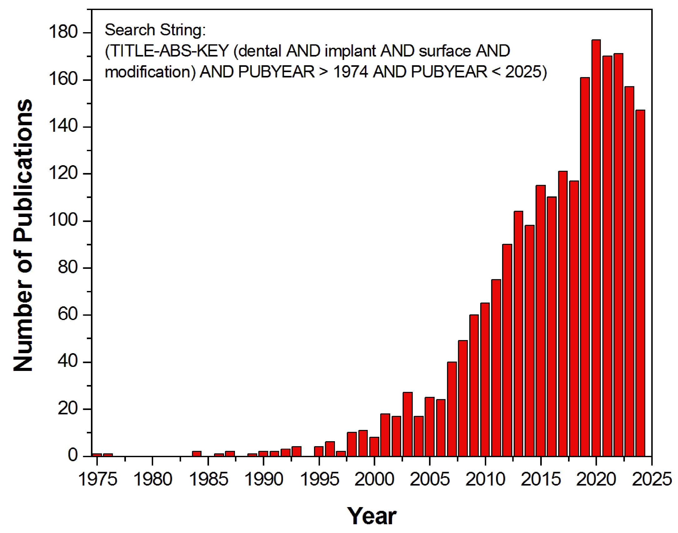
Figure 1. The number of publications on dental implant surface modifications indexed in the Scopus database from 1975 to 2024 (collected on 28 October 2024).
Coatings 15 00109 g001
Figure 2. Osseointegration process of titanium dental implant 3, 7, 14, and 21 days after implantation in mice [94].
Coatings 15 00109 g002
Figure 3. (a) Machined WINSIX dental implant by BioSAF IN [133]; (b,c) SEM image of a titanium implant with machined surface [134].
Coatings 15 00109 g003
Figure 4. (a) IMZ Original dental implant by Spotimplant [148]; (b,c) SEM image of a titanium implant with a TPS surface after 9 years of implant production, with a typical structure resembling molten metal splashes [149].
Coatings 15 00109 g004
Figure 5. (a) Sandblasted Prima Plus 4.1 dental implant by Lifecore Dental [165]; (b,c) SEM image of a titanium implant with a sandblasted surface [134].
Coatings 15 00109 g005
Figure 6. (a) Dental implant type 3i T3 by BIOMET 3i [178]; (b,c) SEM image of a titanium implant with HA surface [162].
Coatings 15 00109 g006
Figure 7. (a) The Osseotite dental implant system from Biomet 3i [187]; (b) SEM image of the Osseotite titanium implant with DE surface at 20,000× magnification [188]; (c) enhanced microscopy image of the Osseotite surface showing platelet activation [189].
Coatings 15 00109 g007
Figure 8. (a) Dental implant of the Standard Implants type with SLA surface by Straumann Institute [198]; (b,c) SEM image of a titanium implant with SLA surface [134].
Coatings 15 00109 g008
Figure 9. (a) Demonstration of the ultrahydrophobic properties of SLA surface and the superhydrophilic properties of SLActive surface. (b) Immersion of dental implants with SLA and SLActive surfaces in water. (c) Implant stability after implantation, with reduction in stability combined with gradual increase in secondary stability leading to a decline in overall stability (stability dip) during the period between weeks 2 and 4. (d) Acceleration of osseointegration between weeks 2 and 4 as a result of using an implant with SLActive surface [207].
Coatings 15 00109 g009aCoatings 15 00109 g009b
Figure 10. (a) Dental implant with Straumann® SLActive® surface (Basel, Switzerland) [209]; (b) SEM image of Roxolid® SLA® surface [210]; (c) SEM image of Roxolid® SLActive® surface [210]; (d) SEM image of Roxolid® SLActive® surface without nanostructures [211]; (e) SEM image of Roxolid® SLActive® surface with nanostructures [211].
Coatings 15 00109 g010
Figure 11. (a) TiUnite® Dental Implant by Nobel Biocare [220]; (b) SEM image of the anodized TiUnite® implant with a porous surface [134]; (c) hemostasis by the newly formed fibrin matrix on the TiUnite surface [224].
Coatings 15 00109 g011
Figure 12. (a) OsseoSpeed dental implant by Astra Tech [244]; (b,c) SEM image of a titanium implant with fluoride-enriched surface [134].
Coatings 15 00109 g012
Figure 13. (a) IMAX NHSI hybrid dental implant by iRES covered with a bioactive hyaluronic acid nanolayer [256]; (b) the hybrid dental implant features a section labeled M that has been machined, while the section labeled A has undergone a sandblasting process followed by a double acid etching treatment [257]; (c) the timeframe image of the Wilhelmy plate experiment [257].
Coatings 15 00109 g013
Figure 14. SEM image of the hybrid dental implant covered with a bioactive hyaluronic acid nanolayer: (a) machined surface in the section labeled M at 20,000× magnification; (b) machined surface in the section labeled M at 50,000× magnification; (c) sandblasted, double acid-etched surface in the section labeled A at 20,000× magnification; (d) sandblasted, double acid-etched surface in the section labeled A at 50,000× magnification [257].
Coatings 15 00109 g014
Figure 15. (a) Tapered Internal Plus (4.5) dental implant with Laser-Lok surface technology by BioHorizons [266]; (b) the Laser-Lok surface at 800× magnification with uniformly shaped microchannels designed to facilitate and enhance tissue growth [267]; (c) a colorized SEM image of a dental implant showing connective tissue that is physically attached and interwoven with the Laser-Lok surface [267].
Coatings 15 00109 g015
Disclaimer/Publisher’s Note: The statements, opinions and data contained in all publications are solely those of the individual author(s) and contributor(s) and not of MDPI and/or the editor(s). MDPI and/or the editor(s) disclaim responsibility for any injury to people or property resulting from any ideas, methods, instructions or products referred to in the content.