Article

Fabrication of PVTF/COL Composite Films and Its Impact on Osteogenic Differentiation

1
State Key Laboratory of Silicon and Advanced Semiconductor Materials, School of Materials Science and Engineering, Zhejiang University, Hangzhou 310058, China
2
Stomatology Hospital, School of Stomatology, Zhejiang University School of Medicine, Zhejiang Province Clinical Research Center for Oral Diseases, Key Laboratory of Oral Biomedical Research of Zhejiang Province, Cancer Center of Zhejiang University, Hangzhou 310006, China
3
Key Laboratory of Acoustics and Vibration Applied Measuring Technology, Zhejiang Key Laboratory of Acoustic Intelligent Sensing and Advanced Measurement, Zhejiang Institute of Quality Sciences, State Administration for Market Regulation, Hangzhou 310013, China
4
Zhejiang Key Laboratory of Acoustic Intelligent Sensing and Advanced Measurement, Zhejiang Institute of Quality Sciences, State Administration for Market Regulation, Hangzhou 310013, China
*
Authors to whom correspondence should be addressed.
Coatings 2025, 15(4), 416; https://doi.org/10.3390/coatings15040416
Submission received: 28 February 2025 / Revised: 23 March 2025 / Accepted: 27 March 2025 / Published: 1 April 2025

Abstract

:
Bone tissue is intrinsically electroactive, and electrical signaling is one of its key regulatory mechanisms. The electroactive poly (vinylidene fluoride trifluoroethylene) (PVTF), due to its piezoelectricity, can provide electrical stimulation to cells, regulating their proliferation and osteogenic differentiation. Collagen I (COL) is the main organic component of bone and is involved in various physiological processes of bone. A crucial question that remains to be explored is whether electroactive materials can meet the requirements for GBR membranes and what synergistic effects electrical signals and collagen’s biochemical signals might have on cellular behavior. In this study, PVTF/COL composite films were prepared using polydopamine (PDA). It was found that collagen modification could increase the surface Kelvin potential of PVTF from −5.07 V to 2.22 V, reduce the WCA from 98.9° to 33.2°, and maintain the tensile strength of PVTF at 24.94 MPa. Additionally, the composite film significantly promoted the adhesion and proliferation of bone marrow stem cells (BMSCs), and the ALP activity on PPC3 films after 7 days was 5.6 times higher than that on P films. This study presents a novel and effective approach for surface modification of PVTF and explores its potential applications in GBR.

1. Introduction

Periodontitis is an infection and inflammation caused by anaerobic bacterial colonies in the oral environment, which can damage the gum and tooth-supporting tissues. According to the Centers for Disease Control and Prevention, 30% of adults aged 47.2 years and older suffer from periodontal disease, with the incidence rising to 65% in adults aged 70.1 years and older [1]. Current treatments for periodontitis mainly target symptoms, such as removing dental plaque and reducing inflammation [2,3]. Although these therapies can slow disease progression, they do not address the reconnection of periodontal tissue to teeth or the restoration of damaged tissues, leading to ongoing tooth function impairment. Furthermore, some treatments may inadvertently damage the periodontal tissue and alveolar bone, contributing to tooth loosening and further tooth loss [4]. The absorption of alveolar bone is an important cause of tooth loss. The ideal treatment for periodontitis involves reconstructing the complex structure of periodontal tissue, including dental bone, alveolar bone, periodontal ligaments, and gingival tissue. Treatment options include bone grafting, guided bone regeneration (GBR), alveolar traction, and others. Among these, GBR is particularly promising, as it avoids the limitations of bone grafts sources and has been widely integrated with implant therapy [5,6,7].
The key principle of GBR is to maintain the space for bone regeneration and mechanically prevent the invasion of surrounding connective tissue, which enhances treatment effectiveness [8,9]. The essential requirements for GBR membranes include biocompatibility, mechanical stability for space maintenance, cell occlusion, tissue integration, and handleability in clinical practice. Currently, GBR membranes are typically classified into two types: absorbable membranes, such as collagen and polylactic acid, and non-absorbable membranes, such as titanium and polytetrafluoroethylene (PTFE). Absorbable membranes, primarily composed of collagen [10], have good bioactivity and are expected to have high therapeutic effects without the need for secondary surgery for removal. However, their mechanical strength is generally weak and further decreases after degradation, which is unfavorable for maintaining the growth space at the bone defect site. Non-absorbable membranes, such as titanium, PTFE, and various polymers [11,12], provide high mechanical strength and can effectively maintain the growth space. These materials are typically biologically inert and function only passively, limiting their therapeutic effect. As a result, they often require further material modifications or coordination with other treatment strategies.
Poly (vinylidene fluoride) (PVDF) is an electroactive material known for its excellent piezoelectricity and biocompatibility. It can alter its physicochemical properties in response to electrical signals or generate electrical signals when subjected to external mechanical stimuli [13,14]. PVDF primarily exists in five crystalline phases: α, β, γ, δ, and ε. The first three phases are more common, with the β-phase being the main contributor to its piezoelectricity [15,16]. By copolymerizing PVDF with trifluoroethylene, the overall crystallinity of the piezoelectric β-phase can be increased due to the steric effect, thereby enhancing electroactivity. This copolymer is referred to as PVTF [17]. Numerous studies have investigated its applications in the biomedical field. For example, Zhang et al. fabricated PVTF films with patterned surface potentials using specially designed electrodes, which promoted osteogenic differentiation of stem cells through integrins [18]. Similarly, Lin et al. generated piezoelectric potentials in PVTFs by applying mechanical deformations, which facilitated bone regeneration via calcium channels [19]. Moreover, bone is a tissue with intrinsic electroactivity [20]. Electrical signals are a regulator of bone tissue and are important for the maintenance of its daily homeostasis and for regeneration and recovery [21,22]. Therefore, it is very promising to use PVTF as a part of GBR membranes and utilize its electroactivity, promising better bone regeneration promotion than bioinert PTFE and titanium. However, the pure PVTF surface has poor hydrophilicity, which may hinder its infiltration in bodily fluids, thereby affecting subsequent cell adhesion and overall therapeutic efficacy.
Collagen refers to a group of at least 29 distinct proteins, making up the most abundant protein component in the extracellular matrix (ECM). It accounts for approximately 20%–30% of the total body protein weight in mammals [23]. In bones, type I collagen is the most prevalent, constituting over 90% of the organic mass, primarily secreted by osteoblasts [24]. Collagen contains numerous specific structural domains that facilitate interactions with cells, such as RGD sequences that bind to integrins [25]. During extracellular matrix formation and degradation, cells can recognize collagen within their microenvironment. As an implant material, collagen not only provides physical support for cell adhesion and growth but also influences cell behavior and fate through receptor-mediated interactions.
Therefore, this study innovatively utilized PDA with wet adhesion to modify collagen onto the surface of PVTF. This material strategy not only enhances the therapeutic effect by synergistically combining the biochemical signal of collagen with the electrical signal of PVTF but also overcomes the challenges posed by the weak mechanical properties of the collagen films and the surface hydrophobicity of PVTF, as shown in Table 1. Moreover, the fabrication strategy of this study is versatile, as PDA adhesion is extensive and efficient. The morphology, surface potential, hydrophilicity, and mechanical properties of the composite film were characterized, and the response behavior of BMSCs on the film surface was investigated, highlighting the potential application of the composite film in guiding bone regeneration.

2. Materials and Methods

2.1. Materials

The poly (vinylidene fluoride-trifluoroethylene) (70/30) powders were purchased from Piezotech (Pierre-Bénite, France). Type Ⅰ Collagen was purchased from Yierkang Co., Ltd. (Beijing, China). N, N-dimethylformamide (DMF) was purchased from Aladdin Biochemical Technology Co., Ltd. (AR, Shanghai, China). Dopamine hydrochloride, Tris, and hydrochloric acid were purchased from Sinopharm Chemical Reagent Co., Ltd. (Shanghai, China).

2.2. Fabrication of PVTF/COL Composite Film

The PVTF film was fabricated using the doctor blade technique, and its thickness was about 50 μm, as previously reported. In brief, 1 g of PVTF powder was added to 5.5 mL of DMF solution and stirred at room temperature for 3 h. The resulting mixture was then spread onto a glass substrate, subjected to heat treatment at 210 °C for 1 h, and subsequently cooled to room temperature in oven. Following preparation, the PVTF films were cut in 1 × 1 cm and exposed to an applied electric field to induce surface charging. Previous studies have shown that polarized PVTF could promote osteogenic differentiation of stem cells, with the sample polarized to D33 = 15 exhibiting the best effect [26]. Since collagen has relatively weak piezoelectric properties, its inclusion in the composite may reduce the overall electrical properties. Therefore, in this study, PVTF was first polarized to D33 = 20 during the material processing stage, whereas the D33 of the original film was 0. Under the influence of external electric field, the electric dipoles within PVTF aligned in a more ordered manner, leading to an enhancement in the overall piezoelectric properties of the film.
A 2 mg/mL aqueous solution of dopamine hydrochloride was prepared, and its pH was adjusted to 8.5 using a 1 M tris buffer. The polarized PVTF films were then immersed in this dopamine hydrochloride solution for 12 h, with continuous stirring, to facilitate the formation of a polydopamine layer on the surface of the films, thus yielding the PP films.
Dissolved type I collagen in a 0.05 M hydrochloric acid solution to obtain collagen solutions. When the concentration is higher than 3 mg/mL, the collagen solution becomes highly viscous, making it challenging to perform relevant experimental operations. Therefore, this study selected collagen solution with concentration of 1, 2, and 3 mg/mL for the following experiment. After rinsing with deionized water, the PP films were subsequently immersed in varying concentrations of collagen solution in 24-well plates, with 1 mL of collagen solution added per 1 × 1 cm film per well, and incubated for 12 h at room temperature to obtain the final composite films, designated as PPCX (X = 1, 2, 3) according to the concentration of collagen solution. The schematic diagram of the preparation process and polarization treatment is shown in Figure 1.

2.3. Characterization of Materials

The surface topography and average roughness (Ra) of the composite films were investigated by field-emission scanning electron microscope (FE-SEM, Hitachi SU-70) and atomic force microscope in tapping mode (AFM, NTEGRA Spectra C), respectively. Anti-collagen I antibody (ab138492, Abcam, Shanghai, China) and Goat Anti-Rabbit IgG H&L (Alexa Fluor 488) (ab150077, Abcam, China) were used to observe collagen I by confocal laser scanning microscope (CLSM 880, Zeiss, Jena, Germany). Fourier transform infrared spectroscopy of the composite films was recorded by an infrared spectrometer with an attenuated total refraction accessory (ATR FT-IR, Nicolet 5700, Thermo Fisher Scientific, Madison, WI, USA). Surface chemical composition of films were investigated with X-ray photoelectron spectroscopy (XPS, Thermo Scientific K-Alpha, Thermo Fisher Scientific, East Hampton, NY, USA) using the Al Ka (1486.6 eV) source at a base pressure of 3.5 × 10⁻⁹ Torr. The piezoelectric coefficient of the composite films was investigated using a quasi-static D33 tester (ZJ-3AN, Institute of Acoustics, Chinese Academy of Science). The surface potential of films was characterized by Kelvin Probe Force Microscopy (KPFM, nanoIR2-fs, Anasys Instruments, Santa Barbara, CA, USA). The surface hydrophilicity of films were tested by a contact angle meter (DCA20, Dataphysics, Filderstadt, Germany).
The tensile and puncture strength of films were tested using the Universal Testing Machine (Z005, ZwickRoell, Ulm, Germany). In the tensile experiments, samples measuring 1 × 3 cm were prepared and clamped with a jig at 1 cm from both ends. The samples were then stretched at a rate of 1.5 mm/min until specimen failure. In the puncture experiment, a 2 × 2 cm sample was prepared and fixed between two flat fixtures. The fixtures have a central hole with a diameter of 1 cm, and a punch with a diameter of 6 mm was used to apply pressure through the hole at a rate of 1.5 mm/min until the sample ruptures.

2.4. Cell Culture

Bone marrow mesenchymal stem cells (BMSCs) were isolated from the tibiae of three-week-old male Sprague–Dawley rats. The BMSCs were cultured in Minimum Essential Medium Alpha (MEM Alpha, GENOM, Jiaxing, China) supplemented with 10% fetal bovine serum (Cellmax, Melbourne, Australia), 1% antibiotic solution containing 10,000 units/mL penicillin, 1% sodium pyruvate, 10,000 μg/mL streptomycin, and 1% MEM non-essential amino acids (Sigma, St. Louis, MO, USA). After enzymatic digestion with trypsin, the cells were seeded at a density of 50,000 cells/well in a 24-well plate, with a single 1 × 1 cm UV-sterilized film placed in each well. Each well contained 500 μL culture medium. The medium was replaced with fresh medium 24 h post seeding and then every two days thereafter. When the primary cells reached approximately 80%–90% confluence, they were passaged.

2.5. Cell Vitality Assays

In accordance with the aforementioned method, BMSCs were cultured on the samples for 1 and 3 days to quantify cell viability and proliferation, using the Cell Counting Kit-8 (CCK-8, Dojindo Laboratories, Kumamoto, Japan) assay. The samples containing cells to be tested were transferred to a new 24-well plate, and 500 μL of culture medium containing 10% CCK-8 reagent was added to each well. After incubating at 37 °C for 2 h in a cell culture incubator, the culture medium was transferred to a 96-well plate, and its absorbance at 450 nm was measured using a microplate reader (Infinite F50, Tecan, Männedorf, Switzerland) to assess cell viability.

2.6. Alkaline Phosphatase (ALP) Assay

The osteogenic differentiation capacity of the BMSCs were determined primarily by measuring ALP activity. The inoculated cells on the surface of films were cultured in an incubator for 7 days, with medium changed every two days. After the end of three days, cells were cultured in medium that induces osteogenic differentiation, which contains an additional 10 mM β-glycerophosphate, 0.1 μM dexamethasone, and 0.25 mM ascorbic acid (all from Sigma, USA), instead of normal medium. Upon arrival at the designated time, the cell on films were lysed by cell lysis buffer (Sigma, USA), and the lysate was used to extract the protein in cells. The total protein content was quantified using the Bicinchoninic Acid (BCA) assay (Pierce 23225, Thermo Fisher, Rockford, IL, USA). The ALP activity of different samples was tested using a LabAssay ALP kit (Wako Pure Chemical Industries, Osaka, Japan).

2.7. Statistical Analysis

All of the data are shown as mean ± standard deviation and were obtained from at least three independent experiments (n ≥ 3). One-way analysis of variance (ANOVA) followed by Tukey’s post hoc test was performed for multiple-group analyses. The statistically significant different in the difference was defined as * p < 0.05, ** p < 0.01, and *** p < 0.001.

3. Results and Discussion

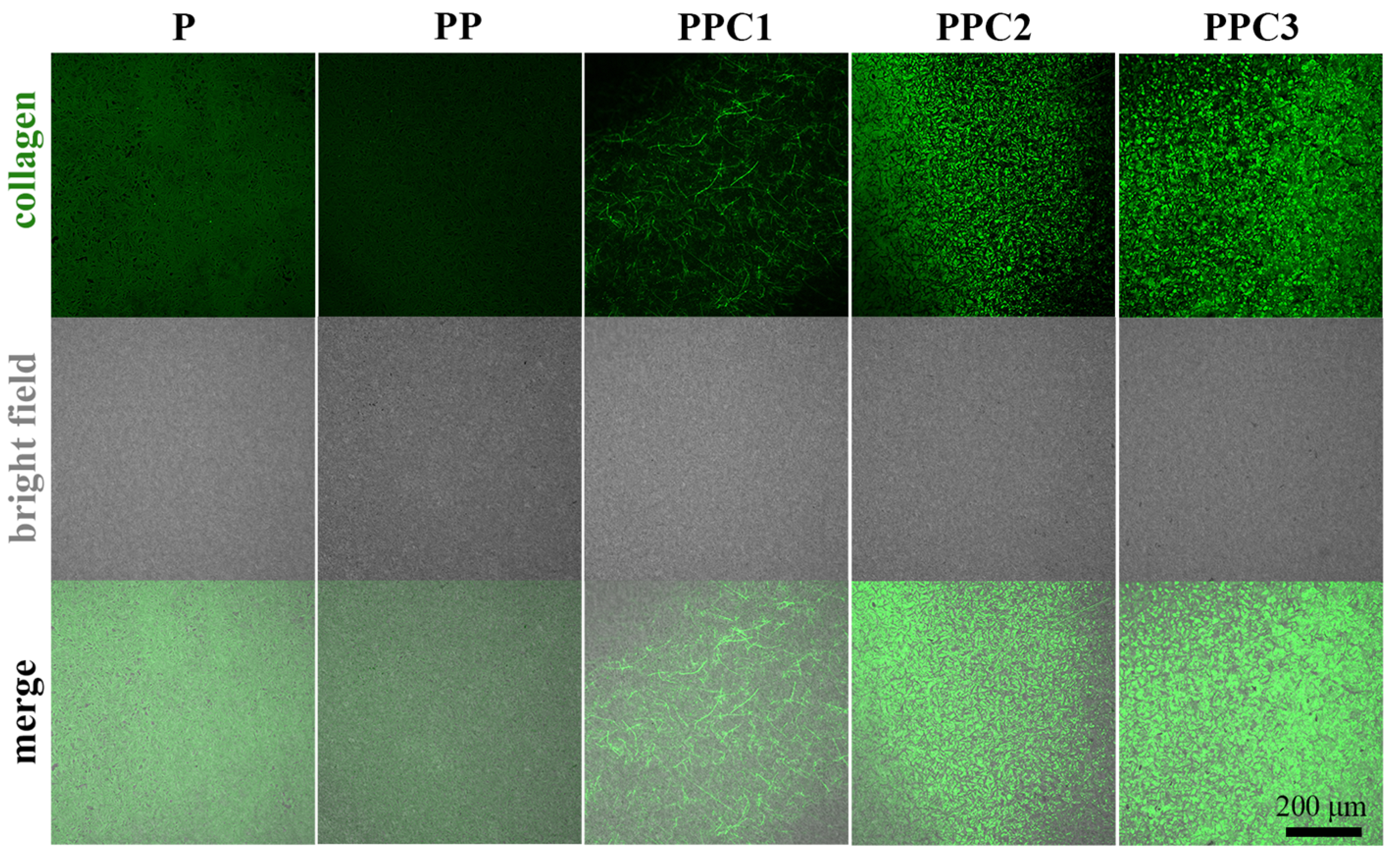
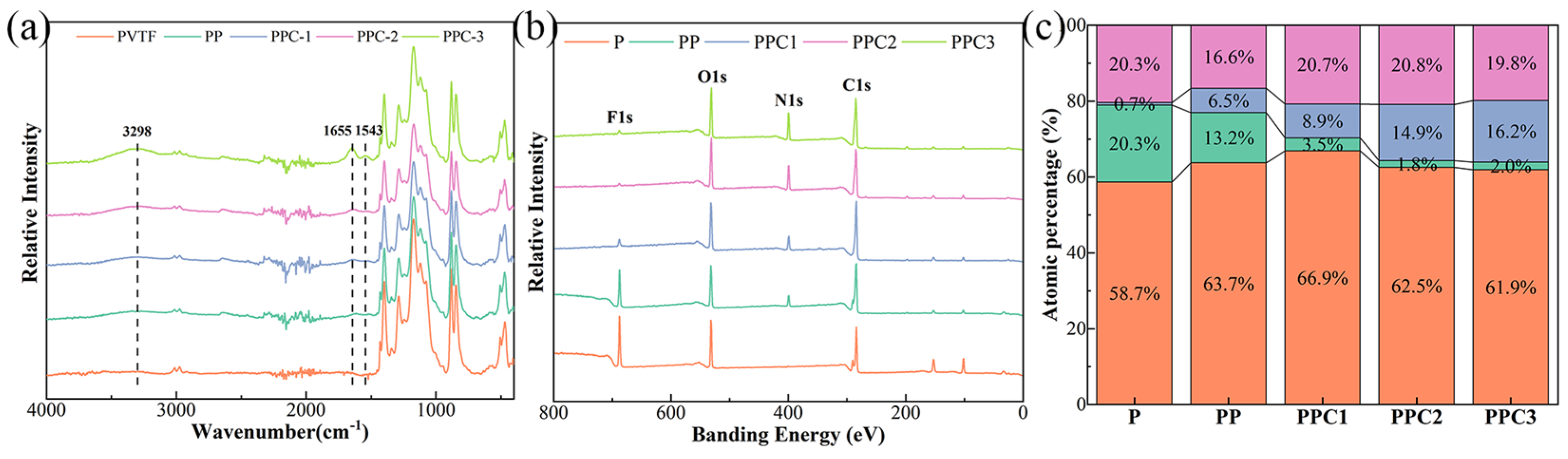
3.1. The Morphology and Composition of PVTF/COL Composite Film

Figure 2a illustrates the surface morphology of different films. The typical crystalline folded morphology of PVTF can be observed in the Figure 2a [27], with no significant alteration seen following the modification with PDA and low concentrations of collagen. However, when the collagen concentration increased to 2 mg/mL, the surface of PVTF showed reduced concavity and became smoother. This effect was further enhanced by higher-concentration collagen solution. Additionally, AFM was employed to characterize the change in surface roughness (Sa) of the films, as shown in Figure 2b. The surface roughness of the films increased slightly after PDA treatment. This was because the rapid self-polymerization of dopamine in solution, which led to the deposition of particles on the film surface rather than the formation of thin layers. The modification of collagen was shown to have a significant effect on surface roughness, with a greater amount of collagen deposited resulting in a smoother film surface (Figure 2b). For example, the PPC3 sample exhibited the lowest roughness (Sa = 331.1 ± 21.6 nm), which was 25% lower than that of the PVTF sample. And the surface roughness of PPC1 and PPC2 was 365.4 ± 21.6 nm and 368.5 ± 30.8 nm, respectively, slightly larger than that of the PPC3 films and smaller than that of the other two films. In the cross-section image of the films, a thin collagen layer, less than 1 μm in thickness, was observed on the surface of the PPC2 and PPC3 films, as indicated by the orange arrows in Figure 2c. This thin layer was not observed on the surface of PPC1 films, which may indicate that the collagen content on it was insufficient to form a thin layer.
To visualize the distribution of collagen on composite films surface, fluorescence images of the composite films were obtained using a confocal laser scanning microscope, as shown in Figure 3. A weak background fluorescence was observed in the P films, which roughly exhibit a typical crystalline folded morphology of PVTF. This fluorescence may arise either from the inherent fluorescent properties of PVTF [28] or from the adsorption of certain dyes due to its surface characteristics [29]. A similar fluorescence pattern was also observed in the PP films. In contrast, the PPC1 sample displayed numerous fibrous structures, likely resulting from collagen self-assembly and subsequent deposition on the film surface [30]. As the collagen solution concentration increased during preparation, patches of green collagen became visible in the PPC2 sample. The black areas on PPC2 films exhibited a crystalline fold-like morphology, which may be attributed to the thin layer of collagen deposition was insufficient to cover the protrusions of PVTF. Finally, the PPC3 sample showed a significant accumulation of collagen, with some areas exhibiting a globular shape, distinguishing it from the PPC2 sample. These results visualize the differences in collagen content on the surface of different films.
The composition of the films was further determined by FT-IR and XPS, and the results are shown in Figure 4. In the FT-IR spectrum, three new absorption peaks appeared after the films were compounded with collagen, all of which were characteristic absorption peaks belonging to collagen. The amide A band of collagen was observed at 3298 cm−1, corresponding to N–H stretching vibrations. The amide I band of collagen at 1655 cm−1 indicated the peptide’s secondary structure and hydrogen bonding between N–H and C=O. The amide II of collagen band at 1543 cm−1 represented N–H bending vibrations coupled with C–N stretching vibrations [31]. In addition, the intensity of these three absorption peaks was positively correlated with the concentration of collagen solution during the preparation process. In the XPS results of Figure 4b, the F absorption peaks weakened and the N absorption peaks strengthened as collagen was incorporated. The elemental content plots also show a similar trend in Figure 4c. This phenomenon was attributed to the fact that the XPS detection depth is approximately 10 nm. As the composite collagen layer thickened, the signals from the PVTF-related elements (F) were increasingly blocked, resulting in the observed trend. These composition test results confirmed the successful preparation of PVTF/PDA composite films with varying collagen contents on surface.

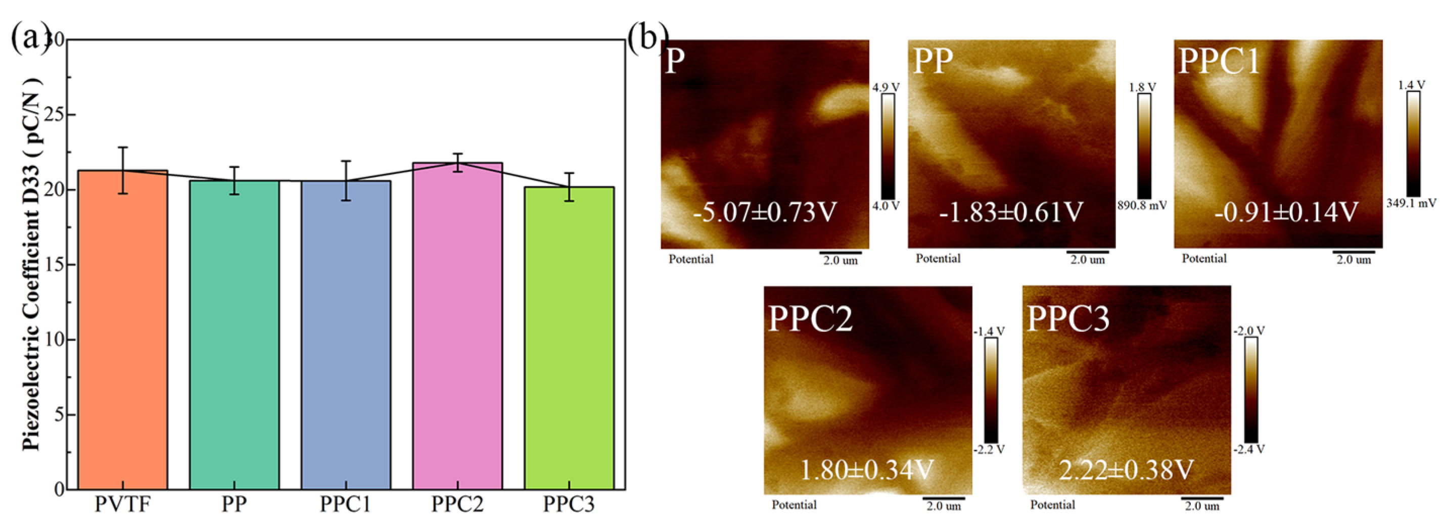
3.2. The Surface Potential and Hydrophilicity of PVTF/COL Composite Film

PVTF is a piezoelectric polymer known for its stable physical property and excellent biocompatibility. And its piezoelectricity can be enhanced through polarization treatment [32]. When used as a GBR membrane at the injury site, the surface potential can exert effects without the need for stress stimulation. In addition, in the presence of external stress, a piezoelectric potential could be generated to further stimulate the cells through the piezoelectric effect. Many studies have shown that PVTF-based electrical signals could promote the differentiation and regeneration of bone tissue. For example, Tang et al. found that surface potential promoted osteogenic differentiation by changing the conformation of adsorbed proteins [26]. During the preparation of films, the PVTF was initially polarized to D33 = 20 via contact polarization (Figure 5a). The test result of the piezoelectric coefficient D33 of various samples indicated that the modification of PDA and collagen did not affect its piezoelectric properties, confirming that the composite film could still generate effective electrical signal stimulation through the piezoelectric effect.
The surface potential test result of the composite film was depicted in Figure 5b. It is important to note that the value obtained by the instrument reflects the probe potential minus the sample surface potential, requiring a reversal of the positive and negative values during analysis. The pure PVTF films exhibited a strong negative surface potential of −5.07 V, which resulted from the regular arrangement of electric dipoles formed by the CF bonds with strong polarity in it. The modification of PDA shifted the surface potential in a positive direction, while collagen modification further increased the surface potential. For instance, the PPC3 films had a surface potential of 2.22 V, which was 7.29 V higher than that of P films. This shift occurred because PDA synthesized at pH 8.5 had a weaker negative charge, probably due to its quinone imine and catechol groups, compared to pure PVTF [33,34]. And the complexation of PDA would result in a change in surface potential towards the intermediate value. The electrical charge of collagen is determined by its isoelectric point, with a positive charge below this point and a negative charge above it. The isoelectric point of collagen varies in the literature [35,36,37] depending on its source, processing methods, and electrolyte solution conditions. For example, Ding et al. reported an isoelectric point of 4.9 for alkali-soluble collagen [38], while Li et al. found that acid-soluble collagen derived from cattle had an isoelectric point ranging from 7.5 to 9.2 under different electrolyte conditions [39]. In this study, acid-soluble collagen was used, which means the collagen carried a certain positive charge. This contributed to a shift in the surface potential of the composite films towards a more positive value after the composite was formed, which is in line with the trend in Figure 5b. The PPC2 and PPC3 films exhibited similar surface potentials, likely because the collagen content was high enough to form a layer, causing the surface potential to approach that of the pure collagen film.
The hydrophilicity of the film surface is crucial for cell adhesion and proliferation, as the initial event after implantation is the infiltration of the material with body fluid, followed by protein adsorption and cell adhesion. The difference in hydrophilicity leads to different protein adsorption in the future, and the protein layer adsorbed on the material surface would mainly determine its biological effects [40]. For example, Parisi et al. found that hydrophilicity promoted the selective adsorption of titanium to HFN but not HSA, which enhanced the adhesion, spreading, and proliferation of cells on it [41]. As shown in Table 2, the water contact angle of the pure PVTF films was 98.9°, which decreased to 59.8° after the deposition of PDA. And it further decreased with an increasing amount of deposited collagen, reaching 33.2° on the PPC3 films. The modification of collagen could effectively enhance the hydrophilicity of PVTF, which is beneficial for its biocompatibility.

3.3. The Tensile Strength and Puncture Strength of PVTF/COL Composite Film

The strength of the composite film is critical for its application in guided bone regeneration, as it requires the ability to maintain a space for ingrowth, cell occlusion, etc. [4,42,43]. In addition, daily physiological activities can exert pressure, making it ideal to use materials with mechanical properties similar to those of physiological tissues to prevent deformation or mismatch. The tensile strength and puncture strength of PPC3 samples were tested, and the results are shown in Figure 6. The reason for not measuring the other groups was that the content of PDA and collagen modified on the PVTF surface was very low, so the strength of the composite films was always mostly provided by PVTF and did not change significantly. The films of varying thicknesses exhibited some variations, but generally, the tensile strength of the composite film was 24.94 ± 0.95 MPa. In comparison, the tensile strengths of collagen membranes from BioGide (0.44 mm thick), Collprotect (0.28 mm thick), and Jason (0.44 mm thick) were reported as 4.8, 13.1, and 13.0 MPa, respectively [44]. Thus, the prepared composite film demonstrated higher tensile strength. The puncture strength of PPC3 with a thickness of about 0.05 mm was 0.54 ± 0.02 N/mm2. It has been reported that the puncture strength of BioGide collagen film (0.4 mm thick) was 1.65 ± 0.45 N/mm2, Creos collagen film (0.2 mm thick) had a puncture strength of 2.81 ± 0.27 N/mm2, and titanium (0.25 mm thick) had a puncture strength of 5.36 ± 0.25 N/mm2 [45]. Since the strength of the composite film is primarily provided by the PVTF component, and its thickness is adjustable, the prepared composite film could exhibit a significant puncture strength at the same thickness with other films. The oral environment is extremely complex, placing higher demands on the mechanical properties of the GBR membrane used. A membrane with too low a mechanical strength cannot maintain the internal growth space, while excessive mechanical strength may cause unnecessary damage due to deformation or mismatch under stress. The specific gold standard still requires further clinical research. The results above indicate that the prepared PVTF/COL film had a higher tensile strength than that of collagen membrane, with a puncture strength between that of collagen membrane and titanium. This mechanical performance suggested that it can meet most clinical application requirements and has practical application potential. Moreover, polymers exhibit exceptional plasticity. Their mechanical strength can be further adjusted through chemical modifications, composites [46], and other approaches to meet specific application requirements.

3.4. The Biocompatibility and Mechanical Properties of PVTF

From the Figure 7a of 1-day cell viability, it can be seen that the PPC3 sample had the highest cell viability, indicating that its surface was most favorable for cell adhesion. This was attributed to the composite of collagen, which improved the surface electrical properties of the film and increased its hydrophilicity. This could promote the binding of adhesion molecules on the cell membrane to corresponding ligands on the material surface, thereby improving the adhesion of cells. On day three, the collagen composite samples demonstrated a significantly higher cell viability, whereas the pure PVTF and PP samples had lower cell viability. This indicates that collagen could significantly promote cell proliferation (Figure 7b). In addition, the amount of total protein in cells measured on day 7 showed a similar pattern (Figure 7c). This means that the collagen still significantly enhanced the bioactivity of films over a longer period of time. However, the P samples may have limited the total number of cells on it due to their strong hydrophobicity, which is not conducive to cell spreading and migration on them [47].
Alkaline phosphatase, an enzyme secreted by osteoblasts, is a specific marker of osteoblast maturation and one of the most commonly used indicators of their secretory function. To a certain extent, the change in the level of ALP activity reflects the degree of osteogenic differentiation of BMSCs. As shown in the Figure 7d, in terms of mean values, the ALP activity of the PPC3 samples was the highest, being 5.6, 1.9, 1.2, and 1.2 times higher than that of the P, PP, PPC1, and PPC2 samples, respectively. All collagen-composited films demonstrated significantly higher ALP activity, suggesting that collagen promoted osteogenic differentiation of BMSCs. Moreover, the effect was more pronounced with higher collagen content. It has been reported that collagen not only provides a suitable physiological microenvironment for cells but is also a promoter and template for osteoblast mineralization [48]. However, no significant difference was observed between the PPC1 and PPC2 samples with varying collagen content. This may be attributed to the fact that as cells proliferated and differentiated, the extracellular matrix became more abundant, gradually modifying the extracellular microenvironment. And collagen is an essential component of the ECM. The lower collagen content on the film surface may primarily act as a biochemical signal for cellular excitation in the early stage, while the ECM secreted by cells takes a dominant role in the later stages. Although PVTF is stable and nearly non-degradable in vivo, collagen may degrade in the in vivo environment, potentially affecting long-term performance. However, distinguishing between collagen modified on the surface of implants and collagen secreted by cells is challenging, which requires more refined experimental designs in the future. In summary, the PVTF/COL composite film possesses mechanical strength suitable for GBR applications and can significantly enhance the biological activity of the films, effectively promoting the adhesion, proliferation, and osteogenic differentiation of BMSCs, as shown in the Table 3.

4. Conclusions

In this study, collagen was modified on the surface of electroactive PVTF to achieve synergistic stimulation of electrical signals and collagen signals to cells on it. And the electrical properties, mechanical properties, and biological activity of the composite film were evaluated. The main conclusions can be summarized as follows:
(1)
Polydopamine effectively facilitated the composite of collagen on the surface of PVTF films, with varying composite amounts achieved by adjusting the concentration of the collagen solution. Although the surface morphology of the composite film showed no significant changes, its hydrophilicity was notably improved, with the water contact angle decreasing from 98.9° for pure PVTF to 33.2° for PPC3;
(2)
The prepared composite film exhibited good mechanical strength, with a tensile strength of 24.94 MPa, 1.9 times higher than that of the Jason collagen membrane. And the puncture strength of composite film was 0.54 N/mm2, which falls between the values of commercial collagen membranes and titanium. The surface potential of the film gradually shifted toward the positive potential direction as collagen recombined, achieving a Kelvin potential of 2.22 V on the PPC3 surface;
(3)
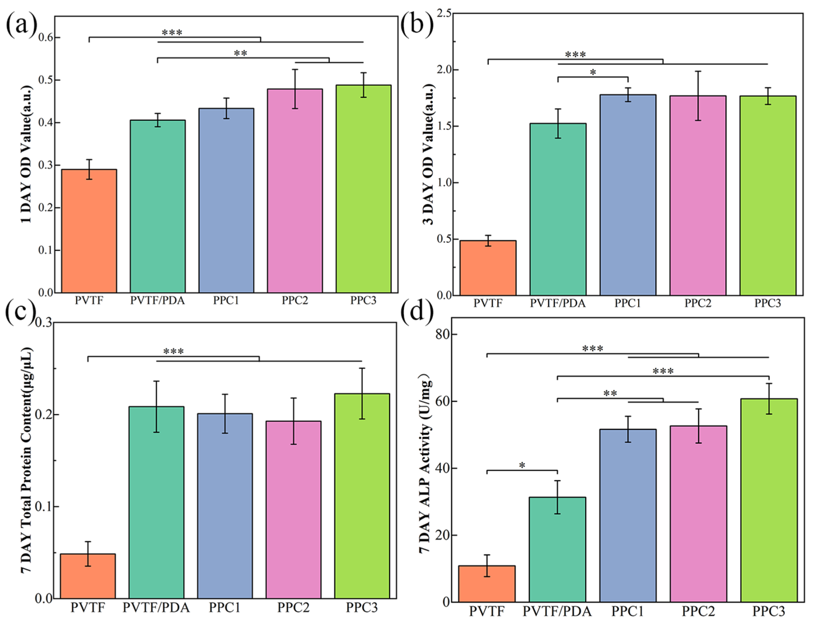
The prepared composite film demonstrates good biocompatibility and bone-promoting effects, with the PPC3 sample showing the best overall performance. Compared to the PVTF sample, the adhesion of cells to the PPC3 sample was 1.7 times higher after 1 day, cell proliferation was 3.6 times higher after 3 days, and ALP activity was 5.6 times higher after 7 days.
Therefore, the composite film prepared in this study meets the mechanical property and biological activity requirements for GBR and has great potential for clinical application. Additionally, while the biological activity of the composite films was evaluated through in vitro cell experiments, further exploration of their therapeutic effects and performance stability in vivo is needed.

Author Contributions

Conceptualization, H.L., W.L. and K.C.; methodology, H.L., C.W., W.L. and K.C.; software, H.L.; validation, H.L.; formal analysis, H.L.; investigation, H.L.; resources, X.C., W.W., X.Y. and K.C.; data curation, X.C. and K.C.; writing—original draft preparation, H.L.; writing—review and editing, H.L., C.W., W.L., X.Y. and K.C.; visualization, X.Y. and K.C.; supervision, X.C., W.W., X.Y. and K.C.; project administration, X.C., W.W., X.Y. and K.C.; funding acquisition, X.C., W.W., X.Y. and K.C. All authors have read and agreed to the published version of the manuscript.

Funding

This research was funded by the National Natural Science Foundation of China, grant numbers 52271252, 32271373, and 82201009. This research was funded by the Science and Technology Plan of State Administration of Market Regulation, grant numbers KJLJ202310, 2023MK048, and 2024MK050.

Institutional Review Board Statement

All animal experiments were approved by the Laboratory Animal Welfare and Ethics Committee of Zhejiang University (Ethics Code: ZJU20220735, approval date 14 February 2022).

Informed Consent Statement

Not applicable.

Data Availability Statement

The authors confirm the data supporting the findings of this study.

Conflicts of Interest

The authors declare no conflicts of interest.

References

  1. Eke, P.I.; Dye, B.A.; Wei, L.; Thornton-Evans, G.O.; Genco, R.J. Prevalence of Periodontitis in Adults in the United States: 2009 and 2010. J. Dent. Res. 2012, 91, 914–920. [Google Scholar] [CrossRef]
  2. Deas, D.E.; Moritz, A.J.; Sagun, R.S., Jr.; Gruwell, S.F.; Powell, C.A. Scaling and root planing vs. conservative surgery in the treatment of chronic periodontitis. Periodontol. 2000 2016, 71, 128–139. [Google Scholar] [CrossRef] [PubMed]
  3. Graziani, F.; Karapetsa, D.; Alonso, B.; Herrera, D. Nonsurgical and surgical treatment of periodontitis: How many options for one disease? Periodontol. 2000 2017, 75, 152–188. [Google Scholar] [CrossRef] [PubMed]
  4. Kim, K.; Su, Y.; Kucine, A.J.; Cheng, K.; Zhu, D. Guided Bone Regeneration Using Barrier Membrane in Dental Applications. ACS Biomater. Sci. Eng. 2023, 9, 5457–5478. [Google Scholar] [CrossRef]
  5. Donos, N.; Mardas, N.; Chadha, V. Clinical outcomes of implants following lateral bone augmentation: Systematic assessment of available options (barrier membranes, bone grafts, split osteotomy). J. Clin. Periodontol. 2008, 35, 173–202. [Google Scholar] [CrossRef]
  6. Chiapasco, M.; Zaniboni, M. Clinical outcomes of GBR procedures to correct peri-implant dehiscences and fenestrations: A systematic review. Clin. Oral Implant. Res. 2009, 20 (Suppl. 4), 113–123. [Google Scholar] [CrossRef]
  7. Bornstein, M.M.; Halbritter, S.; Harnisch, H.; Weber, H.P.; Buser, D. A retrospective analysis of patients referred for implant placement to a specialty clinic: Indications, surgical procedures, and early failures. Int. J. Oral Maxillofac. Implant. 2008, 23, 1109–1116. [Google Scholar]
  8. Alqahtani, A.M. Guided Tissue and Bone Regeneration Membranes: A Review of Biomaterials and Techniques for Periodontal Treatments. Polymers 2023, 15, 3355. [Google Scholar] [CrossRef]
  9. Lei, M.; Wan, H.; Song, J.; Lu, Y.; Chang, R.; Wang, H.; Zhou, H.; Zhang, X.; Liu, C.; Qu, X. Programmable Electro-Assembly of Collagen: Constructing Porous Janus Films with Customized Dual Signals for Immunomodulation and Tissue Regeneration in Periodontitis Treatment. Adv. Sci. 2024, 11, 2305756. [Google Scholar] [CrossRef]
  10. Yan, F.; Yu, M.; He, Y.; Wang, F.; Yang, F.; Zhao, X.; Zheng, Y.; Liu, Y.; Xia, D.; Liu, Y. Hierarchical Mineralized Collagen Coated Zn Membrane to Tailor Cell Microenvironment for Guided Bone Regeneration. Adv. Funct. Mater. 2025, 35, 2412695. [Google Scholar] [CrossRef]
  11. Zeller, A.-N.; Schenk, R.; Bonsmann, M.; Stockbrink, G.; Becher, S.; Pabst, A. Complication rates of guided bone regeneration using titanium-reinforced PTFE membranes: A retrospective analysis. Clin. Oral Investig. 2024, 28, 616. [Google Scholar] [CrossRef]
  12. Kohutiar, M.; Kakošová, L.; Krbata, M.; Janík, R.; Fekiač, J.J.; Breznická, A.; Eckert, M.; Mikuš, P.; Timárová, Ľ. Comprehensive Review: Technological Approaches, Properties, and Applications of Pure and Reinforced Polyamide 6 (PA6) and Polyamide 12 (PA12) Composite Materials. Polymers 2025, 17, 442. [Google Scholar] [CrossRef]
  13. Durán-Rey, D.; Brito-Pereira, R.; Ribeiro, C.; Ribeiro, S.; Sánchez-Margallo, J.A.; Crisóstomo, V.; Irastorza, I.; Silván, U.; Lanceros-Méndez, S.; Sánchez-Margallo, F.M. Development and evaluation of different electroactive poly(vinylidene fluoride) architectures for endothelial cell culture. Front. Bioeng. Biotechnol. 2022, 10, 1044667. [Google Scholar] [CrossRef]
  14. Purushothaman, S.M.; Tronco, M.F.; Kottathodi, B.; Royaud, I.; Ponçot, M.; Kalarikkal, N.; Thomas, S.; Rouxel, D. A review on electrospun PVDF-based nanocomposites: Recent trends and developments in energy harvesting and sensing applications. Polymer 2023, 283, 126179. [Google Scholar] [CrossRef]
  15. Saxena, P.; Shukla, P. A comprehensive review on fundamental properties and applications of poly(vinylidene fluoride) (PVDF). Adv. Compos. Hybrid. Mater. 2021, 4, 8–26. [Google Scholar] [CrossRef]
  16. Jolly, B.; Ullah, W.; Saxena, N.; Sharma, R.; Janyani, V. Structural and microstructural analysis of spin-coated PVDF thin films. Ferroelectrics 2021, 583, 151–161. [Google Scholar] [CrossRef]
  17. Mohammadpourfazeli, S.; Arash, S.; Ansari, A.; Yang, S.; Mallick, K.; Bagherzadeh, R. Future prospects and recent developments of polyvinylidene fluoride (PVDF) piezoelectric polymer; fabrication methods, structure, and electro-mechanical properties. RSC Adv. 2023, 13, 370–387. [Google Scholar] [CrossRef]
  18. Zhang, J.; He, X.; Zhou, Z.; Chen, X.; Shao, J.; Huang, D.; Dong, L.; Lin, J.; Wang, H.; Weng, W.; et al. The osteogenic response to chirality-patterned surface potential distribution of CFO/P(VDF-TrFE) membranes. Biomater. Sci. 2022, 10, 4576–4587. [Google Scholar] [CrossRef]
  19. Lin, W.; Zhou, Z.; Chen, Z.; Xu, K.; Wu, C.; Duan, X.; Dong, L.; Chen, Z.; Weng, W.; Cheng, K. Accelerated Bone Regeneration on the Metal Surface through Controllable Surface Potential. ACS Appl. Mater. Interfaces 2023, 15, 46493–46503. [Google Scholar] [CrossRef]
  20. Heng, B.C.; Bai, Y.; Li, X.; Lim, L.W.; Li, W.; Ge, Z.; Zhang, X.; Deng, X. Electroactive Biomaterials for Facilitating Bone Defect Repair under Pathological Conditions. Adv. Sci. 2023, 10, 2204502. [Google Scholar] [CrossRef]
  21. Liu, Z.; Wan, X.; Wang, Z.L.; Li, L. Electroactive Biomaterials and Systems for Cell Fate Determination and Tissue Regeneration: Design and Applications. Adv. Mater. 2021, 33, 2007429. [Google Scholar] [CrossRef]
  22. Thrivikraman, G.; Lee, P.S.; Hess, R.; Haenchen, V.; Basu, B.; Scharnweber, D. Interplay of Substrate Conductivity, Cellular Microenvironment, and Pulsatile Electrical Stimulation toward Osteogenesis of Human Mesenchymal Stem Cells in vitro. ACS Appl. Mater. Interfaces 2015, 7, 23015–23028. [Google Scholar] [CrossRef] [PubMed]
  23. Miyata, T.; Taira, T.; Noishiki, Y. Collagen engineering for biomaterial use. Clin. Mater. 1992, 9, 139–148. [Google Scholar] [CrossRef]
  24. Gelse, K.; Pöschl, E.; Aigner, T. Collagens—Structure, function, and biosynthesis. Adv. Drug Deliv. Rev. 2003, 55, 1531–1546. [Google Scholar] [CrossRef] [PubMed]
  25. Fang, Z.; Ding, H.; Li, P.; Qiao, H.; Liang, E.; Jia, Y.; Guan, S. Interaction Mechanism of RGD Tripeptide on Different Surfaces of Mg and Mg Alloys: A First-Principles Study. Coatings 2022, 12, 1814. [Google Scholar] [CrossRef]
  26. Tang, B.; Zhang, B.; Zhuang, J.; Wang, Q.; Dong, L.; Cheng, K.; Weng, W. Surface potential-governed cellular osteogenic differentiation on ferroelectric polyvinylidene fluoride trifluoroethylene films. Acta Biomater. 2018, 74, 291–301. [Google Scholar] [CrossRef]
  27. Wang, Z.; He, X.; Tang, B.; Chen, X.; Dong, L.; Cheng, K.; Weng, W. Polarization behavior of bone marrow-derived macrophages on charged P(VDF-TrFE) coatings. Biomater. Sci. 2021, 9, 874–881. [Google Scholar] [CrossRef]
  28. Zhang, Z.; Xiong, J.; He, G.; Dang, D.; Xie, Y.; Wang, Q. Fluorous effect-induced emission of azido substituted poly(vinylidene fluoride) with high photostability and film formation. Polym. Chem. 2020, 11, 1307–1313. [Google Scholar] [CrossRef]
  29. Gholami, S.; Llacuna, J.L.; Vatanpour, V.; Dehqan, A.; Paziresh, S.; Cortina, J.L. Impact of a new functionalization of multiwalled carbon nanotubes on antifouling and permeability of PVDF nanocomposite membranes for dye wastewater treatment. Chemosphere 2022, 294, 133699. [Google Scholar] [CrossRef]
  30. Yue, C.; Ding, C.; Cheng, B.; Du, X.; Su, J. Preparation of Collagen/Aspartic Acid Nanocomposite Fibers and Their Self-Assembly Behaviors. J. Nat. Fibers 2022, 19, 8830–8841. [Google Scholar] [CrossRef]
  31. Riaz, T.; Zeeshan, R.; Zarif, F.; Ilyas, K.; Muhammad, N.; Safi, S.Z.; Rahim, A.; Rizvi, S.a.A.; Rehman, I.U. FTIR analysis of natural and synthetic collagen. Appl. Spectrosc. Rev. 2018, 53, 703–746. [Google Scholar] [CrossRef]
  32. Martins, P.; Lopes, A.C.; Lanceros-Mendez, S. Electroactive phases of poly(vinylidene fluoride): Determination, processing and applications. Prog. Polym. Sci. 2014, 39, 683–706. [Google Scholar] [CrossRef]
  33. Ho, C.-C.; Ding, S.-J. The pH-controlled nanoparticles size of polydopamine for anti-cancer drug delivery. J. Mater. Sci. Mater. Med. 2013, 24, 2381–2390. [Google Scholar] [CrossRef]
  34. Tejido-Rastrilla, R.; Ferraris, S.; Goldmann, W.H.; Grünewald, A.; Detsch, R.; Baldi, G.; Spriano, S.; Boccaccini, A.R. Studies on Cell Compatibility, Antibacterial Behavior, and Zeta Potential of Ag-Containing Polydopamine-Coated Bioactive Glass-Ceramic. Materials 2019, 12, 500. [Google Scholar] [CrossRef] [PubMed]
  35. Wang, L.; Lyu, H.; Zhang, X.; Xiao, Y.; Li, A.; Ma, Z.; Guo, C.; Pei, Y. Revealing the aggregation behaviors of mesostructured collagen by the evaluation of reconstituted collagen performance. Food Hydrocoll. 2022, 130, 107700. [Google Scholar] [CrossRef]
  36. Gonapinuwala, S.T.; Jones, J.R.; Kirk, S.; De Croos, M.; Bronlund, J.E. Collagen Fibrils with Native D-periodicity from Fish Skin: A Simplified Fibrillogenesis Method. Food Bioprocess. Technol. 2024, 18, 3834–3847. [Google Scholar] [CrossRef]
  37. De Oliveira, S.; Miklosic, G.; Veziers, J.; Grastilleur, S.; Coradin, T.; Le Visage, C.; Guicheux, J.; D’este, M.; Hélary, C. Optimizing the physical properties of collagen/hyaluronan hydrogels by inhibition of polyionic complexes formation at pH close to the collagen isoelectric point. Soft Matter 2023, 19, 9027–9035. [Google Scholar] [CrossRef] [PubMed]
  38. Li, C.; Xue, F.; Ding, E. Preparation of polyacrylamide grafted collagen extracted from leather wastes and their application in kaolin flocculation. J. Appl. Polym. Sci. 2015, 132, 41556. [Google Scholar] [CrossRef]
  39. Li, Y.; Asadi, A.; Monroe, M.R.; Douglas, E.P. pH effects on collagen fibrillogenesis in vitro: Electrostatic interactions and phosphate binding. Mater. Sci. Eng. C 2009, 29, 1643–1649. [Google Scholar] [CrossRef]
  40. Ten Brink, T.; Damanik, F.; Rotmans, J.I.; Moroni, L. Unraveling and Harnessing the Immune Response at the Cell–Biomaterial Interface for Tissue Engineering Purposes. Adv. Healthc. Mater. 2024, 13, 2301939. [Google Scholar] [CrossRef]
  41. Parisi, L.; Ghezzi, B.; Bianchi, M.G.; Toffoli, A.; Rossi, F.; Bussolati, O.; Macaluso, G.M. Titanium dental implants hydrophilicity promotes preferential serum fibronectin over albumin competitive adsorption modulating early cell response. Mater. Sci. Eng. C Mater. Biol. Appl. 2020, 117, 111307. [Google Scholar] [CrossRef]
  42. Zhang, K.R.; Gao, H.L.; Pan, X.F.; Zhou, P.; Xing, X.; Xu, R.; Pan, Z.; Wang, S.; Zhu, Y.M.; Hu, B.; et al. Multifunctional Bilayer Nanocomposite Guided Bone Regeneration Membrane. Matter 2019, 1, 770–781. [Google Scholar] [CrossRef]
  43. Chu, X.; Dai, Y.L.; Fu, Z.D.; Wang, J.; Song, J.M.; Dong, Z.B.; Tang, Z.J.; Yan, Y.; Yu, K. Mechanical properties, biodegradation behavior and biocompatibility of novel Zn-based alloy membranes prepared by powder metallurgy for guided bone regeneration. Mater. Today Commun. 2025, 42, 111367. [Google Scholar] [CrossRef]
  44. Ortolani, E.; Quadrini, F.; Bellisario, D.; Santo, L.; Polimeni, A.; Santarsiero, A. Mechanical qualification of collagen membranes used in dentistry. Ann. Ist. Super. Sanita 2015, 51, 229–235. [Google Scholar] [CrossRef] [PubMed]
  45. Birkenfeld, F.; Behrens, E.; Kern, M.; Gassling, V.; Wiltfang, J. Mechanical properties of collagen membranes: Are they sufficient for orbital floor reconstructions? J. Craniomaxillofac. Surg. 2015, 43, 260–263. [Google Scholar] [CrossRef]
  46. Fekiač, J.J.; Krbata, M.; Kohutiar, M.; Janík, R.; Kakošová, L.; Breznická, A.; Eckert, M.; Mikuš, P. Comprehensive Review: Optimization of Epoxy Composites, Mechanical Properties, & Technological Trends. Polymers 2025, 17, 271. [Google Scholar] [CrossRef]
  47. Ayala, R.; Zhang, C.; Yang, D.; Hwang, Y.; Aung, A.; Shroff, S.S.; Arce, F.T.; Lal, R.; Arya, G.; Varghese, S. Engineering the cell–material interface for controlling stem cell adhesion, migration, and differentiation. Biomaterials 2011, 32, 3700–3711. [Google Scholar] [CrossRef]
  48. Li, Z.; Du, T.; Ruan, C.; Niu, X. Bioinspired mineralized collagen scaffolds for bone tissue engineering. Bioact. Mater. 2021, 6, 1491–1511. [Google Scholar] [CrossRef]
Figure 1. Schematic diagram of the preparation process of PVTF/COL composite film.
Coatings 15 00416 g001
Figure 2. (a) SEM images, (b) the surface roughness, and (c) cross-section images of different films, red arrows pointed to the visible collagen layer on PVTF surface.
Coatings 15 00416 g002
Figure 3. Fluorescence images of different films with collagen in green.
Coatings 15 00416 g003
Figure 4. (a) FT-IR spectrum, (b) XPS spectrum, and (c) element content of different films.
Coatings 15 00416 g004
Figure 5. (a) The piezoelectric coefficient D33 and (b) the surface potential results of different films.
Coatings 15 00416 g005
Figure 6. (a) The strain–stress curve and (b) the displacement–load curve of different films.
Coatings 15 00416 g006
Figure 7. The response behavior of BMSCs on the film surface, the cell viability of BMSCs at (a) 1D and (b) 3D, (c) the total protein content of BMSCs at 7D, and (d) the ALP activity of BMSCs at 7D. All data were calculated by the mean ± SD, n ≥ 3, one-way ANOVA, * p < 0.05, ** p < 0.01, and *** p < 0.001.
Coatings 15 00416 g007
Table 1. Bullet point comparison of different GBR films (“+” means weak performance, “−” means strong performance).
SampleBiodegradabilityMechanical PropertiesBioactivityExpected Therapeutic Effect
Collagen+++++
PVTF+++
PVTF/COL+++++(+)
PTFE++
Titanium+++
Table 2. Water contact angle of composite films.
SampleWater Contact Angle (Degree)
PVTF98.9 ± 3.2
PP59.8 ± 5.8
PPC149.6 ± 8.2
PPC245.6 ± 8.3
PPC333.2 ± 8.9
Table 3. The mechanical, surface potential, and biocompatibility properties of composite films.
SampleTensile Strength (MPa)Puncture Strength (N/mm2)Surface
Potential (V)
1D Cell
Viability (a.u.)
3D Cell
Viability (a.u.)
7D ALP
Activity
(U/mg)
PVTF--−5.07 ± 0.730.29 ± 0.020.49 ± 0.0510.89 ± 3.24
PP--−1.83 ± 0.610.41 ± 0.021.52 ± 0.1331.36 ± 4.95
PPC1--−0.91 ± 0.140.43 ± 0.021.78 ± 0.0651.67 ± 3.86
PPC2--1.80 ± 0.340.48 ± 0.051.77 ± 0.2252.66 ± 5.12
PPC324.94 ± 0.950.54 ± 0.022.22 ± 0.380.49 ± 0.031.77 ± 0.0760.80 ± 4.57
Disclaimer/Publisher’s Note: The statements, opinions and data contained in all publications are solely those of the individual author(s) and contributor(s) and not of MDPI and/or the editor(s). MDPI and/or the editor(s) disclaim responsibility for any injury to people or property resulting from any ideas, methods, instructions or products referred to in the content.

