Article

Optimization of Polyvinyl Alcohol-Based Electrospun Fibers with Bioactive or Electroconductive Phases for Tissue-Engineered Scaffolds

by
Zeynep Renkler
1,2,
Iriczalli Cruz Maya
1 and
Vincenzo Guarino
1,*
1
IPCB/CNR, Institute of Polymers, Composites and Biomaterials–Consiglio Nazionale delle Ricerche, Mostra D’Oltremare, Pad. 20, V.le J.F. Kennedy 54, 80125 Naples, Italy
2
Department of Chemical, Materials and Industrial Production Engineering, University of Naples Federico II, 80125 Naples, Italy
*
Author to whom correspondence should be addressed.
Fibers 2023, 11(10), 85; https://doi.org/10.3390/fib11100085
Submission received: 31 August 2023 / Revised: 27 September 2023 / Accepted: 9 October 2023 / Published: 12 October 2023
(This article belongs to the Special Issue Nanofibers: Biomedical Applications)

Abstract

:
The accurate mimicking of the fibrillary structure of the extracellular matrix represents one of the critical aspects of tissue engineering, playing a significant role in cell behavior and functions during the regenerative process. This work proposed the design of PVA-based multi-component membranes as a valuable and highly versatile strategy to support in vitro regeneration of different tissues. PVA can be successfully processed through electrospinning processes, allowing for the integration of other organic/inorganic materials suitable to confer additive bio-functional properties to the fibers to improve their biological response. It was demonstrated that adding polyethylene oxide (PEO) improves fiber processability; moreover, SEM analyses confirmed that blending PVA with PEO or gelatin enables the reduction of fiber size from 1.527 ± 0.66 μm to 0.880 ± 0.30 μm and 0.938 ± 0.245 μm, respectively, also minimizing defect formation. Furthermore, in vitro tests confirmed that gelatin integration allows the formation of bioactive nanofibers with improved biological response in terms of L929 adhesion and proliferation. Lastly, the processability of PVA fibers with conductive phases such as polyvinylpyrrolidone (PVP) or poly(3,4-ethylene dioxythiophene):poly(styrene sulfonate) (PEDOT:PSS) has also been verified. From this perspective, they could be promisingly used to design electroactive composite fibers able to support the regeneration process of electrically stimulated tissues such as nerves or muscles.

1. Introduction

Poly(vinyl alcohol) (PVA) is one of the most recognized bio-sustainable polymers in the biomedical field due to its hydrophilic nature and limited toxicity when in contact with biological fluids [1]. Different molecular weights—from 20,000 to 400,000 g/mol—and hydrolysis degrees—ranging from 86 to 98%—concur to significantly influence its physical/chemical properties (i.e., mechanical properties, melting point, light transparency) and sensitivity to external stimuli (i.e., pH, temperature) [2,3]. PVA is water-soluble, slightly soluble in ethyl alcohol, and insoluble in other organic solvents [4]. Its main benefit is that PVA preferentially dissolves in water near the boiling point [5], thus making it challenging to form stable substrates at room and body temperatures. Moreover, its highly tunable viscoelastic properties make PVA suitable for a large variety of manufacturing processes for fabricating devices in different forms, such as hydrogels, sponges, films, membranes, and fibers [6,7]. It can be easily combined with other polymers or additives to form multicomponent systems (i.e., blends, composites, hybrids) with improved functions to support cell interactions and related biological events [8,9].
In this context, electrospinning has been demonstrated to be an effective technology to fabricate nano-/microfibers by applying a strong electrostatic field to a polymer solution. In particular, the production of biomaterial scaffolds with fibrillary structures that are similar to the extracellular matrix (ECM) of natural tissues makes the use of electrospinning an interesting technology with several advantages including a low cost, tunable properties, reproducibility, and flexibility. Accurately setting up process parameters including the voltage, flow rate, electrode distance, and ambient variables (i.e., temperature, humidity) allows for sage manipulation of a polymer solution towards the fabrication of sub-micrometric fibers [10]. In addition to these parameters, recent efforts have been made to further increase the specific surface area of the fibers by adding porous structures with different collector designs [11,12]. For instance, AC waveform and frequency form high voltage electrospinning techniques have been studied recently as an alternative to the DC electrospinning technique. It has been shown that fiber morphology can be controlled with this technique, and electrospun fibers can be deposited on any type of substrate, as there is no need for an electrically active collector [13,14]. Apart from the morphological properties, the physical and chemical properties of the produced electrospinning-based tissue scaffolds can also be controlled by selecting spinning compositions from different biomaterials.
In this context, the ionotropic behavior of PVA well supports interaction with high-voltage electric fields, promoting the fabrication of sub-micrometric fibers with peculiar topographic signals which play a relevant role in influencing in vitro interactions with cells [15] or bacteria [16,17] and related biological events (i.e., biofilm formation [18] cell shape [19], cross-talking interactions [20], oxygen consumption [21]). In the last decade, PVA has been extensively studied to prepare micro-/submicron-structured membranes via electrospinning for different applications [22]. Although there is a high reproducibility of the electrospinning process that can be variously adapted to a wide range of polymers and polymeric solutions, increasing attention is being devoted to the fabrication of PVA nanofibers for tissue engineering applications, owing to some of its superior properties, such as good fiber-forming ability, thermal stability, good mechanical properties, and low cost [23,24]. Moreover, the preparation of PVA electrospun fibers does not require the use of aggressive organic solvents, drastically limiting the risks associated with toxicity and carcinogenicity, but only aqueous solutions currently offer the most green and sustainable pathway for biomedical applications [25,26].
However, inert bioactivity, poor stability and fibers in this form tend to be rapidly dissolved in any aqueous solution due to the high surface-to-volume ratio of PVA which can compromise the efficiency of electrospun membranes for in vitro use. In this case, the molecular release and in vitro stability of the membranes can be also modulated by different chemical or physical strategies involving different functional groups (i.e., –OH) along its backbone chain to generate inter-chain links to improve its water stability and structural integrity [27]. Also, they can be combined with other polymers to increase the solution’s spinnability. The assembly of multicomponent systems (i.e., blends, composites, hybrids) also offers the opportunity to cover the lack of individual component properties through the benefit of synergistic interactions among them which allow for improvement in their average specific properties and functions [8,9]. In this view, different approaches based on the blending with other polymers or additives, chemical backbones (i.e., copolymer compositions), or surface (i.e., grafting) modifications can be used to modify a wide range of polymer features [28,29].
Herein, PEO—a water-soluble polymer with eco-friendly and benign properties [30,31]—was proposed as a surfactant to stabilize fiber morphology. Moreover, gelatin—a protein obtained by the hydrolysis of native collagen fibers [32]—was offered as a bioactive agent to improve biocompatibility, promote cell interaction, and mimic the composition of fibrous components of the extracellular matrix (ECM). Previous studies have demonstrated that structural proteins, like gelatin alone, exhibit poor workability to form electrospun nanofibers due to strong hydrogen bonding. At the same time, when in combination with other biopolymers, they can efficiently work firstly, by minimizing the jet instability phenomena able to promote the formation of beads along the fibers, and secondly, by improving polymer chain packing, with effects on the ultimate chemical and mechanical stability of the fibers [33,34,35]. Accordingly, structural proteins such as collagen, gelatin, or keratin have been combined with other synthetic polymers to fabricate electrospun scaffolds as ECM analogs [36,37,38,39].
In addition to bioactivity, electroactivity plays an essential role in tissue engineering applications as a complement to maintaining normal biological functions. This is especially so considering that across the cell membrane in the human body, a voltage gradient of −10 to −90 mV is created, which triggers it to transmit signals that alter cell proliferation and differentiation [40]. Such signals can promote the adhesion and proliferation of human cells like neurons, osteoblasts, and myocytes. In this context, electroactive biomaterials can accelerate the regeneration process in nerve tissue, muscle contraction, organs, and bone regeneration in the human body [41,42,43]. In this view, incorporating electroactive phases—i.e., conductive polymers (CPs)—into PVA fibers can be a promising strategy to support the electroconductive mechanisms that regulate cellular adhesion and proliferation. To date, various CPs such as polypyrrole (PPy), poly(3,4-ethylene dioxythiophene) (PEDOT, conductivity up to 103 S/cm [44]), and polyaniline (PANi) were found to be particularly suitable for biomedical use [45,46,47] and electrospinning was successfully employed for the manufacture of composite or blended fibers based on CPs [48,49,50]. Moreover, polyvinylpyrrolidone (PVP) has been studied before in order to improve the conductive properties of PVA as polymer coatings around 0.30 S/cm [51] and there are studies reported in the literature that focused on the effects of CPs to promote the regeneration of electroactive tissues (i.e., muscles, nerves, brain) [52].
In this work, it is proposed that the optimization of process conditions for the fabrication of chemically stable electrospun PVA-based nanofibers by the use of other synthetic polymers with bioactive and/or electroconductive properties is a valid strategy for fabricating multifunctional scaffolds for tissue engineering.

2. Materials and Methods

2.1. Materials

Polyvinyl alcohol (PVA) (Mw 130,000 Da, 99+% hydrolyzed), polyethylene oxide (PEO) (Mv 300,000 Da), gelatin (type B from bovine skin), citric acid (ACS reagent, ≥99.5%), acetic acid (ACS reagent, ≥99.5%), ethanol, 1-ethyl-3-(3-dimethylaminopropyl) carbodiimide (EDC) and N-hydroxy succinimide (NHS), polyvinylpyrrolidone (PVP, average Mw ~1,300,000), poly(3,4-ethylenedioxythiophene), and polystyrene sulfonate (PEDOT:PSS—3.0–4.0% in H2O, high-conductivity grade) were all purchased from Sigma-Aldrich (St. Louis, MO, USA).

2.2. Solution Preparation

Different PVA solutions with different concentrations—i.e., 10 or 12% (w/v)—were prepared to optimize the electrospinning process. The PVA solution was heated to about 80 °C and stirred (120 rpm) for 3 h. PEO (1% w/w with respect to the PVA) was added to the polymer solution (10% w/v), and the mixture was stirred (120 rpm) for 1 h at room temperature. Citric acid was added (10% w/v) to the solution 30 min before electrospinning as a green crosslinking agent. Gelatin was preliminary dissolved in acetic acid (12% w/v) at room temperature (120 rpm for 1 h) and then mixed in PVA aqueous solution (12% w/v) after cooling at room temperature. The solutions were mixed in a 1:2 volume ratio, respectively, and stirred at room temperature for 1 h. Acetic acid was utilized with the gelatin solution to prevent gelation at room temperature [53]. As for the electroconductive fibers, a blend solution was prepared by mixing PVP and PVA aqueous solutions (both 12% w/v) with a volume ratio of 1:1. Alternatively, composite nanofibers were fabricated from an aqueous suspension of PEDOT: PSS into a PVA (10% w/v) aqueous solution with a relative volume ratio of 3:7. All samples with different proportions and treatments are summarized in Table S1.

2.3. Electrospinning Process and Post Processing

The electrospinning setup used was briefly described in Figure 1: a 5 mL plastic syringe with a 27G metallic needle was connected to a high-voltage power supply, used as the source of the electric field, into a grounded chamber containing an aluminum foil-covered collector (NANON01; MECC, Fukuoka, Japan). It was placed into an additive syringe pump (KD Scientific, Holliston, MA, USA), vertically arranged with respect to the collector, to allow a controlled solution feed. An optimal set of process parameters was established in terms of voltage, flow rate, and electrode gap (Figure S1). The distance between the needle tip and the target was held constant at 25 cm. The difference in voltage between the needle and target varied from 19 kV to 21 kV. The flow rate of the polymer solution supplied to the syringe was fixed at 0.3 mL/h. All solutions were electrospun at room temperature under identical conditions for around 4 h, and relative humidity was kept at around 25%.
After the nanofiber production, samples were treated in the oven at different temperatures (90 °C, 110 °C, 130 °C) for 15 min. In the case of the gelatin-filled samples, nanofibers were treated with the EDC/NHS solution in order to prevent protein solubilization in water [54]. EDC/NHS/ethanol solutions (7.5 wt.%) were prepared with a ratio of EDC/NHS of 2/1 (w/w), added into ethanol (VEthanol/VH2O = 95/5) solution at room temperature, and stirred until complete dissolution was ensured. Nanofiber mats were placed into the crosslinking solution for 24 h, then rinsed with deionized water to remove the residual crosslinking solution and left to dry at 25 °C for a further 24 h.

2.4. Morphology

A field-emission scanning electron microscope (Quanta FEG 200 FEI; Eindhoven, The Netherlands) with an accelerating voltage of 10 kV was used to evaluate the electrospun fibers’ morphological features. Small pieces of membranes—rounded samples, 6 mm in diameter—were placed into the sputtering machine and coated with a Pd–Au nanolayer to improve surface electroconductivity—open-source image analysis software was used to measure average fiber diameters on selected SEM images (ImageJ 1.8; Fiji).

2.5. Physical and Chemical Characterization

Differential scanning calorimetry (DSC) (Q2000; TA Instruments) was conducted on PVA samples by single scanning and heating to 250 °C at 10 °C/min. Meanwhile, isothermal analyses at different temperatures (i.e., 90 °C, 110 °C, and 130 °C) were performed on PVA samples to investigate the optimal conditions for fiber crosslinking. Thermogravimetric analysis (TGA Q500; TA Instruments) was carried out under an airflow, within a temperature range from 25 to 600 °C and at a 10 °C/min scanning rate. Fourier-transform infrared spectroscopy (ATR-FTIR Spectrum; PerkiMilan, nElmer; Waltham, MA, USA) was performed from 650 to 4000 cm−1.

2.6. In Vitro Studies

Biocompatibility assays were performed using L929 cell lines (e.g., fibroblasts derived from mice; Sigma-Aldrich, St. Louis, MO, USA). Cells were cultured in a 75 cm2 cell culture flask in Dulbecco’s Modified Eagle Medium (DMEM; Sigma-Aldrich, Milan, Italy), supplemented with 10% of fetal bovine serum (FBS; Sigma-Aldrich, St. Louis, MO, USA), antibiotic solution (streptomycin 100 μg/mL and penicillin 100 U/mL; Sigma-Aldrich, Milan, Italy), and 2 mM of L-glutamine (Sigma-Aldrich, Milan, Italy). The cells were incubated at 37 °C in a humidified atmosphere with 5% CO2 and 95% air. Before the assays, samples were placed in a 24-well cell culture plate, sterilized for 30 min with a solution with 70% ethanol, washed, and air dried.
The Cell Proliferation Kit II (XTT, purchased by Sigma-Aldrich; Roche Diagnostics Deutschland GmbH, Mannheim, Germany) was used for cell adhesion and proliferation. L929 cells were seeded onto PVA, PVA/PEO (+PEO), PVA/gelatin (+Gelatin) in a density of 2 × 10⁴ cells/well and maintained under standard conditions. After 4 and 24 h, samples were washed two times to remove the unattached cells, and a solution of medium with XTT was added to incubate for four hours. After incubation, the supernatant was recovered and placed in a 96-well plate reader. Absorbance measurements were recorded at 450 nm with a plate reader (Wallac Victor 1420; PerkinElmer, Boston, MA, USA). The results of the cell adhesion are presented as a percentage of adhesion with respect to the tissue culture plate (TCP), following the formula (AS/ATCP) × 100, where AS corresponds to the absorbance of the sample and ATCP, to the absorbance of TCP.
The cell–material interaction was evaluated by confocal microscopy. Briefly, L929 cells were seeded onto all groups of fibers at a density of 2 × 10⁴ per well. After 24 h in cell culture, cells were incubated with CellTracker Green CMFDA Dye (5-chloromethyl-fluorescein diacetate) in a serum-free and phenol red-free medium under standard conditions for 1 h. Then, samples were washed with phosphate-buffered saline (PBS) and incubated for 24 h in a complete medium under standard conditions. Subsequently, cells were fixed with 4% of paraformaldehyde (PFA) and stained with DAPI (4′,6-diamidino-2-phenylindole) for 5 min before imaging. The cell morphology and cell interaction of the L929 cells were evaluated by confocal microscopy.
For cell viability, the L929 cells were seeded onto PVA, PVA/PEO (+PEO), and PVA/gelatin (+Gelatin) electrospun fibers at a density of 2.5 × 10⁴ per well and incubated under standard conditions. XTT assay is based on the cleavage of the yellow tetrazolium salt XTT to form a soluble orange formazan dye by living cells. After 1, 3, 7, and 14 days of incubation, cell culture media were removed and replaced with fresh media containing the XTT working solution, according to the manufacturer’s instructions. After four hours of incubation, the supernatant was recovered and placed in a 96-well plate reader to measure the absorbance at 450 nm with a plate reader (Wallac Victor 1420; PerkinElmer, Boston, MA, USA). The measured absorbance directly correlates to the viable cells. The experiments were conducted in triplicate. The cell culture media were changed every two days with fresh media during the experiment.

2.7. Statistical Analysis

Experimental data were presented as the mean ± standard deviation of each treatment. For in vitro assays, statistically significant differences, one-way analysis of variance (ANOVA), followed by Tukey’s post hoc test (p < 0.05) were used.

3. Results and Discussion

The use of nanofibrous membranes with ionotropic behavior represents a powerful strategy for tissue engineering. In this view, electrospinning is certainly the more accredited manufacturing method for the fabrication of membranes with a regular and uniform structure that mimics the fibrillary component of the extracellular matrix of natural tissues. In this work, the use of a bio-sustainable and biocompatible polymer—i.e., PVA—as the main component—was proposed to fabricate multifunctional membranes, by the use of less invasive solvents to not compromise the biological response of cells.
In the first stage, polymer concentration and molecular weight were properly selected in order to control the breakdown of the viscoelastic jet during the electrospinning process with effects on the final fiber structure. In particular, a morphological transition from beads to beaded fibers at lower concentrations (until to 8% w/v) was observed, until fibers were obtained as the PVA concentration increased (10–12% w/v), due to the progressive increase in the molecular entanglement concentration (Figure S2) [55].
Morphological analyses via SEM were conducted in the case of fibers from a higher solution concentration (Figure 2). The PVA nanofibers showed an increase of their fiber diameter from 0.897 ± 0.29 μm (10% w/v) to 1.450 ± 0.37 μm (12% w/v), ascribable to the increase in viscosity and surface tension of the polymer solution (Figure 2a,b).
TGA and DSC analyses (Figure 2c,d) were also performed to investigate the physical and chemical properties of the PVA fibers. TGA highlighted a weight loss along an extended temperature range—from 230 to 420 °C—which can be ascribable to the high polydispersion of molecular chains of used PVA. Moreover, the derivative graph shows three maximum points around 80 °C, 261 °C, and 419 °C that indicate mass loss rates associated with three distinct processes: adsorbed water loss, –OH functionalities, and pyrolysis [56]. Accordingly, the DSC analysis showed a melting temperature of PVA around 220 °C in agreement with the literature [57].
In order to remove some defects caused by co-spraying/dropping phenomena during the electrospinning, small amounts of PEO were added to the polymer solution in order to stabilize the polymer jet during the formation of fibers, in accordance with previous experimental evidence [58,59]. It was verified that PEO plays the role of a viscosifier agent, improving the spinnability of PVA solution without the use of further surfactants [60] and minimizing the formation of droplets along the fibers, not inducing unexpected problems in terms of cytotoxicity [61]. Previous studies have confirmed the good affinity of PEO with PVA in solution due to its water solubility at high concentrations and the ability of its long chains to form physical entanglement [62]. For instance, PEO was mixed into alginate solutions to improve the mechanical strength and stiffness of sodium alginate fibers [63]. This is due to the formation of additional hydrogen bonds between the hydroxyl groups and water molecules that reduce repulsive forces deputed to the formation of defects along the fibers.
In this work, it was proposed to use less than 3% of PEO in PVA solution to reach the right compromise between process stability and the morphological properties of fibers. Indeed, PVA is a polymer soluble in water, and electrospun fibers need to be crosslinked. In this view, a green crosslinking method based on the use of citric acid was optimized. Citric acid is known as a non-toxic and inexpensive compound that was usually used to trigger the crosslinking reaction at elevated temperature—i.e., 120–190 °C so that, it can be used only for a restricted number of biopolymers with appropriate melting point [64]. In order to optimize the crosslinking reaction, differential scanning calorimetry (DSC) analyses were performed by the isothermal route to evaluate the variation in heat flow versus time of membranes at different temperatures (90, 110, 130 °C). Figure S3 shows a comparison of the crosslinking kinetic curves that indicate different thermal behaviors, mainly ascribable to the different exothermic heat released during the thermal reaction. At 130 °C, a higher reaction was detected as remarked by the curve plateau. It was reached after 15 min, which is considered the required time to complete the crosslinking treatment. Fiber morphology was not affected after the crosslinking treatment, as confirmed by optical image analysis (Figure S3b). Moreover, once immersed in water, non-crosslinked fibers completely dissolved (Figure S2c), while crosslinked fibers (Figure S3d) maintained their shape, thus confirming the ability of crosslinking treatment to improve the stability of fibers. Additionally, it was recently reported that the use of CA—in place other toxic crosslinkers—improves the mechanical properties of fibers, compared to uncrosslinked ones [65], thus influencing their biological interface.
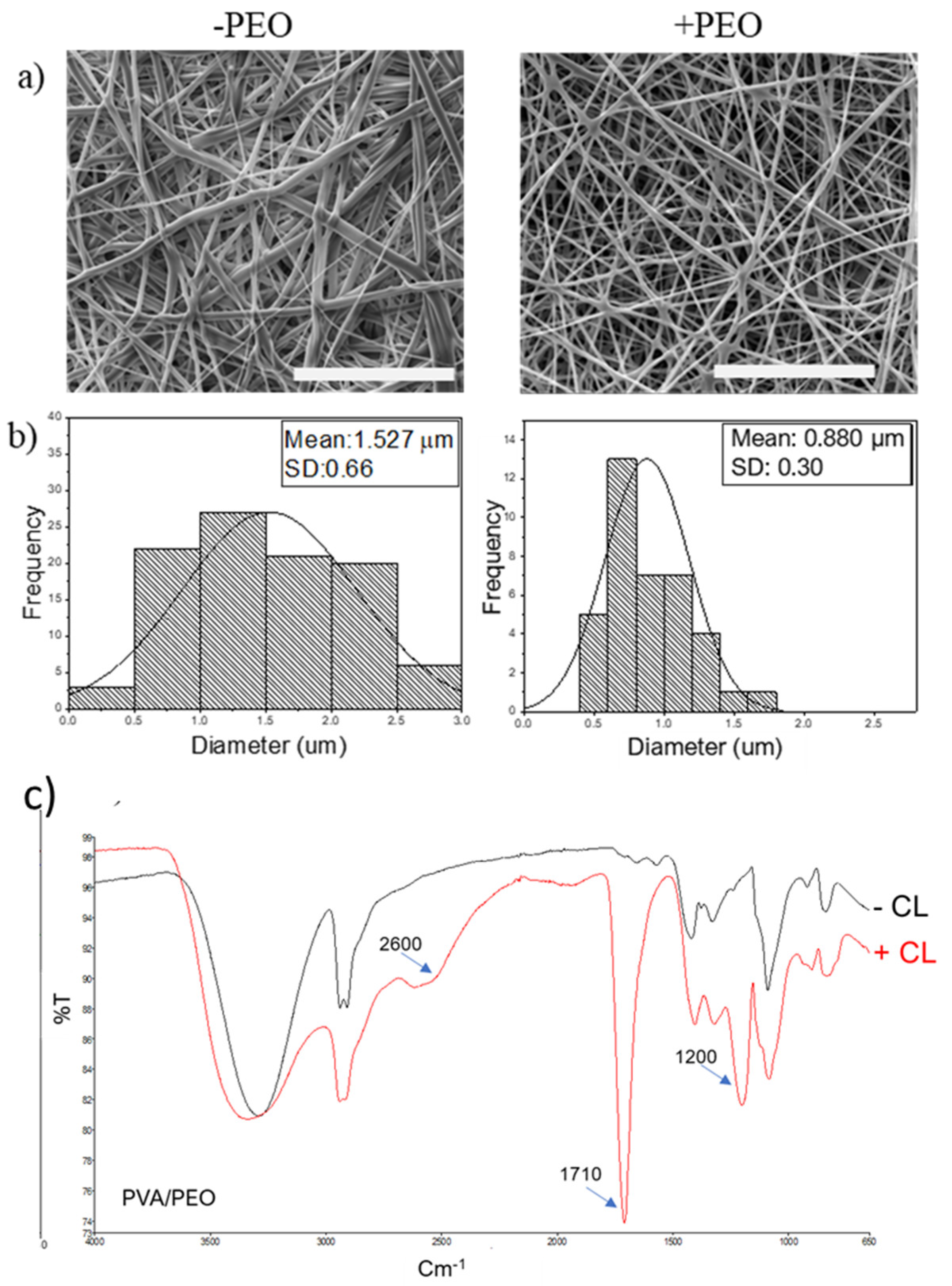
Figure 3 summarizes the morphological studies—qualitative (i.e., SEM analysis) and quantitative (i.e., image analysis)—performed on crosslinked PVA (–PEO) and PVA/PEO (+PEO) nanofibers. It was possible to observe beadless fibers without relevant defects, with a decrease in the diameter of the fibers with PEO, mainly ascribable to the effect of the high molecular weight of PEO, able to improve the packing of PVA chains, with effects in terms of mobility and chain stretching [66,67]. Moreover, it was verified that after thermal crosslinking, PEO is prone to volatize over 100 °C which influences the diameter reduction in fibers [68], in agreement with the image analysis results (Figure 3b).
FTIR analysis was also conducted to obtain further indications regarding the chemical composition of the fibers and interactions among the different phases (Figure 3c). A broad peak around 3550–3200 cm−1 is attributable to the OH stretching vibration from the hydrogen bonds; 2980 and 2800 cm−1 refer to the stretching C–H group. After crosslinking with citric acid (+CL), a higher intensity absorption from 1710 cm−1 due to the C=O stretching from aliphatic ester groups was detected. This is due to the chemical interaction between hydroxyl groups of PVA and carboxyl groups of citric acid [69].
However, an important criticism of PVA fibers still concerns their poor cell adhesion properties, basically due to their lack of cell binding cues needed to promote more efficient cell recognition [70]. In the recent literature, several studies currently focused on using PVA blended with other biopolymers to improve biological and biomechanical properties [71,72,73,74,75]. Among them, gelatin—previously used as a gelling agent in some areas [76]—was mixed with PVA in order to improve its processability. Furthermore, because gelatin has the RGD cell adhesive pattern, it influences the biological affinity of PVA and overcomes bioactivity limits [77]. Also in this case, the use of benign solvents—i.e., mixtures of water and acetic acid—to dissolve PVA/gelatin with a ratio equal to 8:4 allowed for a good balance between processability and biorecognition to be reached.
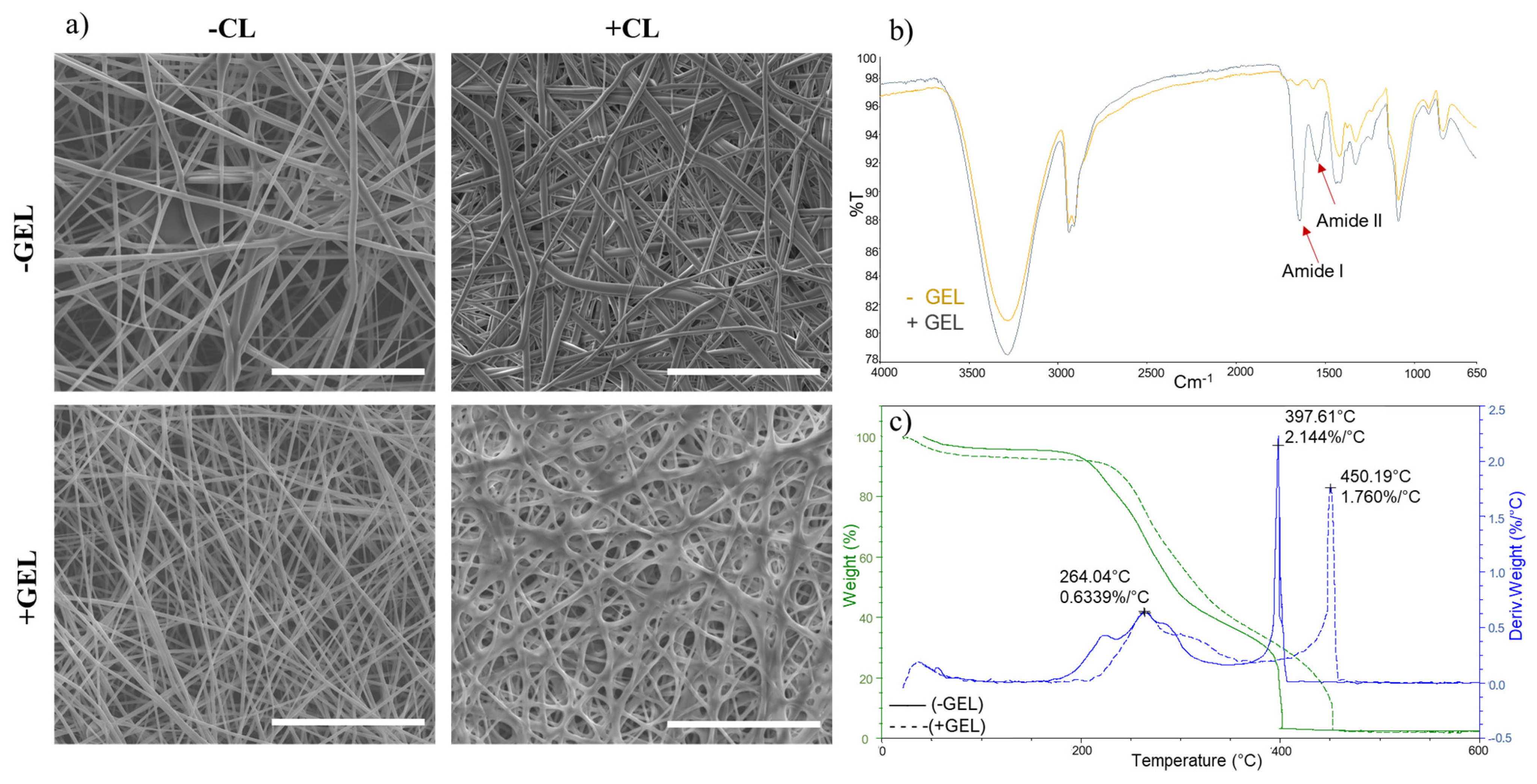
Figure 4a summarizes the morphological properties of PVA/gelatin fibers (+GEL, −CL). The presence of polar macromolecules in gelatin played an active role in the stretching of the polymer jet during the process, promoting the formation of fibers with a mean diameter of 0.938 ± 0.245 μm, lower than those of PVA nanofibers, equal to 1.450 ± 0.37 μm. Since PVA and gelatin are soluble in water, a crosslinking treatment was also required. However, there are some reports that found that citric acid was not able to improve the water resistance of gelatin fibers [78], with a decay in their mechanical properties in vitro [79]. In order to preserve the fibers’ stability in aqueous environments, a green crosslinker (i.e., EDC/NHS) was used. The EDC/NHS crosslinking method can be performed under mild conditions to preserve the biocompatibility of gelatin [80]. Figure 4 shows that fiber organization and porous structure were preserved during crosslinking treatment (+GEL, +CL).
In order to evaluate the presence of gelatin in the fibers, FTIR characterization was conducted to analyze the contribution of gelatin on the characteristic peaks of PVA (Figure 4b). Additional bands associated with proteins were observed at 1647 cm−1 (amide I) and 1539 cm−1 (amide II) in the spectra of PVA/gelatin nanofiber mats [81]. Therefore, amine groups on the biomaterial surface can mediate the focal adhesion function and signaling in the cell adhesion process. It can also be seen that apart from the characteristic peaks of the gelatin, bands are overlapping, and none of the characteristic peaks of acetic acid (around 1700 cm−1 for C=O stretching and 1270 cm−1 for C–O stretching) are observed in the FTIR spectra [82].
The thermal decomposition profiles of PVA and PVA/gelatin analyzed by TGA are presented in Figure 4c. The TGA curves of PVA showed a thermal decomposition profile in the temperature range of 170–420 °C due to the degradation of side groups (–OH), consistent with previous reports [83,84]. The weight loss during the second decomposition step was around 92.8%. There was also an initial weight loss of 4.48%, which could have been caused by the evaporation of water. In the case of the PVA/gelatin nanofiber mats, the initial water loss increased due to the two hydrophilic components in fibers. Since gelatin is more hydrophilic than PVA, the amount of absorbed water is expected to be increased, and weight loss at the first decomposition step increased (7.58%). PVA/gelatin membranes start to degrade at higher temperatures than pure PVA. The weight loss at the second decomposition step (89.86%) was less than PVA, which means that blending with gelatin increased the thermal stability of PVA at this step. It is noteworthy that the improved thermal stability of the PVA/gelatin membranes with respect to the pure PVA could have been related to the temperature shift during the decomposition stages, also confirmed by the derivative curves.
The in vitro response of fibroblasts was investigated according to previous studies [85] to demonstrate the contribution of the presence of gelatin in the fibers. Cell adhesion onto PVA-based fibers was evaluated in culture after 4 and 24 h (Figure 5a). The results showed that over 70% of cells were able to adhere to all groups. In particular, nanofibers with PEO showed smaller diameters, thus providing more surface area for cells to attach after 24 h, while cell adhesion onto the PVA fibers was comparable. In this context, the presence of gelatin allowed for improved cell adhesion just after 24 h, due to the presence of the RGD sequence which balanced the lack of binding sequences of PVA [86]. This was confirmed by the confocal images taken after 24 h in cell culture (Figure 5b) which show the effect of the binding motifs of gelatin on the fibers during cell interaction; L929 cells adhered well and spread along the electrospun fibers differently from those in contact with the PVA fibers, which showed a preferentially rounded shape.
The cell proliferation onto PVA-based nanofibers was evaluated at 1, 3, 7, and 14 days (Figure 5c). For all groups, cell proliferation was maintained over time. PVA was blended with bioactive molecules or natural polymers to improve its cellular interaction and biological response [73,87]. In particular, the PVA fibers with gelatin showed a higher proliferation rate until 14 days, with a slight decrease after 3 days compared to the other PVA-based fibers. PVA-based electrospun fibers are biocompatible materials able to support cell proliferation for a longer time due to their good stability. Moreover, gelatin in the PVA-based fibers counteracted the lack of PVA bioactivity by improving adhesion and cell growth, thus providing biochemical signals that may have contributed to creating microenvironmental conditions suitable to support tissue regeneration.
Recent studies have focused on the idea that regenerative processes can be supported and accelerated by electrical stimuli which can facilitate the repair of the injured tissues [40]. For this purpose, the use of intrinsically electroconductive polymers is rapidly growing for different biomedical applications, from medicine to biosensing [49,50,88]. In this view, the fabrication of PVA fibers by integrating material phases with different electroconductive properties was preliminarily investigated (i.e., PVP and PEDOT:PS) (Figure 6).
PVP is a water-soluble polymer with good environmental stability, biocompatibility, and amphiphilic properties particularly suitable to be mixed with hydrogels and polyelectrolyte solutions. Accordingly, the PVA and PVP solutions have shown good miscibility according to theoretical studies and molecular dynamics simulations, where their blending is attributable to hydrogen interactions between the hydroxyl groups of PVA and proton-accepting carbonyl groups in pyrrolidone rings [89,90]. The good blending of PVA and PVP facilitates the formation of fibers by applying a high-voltage electric field [91]. The use of PVP with a high molecular weight (i.e., Mw 1,300,000 kDa) assured strong, cohesive forces directly associated with a high viscosity. At the same time, ionotropic interactions among the polymer phases concurred to promote the stretching of polymer chains, with a reduction of the average fiber diameter—1.188 ± 0.41 μm—with respect to that of PVA (1.450 ± 0.37 μm) (Figure 6a).
Although the electrical conductivity of PVP is not as relevant in comparison with inorganic materials, the addition of other compounds to form electrolyte solutions allows for drastically improved electrical and mechanical properties [92], making electrospun fibers suitable for actively supporting the transport of electrical/molecular signals for different approaches in skin engineering and wound dressing [93,94].
In the latter case, PEDOT is a biocompatible conductive polymer doped with PSS to obtain a water-soluble polyelectrolyte system with enhanced stability. Recently, thin films of PEDOT:PSS/PVA have been fabricated to monitor human activities (i.e., joint muscle motions, arterial pulsation, and voice vibration) and can distinguish complex and diverse bending motions. In combination with bioactive proteins, they have been used to fabricate composite scaffolds for bone [95]. Moreover, conductive polymers can be used to coat electrospun fibers to increase the electrical conductivity of fibers for neural applications. Alternatively, PEDOT:PSS can be blended with other polymers before electrospinning. In the latter case, PEDOT:PSS particle suspension can be combined with PVA aqueous solutions to form composite nanofibers with electroconductive properties to regenerate electroactive tissues or design real-time electrical stimulation devices [96]. As a function of the relative amount used and their distribution in a matrix, it is possible to control the semiconducting properties of sensing particles corroborated by their interactions with an ionotropic matrix [97]. Hence, the accurate control of particle weight fractions and spatial dispersion enabled the realization of electrospun fibers; moreover, the diameter of the fibers decreased by adding PEDOT:PSS—i.e., 0.872 ± 0.22 μm—due to the increase in electrical interactions of the polymer chains under the applied electric field (Figure 6b). However, an accurate manufacturing strategy cannot ignore the relevance of side effects in terms of cell toxicity, due to the uncontrolled delivery of particles, which can easily be engulfed by cells in vitro through their cellular membranes [98,99]. In this view, further investigations will be performed to optimize the PEDOT:PSS amount to be included in PVA electrospun nanofibers. This is a key requisite to impart bio-functional properties, suitable for the fabrication of bioengineered devices (i.e., neural guides) able to efficiently transfer electrical stimuli, as a potential alternative to commercial synthetic ones.

4. Conclusions

This study investigated the optimization of multicomponent PVA-based fibers to be used as multifunctional scaffolds. It was demonstrated that the addition of PEO aids in improving fiber processability, reducing fiber size and defect formation. Blending with natural proteins such as gelatin improves the biological response regarding fibroblast adhesion and proliferation. Most interestingly, PVA can be easily processed with different intrinsically electroconductive organic phases, such as polyvinylpyrrolidone (PVP) or poly(3,4-ethylenedioxythiophene) (PEDOT), to design fibrous electroactive membranes with controlled morphological properties. To put this in perspective, composite fibers could be successfully used to create multifunctional substrates that, as a function of the peculiar properties (i.e., bioactivity, electrical conductivity) of their different compositional phases, might be able to simultaneously promote cellular events towards a more accurate control of the in vitro regeneration process with possibilities in tissue regeneration.

Supplementary Materials

The following supporting information can be downloaded at: https://www.mdpi.com/article/10.3390/fib11100085/s1, Figure S1: Comparison of optical images of electrospun PVA fibers produced by using different process parameters (flow rate, Voltage, Electrode gap): (a) 0.3 mL/h, 20kV, 25 cm; (b) 0.1 mL/h, 20 kV, 25 cm; (c) 0.3 mL/h, 18 kV, 25 cm; (d) 0.3 mL/h, 22 kV, 25 cm and (e) 0.3 mL/h, 20 kV, 15 cm. (Scale bar 50 μm). Black arrows indicate the presence of defects; Figure S2: Optical images of electrospun fibers from PVA aqueous solutions at different concentrations: (a) 6% w/v and (b) 8%—beaded fibres; (c) 10% w/v and (d) 12% w/v—beadless fibers (Scale bar: 50 μm); Figure S3: Thermal crosslinking of PVA/PEO nanofibers: (a) isothermal analysis at 90 °C (green), 110 °C (blue), 130 °C (red) and (b) optical images of PVA fibers before (b) and after immersion in water of PVA fibers non-crosslinked (c), and PVA crosslinked (d) fibers. (Scale bar: 50 μm); Table S1: Summary of PVA based electrospun fibers.

Author Contributions

Conceptualization, V.G.; methodology, Z.R. and I.C.M.; investigation, Z.R. and I.C.M.; resources, V.G.; data curation, Z.R. and I.C.M.; writing—original draft preparation, Z.R.; writing—review and editing, I.C.M. and V.G.; supervision, V.G.; funding acquisition, V.G. All authors have read and agreed to the published version of the manuscript.

Funding

MSCA-ITN-2020-ASTROTECH (n. GA956325).

Data Availability Statement

Not applicable.

Acknowledgments

Morphological analyses were performed by the LAMEST Labs of the National Research Council. The authors would also like to thank Maria Rosaria Marcedula for her technical support with the physical and chemical characterizations. N.Z.R. and I.C.M. equally contributed to this work.

Conflicts of Interest

The authors declare no conflict of interest.

References

  1. Teixeira, M.A.; Amorim, M.T.P.; Felgueiras, H.P. Poly(Vinyl Alcohol)-Based Nanofibrous Electrospun Scaffolds for Tissue Engineering Applications. Polymers 2019, 12, 7. [Google Scholar] [CrossRef] [PubMed]
  2. Rivera-Hernández, G.; Antunes-Ricardo, M.; Martínez-Morales, P.; Sánchez, M.L. Polyvinyl Alcohol Based-Drug Delivery Systems for Cancer Treatment. Int. J. Pharm. 2021, 600, 120478. [Google Scholar] [CrossRef] [PubMed]
  3. Dalyan, O.; Öztürk, Ö.F.; Pişkin, M. Toxicity of Polyvinyl Alcohols in Medicinal Chemistry. MANAS J. Eng. 2021, 9, 129–135. [Google Scholar] [CrossRef]
  4. Mirjalili, M.; Zohoori, S. Review for Application of Electrospinning and Electrospun Nanofibers Technology in Textile Industry. J. Nanostruct. Chem. 2016, 6, 207–213. [Google Scholar] [CrossRef]
  5. Mahmud, M.M.; Perveen, A.; Matin, M.A.; Arafat, M.T. Effects of Binary Solvent Mixtures on the Electrospinning Behavior of Poly (Vinyl Alcohol). Mater. Res. Express 2018, 5, 115407. [Google Scholar] [CrossRef]
  6. Parhi, R. Fabrication and Characterization of PVA-Based Green Materials. In Advanced Green Materials; Ahmed, S.B.T.-A.G.M., Ed.; Elsevier: Amsterdam, The Netherlands, 2021; pp. 133–177. ISBN 978-0-12-819988-6. [Google Scholar]
  7. Waheed, S.; Butcher, A.L.; Oyen, M.L. The Viscoelastic Response of Electrospun Poly(Vinyl Alcohol) Mats. J. Mech. Behav. Biomed. Mater. 2018, 77, 383–388. [Google Scholar] [CrossRef]
  8. Peresin, M.S.; Habibi, Y.; Zoppe, J.O.; Pawlak, J.J.; Rojas, O.J. Nanofiber Composites of Polyvinyl Alcohol and Cellulose Nanocrystals: Manufacture and Characterization. Biomacromolecules 2010, 11, 674–681. [Google Scholar] [CrossRef]
  9. Moradipour, P.; Limoee, M.; Janfaza, S.; Behbood, L. Core-Shell Nanofibers Based on Polycaprolactone/Polyvinyl Alcohol and Polycaprolactone/Collagen for Biomedical Applications. J. Pharm. Innov. 2022, 17, 911–920. [Google Scholar] [CrossRef]
  10. Renkler, N.Z.; Cruz-Maya, I.; Bonadies, I.; Guarino, V. Electro Fluid Dynamics: A Route to Design Polymers and Composites for Biomedical and Bio-Sustainable Applications. Polymers 2022, 14, 4249. [Google Scholar] [CrossRef]
  11. Wang, P.; Lv, H.; Cao, X.; Liu, Y.; Yu, D.-G. Recent Progress of the Preparation and Application of Electrospun Porous Nanofibers. Polymers 2023, 15, 921. [Google Scholar] [CrossRef]
  12. Wang, Q.; Ma, J.; Chen, S.; Wu, S. Designing an Innovative Electrospinning Strategy to Generate PHBV Nanofiber Scaffolds with a Radially Oriented Fibrous Pattern. Nanomaterials 2023, 13, 1150. [Google Scholar] [CrossRef] [PubMed]
  13. Sivan, M.; Madheswaran, D.; Valtera, J.; Kostakova, E.K.; Lukas, D. Alternating Current Electrospinning: The Impacts of Various High-Voltage Signal Shapes and Frequencies on the Spinnability and Productivity of Polycaprolactone Nanofibers. Mater. Des. 2022, 213, 110308. [Google Scholar] [CrossRef]
  14. Sivan, M.; Madheswaran, D.; Hauzerova, S.; Novotny, V.; Hedvicakova, V.; Jencova, V.; Kostakova, E.K.; Schindler, M.; Lukas, D. AC Electrospinning: Impact of High Voltage and Solvent on the Electrospinnability and Productivity of Polycaprolactone Electrospun Nanofibrous Scaffolds. Mater. Today Chem. 2022, 26, 101025. [Google Scholar] [CrossRef]
  15. Mao, Y.; Shen, W.; Wu, S.; Ge, X.; Ao, F.; Ning, Y.; Luo, Y.; Liu, Z. Electrospun Polymers: Using Devices to Enhance Their Potential for Biomedical Applications. React. Funct. Polym. 2023, 186, 105568. [Google Scholar] [CrossRef]
  16. Hamdan, N.; Yamin, A.; Hamid, S.A.; Khodir, W.K.W.A.; Guarino, V. Functionalized Antimicrobial Nanofibers: Design Criteria and Recent Advances. J. Funct. Biomater. 2021, 12, 59. [Google Scholar] [CrossRef]
  17. Maleki, H.; Mathur, S.; Klein, A. Antibacterial Ag Containing Core-shell Polyvinyl Alcohol-poly (Lactic Acid) Nanofibers for Biomedical Applications. Polym. Eng. Sci. 2020, 60, 1221–1230. [Google Scholar] [CrossRef]
  18. Lencova, S.; Svarcova, V.; Stiborova, H.; Demnerova, K.; Jencova, V.; Hozdova, K.; Zdenkova, K. Bacterial Biofilms on Polyamide Nanofibers: Factors Influencing Biofilm Formation and Evaluation. ACS Appl. Mater. Interfaces 2021, 13, 2277–2288. [Google Scholar] [CrossRef] [PubMed]
  19. Jhala, D.; Rather, H.; Vasita, R. Polycaprolactone–Chitosan Nanofibers Influence Cell Morphology to Induce Early Osteogenic Differentiation. Biomater. Sci. 2016, 4, 1584–1595. [Google Scholar] [CrossRef]
  20. Erickson, A.; Chiarelli, P.A.; Huang, J.; Levengood, S.L.; Zhang, M. Electrospun Nanofibers for 3-D Cancer Models, Diagnostics, and Therapy. Nanoscale Horiz. 2022, 7, 1279–1298. [Google Scholar] [CrossRef]
  21. Guaccio, A.; Guarino, V.; Perez, M.A.A.; Cirillo, V.; Netti, P.A.; Ambrosio, L. Influence of Electrospun Fiber Mesh Size on HMSC Oxygen Metabolism in 3D Collagen Matrices: Experimental and Theoretical Evidences. Biotechnol. Bioeng. 2011, 108, 1965–1976. [Google Scholar] [CrossRef] [PubMed]
  22. Park, J.-C.; Ito, T.; Kim, K.-O.; Kim, K.-W.; Kim, B.-S.; Khil, M.-S.; Kim, H.-Y.; Kim, I.-S. Electrospun Poly(Vinyl Alcohol) Nanofibers: Effects of Degree of Hydrolysis and Enhanced Water Stability. Polym. J. 2010, 42, 273–276. [Google Scholar] [CrossRef]
  23. Al-Abduljabbar, A.; Farooq, I. Electrospun Polymer Nanofibers: Processing, Properties, and Applications. Polymers 2023, 15, 65. [Google Scholar] [CrossRef]
  24. Baykara, T.; Taylan, G. Coaxial Electrospinning of PVA/Nigella Seed Oil Nanofibers: Processing and Morphological Characterization. Mater. Sci. Eng. B 2021, 265, 115012. [Google Scholar] [CrossRef]
  25. Alazab, M.; Mitchell, G.R.; Davis, F.J.; Mohan, S.D. Sustainable Electrospinning of Nanoscale Fibres. Procedia Manuf. 2017, 12, 66–78. [Google Scholar] [CrossRef]
  26. Aslam Khan, M.U.; Abd Razak, S.I.; Al Arjan, W.S.; Nazir, S.; Sahaya Anand, T.J.; Mehboob, H.; Amin, R. Recent Advances in Biopolymeric Composite Materials for Tissue Engineering and Regenerative Medicines: A Review. Molecules 2021, 26, 619. [Google Scholar] [CrossRef] [PubMed]
  27. Rynkowska, E.; Fatyeyeva, K.; Marais, S.; Kujawa, J.; Kujawski, W. Chemically and Thermally Crosslinked PVA-Based Membranes: Effect on Swelling and Transport Behavior. Polymers 2019, 11, 1799. [Google Scholar] [CrossRef] [PubMed]
  28. Imre, B.; Pukánszky, B. Compatibilization in Bio-Based and Biodegradable Polymer Blends. Eur. Polym. J. 2013, 49, 1215–1233. [Google Scholar] [CrossRef]
  29. Mares-Bou, S.; Serrano, M.-A.; Gómez-Tejedor, J.A. Core–Shell Polyvinyl Alcohol (PVA) Base Electrospinning Microfibers for Drug Delivery. Polymers 2023, 15, 1554. [Google Scholar] [CrossRef] [PubMed]
  30. Vacheethasanee, K.; Wang, S.; Qiu, Y.; Marchant, R.E. Poly(Ethylene Oxide) Surfactant Polymers. J. Biomater. Sci. Polym. Ed. 2004, 15, 95–110. [Google Scholar] [CrossRef]
  31. Liu, H.; Zuo, B. Structure and Sound Absorption Properties of Spiral Vane Electrospun PVA/PEO Nanofiber Membranes. Appl. Sci. 2018, 8, 296. [Google Scholar] [CrossRef]
  32. Gómez-Guillén, M.C.; Giménez, B.; López-Caballero, M.E.; Montero, M.P. Functional and Bioactive Properties of Collagen and Gelatin from Alternative Sources: A Review. Food Hydrocoll. 2011, 25, 1813–1827. [Google Scholar] [CrossRef]
  33. Gaspar-Pintiliescu, A.; Stanciuc, A.-M.; Craciunescu, O. Natural Composite Dressings Based on Collagen, Gelatin and Plant Bioactive Compounds for Wound Healing: A Review. Int. J. Biol. Macromol. 2019, 138, 854–865. [Google Scholar] [CrossRef] [PubMed]
  34. Zavan, B.; Gardin, C.; Guarino, V.; Rocca, T.; Cruz Maya, I.; Zanotti, F.; Ferroni, L.; Brunello, G.; Chachques, J.-C.; Ambrosio, L.; et al. Electrospun PCL-Based Vascular Grafts: In Vitro Tests. Nanomaterials 2021, 11, 751. [Google Scholar] [CrossRef]
  35. Ferraris, S.; Spriano, S.; Scalia, A.C.; Cochis, A.; Rimondini, L.; Cruz-Maya, I.; Guarino, V.; Varesano, A.; Vineis, C. Topographical and Biomechanical Guidance of Electrospun Fibers for Biomedical Applications. Polymers 2020, 12, 2896. [Google Scholar] [CrossRef] [PubMed]
  36. Cirillo, V.; Guarino, V.; Alvarez-Perez, M.A.; Marrese, M.; Ambrosio, L. Optimization of Fully Aligned Bioactive Electrospun Fibers for “In Vitro” Nerve Guidance. J. Mater. Sci. Mater. Med. 2014, 25, 2323–2332. [Google Scholar] [CrossRef]
  37. Abdul Khodir, W.K.W.; Abdul Razak, A.H.; Ng, M.H.; Guarino, V.; Susanti, D. Encapsulation and Characterization of Gentamicin Sulfate in the Collagen Added Electrospun Nanofibers for Skin Regeneration. J. Funct. Biomater. 2018, 9, 36. [Google Scholar] [CrossRef]
  38. Vineis, C.; Cruz Maya, I.; Mowafi, S.; Varesano, A.; Sánchez Ramírez, D.O.; Abou Taleb, M.; Tonetti, C.; Guarino, V.; El-Sayed, H. Synergistic Effect of Sericin and Keratin in Gelatin Based Nanofibers for In Vitro Applications. Int. J. Biol. Macromol. 2021, 190, 375–381. [Google Scholar] [CrossRef] [PubMed]
  39. Cruz-Maya, I.; Guarino, V.; Almaguer-Flores, A.; Alvarez-Perez, M.A.; Varesano, A.; Vineis, C. Highly Polydisperse Keratin Rich Nanofibers: Scaffold Design and In Vitro Characterization. J. Biomed. Mater. Res. Part A 2019, 107, 1803–1813. [Google Scholar] [CrossRef]
  40. Ning, C.; Zhou, Z.; Tan, G.; Zhu, Y.; Mao, C. Electroactive Polymers for Tissue Regeneration: Developments and Perspectives. Prog. Polym. Sci. 2018, 81, 144–162. [Google Scholar] [CrossRef] [PubMed]
  41. Dong, R.; Ma, P.X.; Guo, B. Conductive Biomaterials for Muscle Tissue Engineering. Biomaterials 2020, 229, 119584. [Google Scholar] [CrossRef]
  42. Heng, B.C.; Bai, Y.; Li, X.; Lim, L.W.; Li, W.; Ge, Z.; Zhang, X.; Deng, X. Electroactive Biomaterials for Facilitating Bone Defect Repair under Pathological Conditions. Adv. Sci. 2023, 10, 2204502. [Google Scholar] [CrossRef] [PubMed]
  43. Palza, H.; Zapata, P.A.; Angulo-Pineda, C. Electroactive Smart Polymers for Biomedical Applications. Materials 2019, 12, 277. [Google Scholar] [CrossRef] [PubMed]
  44. Song, J.; Ma, G.; Qin, F.; Hu, L.; Luo, B.; Liu, T.; Yin, X.; Su, Z.; Zeng, Z.; Jiang, Y.; et al. High-Conductivity, Flexible and Transparent PEDOT:PSS Electrodes for High Performance Semi-Transparent Supercapacitors. Polymers 2020, 12, 450. [Google Scholar] [CrossRef]
  45. Guo, B.; Glavas, L.; Albertsson, A.-C. Biodegradable and Electrically Conducting Polymers for Biomedical Applications. Prog. Polym. Sci. 2013, 38, 1263–1286. [Google Scholar] [CrossRef]
  46. Zubair, N.A.; Rahman, N.A.; Lim, H.N.; Zawawi, R.M.; Sulaiman, Y. Electrochemical Properties of PVA–GO/PEDOT Nanofibers Prepared Using Electrospinning and Electropolymerization Techniques. RSC Adv. 2016, 6, 17720–17727. [Google Scholar] [CrossRef]
  47. Devi, C.; Gellanki, J.; Pettersson, H.; Kumar, S. High Sodium Ionic Conductivity in PEO/PVP Solid Polymer Electrolytes with InAs Nanowire Fillers. Sci. Rep. 2021, 11, 20180. [Google Scholar] [CrossRef] [PubMed]
  48. Serrano-Garcia, W.; Cruz-Maya, I.; Melendez-Zambrana, A.; Ramos-Colon, I.; Pinto, N.J.; Thomas, S.W.; Guarino, V. Optimization of PVDF-TrFE Based Electro-Conductive Nanofibers: Morphology and In Vitro Response. Materials 2023, 16, 3106. [Google Scholar] [CrossRef]
  49. Cruz-Maya, I.; Zuppolini, S.; Zarrelli, M.; Mazzotta, E.; Borriello, A.; Malitesta, C.; Guarino, V. Polydopamine-Coated Alginate Microgels: Process Optimization and In Vitro Validation. J. Funct. Biomater. 2022, 14, 2. [Google Scholar] [CrossRef]
  50. Saracino, E.; Zuppolini, S.; Guarino, V.; Benfenati, V.; Borriello, A.; Zamboni, R.; Ambrosio, L. Polyaniline Nano-Needles into Electrospun Bio Active Fibres Support In Vitro Astrocyte Response. RSC Adv. 2021, 11, 11347–11355. [Google Scholar] [CrossRef]
  51. Sarip, M.N.; Noor, M.F.H.M.; Ahmad, Z.; Shuhaime, N.; Dahan, R.M.; Arshad, A.N.; Ismail, W.I.N.W. Conductivity Study of Polyvinyl Alcohol/Polyvinyl Pyrrolidone (PVA/PVP)-KOH Coatings System. AIP Conf. Proc. 2018, 2031, 20022. [Google Scholar] [CrossRef]
  52. Yu, R.; Zhang, H.; Guo, B. Conductive Biomaterials as Bioactive Wound Dressing for Wound Healing and Skin Tissue Engineering. Nano-Micro Lett. 2021, 14, 1. [Google Scholar] [CrossRef] [PubMed]
  53. Chi, H.Y.; Chang, N.Y.; Li, C.; Chan, V.; Hsieh, J.H.; Tsai, Y.-H.; Lin, T. Fabrication of Gelatin Nanofibers by Electrospinning—Mixture of Gelatin and Polyvinyl Alcohol. Polymers 2022, 14, 2610. [Google Scholar] [CrossRef] [PubMed]
  54. Liu, R.; Ming, J.; Zhang, H.; Zuo, B. EDC/NHS Crosslinked Electrospun Regenerated Tussah Silk Fibroin Nanofiber Mats. Fibers Polym. 2012, 13, 613–617. [Google Scholar] [CrossRef]
  55. Yousefzadeh, M. Modeling and Simulation of the Electrospinning Process. In Electrospun Nanofibers; Afshari, M.B.T.-E.N., Ed.; Elsevier: Amsterdam, The Netherlands, 2017; pp. 277–301. [Google Scholar]
  56. Sharma, S.K.; Prakash, J.; Pujari, P.K. Effects of the Molecular Level Dispersion of Graphene Oxide on the Free Volume Characteristics of Poly(Vinyl Alcohol) and Its Impact on the Thermal and Mechanical Properties of Their Nanocomposites. Phys. Chem. Chem. Phys. 2015, 17, 29201–29209. [Google Scholar] [CrossRef]
  57. Yang, Q.; Guo, J.; Zhang, S.; Guan, F.; Yu, Y.; Feng, S.; Yao, Q.; Bao, D. Improved Biomedical Bioactivity of Polyvinyl Alcohol/Polyethylene Oxide Composite System-Based Nanofiber Membranes via Incorporating Antarctic Krill Protein. Eur. Polym. J. 2023, 187, 111888. [Google Scholar] [CrossRef]
  58. Jia, L.; Qin, X. The Effect of Different Surfactants on the Electrospinning Poly(Vinyl Alcohol) (PVA) Nanofibers. J. Therm. Anal. Calorim. 2013, 112, 595–605. [Google Scholar] [CrossRef]
  59. Zheng, J.-Y.; Zhuang, M.-F.; Yu, Z.-J.; Zheng, G.-F.; Zhao, Y.; Wang, H.; Sun, D.-H. The Effect of Surfactants on the Diameter and Morphology of Electrospun Ultrafine Nanofiber. J. Nanomater. 2014, 2014, 689298. [Google Scholar] [CrossRef]
  60. Taemeh, M.A.; Shiravandi, A.; Korayem, M.A.; Daemi, H. Fabrication Challenges and Trends in Biomedical Applications of Alginate Electrospun Nanofibers. Carbohydr. Polym. 2020, 228, 115419. [Google Scholar] [CrossRef]
  61. Avossa, J.; Herwig, G.; Toncelli, C.; Itel, F.; Rossi, R.M. Electrospinning Based on Benign Solvents: Current Definitions, Implications and Strategies. Green Chem. 2022, 24, 2347–2375. [Google Scholar] [CrossRef]
  62. Hirsch, E.; Pantea, E.; Vass, P.; Domján, J.; Molnár, M.; Suhajda, Á.; Andersen, S.K.; Vigh, T.; Verreck, G.; Marosi, G.J.; et al. Probiotic Bacteria Stabilized in Orally Dissolving Nanofibers Prepared by High-Speed Electrospinning. Food Bioprod. Process. 2021, 128, 84–94. [Google Scholar] [CrossRef]
  63. Shen, W.; Hsieh, Y.-L. Biocompatible Sodium Alginate Fibers by Aqueous Processing and Physical Crosslinking. Carbohydr. Polym. 2014, 102, 893–900. [Google Scholar] [CrossRef] [PubMed]
  64. Salihu, R.; Abd Razak, S.I.; Zawawi, N.A.; Kadir, M.R.; Ismail, N.I.; Jusoh, N.; Mohamad, M.R.; Nayan, N.H. Citric Acid: A Green Cross-Linker of Biomaterials for Biomedical Applications. Eur. Polym. J. 2021, 146, 110271. [Google Scholar] [CrossRef]
  65. Nataraj, D.; Reddy, R.; Reddy, N. Crosslinking Electrospun Poly (Vinyl) Alcohol Fibers with Citric Acid to Impart Aqueous Stability for Medical Applications. Eur. Polym. J. 2020, 124, 109484. [Google Scholar] [CrossRef]
  66. Lian, Z.; Ye, L. Structure and Properties of PVA/PEO Hydrogel Prepared by Freezing/Thawing Method. J. Thermoplast. Compos. Mater. 2011, 26, 912–922. [Google Scholar] [CrossRef]
  67. Liu, H.; Zuo, B. Sound Absorption Property of PVA/PEO/GO Nanofiber Membrane and Non-Woven Composite Material. J. Ind. Text. 2019, 50, 512–525. [Google Scholar] [CrossRef]
  68. Zhang, J.; Li, Y.; Wu, H.; Wang, C.; Salleh, K.M.; Li, H.; Zakaria, S. Thermally Treated Berberine-Loaded SA/PVA/PEO Electrospun Microfiber Membranes for Antibacterial Wound Dressings. Polymers 2022, 14, 4473. [Google Scholar] [CrossRef]
  69. Yu, D.; Feng, Y.-Y.; Xu, J.-X.; Kong, B.-H.; Liu, Q.; Wang, H. Fabrication, Characterization, and Antibacterial Properties of Citric Acid Crosslinked PVA Electrospun Microfibre Mats for Active Food Packaging. Packag. Technol. Sci. 2021, 34, 361–370. [Google Scholar] [CrossRef]
  70. Pathan, S.G.; Fitzgerald, L.M.; Ali, S.M.; Damrauer, S.M.; Bide, M.J.; Nelson, D.W.; Ferran, C.; Phaneuf, T.M.; Phaneuf, M.D. Cytotoxicity Associated with Electrospun Polyvinyl Alcohol. J. Biomed. Mater. Res. B Appl. Biomater. 2015, 103, 1652–1662. [Google Scholar] [CrossRef]
  71. Lv, H.; Zhao, M.; Li, Y.; Li, K.; Chen, S.; Zhao, W.; Wu, S.; Han, Y. Electrospun Chitosan–Polyvinyl Alcohol Nanofiber Dressings Loaded with Bioactive Ursolic Acid Promoting Diabetic Wound Healing. Nanomaterials 2022, 12, 2933. [Google Scholar] [CrossRef]
  72. Asiri, A.; Saidin, S.; Sani, M.H.; Al-Ashwal, R.H. Epidermal and Fibroblast Growth Factors Incorporated Polyvinyl Alcohol Electrospun Nanofibers as Biological Dressing Scaffold. Sci. Rep. 2021, 11, 5634. [Google Scholar] [CrossRef]
  73. Sanchez Ramirez, D.O.; Cruz-Maya, I.; Vineis, C.; Tonetti, C.; Varesano, A.; Guarino, V. Design of Asymmetric Nanofibers-Membranes Based on Polyvinyl Alcohol and Wool-Keratin for Wound Healing Applications. J. Funct. Biomater. 2021, 12, 76. [Google Scholar] [CrossRef] [PubMed]
  74. Koosha, M.; Mirzadeh, H. Electrospinning, Mechanical Properties, and Cell Behavior Study of Chitosan/PVA Nanofibers. J. Biomed. Mater. Res. A 2015, 103, 3081–3093. [Google Scholar] [CrossRef] [PubMed]
  75. Deng, Y.; Zhang, X.; Zhao, Y.; Liang, S.; Xu, A.; Gao, X.; Deng, F.; Fang, J.; Wei, S. Peptide-Decorated Polyvinyl Alcohol/Hyaluronan Nanofibers for Human Induced Pluripotent Stem Cell Culture. Carbohydr. Polym. 2014, 101, 36–39. [Google Scholar] [CrossRef] [PubMed]
  76. Qiao, C.; Ma, X.; Zhang, J.; Yao, J. Molecular Interactions in Gelatin/Chitosan Composite Films. Food Chem. 2017, 235, 45–50. [Google Scholar] [CrossRef] [PubMed]
  77. Davidenko, N.; Schuster, C.F.; Bax, D.V.; Farndale, R.W.; Hamaia, S.; Best, S.M.; Cameron, R.E. Evaluation of Cell Binding to Collagen and Gelatin: A Study of the Effect of 2D and 3D Architecture and Surface Chemistry. J. Mater. Sci. Mater. Med. 2016, 27, 148. [Google Scholar] [CrossRef]
  78. Banner, J.; Dautzenberg, M.; Feldhans, T.; Hofmann, J.; Plümer, P.; Ehrmann, A. Water Resistance and Morphology of Electrospun Gelatine Blended with Citric Acid and Coconut Oil. Tekstilec 2018, 61, 129–135. [Google Scholar] [CrossRef]
  79. Mahnama, H.; Dadbin, S.; Frounchi, M.; Rajabi, S. Preparation of Biodegradable Gelatin/PVA Porous Scaffolds for Skin Regeneration. Artif. Cells Nanomed. Biotechnol. 2017, 45, 928–935. [Google Scholar] [CrossRef] [PubMed]
  80. Ghassemi, Z.; Slaughter, G. Cross-Linked Electrospun Gelatin Nanofibers for Cell-Based Assays. In Proceedings of the 2018 40th Annual International Conference of the IEEE Engineering in Medicine and Biology Society (EMBC), Honolulu, HI, USA, 18–21 July 2018; pp. 6088–6091. [Google Scholar] [CrossRef]
  81. Damayanti, R.; Alfian, Z.; Zulfajri, M. Biosynthesis of Silver Nanoparticles Loaded PVA/Gelatin Nanocomposite Films and Their Antimicrobial Activities. Inorg. Chem. Commun. 2022, 144, 109948. [Google Scholar] [CrossRef]
  82. Erencia, M.; Cano, F.; Tornero, J.A.; Fernandes, M.M.; Tzanov, T.; Macanás, J.; Carrillo, F. Electrospinning of Gelatin Fibers Using Solutions with Low Acetic Acid Concentration: Effect of Solvent Composition on Both Diameter of Electrospun Fibers and Cytotoxicity. J. Appl. Polym. Sci. 2015, 132, 42115. [Google Scholar] [CrossRef]
  83. Gomaa, M.M.; Hugenschmidt, C.; Dickmann, M.; Abdel-Hady, E.E.; Mohamed, H.F.M.; Abdel-Hamed, M.O. Crosslinked PVA/SSA Proton Exchange Membranes: Correlation between Physiochemical Properties and Free Volume Determined by Positron Annihilation Spectroscopy. Phys. Chem. Chem. Phys. 2018, 20, 28287–28299. [Google Scholar] [CrossRef] [PubMed]
  84. Azeez Betti, N. Thermogravimetric Analysis on PVA / PVP Blend Under Air Atmosphere. Eng. Technol. J. 2016, 34, 2433–2442. [Google Scholar] [CrossRef]
  85. Bello, J.S.; Cruz-Maya, I.; González-Alva, P.; Alvarez-Perez, M.A.; Guarino, V. Electro- and Nonelectro-Assisted Spinning Technologies for In Vitro and In Vivo Models. In Nanomaterials for Theranostics and Tissue Engineering; Rossi, F., Rainer, A.B., Eds.; Elsevier: Amsterdam, The Netherlands, 2020; pp. 175–204. ISBN 978-0-12-817838-6. [Google Scholar]
  86. Cruz-Maya, I.; Varesano, A.; Vineis, C.; Guarino, V. Comparative Study on Protein-Rich Electrospun Fibers for In Vitro Applications. Polymers 2020, 12, 1671. [Google Scholar] [CrossRef]
  87. García-Hernández, A.B.; Morales-Sánchez, E.; Calderón-Domínguez, G.; de la, P.; Salgado-Cruz, M.; Farrera-Rebollo, R.R.; Vega-Cuellar, M.Á.; García-Bórquez, A. Hydrolyzed Collagen on PVA-Based Electrospun Membranes: Synthesis and Characterization. J. Appl. Polym. Sci. 2021, 138, 51197. [Google Scholar] [CrossRef]
  88. Serrano-Garcia, W.; Bonadies, I.; Thomas, S.W.; Guarino, V. New Insights to Design Electrospun Fibers with Tunable Electrical Conductive–Semiconductive Properties. Sensors 2023, 23, 1606. [Google Scholar] [CrossRef] [PubMed]
  89. Cassu, S.N.; Felisberti, M.I. Poly(Vinyl Alcohol) and Poly(Vinylpyrrolidone) Blends: 2. Study of Relaxations by Dynamic Mechanical Analysis. Polymer 1999, 40, 4845–4851. [Google Scholar] [CrossRef]
  90. Wei, Q.; Zhang, Y.; Wang, Y.; Chai, W.; Yang, M. Measurement and Modeling of the Effect of Composition Ratios on the Properties of Poly(Vinyl Alcohol)/Poly(Vinyl Pyrrolidone) Membranes. Mater. Des. 2016, 103, 249–258. [Google Scholar] [CrossRef]
  91. Virginia, C.; Khasanah, A.; Jauhari, J.; Sriyanti, I. Electrospinning and Characterization Nanofibers and Nano Particle of Polyvinylpyrrolidone. IOP Conf. Ser. Mater. Sci. Eng. 2020, 850, 12039. [Google Scholar] [CrossRef]
  92. Basha, S.K.; Sundari, G.; Vijay Kumar, K. Electrical Conductivity, Transport and Discharge Characteristics of a Sodium Acetate Trihydrate Complexed with Polyvinyl Alcohol for Electrochemical Cell. Int. J. ChemTech Res. 2015, 8, 803–810. [Google Scholar]
  93. Gökmeşe, F.; Uslu, İ.; Aytimur, A. Preparation and Characterization of PVA/PVP Nanofibers as Promising Materials for Wound Dressing. Polym. Plast. Technol. Eng. 2013, 52, 1259–1265. [Google Scholar] [CrossRef]
  94. Rahmani, F.; Ziyadi, H.; Baghali, M.; Luo, H.; Ramakrishna, S. Electrospun PVP/PVA Nanofiber Mat as a Novel Potential Transdermal Drug-Delivery System for Buprenorphine: A Solution Needed for Pain Management. Appl. Sci. 2021, 11, 2779. [Google Scholar] [CrossRef]
  95. Shahini, A.; Yazdimamaghani, M.; Walker, K.J.; Eastman, M.A.; Hatami-Marbini, H.; Smith, B.J.; Ricci, J.L.; Madihally, S.V.; Vashaee, D.; Tayebi, L. 3D Conductive Nanocomposite Scaffold for Bone Tissue Engineering. Int. J. Nanomed. 2014, 9, 167–181. [Google Scholar] [CrossRef]
  96. Tandon, B.; Magaz, A.; Balint, R.; Blaker, J.J.; Cartmell, S.H. Electroactive Biomaterials: Vehicles for Controlled Delivery of Therapeutic Agents for Drug Delivery and Tissue Regeneration. Adv. Drug Deliv. Rev. 2018, 129, 148–168. [Google Scholar] [CrossRef]
  97. Kim, K.; Yoo, H.; Lee, E.K. New Opportunities for Organic Semiconducting Polymers in Biomedical Applications. Polymers 2022, 14, 2960. [Google Scholar] [CrossRef] [PubMed]
  98. Latonen, R.-M.; Cabrera, J.A.W.; Lund, S.; Kosourov, S.; Vajravel, S.; Boeva, Z.; Wang, X.; Xu, C.; Allahverdiyeva, Y. Electrospinning of Electroconductive Water-Resistant Nanofibers of PEDOT–PSS, Cellulose Nanofibrils and PEO: Fabrication, Characterization, and Cytocompatibility. ACS Appl. Bio Mater. 2021, 4, 483–493. [Google Scholar] [CrossRef] [PubMed]
  99. Pisesweerayos, P.; Dangtip, S.; Supaphol, P.; Srikhirin, T. Electrically Conductive Ultrafine Fibers of PVA-PEDOT/PSS and PVA-AgNPs by Means of Electrospinning. Adv. Mater. Res. 2014, 1033–1034, 1024–1035. [Google Scholar] [CrossRef]
Figure 1. Scheme of fabrication of different multicomponent nanofibrous membranes (i.e., bioactive, electroactive) as tissue-engineered scaffolds.
Fibers 11 00085 g001
Figure 2. PVA electrospun fibers: SEM images and fiber diameter distribution of PVA at (a) 10% w/v and (b) 12% w/v (Magnification: 3000×, Scale bar: 40 μm). (c) TGA and (d) DSC analyses.
Fibers 11 00085 g002
Figure 3. Crosslinked PVA (–PEO) and PVA/PEO (+PEO) electrospun fibers: (a) SEM images (Magnification: 3000×, Scale bar: 40 μm) and (b) distribution of diameter of fibers. (c) FTIR analysis of samples with (red, +CL) or without (black, –CL) crosslinking.
Fibers 11 00085 g003
Figure 4. PVA (–GEL) and PVA/gelatin (+GEL) electrospun nanofibers with (+CL) and without (–CL) crosslinking: (a) SEM images (Magnification: 3000×, Scale bar: 40 μm); (b) FTIR; and (c) TGA analyses.
Fibers 11 00085 g004
Figure 5. Cell adhesion assays: (a) cell adhesion of L929 cells onto electrospun fibers after 4 and 24 h, with respect to the control (TCP) (* p < 0.05); (b) morphology of living L929 cells (green) and DAPI (blue) after 24 h seeding onto PVA, PVA/PEO, and PVA/gelatin electrospun fibers (Scale bar: 200 μm); (c) in vitro cell proliferation of L929 cells evaluated by XTT assay after 1, 3, 7, and 14 days in cell culture. Results are presented as means ± standard error (* p < 0.05, ** p < 0.01).
Fibers 11 00085 g005
Figure 6. PVA-based nanofibers including electroconductive phases: (a) PVP and (b) PEDOT:PSS (Magnification: 6000×, Scale bar: 20 μm).
Fibers 11 00085 g006
Disclaimer/Publisher’s Note: The statements, opinions and data contained in all publications are solely those of the individual author(s) and contributor(s) and not of MDPI and/or the editor(s). MDPI and/or the editor(s) disclaim responsibility for any injury to people or property resulting from any ideas, methods, instructions or products referred to in the content.