Article

Amazing Discoveries of Benthic Fauna from the Abyssal Zone of Lake Baikal

by
Ilya G. Kondratov
,
Tatiana Ya. Sitnikova
,
Irina A. Kaygorodova
,
Natalia N. Denikina
*,
Vadim V. Annenkov
,
Igor V. Khanaev
,
Sergei V. Kirilchik
,
Ivan A. Nebesnykh
and
Elena V. Dzyuba
Limnological Institute, Siberian Branch of the Russian Academy of Sciences, Ulan-Batorskaya Str. 3, 664033 Irkutsk, Russia
*
Author to whom correspondence should be addressed.
Biology 2021, 10(10), 972; https://doi.org/10.3390/biology10100972
Submission received: 6 August 2021 / Revised: 23 September 2021 / Accepted: 23 September 2021 / Published: 27 September 2021
(This article belongs to the Special Issue Biodiversity Patterns)

Abstract

:

Simple Summary

Lake Baikal is the deepest and oldest freshwater ecosystem in the world. Since more than half of the currently known species of aquatic organisms inhabiting Lake Baikal are endemic, it is called a natural laboratory for the study of species diversity and evolution. However, despite many years of research, the true biodiversity of the lake is clearly insufficiently studied. As before, the deep-water zones of the lake remain white spots: there is little information about small benthic organisms, especially sessile forms. Therefore, when, for the first time, during deep-sea trawls, fragments of packaging plastic were raised from the bottom of Lake Baikal, the main goal was to determine the diversity of aquatic organisms inhabiting it. Analysis of the standard fragment of the mitochondrial genome allowed the obtaining of the first nucleotide data for the endemic Baikal bryozoans Hislopia placoides, two new species of leeches (Rhynchobdellida, Piscicolidae) and an unknown representative of stramenopiles that is genetically equidistant from the fungi-like organisms, Oomycetes and Chromulinales (Chrysophyta). Nucleotide data of H. placoides indirectly confirmed the hypothesis of the partial origin of Baikal fauna from Tethys derivatives. Thus, the abyssal zone of Baikal is an inexhaustible source of information about biodiversity and the evolution of life on the planet.

Abstract

Lake Baikal is a natural laboratory for the study of species diversity and evolution, as a unique freshwater ecosystem meeting the all of the main criteria of the World Heritage Convention. However, despite many years of research, the true biodiversity of the lake is clearly insufficiently studied, especially that of deep-water benthic sessile organisms. For the first time, plastic waste was raised from depths of 110 to 190 m of Lake Baikal. The aim of this study was to examine the biological community inhabiting the plastic substrate using morphological and molecular genetic analysis. Fragments of plastic packaging materials were densely populated: bryozoans, leeches and their cocoons, capsules of gastropod eggs, and turbellaria cocoons were found. All the data obtained as a result of an analysis of the nucleotide sequences of the standard bar-coding fragment of the mitochondrial genome turned out to be unique. Our results demonstrate the prospects for conducting comprehensive studies of artificial substrates to determine the true biodiversity of benthos in the abyssal zone of Lake Baikal.

Graphical Abstract

1. Introduction

Lake Baikal is located in a rift depression on the border of Eastern Siberia and Central Asia and is the deepest lake on the planet (1.642 m) and the largest reservoir of unfrozen surface fresh water (23,000 km3). Baikal water is slightly mineralised (the sum of the major ions ~ 96 mg/L) with an unessential content of biogenic elements and high oxygenation (from 11.9 mg/L in the upper layers to 9.5 mg/L at a depth of 1600 m) [1]. These conditions ensured the formation of uniquely rich and diverse biota in the lake. More than half of the currently known species of hydrobionts inhabiting Baikal are endemic. Thus, Lake Baikal is a natural laboratory for the study of species diversity and evolution [2], as a unique freshwater ecosystem meeting the all of the main criteria of the World Heritage Convention [3].
However, despite many years of research, the true biodiversity of the lake is clearly insufficiently studied. To the greatest extent, this statement applies to small-sized deep-sea benthic sessile organisms. The reasons for this are obvious: at present, the collection of samples from great depths is possible only with the help of a dredge or an ichthyological beam trawl. Taking into account the peculiarities of the soils and the bottom relief of Lake Baikal [1], the collection of benthic sessile organisms is very problematic. At the same time, the integrity of the samples suffers; often, when lifting, the bodies of aquatic organisms are frayed by stones, sand, and clay. For the same reason, nucleotide data are absent even for long-described organisms.
In the past decade, scientists have been increasingly recording the consequences of negative anthropogenic impacts on the ecology in the coastal zone of the lake, which has manifested as a change in the dominant species of macrophytes, a huge accumulation of filamentous Zygnematales algae and benthic cyanobacteria, as well as disease and death of the endemic Lubomorskiidae sponges [4,5,6,7,8]. The negative anthropogenic impact on the lake ecosystem is not only due to pollution by industrial and domestic sewage but also a rapid increase in the amount of municipal solid waste, the most obvious component of which is plastic. Products made of polymer materials resistant to degradation and decomposition are the most common type of waste within the recreational areas of the coastal zones of Lake Baikal [9].
Previously, it was shown that human activities often result in the occurrence of new habitats for various groups of invertebrates in aquatic ecosystems [10,11,12]. Invertebrates colonise anthropogenic substrates, including a variety of plastics [13]. Plastic waste carried with storms and currents from the coast of Lake Baikal into its abyssal part has not been previously found.
The collecting of plastic waste during trawling in the abyssal zone of the lake initiated the present study. For the first time, we received a unique material for research: samples of benthic sessile organisms from depths of more than 100 m.
The aim of this study was to examine the biological community inhabiting the plastic substrate using morphological and molecular genetic analysis.
Fragments of plastic packaging materials were densely populated; bryozoans, leeches and their cocoons, capsules of gastropod eggs, and turbellaria cocoons were found. The mitochondrial gene of the first subunit of cytochrome C oxidase (COI), a universal marker widely used in DNA barcoding techniques, was chosen as a genetic marker for identifying biological objects [14,15]. All determined nucleotide sequences were found to be unique.
The research results indicate the need for the monitoring of plastic waste pollution of the lake’s abyssal zones aimed at replenishing knowledge about the biodiversity of the lake using comprehensive methods, as well as determining its impact on the ecology of hydrobionts.

2. Materials and Methods

2.1. Study Site and Sample Collection

Deepwater sampling (depths from 100 to 900 m) was carried out from onboard the research vessel ‘G. Yu. Vereshchagin’ in September 2019. Demersal trawling using an ichthyological beam trawl was repeated 15 times at eight sites of Lake Baikal (Table 1, Figure 1).
Plastic waste was only found at two sites: the area of the Selenga River delta (52°17.270′ N/106°03.910′ E; 52°18.600′ N/106°06.095′ E) at depths of 111 to 133 m, and in the northern section of the Maloye More strait (53°25.280′ N, 107°45.085′ E; 53°24.821′ N, 107°44.079′ E) at depths of 175 to 188 m (Figure 1, sampling points). Amphipods and leeches parasitizing them, planarians, gastropods with their egg capsules on their shell, and cottoid fish were present in the trawls.
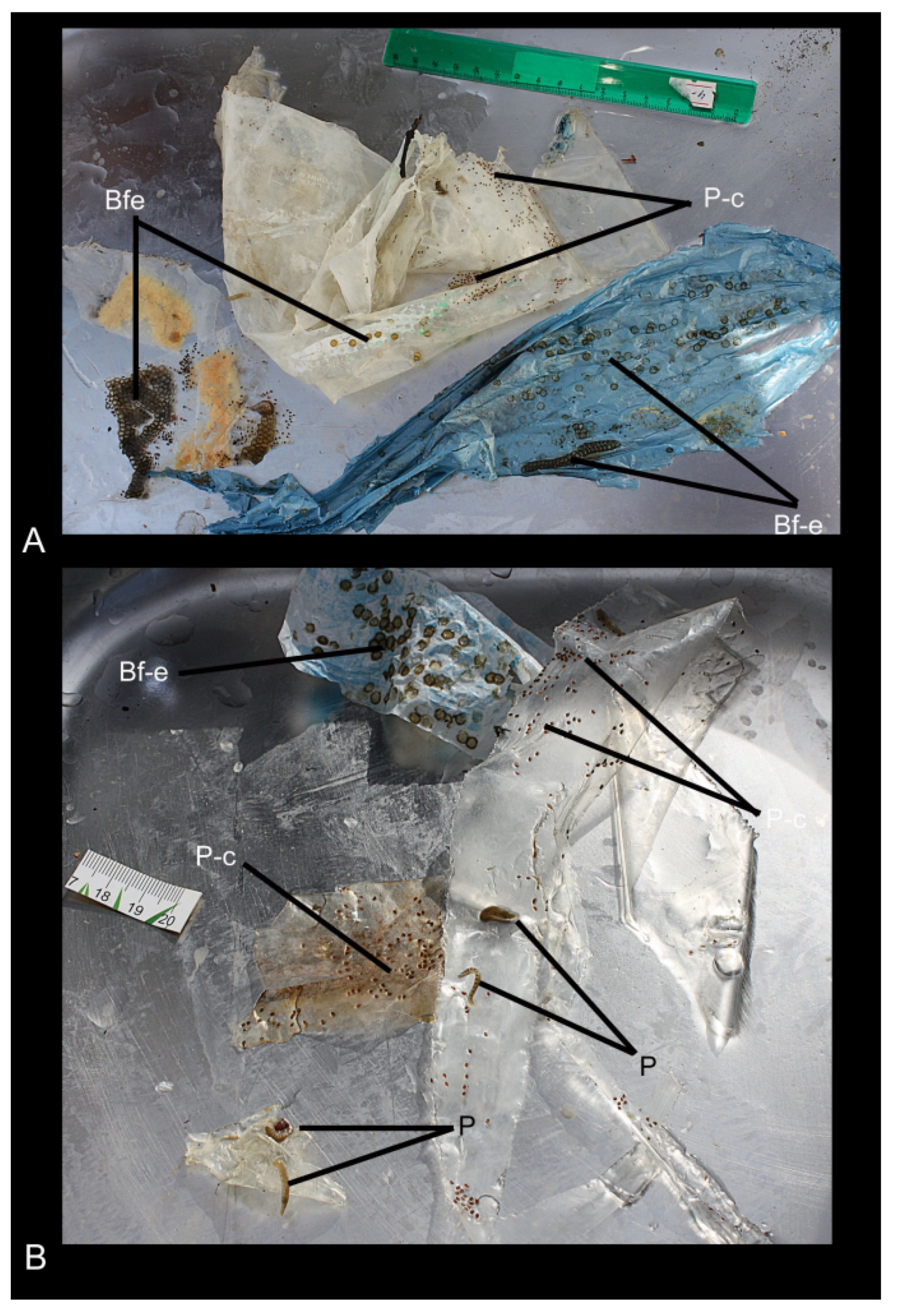
The waste consisted of five fragments of packing material, colourless, white, and blue plastic, with an area of 0.02 m2 to 1.2 m2 (Figure 2). During the primary examination, the leeches and bryozoans, the cocoons of the leeches, and the egg capsules of gastropods found on the waste fragments were photographed; the gastropod eggs were immediately identified. The leech cocoons and bryozoans were fixed with 80% ethanol for molecular analyses. Additionally, the leech cocoons were fixed with 4% paraformaldehyde in 0.05% phosphate buffer for morphological study using scanning electron microscopy (SEM). The plastic waste was frozen and transported to the laboratory for further research.

2.2. Plastic Composition Experimental Analysis

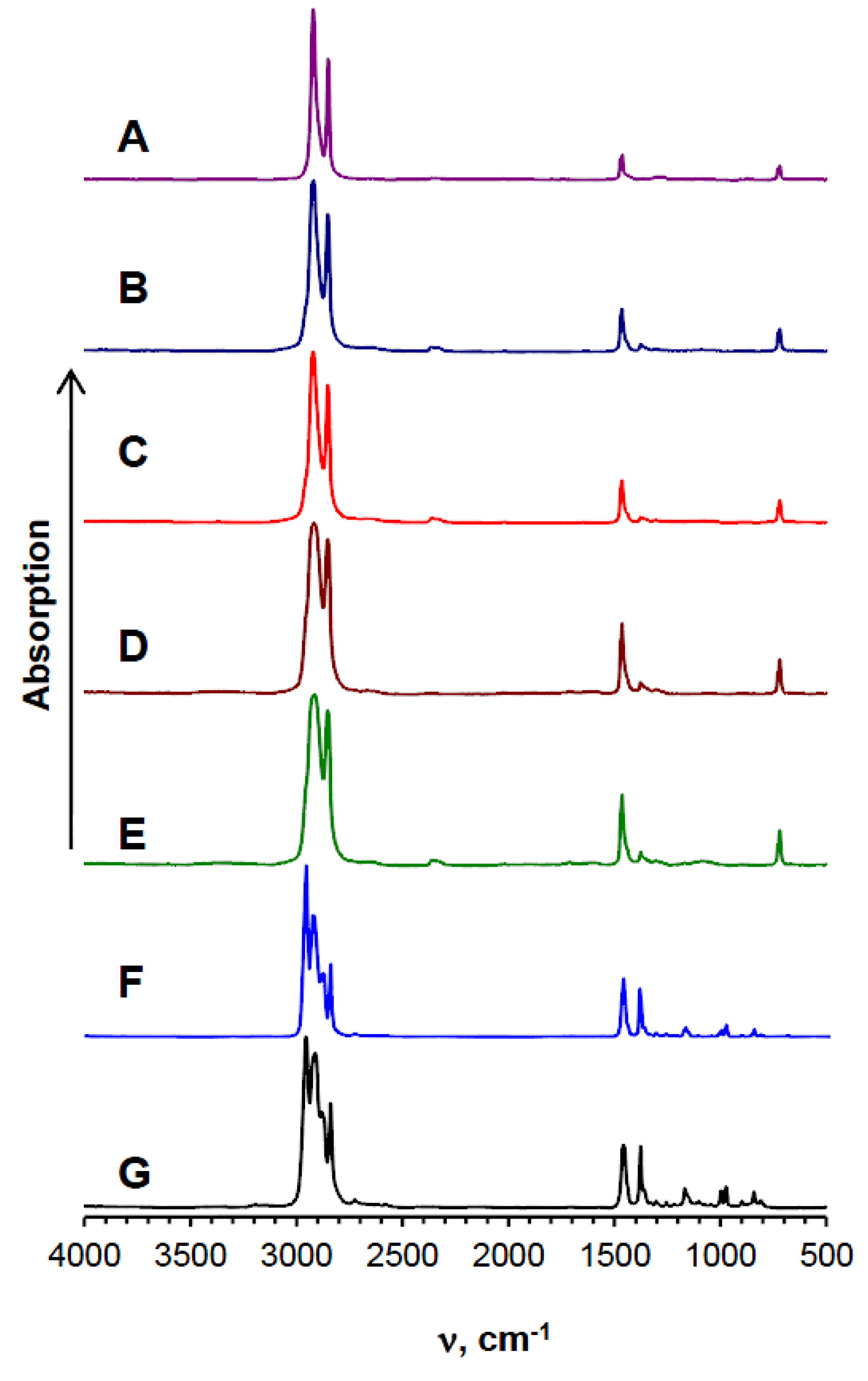
Fourier-transform infrared spectra (FT-IR) were recorded on an Infralum FT-801 spectrophotometer (SIMEX, Novosibirsk, Russia) in the transmission mode. Before measurement, the films were gently stretched to obtain a thickness that allows spectra with absorption below 1.5 to be obtained. The samples from Lake Baikal were thoroughly washed with SDS solution, then washed with deionised water and dried in a vacuum before the study began. The standard polyethylene and polypropylene spectra were obtained from Aldrich samples (Catalogues Nos. 42805-1 and 45214-9, respectively) cast on the Teflon surface from the melt.

2.3. Morphological Analysis

Ten cocoons (5 from the Selenga region and 5 from the Maloye More strait) were rinsed in a liquid of chlorine bleach, washed with distilled water and alcohol, then kept for 12 h in hexamethyldisilazane, according to the method of Saha et al. [16], then dried, gold plated, and examined using SEM (Quanta 200, K. Zeiss).

2.4. DNA Analysis

Total DNA was extracted using the DNA-sorb B commercial kit (Research Institute of Epidemiology, Rospotrebnadzor, Moscow, Russia) according to the manufacturer’s protocol.
A cytochrome C oxidase subunit I (COI) gene fragment was amplified using a pair of universal primers, LCO1490/HCO2198 [17], and Phusion High-Fidelity DNA Polymerase (Thermo Fisher Scientific, Carlsbad, CA, USA).
The amplified fragments were cloned into the pJET 1.2 (Fermentas) plasmid vector and analysed by PCR with the recommended pJET1.2 Forward/pJET1.2 Revers primers. The nucleotide sequences of both chains were determined through direct sequencing using the BigDye Terminator v3.1 Cycle Sequencing Kit on a 3500 Genetic Analyzer (Thermo Fisher Scientific), according to the manufacturer’s protocol. All nucleotide sequences were deposited into the GenBank international database under the following numbers: MN854831–MN854837, MN863382–MN863388, and MN866419.
A basic local alignment search [18] was applied to compare the obtained nucleotide sequences of the COI gene fragments with available sequence databases and calculate the statistical significance of the matches. Alignment of the obtained sets of nucleotide sequences was carried out using CLUSTAL W [19].
Visualisations of phylogenetic relationships and calculations of inter- and intragroup genetic distances were implemented using MEGA7.0, the maximum likelihood method [20]. Molecular evolution models were selected using the ModelTest-NG algorithm [21]. The best-fit models for the substitution patterns were GTR+G+I for leeches and Tamura 3-parameter+G+I for bryozoans [22,23].
The phylogenetic analyses of leeches and bryozoans involved 45 and 99 nucleotide sequences, respectively (Table 2 and Table 3). The sequence length of both datasets was 658 bp.

3. Results

3.1. The Chemical Composition of Plastic Waste

FT–IR spectra of the plastic samples collected in the Maloye More strait (Figure 3B–D) and the colourless plastic collected in the Selenga area (Figure 3E) corresponded to the standard spectrum of polyethylene (Figure 3A); the spectra of the blue plastic (Figure 3G) corresponded to the standard polypropylene spectrum (Figure 3F).

3.2. Waste-Associated Macroorganisms

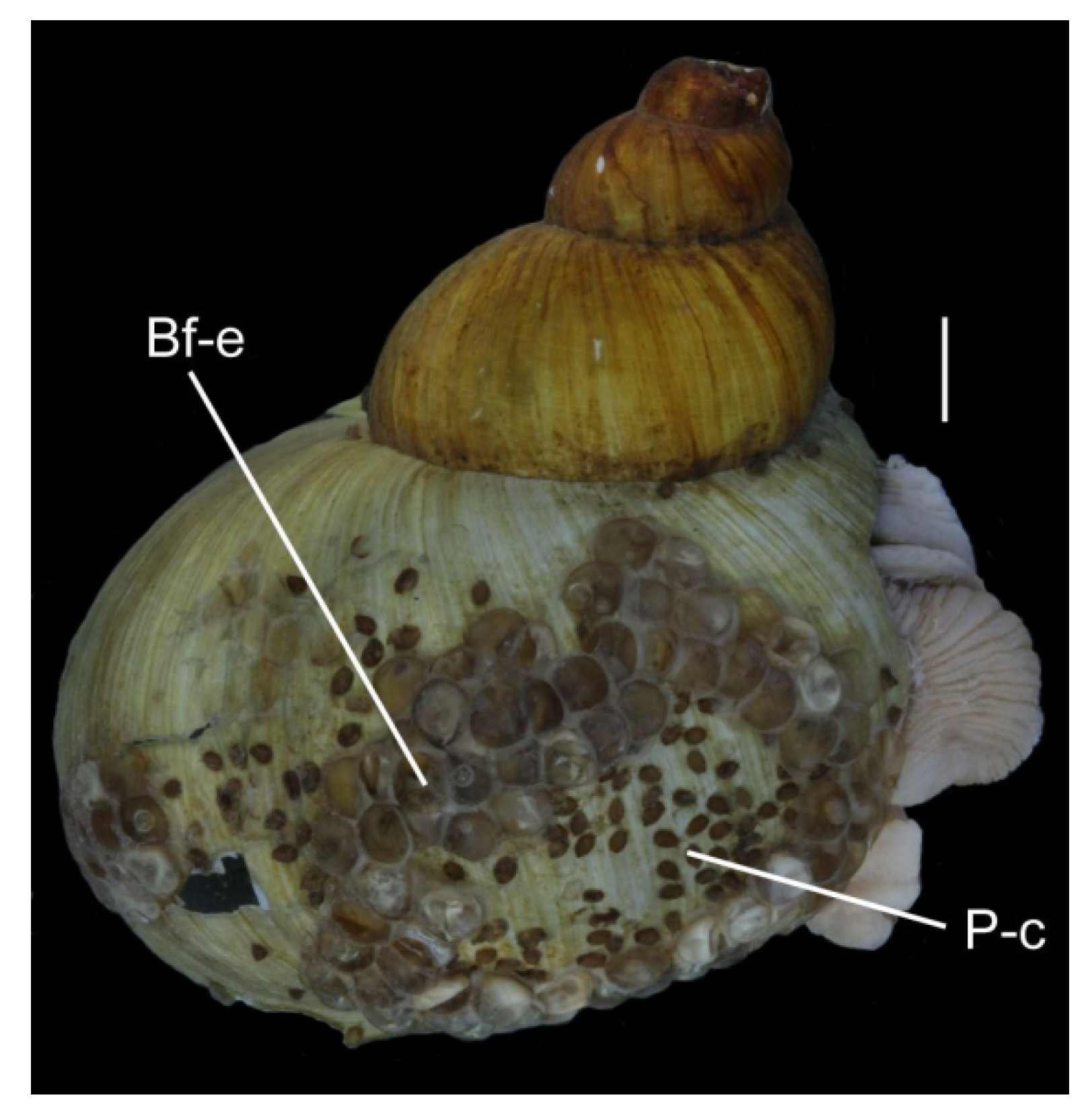
The cocoons of leeches and egg masses of gastropods found were unevenly distributed on the waste fragments (Figure 2); the coverage ranged from 10% (white and colourless plastic) to almost 50% (blue plastic). Some areas of polyethylene fragments of both colours were completely free of eggs. Large egg capsules (approximately 5 mm in diameter) in the form of a smooth hemisphere belonged to the Baikal endemic gastropods, Benedictia fragilis, whose adult individuals were collected together with the waste in the trawls. Similar egg capsules and leech cocoons attended the shells of this gastropod species (Figure 4).
On the plastic surface, egg capsules of snails were located both individually and in dense groups. There were 186 capsules on the plastic with an area of 1.2 m2. The proportion of capsules with embryos at the first stages of development was approximately 2%; the rest of the capsules were empty.
Leech cocoons were located close to the egg capsules of the snails, as well as at a distance from them, and did not form dense groups; there were from one to three cocoons in an area of 1 cm2. The globular cocoons (3 to 5 mm in diameter) of small Turbellaria were empty, dilapidated on the waste.
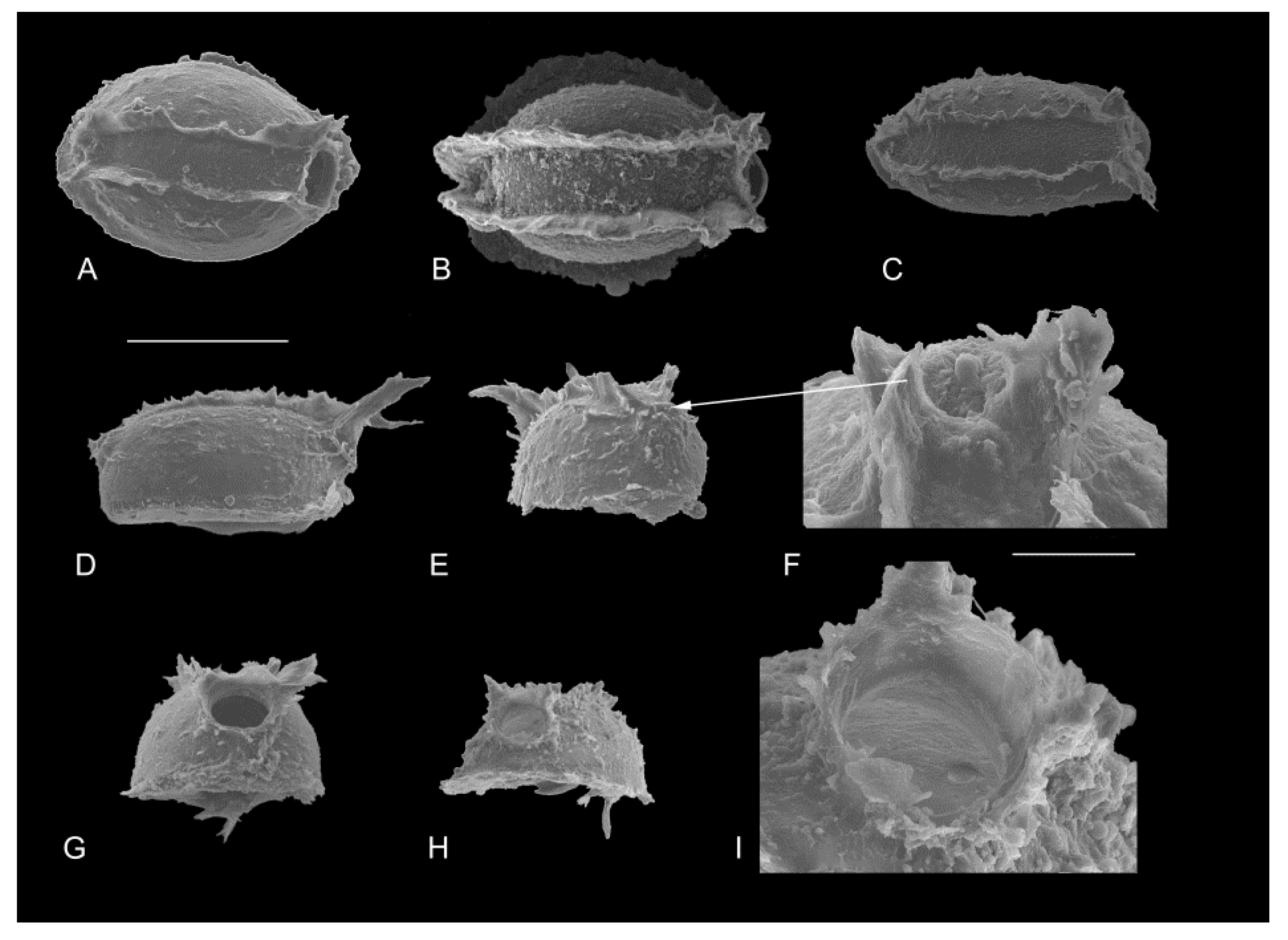
The cocoons of leeches from the northern region of the Maloye More strait were oblong; their length ranged from 889 μm to 1 mm (1.010 ± 1.175 μm), width—from 430 to 725 μm (594 ± 82), and height—from 247 to 600 μm (434 ± 102). Each cocoon had one aperture covered by an operculum, the diameter of which varied from 115 to 200 μm (168 ± 28; n = 10). The dense sheath of the cocoons was sculptured in the form of two parallel protuberances (Figure 5A–C), with the distance between ranging from 175 to 250 μm (216 ± 33; n = 8). The round operculum had two protuberances of different lengths; the posterior part of some cocoons had short protuberances (Figure 5F). The cocoons of leeches from the Selenga area had similar sizes and morphologies.
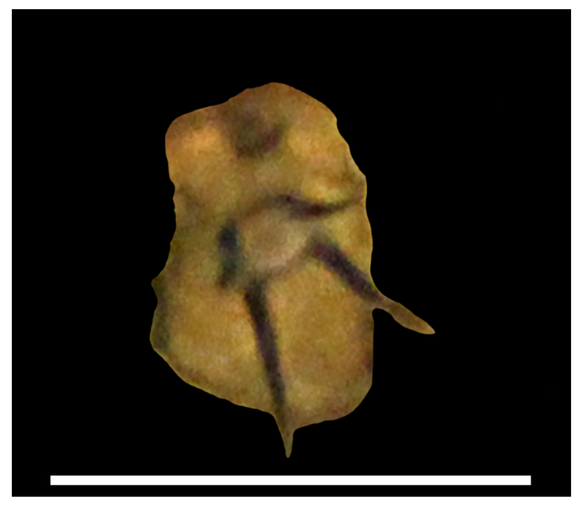
Bryozoans were found on the polyethylene that was lifted in the Selenga area; some of them were between empty cocoons of leeches. The colonies of bryozoans were short, consisting of three to five zooids with chitin exoskeleton (Figure 6), and similar to Hislopia placoides (Korotneff, 1901) in morphology.

3.3. Identification of Cocoons

Seven nucleotide sequences of the COI gene fragment (MN854831-MN854837) were obtained from the Maloye More cocoons containing eggs.
Cocoon COI sequences were searched by homology using the BLASTN 2.11.0+ [18], through the NCBI online platform (https://blast.ncbi.nlm.nih.gov; accessed on 26 May 2021). The search found 100 sequences belonging to Rhynchobdellida. The top of the list was formed by 33 different Piscicolidae species with the percent identity ranging between 82.07–90.51%. All of them were included in phylogenetic analysis as a comparison group, whereas representatives of Glossiphoniidae, another Rhynchobdellida family, were used as the outgroup.
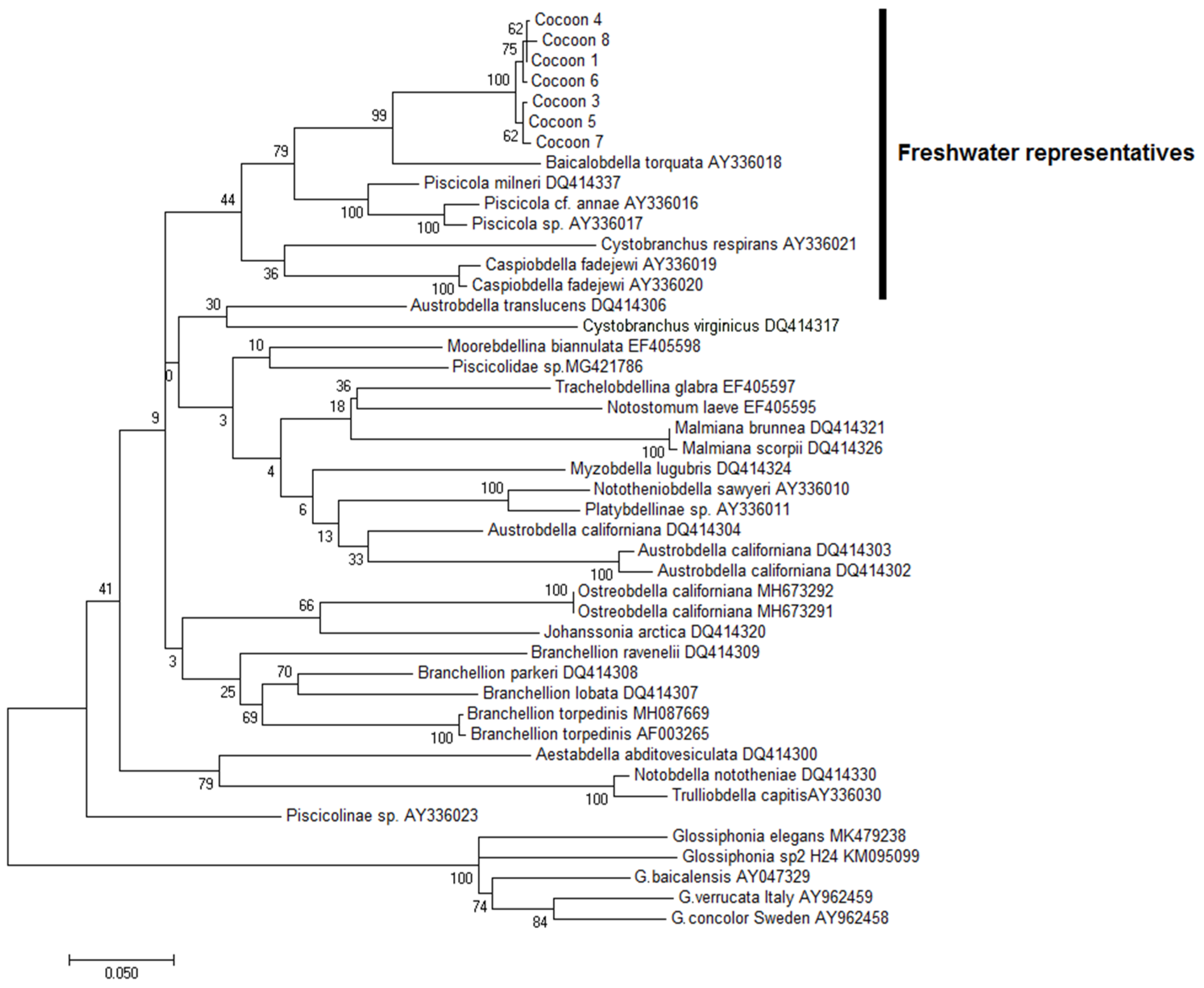
The phylogenetic tree showed that the group of the leech cocoon sequences form a single lineage within the freshwater representatives of the Piscicolidae branch with a high probability (99–100%) (Figure 7). COI of cocoons formed a separate clade and appeared to be closely related to the Baikal species, Baicalobdella torquata (Grube, 1871) (Figure 7).
The genetic polymorphism within the cocoon sequences is 0.42 ± 0.18% of nucleotide substitutions per site. Since these values coincide with the intraspecific differences typical for rhynchobdellid leeches [24], we should conclude that all cocoons most likely belong to the same species. There are 10 variable sites in the set of cocoons sequences: five transitions and five transversions. Some point mutations of the COI gene fragment led to amino acid replacements, including significant ones. Thus, hydrophobic phenylalanine in one of the sequences is replaced by hydrophilic serine. This fact can evidence the genetic plasticity of the test species.
The genetic distance between the cocoons and the B. torquata sequence is 9.85 ± 0.9%, which is comparable with the genetic distances between other Piscicolidae species (Table 4). According to the hypothesis underlying the DNA barcoding approach [14], this level of genetic distances is sufficient to conclude that these phylogenetic groups are taxonomically independent and therefore belong to different species. Unfortunately, the international base of publicly available DNA sequences contains a single nucleotide sequence of the COI belonging to the Baikal piscine leech—B. torquata (GenBank Accession No. AY336018).

3.4. Detection of Stramenopiles

In the associations with the empty leech cocoons from the Selenga River area, we obtained the nucleotide sequence (650 bp) of the COI mtDNA gene belonging to the stramenopiles clade (GenBank Accession MN866419). This sequence is genetically equidistant from both the fungi-like organism Phytopythium paucipapillatum Langenhoven, 2017 (SL-2017a strain STE-U7848; GenBank Accession No. KX372747) (Pythiaceae: Peronosporales), with a p-distance of 23%, and Pedospumella sinomuralis Boenigk et Grossmann 2016 (GenBank Accession No. KF697349) (Chrysophyta: Chromulinales), with a p-distance of 22%.

3.5. Identification of Bryozoans

The newly sequenced COI gene fragments from Baikal bryozoans (GenBank Accession Nos.MN863382-MN863388) differed in single-point substitutions (eight transitions and one transversion) that did not lead to significant amino acid replacements. The genetic distances within the group of bryozoans belonging to H. placoides sequences were 0.39 ± 0.12%, which, by analogy with the cocoons of leeches, indicates their genetic homogeneity and, as a consequence, their belonging to the same species. The COI sequences of the H. placoides clustered in a separate clade (Figure 8) and appeared to be relative of marine bryozoans of the genus Amathia Lamouroux, 1812 (Ctenostomatida: Vesiculariidae) (p-distance with A. imbricata 20.8 ± 2.0%; A. vidovici 20.8 ± 1.9%; A. distans 20.8 ± 2.0%) (Table 5).

4. Discussion

The presence of plastic waste at two sites of Central Baikal is not accidental, because they are subject to the influence of intense human activity. The Maloye More strait and the Selenga area are the main fishing areas where, before the introduction of the governmental ban on catching the Baikal omul Coregonus migratorius (Georgi, 1775) in 2017, commercial fishing was carried out. Nowadays, in addition to illegal fishing and recreational fishing, tourist camps and unorganised tourism contribute to pollution of the lake. Municipal solid waste from the coasts and ice cover of the lake, as well as from its inflowing rivers enters Lake Baikal. The detection of attached forms (bryozoans) and egg clutches (including empty and dilapidated) of benthic hydrobionts on the surfaces of packing materials indicates that the waste has been at the bottom of the lake for quite a long time. Unfortunately, there are no data on the time of hatching from the cocoons of Baikal Piscicolidae. However, earlier, under experimental conditions, it was shown that the hatching time of Myzobdella lugubris (Hirudinidae, Piscicolidae) is 48 ± 7 days at 17 °C, and at a temperature of 22 °C, the hatching time is 28 ± 5 days [25]. Taking into account the temperature regime at the bottom of Lake Baikal, we can assume with a fair degree of confidence that the plastic has been there for several months.
Piscine leeches and gastropods are oviparous, needing a substrate for the eggs to attach. However, it is obvious that there is a shortage of solid substrates in the Maloye More strait and in the Selenga area, since the bottom here, especially at the depth zone from 100 to 200 m, is composed of silt and silty sands [26].
The gastropods B. fragilis, with large shells (up to 6 cm in height), widespread in Lake Baikal at depths from 30 to 1300 m, are common inhabitants of the silt sands of the lake. In the absence of hard surfaces, they adapted, attaching their eggs to the smooth shell of their own species [27].
To date, there is no information about natural substrates used by the Baikal endemic leeches for attaching cocoons. Nevertheless, freshwater leeches avoid biotopes with soft and unstable substrates such as ooze and sand [11,28]. Piscine leeches usually attach cocoons to aquatic plants, rocky substrates, sunken trees, shells of molluscs, and exoskeletons of crustaceans [29,30,31]. Some species lay cocoons on various artificial substrates that enter aquatic ecosystems as a result of human activity [11,32]. At the same time, the density of cocoons, for example, for the species of the family Erpobdellidae on artificial substrates is several times higher than on natural ones [33]. The presence of cocoons on the B. fragilis shells and plastic waste (Figure 1) indicates that the Baikal endemic leeches can use any available solid substrate to attach their cocoons.
In the family Piscicolidae from the Maloye More strait, three species of Baikal endemic leeches were described: Baicalobdella torquata, B. cottidarum Dogiel, 1957, and Codonobdella sp. [34]. Recently, Matveenko and Kaygorodova [35] suggested the existence of two cryptic species morphologically corresponding to B. torquata. Our analysis has revealed that the investigated cocoons likely do not belong to B. torquata, whose nucleotide sequences are represented in GenBank. Moreover, representatives of the genus Baicalobdella are parasites of littoral amphipods and cottoid fishes; they were found at depths of 3 to 130 m [36]. It can be assumed that the cocoons that we found on the plastic waste could belong to another Baikal leech, Codonobdella sp., exploiting deep-water amphipods and ecologically confined to the sublittoral zone of Lake Baikal [34].
Since the samples of cocoons were taken within a depth range from 175 to 188 m, we assume that the polyethylene waste with the cocoons was initially located in the shallow zone of the lake and carried deeper by currents. The same cause can also explain the presence of bryozoans deeper than 150 m, the maximum depth mentioned by Vinogradov [37] for H. placoides.
Bryozoans are colonial animals leading a sessile lifestyle. Stones, aquatic plants, sunken wood, shells of molluscs, crayfish shells, and other objects, including plastic, serve as substrates for bryozoans. The experiment has revealed that larvae of the Bugula bryozoans choose plastic rather than wooden surfaces [38]. The genetic relationship of H. placoides with the marine representatives of Ctenostomatida indirectly confirms the opinion of Pelseneer [39] about their relatively recent divergence. The absence of COI mtDNA sequences of Hislopia from other lakes in GenBank does not allow us to clarify the taxonomic status of the Baikal bryozoans with a chitin exoskeleton and explain their origin in Lake Baikal. Large genetic distances between species within Ctenostomatida (Table 4) indicate that these animals are poorly studied. It is noteworthy that the accumulation of information about nucleotide sequences of bryozoans often leads to the identification of novel cryptic species or a redescription of the known ones [40,41]. Since H. placoides has been described in four morphotypes inhabiting different depths [36], we can assume that several Hislopia species exist in Lake Baikal. Of course, this hypothesis requires further research.
The egg cocoon shells of leeches and gastropods are composed of polysaccharides and fibrous proteins [34,42,43,44] and serve as substrates for colonies of various microorganisms and small fouling organisms. The detected sequence of stramenopiles appeared to be genetically equidistant from the fungi-like organisms Oomycetes and Chromulinales of Chrysophyta, being new for Lake Baikal. The closest relative from oomycetes, Phytopythium paucipapillatum, was isolated in South Africa, and may be a soil inhabitant [45]. The genus Phytopythium includes more than 20 species, most of which are saprophytic [46]. Pedospumella sinomuralis soil colourless Chrysophyta was described in China, 795 m asl [47]. Notably, in the water column of Lake Baikal, stramenopiles were the most diverse group (especially Chrysophyceae), encompassing 562 OTUs of 18S rDNA [48]. According to Yi et al. [49], Baikal microeukaryote diversity is very high and ecologically differentiated. The bottom stramenopiles have not been studied at all in Lake Baikal. The study of the organisms that are not available by conventional collection methods but inhabit plastic waste could lead to unique discoveries.
Despite the fact that our results on the community of benthic organisms associated with plastic waste are geographically limited, they can contribute to the understanding of the mechanisms of the spread of animals in previously uncharacteristic biotopes. At present, this is especially important when studying the distribution of invasive species [50] and the creation of new habitats for animals [51].
The endemic invertebrates effectively use plastic waste to attach cocoons and egg masses. Coming from shallow areas to great depths of Lake Baikal, plastic waste can contribute to the distribution of animals to previously untypical biotopes. The research results indicate the need for the monitoring of plastic waste pollution of the lake’s abyssal zones, aimed at replenishing knowledge about the biodiversity of the lake using comprehensive methods as well as determining the impact of waste pollution on the ecology of hydrobionts. It is necessary to conduct a qualitative and quantitative assessment of micro- and macroeukaryotes inhabiting plastic and an experimental study of the rate of colonisation of organisms and the development of communities of microorganisms, including those capable of destroying plastic.

Author Contributions

Conceptualization, E.V.D., N.N.D., T.Y.S. and I.G.K.; methodology, I.G.K., I.V.K., S.V.K., I.A.N. and T.Y.S.; software, N.N.D. and I.A.K.; validation, T.Y.S., N.N.D. and I.A.K.; formal analysis, N.N.D. and I.A.K.; investigation, I.G.K., T.Y.S., N.N.D., V.V.A. and I.A.K.; resources, I.V.K. and E.V.D.; data curation, E.V.D.; writing—original draft preparation, I.G.K., T.Y.S., N.N.D., I.A.K. and E.V.D.; writing—review and editing, N.N.D. and E.V.D.; visualization, T.Y.S., N.N.D. and I.A.K.; supervision, E.V.D.; project administration, E.V.D.; funding acquisition, I.V.K. and E.V.D. All authors have read and agreed to the published version of the manuscript.

Funding

This research received no external funding.

Institutional Review Board Statement

Not applicable.

Informed Consent Statement

Not applicable.

Data Availability Statement

The data on the obtained nucleotide sequences are available at the GenBank (MN854831-MN854837, MN863382-MN863388 and MN866419).

Acknowledgments

The authors are grateful to V.l. Egorov for help working in the Electron Microscopy Instrument Centre of the Joint Centre for Ultramicroanalysis, Limnological Institute, Siberian Branch, Russian Academy of Sciences. This study was performed within the framework of the State Tasks Nos. 0279-2021-0005, 0279-2019-0004, 0279-2021-0007.

Conflicts of Interest

The authors declare no conflict of interest.

References

  1. Kozhova, O.M.; Izmest’eva, L.R. Lake Baikal. In Evolution and Biodiversity; Backhuys Publishers: Leiden, The Netherlands, 1998; p. 447. [Google Scholar]
  2. Taliev, D.N. Sculpins Fishes of Lake Baikal (Cottoidei); Publishing House of the Academy of Sciences of the USSR: Moscow, Russia, 1955; p. 602. [Google Scholar]
  3. UNESCO. Operational Guidelines for the Implementation of the World Heritage Convention; UNESCO: Paris, France, 1999. [Google Scholar]
  4. Kravtsova, L.S.; Izhboldina, L.A.; Khanaev, I.V.; Pomazkina, G.V.; Domysheva, V.M.; Kravchenko, O.S.; Grachev, M.A. Disturbances of the vertical zoning of green algae in the coastal part of the Listvennichnyi Gulf of Lake Baikal. Dokl. Biol. Sci. 2012, 448, 227–229. [Google Scholar] [CrossRef]
  5. Kravtsova, L.S.; Izhboldina, L.A.; Khanaev, I.V.; Pomazkina, G.V.; Rodionova, E.V.; Domysheva, V.M.; Sakirko, M.V.; Tomberg, I.V.; Kostornova, T.Y.; Kravchenko, O.S.; et al. Nearshore benthic blooms of filamentous green algae in Lake Baikal. J. Great Lakes Res. 2014, 40, 441–448. [Google Scholar] [CrossRef]
  6. Timoshkin, O.A.; Samsonov, D.P.; Yamamuro, M.; Moore, M.V.; Belykh, O.I.; Malnik, V.V.; Sakirko, M.V.; Shirokaya, A.A.; Bondarenko, N.A.; Domysheva, V.M.; et al. Rapid ecological change in the coastal zone of Lake Baikal (East Siberia): Is the site of the world’s greatest freshwater biodiversity in danger? J. Great Lakes Res. 2016, 42, 487–497. [Google Scholar] [CrossRef] [Green Version]
  7. Timoshkin, O.A.; Bondarenko, N.A.; Kulikova, N.N.; Lukhnev, A.G.; Maximova, N.V.; Malnik, V.V.; Moore, M.V.; Nepokrytykh, A.V.; Obolkina, L.A.; Rozhkova, N.A.; et al. Protection of Lake Baikal requires more stringent, not more lenient, environmental regulation. J. Great Lakes Res. 2019, 45, 401–402. [Google Scholar] [CrossRef]
  8. Volkova, E.A.; Bondarenko, N.A.; Timoshkin, O.A. Morphotaxonomy, distribution and abundance of Spirogyra (Zygnematophyceae, Charophyta) in Lake Baikal, East Siberia. Phycologia 2018, 57, 298–308. [Google Scholar] [CrossRef]
  9. Potapskaya, N.V.; Kulikova, N.N.; Timoshkin, O.A.; Zaitseva, E.P.; Nepokrytykh, A.V.; Mal’nik, V.V. Estimation of consumer waste accumulation in the coastal area of Baikal and Selenga delta. Geogr. Nat. Resour. 2016, 1, 62–69. [Google Scholar]
  10. Chapman, M.G.; Clynick, B.G. Experiments testing the use of waste material in estuaries as habitat for subtidal organisms. J. Exp. Mar. Biol. Ecol. 2006, 338, 164–178. [Google Scholar] [CrossRef]
  11. Czarnecka, M.; Poznanska, M.; Kobak, J.; Wolnomiejski, N. The role of solid waste materials as habitats for macroinvertebrates in a lowland dam reservoir. Hydrobiologia 2009, 635, 125–135. [Google Scholar] [CrossRef]
  12. Kennedy, K.T.M.; El-Sabaawi, R.W. Decay patterns of invasive plants and plastic trash in urban streams. Urban Ecosyst. 2018, 21, 817–830. [Google Scholar] [CrossRef]
  13. Booth, A.J.; Kadye, W.T.; Vu, T.; Wright, M. Rapid colonisation of artificial substrates by macroinvertebrates in a South African lentic environment. Afr. J. Aquat. Sci. 2013, 38, 175–183. [Google Scholar] [CrossRef]
  14. Hebert, P.; Cywinska, A.; Ball, S.; Dewaard, J. Biological identifications through DNA barcodes. Proc. R. Soc. B 2003, 270, 313–321. [Google Scholar] [CrossRef] [PubMed] [Green Version]
  15. Hebert, P.; Hollingsworth, P.; Hajibabaei, M. From writing to reading the encyclopedia of life. Philos. Trans. R. Soc. Lond. B Biol. Sci. 2016, 371, 20150321. [Google Scholar] [CrossRef] [PubMed] [Green Version]
  16. Saha, R.K.; Biswas, A.; Goswami, U. A simple method for scanning electron microscopy (SEM) study of Cladocera: Bosmina (Bosmina) tripurae. WJFMS 2011, 3, 71–78. [Google Scholar]
  17. Folmer, O.; Black, M.; Hoeh, W.; Lutz, R.; Vrijenhoek, R. DNA primers for amplification of mitochondrial cytochrome c oxidase subunit I from diverse metazoan invertebrates. Mol. Mar. Biol. Biotechnol. 1994, 3, 294–299. [Google Scholar] [PubMed]
  18. Zhang, Z.; Schwartz, S.; Wagner, L.; Miller, W. A greedy algorithm for aligning DNA sequences. J. Comput. Biol. 2000, 7, 203–214. [Google Scholar] [CrossRef]
  19. Larkin, M.A.; Blackshields, G.; Brown, N.P.; Chenna, R.; McGettigan, P.A.; McWilliam, H.; Valentin, F.; Wallace, I.M.; Wilm, A.; Lopez, R.; et al. Clustal W and Clustal X version 2.0. Bioinformatics 2007, 23, 2947–2948. [Google Scholar] [CrossRef] [Green Version]
  20. Kumar, S.; Stecher, G.; Tamura, K. MEGA7: Molecular evolutionary genetics analysis version 7.0 for bigger datasets. Mol. Biol. Evol. 2016, 33, 1870–1874. [Google Scholar] [CrossRef] [Green Version]
  21. Darriba, D.; Posada, D.A.; Kozlov, M.; Stamatakis, A.; Morel, B.; Flouri, T. ModelTest-NG: A new and scalable tool for the selection of DNA and protein evolutionary models. Mol. Biol. Evol. 2019, 37, 291–294. [Google Scholar] [CrossRef] [Green Version]
  22. Tamura, K. Estimation of the number of nucleotide substitutions when there are strong transition-transversion and G + C-content biases. Mol. Biol. Evol. 1992, 9, 678–687. [Google Scholar]
  23. Tamura, K.; Nei, M. Estimation of the number of nucleotide substitutions in the control region of mitochondrial DNA in humans and chimpanzees. Mol. Biol. Evol. 1993, 10, 512–526. [Google Scholar]
  24. Kaygorodova, I.; Bolbat, N.; Bolbat, A. Species delimitation through DNA barcoding of freshwater leeches of the Glossiphonia genus (Hirudinea: Glossiphoniidae) from Eastern Siberia, Russia. J. Zoolog. Syst. Evol. Res. 2020, 58, 1437–1446. [Google Scholar] [CrossRef]
  25. Saglam, N.; Saunders, R.; Lang, S.A.; Shain, D.H. Phylogeny and cocoon production in the parasitic leech Myzobdella lugubris Leidy, 1851 (Hirudinidae, Piscicolidae). Acta Parasitol. 2018, 63, 15–26. [Google Scholar] [CrossRef] [PubMed]
  26. Karabanov, E.B.; Sideleva, V.G.; Izhboldina, L.A.; Melnik, N.G.; Zubin, A.A.; Zubina, L.V.; Smirnov, N.V.; Parfyonova, V.V.; Fedorova, O.A.; Gorbunova, L.A.; et al. Underwater Landscapes of Baikal; Nauka: Novosibirsk, Russia, 1990; p. 183. [Google Scholar]
  27. Sitnikova, T.Y.; Shimaraev, M.N. Deep-water “dwarfs” and “giants” among endemic Baikal gastropods. Zh. Obs. Biol. 2001, 62, 226–238. [Google Scholar]
  28. Brönmark, C. Leech predation on juvenile freshwater snails: Effects of size, species and substrate. Oecologia 1992, 91, 526–529. [Google Scholar] [CrossRef] [PubMed]
  29. Sawyer, R.T. Leech Biology and Behavior: Feeding Biology, Ecology, and Systematics, 1st ed.; Oxford University Press: Oxford, UK, 1986; Volume II. [Google Scholar]
  30. Young, J.O. Intra- and interspecific predation of the cocoon of Erpobdella octoculata (L.) (Annelida: Hirudinea). Hydrobiologia 1988, 169, 85–89. [Google Scholar] [CrossRef]
  31. Nagasawa, K.; Fujiwara, K. Two Piscicolid leeches (Hirudinida) and their cocoons on Snow Crabs Chionoecetes opilio in Japan, with the first record of Johanssonia arctica from the Sea of Japan. Biogeography 2008, 10, 65–72. [Google Scholar]
  32. Adamiak-Brud, Z.; Jabłonska-Barna, I.; Bielecki, A.; Terlecki, J. Settlement preferences of leeches (Clitellata: Hirudinida) for different artificial substrates. Hydrobiologia 2015, 758, 275–286. [Google Scholar] [CrossRef] [Green Version]
  33. Koperski, P. Stone-dwelling leeches (Hirudinea, Clitellata) of Lake Hancza (Poland): Different sampling methods determine different taxonomic structures. Pol. J. Ecol. 2003, 51, 353–361. [Google Scholar]
  34. Kaygorodova, I.A. Annotated checklist of the leech species diversity in the Maloe More Strait of Lake Baikal, Russia. ZooKeys 2015, 545, 37–52. [Google Scholar] [CrossRef]
  35. Matveenko, E.Y.; Kaygorodova, I.A. Ecological zonation of the Baikal endemic piscine leeches of the genus Baicalobdella (Hirudinea, Piscicolidae). Limnol. Freshw. Biol. 2020, 4, 801–802. [Google Scholar] [CrossRef]
  36. Snimschikova, L.N. The Lists of species of animals and plants inhabiting in Baikal. Hirudinea. In Lake Baikal: Evolution and Biodiversity; Kozhova, O.M., Izmest’eva, L.R., Eds.; Backuys Publishers: Leiden, The Netherlands, 1998; pp. 369–370. [Google Scholar]
  37. Vinogradov, A.V. Eurystomata and Phylactolaemata fauna at the main relict continental water-bodies of Euroasia. Izv. RAS SamSC 2008, 10, 531–546. [Google Scholar]
  38. Pinochet, J.; Urbina, M.A.; Lagos, M.E. Marine invertebrate larvae love plastics: Habitat selection and settlement on artificial substrates. Environ. Pollut. 2020, 257, 113571. [Google Scholar] [CrossRef] [PubMed]
  39. Pelseneer, P. The origin of fresh water animals [L’origine des animaux d’eau douce]. Acad. Analecta 1906, 12, 699–741. [Google Scholar]
  40. Fehlauer-Ale, K.H.; Vieira, L.M.; Winston, J.E. Molecular and morphological characterization of Amathia distans Busk and Amathia brasiliensis Busk (Bryozoa: Ctenostomata) from the tropical and subtropical Western Atlantic. Zootaxa 2011, 2962, 49–62. [Google Scholar] [CrossRef]
  41. Waeschenbach, A.; Vieira, L.M.; Reverter-Gil, O.; Souto-Derungs, J.; Nascimento, K.B.; Fehlauer-Ale, K.H. A phylogeny of Vesiculariidae (Bryozoa, Ctenostomata) supports synonymization of three genera and reveals possible cryptic diversity. Zool. Scr. 2015, 44, 667–683. [Google Scholar] [CrossRef]
  42. Mason, T.A.; McIlroy, J.P.; Shain, D.H. A cysteine-rich protein in the Theromyzon (Annelida: Hirudinea) cocoon membrane. FEBS J. 2004, 561, 167–172. [Google Scholar] [CrossRef] [Green Version]
  43. Coleman, J.; Shain, D.H. Clitellate cocoons and their secretion. In Annelids in Modern Biology; Shain, D.H., Ed.; Wiley-Blackwell: Hoboken, NJ, USA, 2009; Volume 18, pp. 328–346. [Google Scholar]
  44. Rossi, A.M.; Saidel, W.M.; Gravante, C.J.; Sayers, C.W.; Shain, D.H. Mechanics of cocoon secretion in a segmented worm (Annelida: Hirudinidae). Micron 2016, 86, 30–35. [Google Scholar] [CrossRef] [Green Version]
  45. Langenhoven, S.D.; Halleen, F.; Spies, C.; Stempien, E.; Mostert, L. Detection and quantification of black foot and crown and root rot pathogens in grapevine nursery soils in the Western Cape of South Africa. Phytopathol. Mediterr. 2018, 57, 519–537. [Google Scholar]
  46. Tkaczyk, M. Phytopythium: Origin, differences and meaning in modern plant pathology. Folia For. Pol. A 2020, 62, 227–232. [Google Scholar] [CrossRef]
  47. Grossmann, L.; Bock, C.; Schweikert, M.; Boenigk, J. Small but manifold-hidden diversity in “Spumella-like Flagellates”. J. Eukaryot. Microbiol. 2016, 63, 419–439. [Google Scholar] [CrossRef] [PubMed] [Green Version]
  48. Annenkova, N.V.; Giner, C.R.; Logares, R. Tracing the origin of planktonic protists in an Ancient Lake. Microorganisms 2020, 8, 543. [Google Scholar] [CrossRef] [PubMed] [Green Version]
  49. Yi, Z.; Berney, C.; Hartikainen, H.; Mahamdallie, S.; Gardner, M.; Boenigk, J.; Cavalier-Smith, T.; Bass, D. High-throughput sequencing of microbial eukaryotes in Lake Baikal reveals ecologically differentiated communities and novel evolutionary radiations. FEMS Microbiol. Ecol. 2017, 93, 1–19. [Google Scholar] [CrossRef] [PubMed]
  50. Rech, S.; Borrell Pichs, Y.J.; García-Vazquez, E. Anthropogenic marine litter composition in coastal areas may be a predictor of potentially invasive rafting fauna. PLoS ONE 2018, 13, e0191859. [Google Scholar] [CrossRef] [PubMed] [Green Version]
  51. Thushari, G.G.N.; Senevirathna, J.D.M. Plastic pollution in the marine environment. Heliyon 2020, 6, e04709. [Google Scholar] [CrossRef] [PubMed]
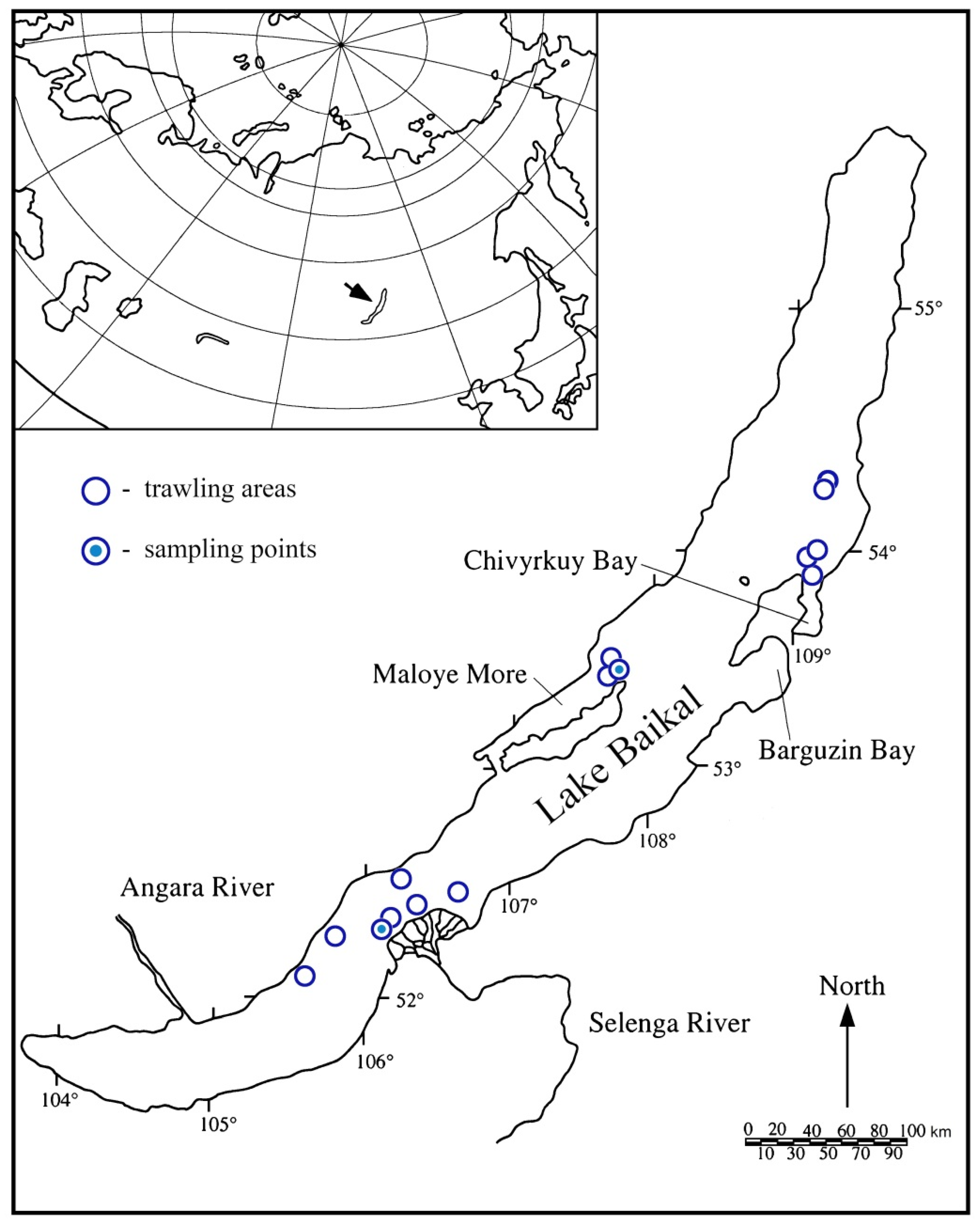
Figure 1. Lake Baikal: trawl map.
Biology 10 00972 g001
Figure 2. Fragments of packing materials with cocoons of leeches (P-c), leeches (P), and egg capsules (Bf-e) of Benedictia fragilis (Dybowski, 1875). (A)—from northern region of the Maloye More strait; (B)—from the Selenga area.
Biology 10 00972 g002
Figure 3. Fourier-transform infrared spectra of standard polyethylene (A), colourless (B), white (C), and blue (D) films from the Maloe More strait, colourless (E) films from the Selenga area, standard polypropylene (F), and a blue film (G) from the Selenga area.
Biology 10 00972 g003
Figure 4. The shell of Benedictia fragilis with egg capsules (Bf-e) laid by a snail on the same species and cocoons of leeches (P-c). Scale: 5 mm.
Biology 10 00972 g004
Figure 5. Leech cocoons collected from plastic waste: top view (AC); side view (D); the back side of the cocoon (E,F); the front part of the cocoons (GI); empty cocoons without operculum (A,G); cocoons with operculum (F,H,I); enlarged operculum (I). Scale: 500 μm (AE,G,H); 100 μm (F,I).
Biology 10 00972 g005
Figure 6. Zooid of bryozoans, Hislopia placoides (Korotneff, 1901). Scale: 1 mm.
Biology 10 00972 g006
Figure 7. Maximum likelihood phylogenetic tree visualizing the phylogenetic position of the studied leech cocoons.
Biology 10 00972 g007
Figure 8. Maximum likelihood phylogenetic tree of Ctenostomatida obtained from 99 nucleotide sequences of COI fragment. Bootstrap values are shown next to the branches (1.000 replicates).
Biology 10 00972 g008
Table 1. Places and coordinates of trawls.
TrawlPlace NameCoordinates down (N/E)Coordinates up (N/E) Depths (m)
1Selenga River delta52°17.270′/106°03.910′52°18.600′/106°06.095′111 to 133
2Selenga River delta52°19.910′/106°07.441′52°22.280′/106°10.015′142 to 181
3Selenga River delta52°24.933′/106°13.027′52°26.217′/106°16.940′271 to 276
4Proval Bay52°30.502′/106°36.704′52°31.196′/106°40.088′553 to 478
5Chivyrkuy Bay53°55.900′/109°08.473′53°58.341′/109°05.869′790 to 760
6Chivyrkuy Bay53°56.260′/109°06.425′53°57.765′/109°07.358′785 to 810
7Chivyrkuy Bay53°49.813′/109°08.584′53°50.787′/109°03.886′125 to 180
8Davsha54°23.662′/109°14.127′54°23.091′/109°15.727′700 to 715
9Davsha54°21.745′/109°14.749′54°21.006′/109°12.765′715 to 715
10Northern section of the Maloye More strait53°26.746′/107°44.691′53°26.496′/107°43.012′313 to 300
11Northern section of the Maloye More strait53°25.280′/107°45.085′53°24.821′/107°44.079′175 to 188
12Northern section of the Maloye More strait53°24.979′/107°44.783′53°24.585′/107°43.074′163 to 186
13Buguldeyka52°30.870′/106°07.467′52°29.179′/106°07.226′360 to 395
14Peschanaya52°15.864′/105°50.365′52°14.974′/105°49.767′1005 to 1015
15Verkhniye Khomuty52°06.481′/105°37.545′52°05.767′/105°39.826′1136 to 1200
Table 2. Most relative sequences of Hirudinea selected by BLAST.
DescriptionMax ScoreQuery CoverPer. IdentAccession
Baicalobdella torquata83395%90.51%AY336018
Piscicola milneri81399%88.82%DQ414337
Piscicola cf. annae77194%88.71%AY336016
Moorebdellina biannulata76399%87.46%EF405598
Piscicola sp.74593%88.12%AY336017
Branchellion parkeri72599%86.40%DQ414308
Austrobdella translucens72599%86.40%DQ414306
Nototheniobdella sawyeri70899%86.04%AY336010
Branchellion torpedinis69998%85.84%MH087669
Caspiobdella fadejewi68994%86.46%AY336019
Piscicolidae sp.68898%85.58%MG421786
Branchellion torpedinis68697%85.69%AF003265
Caspiobdella fadejewi68294%86.26%AY336020
Cystobranchus respirans66994%85.90%AY336021
Trachelobdellina glabra64799%84.31%EF405597
Branchellion lobata63699%83.99%DQ414307
Aestabdella abditovesiculata63699%84.08%DQ414300
Piscicolinae sp.63497%84.36%AY336023
Myzobdella lugubris63099%83.91%DQ414324
Austrobdella californiana63099%83.86%DQ414304
Johanssonia arctica62599%83.71%DQ414320
Cystobranchus virginicus62599%83.69%DQ414317
Branchellion ravenelii62398%83.76%DQ414309
Notostomum laeve61999%83.56%EF405595
Ostreobdella californiana61697%83.72%MH673292
Ostreobdella californiana61697%83.72%MH673291
Malmiana brunnea59799%82.98%DQ414321
Austrobdella californiana59799%82.96%DQ414303
Platybdellinae sp.59784%85.66%AY336011
Malmiana scorpii58699%82.68%DQ414326
Notobdella nototheniae57998%82.60%DQ414330
Austrobdella californiana56494%82.93%DQ414302
Trulliobdella capitis55698%82.07%AY336030
Glossiphonia elegans55598%82.00%MK479238
Glossiphonia complanata52798%81.27%JQ821635
Table 3. Most relative sequences of Bryozoa: Ctenostomatida selected by BLAST (Max Score > 460).
DescriptionMax ScoreQuery CoverPer. IdentAccession
Amathia evelinae60199%83.41%KM373436
Amathia vidovici60199%83.36%KM373394
Amathia cf. vidovici60199%83.36%JF490059
Amathia distans60199%83.28%JF490058
Amathia imbricata59999%83.16%KM373430
Amathia vidovici59599%83.21%KM373419
Amathia vidovici59599%83.21%KM373415
Amathia vidovici59599%83.21%KM373414
Amathia vidovici59599%83.21%KM373413
Amathia vidovici59599%83.21%KM373412
Amathia vidovici59599%83.21%KM373411
Amathia vidovici59599%83.21%KM373410
Amathia vidovici59599%83.21%KM373406
Amathia vidovici59599%83.21%KM373404
Amathia vidovici59599%83.21%KM373401
Amathia vidovici59599%83.21%KM373399
Amathia vidovici59599%83.21%KM373395
Amathia vidovici59599%83.21%KM373379
Amathia vidovici59599%83.21%KM373368
Amathia vidovici59599%83.21%KM373367
Amathia vidovici59599%83.21%KM373366
Amathia vidovici59398%83.26%KM373370
Amathia vidovici58898%83.05%KM373421
Amathia vidovici58896%83.41%KM373418
Amathia vidovici58897%83.26%KM373396
Amathia vidovici58898%83.05%KM373380
Amathia vidovici58897%83.26%KM373374
Amathia vidovici58897%83.26%KM373373
Amathia vidovici58897%83.26%KM373372
Amathia vidovici58897%83.26%KM373369
Amathia vidovici58898%83.05%KM373365
Amathia vidovici58898%83.05%KM373364
Amathia vidovici58497%83.20%KM373417
Amathia vidovici58497%83.10%KM373400
Amathia imbricata58099%82.55%KM373431
Amathia vidovici58096%83.23%KM373397
Amathia vidovici57996%83.20%KM373362
Amathia vidovici57996%83.20%KM373361
Amathia vidovici57795%83.25%KM373420
Amathia vidovici57796%83.12%KM373382
Amathia vidovici57796%83.12%KM373381
Amathia vidovici57796%83.12%KM373376
Amathia vidovici57796%83.12%KM373375
Amathia vidovici57795%83.25%KM373371
Amathia maxima57198%82.72%KM373432
Amathia vidovici57196%82.92%KM373402
Amathia vidovici57196%82.97%KM373384
Amathia vidovici57196%82.94%KM373359
Amathia vidovici56695%82.97%KM373378
Amathia vidovici53895%81.86%KM373377
Bowerbankia gracilis51898%81.25%FJ196083
Amathia vidovici51071%86.20%KM373407
Amathia vidovici51071%86.20%KM373405
Anguinella palmata50198%80.79%JN680973
Amathia vidovici49971%85.77%KM373393
Amathia vidovici49971%85.77%KM373391
Amathia vidovici49971%85.77%KM373360
Amathia vidovici49295%80.57%KM373383
Anguinella palmata46498%79.69%KM373422
Table 4. Estimates of evolutionary divergence over sequence pairs between cocoons group and nearest related sequences. Standard error estimates are shown above the diagonal.
Cocoons 0.0140.0140.0130.014
Piscicola cf. annae AY3360160.11641 0.0060.0150.010
Piscicola sp. AY3360170.124250.023 0.0150.009
Baicalobdella torquata AY3360180.098590.1230.129 0.014
Piscicola milneri DQ4143370.116760.0590.0520.115
Table 5. Estimates of evolutionary divergence over sequence pairs between Hislopia placoides group and other representatives Ctenostomatida.
Hislopia placoides 0.0190.0220.0200.0220.0190.0230.0210.0200.0300.0230.0230.0220.0240.0220.0250.0270.0250.0320.0300.0220.0220.039
Amathia vidovici
2 group
0.208 0.0130.0120.0170.0170.0140.0140.0170.0300.0150.0160.0140.0240.0140.0150.0200.0250.0330.0300.0220.0210.038
Amathia vidovici
1 group
0.2360.115 0.0160.0200.0170.0160.0150.0200.0320.0160.0190.0170.0250.0150.0170.0220.0280.0380.0360.0250.0230.039
Amathia distans0.2080.1110.136 0.0190.0170.0170.0160.0170.0330.0150.0160.0140.0250.0150.0170.0210.0240.0380.0330.0230.0220.040
Amathia tertia0.2370.1750.2030.179 0.0200.0190.0200.0150.0290.0160.0190.0160.0270.0180.0200.0230.0250.0320.0320.0240.0200.043
Amathia evelinae0.2170.1920.1870.1900.220 0.0200.0190.0190.0300.0190.0210.0190.0230.0180.0220.0220.0220.0350.0320.0230.0200.036
Amathia pustulosa0.2440.1270.1440.1610.1870.234 0.0170.0190.0290.0150.0170.0170.0260.0170.0190.0200.0270.0310.0320.0250.0240.037
Amathia maxima0.2240.1270.1210.1400.1930.2050.153 0.0200.0300.0160.0190.0150.0260.0160.0190.0210.0260.0370.0320.0230.0210.043
Amathia imbricata0.2080.1690.1980.1660.1250.2110.1940.196 0.0310.0170.0180.0180.0250.0160.0200.0220.0240.0340.0340.0240.0240.040
Amathia gracilis0.3680.3930.3950.4080.3570.3890.3770.3800.390 0.0300.0310.0300.0270.0320.0310.0260.0300.0270.0270.0280.0300.030
Amathia citrina0.2430.1440.1500.1350.1650.2150.1320.1400.1770.389 0.0160.0140.0260.0140.0160.0200.0260.0350.0320.0240.0220.043
Amathia crispa0.2380.1540.1810.1530.1850.2290.1570.1780.1800.3920.143 0.0170.0270.0170.0200.0210.0260.0370.0340.0240.0230.040
Vesicularia spinosa0.2350.1270.1430.1280.1540.2060.1640.1200.1620.3680.1230.156 0.0260.0130.0170.0190.0240.0340.0320.0220.0220.041
Anguinella palmata0.2590.2710.2720.2680.2990.2720.2840.2720.2590.3450.2880.3050.263 0.0260.0270.0280.0250.0290.0260.0240.0260.035
Amathia
semiconvoluta
0.2240.1250.1180.1230.1750.1900.1540.1300.1530.3980.1190.1530.1090.280 0.0170.0200.0240.0360.0340.0250.0210.042
Amathia lendigera0.2610.1430.1640.1570.2060.2300.1820.1770.1970.3810.1480.1900.1630.2800.156 0.0220.0250.0370.0340.0270.0240.041
Amathia convoluta0.3100.2100.2320.2130.2530.2740.2140.2260.2440.3420.2090.2310.1890.3130.2100.230 0.0260.0270.0290.0260.0240.036
Alcyonidium
verrilli
0.2650.2760.2970.2620.2810.2550.2850.2640.2690.3790.2810.2810.2440.2750.2520.2650.312 0.0340.0310.0260.0250.036
Alcyonidium
mamillatum
0.3650.3940.4190.4200.3750.4120.3500.4090.4020.3360.3880.4160.3800.3200.4060.4000.3180.399 0.0290.0340.0300.036
Triticella flava0.3500.3570.3980.3750.3620.3660.3720.3530.3730.3230.3600.3880.3600.2660.3790.3860.3350.3490.331 0.0260.0310.033
Triticella
pedicellata
0.2450.2420.2700.2450.2440.2570.2660.2400.2630.3550.2590.2680.2330.2560.2660.2840.2930.2740.3770.266 0.0220.037
Bowerbankia
gracilis
0.2390.2100.2340.2150.2090.2200.2390.2070.2350.3750.2250.2510.2230.2910.2150.2430.2520.2560.3550.3360.233 0.035
Flustrellidra hispida0.4810.4630.4540.4640.4900.4460.4350.4790.4930.3920.5110.4710.4810.4290.4780.4690.4480.4160.4190.3900.4470.412
Publisher’s Note: MDPI stays neutral with regard to jurisdictional claims in published maps and institutional affiliations.