Article

Co-Existence of Free-Living Amoebae and Potential Human Pathogenic Bacteria Isolated from Rural Household Water Storage Containers

by
Natasha Potgieter
1,*,
Clarissa van der Loo
2 and
Tobias George Barnard
2
1
One Health Research Group, Department of Biochemistry and Microbiology, Faculty of Science, Engineering and Agriculture, University of Venda, Thohoyandou 0950, Limpopo Province, South Africa
2
Water and Health Research Centre, Doornfontein Campus, University of Johannesburg, Johannesburg 2112, Gauteng, South Africa
*
Author to whom correspondence should be addressed.
Biology 2021, 10(12), 1228; https://doi.org/10.3390/biology10121228
Submission received: 16 October 2021 / Revised: 7 November 2021 / Accepted: 8 November 2021 / Published: 25 November 2021
(This article belongs to the Section Microbiology)

Abstract

:

Simple Summary

In many households in rural communities, water needed for drinking and cooking is fetched from rivers, fountains, or boreholes shared by the community. The water is then stored in various storage containers for several days without treatment and exposed to several conditions that could potentially contaminate the water and cause diseases. If the storage containers are not regularly and properly cleaned, biofilms can form inside the containers. Several microorganisms can be found inside the biofilm that can potentially cause diseases in humans. One such group of organisms is called free-living amoebae, which graze on the bacteria found inside the biofilm. Several of these potentially harmful bacteria have adapted and can survive inside these free-living amoebae and potentially cause diseases when ingested by humans.

Abstract

This study investigated the co-existence of potential human pathogenic bacteria and free-living amoebae in samples collected from stored water in rural households in South Africa using borehole water as a primary water source. Over a period of 5 months, a total of 398 stored water and 392 biofilm samples were collected and assessed. Free-living amoebae were identified microscopically in 92.0% of the water samples and 89.8% of the biofilm samples. A further molecular identification using 18S rRNA sequencing identified Vermamoeba vermiformis, Entamoeba spp., Stenamoeba spp., Flamella spp., and Acanthamoeba spp. including Acanthamoeba genotype T4, which is known to be potentially harmful to humans. Targeted potential pathogenic bacteria were isolated from the water samples using standard culture methods and identified using 16S rRNA sequencing. Mycobacterium spp., Pseudomonas spp., Enterobacter spp., and other emerging opportunistic pathogens such as Stenotrophomonas maltophilia were identified. The results showed the importance of further studies to assess the health risk of free-living amoebae and potential human pathogenic bacteria to people living in rural communities who have no other option than to store water in their homes due to water shortages.

1. Introduction

Statistics South Africa [1] reported that only 88.2% of households have access to an improved drinking water source in South Africa. Many households in rural and peri-urban areas of South Africa have no other choice but to store water to use for drinking, cooking, and other household chores [2]. Water quality deteriorates during household storage due to physical, chemical, and microbiological processes [2,3,4,5,6]. If the storage containers are not properly or regularly cleaned and hygiene behaviours are poor inside the household, a biofilm forms inside the container [7,8,9].
The presence of biofilms in water storage containers is a major contribution in the survival and transmission of free-living amoebae (FLA) and potential amoeba-resistant bacteria (ARB)—which include human pathogens—in stored water containers, especially in large water storage tanks [10].
Biofilms can harbour potentially pathogenic microorganisms that can cause infections if not removed properly [11]. Amoebae are found in biofilms due to their ability to strongly adhere to the sub-stratum through their large area of contact [12] and they are also able to graze on the bacterial biofilm [13]. The deterioration of stored water [14,15] and the formation of biofilms within water storage containers [9,16,17] have been investigated; many of these studies have been in low-income countries [18,19,20,21] but very few studies have been done in South Africa. In 2002, Momba and Kaleni [22] demonstrated the direct relationship between the material of the storage container, the quality of the intake water, and the degree of bacterial regrowth. They found that total coliforms grew at a higher rate on polyethylene than on galvanised steel; however, steel is vastly more expensive than plastic and most people living in rural communities can only afford plastic storage vessels [22], of which most are old used food or paint containers. Other studies in South Africa have investigated the composition, biomass, adhesion mechanisms, and resistance profiles of biofilms in water storage containers but the focus has been on total coliforms, E. coli, Salmonella, Clostridium perfringens [22], somatic coliphages [3], heterotrophic bacteria [17,23] and faecal coliforms [23,24].
Unfortunately, there is very little literature on studies carried out on FLA biofilms in drinking water storage containers globally. Taravaud et al. [25] found a marked increase in the FLA biofilm density at the top levels of drinking water storage towers. Stockman et al. [26] enumerated FLA in (municipal) household water. Rohr et al. [27] and Muchesa et al. [28,29] focused on FLA in hospital water networks; however, most researchers focus on the bacterium Legionella pneumophila and its interaction with FLA within biofilms [30,31,32,33,34] rather than on other known ARBs or human pathogens. The aim of this study was to investigate the prevalence of FLA and potential human pathogenic bacteria in stored household water as well as the prevalence of FLA present in biofilms within household water storage containers used in South African rural households.

2. Materials and Methods

2.1. Study Area

A total of 82 households in seven informal settlements on the border between the Gauteng and Mpumalanga provinces in South Africa were randomly selected and included in this study. All households used borehole water as their primary water source, collected from either a central free-standing tap outside the home, a windmill, or a large water storage tank (with a capacity of between 260 L and 10,000 L). Distances to the water collection points from the households varied between the settlements and ranged from a few meters to approximately one kilometre. Households would typically collect drinking water using 20 L plastic buckets that were reused food-grade plastic buckets that previously contained cooking oil. The buckets were cleaned and repurposed by the households to store water for drinking, cooking, and washing purposes (Figure 1A,B).

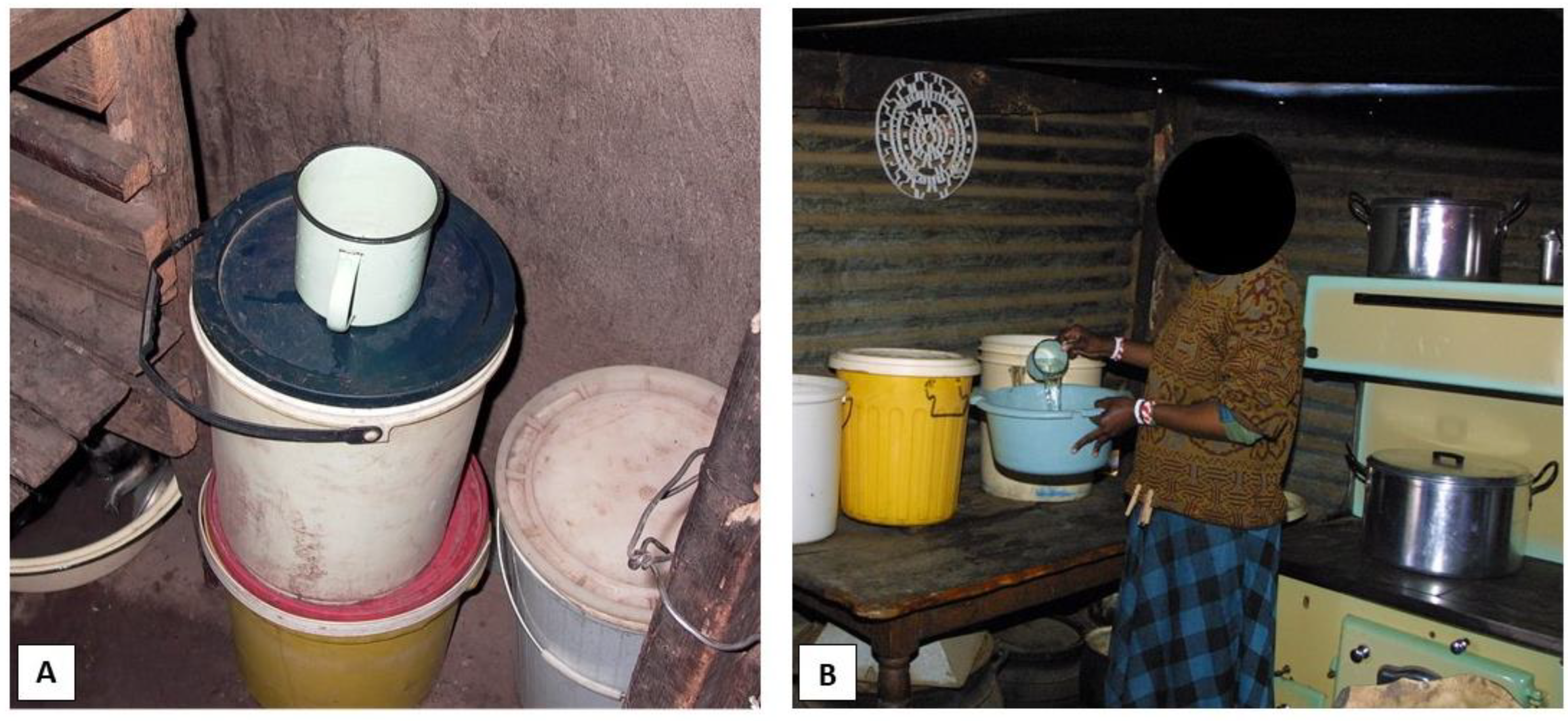
2.2. Sample Collection

All samples were collected once a month from each study household over a period of 5 months between March and July of 2016. Water was collected from the 20 L household storage buckets using the cup/jug that the household normally used for water retrieval (Figure 1A) and transferred into sterile 1000 mL collection containers that were clearly marked with the settlement name, house number, and date. Biofilm samples were collected by swabbing an area of 10 × 10 cm on the inside of the water storage container with a sterile swab (COPAN, Murrieta, CA, USA). The swab was inserted into the water under the waterline, as far down as possible (approximately 15 cm) without the person sampling touching the water with their hand (Figure 2). The swab was placed back in its container and clearly marked with the settlement, house number, and the date collected. All samples were refrigerated at 4 °C and transported to the laboratory early the next morning for the analysis.

2.3. Isolation and Identification of Potential Human Pathogenic Bacteria

Amoebal enrichment suspensions (100 µL of each sample) were inoculated onto different selective media for the isolation of potential harmful bacteria The study only focused on potential harmful bacteria including Salmonella, Shigella, Legionella, Mycobacterium, E. coli, Vibrio, and Coliform bacteria based on a review article of ARB by Greub and Raoult [35]. The selective media types used to isolate these bacteria included a BrillianceTM E. coli/coliform selective medium for Escherichia coli and other coliform bacteria (Oxoid CM1046B); a xylose lysine deoxycholate (XLD) agar for Salmonella and Shigella spp. (Oxoid CM0469B); a thiosulphate citrate bile salts sucrose (TCBS) agar for pathogenic Vibrio spp. (Oxoid CM0333B); a charcoal yeast extract (CYE) agar (Oxoid CM0655B) with a Legionella buffered charcoal yeast extract (BCYE) growth supplement for Legionellae spp. (Oxoid SR0110A); and Middlebrook 7H9 media with an oleic albumin dextrose catalase (OADC) growth supplement for Mycobacterium spp. For each type of selective media, positive controls were used to ensure that the media would serve as an accurate source of information regarding the unknown organisms cultured on them and included Vibrio cholerae O139 (NSCC), Salmonella enteritidis (NSCC), Shigella boydii (NCTC 9329), Escherichia coli (ATCC 43888), Legionella pneumophila (ATCC 33152), and Mycobacterium avium (NSCC). All incubations were done as per the specifications of the manufacturers. The Ziehl–Neelsen stain [36] was used to confirm Mycobacterium spp. Non-specific colonies on the selective media were picked and re-plated onto nutrient agar (Sigma-Aldrich 70148) to isolate single colonies for further identification. Likewise, single colonies identified using selective media as presumptive Mycobacterium, Shigella, Legionella, Salmonella, Shigella, E. coli, or coliform species were also picked and streaked on nutrient agar for purification before a molecular identification. All molecular identifications were performed by Inqaba Biotechnical Industries (Pty) Ltd. (Pretoria, South Africa). Only the 16S rRNA gene was investigated using primers sourced from the literature. These primers amplify almost the entire length of the gene [37] and the target region is conserved in many bacterial species [38]. For the Mycobacterium spp. identification, the forward primer pA (5′-AGAGTTTGATCCTGGCTCAG-3′) and the reverse primer MSHE (5′-GCGACAAACCACCTACGAG-3′) were used [39]; for Legionella spp., the forward primer LEG 225 (5′-AAGATTAGCCTGCGTCCGAT-3′) and the reverse primer LEG 858 (5′-GTCAACTTATCGCGTTTGCT-3′) were used [40]; and for the identification of the colonies from the nutrient agar, XLD, and Brilliance E. coli/coliform media, 16S rRNA primer pairs that included the forward primer 27 (5′-AGAGTTTGATCMTGGCTCAG-3′) and the reverse primer 1492 (5′-CGGTTACCTTGTTACGACTT-3′) were used [41].

2.4. Isolation of FLA from the Water Samples

The FLA were then isolated using the amoebal enrichment method described by Thomas et al. [42]. All water samples (500 mL of each sample) were filtered through 0.45 µm nitrocellulose filters (Millipore, Burlington, MA, USA) with a filter manifold (Sartorius, Goettingen, Germany). The filter was aseptically placed face-side down onto a non-nutrient agar (NNA) plate and covered with a layer of heat-killed Escherichia coli (type strain, ATCC 25,922), which served as a food source for the amoeba; a few drops of Page’s amoebal saline (PAS) were then added to aid mobility and the plates were incubated aerobically at 33 °C [42]. The NNA plates were checked daily for the appearance of amoebal cysts using a 10× light microscope. A disposable Pasteur pipette was used to cut small plugs from the plates; these plugs were placed onto new NNA plates (covered with heat-killed E. coli) and one drop of PAS was added to each plug. This process purified the amoebae as other organisms remained on the original plate. These were sub-cultured onto new NNA plates with E. coli and PAS until the culture was axenic. The sub-cultured plates were flooded with 2 mL of PAS and a sterile plastic loop was used to gently scrape the axenic amoebae from the plate surface. This suspension was transferred to clearly marked sterile 2 mL Eppendorf® tubes. Before freezing the 2 mL tubes for a later analysis, 1 mL of the amoebal suspension was lysed by passaging the suspension through a 27-gauge syringe and vortexed at 2500 rpm (Vortex Genie® 2-Mixer 240 V, 50 Hz Scientific Industries, Bohemia, New York, NY, USA) to release the ARB. The lysed amoebae were stored in a different sterile marked tube at −70 °C.

2.5. Isolation of FLA from the Biofilm Samples

Each biofilm swab was vortexed in its individual sterile tube for 30 s with 1 mL of Page’s amoebal saline (PAS) and then centrifuged for 10 min at 800× g. Of the supernatant, 200 µL was inoculated onto a 24-well plate and the pellet was suspended in 100 µL PAS and plated onto a non-nutrient agar (NNA) plate seeded with heat-killed E. coli. The 24-well plate was centrifuged for 30 min at 1500× g and incubated aerobically at 33 °C for three days as per Thomas et al. [42] with the following exceptions: after the three days, when the amoebae had attached to the plate, the supernatant was carefully removed (without disturbing the cells) and discarded. A fresh volume of 100 µL PAS was added to the layer of FLA cells in the 24-well plates and passaged by pipette until well-mixed then again incubated aerobically at 33 °C for three days. The 24-well plate co-cultures were washed, as previously described, and read by an inverted microscope to observe the FLA in their natural state.

2.6. FLA Identification

Only a subset (±10%) of the isolates that were positive for FLA by inverted microscopy were randomly selected from the samples containing FLA and Acanthamoeba cysts to send for an 18S rRNA PCR and sequencing, which were performed by the Department of Hygiene, Social, and Environmental Medicine at Ruhr University, Bochum, Germany.
The selected samples stored after the initial amoebal enrichment were freshly inoculated onto NNA plates and allowed to grow. The plates were sealed individually with Parafilm M® before they were packaged and sent to our collaborators in Germany for PCR and sequencing. Amoebal DNA was extracted from 200 μL of the prepared amoebae suspension in the amoebae-positive plates using a QIAamp DNA Blood Mini Kit (Qi-agen, Hilden, Germany) as per manufacturer’s protocol. The nucleic acid was eluted in 100 μL of an elution buffer into a sterile clearly marked 1.5 mL microcentrifuge tube and stored at −20 °C. The FLA genotyping was performed using the forward primers Ami6F1 (5′-CCAGCTCCAATAGCGTATATT-3′) and Ami6F2 (5′-CCAGCTCCAAGAGTGTATATT-3′) as well as reverse primer Ami9R (5′-GTTGAGTCGAATTAAGCCGC-3′) to amplify the 18S rRNA gene [42]. For the Acanthamoeba genotype identification, the amplification was performed with the primer set JDP1 (5′-GGCCCAGATCGTTTACCGTGAA-3′) and JDP2 (5′-TCTCACAAGCTGCTAGGGGAGTCA-3′) [43].

3. Results

A total of 97% (398/410) household stored water samples and 96% (392/410) biofilm swabs were collected in this study over the 5 month study period because a few household members were absent on the collection day and samples in a specific month could not be collected.
When viewed under an inverted microscope, 92% (366/398) of the water samples and 89.8% (352/392) of the biofilm samples contained FLA. The FLA observed were grouped into either trophozoites, round cysts, or Acanthamoeba spp. cysts based on the morphology only (Table 1). FLA were present in both the biofilm and stored container water as either a single form of FLA or in a combination (present as a cyst, trophozoites, or both in one sample).
Approximately 10% of presumptive positive samples were selected and sent for an 18S rRNA PCR and sequencing in Germany, of which the water samples comprised 11.5% (42/366) and the biofilm samples 9.7% (34/352) of the total number of samples. These samples included a total of 41 samples previously identified as trophozoites and 35 samples previously identified as presumptive Acanthamoeba cysts (Table 2).
Table 3 indicates the molecular identification of Acanthamoeba spp. and other FLA from the subset samples sent for the 18S rRNA PCR and sequencing. A total of 57.1% (16/28) of the water samples and 53.8% (7/13) of the biofilm samples tested with the FLA primer set yielded positive FLA results. With the specific Acanthamoeba spp. primer set, 42.9% (6/14) of the water samples and 19.0% (4/21) of the biofilm samples were FL-positive.
From the 76 samples sequenced, FLA (n = 50) were identified in only 33 of the samples. In addition, 37.0% (20/54) of Vermamoeba spp. and 29.6% (16/54) of Acanthamoeba spp. were the most abundant of the amoebae isolated (Table 4).
Table 5 lists the ARBs isolated from the stored water and confirmed with the 16S rRNA PCR and sequencing. Of the 398 water samples tested, 28.4% (113/398) yielded results for the presence of potential human bacteria with Mycobacterium spp. comprising the largest part of the isolates with 48.7% (55/113).
The 43 samples without positive FLA results either contained slime moulds or could not be amplified after repeated PCR attempts. The slime moulds belong to the group Mycetozoa that form part of the super-group Amoebozoa and comprise numerous amoebae [44] that can harbour bacteria [45].

4. Discussion

Although people are supplied with a safe water source, several practices can contaminate the water from the point of collection to the point of consumption (e.g., type of storage container used, cleanliness of the container, the handling of the water during collection and storage, water treatment practices at the household) [14,15]. However, most of these studies concentrated on the prevalence of indicator coliform bacteria and Escherichia coli whereas a few studies have investigated the survival of certain viruses and parasites in different storage water [46]. Very few studies have been conducted to provide information of the prevalence of FLA and potential human pathogenic bacteria in water stored at the household at the point of consumption.
In this study, all the households used wide-mouthed pre-used food containers to store their household water in (Figure 2). Several studies have proved that wide-mouthed domestic water storage vessels are more prone to contamination by unclean hands, dirty cups, and other water retrieval methods than screw-top containers (with their lids closed), which have a positive effect on the water quality [3,15,47,48,49,50]. It was also seen that the cup/jug (metal, glass, or plastic) used to retrieve the water from the storage vessel was often kept uncovered near the storage vessel and thus exposed to dust, flies, and unwashed hands, similar to the results of other studies in South Africa [3]. Other potential sources of groundwater contamination could be from agricultural activities and pit latrines [51] and a few studies have shown that the material of the containers could assist in the survival and transmission of potential pathogenic microorganisms [52,53].
Most of the FLA identified in this study were consistent with the environment from which the samples were taken (water and/or soil, or animal hosts). Vermamoeba spp. were one of the most abundant of the FLA isolated in this study (Table 4). Vermamoeba vermiformis has been isolated from various water sources both natural and man-made [54,55] as well as bat guano [56]. It has also proved to be a double threat as it may act as a potential pathogen in humans [57,58,59] and is also an important reservoir and vector for Legionella pneumophila [60], Mycobacterium spp. [58,61], and Stenotrophomonas maltophilia [54,62]. The other abundant FLA isolated in this study was Acanthamoeba spp. (Table 5); isolates containing genotype T4 have most frequently been isolated from human infections [63,64,65] because the T4 genotype is more transmissible and more virulent than other genotypes [66]. Other studies also reported Acanthamoeba spp. and A. castellanii str. Neff in soil samples [67]. The soil amoebae isolated from the samples included Stenamoeba spp. [68] and Stenamoeba berchidia [69]. Several FLA are known waterborne amoebae such as Vexillefera westveldii [70] and Korotnevella hemistylolepis [71]; Flamella spp. can be found in saltwater, freshwater, and soil samples [72]. Usually, FLA are ubiquitous and have been isolated from various natural sources such as freshwater, saltwater, dust, air, and soil [73,74]. Loret and Greub [75] collated data from recent studies and found that 62% of surface water and 71% of groundwater samples contained FLA. They often live in water–soil, water–air, and water–plant interfaces as well as on biofilms. Several FLA species attach to particulate matter to feed but others prefer to move in their planktonic phase [35,73,74,75]. Limitations of this study were the selection of the primer sets used for the identification of the FLA and the FLA enrichment cultivation method used that was selective in the isolation of the FLA from the water samples. Further studies with other isolation methods and different primers are needed to show the prevalence of other FLA in the water and biofilm samples.
Several bacteria are ingested by FLA and digested as a food source [35] whereas others have adapted to survive and grow within the amoebae by resisting the microbiocidal mechanisms of FLA and are termed amoeba-resistant bacteria [76]. These potential harmful bacteria are protected from adverse conditions until they excyst the amoebae [35,77]. These bacteria can persist within the FLA if the defence mechanisms of the amoebal cell are inefficient or impaired, which leads to a symbiotic relationship [78]. In a review by Thomas et al. [79], 102 of these bacterial species (all human pathogens and capable of survival in FLA) were identified. A few of the genera include Achromobacter, Klebsiella, Mycobacterium, Legionella, Stenotrophomonas, Enterobacter, and Pseudomonas [80] A wide range of potential human pathogenic bacteria were identified from the container-stored water samples in this study. Future studies could include determining accurately if these potential human bacteria species are ARBs retrieved from FLA. A few of the potential human pathogenic bacteria seen in this study are known human pathogens and are particularly fond of causing hospital-acquired infections such as K. oxytoca [80] and S. maltophilia [81] whereas several bacteria identified in this study are found naturally in the environment such as E. amnigenus [82] and M. salmoniphilum [83] in animals and P. kilonensis [84] and P. koreensis [85] in soil. Numerous Mycobacterium spp. were isolated from the stored water samples. Mycobacterial species can live and grow in amoebae for years in a truly symbiotic relationship [86]; it was also proven that Acanthamoeba castellanii can amass several different mycobacterial species within their cytoplasm [87].

5. Conclusions

The results from this study showed the prevalence and co-existence of FLA and potential harmful bacteria which could be a health concern, that were found in household-stored water and containers, highlighting the need for improved hygiene education and intervention programmes on water collection and treatment practices at a household level. Many of the FLA and potential harmful bacterial spp isolated from the stored drinking water and the container biofilms were potential pathogens and a health risk to vulnerable individuals who could drink the water without any prior treatment. In addition, to our knowledge, this is the first study in South Africa to isolate FLA and potential harmful bacteria present in storage container biofilms as well as identify them at a species level. Several recommendations can, therefore, be made from the results of this study. Firstly, further studies are needed to examine the prevalence, behaviour, and possible disinfection of FLA in both the planktonic and biofilm states. Secondly, further studies are needed to examine the removal of these organisms from the storage containers to protect the communities that rely on the water, especially with the increase in demand for groundwater in semi-arid areas such as South Africa. Thirdly, further studies are needed to examine the causal links between the organisms found and the clinical symptoms presented by the users of the water (if any) as well as exploring the possibility of changing conventional water testing by means of indicator organisms alone to include more organisms that have proven to be emerging pathogens. Lastly, more studies are needed to assess the effect of global warming on the survival and distribution of pathogenic amoebae and to determine the microbiomes inside these amoebae to increase our understanding of the health risks of amoeba protection strategies in drinking water.

Author Contributions

Conceptualisation, N.P. and T.G.B.; data curation, N.P., C.v.d.L. and T.G.B.; formal analysis, N.P., C.v.d.L. and T.G.B.; funding acquisition, N.P. and T.G.B.; investigation, N.P., C.v.d.L. and T.G.B.; methodology, N.P., C.v.d.L. and T.G.B.; project administration, N.P. and T.G.B.; resources, N.P. and T.G.B.; supervision, N.P. and T.G.B.; validation, N.P. and T.G.B.; visualisation, N.P. and T.G.B.; writing—original draft, N.P. and T.G.B.; writing—review and editing, N.P., C.v.d.L. and T.G.B. All authors have read and agreed to the published version of the manuscript.

Funding

The research was funded by the National Research Foundation of South Africa for Doctoral Funding, a University of Johannesburg Research Committee Research Grant (Water and Health Research), a University of Venda Research Project Committee Grant, and the Water Research Commission (Project number: 1653).

Institutional Review Board Statement

Not applicable.

Informed Consent Statement

Informed consent was obtained from all subjects involved in the study. A female adult within each of the randomly selected households gave consent to partake in this study. A detailed explanation of the study was given and a consent form was administered by trained field workers who translated the forms from English into the home language of the participants. Participation in the study was voluntary and the participants were free to withdraw at any point during the study period.

Data Availability Statement

The data presented in this study are available on request from the corresponding author.

Acknowledgments

The authors want to sincerely thank the household members of the sampling communities for allowing the researchers to perform this study and take samples from their households as well as our collaborators at the Department of Hygiene, Social and Environmental Medicine at Ruhr University, Bochum (Germany) for their PCR and sequencing work, D Bartie for her valuable advice during the field and laboratory part of the study, and the National Institute of Occupational Health (Department of Immunology and Microbiology) for the use of their laboratories and equipment.

Conflicts of Interest

The authors declare no conflict of interest.

References

  1. Stats SA. General Household Survey. (Report No. P0318). Pretoria, South Africa; 2019. Available online: https://www.statssa.gov.za/publications/P0318/P03182019.pdf (accessed on 8 October 2021).
  2. Slavik, I.; Oliveira, K.R.; Cheung, P.B.; Uhl, W. Water quality aspects related to domestic drinking water storage tanks and consideration in current standards and guidelines throughout the world—A review. J. Water Health 2020, 18, 439–463. [Google Scholar] [CrossRef]
  3. Jagals, P.; Grabow, W.O.K.; Williams, E. The effect of supplied water quality on human health in an urban development with limited basic -subsistence facilities. Water SA 1997, 23, 373–378. [Google Scholar]
  4. Momba, M.N.B.; Notshe, T.L. The effect of long storage and household containers on the microbiological quality of drinking water in rural communities of South Africa. J. Water Supply Res. Technol. 2003, 52, 67–76. [Google Scholar] [CrossRef]
  5. Potgieter, N.; Mudau, L.S.; Maluleke, F.R.S. The microbiological quality of ground water sources used by rural communities in the Limpopo Province, South Africa. Water Sci. Technol. 2006, 54, 371–377. [Google Scholar] [CrossRef] [PubMed]
  6. Esterhuizen, L.; Fossey, A.; Potgieter, E. Groundwater quality on dairy farms in central South Africa. Water SA 2015, 41, 194–198. [Google Scholar] [CrossRef]
  7. Costerton, J.W.; Lewandowski, Z.; Caldwell, D.E.; Korber, D.R.; Lappin-Scott, H.M. Microbial Biofilms. Annu. Rev. Microbiol. 1995, 49, 711–742. [Google Scholar] [CrossRef] [PubMed]
  8. Berlanga Herranz, M.; Guerrero, R. Living together in biofilms: The microbial cell factory and its biotechnological implications. Microbial Cell Factories 2016, 15, 165. [Google Scholar] [CrossRef] [PubMed] [Green Version]
  9. Budeli, P.; Moropeng, R.C.; Mpenyana-Monyatsi, L.; Momba, M.N.B. Inhibition of biofilm formation on the surface of water storage containers using biosand zeolite silver-impregnated clay granular and silver impregnated porous pot filtration systems. PLoS ONE 2018, 13, e0194715. [Google Scholar] [CrossRef] [Green Version]
  10. Barbeau, J.; Buhler, T. Biofilms augment the number of free-living amoebae in dental unit waterlines. Res. Microbiol. 2001, 152, 753–760. [Google Scholar] [CrossRef]
  11. Costerton, J.W.; Stewart, P.S.; Greenberg, E.P. Bacterial biofilms: A common cause of persistent infections. Science 1999, 284, 1318–1322. [Google Scholar] [CrossRef] [Green Version]
  12. Arndt, H.; Schmidt-Denter, K.; Auer, B.; Weitere, M. Protozoans and Biofilms. In Fossil and Recent Biofilms; Krumbein, W.E., Paterson, D.M., Zavarzin, G.A., Eds.; Kluwer Academic Publishing: Dordrecht, The Netherlands, 2003; pp. 173–189. [Google Scholar]
  13. Jackson, S.M.; Jones, E.B.G. Interactions within biofilms: The disruption of biofilm structure by protozoa. Kieler Meeresforsch. Sonderh. 1991, 8, 264–268. [Google Scholar]
  14. Murcott, S. Implementation, Critical Factors and Challenges to Scale-Up of Household Drinking Water Treatment and Safe Storage Systems. In Background Paper on Household Water Treatment and Safe Storage (HWTS) for the Electronic Conference. USAID/Hygiene Improvement Project (HIP). 2006. Available online: http://hip.fhi360.org/file/6114/20060501-HWTS-Murcott.pdf (accessed on 8 October 2021).
  15. Levy, K.; Nelson, K.L.; Hubbard, A.; Eisenberg, J.N. Following the water: A controlled study of drinking water storage in northern coastal Ecuador. Environ. Health Perspect. 2008, 116, 1533–1540. [Google Scholar] [CrossRef] [PubMed]
  16. Jagals, P.; Jagals, C.; Bokako, T. The effect of container-biofilm on the microbiological quality of water used from plastic household containers. J. Water Health 2003, 1, 101–108. [Google Scholar] [CrossRef] [PubMed] [Green Version]
  17. Van der Merwe, V.; Duvenage, S.; Korsten, L. Comparison of biofilm formation and water quality when water from different sources was stored in large commercial water storage tanks. J. Water Health 2013, 11, 30–40. [Google Scholar] [CrossRef] [PubMed]
  18. Lantagne, D.; Clasen, T. Point-of-use water treatment in emergency response. Waterlines 2012, 31, 30–52. [Google Scholar] [CrossRef] [Green Version]
  19. Oloruntoba, E.O.; Babalola, T.F.; Morakinyo, O.M.; Mumuni, A. Effects of improved storage containers on the bacteriological quality of household drinking water in low-income urban communities in Ibadan, Nigeria. Water Supply 2016, 16, 378–387. [Google Scholar] [CrossRef]
  20. McGuinness, S.L.; O’Toole, J.; Barker, S.F.; Forbes, A.B.; Boving, T.B.; Giriyan, A.; Patil, K.; D’Souza, F.; Vhaval, R.; Cheng, A.C.; et al. Household Water Storage Management, Hygiene Practices, and Associated Drinking Water Quality in Rural India. Environ. Sci. Technol. 2020, 54, 4963–4973. [Google Scholar] [CrossRef]
  21. Soliman, M.Y.; van Halem, D.; Medema, G. Virus removal by ceramic pot filter disks: Effect of biofilm growth and surface cleaning. Int. J. Hyg. Environ. Health 2019, 224, 113438. [Google Scholar] [CrossRef]
  22. Momba, M.N.; Kaleni, P. Regrowth and survival of indicator microorganisms on the surfaces of household containers used for the storage of drinking water in rural communities of South Africa. Water Res. 2002, 36, 3023–3028. [Google Scholar] [CrossRef]
  23. Momba, M.N.B.; Mnqumevu, B.V. Detection of Faecal Coliforms and Heterotrophic Plate Count Bacteria Attached to Household Containers during the Storage of Drinking Groundwater in Rural Communities. In Proceedings of the WISA 2000 Biennial Conference, Sun City, South Africa, 28 May–1 June 2000; pp. 21–28. Available online: https://wisa.org.za (accessed on 20 October 2021).
  24. Wright, J.; Gundry, S.; Conroy, R.M. Household drinking water in developing countries: A systematic review of microbiological contamination between source and point-of-use. Trop. Med. Int. Health 2004, 9, 106–117. [Google Scholar] [CrossRef] [Green Version]
  25. Taravaud, A.; Ali, M.; Lafosse, B.; Nicolas, V.; Féliers, C.; Thibert, S.; Lévi, Y.; Loiseau, P.; Pomel, S. Enrichment of free-living amoebae in biofilms developed at upper water levels in drinking water storage towers: An inter- and intra-seasonal study. Sci. Total. Environ. 2018, 633, 157–166. [Google Scholar] [CrossRef] [PubMed]
  26. Stockman, L.J.; Wright, C.J.; Visvesvara, G.S.; Fields, B.S.; Beach, M.J. Prevalence of Acanthamoeba spp. and other free-living amoebae in household water, Ohio, USA—1990–1992. Parasitol. Res. 2010, 108, 621–627. [Google Scholar] [CrossRef]
  27. Rohr, U.; Weber, S.; Michel, R.; Selenka, F.; Wilhelm, M. Comparison of Free-Living Amoebae in Hot Water Systems of Hospitals with Isolates from Moist Sanitary Areas by Identifying Genera and Determining Temperature Tolerance. Appl. Environ. Microbiol. 1998, 64, 1822–1824. [Google Scholar] [CrossRef] [PubMed] [Green Version]
  28. Muchesa, P.; Barnard, T.G.; Bartie, C. The prevalence of free-living amoebae in a South African hospital water distribution system. S. Afr. J. Sci. 2015, 111, 1–3. [Google Scholar] [CrossRef]
  29. Muchesa, P.; Leifels, M.; Jurzik, L.; Hoorzook, K.B.; Barnard, T.G.; Bartie, C. Coexistence of free-living amoebae and bacteria in selected South African hospital water distribution systems. Parasitol. Res. 2016, 116, 155–165. [Google Scholar] [CrossRef] [PubMed]
  30. Kuiper, M.W.; Wullings, B.A.; Akkermans, A.D.L.; Beumer, R.R.; Van der Kooij, D. Intracellular Proliferation of Legionella pneumophila in Hartmanella vermiformis in Aquatic Biofilms Grown on Plasticized Polyvinyl Chloride. Appl. Environ. Microbiol. 2004, 70, 6826–6833. [Google Scholar] [CrossRef] [Green Version]
  31. van der Kooij, D.; Veenendaal, H.R.; Scheffer, W.J. Biofilm formation and multiplication of Legionella in a model warm water system with pipes of copper, stainless steel and cross-linked polyethylene. Water Res. 2005, 39, 2789–2798. [Google Scholar] [CrossRef]
  32. Lau, H.Y.; Ashbolt, N.J. The role of biofilms and protozoa in Legionella pathogenesis: Implications for drinking water. J. Appl. Microbiol. 2009, 107, 368–378. [Google Scholar] [CrossRef]
  33. Muchesa, P.; Lelfels, M.; Jurzik, L.; Barnard, T.G.; Bartle, C. Detection of amoeba-associated Legionella pneumophila in hospital water networks of Johannesburg. South. Afr. J. Infect. Dis. 2018, 33, 72–75. [Google Scholar] [CrossRef]
  34. Shaheen, M.; Scott, C.; Ashbolt, N.J. Long-term persistence of infectious Legionella with free-living amoebae in drinking water biofilms. Int. J. Hyg. Environ. Health 2019, 222, 678–686. [Google Scholar] [CrossRef]
  35. Greub, G.; Raoult, D. Microorganisms Resistant to Free-Living Amoebae. Clin. Microbiol. Rev. 2004, 17, 413–433. [Google Scholar] [CrossRef] [Green Version]
  36. International Union against Tuberculosis and Lung Disease. Technical guide for sputum examination for tuberculosis by direct microscopy. Bull. Int. Union Tuberc. Lung Dis. 1978, 2, 4–16. [Google Scholar]
  37. Frank, J.A.; Reich, C.I.; Sharma, S.; Weisbaum, J.S.; Wilson, B.A.; Olsen, G.J. Critical evaluation of two primers commonly used for amplification of bacterial 16S rRNA genes. Appl. Environ. Microbiol. 2008, 74, 2461–2470. [Google Scholar] [CrossRef] [Green Version]
  38. Fredriksson, N.J.; Hermansson, M.; Wilén, B.-M. The Choice of PCR Primers Has Great Impact on Assessments of Bacterial Community Diversity and Dynamics in a Wastewater Treatment Plant. PLoS ONE 2013, 8, e76431. [Google Scholar] [CrossRef] [PubMed]
  39. Hughes, M.S.; Ball, N.W.; Beck, L.A.; de Lisle, G.W.; Skuce, R.A.; Neill, S.D. Determination of the etiology of presumptive feline leprosy by 16S rRNA gene analysis. J. Clin. Microbiol. 1997, 35, 2464–2467. [Google Scholar] [CrossRef] [PubMed] [Green Version]
  40. Miyamoto, H.; Yamamoto, H.; Arima, K.; Fujii, J.; Maruta, K.; Izu, K.; Shiomori, T.; Yoshida, S. Development of a new semi-nested PCR method for detection of Legionella species and its application to surveillance of legionellae in hospital cooling tower water. Appl. Environ. Microbiol. 1997, 63, 2489–2494. [Google Scholar] [CrossRef] [PubMed] [Green Version]
  41. Altschul, S.F.; Madden, T.L.; Schäffer, A.A.; Zhang, J.; Zhang, Z.; Miller, W.; Lipman, D.J. Gapped BLAST and PSI-BLAST: A new generation of protein database search programs. Nucleic Acids Res. 1997, 25, 3389–3402. [Google Scholar] [CrossRef] [Green Version]
  42. Thomas, V.; Herrera-Rimann, K.; Blanc, D.S.; Greub, G. Biodiversity of amoebae and amoebae-resisting bacteria in a hospital water network. Appl. Environ. Microbiol. 2006, 72, 2428–2438. [Google Scholar] [CrossRef] [PubMed] [Green Version]
  43. Schroeder, J.M.; Booton, G.C.; Hay, J.; Niszl, I.A.; Seal, D.V.; Markus, M.B.; Fuerst, P.A.; Byers, T.J. Use of Subgenic 18S Ribosomal DNA PCR and Sequencing for Genus and Genotype Identification of Acanthamoebae from Humans with Keratitis and from Sewage Sludge. J. Clin. Microbiol. 2001, 39, 1903–1911. [Google Scholar] [CrossRef] [PubMed] [Green Version]
  44. Schilde, C.; Schaap, P. The Amoebozoa. Methods Mol. Biol. 2013, 983. [Google Scholar] [CrossRef] [Green Version]
  45. Denoncourt, A.M.; Paquet, V.E.; Charette, S.J. Potential role of bacteria packaging by protozoa in the persistence and transmission of pathogenic bacteria. Front. Microbiol. 2014, 5, 240. [Google Scholar] [CrossRef] [Green Version]
  46. WHO. Combating Waterborne Disease at the Household Level: The International Network to Promote Household Water Treatment and Safe Storage; World Health Organization: Geneva, Switzerland, 2007; Available online: http://www.who.int/household_water/advocacy/combating_disease.pdf (accessed on 8 October 2021).
  47. Mintz, E.D.; Reiff, F.M.; Tauxe, R.V. Safe water treatment and storage in the home. A practical new strategy to prevent waterborne disease. JAMA 1995, 273, 948–953. [Google Scholar] [CrossRef] [PubMed]
  48. Trevett, A.F.; Carter, R.C.; Tyrrel, S. Water quality deterioration: A study of household drinking water quality in rural Honduras. Int. J. Environ. Health Res. 2004, 14, 273–283. [Google Scholar] [CrossRef] [PubMed] [Green Version]
  49. Oswald, W.E.; Bern, C.; Cabrera, L.; Lescano, A.; Gilman, R.H.; Calderon, M.M. Fecal Contamination of Drinking Water within Peri-Urban Households, Lima, Peru. Am. J. Trop. Med. Hyg. 2007, 77, 699–704. [Google Scholar] [CrossRef] [PubMed] [Green Version]
  50. Adane, M.; Mengistie, B.; Medhin, G.; Kloos, H.; Mulat, W. Piped water supply interruptions and acute diarrhea among under-five children in Addis Ababa slums, Ethiopia: A matched case-control study. PLoS ONE 2017, 12, e0181516. [Google Scholar] [CrossRef] [Green Version]
  51. Odiyo, J.O.; Mathoni, M.M.; Makungo, R. Health Risks and Potential Sources of Contamination of Groundwater Used by Public Schools in Vhuronga 1, Limpopo Province, South Africa. Int. J. Environ. Res. Public Health 2020, 17, 6912. [Google Scholar] [CrossRef]
  52. Schafer, C.A.; Mihelcic, J.R. Effect of storage tank material and maintenance on domestic water quality. J. Am. Water Works Assoc. 2012, 104, E521–E529. [Google Scholar] [CrossRef]
  53. Akuffo, I.; Cobbina, S.J.; Alhassan, E.H.; Nkoom, M. Assessment of the quality of water before and after storage in the Nyankpala Community of the Tolon-Kumbungu District, Ghana. Int. J. Scient. Technol. Res. 2013, 2, 221–227. [Google Scholar]
  54. Pagnier, I.; Valles, C.; Raoult, D.; La Scola, B. Isolation of Vermamoeba vermiformis and associated bacteria in hospital water. Microb. Pathog. 2015, 80, 14–20. [Google Scholar] [CrossRef]
  55. Javanmard, E.; Niyyati, M.; Lorenzo-Morales, J.; Lasjerdi, Z.; Behniafar, H.; Mirjalali, H. Molecular identification of waterborne free living amoebae (Acanthamoeba, Naegleria and Vermamoeba) isolated from municipal drinking water and environmental sources, Semnan province, north half of Iran. Exp. Parasitol. 2017, 183, 240–244. [Google Scholar] [CrossRef]
  56. Mulec, J.; Dietersdorfer, E.; Üstüntürk-Onan, M.; Walochnik, J. Acanthamoeba and other free-living amoebae in bat guano, an extreme habitat. Parasitol. Res. 2015, 115, 1375–1383. [Google Scholar] [CrossRef] [PubMed]
  57. Lorenzo-Morales, J.; Martínez-Carretero, E.; Batista, N.; Álvarez-Marín, J.; Bahaya, Y.; Walochnik, J.; Valladares, B. Early diagnosis of amoebic keratitis due to a mixed infection with Acanthamoeba and Hartmannella. Parasitol. Res. 2007, 102, 167–169. [Google Scholar] [CrossRef]
  58. Cabello-Vílchez, A.M.; Mena, R.; Zuñiga, J.; Cermeño, P.; Martín-Navarro, C.M.; González, A.C.; Arencibia, A.L.; Reyes-Batlle, M.; Piñero, J.; Valladares, B.; et al. Endosymbiotic Mycobacterium chelonae in a Vermamoeba vermiformis strain isolated from the nasal mucosa of an HIV patient in Lima, Peru. Exp. Parasitol. 2014, 145, S127–S130. [Google Scholar] [CrossRef] [PubMed]
  59. Walochnik, J.; Scheikl, U.; Haller-Schober, E. Twenty Years of Acanthamoeba Diagnostics in Austria. J. Eukaryot. Microbiol. 2014, 62, 3–11. [Google Scholar] [CrossRef] [Green Version]
  60. Wadowsky, R.M.; Butler, L.J.; Cook, M.K.; Verma, S.M.; A Paul, M.; Fields, B.S.; Keleti, G.; Sykora, J.L.; Yee, R.B. Growth-supporting activity for Legionella pneumophila in tap water cultures and implication of hartmannellid amoebae as growth factors. Appl. Environ. Microbiol. 1988, 54, 2677–2682. [Google Scholar] [CrossRef] [PubMed] [Green Version]
  61. Delafont, V.; Rodier, M.-H.; Maisonneuve, E.; Cateau, E. Vermamoeba vermiformis: A Free-Living Amoeba of Interest. Microb. Ecol. 2018, 76, 991–1001. [Google Scholar] [CrossRef] [PubMed]
  62. Cateau, E.; Delafont, V.; Hechard, Y.; Rodier, M.H. Free-living amoebae: What part do they play in healthcare-associated infections? J. Hospital Infec. 2014, 87, 131–140. [Google Scholar] [CrossRef] [PubMed]
  63. Khan, N.A. Acanthamoeba: Biology and increasing importance in human health. FEMS Microbiol. Rev. 2006, 30, 564–595. [Google Scholar] [CrossRef] [PubMed] [Green Version]
  64. Maciver, S.K.; Asif, M.; Simmen, M.W.; Lorenzo-Morales, J. A systematic analysis of Acanthamoeba genotype frequency correlated with source and pathogenicity: T4 is confirmed as a pathogen-rich genotype. Eur. J. Protistol. 2013, 49, 217–221. [Google Scholar] [CrossRef]
  65. Arnalich-Montiel, F.; Lumbreras-Fernández, B.; Martín-Navarro, C.M.; Valladares, B.; Lopez-Velez, R.; Morcillo-Laiz, R.; Lorenzo-Morales, J. Influence of Acanthamoeba Genotype on Clinical Course and Outcomes for Patients with Acanthamoeba Keratitis in Spain. J. Clin. Microbiol. 2014, 52, 1213–1216. [Google Scholar] [CrossRef] [Green Version]
  66. Siddiqui, R.; Khan, N.A. War of the microbial worlds: Who is the beneficiary in Acanthamoeba–bacterial interactions? Exp. Parasitol. 2012, 130, 311–313. [Google Scholar] [CrossRef] [PubMed]
  67. Neff, R.J. Purification, Axenic Cultivation, and Description of a Soil Amoeba, Acanthamoeba sp. J. Protozool. 1957, 4, 176–182. [Google Scholar] [CrossRef]
  68. Page, F.C. Platyamoeba stenopodia n. g., n. sp., a freshwater amoeba. J. Protozool. 1969, 16, 437–441. [Google Scholar] [CrossRef] [PubMed]
  69. Geisen, S.; Weinert, J.; Kudryavtsev, A.; Glotova, A.; Bonkowski, M.; Smirnov, A. Two new species of the genus Stenamoeba (Discosea, Longamoebia): Cytoplasmic MTOC is present in one more amoebae lineage. Eur. J. Protistol. 2014, 50, 153–165. [Google Scholar] [CrossRef] [PubMed]
  70. Van Wichelen, J.; D’Hondt, S.; Claeys, M.; Vyverman, W.; Berney, C.; Bass, D.; Vanormelingen, P. A Hotspot of Amoebae Diversity: 8 New Naked Amoebae Associated with the Planktonic Bloom-forming Cyanobacterium Microcystis. Acta Protozool. 2015, 55, 61–87. [Google Scholar]
  71. O'Kelly, C.J.; Peglar, M.T.; Black, M.; Sawyer, T.K.; Nerad, T.A. Korotnevella hemistylolepis N. Sp. and Korotnevella monacantholepis N. Sp. (Paramoebidae), Two New Scale-covered Mesohaline Amoebae. J. Eukaryot. Microbiol. 2001, 48, 655–662. [Google Scholar] [CrossRef] [PubMed]
  72. Michel, R.; Smirnov, A. The genus Flamella Schaeffer, 1926 (lobosea, gymnamoebia), with description of two new species. Eur. J. Protistol. 1999, 35, 403–410. [Google Scholar] [CrossRef]
  73. Rodríguez-Zaragoza, S. Ecology of free-living amoebae. Crit. Rev. Microbiol. 1994, 20, 225–241. [Google Scholar] [CrossRef] [PubMed]
  74. Marshall, M.M.; Naumovitz, D.; Ortega, Y.; Sterling, C.R. Waterborne protozoan pathogens. Clin. Microbiol. Rev. 1997, 10, 67–85. [Google Scholar] [CrossRef]
  75. Loret, J.; Greub, G. Free-living amoebae: Biological by-passes in water treatment. Int. J. Hyg. Environ. Health 2010, 213, 167–175. [Google Scholar] [CrossRef]
  76. Matz, C.; Kjelleberg, S. Off the hook—How bacteria survive protozoan grazing. Trends Microbiol. 2005, 13, 302–307. [Google Scholar] [CrossRef]
  77. Molmeret, M.; Horn, M.; Wagner, M.; Santic, M.; Kwaik, Y.A. Amoebae as Training Grounds for Intracellular Bacterial Pathogens. Appl. Environ. Microbiol. 2005, 71, 20–28. [Google Scholar] [CrossRef] [PubMed] [Green Version]
  78. Winiecka-Krusnell, J.; Linder, E. Bacterial infections of free living amebae. Res. Microbiol. 2001, 152, 613–619. [Google Scholar] [CrossRef]
  79. Thomas, V.; McDonnel, G.; Denyer, S.P.; Maillard, J.-Y. Free-living amoebae and their intracellular pathogenic microorganisms: Risk for water quality. FEMS Microbiol. Rev. 2010, 34, 231–259. [Google Scholar] [CrossRef] [PubMed] [Green Version]
  80. Lowe, C.; Willey, B.; O’Shaughnessy, A.; Lee, W.; Lum, M.; Pike, K.; Larocque, C.; Dedier, H.; Dales, L.; Moore, C.; et al. Outbreak of Extended-Spectrum β-Lactamase–producing Klebsiella oxytoca Infections Associated with Contaminated Handwashing Sinks1. Emerg. Infect. Dis. 2012, 18, 1242–1247. [Google Scholar] [CrossRef] [PubMed]
  81. Brooke, J.S. Stenotrophomonas maltophilia: And Emerging Global Opportunistic Pathogen. Clin. Microbiol. Rev. 2012, 25, 2–41. [Google Scholar] [CrossRef] [PubMed] [Green Version]
  82. Murugaiyan, J.; Krueger, K.; Roesler, U.; Weinreich, J.; Schierack, P. Assessment of species and antimicrobial resistance among Enterobacteriaceae isolated from mallard duck faeces. Environ. Monit. Assess. 2015, 187, 127. [Google Scholar] [CrossRef]
  83. Ross, A.J. Mycobacterium salmoniphilum sp. nov. from salmonoid fishes. Am. Rev. Resp. Dis. 1960, 81, 241–250. [Google Scholar]
  84. Sikorski, J.; Stackebrandt, E.; Wackernagel, W. Pseudomonas kilonensis sp. nov., a bacterium isolated from agricultural soil. Int. J. Syst. Evol. Microbiol. 2001, 51, 1549–1555. [Google Scholar] [CrossRef] [Green Version]
  85. Kwon, S.W.; Kim, J.S.; Park, I.C.; Yoon, S.H.; Park, D.H.; Lim, C.K.; Go, S.J. Pseudomonas koreensis sp. nov., Pseudomonas umsongensis sp. nov. and Pseudomonas jinjuensis sp. nov., novel species from farm soils in Korea. Int. J. Syst. Evol. Microbiol. 2003, 53, 21–27. [Google Scholar] [CrossRef] [Green Version]
  86. Yu, H.S.; Jeong, H.J.; Hong, Y.C.; Seol, S.Y.; Chung, D.I.; Kong, H.H. Natural occurrence of Mycobacterium as an endosymbiont of Acanthamoeba isolated from a contact lens storage case. Korean J. Parasitol. 2007, 45, 11–18. [Google Scholar] [CrossRef] [PubMed]
  87. Krishna Prasad, B.N.; Gupta, S.K. Preliminary report on the engulfment and retention of mycobacteria by trophozoites of axenically grown Acanthamoeba castellanii Douglas, 1930. Curr. Sci. 1978, 47, 245–247. [Google Scholar]
Figure 1. Water storage container in study group. (A) Typical water storage buckets in the households with a retrieval cup on top. (B) A female study participant decanting water from the water storage container.
Biology 10 01228 g001
Figure 2. A swab taken from water storage container.
Biology 10 01228 g002
Table 1. Presumptive FLA identification by inverted microscope.
Presumptive Identification of FLA TypesWater in Storage Container (n = 398)Biofilm in Storage Container (n = 392)
TrophozoitesAcanthamoeba cystsRound cysts
26.9% (107/398)89.2% (355/398)32.2% (128/398)
13.8% (54/392)79.6% (312/392)48.5% (190/392)
Table 2. FLA selected for an 18S rRNA PCR and sequencing.
FLA TypeWater (n = 42)Biofilm (n = 34)
Trophozoites (n = 42)2813
Presumptive Acanthamoeba (n = 43)1421
Table 3. Samples positive for FLA according to the sample types and primer sets.
Primer Set UsedWater in Storage Container (n = 42)Biofilm in Storage Container (n = 34)
Ami6F1, Ami6F2, Ami9R (FLA) [42] 38.1% (16/42)20.6% (7/34)
JDP1, JDP2 (Acanthamoeba spp.) [44]14.3% (6/42)11.8% (4/34)
Table 4. FLA identified using 18S rRNA sequencing from storage container biofilm and water samples.
FLA Types
Isolated
Total nr of FLA
Isolated (n)
FLA Isolated form Stored WaterFLA Isolated
from Biofilm
Amoebozoa spp.532
Acanthamoeba spp.532
Genotype T3110
Genotype T4531
Genotype T15110
Genotype T16422
Flamella spp.220
F. fluviatalis110
Korotnevella spp.
Korotnevella hemistylolepis101
Lobosea spp.110
Stenamoeba spp.220
S. berchidia101
Vermamoeba spp.
V. vermiformis20164
Vexillifera spp.
V. westveldii110
Total503614
Table 5. Potential human bacteria pathogens isolated in stored household water samples.
Potential Human Pathogen Bacteria Isolated
Pseudomonas spp.:
Pseudomonas fluorescens
Pseudomonas geniculata
Pseudomonas kilonensis
Pseudomonas koreensis
Pseudomonas poae
Pseudomonas tremae
Pseudomonas vancouverensis
Pseudomonas poae/tolaasii
Pseudomonas putida
Arthobacter spp.:
Arthrobacter nicotinovorans
Mycobacterium spp.:
Mycobacterium chlorophenolicum
Mycobacterium chubuense
Mycobacterium elephantis
Mycobacterium fallax
Mycobacterium farcinogenes
Mycobacterium florentinum
Mycobacterium gilvum
Mycobacterium intermedium
Mycobacterium llatzerense
Mycobacterium noviomagense
Mycobacterium pallens
Mycobacterium poriferae
Mycobacterium psychrotolerans
Mycobacterium rhodesiae
Mycobacterium salmoniphilum
Mycobacterium smegmatis
Mycobacterium tokaiense
Mycobacterium triplex
Caulobacter spp.:
Caulobacter segnis
Klebsiella spp.:
Klebsiella oxytoca
Klebsiella variicola
Enterobacter spp.:
Enterobacter amnigenus
Enterobacter asburiae
Enterobacter cancerogenus
Enterobacter kobei
Enterobacter ludwigii
Paenibacillus spp.:
Paenibacillus validus
Pragia spp.:
Pragia fontium
Microbacterium spp.:
Microbacterium spp.
Microbacterium oxydans
Microbacterium paraoxydans
Achromobacter spp.:
Achromobacter insolitus
Achromobacter marplatensis
Achromobacter spanius
Rhodococcus spp.:
Rhodococcus erythropolis
Sarratia spp.:
Serratia ureilytica
Stenotrophomonas spp.:
Stenotrophomonas maltophilia
Mixed bacterial spp.:
Alcaligenes faecalis and Achromobacter marplatensis
Publisher’s Note: MDPI stays neutral with regard to jurisdictional claims in published maps and institutional affiliations.

