Article

The Arabidopsis Receptor-like Kinase CAP1 Promotes Shoot Growth under Ammonium Stress

State Key Laboratory of Crop Stress Adaptation and Improvement, School of Life Sciences, Henan University, Kaifeng 475004, China
*
Authors to whom correspondence should be addressed.
Biology 2022, 11(10), 1452; https://doi.org/10.3390/biology11101452
Submission received: 31 August 2022 / Revised: 23 September 2022 / Accepted: 29 September 2022 / Published: 2 October 2022
(This article belongs to the Section Plant Science)

Abstract

:

Simple Summary

Understanding the underlying mechanisms of NH4+ toxicity is essential for improving nitrogen use efficiency. Although numerous genes and factors have been identified to function in modulating the response to NH4+ stress, NH4+ toxicity remains poorly characterized. Our work reported here demonstrated a new role for CAP1 in shoot growth in response to NH4+ stress. The enhanced sensitivity of the cap1-1 mutant to NH4+ stress is linked with the role of CAP1 in regulation cell wall loosening and ROS accumulation.

Abstract

High levels of ammonium (NH4+) in soils inhibit plant growth and nitrogen utilization efficiency. Elucidating the underlying mechanisms of NH4+ toxicity is essential for alleviating the growth inhibition caused by high NH4+. Our previous work showed that [Ca2+]cyt-associated protein kinase 1 (CAP1) regulates root hair growth in response to NH4+ in Arabidopsis thaliana, and the cap1-1 mutant produces short root hairs under NH4+ stress conditions. However, it is unclear whether CAP1 functions in other physiological processes in response to NH4+. In the present study, we found that CAP1 also plays a role in attenuating NH4+ toxicity to promote shoot growth. The cap1-1 mutant produced smaller shoots with smaller epidermal cells compared with the wild type in response to NH4+ stress. Disruption of CAP1 enhanced the NH4+-mediated inhibition of the expression of cell enlargement-related genes. The cap1-1 mutant showed elevated reactive oxygen species (ROS) levels under NH4+ stress, as well as increased expression of respiratory burst oxidase homologue genes and decreased expression of catalase genes compared with the wild type. Our data reveal that CAP1 attenuates NH4+-induced shoot growth inhibition by promoting cell wall extensibility and ROS homeostasis, thereby highlighting the role of CAP1 in the NH4+ signal transduction pathway.

1. Introduction

Plant growth is closely related to the availability of mineral nutrients, particularly nitrogen (N) [1]. To produce 1 kg of dry biomass, most non-legume plants need to take up 20–50 g of N [2]. However, N in soil is often insufficient to support optimum plant growth, and therefore, N fertilizer is commonly applied in cropping systems.
The two major forms of inorganic N in the soil are nitrate (NO3) and ammonium (NH4+). Large quantities of N fertilizer (NH3/NH4) have been applied to agricultural soils to improve crop yields, which results in NH4+ toxicity. The typical concentration of NH4+ reported in soils ranges from 2 to 20 mM, and is even higher in some cases (40 mM) [3,4]. Although NH4+ requires less energy for assimilation than NO3, the toxicity caused by excessive NH4+ (millimolar concentrations) is a big problem in agriculture [5,6]. For most plants, the application of NH4+ as the sole N source causes severe growth suppression. NH4+ toxicity commonly results in root and shoot growth inhibition, which includes biomass reduction, oxidative stress with overproduction of reactive oxygen species (ROS), and disturbance of pH gradients or ionic imbalances [5,6,7,8,9]. To address this agronomic problem, it is essential to thoroughly understand the mechanisms of NH4+ toxicity.
Several mechanisms that are mainly dependent on the physiological process have been elucidated, including rhizosphere acidification, energy consumption for transmembrane cycling of NH4+, ion imbalance, hormone alterations, and oxidative stress [5,10]. Furthermore, the role of NH4+ as a photophosphorylation uncoupler impairs photosynthetic processes [11,12].
ROS have important roles in the response to various environmental stresses, and high levels of ROS induce oxidative defense responses and programmed cell death [13,14,15]. ROS participate in NH4+ toxicity, as elevated levels of ROS in response to high NH4+ trigger a series of molecular pathways [16,17]. Both roots and leaves of Arabidopsis thaliana overproduce ROS in response to excess NH4+, and these ROS are responsible for the high-NH4+-induced growth inhibition [7,18,19].
ROS can be produced through many routes [20], and NADPH oxidases are a key source of ROS production. Plant NADPH oxidases are also called respiratory burst oxidase homologues (RBOHs) [21,22]. A. thaliana has 10 RBOHs (A-J); these plasma membrane-localized enzymes catalyze the formation of oxygen radicals (O2) in the apoplast, and then, O2 is transformed into hydrogen peroxide (H2O2) by an enzymatic dismutation step. Many abiotic and biotic stresses induce the expression of RBOH genes, such as anoxia/hypoxia, drought, and N stress [21,23]. NADPH oxidases may function as the source of ROS in NH4+-stressed plants.
To relieve the deleterious effects of ROS, the activities of ROS scavenging enzymes can be up-regulated, such as catalase (CAT), glutathione reductase (GR), and superoxide dismutase (SOD) [24,25,26,27]. Moreover, high NH4+ levels alter the apoplastic pH [28], up-regulating the activities of cell wall peroxidases (PODs) [29], which can also scavenge ROS.
A practical approach to understanding the mechanisms of NH4+ toxicity is the isolation and characterization of NH4+-responsive mutants. To explore the underlying mechanism of NH4+-triggered ROS accumulation, forward genetic screens have been used to identify genes involved in modulating ROS levels in leaves under NH4+ stress, including ammonium overly sensitive 1 (AMOS1)/ethylene-dependent, gravitropism-deficient, and yellow-green-like protein 1 (EGY1) and ammonium tolerance 1 (AMOT1)/ethylene insensitive 3 (EIN3) [19,30].
NH4+-mediated suppression of growth may be attributable to repression of cell expansion, which is associated with altered expression of cell wall modifying genes. The expansins (EXPs) and endopolygalacturonases (PGs) genes are related with cell wall assembly, members of these family have been proposed function in growth regulation to NH4+ [29]. EXPs modulate cell extensibility, they loosen the adhesion between cellulose and hemicelluloses, which lead to cell wall relaxation. EXPs are of greater importance for controlling the loosening of cell wall polymers during cell expansion. PGs are a kind of pectin-hydrolyzing enzymes which also function in cell wall relaxation by hydrolyzing the pectin network.
Receptor-like kinases have been showed function in regulating cell growth to environmental signal. Members of Catharanthus roseus receptor-like kinase (CrRLK) are highly expressed in elongating tissues and they have been suggested to play role in regulation cell expansion growth [31]. Feronia (FER), an intensive studied CrRLK gene, encodes a cell wall sensor receptor-like kinases and is essential for cell expansion and growth [32]. The lower expression of FER has been observed for seedlings treated with NH4+ [29], which proposed that FER may involve in the disturbed cell growth to NH4+. Our previous work showed that the receptor-like kinase [Ca2+]cyt-associated protein kinase1 (CAP1), a member of CrRLKs, is involved in NH4+-regulated root hair growth; the deficiency of CAP1 caused short and abnormal root hairs in A. thaliana [33]. Here, we demonstrated that CAP1 also plays a role in shoot growth under NH4+ stress. After culture for about one month in vermiculite, plants were treated with Murashige and Skoog (MS) liquid medium containing different levels of NH4+. The cap1-1 mutants showed smaller leaves and smaller epidermal cells compared to the wild type in response to the high NH4+ treatments. Our analyses of cell enlargement- and ROS homeostasis-related gene expression, and the ROS content in response to NH4+ stress suggested that CAP1 attenuates the NH4+-inhibited shoot growth by promoting cell wall extensibility and ROS homeostasis.

2. Materials and Methods

2.1. Plant Materials and Growth Conditions

All A. thaliana lines used in this work were in the Col-0 background. All seeds were surface sterilized and stratified at 4 °C for 3 d in the dark, and then grown on plates with Murashige and Skoog (MS) medium containing 3% (w/v) sucrose and 0.6% agar. After growing for 7–10 days, 8–10 seedlings for each line were transplanted to a pot containing only vermiculite for further growth, three technical replicates were performed for each treatment, and every experiment repeated at least three times. During this period, same volume solutions containing different concentrations of NH4+ were applied to the respective plants for about 4 weeks followed by analysis. The growth conditions were 22 °C and the fluency rate of white light was ~80–100 μmol m−2s−1. The photoperiod was 16 h/8 h dark.
Media used in this work are all modified from the MS medium. The nitrogen in MS medium consists of 37.6 mM NO3 (KNO3) and 20.6 mM NH4+ (NH4Cl). The 4 different media we used were named according their NH4+ concentration. The first only consists of 37.6 mM NO3 (KNO3), so we nominated it as 0 mM. The second is the MS medium, as above, we nominated it as 20.6 mM. The third and the fourth are the MS medium supplemented with 41.2 mM NH4+ or 61.8 mM NH4+ (NH4Cl), correspondingly, these two were designated as 41.2 mM or 61.8 mM.

2.2. Leaf Area and Epidermal Cell Area Measurements

To investigate the different shoot growth, we first measured the leaf area of the wild type and the cap1-1 mutant under different concentrations of NH4+. The 3rd leaves were scanned, and leaf area were measured by Image J.
In order to analyze the development of leaf cells, we stained the leaf cells with FM4-64. The 3rd leaves were placed in distilled water containing 4 μM FM4-64 at room temperature for 5–10 min in the dark. Images were acquired with a confocal microscope (LSM710; Zeiss), and the epidermal cell area was measured by Image J.

2.3. Gene Expression Analysis

For analyzing the different leaf epidermal cell expansion between wild-type and cap1-1 plants, EXP1 and EXP17, PG1 and a putative PG gene (P-PG, At2g43890), and THESSEUS1 (THE1) and FER genes were selected, which have been reported that may function in ammonium regulated cell expansion [29]. The 3rd leaf of WT and cap1-1 plants were harvested, and the relative transcription levels were quantified by quantitative real-time PCR (qRT-PCR).
To compare the ROS related genes expression to NH4+ stress in wild type and cap1-1, expression of respiratory burst oxidase homologues (RBOHs) and catalase genes (CAT1, CAT2, and CAT3) were analyzed. Seedlings grown on MS medium for 5–7 days were transplanted to NH4+-free MS medium for 2 days. Then the seedlings were treated with 3 mM NH4Cl for 30 min, and about 50–100 mg seedlings were collected.
Total RNA was extracted using the TRIzol Reagent (Invitrogen) in accordance with the manufacturer’s protocol. The cDNAs were used as templates in qRT-PCR using ChamQTM Universal SYBR qPCR Master Mix (Vazyme) with gene-specific primers and the internal control (UBC9). Three biological replicates and three technical replicates were performed for each treatment. The primer pairs used for qRT-PCR are shown in Table 1. The qRT-PCR analyses were performed on an ABI Step One Plus instrument.

2.4. DAB staining and Image Analysis

Histochemical staining of H2O2 was performed as previously described [34] with minor modifications. Leaves were vacuum infiltrated with 1 mg/mL 3,3′-diaminobenzidine (DAB) in 2 M HCl buffer, pH 3.0. Samples were incubated for 5–6 h at room temperature in the dark. Following the incubation, the DAB staining solution was replaced with bleaching solution (ethanol:acetic acid:glycerol = 3:1:1). The samples were placed in an oven (65 °C) for 15 min and then replaced with bleaching solution, again, until the chlorophyll was completely bleached. This will bleach out the chlorophyll but leave the brown precipitate formed by the reaction of DAB with H2O2. Samples were photographed with a stereomicroscope (Olympus, SZX16). Intensities of the DAB-stained zones were quantified using Image J software. All staining and image analysis procedures were repeated at least three times.

3. Results

3.1. Deficiency of CAP1 Enhances NH4+-Mediated Inhibition of Shoot Growth in A. thaliana

In a previous study, we observed that A. thaliana CAP1 plays a role in root hair growth in response to NH4+. In order to further explore the function of CAP1 in other tissues under NH4+ stress, we observed the phenotype of wild-type and cap1-1 plants under different concentrations of NH4+.
After letting the plants grow for about 4 weeks, we found no obvious differences in growth between wild-type and cap1-1 plants in the absence of NH4+ (0 mM NH4+). However, with MS medium (20.6 mM NH4+), we observed a reduction in the cap1-1 shoot size, and an increase in the wild-type shoot (Figure 1A). When the NH4+ concentration of the MS medium was increased from 20.6 mM to 61.8 mM, the shoot size of both the wild-type and cap1-1 plants decreased. We found significant differences in shoot growth between these two genotypes as the NH4+ concentration of the medium increased. The average fresh weight of cap1-1 shoots dropped from about 0.063 g/plant under the NO3 condition (0 mM NH4+) to 0.019 g/plant at 61.8 mM NH4+, while that of the wild type decreased from 0.073 to 0.044 g/plant, respectively (Figure 1B). The inhibition of shoot growth is a classic symptom of NH4+ toxicity. The significant reduction of shoot growth under NH4+ stress in the cap1-1 mutant suggested that the disruption of CAP1 caused hypersensitivity to NH4+.

3.2. The cap1-1 Mutant Produces Smaller Leaves under High NH4+

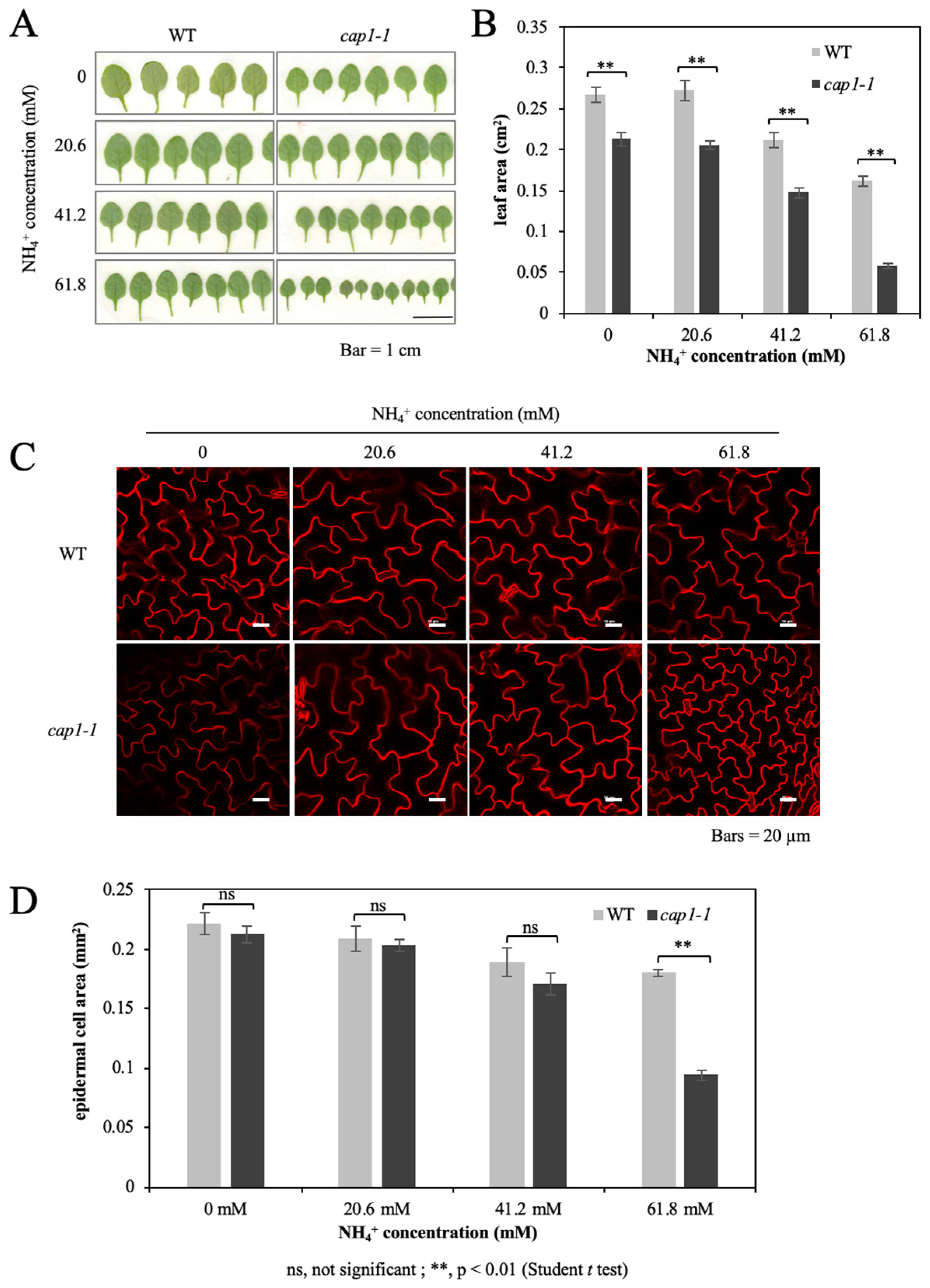
The above experimental results showed that the absence of CAP1 enhances the NH4+-mediated inhibition of plant growth. We further measured the leaf area. When the NH4+ concentration was increased to more than 41.2 mM, the area of the 3rd leaf of both the wild type and the cap1-1 mutant decreased, and the decrease in the cap1-1 mutant was more drastic; the leaf size of the cap1-1 mutant was about 69% of that of the wild type at 41.2 mM, and only 36% at 61.8 mM (Figure 2A,B). Moreover, leaf area for wild type showed no distinct difference between the treatments of MS medium and the MS medium without ammonium (0 mM NH4+), and the same for cap1-1 mutant (Figure 2B).
FM4-64 is a kind of membrane dye, which can emit high intensity fluorescence after specific binding with plasma membrane. We stained the 3rd leaf with FM4-64 to visualize the size of epidermal cells (Figure 2C). After taking pictures under the fluorescence microscope, we measured the epidermal cell area. No significant difference in single epidermis cell area was found for wild type and cap1-1 plants grown on MS and the NH4+-free medium (Figure 2D), suggesting that the deletion of NH4+ do not influence the leaf epidermal cell grow. However, difference between both lines was observed from the treatment with 41.2 mM NH4+, and substantial difference was displayed for plants grown on 61.8 mM NH4+ (Figure 2D). The average single epidermis cell area of the wild type decreased from 0.21 mm2 in the MS condition to 0.18 mm2 when treated with 61.8 mM NH4+, whereas that of the cap1-1 mutant decreased from 0.20 mm2 to 0.09 mm2, respectively (Figure 2D). cap1-1 leaf cell expansion is more sensitive to high level of NH4+. The decrease of the cap1-1 epidermis cell area was much higher than in the wild type and is a main reason for the reduced cap1-1 shoot growth under NH4+ stress, suggesting that the involvement of CAP1 in the NH4+ suppressed leaf growth may be accomplished partially by influencing cell expansion.

3.3. Deficiency of CAP1 Enhanced the Downregulation of Cell Enlargement-Related Genes under NH4+ Stress

Several cell wall enlargement genes related with ammonium-influenced plant growth have been reported [29], to explore whether these genes are involved in CAP1-regulated leaf growth to ammonium, we harvested the 3rd leaf of plants and compared the expression of these genes in wild type and cap1-1 by qRT-PCR. No significant difference in epidermis cell area between wild type and cap1-1 mutant showed for plants cultured on MS medium, while a marked difference can be seen under MS medium supplemented with 61.8 mM NH4+ treatment, so plants grown on MS medium and MS medium supplemented with 61.8 mM NH4+ were analyzed.
EXPs are engaged in cell wall loosening, the expression of EXP1 and EXP17 was determined. The disruption of CAP1 inhibits the expression of EXP1 and EXP17 (Figure 3A). In addition, as the concentration of NH4+ raised to 61.8 mM, the expression of EXP1 and EXP17 was declined for both lines (Figure 3A). However, more severe inhibition of both EXP1 and EXP17 expression was detected in the cap1-1 mutant than in the wild type by comparing treatments between MS medium and 61.8 mM NH4+ medium. The expression of EXP1 dropped about 68% for the wild type, and in contrast, nearly 89% for cap1-1 mutant. In addition, the expression of EXP17 dropped 28% for the wild type, while 71% for cap1-1 mutant (Figure 3A).
Expression of pectin-hydrolyzing enzymes PG1 and P-PG in both wild type and cap1-1 mutant has also been studied. Their expression was lower in cap1-1 plants for both growth condition (Figure 3B). The transcription for PG1 decreased about 60% for both wild type and cap1-1 mutant by comparing 61.8 mM NH4+ condition with the MS condition. While P-PG decreased more in cap1-1 plants (about 52%) than in the wild type (only about 3%) treated with 61.8 mM NH4+ (Figure 3B). The lower expression levels of these genes in the cap1-1 mutant compared to the wild type were in accordance with the more severe impairment of shoot growth in the mutant under NH4+ stress.
We also determined the expression of FER and Thesseus1 (THE1), two members of the CrRLK genes. FER is thought to be associated with regulation of signaling during cell elongation. THE1 is called a cell wall integrity sensor kinase. The expression of FER in the cap1-1 mutant decreased by approximately 70% under high ammonium medium (61.8 mM NH4+) compared with the MS medium condition, while in the wild type, the expression of FER decreased by only about 43% (Figure 3C). Similar to the expression of FER, the expression of THE1 in the cap1-1 mutant decreased by nearly 68% in the 61.8 mM NH4+ treatment compared with the MS condition, but only decreased by about 40% in the wild type (Figure 3C).

3.4. Deficiency of CAP1 Causes Higher NH4+-Induced ROS Levels in Shoots

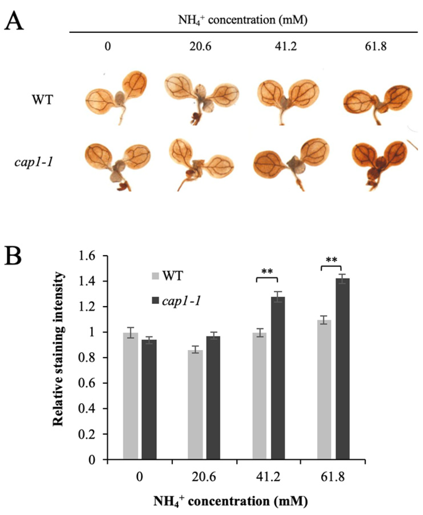
High NH4+ induces an increase in ROS in plants. However, the biological mechanism of NH4+-induced ROS accumulation remains largely unknown. 3,3′-diaminobenzidine (DAB) staining is used to detect H2O2 in plants. We observed stronger staining in cap1-1 shoots compared with the wild type under NH4+ treatment, and the intensity of staining increased with increased NH4+ (Figure 4A). The average DAB intensity in the cap1-1 mutant was about 1.3-fold of the wild type with 61.8 mM NH4+ (Figure 4B). This suggested that disruption of CAP1 leads to increased accumulation of ROS under high NH4+, and conversely, that CAP1 inhibits the NH4+-induced ROS accumulation in leaves.

3.5. CAP1 Regulates the Transcription of AtRBOH and CAT Genes in Response to NH4+

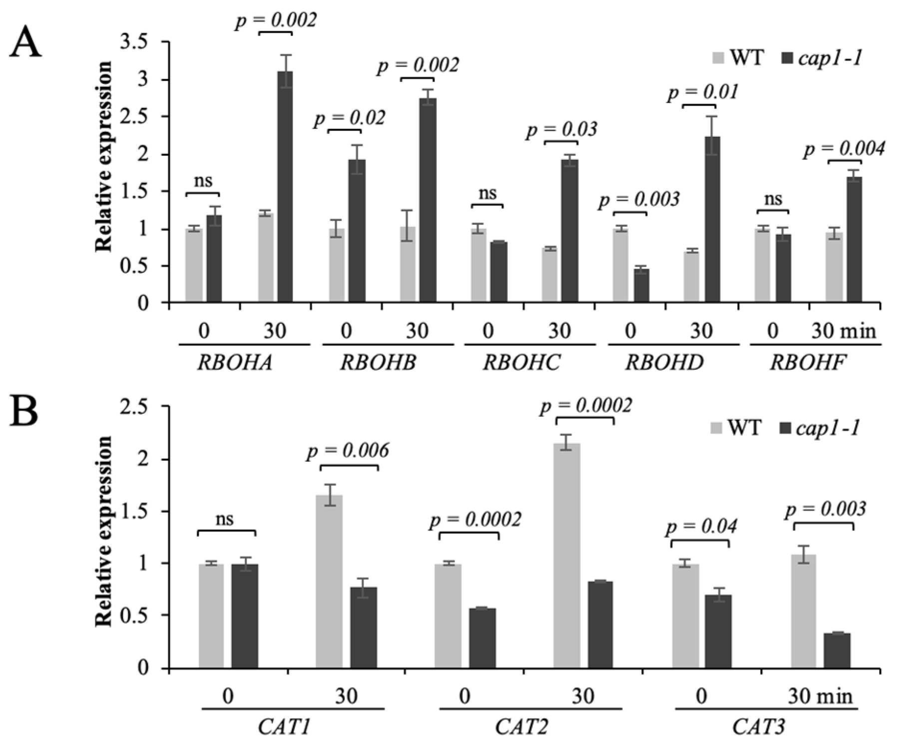
ROS can be generated in the apoplast via the activity of NADPH oxidases under stress [22]. Therefore, we also detected the relative transcript levels of RBOH genes under NH4+ stress by qRT-PCR. In the wild type, the expression of RBOHA, RBOHB, RBOHC, RBOHD, and RBOHF was similar at 0 and 30 min of NH4+ treatment, while their expression was significantly increased after 30 min of NH4+ treatment in the cap1-1 mutant (Figure 5A).
We next quantified the expression of catalase genes (CAT1, CAT2, and CAT3) by qRT-PCR. The expression of CAT1 and CAT2 increased in the wild type under NH4+ stress, but no change was detected for CAT3. However, in the cap1-1 mutant, the expression of CAT1 and CAT3 decreased, and was significantly lower than in the wild type under NH4+ treatment (Figure 5B). Although a small increase has been observed for CAT2 expression in the cap1-1 mutant under NH4+ stress, it was still significantly lower than in the wild type (Figure 5B). All these results indicated that CAP1 regulates NH4+-mediated ROS accumulation through affecting the expression of CAT and RBOH genes.

3.6. CAP1 Transgenic Lines Suppress the Hypersensitivity of cap1-1 Mutants to NH4+

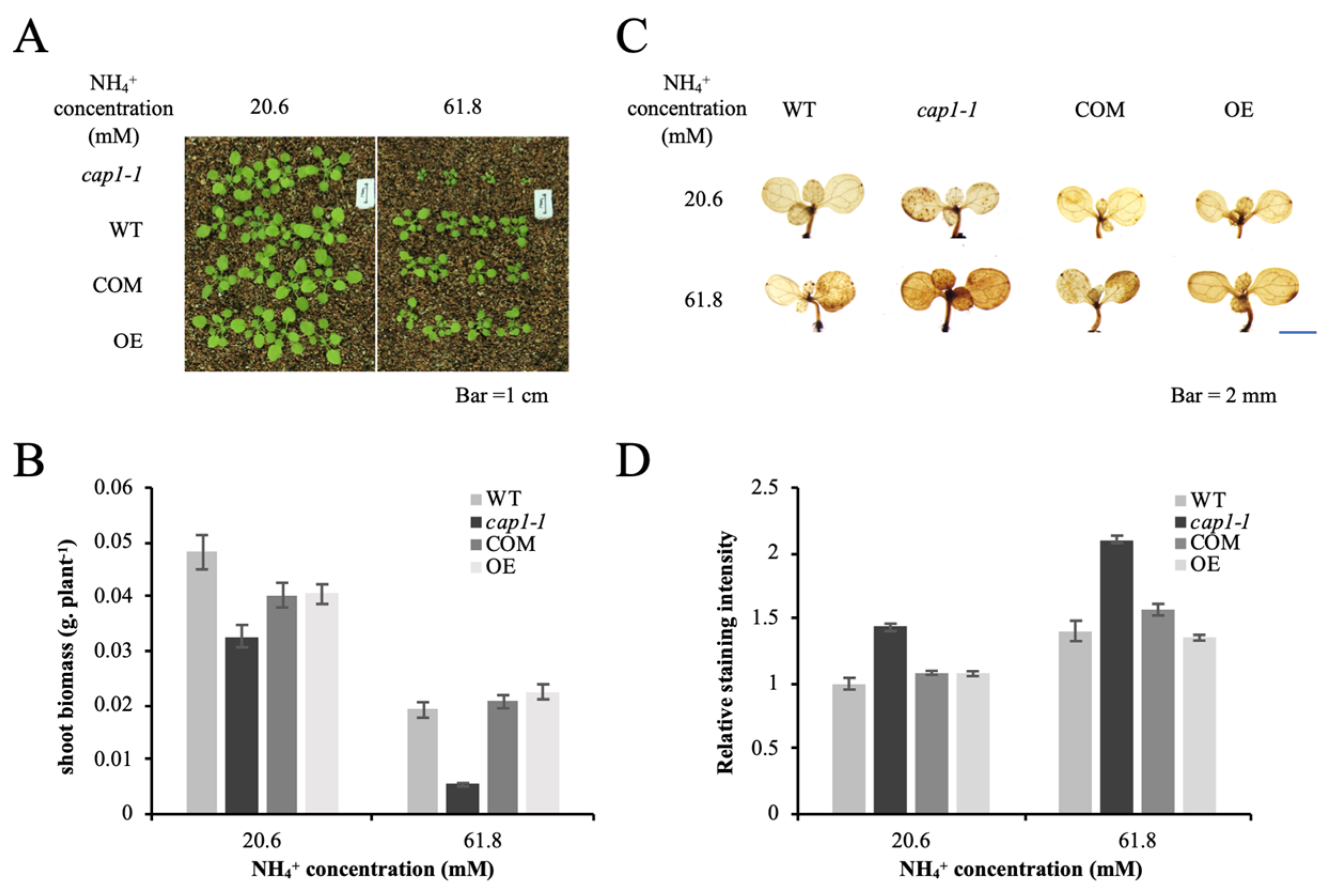
The enhanced shoot sensitivity to NH4+ of cap1-1 revealed that the deletion of CAP1 gene results this hypersensitivity. We further confirmed this by using the transgenic lines containing AtCAP1 promoter: CAP1 fusions (complementary lines, COM) or 35S:CAP1 fusions (over expression lines, OE) in the cap1-1 background [31,33]. Both the COM and the OE lines exhibited similar shoot growth to that of the wild type under NH4+ stress, and only the cap1-1 mutant exhibited obviously smaller shoots to higher NH4+ (61.8 mM) (Figure 6A). Average shoot biomass for both transgenic plants showed no difference compared with that of the wild type to NH4+ stress (Figure 6B). These results further proved that CAP1 plays a role in regulating shoot growth to high level of NH4+.
Furthermore, ROS levels were also studied for seedlings. DAB staining for both OE and the COM transgenic seedlings were weaker than that of the cap1-1 mutant, whereas the staining patterns of both transgenic lines are similar as the wild type to both treatment of NH4+ (Figure 6C,D). These staining results further confirmed that the disruption of CAP1 gene causes accumulation of ROS to NH4+ stress, that in turn suppress shoot growth.

4. Discussion

NH4+-mediated growth retardation is a major issue in cropping systems. The most visible phenotype of NH4+ toxicity is reduced growth of roots and leaves [5]. Genes that function in NH4+ toxicity have been discovered through mutant analysis. Studies on the Arabidopsis NH4+-sensitive mutant hsn1 (hypersensitivity to NH4+ 1)/vtc1 (vitamin c1) showed that the protein N-glycosylation mediated by GDP–mannose pyrophosphorylases (GMPase) may regulate root elongation in response to NH4+ stress [35,36,37]. The mutant of AMOS1/EGY1 displays severe chlorosis under NH4+ stress. During NH4+ stress, the AMOS1-dependent retrograde signaling pathway, in which signals from the chloroplast modulate nuclear gene expression, is integrated with abscisic acid signaling. The elevated H2O2 induced by NH4+ was markedly lower in amos1 seedlings than in the wild type [30]. AMOS2 plays a role in NH4+-mediated root and shoot growth, and severely suppressed shoot biomass and inhibition of root growth were observed in the amos2 mutant [38]. AMOS2 was reported to function in NH4+ stress mainly by controlling cation homeostasis, such as potassium (K+), calcium (Ca2+), and magnesium (Mg2+) [38]. AMOT1/EIN3 has been reported to function in the NH4+-induced impairment of shoot growth through positively mediating the NH4+-induced H2O2 accumulation in leaves. Enhanced tolerance to NH4+ was observed for the amot1 mutant, which showed increased shoot growth compared to the wild type [38]. Although these genes have been implicated in NH4+ toxicity, the current understanding of their roles in NH4+ toxicity remains limited. Previous studies showed that CAP1 functions in NH4+-related root hair growth; the knockout mutant cap1-1 displays normal root hairs only when grown on NH4+-free medium [33]. In this study, we investigated the growth of the cap1-1 mutant under different levels of NH4+. The significantly enhanced downregulation of leaf biomass and increase in ROS levels in response to NH4+ in the cap1-1 mutant compared with the wild type suggested that CAP1 functions in attenuating the sensitivity to NH4+ by negatively mediating NH4+-induced H2O2 accumulation.
The high levels of NH4+ inhibited cap1-1 shoot growth (Figure 1 and Figure 2), which suggests a function of CAP1 in leaf expansion under high NH4+. Severe suppression of leaf growth in response to NH4+ has been attributed to suppression of both cell enlargement and cell division. The NH4+-mediated repression of tobacco (Nicotiana tabacum) leaf growth is related with both cell growth and cell number [39], while root growth suppression is more attributed to the suppression of cell growth [35,40]. In our study, the significantly smaller cell area of cap1-1 mutant leaves suggested that cell growth is altered under NH4+ stress in A. thaliana. As cell growth is limited by the cell wall, cell wall loosening enables cell enlargement. One of the factors controlling cell enlargement at the individual cell level is the presence of the cell wall. Cell extensibility is closely related with the cell wall architecture. Proteins or enzymes involved in cell wall structure can modulate cell extensibility; for example, pectin hydrolyzing enzymes and EXP. Altered activities of cell wall-modifying proteins is one reason for the NH4+-triggered growth retardation in plants, and EXP and PG have been implicated in NH4+-inhibited cell growth [29]. Our results suggested that the decrease of leaf area of the cap1-1 mutant is partly due to the decrease of cell area when exposed to NH4+ stress. The inhibited growth is related with cell wall structure, and the smaller leaf area of cap1-1 mutant may suggest the different expression pattern for genes related with cell growth. The transcript levels of EXP1, EXP17, and two PG genes were markedly decreased in the cap1-1 mutant in response to increasing concentrations of NH4+, but to a lesser extent in the wild type (Figure 3), suggesting that CAP1 plays a role in regulating cell wall extensibility in response to NH4+ by influencing the expression of these genes. The lower activity and/or expression of pectin hydrolyzing enzymes and expansins may limit cell wall expansion. Although we did not detect their protein activities in this study, the lower expression of these genes in the cap1-1 mutant in response to NH4+ compared to the wild type suggests that they are involved in the NH4+-mediated shoot growth impairment in the mutant.
CAP1 belongs to the CrRLK family. Other genes of this family have also been reported to participate in regulating plant growth, and plants with mutations of CrRLK family genes show growth inhibition phenotypes [31]. Among the CrRLK genes, FER is essential for pollen tube and root hair growth [41,42,43], and THE1 is responsible for cell elongation [44]. The expression of both genes decreased in the wild type and cap1-1 plants under high NH4+ (Figure 3), suggesting that FER and THE1 are involved in NH4+ stress. Further, the lower expression of these genes in the cap1-1 mutant compared with the wild type were in accordance with the markedly inhibited cell growth in the mutant (Figure 3), further suggesting that CAP1 plays an indispensable role in attenuating NH4+-inhibited cell expansion.
ROS are generated by various environmental stresses. Increased ROS in response to high NH4+ is a common symptom of NH4+ toxicity [7], and has been reported in both leaves and roots [16,24,25,45,46], which will induce oxidative defense responses. However, the mechanisms underlying NH4+-induced ROS accumulation are not clear [9,19]. AMOS1/EGY1 and AMOT1/EIN3 have been reported to function in regulating H2O2 metabolism in response to NH4+ stress [19,30]. In this study, although we observed elevated H2O2 levels in both wild-type and cap1-1 seedlings under NH4+ stress, the cap1-1 mutant showed markedly higher levels in our DAB staining assay (Figure 4 and Figure 6). The increased level of H2O2 in the cap1-1 mutant suggested that CAP1 plays a role in ROS homeostasis under NH4+ stress (Figure 4 and Figure 6). As it is reported that ROS affected the transcription for cell wall remodeling enzymes [47], the perturbed ROS in the mutant may be one of the reasons that influence expression of genes is related to cell wall extensibility. However, correlation between CAP1 with the cell enlargement will need further study, the underlying mechanisms by which this process is accomplished may related with CAP1-mediated maintenance of ROS homeostasis to NH4+ stress.
A prominent source of H2O2 production is the NADPH oxidases [14]. It has been reported that expression of RBOHD was not induced under NH4+ stress in wild-type leaves [48]. Additionally, Li [19] found that the expression of RBOHA, RBOHB, RBOHD, and RBOHF was not induced by NH4+ in wild-type leaves. Similarly, the transcript levels of the AtRBOH genes investigated in the present study remained relatively unchanged in the wild type under NH4+ stress (Figure 5). However, the expression of RBOHA, RBOHB, RBOHC, RBOHD, and RBOHF was strongly induced in the cap1-1 mutant after treatment with NH4+ (Figure 5). These results indicated that ROS generated by these AtRBOHs may function in NH4+ stress through a CAP1-dependent pathway. Concurrently, this induced oxidative stress can trigger ROS scavenging pathways. The relatively lower transcript levels of CAT genes in the cap1-1 mutant compared with the wild type under high NH4+ also suggested the function of CAP1 in NH4+-mediated ROS homeostasis (Figure 5). The expression patterns of these genes suggested a role for CAP1 in maintaining ROS homeostasis in response to NH4+ stress by balancing ROS production and scavenging. The higher ROS content of the cap1-1 mutant under NH4+ stress leads to more severe shoot growth reduction than in the wild type.

5. Conclusions

In conclusion, understanding the underlying mechanisms of NH4+ toxicity is essential for improving nitrogen use efficiency. Although numerous genes and factors have been identified to function in modulating the response to NH4+ stress, NH4+ toxicity remains poorly characterized. The work reported here demonstrated a new role for CAP1 in shoot growth in response to NH4+ stress (Figure 7). The enhanced sensitivity of the cap1-1 mutant to NH4+ stress is linked with the role of CAP1 in regulation cell wall loosening and ROS accumulation.

Author Contributions

X.M. and L.B. conceived the project. Q.Y., N.D., H.Y., F.F., Y.X., C.W., and Y.Y. performed the experiments. X.M. and L.B. analyzed the data and wrote the manuscript. All authors have read and agreed to the published version of the manuscript.

Funding

This work was supported by the National Natural Science Foundation of China (31970198 and 31900239).

Institutional Review Board Statement

Not applicable.

Informed Consent Statement

Not applicable.

Data Availability Statement

Not applicable.

Conflicts of Interest

The authors declare no conflict of interest.

Abbreviations

CAP1: [Ca2+]cyt-ASSOCIATED PROTEIN KINASE1; N: Nitrogen; NO3: Nitrate; NH4+: Ammonium; ROS: Reactive oxygen species; RBOHs: Respiratory burst oxidase homologues; CAT: Catalase; GR: Glutathione reductase; SOD: Superoxide dismutase; PODs: Peroxidases; AMOS1: AMMONIUM OVERLY SENSITIVE1; EGY1: ETHYLENE-DEPENDENT, GRAVITROPISM-DEFICIENT, AND YELLOW-GREEN-LIKE PROTEIN1; AMOT1: AMMONIUM TOLERANCE 1; EXP: expansins; PG: Endopolygalacturonases; CrRLKs: Catharanthus roseus receptor-like kinases; FER: FERONIA; THE1: THESSEUS1; DAB: 3,3′-diaminobenzidine; NUE: Nitrogen utilization efficiency; GMPase: GDP–mannose pyrophosphorylases; K+: Potassium; Ca2+: Calcium; Mg2+: Magnesium.

References

  1. Miller, A.J.; Cramer, M.D. Root nitrogen acquisition and assimilation. Plant Soil 2005, 274, 1–36. [Google Scholar] [CrossRef]
  2. Sanchez-Zabala, J.; Gonzalez-Murua, C.; Marino, D. Mild ammonium stress increases chlorophyll content in Arabidopsis thaliana. Plant Signal. Behav. 2015, 10, e991596. [Google Scholar] [CrossRef] [PubMed] [Green Version]
  3. Glass, A.D.; Britto, D.T.; Kaiser, B.N.; Kinghorn, J.R.; Kronzucker, H.J.; Kumar, A.; Okamoto, M.; Rawat, S.; Siddiqi, M.Y.; Unkles, S.E.; et al. The regulation of nitrate and ammonium transport systems in plants. J. Exp. Bot. 2002, 53, 855–864. [Google Scholar] [CrossRef] [PubMed] [Green Version]
  4. Kronzucker, H.J.; Siddiqi, M.Y.; Glass, A.D.M.; Britto, D.T. Root ammonium transport efficiency as a determinant in forest colonization patterns: An hypothesis. Physiol. Plant. 2003, 117, 164–170. [Google Scholar] [CrossRef] [Green Version]
  5. Britto, D.T.; Kronzucker, H.J. NH4+ toxicity in higher plants: A critical review. J. Plant Physiol. 2002, 159, 567–584. [Google Scholar] [CrossRef] [Green Version]
  6. Li, B.; Li, G.; Kronzucker, H.J.; Baluska, F.; Shi, W. Ammonium stress in Arabidopsis: Signaling, genetic loci, and physiological targets. Trends Plant Sci. 2014, 19, 107–114. [Google Scholar] [CrossRef] [PubMed]
  7. Liu, Y.; von Wirén, N. Ammonium as a signal for physiological and morphological responses in plants. J. Exp. Bot. 2017, 68, 2581–2592. [Google Scholar] [CrossRef] [Green Version]
  8. Esteban, R.; Ariz, I.; Cruz, C.; Moran, J.F. Review: Mechanisms of ammonium toxicity and the quest for tolerance. Plant Sci. 2016, 248, 92–101. [Google Scholar] [CrossRef] [Green Version]
  9. Bittsánszky, A.; Pilinszky, K.; Gyulai, G.; Komives, T. Overcoming ammonium toxicity. Plant Sci. 2015, 231, 184–190. [Google Scholar] [CrossRef]
  10. Di, D.W.; Sun, L.; Zhang, X.N.; Li, G.J.; Kronzucker, H.J.; Shi, W.M. Involvement of auxin in the regulation of ammonium tolerance in rice (Oryza sativa L.). Plant Soil 2018, 432, 373–387. [Google Scholar] [CrossRef]
  11. Raab, T.K.; Terry, N. Nitrogen Source Regulation of Growth and Photosynthesis in Beta vulgaris L. Plant Physiol. 1994, 105, 1159–1166. [Google Scholar] [CrossRef] [PubMed] [Green Version]
  12. Gerendás, J.; Zhu, Z.; Bendixen, R.; Ratcliffe, R.G.; Sattelmacher, B. Physiological and biochemical processes related to ammonium toxicity in higher plants. J. Plant Nutr. Soil Sci. 1997, 160, 239–251. [Google Scholar] [CrossRef]
  13. Jin, M.; Guo, M.; Yue, G.; Li, J.; Yang, S.; Zhao, P.; Su, Y. An unusual strategy of stomatal control in the desert shrub Ammopiptanthus mongolicus. Plant Physiol. Biochem. 2018, 125, 13–26. [Google Scholar] [CrossRef] [PubMed]
  14. Mittler, R.; Blumwald, E. The Roles of ROS and ABA in Systemic Acquired Acclimation. Plant Cell 2015, 27, 64–70. [Google Scholar] [CrossRef] [PubMed] [Green Version]
  15. Mittler, R. Oxidative stress, antioxidants and stress tolerance. Trends Plant Sci. 2002, 7, 405–410. [Google Scholar] [CrossRef]
  16. Yang, S.Y.; Hao, D.L.; Jin, M.; Li, Y.; Liu, Z.T.; Huang, Y.N.; Chen, T.X.; Su, Y.H. Internal ammonium excess induces ROS-mediated reactions and causes carbon scarcity in rice. BMC Plant Biol. 2020, 20, 143. [Google Scholar] [CrossRef] [PubMed]
  17. Zhuang, K.; Shi, D.L.; Hu, Z.B.; Xu, F.L.; Chen, Y.H.; Shen, Z.G. Subcellular accumulation and source of O2 and H2O2 in submerged plant Hydrilla verticillata (L.f.) Royle under NH4+-N stress condition. Aquat. Toxicol. 2019, 207, 1–12. [Google Scholar] [CrossRef]
  18. Sun, L.; Di, D.W.; Li, G.; Kronzucker, H.J.; Wu, X.; Shi, W. Endogenous ABA alleviates rice ammonium toxicity by reducing ROS and free ammonium via regulation of the SAPK9-bZIP20 pathway. J. Exp. Bot. 2020, 71, 4562–4577. [Google Scholar] [CrossRef]
  19. Li, G.; Zhang, L.; Wang, M.; Di, D.; Kronzucker, H.J.; Shi, W. The Arabidopsis AMOT1/EIN3 gene plays an important role in the amelioration of ammonium toxicity. J. Exp. Bot. 2019, 70, 1375–1388. [Google Scholar] [CrossRef] [Green Version]
  20. Smirnoff, N.; Arnaud, D. Hydrogen peroxide metabolism and functions in plants. New Phytol. 2019, 221, 1197–1214. [Google Scholar] [CrossRef] [Green Version]
  21. Sagi, M.; Fluhr, R. Production of reactive oxygen species by plant NADPH oxidases. Plant Physiol. 2006, 141, 336–340. [Google Scholar] [CrossRef] [PubMed] [Green Version]
  22. Mittler, R.; Vanderauwera, S.; Gollery, M.; Van Breusegem, F. Reactive oxygen gene network of plants. Trends Plant Sci. 2004, 9, 490–498. [Google Scholar] [CrossRef] [PubMed]
  23. Lee, S.; Seo, P.J.; Lee, H.J.; Park, C.M. A NAC transcription factor NTL4 promotes reactive oxygen species production during drought-induced leaf senescence in Arabidopsis. Plant J. 2012, 70, 831–844. [Google Scholar] [CrossRef]
  24. Xie, Y.J.; Mao, Y.; Xu, S.; Zhou, H.; Duan, X.L.; Cui, W.T.; Zhang, J.; Xu, G.H. Heme-heme oxygenase 1 system is involved in ammonium tolerance by regulating antioxidant defence in Oryza sativa. Plant Cell Environ. 2015, 38, 129–143. [Google Scholar] [CrossRef] [PubMed]
  25. Patterson, K.; Cakmak, T.; Cooper, A.; Lager, I.; Rasmusson, A.G.; Escobar, M.A. Distinct signalling pathways and transcriptome response signatures differentiate ammonium- and nitrate-supplied plants. Plant Cell Environ. 2010, 33, 1486–1501. [Google Scholar] [CrossRef] [Green Version]
  26. Wang, C.; Zhang, S.H.; Wang, P.F.; Hou, J.; Li, W.; Zhang, W.J. Metabolic adaptations to ammonia-induced oxidative stress in leaves of the submerged macrophyte Vallisneria natans (Lour.) Hara. Aquat. Toxicol. 2008, 87, 88–98. [Google Scholar] [CrossRef]
  27. Nimptsch, J.; Pflugmacher, S. Ammonia triggers the promotion of oxidative stress in the aquatic macrophyte Myriophyllum mattogrossense. Chemosphere 2007, 66, 708–714. [Google Scholar] [CrossRef]
  28. Husted, S.; Schjoerring, J.K. Apoplastic pH and Ammonium Concentration in Leaves of Brassica napus L. Plant Physiol. 1995, 109, 1453–1460. [Google Scholar] [CrossRef]
  29. Podgorska, A.; Burian, M.; Gieczewska, K.; Ostaszewska-Bugajska, M.; Zebrowski, J.; Solecka, D.; Szal, B. Altered Cell Wall Plasticity Can Restrict Plant Growth under Ammonium Nutrition. Front. Plant Sci. 2017, 8, 1344. [Google Scholar] [CrossRef] [Green Version]
  30. Li, B.; Li, Q.; Xiong, L.; Kronzucker, H.J.; Kramer, U.; Shi, W. Arabidopsis plastid AMOS1/EGY1 integrates abscisic acid signaling to regulate global gene expression response to ammonium stress. Plant Physiol. 2012, 160, 2040–2051. [Google Scholar] [CrossRef]
  31. Nissen, K.S.; Willats, W.G.T.; Malinovsky, F.G. Understanding CrRLK1L Function: Cell Walls and Growth Control. Trends Plant Sci. 2016, 21, 516–527. [Google Scholar] [CrossRef] [PubMed]
  32. Cheung, A.Y.; Wu, H.M. THESEUS 1, FERONIA and relatives: A family of cell wall-sensing receptor kinases? Curr. Opin. Plant Biol. 2011, 14, 632–641. [Google Scholar] [CrossRef] [PubMed]
  33. Bai, L.; Ma, X.; Zhang, G.; Song, S.; Zhou, Y.; Gao, L.; Miao, Y.; Song, C.P. A Receptor-Like Kinase Mediates Ammonium Homeostasis and Is Important for the Polar Growth of Root Hairs in Arabidopsis. Plant Cell 2014, 26, 1497–1511. [Google Scholar] [CrossRef] [PubMed] [Green Version]
  34. Daudi, A.; O’Brien, J.A. Detection of Hydrogen Peroxide by DAB Staining in Arabidopsis Leaves. Bio-Protocol 2012, 2, e263. [Google Scholar] [CrossRef] [Green Version]
  35. Li, Q.; Li, B.H.; Kronzucker, H.J.; Shi, W.M. Root growth inhibition by NH4+ in Arabidopsis is mediated by the root tip and is linked to NH4+ efflux and GMPase activity. Plant Cell Environ. 2010, 33, 1529–1542. [Google Scholar] [CrossRef]
  36. Barth, C.; Gouzd, Z.A.; Steele, H.P.; Imperio, R.M. A mutation in GDP-mannose pyrophosphorylase causes conditional hypersensitivity to ammonium, resulting in Arabidopsis root growth inhibition, altered ammonium metabolism, and hormone homeostasis. J. Exp. Bot. 2010, 61, 379–394. [Google Scholar] [CrossRef] [Green Version]
  37. Qin, C.; Qian, W.; Wang, W.; Wu, Y.; Yu, C.; Jiang, X.; Wang, D.; Wu, P. GDP-mannose pyrophosphorylase is a genetic determinant of ammonium sensitivity in Arabidopsis thaliana. Proc. Natl. Acad. Sci. USA 2008, 105, 18308–18313. [Google Scholar] [CrossRef] [Green Version]
  38. Li, G.; Dong, G.; Li, B.; Li, Q.; Kronzucker, H.J.; Shi, W. Isolation and characterization of a novel ammonium overly sensitive mutant, amos2, in Arabidopsis thaliana. Planta 2012, 235, 239–252. [Google Scholar] [CrossRef]
  39. Walch-Liu, P.; Neumann, G.; Bangerth, F.; Engels, C. Rapid effects of nitrogen form on leaf morphogenesis in tobacco. J. Exp. Bot. 2000, 51, 227–237. [Google Scholar] [CrossRef] [Green Version]
  40. Liu, Y.; Lai, N.W.; Gao, K.; Chen, F.J.; Yuan, L.X.; Mi, G.H. Ammonium Inhibits Primary Root Growth by Reducing the Length of Meristem and Elongation Zone and Decreasing Elemental Expansion Rate in the Root Apex in Arabidopsis thaliana. PLoS ONE 2013, 8, e61031. [Google Scholar] [CrossRef]
  41. Duan, Q.; Kita, D.; Li, C.; Cheung, A.Y.; Wu, H.M. FERONIA receptor-like kinase regulates RHO GTPase signaling of root hair development. Proc. Natl. Acad. Sci. USA 2010, 107, 17821–17826. [Google Scholar] [CrossRef] [PubMed] [Green Version]
  42. Guo, H.; Li, L.; Ye, H.; Yu, X.; Algreen, A.; Yin, Y. Three related receptor-like kinases are required for optimal cell elongation in Arabidopsis thaliana. Proc. Natl. Acad. Sci. USA 2009, 106, 7648–7653. [Google Scholar] [CrossRef] [PubMed] [Green Version]
  43. Escobar-Restrepo, J.M.; Huck, N.; Kessler, S.; Gagliardini, V.; Gheyselinck, J.; Yang, W.C.; Grossniklaus, U. The FERONIA receptor-like kinase mediates male-female interactions during pollen tube reception. Science 2007, 317, 656–660. [Google Scholar] [CrossRef] [PubMed]
  44. Hématy, K.; Sado, P.E.; Van Tuinen, A.; Rochange, S.; Desnos, T.; Balzergue, S.; Pelletier, S.; Renou, J.P.; Hofte, H. A receptor-like kinase mediates the response of Arabidopsis cells to the inhibition of cellulose synthesis. Curr. Biol. 2007, 17, 922–931. [Google Scholar] [CrossRef] [PubMed] [Green Version]
  45. Huang, L.; Lu, Y.Y.; Gao, X.; Du, G.; Ma, X.X.; Liu, M.; Guo, J.S.; Chen, Y.P. Ammonium-induced oxidative stress on plant growth and antioxidative response of duckweed (Lemna minor L.). Ecol. Eng. 2013, 58, 355–362. [Google Scholar] [CrossRef]
  46. Wang, C.; Zhang, S.H.; Wang, P.F.; Li, W.; Lu, J. Effects of ammonium on the antioxidative response in Hydrilla verticillata (L.f.) Royle plants. Ecotoxicol. Environ. Saf. 2010, 73, 189–195. [Google Scholar] [CrossRef]
  47. Tenhaken, R. Cell wall remodeling under abiotic stress. Front. Plant Sci. 2014, 5, 771. [Google Scholar] [CrossRef] [Green Version]
  48. Podgórska, A.; Ostaszewska, M.; Gardeström, P.; Rasmusson, A.G.; Szal, B. In comparison with nitrate nutrition, ammonium nutrition increases growth of the frostbite1 Arabidopsis mutant. Plant Cell Environ. 2015, 38, 224–237. [Google Scholar] [CrossRef]
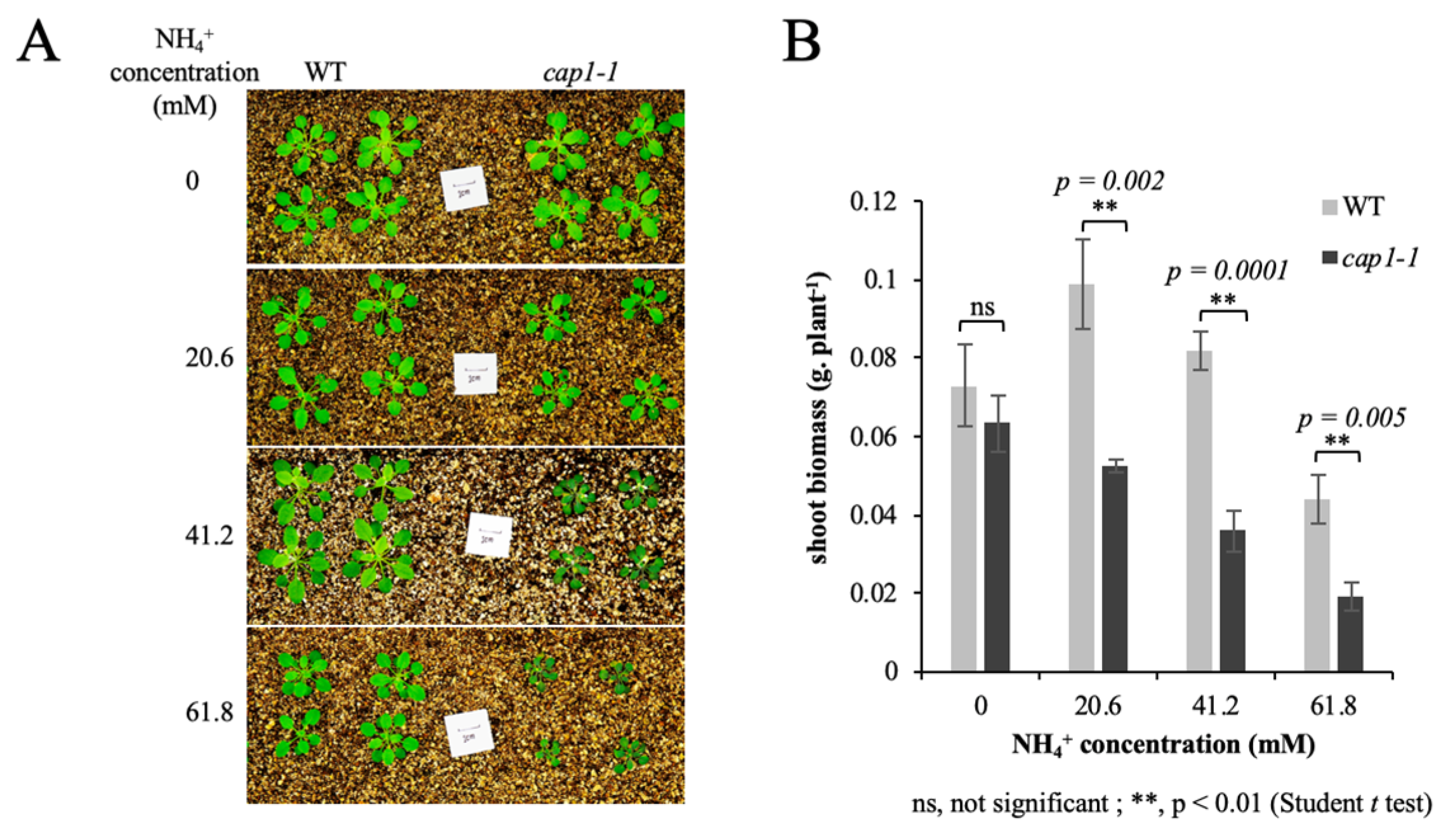
Figure 1. Shoot phenotypes of wild-type (WT) and cap1-1 plants growing on media with different concentrations of NH4+. (A) Growth of the WT and the cap1-1 mutant in response to different growth condition. Scale bars = 1 cm. (B) Shoot fresh weights of WT and cap1-1 plants in (A) were averaged. Seven-day-old seedlings were transplanted to a pot containing only vermiculite. Solutions containing 0, 20.6 mM, 41.2 mM, or 61.8 mM NHCl4 were applied to the respective plants for about 4 weeks, and then pictures were taken. Values are the means ± SE, n = 8–11. (Independent samples t-test; ns, not significant; **, p < 0.01.)
Biology 11 01452 g001
Figure 2. Leaf area and cell size of WT and cap1-1 plants under different growth concentrations. (A) The 3rd leaf phenotype and (B) leaf area in WT and cap1-1 plants grown on 4 different media with 0, 20.6, 41.2, and 61.8 mM NH4+. (C) The epidermis of the 3rd leaf was stained with FM4-64 and scanned by a confocal microscope. (D) Epidermal cell area of the 3rd leaf under MS medium without ammonium (0 mM NH4+), MS medium (20.6 mM NH4+), and MS medium with 41.2 or 61.8 mM NH4+ treatment. Seven-day-old seedlings were transplanted to a pot containing only vermiculite. Solutions containing 0, 20.6, 41.2, and 61.8 mM NHCl4 were applied to the respective plants for about 4 weeks. Values are the means ± SE, n = 8–11. (Independent samples t-test; ns, not significant; **, p < 0.01.)
Biology 11 01452 g002
Figure 3. The expression of cell expansion-related genes in WT and cap1-1 plants. Seven-day-old seedlings were transplanted to a pot containing only vermiculite with MS medium (20.6 mM NH4+) or MS medium with high content of ammonium (61.8 mM NH4+) and grew for about 4 weeks, and then 3rd leaf of WT and cap1-1 plants were harvested. (A) Relative transcript levels for EXP1 and EXP17, (B) PG1 and P-PG, and (C) THE1 and FER in WT and cap1-1 plants are determined by qRT-PCR. The value for each gene in the WT at MS medium (20.6 mM NH4+) was set as 1. Data are presented as means ± SE of three replicates (independent samples t-test; p < 0.05; ns, not significant).
Biology 11 01452 g003
Figure 4. Effects of CAP1 on NH4+-induced H2O2 accumulation in shoots. (A) Detection of H2O2 levels in WT and cap1-1 leaves. Seven-day-old seedlings were grown on 4 different media with 0, 20.6, 41.2, and 61.8 mM NH4+ for 3 d, and then DAB staining of shoots was performed. (B) The mean relative DAB staining intensity in the WT and cap1-1 leaves in (A). WT grown on MS medium without ammonium (0 mM NH4+) was set as 1. Values are the means ± SE, n = 24–33 (independent samples t-test; **, p < 0.01).
Biology 11 01452 g004
Figure 5. The expression of NADPH oxidase and CAT genes in WT and cap1-1 plants to NH4+ treatment. (A) Relative transcript levels for RBOHA, RBOHB, RBOHC, RBOHD, and RBOHF, (B) and CAT genes (CAT1, CAT2, and CAT3) in WT and cap1-1 plants under 3 mM NH4Cl treatment for 0 and 30 min quantified by qRT-PCR. Five-day-old plants grown on MS medium were transferred to MS NH4+-free medium for 2 days, then treated with 3 mM NH4Cl. The value for each gene in the WT at 0 min was set as 1. Data are presented as means ± SE of three replicates (independent samples t-test; p < 0.05; ns, not significant).
Biology 11 01452 g005
Figure 6. CAP1 transgene line suppress the hypersensitivity of cap1-1 to NH4+. (A) Growth of the OE, COM, WT, and the cap1-1 mutant in response to different growth condition. Scale bars = 1 cm. (B) Shoot fresh weights of WT and cap1-1 plants in (A) were averaged. Seven-day-old seedlings were transplanted to a pot containing only vermiculite. Solutions containing 20.6 mM and 61.8 mM NHCl4 were applied to the respective plants for about 4 weeks, and then pictures were taken. Values are the means ± SE, n = 8–11. (Independent samples t-test.) (C) Detection of H2O2 levels in WT and cap1-1 leaves. Seven-day-old seedlings were grown on 2 different media with 20.6 and 61.8 mM NH4+ for 3 d, and then DAB staining of shoots was performed. (D) The mean relative DAB staining intensity in the WT and cap1-1 leaves in (C). WT grown on MS medium (20.6 mM NH4+) was set as 1. Values are the means ± SE, n = 24–33 (independent samples t-test).
Biology 11 01452 g006
Figure 7. A proposed model for the role of CAP1 in shoot growth in response to NH4+ stress. Under NH4+ stress, CAP1 regulates the expression of RBOH, CAT, and cell expansion-related genes to maintain ROS homeostasis and cell wall expansion to allow shoot growth. Deficiency of CAP1 enhances the downregulation of cell expansion-related genes and the accumulation of ROS in shoots under NH4+ stress, which results in shoot growth inhibition.
Biology 11 01452 g007
Table 1. The primer sequences of qRT-PCR.
GenePrimer Sequence
EXP1
(At1g69530)
F: GAAGAGTGCCGTGCGTGAG
R: TAGGTTGAAGTAAGAGTGTCCGTTT
EXP17
(At4g01630)
F: GCCTCTGGTACAATGGGTGG
R: TGAGCAAAGTTCGGTGGACA
PG1
(At3g26610)
F: CCAATGAAACCAACGGCACT
R: TGACTTTGACTCCATCCGAATC
Putative-PG
(At2g43890)
F: ACAGGGTTCTGGAGTGAAGATTAGTC
R: TCGCTTGGCACGGATTACT
FER
(At3g51550)
F: TGCCGTCACTTCTCGTTTGC
R: CTTGCTCGGACATTGGGTTG
THE1
(At5g54380)
F: TCAAGAAGGCGGTAATGGACA
R: CAACCCCAAGCAACGAACTC
RBOHA
(At5g07390)
F: AGGGGTCGTTTGACTGGTTC
R: CTCGTAAACGCTGGTGCAGT
RBOHB
(At1g09090)
F: CGAGGTGATGGGCTACTGTG
R: TGTTCCTACAAACGGGCAAG
RBOHC
(At5g51060)
F: GAGAACAGTCAGAGAACACGTA
R: TTCGTTTCAGCAAAGTAAGCTC
RBOHD
(At5g47910)
F: GCTCCGTGCTTTCAGATCAA
R: TTTGAATCCTTGTGGCTTCG
RBOHF
(At1g64060)
F: GCCGACGAAACAACAAAGAA
R: CACCAATGCCAAGACCAACT
CAT1
(At1g20630)
F: CTGGGATTCAGACAGGCAAGA
R: AACCAAACCGTAAGAGGAGCAT
CAT2
(At4g35090)
F: CCCGTGTCTTCTCCTATGCC
R: TAACCTCCTCGTCCCTGTGC
CAT3
(At1g20620)
F: AATCACAGCCACGCCACTAA
R: TCAGAACCAAGCGACCAACC
UBC9
(At4g27960)
F: CACAATTTCCAAGGTGCTGCTATCG
R: GTACATGTGAGCTATCTCAGGGACCAAA
Publisher’s Note: MDPI stays neutral with regard to jurisdictional claims in published maps and institutional affiliations.